Embed Size (px)
Citation preview
ORIGINAL RESEARCHADULT BRAIN
Clinical, Imaging, and Lab Correlates of Severe COVID-19Leukoencephalopathy
O. Rapalino, A. Pourvaziri, M. Maher, A. Jaramillo-Cardoso, B.L. Edlow, J. Conklin, S. Huang, B. Westover,J.M. Romero, E. Halpern, R. Gupta, S. Pomerantz, P. Schaefer, R.G. Gonzalez, S.S. Mukerji, and M.H. Lev
ABSTRACT
BACKGROUND AND PURPOSE: Patients infected with the Severe Acute Respiratory Syndrome coronavirus 2 (SARS-CoV-2) can de-velop a spectrum of neurological disorders, including a leukoencephalopathy of variable severity. Our aim was to characterizeimaging, lab, and clinical correlates of severe coronavirus disease 2019 (COVID-19) leukoencephalopathy, which may provide insightinto the SARS-CoV-2 pathophysiology.
MATERIALS AND METHODS: Twenty-seven consecutive patients positive for SARS-CoV-2 who had brain MR imaging following in-tensive care unit admission were included. Seven (7/27, 26%) developed an unusual pattern of “leukoencephalopathy with reduceddiffusivity” on diffusion-weighted MR imaging. The remaining patients did not exhibit this pattern. Clinical and laboratory indices,as well as neuroimaging findings, were compared between groups.
RESULTS: The reduced-diffusivity group had a significantly higher body mass index (36 versus 28 kg/m2, P , .01). Patients withreduced diffusivity trended toward more frequent acute renal failure (7/7, 100% versus 9/20, 45%; P ¼ .06) and lower estimatedglomerular filtration rate values (49 versus 85 mL/min; P ¼ .06) at the time of MRI. Patients with reduced diffusivity also showedlesser mean values of the lowest hemoglobin levels (8.1 versus 10.2 g/dL, P , .05) and higher serum sodium levels (147 versus139 mmol/L, P ¼ .04) within 24 hours before MR imaging. The reduced-diffusivity group showed a striking and highly reproduci-ble distribution of confluent, predominantly symmetric, supratentorial, and middle cerebellar peduncular white matter lesions(P , .001).
CONCLUSIONS:Our findings highlight notable correlations between severe COVID-19 leukoencephalopathy with reduced diffusivityand obesity, acute renal failure, mild hypernatremia, anemia, and an unusual brain MR imaging white matter lesion distribution pat-tern. Together, these observations may shed light on possible SARS-CoV-2 pathophysiologic mechanisms associated with leukoen-cephalopathy, including borderzone ischemic changes, electrolyte transport disturbances, and silent hypoxia in the setting of theknown cytokine storm syndrome that accompanies severe COVID-19.
ABBREVIATIONS: BMI ¼ body mass index; COVID-19 ¼ coronavirus disease 2019; ICU ¼ intensive care unit; RT-PCR ¼ reverse transcription polymerasechain reaction; SARS-CoV-2 ¼ Severe Acute Respiratory Syndrome coronavirus 2; SOFA ¼ Sequential Organ Failure Assessment
Among the neurologic disorders associated with SevereAcute Respiratory Syndrome coronavirus-2 (SARS-CoV-
2)1-3 infection, there have been several reports of diffuse whitematter abnormalities, including a “leukoencephalopathy with
reduced diffusivity” on diffusion-weighted MR imaging.4 This
pattern of severe, bilateral white matter involvement appears to
develop late in the course of coronavirus disease 2019 (COVID-
19) in critically ill patients and may be related to the prolongedhypoxemia that these patients experience, often even while
asymptomatic.5
Indeed, although leukoencephalopathy can result from adiverse group of genetic, toxic/metabolic, inflammatory, andinfectious conditions, several well-described leukoencephal-opathy syndromes may have direct relevance to COVID-19pathophysiology. These disorders, which are associated withdistinct clinical features, imaging patterns, and laboratoryfindings, include but are not limited to both delayed posthy-poxic leukoencephalopathy (which often develops days or
Received August 25, 2020; accepted after revision October 28.
From the Departments of Radiology (O.R., A.P., M.M., A.J.-C., J.C., S.H., J.M.R., R.G.,S.P., P.S., R.G.G., M.H.L.) and Neurology (B.L.E., B.W., S.S.M.), and Institute forTechnology Assessment (E.H.), Massachusetts General Hospital, Boston,Massachusetts.
Please address correspondence to Otto Rapalino, MD, Department of Radiology,Massachusetts General Hospital, 55 Fruit St, Boston, MA 02114; e-mail:[email protected]
Indicates open access to non-subscribers at www.ajnr.org
Indicates article with online supplemental data.
http://dx.doi.org/10.3174/ajnr.A6966
632 Rapalino Apr 2021 www.ajnr.org
weeks following an initial, typically catastrophic, globalhypoxic event, such as carbon monoxide poisoning, drown-ing, opioid overdose, or other causes of cardiac arrest)6-9 andsepsis-related leukoencephalopathy (which occurs in criticallyill patients and is likely due to deranged blood-brain barrierpermeability caused by inflammatory mediators, allowing pas-sage of cytokines and other neurotoxins into the cerebralwhite matter).10-13
Review of the current literature suggests possible roles for“silent hypoxia” and/or “cytokine storm” in the developmentof severe COVID-19-related leukoencephalopathy;5,14,15 thepaucity of postmortem studies to date contributes to thisuncertainty.16 Our purpose, therefore, has been to characterizethe clinical, imaging, and laboratory correlates of COVID-19leukoencephalopathy, which may provide insight into theSARS-CoV-2 pathophysiologic mechanisms of severe whitematter cellular injury.
MATERIALS AND METHODSStudy DesignWe performed an observational, retrospective study of consec-utive patients admitted to Massachusetts General Hospitalbetween February 1, 2020, and May 12, 2020. Data collectionwas approved by the institutional review board and followedHealth Insurance Portability and Accountability Act guide-lines. The institutional review board approved a waiver ofinformed consent for this retrospective analysis.
Patient CohortsForty-seven consecutive adult patients positive for SARS-CoV-2 who had brain MR imaging were identified by overlappingan institutional database of adult patients with COVID-19
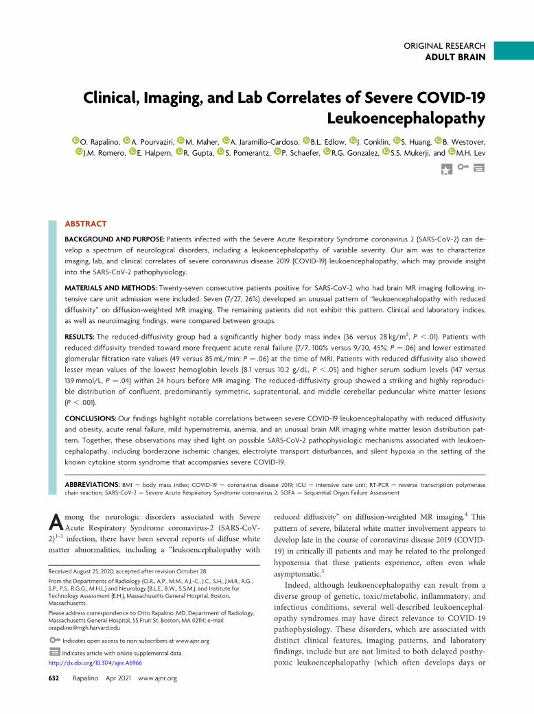
(n= 7146) and a radiology database ofbrain MR imaging studies (n = 2266)acquired between February 1, 2020,and May 12, 2020. SARS-CoV-2 infec-tion was confirmed by at least 1 posi-tive real-time reverse transcriptionpolymerase chain reaction (RT-PCR)test. Inclusion criteria were the follow-ing: 1) an assay positive for SARS-CoV-2; 2) age 18 years or older; 3)admission to the intensive care unit(ICU); and 4) brain MR imagingperformed .24 hours following ICUadmission (Fig 1). Patients not admittedto the ICU, younger than 18 years of age,or having undergone brain MR imagingbefore their ICU admission wereexcluded. One patient who had a brainMR imaging at the time of the ICUadmission was excluded due to thepresence of a pre-existent neurologiccondition (suspected central pontinemyelinolysis). During data collection,308 patients (202 men, 66%) withCOVID-19 were admitted to the ICU.
Of 47 patients positive for SARS-CoV-2 who had brain MRimaging, 28 (28/47, 60%) were admitted to the ICU (Fig 1).
Definition of Baseline and Clinical VariablesDemographic and baseline clinical characteristics were recorded,including the duration of symptoms before admission and thenumber of days from ICU admission to MR imaging acquisition,as well as documentation of organ involvement and standard lab-oratory data both at the time of ICU admission and within24 hours before MR imaging acquisition. The Sequential OrganFailure Assessment (SOFA) score was calculated for every patientat ICU admission. All data were collected using predeterminedguidelines from the patients’ electronic medical records. Baselineand ICU data collection was performed blinded to the imagingfindings, including the diffusion abnormalities.
Imaging ProtocolThe brain MR imaging studies were performed on 1.5T (ExciteHDx; GE Healthcare) and 3T (Magnetom Skyra or Prisma;Siemens) scanners. The sequences and acquisition parameters ofthe typical MR imaging protocol included the following: axial dif-fusion (voxel size ¼ 1.4 � 1.4 � 5.0mm, TR¼ 5000 ms, TE¼96.0ms, b-values ¼ 0 and 1000 s/mm2), axial SWI (voxel size ¼0.8 � 0.8 � 1.8mm), axial FLAIR (voxel size ¼ 0.4 � 0.4 �4.0mm, TR¼ 9000ms, TE¼ 85.0ms, TI¼ 2500ms), axial T1(voxel size ¼ 0.8 � 0.8 � 4.0mm, TR¼ 220ms, TE ¼ 2.46ms),sagittal MPRAGE (voxel size ¼ 1.0 � 1.0 � 1.0mm, TR¼2530ms, multiple TE values), and axial T2 BLADE (Siemens)(voxel size ¼ 0.7 � 0.7 � 7.0mm, TR ¼ 5500 ms, TE ¼ 17 ms).Patients without gadolinium contraindications were administered0.1mmol/kg of gadoterate meglumine (Dotarem; Guerbet). Thefollowing sequences were acquired following contrast
FIG 1. Flow chart of the patient-selection process.
AJNR Am J Neuroradiol 42:632–38 Apr 2021 www.ajnr.org 633
administration: axial T1-2D (voxel size ¼ 0.8 � 0.8 � 4.0mm,TR¼ 220ms, TE ¼ 2.46ms) and sagittal MPRAGE (voxel size ¼1.0� 1.0� 1.0mm, TR¼ 2530ms, multiple TE values).
Imaging EvaluationImaging was reviewed using the PACS by 2 neuroradiologists(O.R., M.M.), blinded to the clinical and laboratory findings.Discrepancies were resolved by consensus. Patients were dicho-tomized into 2 discrete groups: those with-versus-without “leu-koencephalopathy with reduced diffusivity,” based on thepresence or absence of white matter lesions with reduced diffusiv-ity not consistent with an established pattern of ischemic infarc-tion. The extent of supra- and infratentorial WM involvementwas qualitatively graded for each patient on the basis of a 4-pointLikert score (0=normal, 1 =mild, 2 =moderate, and 3= severe).The anatomic distribution of white matter signal abnormalitieswas recorded. The presence and number of microhemorrhageswere assessed on SWI sequences when available (n=24) or ongradient recalled-echo sequences (n=3). Additional neuroimag-ing findings such as abnormal gray matter signal or intracranialenhancement were collected.
Statistical AnalysisDescriptive statistics were performed to summarize categoric var-iables as percentages and continuous variables using means andproportions, range, and 95% confidence intervals. Q-Q plots andShapiro-Wilk tests were implemented for testing normality.Nonparametric data were described using median and interquar-tile range. For comparison of means of continuous variables withnormal distribution, the Student t test was used. The Mann-WhitneyU test was chosen for comparison of nonparametric var-iables. The Fisher exact test was used to analyze the association ofclinical and laboratory findings between the groups with-versus-without reduced diffusivity. Differences and 95% confidenceintervals were estimated using the t test for all variables reported.P values ,.05 were considered significant. Analyses were con-ducted using SPSS, Version 24 (IBM).
RESULTSPatient Cohort CharacteristicsTwenty-seven patients (20/27 men, 74%; mean age, 63 years; 95%CI, 57–69 years) were included in the analysis. Seven (7/27, 26%)were identified as having leukoencephalopathy with reduced dif-fusivity (5/7 men, 71%; mean age, 63 years; 95% CI, 55–71 years);the remaining 20 in the control group did not exhibit this pattern(15/20 men, 75%; mean age, 63 years; 95% CI, 58–69 years)(Table 1 in the Online Supplemental Data). There were no signifi-cant differences in the percentage of Asian (0% vs. 5%), Black/African American (14% vs. 30%), White (29% vs. 30%), orHispanic (57% vs. 35%) between the reduced diffusivity and con-trol groups. The patients with reduced diffusivity compared withthe control group had a significantly higher BMI (36 versus28 kg/m2, P, .01). Other baseline characteristics, including priortobacco use, concurrent illnesses, symptoms before admission,previous cardiac arrest, and number of days symptomatic(including dyspnea), were similar per the Online SupplementalData. Except for a single patient in the control group, all patients
were intubated at ICU admission. One patient in the controlgroup (1/20, 5%) received extracorporeal membrane oxygen-ation. Four patients in the reduced-diffusivity group and 5 in thecontrol group were treated with nitric oxide (4/7, 57%, versus5/20, 25%; P ¼ .17). The most common comorbidities were ob-structive sleep apnea (5/7, 71.4%, versus 14/20, 70%; P ¼ .26),diabetes (4/7, 57%, versus 8/20, 40%; P ¼ .66), and hyperten-sion (3/7, 43%, versus 13/20, 65%; P ¼ .39) (Table 1 in theOnline Supplemental Data).
ICU Clinical DataPatients in the reduced-diffusivity group trended toward morefrequent acute renal failure documented in the ICU clinical notes(7/7, 100%, versus 9/20, 45%; P ¼ .06) and lower estimated glo-merular filtration rate values (49 versus 85mL/min, P ¼ .06) atMR imaging (Table 2 in the Online Supplemental Data). Therewere no significant differences between the 2 groups in the fre-quency of septic shock (6/7, 85.7%, versus 18/20, 90.0%), acuteliver failure (1/7, 14.3%, versus 2/20, 10.0%), cardiac involvement(2/7, 28.6%, versus 2/20, 10.0%), ischemic bowel (1/7, 14.3%, ver-sus 2/20, 10.0%), or other clinical and lab indices, including overtDisseminated Intravascular Coagulation, as listed in table 2 of theOnline Supplemental Data. The reduced-diffusivity groupshowed significantly depressed mean values of the lowest hemo-globin levels (8.1 versus 10.2 g/dL, P , .05), elevated serum so-dium levels (147 versus 139mmol/L, P¼ .04), and a trend towardelevated highest D-dimers (4080 versus 2386ng/L, P ¼ .09), allobtained within 24 hours before MR imaging. Other clinical vari-ables, including SOFA scores at admission, sedatives, nitric oxidetreatment, and ICU length of stay (up through the time of datacollection), were not significantly different between the 2 groups(Table 1 in the Online Supplemental Data).
One patient in the reduced-diffusivity group and one in thecontrol group underwent lumbar puncture for CSF analysis. Thepatient in the control group was diagnosed with CNS cryptococ-cosis in the setting of HIV and SARS-CoV-2 co-infection; theCSF sample from this patient showed 108 white blood cells andprotein levels at 141mg/dL. CSF samples from the patient in thereduced-diffusivity group were without pleocytosis (#4 cells)and had mean protein levels of 152mg/dL.
Imaging FindingsThe extent of bilateral, supra- and infratentorial white matterabnormalities was significantly greater in the reduced-diffusivitygroup than in the control group (median Likert scores, 3, 2 versus1, 0, respectively; P, .002). White matter lesions in the reduced-diffusivity group were also more likely than in controls toshow an unusual, confluent pattern of supratentorial (P , .001)and middle cerebellar peduncular (6/7, 86%, versus 1/20, 5%;P , .001) involvement (Online Supplemental Data, Figs 2–4).Cerebellar white matter (P ¼ .002) and brain stem (P ¼ .04)lesions were significantly more common in the reduced-diffusiv-ity group than in the control group, whereas white matter lesionstrended toward a predominantly symmetric pattern in thereduced-diffusivity group (7/7, 100%, versus 11/19, 58%; P ¼ .06).There was no significant difference between groups regarding U-fiber or corpus callosal involvement, percentage of patients with
634 Rapalino Apr 2021 www.ajnr.org
.10 microbleeds, cortical laminar necrosis, or abnormal deep graymatter signal changes (all P. .25), per Online Supplemental Data.
Contrast was administered to 4/7 (57%) patients in thereduced-diffusivity group and 7/20 (35%) in the control group.One patient in each group had abnormal intraparenchymalenhancement (focal cortical enhancement in a region of laminarnecrosis for a patient in the reduced-diffusivity group, and a smallenhancing focus for a patient with cerebral cryptococcosis associ-ated with HIV/SARS-CoV-2 co-infection in the control group).No definite abnormal leptomeningeal enhancement was presentin any of our patients.
DISCUSSIONOur results show that in a consecutive cohort of adult patientspositive for SARS-CoV-2 in the ICU requiring post-admissionbrain MR imaging, severe COVID-19 leukoencephalopathy withreduced diffusivity was associated with obesity, acute renal failure,mild hypernatremia, anemia, and an unusual, striking, and abnor-mal brain white matter lesion distribution pattern on MR imaging,featuring diffuse, confluent, predominantly symmetric supratento-rial and middle cerebellar peduncular lesions. Although there wereno significant differences in age, gender, race, or ethnicity betweenthe reduced diffusivity and control groups, in both groups, olderage (mean 63 years) and male sex (71–75%) were predominant.
Several articles to date have reported the occurrence of whitematter signal abnormalities in patients with severe COVID-19.4,17-19
Kandemirli et al17 observed 3 (of 12) critically ill patients with sub-cortical or deep white matter lesions but without reduced diffusivity.More recently, Radmanesh et al4 described a series of patients with
leukoencephalopathy, reduced diffusivity,and microhemorrhages. A systematicanalysis of the risk factors and laboratoryand clinical variables associated with thisunusual leukoencephalopathy, however,has not previously been emphasized inthe literature.3
We saw no significant differencesbetween groups regarding the durationof subjective dyspnea before ICU admis-sion, the lowest reported pulse oximetrybefore admission, PaO2/FiO2 ratios onadmission, the need for extracorporealmembrane oxygenation, or worst arte-rial partial pressure of oxygen (PO2)and lowest oxygen saturation duringthe first 24 hours in the ICU (OnlineSupplemental Data). These similaritiesin baseline oxygenation parameterswere surprising, given that hypoxiahas been hypothesized to be a majorculprit in the pathophysiology of thesewhite matter changes. Indeed, oxygen-ation indices (worst arterial PO2 andlowest O2 saturation oxygenation) onthe day of MR imaging were lower inthe control group, likely reflecting that
45% of those patients had been extubated by that time (versus14% extubated in the reduced-diffusivity group, OnlineSupplemental Data).
Except for body mass index (BMI), our reduced-diffusivity andcontrol groups were well-matched (Online Supplemental Data). Thereduced-diffusivity group had a significantly higher mean BMI of 36versus 28 in the control group (corresponding to “moderately obese”versus “overweight,” respectively). Although there is no estab-lished direct link between obesity and leukoencephalop-athy, obesity is a known risk factor for insulin resistance,lacunar infarcts, and chronic small-vessel disease.20-22 The effectof obesity on arteriosclerosis might be a contributing factor tothe increasingly recognized thrombotic microangiopathy andendotheliitis associated with SARS-CoV-2 infection.23 Lampe etal24 described visceral obesity being associated with a predomi-nance of deep white matter (rather than periventricular)MR imaging abnormalities and suggested a potential rolefor interleukin 6 and other proinflammatory cytokines inthe development of white matter pathology. The microan-giopathic effect of obesity (with potential superimposedeffects mediated by cytokines) might further compromiseborderzone regions located in the deep cerebral white mat-ter and middle cerebellar peduncles.25
Several clinical variables were different between our 2 cohorts,including a lower mean value for the lowest hemoglobin level within24 hours before MR imaging (8.1 versus 10.2, P , .05), mildlyhigher mean values of the most elevated serum sodium at the sametime point (147 versus 139, P, .04), and a trend toward more acuterenal dysfunction (glomerular filtration rate = 49 versus 85mL/min;P , .06) in the reduced-diffusivity group. Other organ system
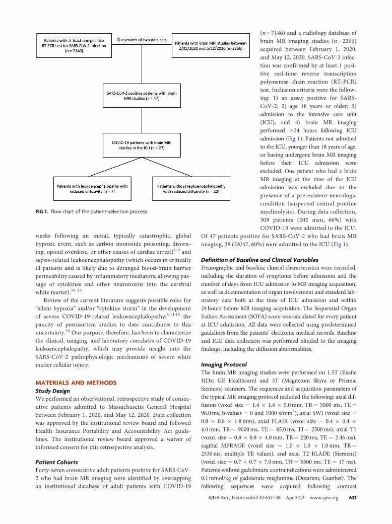
FIG 2. A 64-year-old man with a history of diabetes and hyperlipidemia, admitted with COVID-19–related hypoxemic respiratory failure and worsening septic shock. Axial DWI (A–C) and ADCimages (D–F) show extensive, bilateral, predominantly symmetric, DWI-hyperintense signal in thewhite matter of the bilateral middle cerebellar peduncles, corona radiata, and centrum semi-ovale, with corresponding dark signal intensity on the ADC images (yellow arrows), reflectingleukoencephalopathic changes with marked, confluent reduced diffusivity.
AJNR Am J Neuroradiol 42:632–38 Apr 2021 www.ajnr.org 635
damage was not significantly different between groups, includingcardiac events, acute liver failure, overt Disseminated IntravascularCoagulation, ischemic bowel, and mean number of systems affected(Online Supplemental Data).
Although these differences may simply reflect an epiphenome-non, it is intriguing to speculate whether cytokine effects, which
could also be related to obesity, might con-tribute to some of these abnormal lab indi-ces. For example, interleukin 6 and otherproinflammatory cytokines (eg, interleukin1 and tumor necrosis factor-a) are knownto have a central role in the anemia seenduring systemic inflammatory processes,impacting iron homeostasis and the retic-uloendothelial system as well as impairingregulatory feedback of iron absorption inthe gastrointestinal tract.26,27 Interleukin6 has been shown to mediate low ironlevels during inflammatory states via theproduction of hepcidin.28 These cytokinesmight also modulate the transcription ofthe erythropoietin (EPO) gene and inhibiterythroid progenitor cells in the bonemarrow.26
The modest hypernatremia at thetime of MR imaging is more challengingto explain. Both groups had similar criti-cal illness severity (reflected by SOFAscores of 9.3 and 7, respectively; P¼ .09)and had no reported differences in waterintake or extrarenal water losses (gastro-intestinal- or skin-mediated). Althoughthis finding might reflect hypothalamicinvolvement with impaired water home-ostasis, renal dysfunction in COVID-19has more often been reported to result inhyponatremia.29,30
The pattern of MR imaging signalabnormalities we observed is striking.This pattern of diffuse, confluent, pre-dominantly symmetric supra- and infra-tentorial involvment with middlecerebellar peduncle lesions (seen in 6/7cases, 86%) is unusual and noteworthy.The neuroanatomic distribution of theseabnormalities appears more selectivethan those reported in cases of delayedposthypoxic leukoencephalopathy at ourinstitution and in the literature. Delayedposthypoxic encephalopathy is oftenmore extensive, with subcortical whitematter involvement and less frequentinfratentorial involvement.7,31 None ofthe patients in our reduced-diffusivitygroup had a history of recent cardiacarrest, though 1 patient in the controlgroup had an episode of cardiac arrest
without developing these findings.There have been multiple reports of white matter abnormal-
ities associated with sepsis and critical illness. Sharshar et al12
described white matter lesions in 5/9 (55%) patients with septicshock and clinical brain dysfunction (mean SOFA, 8). Polito et al11
published a similar study of 71 patients with septic shock and
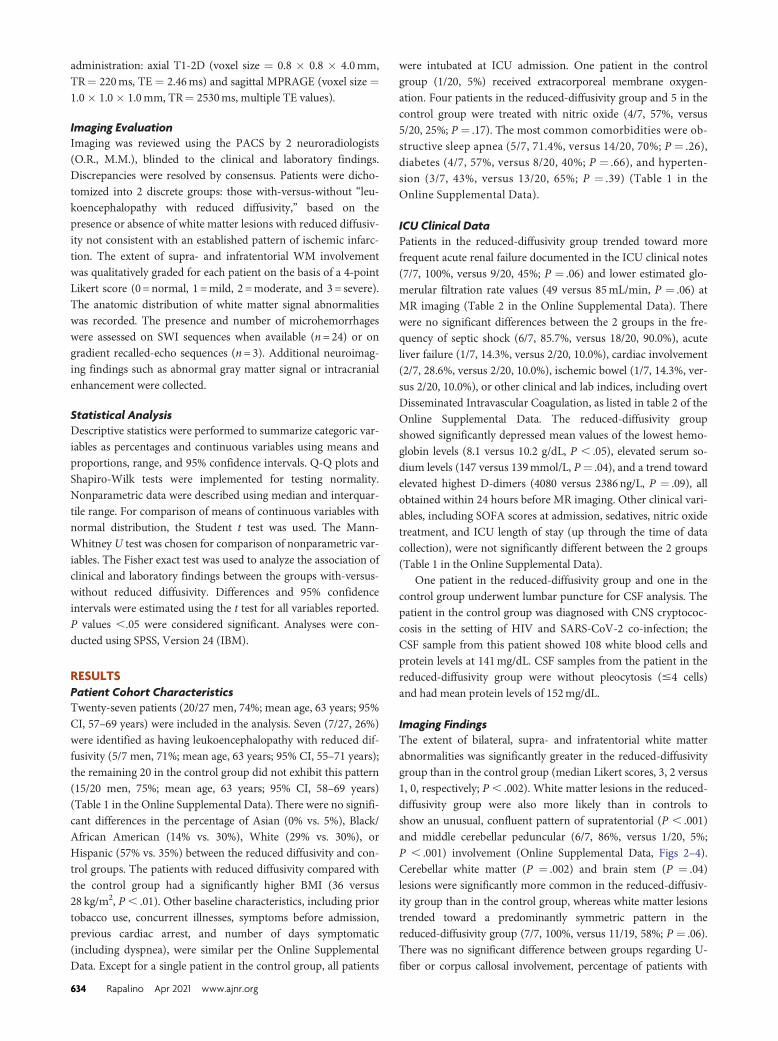
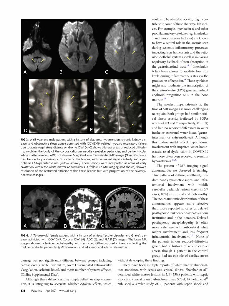
FIG 3. A 63-year-old male patient with a history of diabetes, hypertension, chronic kidney dis-ease, and obstructive sleep apnea admitted with COVID-19–related hypoxic respiratory failuredue to acute respiratory distress syndrome. DWI (A–C) shows bilateral areas of reduced diffusiv-ity, involving the body of the corpus callosum, middle cerebellar peduncles, and periventricularwhite matter (arrows, ADC not shown). Magnified axial T2-weighted MR images (D and E) show apeculiar cavitary appearance of some of the lesions, with decreased signal centrally and a pe-ripheral T2-hyperintense rim (yellow arrows). These lesions were interpreted as areas of earlycavitation within the white matter abnormalities. A follow-up MR imaging (not shown) showedresolution of the restricted diffusion within these lesions but with progression of the cavitary/necrotic changes.
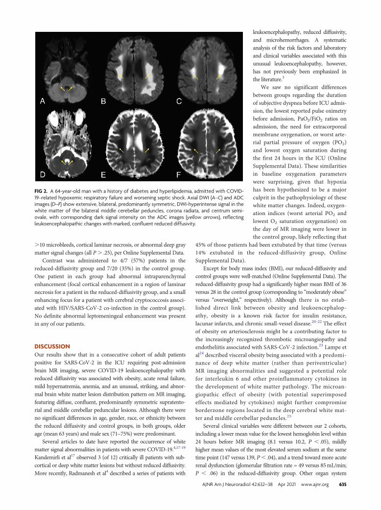
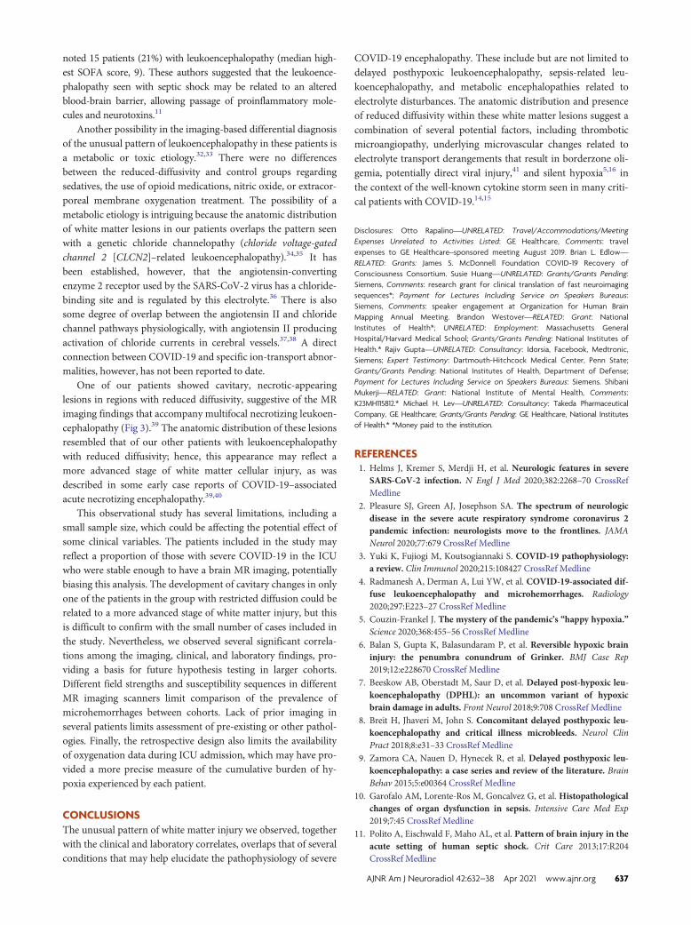
FIG 4. A 76-year-old female patient with a history of schizoaffective disorder and Grave’s dis-ease, admitted with COVID-19. Coronal DWI (A), ADC (B), and FLAIR (C) images. The brain MRimages showed a leukoencephalopathy with restricted diffusion, predominantly affecting themiddle cerebellar peduncles (yellow arrows) and adjacent cerebellar white matter.
636 Rapalino Apr 2021 www.ajnr.org
noted 15 patients (21%) with leukoencephalopathy (median high-est SOFA score, 9). These authors suggested that the leukoence-phalopathy seen with septic shock may be related to an alteredblood-brain barrier, allowing passage of proinflammatory mole-cules and neurotoxins.11
Another possibility in the imaging-based differential diagnosisof the unusual pattern of leukoencephalopathy in these patients isa metabolic or toxic etiology.32,33 There were no differencesbetween the reduced-diffusivity and control groups regardingsedatives, the use of opioid medications, nitric oxide, or extracor-poreal membrane oxygenation treatment. The possibility of ametabolic etiology is intriguing because the anatomic distributionof white matter lesions in our patients overlaps the pattern seenwith a genetic chloride channelopathy (chloride voltage-gatedchannel 2 [CLCN2]–related leukoencephalopathy).34,35 It hasbeen established, however, that the angiotensin-convertingenzyme 2 receptor used by the SARS-CoV-2 virus has a chloride-binding site and is regulated by this electrolyte.36 There is alsosome degree of overlap between the angiotensin II and chloridechannel pathways physiologically, with angiotensin II producingactivation of chloride currents in cerebral vessels.37,38 A directconnection between COVID-19 and specific ion-transport abnor-malities, however, has not been reported to date.
One of our patients showed cavitary, necrotic-appearinglesions in regions with reduced diffusivity, suggestive of the MRimaging findings that accompany multifocal necrotizing leukoen-cephalopathy (Fig 3).39 The anatomic distribution of these lesionsresembled that of our other patients with leukoencephalopathywith reduced diffusivity; hence, this appearance may reflect amore advanced stage of white matter cellular injury, as wasdescribed in some early case reports of COVID-19–associatedacute necrotizing encephalopathy.39,40
This observational study has several limitations, including asmall sample size, which could be affecting the potential effect ofsome clinical variables. The patients included in the study mayreflect a proportion of those with severe COVID-19 in the ICUwho were stable enough to have a brain MR imaging, potentiallybiasing this analysis. The development of cavitary changes in onlyone of the patients in the group with restricted diffusion could berelated to a more advanced stage of white matter injury, but thisis difficult to confirm with the small number of cases included inthe study. Nevertheless, we observed several significant correla-tions among the imaging, clinical, and laboratory findings, pro-viding a basis for future hypothesis testing in larger cohorts.Different field strengths and susceptibility sequences in differentMR imaging scanners limit comparison of the prevalence ofmicrohemorrhages between cohorts. Lack of prior imaging inseveral patients limits assessment of pre-existing or other pathol-ogies. Finally, the retrospective design also limits the availabilityof oxygenation data during ICU admission, which may have pro-vided a more precise measure of the cumulative burden of hy-poxia experienced by each patient.
CONCLUSIONSThe unusual pattern of white matter injury we observed, togetherwith the clinical and laboratory correlates, overlaps that of severalconditions that may help elucidate the pathophysiology of severe
COVID-19 encephalopathy. These include but are not limited todelayed posthypoxic leukoencephalopathy, sepsis-related leu-koencephalopathy, and metabolic encephalopathies related toelectrolyte disturbances. The anatomic distribution and presenceof reduced diffusivity within these white matter lesions suggest acombination of several potential factors, including thromboticmicroangiopathy, underlying microvascular changes related toelectrolyte transport derangements that result in borderzone oli-gemia, potentially direct viral injury,41 and silent hypoxia5,16 inthe context of the well-known cytokine storm seen in many criti-cal patients with COVID-19.14,15
Disclosures: Otto Rapalino—UNRELATED: Travel/Accommodations/MeetingExpenses Unrelated to Activities Listed: GE Healthcare, Comments: travelexpenses to GE Healthcare–sponsored meeting August 2019. Brian L. Edlow—RELATED: Grants: James S. McDonnell Foundation COVID-19 Recovery ofConsciousness Consortium. Susie Huang—UNRELATED: Grants/Grants Pending:Siemens, Comments: research grant for clinical translation of fast neuroimagingsequences*; Payment for Lectures Including Service on Speakers Bureaus:Siemens, Comments: speaker engagement at Organization for Human BrainMapping Annual Meeting. Brandon Westover—RELATED: Grant: NationalInstitutes of Health*; UNRELATED: Employment: Massachusetts GeneralHospital/Harvard Medical School; Grants/Grants Pending: National Institutes ofHealth.* Rajiv Gupta—UNRELATED: Consultancy: Idorsia, Facebook, Medtronic,Siemens; Expert Testimony: Dartmouth-Hitchcock Medical Center, Penn State;Grants/Grants Pending: National Institutes of Health, Department of Defense;Payment for Lectures Including Service on Speakers Bureaus: Siemens. ShibaniMukerji—RELATED: Grant: National Institute of Mental Health, Comments:K23MH115812.* Michael H. Lev—UNRELATED: Consultancy: Takeda PharmaceuticalCompany, GE Healthcare; Grants/Grants Pending: GE Healthcare, National Institutesof Health.* *Money paid to the institution.
REFERENCES1. Helms J, Kremer S, Merdji H, et al. Neurologic features in severe
SARS-CoV-2 infection. N Engl J Med 2020;382:2268–70 CrossRefMedline
2. Pleasure SJ, Green AJ, Josephson SA. The spectrum of neurologicdisease in the severe acute respiratory syndrome coronavirus 2pandemic infection: neurologists move to the frontlines. JAMANeurol 2020;77:679 CrossRef Medline
3. Yuki K, Fujiogi M, Koutsogiannaki S. COVID-19 pathophysiology:a review. Clin Immunol 2020;215:108427 CrossRef Medline
4. Radmanesh A, Derman A, Lui YW, et al. COVID-19-associated dif-fuse leukoencephalopathy and microhemorrhages. Radiology2020;297:E223–27 CrossRef Medline
5. Couzin-Frankel J. The mystery of the pandemic’s “happy hypoxia.”Science 2020;368:455–56 CrossRef Medline
6. Balan S, Gupta K, Balasundaram P, et al. Reversible hypoxic braininjury: the penumbra conundrum of Grinker. BMJ Case Rep2019;12:e228670 CrossRef Medline
7. Beeskow AB, Oberstadt M, Saur D, et al. Delayed post-hypoxic leu-koencephalopathy (DPHL): an uncommon variant of hypoxicbrain damage in adults. Front Neurol 2018;9:708 CrossRef Medline
8. Breit H, Jhaveri M, John S. Concomitant delayed posthypoxic leu-koencephalopathy and critical illness microbleeds. Neurol ClinPract 2018;8:e31–33 CrossRef Medline
9. Zamora CA, Nauen D, Hynecek R, et al. Delayed posthypoxic leu-koencephalopathy: a case series and review of the literature. BrainBehav 2015;5:e00364 CrossRef Medline
10. Garofalo AM, Lorente-Ros M, Goncalvez G, et al. Histopathologicalchanges of organ dysfunction in sepsis. Intensive Care Med Exp2019;7:45 CrossRef Medline
11. Polito A, Eischwald F, Maho AL, et al. Pattern of brain injury in theacute setting of human septic shock. Crit Care 2013;17:R204CrossRef Medline
AJNR Am J Neuroradiol 42:632–38 Apr 2021 www.ajnr.org 637
12. Sharshar T, Carlier R, Bernard F, et al. Brain lesions in septic shock:a magnetic resonance imaging study. Intensive Care Med2007;33:798–806 CrossRef Medline
13. Shindo A, Suzuki K, Iwashita Y, et al. Sepsis-associated encephalop-athy with multiple microbleeds in cerebral white matter. Am J Med2018;131:e297–98 CrossRef Medline
14. Mehta P, McAuley DF, Brown M, et al; HLH Across SpecialityCollaboration, UK. COVID-19: consider cytokine storm syndromesand immunosuppression. Lancet 2020;395:1033–34 CrossRefMedline
15. Moore BJ, June CH. Cytokine release syndrome in severe COVID-19. Science 2020;368:473–74 CrossRef Medline
16. Solomon IH, Normandin E, Bhattacharyya S, et al. Neuropathologicalfeatures of Covid-19. N Engl J Med 2020;383:989–92 CrossRefMedline
17. Kandemirli SG, Dogan L, Sarikaya ZT, et al. Brain MRI findings inpatients in the intensive care unit with COVID-19 infection.Radiology 2020;297:E232–35 CrossRef Medline
18. Sachs JR, Gibbs KW, Swor DE, et al. COVID-19-associated leukoen-cephalopathy. Radiology 2020;296:E184–85 CrossRef Medline
19. Lang M, Buch K, Li MD, et al. Leukoencephalopathy associatedwith severe COVID-19 infection: sequela of hypoxemia? AJNR AmJ Neuroradiol 2020;41:1641–45 CrossRef Medline
20. Dearborn JL, Schneider AL, Sharrett AR, et al. Obesity, insulin re-sistance, and incident small vessel disease on magnetic resonanceimaging: Atherosclerosis Risk in Communities Study. Stroke2015;46:3131–36 CrossRef Medline
21. Hakim AM. Small vessel disease. Front Neurol 2019;10:1020CrossRef Medline
22. Ter Telgte A, Wiegertjes K, Gesierich B, et al. Contribution of acuteinfarcts to cerebral small vessel disease progression. Ann Neurol2019;86:582–92 CrossRef Medline
23. Varga Z, Flammer AJ, Steiger P, et al. Endothelial cell infection andendotheliitis in COVID-19. Lancet 2020;395:1417–18 CrossRefMedline
24. Lampe L, Zhang R, Beyer F, et al. Visceral obesity relates to deepwhite matter hyperintensities via inflammation. Ann Neurol2019;85:194–203 CrossRef Medline
25. Kataoka H, Izumi T, Kinoshita S, et al. Infarction limited to bothmiddle cerebellar peduncles. J Neuroimaging 2011;21:e171–72CrossRef Medline
26. Hayden SJ, Albert TJ, Watkins TR, et al. Anemia in critical illness:insights into etiology, consequences, and management. Am J RespirCrit Care Med 2012;185:1049–57 CrossRef Medline
27. Houston DS. Hepcidin and the anemia of critical illness. Crit CareMed 2018;46:1030–31 CrossRef Medline
28. Nemeth E, Rivera S, Gabayan V, et al. IL-6 mediates hypoferremiaof inflammation by inducing the synthesis of the iron regulatoryhormone hepcidin. J Clin Invest 2004;113:1271–76 CrossRef Medline
29. Lippi G, South AM, Henry BM. Electrolyte imbalances in patientswith severe coronavirus disease 2019 (COVID-19). Ann ClinBiochem 2020;57:262–65 CrossRef Medline
30. Puelles VG, Lutgehetmann M, Lindenmeyer MT, et al. Multiorganand renal tropism of SARS-CoV-2. N Engl J Med 2020;383:590–92CrossRef Medline
31. Meyer MA. Delayed post-hypoxic leukoencephalopathy: casereport with a review of disease pathophysiology. Neurol Int 2013;5:e13 CrossRef Medline
32. de Oliveira AM, Paulino MV, Vieira AP, et al. Imaging patterns oftoxic and metabolic brain disorders. Radiographics 2019;39:1672–95CrossRef Medline
33. Sener RN.Diffusion magnetic resonance imaging patterns in meta-bolic and toxic brain disorders. Acta Radiol 2004;45:561–70CrossRef Medline
34. Gaitan-Penas H, Apaja PM, Arnedo T, et al. Leukoencephalopathy-causing CLCN2mutations are associated with impaired Cl(-) chan-nel function and trafficking. J Physiol 2017;595:6993–7008 CrossRefMedline
35. Guo Z, Lu T, Peng L, et al. CLCN2-related leukoencephalopathy: acase report and review of the literature. BMC Neurol 2019;19:156CrossRef Medline
36. Rushworth CA, Guy JL, Turner AJ. Residues affecting the chlorideregulation and substrate selectivity of the angiotensin-convertingenzymes (ACE and ACE2) identified by site-directed mutagenesis.FEBS J 2008;275:6033–42 CrossRef Medline
37. Li RS, Wang Y, Chen HS, et al. TMEM16A contributes to angioten-sin II-induced cerebral vasoconstriction via the RhoA/ROCK sig-naling pathway.Mol Med Rep 2016;13:3691–99 CrossRef Medline
38. Nelson MT, Conway MA, Knot HJ, et al. Chloride channel blockersinhibit myogenic tone in rat cerebral arteries. J Physiol1997;502:259–64 CrossRef Medline
39. Premji S, Kang L, Rojiani MV, et al.Multifocal necrotizing leukoen-cephalopathy: expanding the clinicopathologic spectrum. JNeuropathol Exp Neurol 2019;78:340–47 CrossRef Medline
40. Poyiadji N, Shahin G, Noujaim D, et al. COVID-19-associated acutehemorrhagic necrotizing encephalopathy: imaging features.Radiology 2020;296:E119–20 CrossRef Medline
41. Paniz-Mondolfi A, Bryce C, Grimes Z, et al. Central nervous systeminvolvement by severe acute respiratory syndrome coronavirus-2(SARS-CoV-2). J Med Virol 2020;92:699–702 CrossRef Medline
638 Rapalino Apr 2021 www.ajnr.org