Embed Size (px)
Citation preview
Teresa Gómez-Isla, MD, PhDMassachusetts General Hospital
Harvard Medical SchoolMassachusetts ADRC
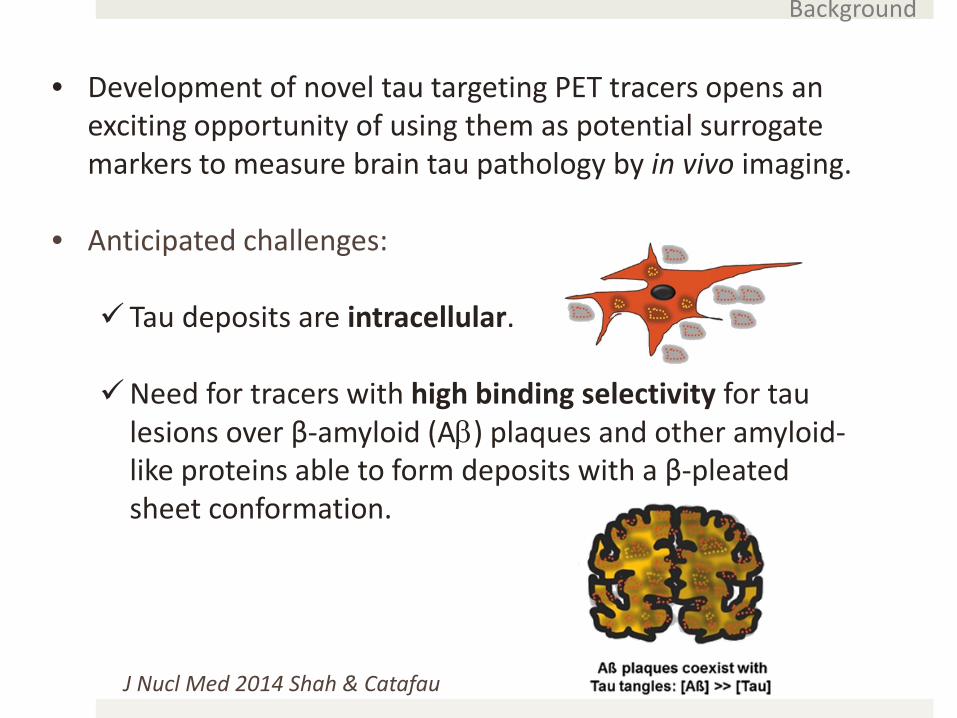
• Development of novel tau targeting PET tracers opens an exciting opportunity of using them as potential surrogate markers to measure brain tau pathology by in vivo imaging.
• Anticipated challenges:
Tau deposits are intracellular.
Need for tracers with high binding selectivity for tau lesions over β-amyloid (Aβ) plaques and other amyloid-like proteins able to form deposits with a β-pleated sheet conformation.
Background
J Nucl Med 2014 Shah & Catafau
Introduction
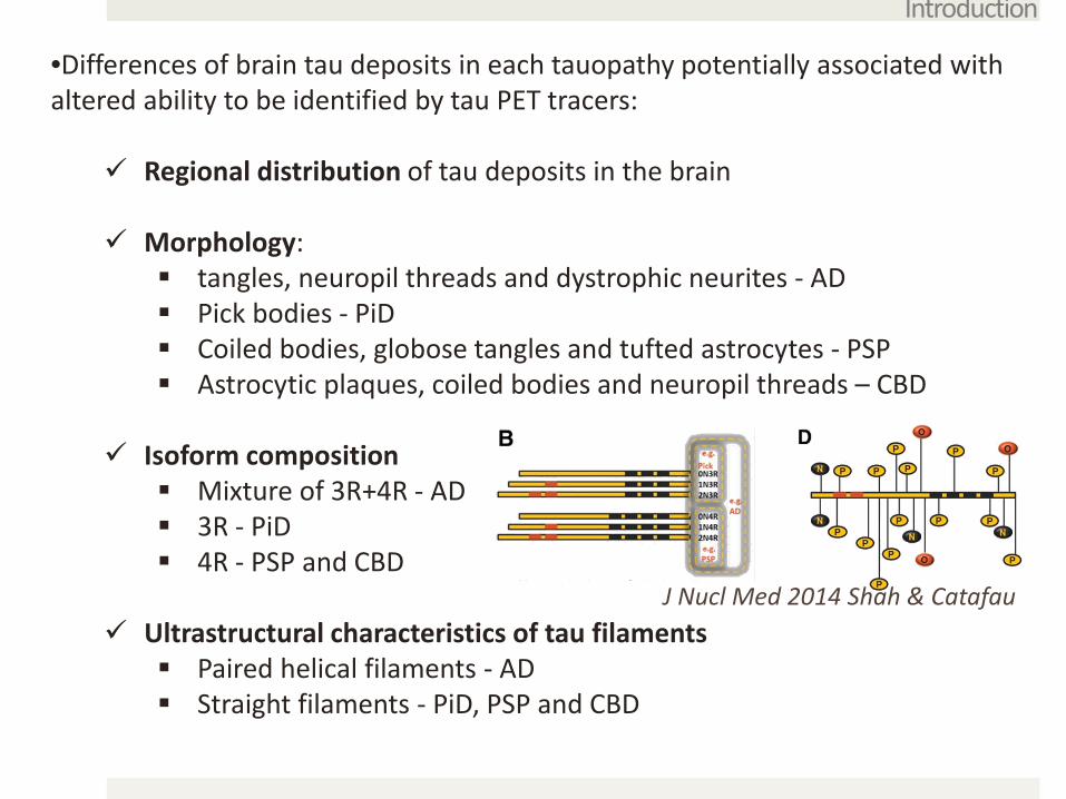
•Differences of brain tau deposits in each tauopathy potentially associated with altered ability to be identified by tau PET tracers:
Regional distribution of tau deposits in the brain
Morphology: tangles, neuropil threads and dystrophic neurites - AD Pick bodies - PiD Coiled bodies, globose tangles and tufted astrocytes - PSP Astrocytic plaques, coiled bodies and neuropil threads – CBD
Isoform composition Mixture of 3R+4R - AD 3R - PiD 4R - PSP and CBD
Ultrastructural characteristics of tau filaments Paired helical filaments - AD Straight filaments - PiD, PSP and CBD
J Nucl Med 2014 Shah & Catafau
Introduction
J Nucl Med 2014 Shah&Catafau
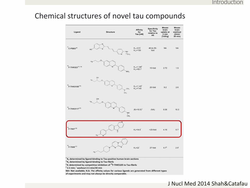
Chemical structures of novel tau compounds
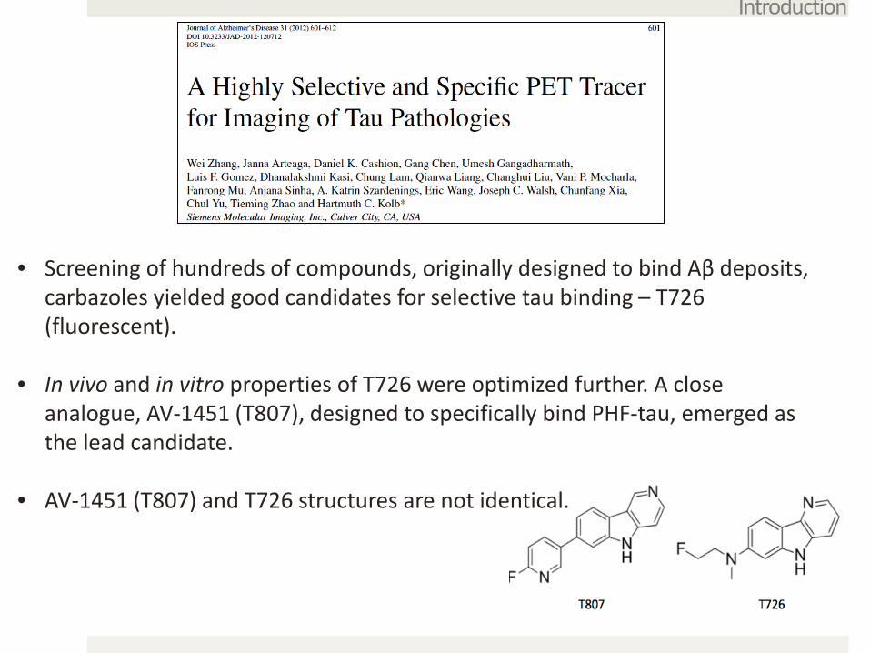
• Screening of hundreds of compounds, originally designed to bind Aβ deposits, carbazoles yielded good candidates for selective tau binding – T726 (fluorescent).
• In vivo and in vitro properties of T726 were optimized further. A close analogue, AV-1451 (T807), designed to specifically bind PHF-tau, emerged as the lead candidate.
• AV-1451 (T807) and T726 structures are not identical.
Introduction
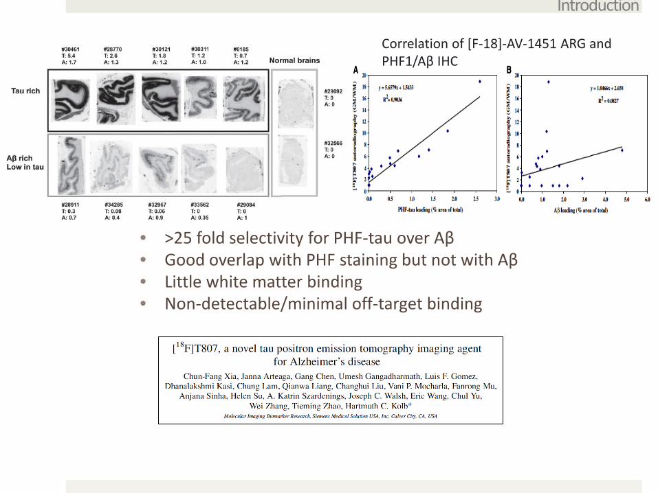
• >25 fold selectivity for PHF-tau over Aβ • Good overlap with PHF staining but not with Aβ• Little white matter binding• Non-detectable/minimal off-target binding
Introduction
Correlation of [F-18]-AV-1451 ARG and PHF1/Aβ IHC
HCS MCI mild AD severe AD
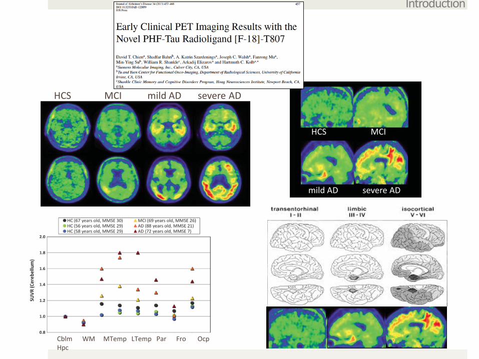
Cblm WM MTemp LTemp Par Fro OcpHpc
Introduction
HCS MCI
mild AD severe AD
• We aimed at examining region and substrate specific autoradiographic binding patterns of [F-18]-AV-1451 in brain postmortem tissue samples representing a diverse spectrum of neurodegenerative diseases in order to:
Validate the site/s of [F-18]-AV-1451 binding. Determine whether there is any off-target binding.
Early time in tau imaging, yet this compound is quickly making its way into use in clinical research including secondary prevention trials in AD like the A4 trial.
Goal
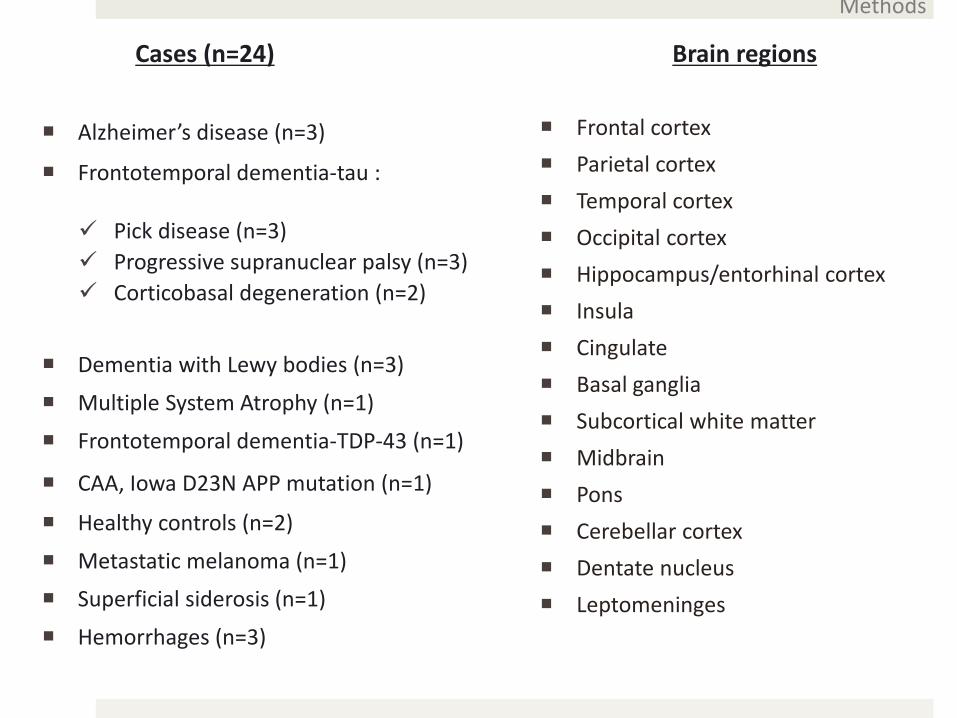
Cases (n=24)
Alzheimer’s disease (n=3)
Frontotemporal dementia-tau :
Pick disease (n=3) Progressive supranuclear palsy (n=3) Corticobasal degeneration (n=2)
Dementia with Lewy bodies (n=3)
Multiple System Atrophy (n=1)
Frontotemporal dementia-TDP-43 (n=1)
CAA, Iowa D23N APP mutation (n=1)
Healthy controls (n=2)
Metastatic melanoma (n=1)
Superficial siderosis (n=1)
Hemorrhages (n=3)
Brain regions
Frontal cortex Parietal cortex Temporal cortex Occipital cortex Hippocampus/entorhinal cortex Insula Cingulate Basal ganglia Subcortical white matter Midbrain Pons Cerebellar cortex Dentate nucleus Leptomeninges
Methods

(1) [F-18]-AV-1451 phosphor screen autoradiography
• Frozen sections thawed + fixed with Methanol x 20min
• [F-18]-AV-1451 (20μCi/ml) 60min incubation + washes with PBS and EtOH + air dried
• Adjacent sections with identical conditions + unlabeled AV-1451 (1μM) (“blocking”) to saturate available tau binding sites
• Phosphor screen (MultiSensitivePhosphor Screen, PerkinElmer Lifeand Analytic Sciences) overnight
• Imaging system (Cyclone Plus Storage Phosphor Scanner, Perkin Elmer Life and Analytic Sciences)
Methods

Methods
A photographic nuclear emulsion consists of a thin layer of radiation-sensitive silver crystals suspended in a gelatin matrix which is supported by a glass plate.
When silver grains are struck by radiation (photons or electrons), they are altered chemically to form a latent image of the silver crystals.
(2) [F-18]-AV-1451 nuclear emulsion autoradiography
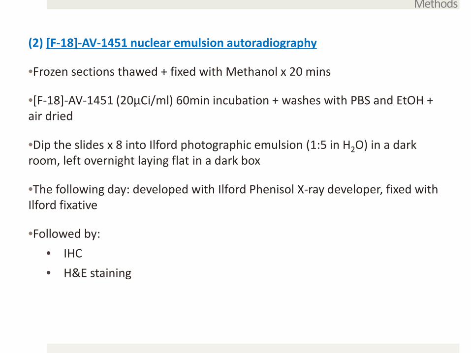
(2) [F-18]-AV-1451 nuclear emulsion autoradiography
•Frozen sections thawed + fixed with Methanol x 20 mins
•[F-18]-AV-1451 (20μCi/ml) 60min incubation + washes with PBS and EtOH + air dried
•Dip the slides x 8 into Ilford photographic emulsion (1:5 in H2O) in a dark room, left overnight laying flat in a dark box
•The following day: developed with Ilford Phenisol X-ray developer, fixed with Ilford fixative
•Followed by:• IHC • H&E staining
Methods

Methods
(3) [H3]-AV-1451 binding assay
•Fresh-frozen human brain samples are homogenized at RT in PBS (137 mM NaCl, 3 mM KCl, 10 mM sodium phosphate, pH 7.0) at a concentration of 10 mg of brain per milliliter.
•10 mg/ml frozen brain homogenate aliquots were thawed and diluted 10-fold in binding buffer to 1 mg/ml.
•500 μl of appropriate concentrations of non-radioactive compound combined with 400 μl of [H-3]-AV-1451 in a volume of 900 ml of binding buffer. The final concentration of [H-3]-AV-1451 is typically 1-2 nM.
•After incubation at RT for 60 min, the binding mixture is filtered through a Whatman GF/B glass filter and washed 5 times with 3 ml binding buffer.
•Filters are counted in Cytoscint-ES after sitting in the cocktail overnight.
•Complete (100%) inhibition of binding is defined as the number of counts displaced by 3 μM non-radioactive T807.

(1) [F-18]-AV-1451 phosphor screen autoradiography: AD vs controls
Marquie et al. Ann Neurol 2015
Results
Normal AD dementia
Tau (AV-1451)
PET images are courtesy of Dr. Keith Johnson
Very strong binding in the HF and EC, frontal , temporal, parietal and occipital cortices in brain slices containing robust loads of NTF from AD brains. Binding was almost completely blocked by cold tracer. No binding in control brains free of NFT pathology.
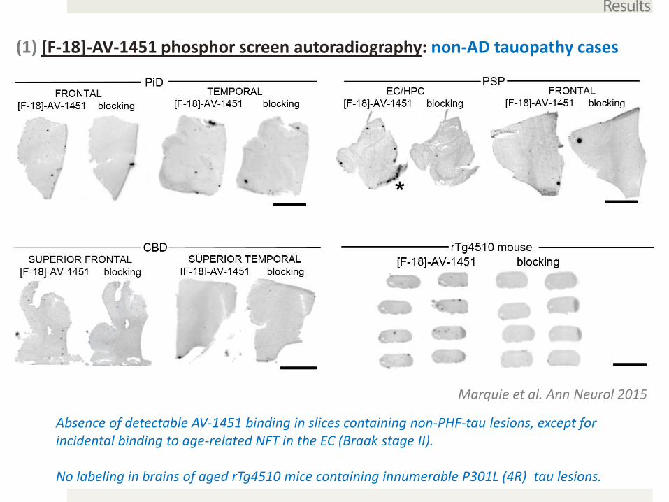
(1) [F-18]-AV-1451 phosphor screen autoradiography: non-AD tauopathy cases
Results
Marquie et al. Ann Neurol 2015
Absence of detectable AV-1451 binding in slices containing non-PHF-tau lesions, except for incidental binding to age-related NFT in the EC (Braak stage II).
No labeling in brains of aged rTg4510 mice containing innumerable P301L (4R) tau lesions.
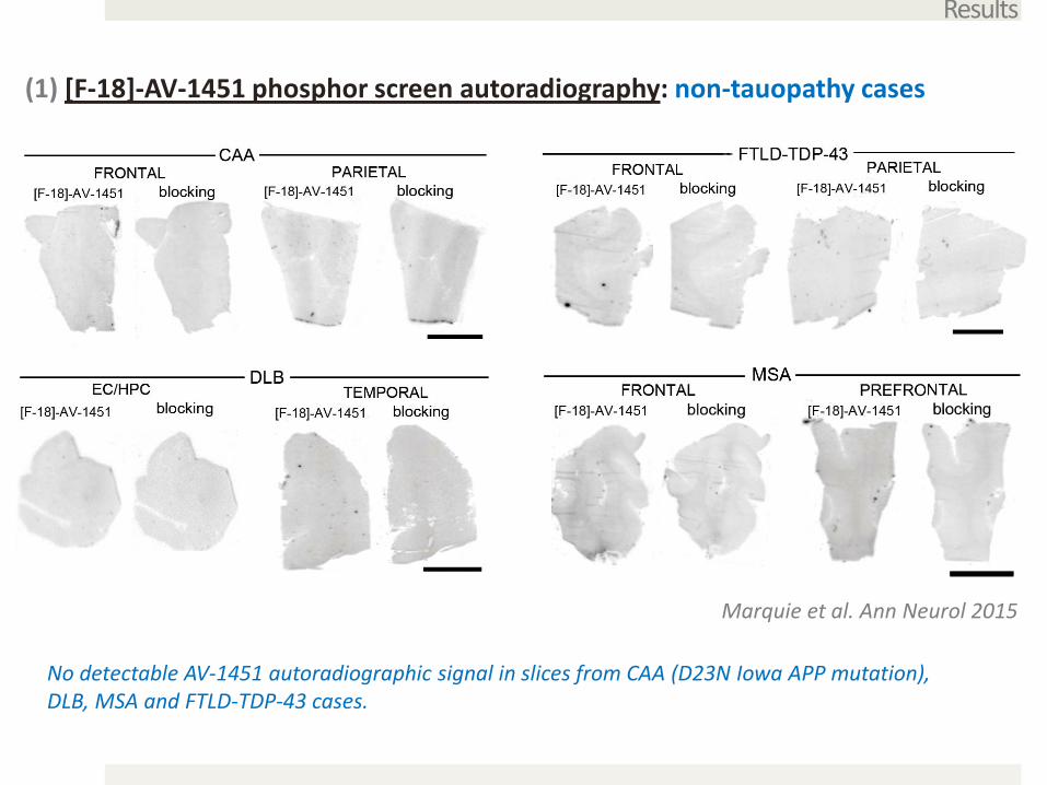
(1) [F-18]-AV-1451 phosphor screen autoradiography: non-tauopathy cases
Results
Marquie et al. Ann Neurol 2015
No detectable AV-1451 autoradiographic signal in slices from CAA (D23N Iowa APP mutation), DLB, MSA and FTLD-TDP-43 cases.
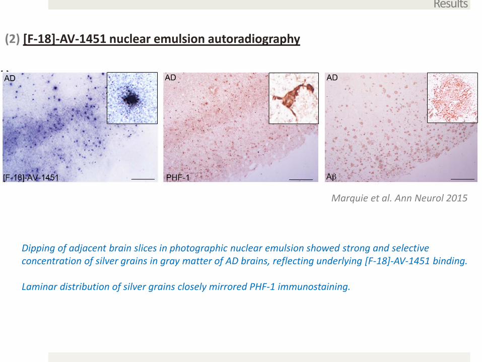
(2) [F-18]-AV-1451 nuclear emulsion autoradiography
Results
Marquie et al. Ann Neurol 2015
Dipping of adjacent brain slices in photographic nuclear emulsion showed strong and selective concentration of silver grains in gray matter of AD brains, reflecting underlying [F-18]-AV-1451 binding.
Laminar distribution of silver grains closely mirrored PHF-1 immunostaining.
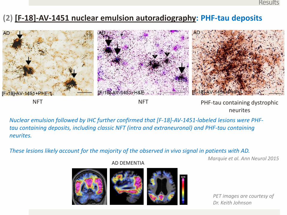
(2) [F-18]-AV-1451 nuclear emulsion autoradiography: PHF-tau deposits
Results
NFT PHF-tau containing dystrophic neurites
NFT
Marquie et al. Ann Neurol 2015
Nuclear emulsion followed by IHC further confirmed that [F-18]-AV-1451-labeled lesions were PHF-tau containing deposits, including classic NFT (intra and extraneuronal) and PHF-tau containing neurites.
These lesions likely account for the majority of the observed in vivo signal in patients with AD.
AD DEMENTIA
PET images are courtesy of Dr. Keith Johnson
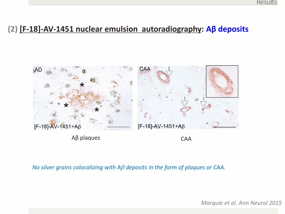
(2) [F-18]-AV-1451 nuclear emulsion autoradiography: Aβ deposits
Results
Marquie et al. Ann Neurol 2015
Aβ plaques CAA
No silver grains colocalizing with Aβ deposits in the form of plaques or CAA.
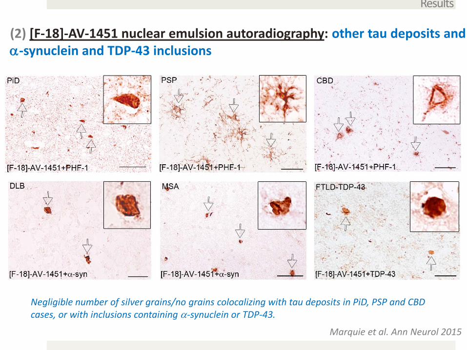
(2) [F-18]-AV-1451 nuclear emulsion autoradiography: other tau deposits and α-synuclein and TDP-43 inclusions
Results
Marquie et al. Ann Neurol 2015
Negligible number of silver grains/no grains colocalizing with tau deposits in PiD, PSP and CBD cases, or with inclusions containing α-synuclein or TDP-43.
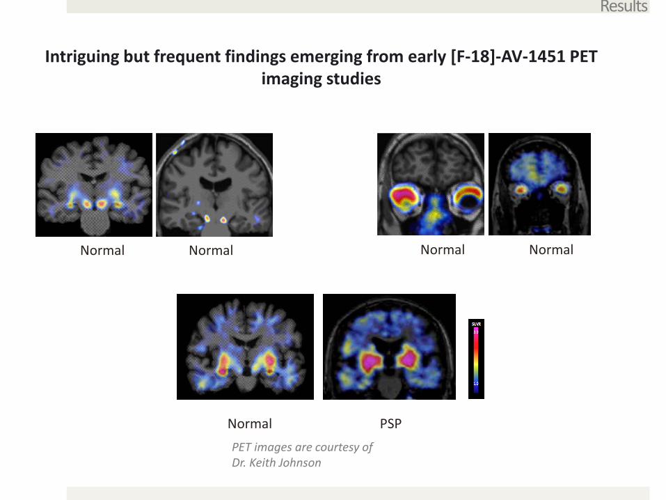
Intriguing but frequent findings emerging from early [F-18]-AV-1451 PET imaging studies
Results
PET images are courtesy of Dr. Keith Johnson
Normal Normal Normal Normal
Normal PSP
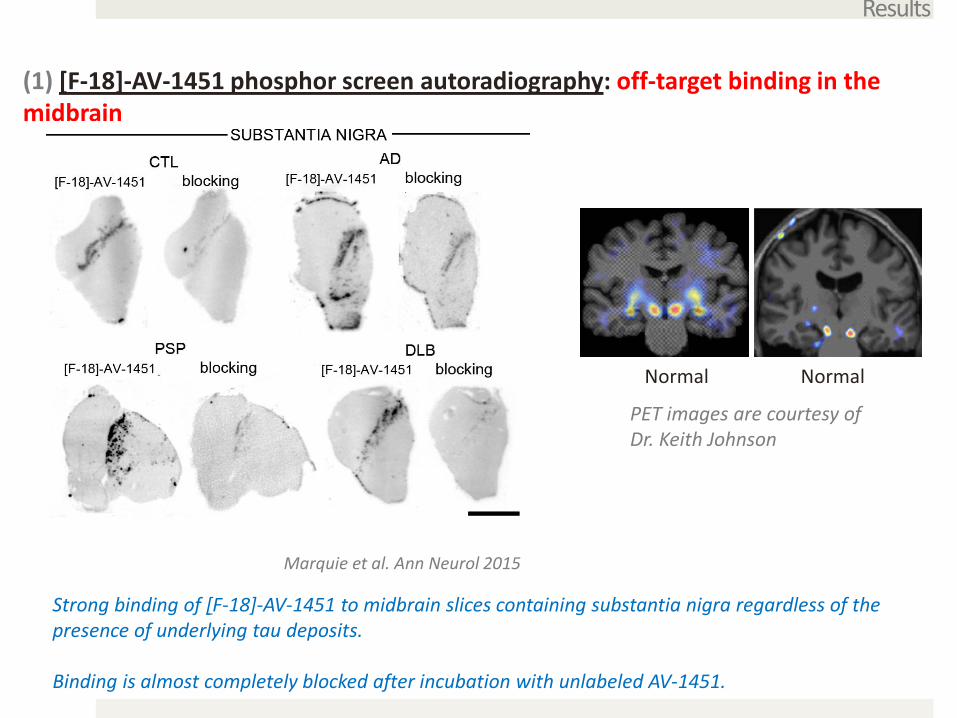
(1) [F-18]-AV-1451 phosphor screen autoradiography: off-target binding in the midbrain
Results
Marquie et al. Ann Neurol 2015
PET images are courtesy of Dr. Keith Johnson
Normal Normal
Strong binding of [F-18]-AV-1451 to midbrain slices containing substantia nigra regardless of the presence of underlying tau deposits.
Binding is almost completely blocked after incubation with unlabeled AV-1451.
Results
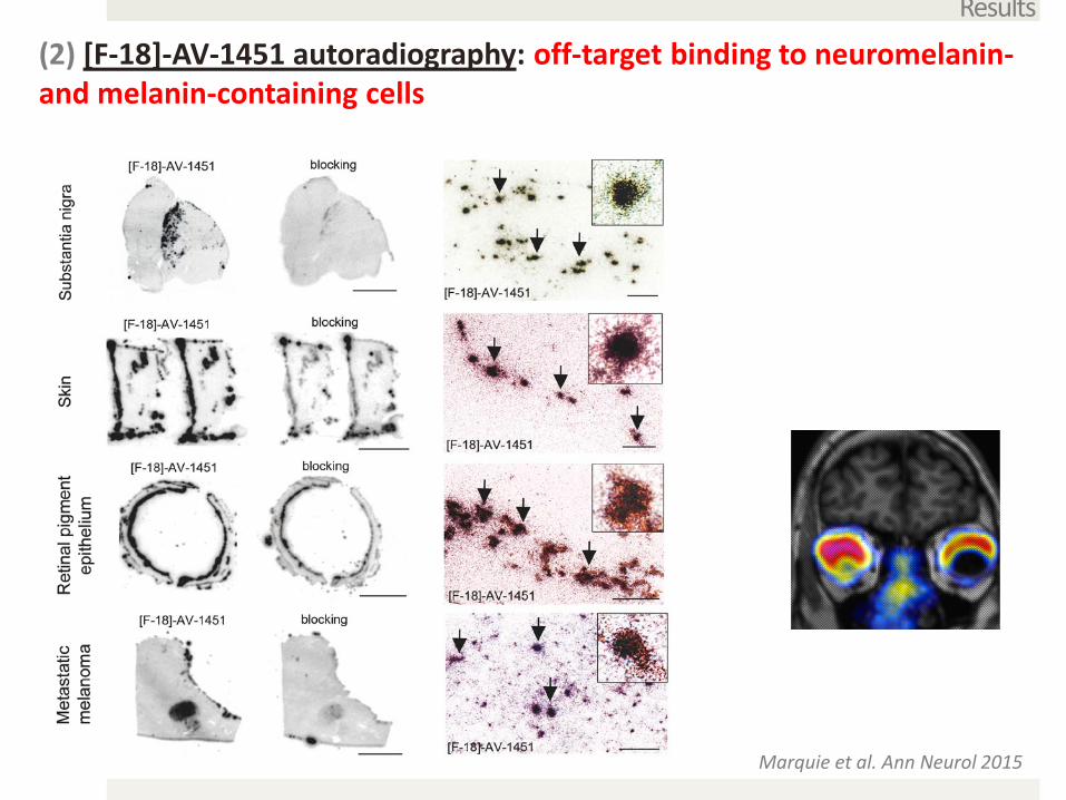
(2) [F-18]-AV-1451 autoradiography: off-target binding to neuromelanin-and melanin-containing cells
Marquie et al. Ann Neurol 2015
Results
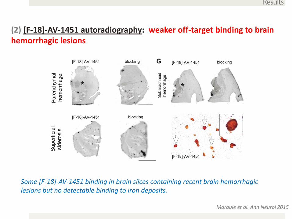
(2) [F-18]-AV-1451 autoradiography: weaker off-target binding to brain hemorrhagic lesions
Marquie et al. Ann Neurol 2015
Some [F-18]-AV-1451 binding in brain slices containing recent brain hemorrhagic lesions but no detectable binding to iron deposits.
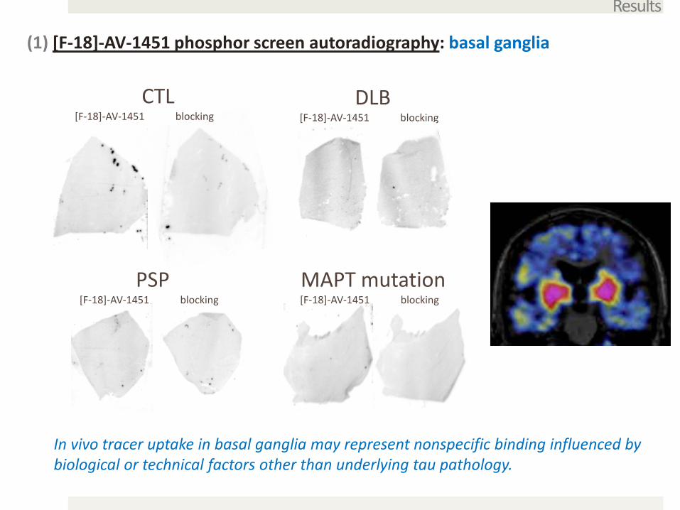
(1) [F-18]-AV-1451 phosphor screen autoradiography: basal ganglia
Results
MAPT mutation[F-18]-AV-1451 blocking
PSP[F-18]-AV-1451 blocking
DLB[F-18]-AV-1451 blocking
CTL[F-18]-AV-1451 blocking
In vivo tracer uptake in basal ganglia may represent nonspecific binding influenced by biological or technical factors other than underlying tau pathology.
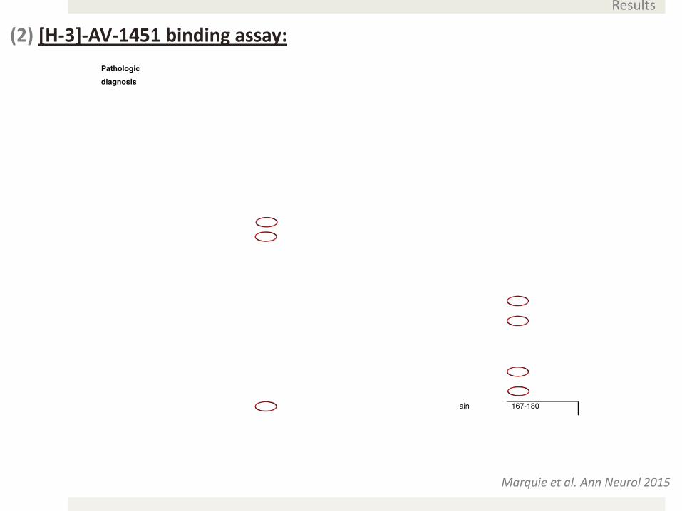
(2) [H-3]-AV-1451 binding assay:
Marquie et al. Ann Neurol 2015
Results
Conclusions
Our data derived from [F-18]-AV-1451-sensitive autoradiography and [H-3]-AV-1451 in vitro binding assays suggest that this tracer strongly binds to lesions primarily made of tau in the form of PHF (e g. neurofibrillary tangles and PHF-tau containing neurites).
[F-18]-AV-1451 does not bind to a significant extent to neuronal and glial inclusions mainly composed of straight tau filaments (non-AD tauopathies) or to Aβ deposits, α-synuclein and TDP-43 inclusions.
[F-18]-AV-1451 shows strong off-target binding to neuromelanin- and melanin-containing cells, and some weaker off-target binding to brain hemorrhagic lesions.
MADRC
Marta Marquie-SayaguesCharles VanderburgElizabeth BienLisa RycynaMatthew FroschBradley HymanKeith JohnsonBradford DickersonStephen GompertsIsabel CostantinoChris William
Radiology Department-MGH
Marc Normandin
ADRC University of Pittsburgh
Bill KlunkChester MathisMilos IkonomovicManik Debnath
Funding support :
• NIH Grant U01 AG016976• NIH Grant R01 AG43511• P50AG016574• ASISA foundation, Spain
Acknowledgements
*Avid/Lilly provided AV-1451 fluorescent analogues* Dr. Peter Davies kindly provided PHF1 antibody

How do we reconcile our findings with some findings communicated by others from early [F-18]-AV-1451 PET imaging studies in PSP patients?


![[Review] the Neuropathology of Drug Abuse_2011](https://img.pdfslide.us/doc/110x75/5477cd8b5806b5ed188b468d/review-the-neuropathology-of-drug-abuse2011.jpg)