Embed Size (px)
Citation preview

Clinical Image Gallery Next Generation – Volume 1

After long experience with the fi rst generation, a next generation Aquilion ONETM
was recently installed in our department. Four years ago we were excited by
the new applications offered by the 16 cm detector coverage, the decrease
in dose and the excellent image quality. With the new Aquilion ONE we are
at the latest hardware level, the image quality has improved again and the
patient doses were reduced once more. The new software comes with
Adaptive Diagnosis offering us the best solutions for a wide variety of clinical
questions. For example, when scanning metal hip implants SEMAR reduces
artifacts caused by metal so that we easily can diagnose the surrounding
areas. Variable Helical Pitch combines fast and low pitch in one continuous
scan, simplifying scan procedures, reducing contrast media and shortening
scan times. With SURESubtractionTM Lung we can visualize for example the effect
of lung emboli in detail which can affect treatment. With the new Aquilion ONE
we have state-of-the-art technology to offer our patients the best possible care.
Dr. Russell Bull
Royal Bournemouth Hospital, Bournemouth,
United Kingdom
2
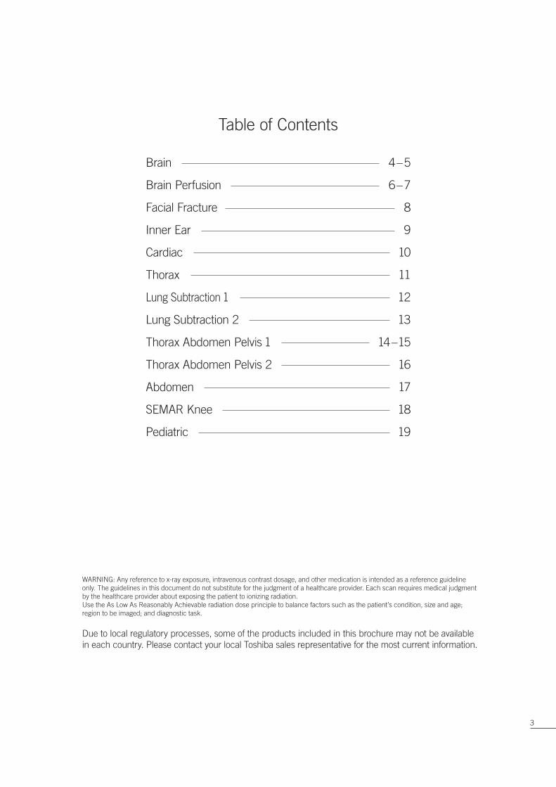
Brain 4 – 5
Brain Perfusion 6 – 7
Facial Fracture 8
Inner Ear 9
Cardiac 10
Thorax 11
Lung Subtraction 1 12
Lung Subtraction 2 13
Thorax Abdomen Pelvis 1 14 – 15
Thorax Abdomen Pelvis 2 16
Abdomen 17
SEMAR Knee 18
Pediatric 19
Table of Contents
Due to local regulatory processes, some of the products included in this brochure may not be available
in each country. Please contact your local Toshiba sales representative for the most current information.
WARNING: Any reference to x-ray exposure, intravenous contrast dosage, and other medication is intended as a reference guideline
only. The guidelines in this document do not substitute for the judgment of a healthcare provider. Each scan requires medical judgment
by the healthcare provider about exposing the patient to ionizing radiation.
Use the As Low As Reasonably Achievable radiation dose principle to balance factors such as the patient’s condition, size and age;
region to be imaged; and diagnostic task.
3

Brain
A native brain scan of a 32-year-old trauma patient was performed with multiple wide volumes of 80 x 0.5 mm.
The wide volume scan mode provides an excellent white/gray matter differentiation, while 3D reconstructions
and MPR images are of high quality.
4

Scan Mode Collimation Pitch kVp mAsRotation
Time (s)
Scan Range
(mm)
Dose
Reduction
CTDIvol
(mGy)
DLP
(mGy·cm)
Effective
Dose (mSv)k
Wide-Volume 0.5 mm x 80 / 120 250 1 151AIDR 3D
Standard55.0 1034.3 2.17 0.0021
5
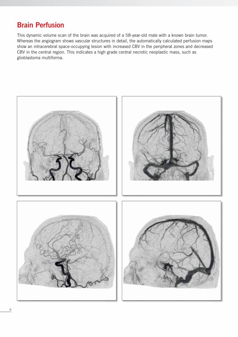
This dynamic volume scan of the brain was acquired of a 58-year-old male with a known brain tumor.
Whereas the angiogram shows vascular structures in detail, the automatically calculated perfusion maps
show an intracerebral space-occupying lesion with increased CBV in the peripheral zones and decreased
CBV in the central region. This indicates a high grade central necrotic neoplastic mass, such as
glioblastoma multiforma.
Brain Perfusion
6

Scan Mode Collimation Pitch kVp mAsRotation
Time (s)
Scan Range
(mm)
Dose
Reduction
CTDIvol
(mGy)
DLP
(mGy·cm)
Effective
Dose (mSv)k
Dynamic
Volume0.5 mm x 320 / 80
SUREExposureTM 3D
Standard0.35 160
AIDR 3D
Standard137.47 2199.6 4.62 0.0021
7

Scan Mode Collimation Pitch kVp mAsRotation
Time (s)
Scan Range
(mm)
Dose
Reduction
CTDIvol
(mGy)
DLP
(mGy·cm)
Effective
Dose (mSv)k
Volume 0.5 mm x 80 / 100 25 0.5 140AIDR 3D
Standard2.9 40.9 0.086 0.0021
Multiple fractures of the facial bones are shown in this scan of a 29-year-old male. This scan was acquired
in only one rotation of 0.5 seconds allowing the clinician to make an instant diagnosis in a trauma situation
where time is critical.
Facial Fracture
8

Scan Mode Collimation Pitch kVp mAsRotation
Time (s)
Scan Range
(mm)
Dose
Reduction
CTDIvol
(mGy)
DLP
(mGy·cm)
Effective
Dose (mSv)k
Volume 0.5 mm x 80 / 120 100 0.5 40AIDR 3D
Standard21.1 84.5 0.18 0.0021
This scan of the inner ear is acquired in just one rotation of 0.5 seconds. With a slice thickness of only
0.5 mm, a very high spatial resolution is reached which results in this detailed image of the inner ear.
Inner Ear
9
Scan Mode Collimation Pitch kVp mAsRotation
Time (s)
Scan Range
(mm)
Dose
Reduction
CTDIvol
(mGy)
DLP
(mGy·cm)
Effective
Dose (mSv)k
Volume 0.5 mm x 320 / 100SUREExposure 3D
Standard0.35 100
AIDR 3D
Standard4.9 58.7 0.82 0.014
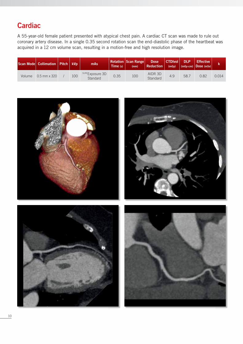
A 55-year-old female patient presented with atypical chest pain. A cardiac CT scan was made to rule out
coronary artery disease. In a single 0.35 second rotation scan the end-diastolic phase of the heartbeat was
acquired in a 12 cm volume scan, resulting in a motion-free and high resolution image.
Cardiac
10

Scan Mode Collimation Pitch kVp mAsRotation
Time (s)
Scan Range
(mm)
Dose
Reduction
CTDIvol
(mGy)
DLP
(mGy·cm)
Effective
Dose (mSv)k
Helical 0.5 mm x 80 0.812 120SUREExposure 3D
Standard0.35 447
AIDR 3D
Standard3.1 144.2 2.02 0.014
This oncology follow-up scan reconstructed with hybrid fi lter shows low contrast soft tissue and high contrast
sharp lungs within one reconstruction. Together with the integration of AIDR 3D into SUREExposure 3D the scan
is automatically made with the lowest dose possible.
Thorax
11
Scan Mode Collimation Pitch kVp mAsRotation
Time (s)
Scan Range
(mm)
Dose
Reduction
CTDIvol
(mGy)
DLP
(mGy·cm)
Effective
Dose (mSv)k
Helical 0.5 mm x 80 0.821 100SUREExposure 3D
Standard0.35 312
AIDR 3D
Standard1.40 47.80 0.67 0.014
Helical 0.5 mm x 80 0.821 100SUREExposure 3D
Standard0.35 312
AIDR 3D
Standard2.20 76.30 1.07 0.014
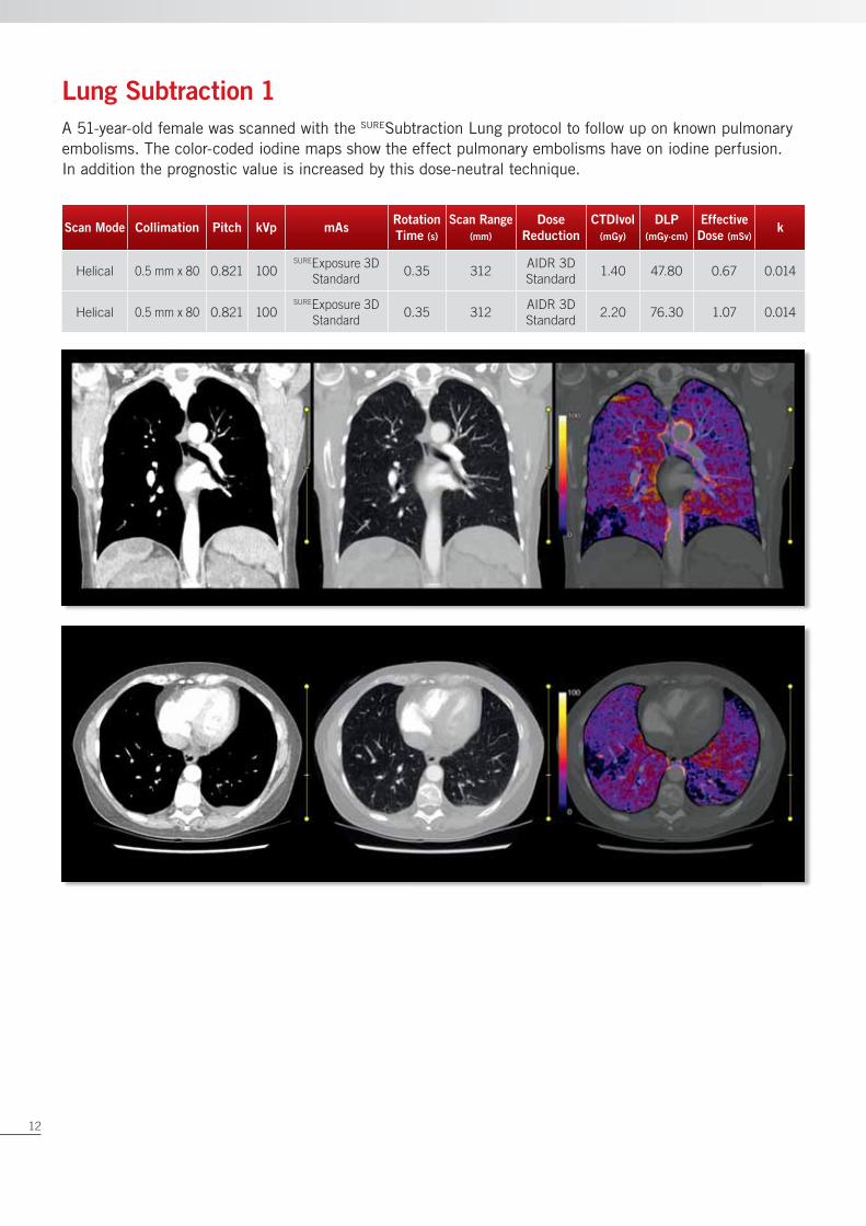
A 51-year-old female was scanned with the SURESubtraction Lung protocol to follow up on known pulmonary
embolisms. The color-coded iodine maps show the effect pulmonary embolisms have on iodine perfusion.
In addition the prognostic value is increased by this dose-neutral technique.
Lung Subtraction 1
12

Scan Mode Collimation Pitch kVp mAsRotation
Time (s)
Scan Range
(mm)
Dose
Reduction
CTDIvol
(mGy)
DLP
(mGy·cm)
Effective
Dose (mSv)k
Helical 0.5 mm x 80 0.821 100SUREExposure 3D
Standard0.35 316
AIDR 3D
Standard4.5 156 2.18 0.014
Helical 0.5 mm x 80 0.821 100SUREExposure 3D
Standard0.35 316
AIDR 3D
Standard7.9 276.9 3.88 0.014
This 72-year-old male presented with a suspicion for pulmonary embolisms. A CT scan was made using the
dose-neutral SURESubtraction Lung protocol. Lung embolisms were found on the right and left side. The color-
coded iodine maps clearly show perfusion defects due to the embolisms.
Lung Subtraction 2
13

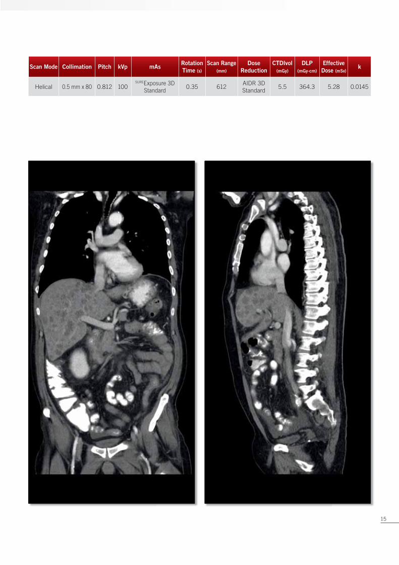
Multiple liver lesions are shown in this TAP scan of a 48-year-old patient. With the excellent low contrast
performance all structures in the abdomen are well visible.
Thorax Abdomen Pelvis 1
14
Scan Mode Collimation Pitch kVp mAsRotation
Time (s)
Scan Range
(mm)
Dose
Reduction
CTDIvol
(mGy)
DLP
(mGy·cm)
Effective
Dose (mSv)k
Helical 0.5 mm x 80 0.812 100SUREExposure 3D
Standard0.35 612
AIDR 3D
Standard5.5 364.3 5.28 0.0145
15

Scan Mode Collimation Pitch kVp mAsRotation
Time (s)
Scan Range
(mm)
Dose
Reduction
CTDIvol
(mGy)
DLP
(mGy·cm)
Effective
Dose (mSv)k
Helical 0.5 mm x 80 0.812 100SUREExposure 3D
Standard0.35 645
AIDR 3D
Standard5.64 396.04 5.74 0.0145
A thorax, abdomen and pelvis scan was required of this 60-year-old male patient following up on treatment. Using
a fast rotation time of 0.35 seconds for routine scans gives motion-free images. Both low contrast soft tissue
and high contrast sharp lungs are reconstructed with one hybrid fi lter for fast diagnosis and effi cient workfl ow.
Thorax Abdomen Pelvis 2
16

Scan Mode Collimation Pitch kVp mAsRotation
Time (s)
Scan Range
(mm)
Dose
Reduction
CTDIvol
(mGy)
DLP
(mGy·cm)
Effective
Dose (mSv)k
Helical 0.5 mm x 80 0.821 120SUREExposure 3D
Standard0.35 408
AIDR 3D
Standard4.8 210.6 3.16 0.015
This urogram made of a 72-year-old female shows hydronephrosis and clearly visible stenosis of the left side
urether. The superb low contrast resolution in the venous phase shows the different abdominal structures in
high detail.
Abdomen
17

Scan Mode Collimation Pitch kVp mAsRotation
Time (s)
Scan Range
(mm)
Dose
Reduction
CTDIvol
(mGy)
DLP
(mGy·cm)
Effective
Dose (mSv)k
Volume 0.5 mm x 256 / 120 82 0.5 128AIDR 3D
Standard7.50 95.9 0.077 0.0008
A 67-year-old female patient with total knee prosthesis was sent for follow-up CT. The 128 mm volume scan
was made in a single rotation using a low dose AIDR 3D scan protocol. Reconstruction was done with Single
Energy Metal Artifact Reduction (SEMAR). This technique reduces metal artifacts extremely, while the knee
and surrounding tissue can be diagnosed with improved accuracy.
SEMAR Knee
Original Image SEMAR Image
18

Scan Mode Collimation Pitch kVp mAsRotation
Time (s)
Scan Range
(mm)
Dose
Reduction
CTDIvol
(mGy)
DLP
(mGy·cm)
Effective
Dose (mSv)k
Volume 0.5 mm x 320 / 100SUREExposure 3D
Standard0.35 160
AIDR 3D
Standard1.50 23.90 0.35 0.0145
A CT scan of the thorax was performed in this 2-day-old child after reanimation. Sedation of the child was not
needed as the whole thorax was scanned in one single rotation of 16 cm without table movement. Integration
of AIDR 3D into automatic exposure control ensured the lowest possible dose.
Pediatric
19
Toshiba Medical Systems Corporation meets internationally recognized
standards for Quality Management System ISO 9001, ISO 13485.
Toshiba Medical Systems Corporation Nasu Operations meets the
Environmental Management System standard ISO 14001.
Made for Life, Aquilion ONE , SUREExposure and SURESubtraction are
trademarks of Toshiba Medical Systems Corporation.
Printed in Europe
www.toshiba-medical.eu
©Toshiba Medical Systems Corporation 2013. All rights reserved.
Design and specifications are subject to change without notice.
Model number: MCACT0001EUC 2013-10
TOSHIBA MEDICAL SYSTEMS EUROPE