Embed Size (px)
Citation preview
Brain-wide representations of ongoing behavior:a universal principle?Harris S Kaplan1,2,3 and Manuel Zimmer1,2
Available online at www.sciencedirect.com
ScienceDirect
Recent neuronal activity recordings of unprecedented breadth
and depth in worms, flies, and mice have uncovered a
surprising common feature: brain-wide behavior-related
signals. These signals pervade, and even dominate, neuronal
populations thought to function primarily in sensory
processing. Such convergent findings across organisms
suggest that brain-wide representations of behavior might be a
universal neuroscientific principle. What purpose(s) do these
representations serve? Here we review these findings along
with suggested functions, including sensory prediction,
context-dependent sensory processing, and, perhaps most
speculatively, distributed motor command generation. It
appears that a large proportion of the brain’s energy and
coding capacity is used to represent ongoing behavior;
understanding the function of these representations should
therefore be a major goal in neuroscience research.
Addresses1Department of Neuroscience and Developmental Biology, University of
Vienna, Althanstrasse 14, 1090 Vienna, Austria2Research Institute of Molecular Pathology (IMP), Vienna Biocenter
(VBC), Campus-Vienna-Biocenter 1, 1030 Vienna, Austria
Corresponding author: Kaplan, Harris S ([email protected])3 Present address: Department of Molecular and Cellular Biology,
Harvard University, Cambridge, MA, USA.
Current Opinion in Neurobiology 2020, 64:60–69
This review comes from a themed issue on Systems neuroscience
Edited by Dan Feldman and Kristin Scott
For a complete overview see the Issue and the Editorial
Available online 20th March 2020
https://doi.org/10.1016/j.conb.2020.02.008
0959-4388/ã 2020 Published by Elsevier Ltd.
IntroductionRecent advances in calcium imaging and electrophysiologi-
cal techniques have enabled increasingly widespread high-
resolution neuronal activity recordings, spanning much of a
single brainarea [1], severalbrainareas simultaneously [2–4],
or even entire brains of smaller organisms [5–10]. One
powerful approach is to use these techniques for a discov-
ery-driven or data-driven, rather than hypothesis-driven,
analysis of brain dynamics: we can ask, under a given set
of conditions, what are the brain’s major activities and
computations? Thus far, the answer has been resoundingly
clear, and surprising: signals primarily relate to the animal’s
Current Opinion in Neurobiology 2020, 64:60–69
own ongoing behavior [1–3,8,11]. This internal representa-
tion of behavior goes beyond simple behavioral state modu-
lation. Instead, brain-wide behavior representations are both
quantitative (co-varying with behavior in a graded continu-
ous manner) and multi-dimensional (uniquely representing
several aspects of behavior simultaneously). Here we sum-
marize these cross-species findings and discuss potential
functions. We are motivated by a few recent studies that
examine brain-wide signals in mice [1–3], flies [8], and
worms [11]. However, these studies are complemented by
a larger body of work that examined the function of a few
behavior-related signals in specific sensory brain regions (see
Refs. [12,13] for more systematic reviews). Below, we syn-
thesize this literature to discuss crucial questions regarding
the source and function of brain-wide behavior-related
signals. We expect the answers to be at least partly spe-
cies-specific or modality-specific. Still, common principles
may emerge from such a cross-species comparison.
Brain-wide representations of behavioracross speciesThree recent studies in mice [1–3] reported some of the
largest-scale neuronal activity recordings to date. Using
various techniques and experimental settings, the authors
consistently found that a large proportion of brain activity
correlates with the animal’s ongoing behavior. This was
the case in cortical (including primary sensory) as well as
subcortical areas. Stringer et al. [1] reported strong neuro-
nal activity relationships with spontaneous behavior
either in the dark or during passive viewing of visual
stimuli, using two techniques: single-cell resolution two-
photon calcium imaging of excitatory V1 neurons, and
neuropixel probes [4] in cortical and extra-cortical areas
(Figure 1a). Musall et al. [2] and Salkoff et al. [3] reported
widespread neuronal activity correlations to instructed
and especially uninstructed movements during decision-
making tasks; both studies used widefield calcium imag-
ing in either excitatory only [2] or both excitatory and
inhibitory neurons [3]. While the widefield imaging used
in both studies [2,3] lacks single-cell resolution, Musall
et al. [2] complemented these data with two-photon
recordings of several areas as well as neuropixel probe
recordings, both of which largely confirmed the findings
from widefield imaging.
Earlier studies had reported behavior-related signals in
primary sensory cortex (e.g. locomotion signals in visual
cortex [14], discussed below), but these three studies [1–3]
may portend a sea change in our understanding of the
mouse brain, for at least two reasons. First, they show that
www.sciencedirect.com
Brain-wide motor representations Kaplan and Zimmer 61
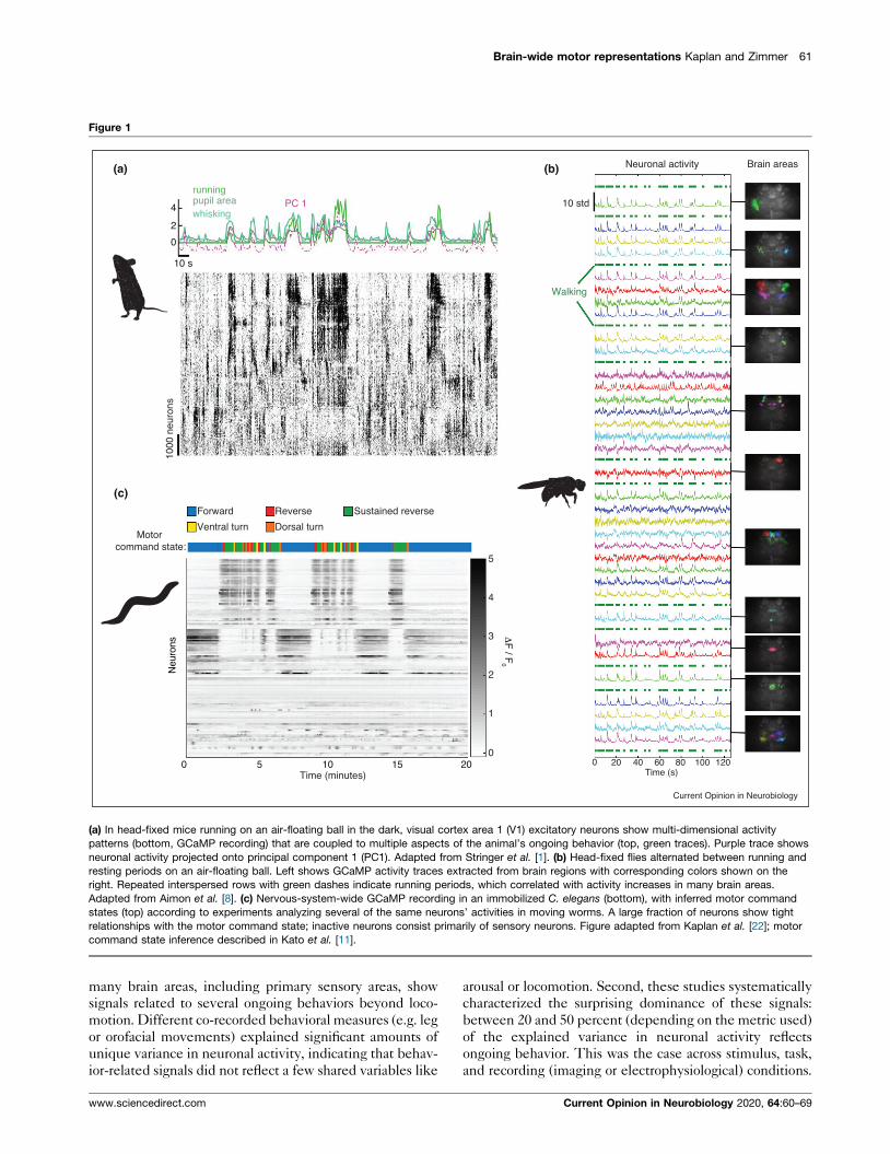
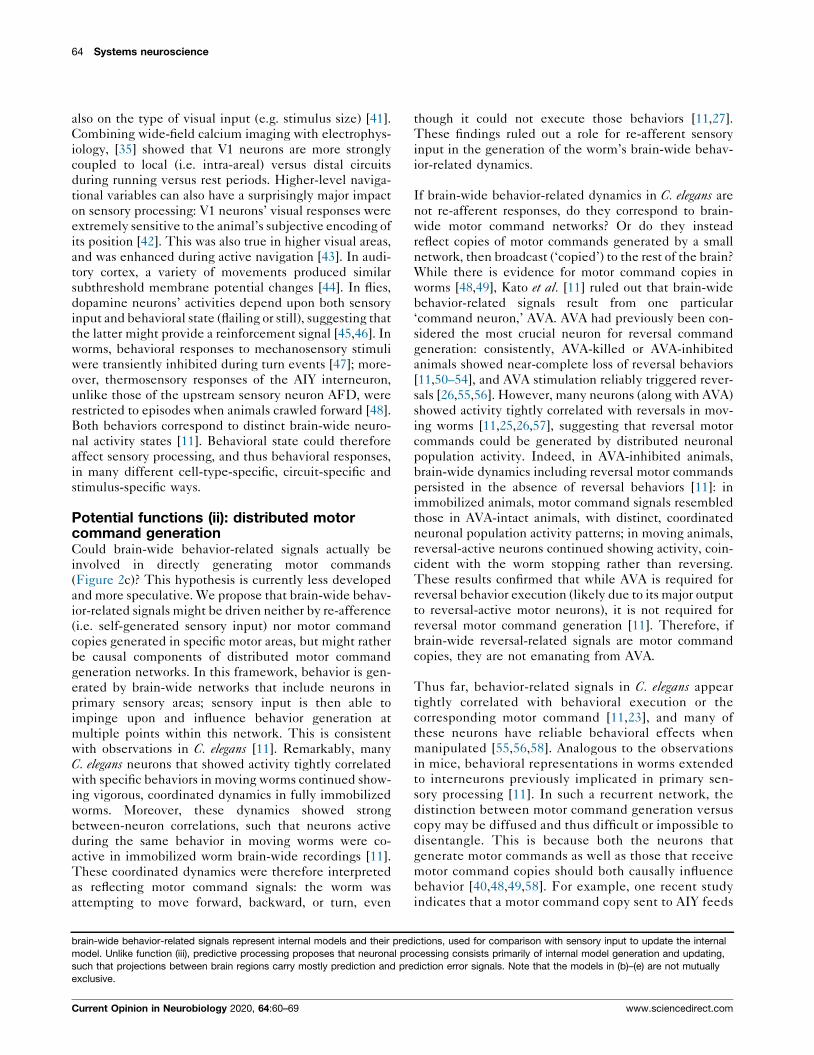
Figure 1
(c)
00 20 40 60 80 100 120
Time (s)
1
2
3
4
5
ΔF
/ F0
Motor command state:
Neu
rons
0 5 10 2015Time (minutes)
1000
neu
rons
10 s
2
4
0
PC 1running
whiskingpupil area
Forward Reverse Sustained reverse
Ventral turn Dorsal turn
(a) (b)
Walking
Neuronal activity
10 std
Brain areas
Current Opinion in Neurobiology
(a) In head-fixed mice running on an air-floating ball in the dark, visual cortex area 1 (V1) excitatory neurons show multi-dimensional activity
patterns (bottom, GCaMP recording) that are coupled to multiple aspects of the animal’s ongoing behavior (top, green traces). Purple trace shows
neuronal activity projected onto principal component 1 (PC1). Adapted from Stringer et al. [1]. (b) Head-fixed flies alternated between running and
resting periods on an air-floating ball. Left shows GCaMP activity traces extracted from brain regions with corresponding colors shown on the
right. Repeated interspersed rows with green dashes indicate running periods, which correlated with activity increases in many brain areas.
Adapted from Aimon et al. [8]. (c) Nervous-system-wide GCaMP recording in an immobilized C. elegans (bottom), with inferred motor command
states (top) according to experiments analyzing several of the same neurons’ activities in moving worms. A large fraction of neurons show tight
relationships with the motor command state; inactive neurons consist primarily of sensory neurons. Figure adapted from Kaplan et al. [22]; motor
command state inference described in Kato et al. [11].
many brain areas, including primary sensory areas, show
signals related to several ongoing behaviors beyond loco-
motion. Different co-recorded behavioral measures (e.g. leg
or orofacial movements) explained significant amounts of
unique variance in neuronal activity, indicating that behav-
ior-related signals did not reflect a few shared variables like
www.sciencedirect.com
arousal or locomotion. Second, these studies systematically
characterized the surprising dominance of these signals:
between 20 and 50 percent (depending on the metric used)
of the explained variance in neuronal activity reflects
ongoing behavior. This was the case across stimulus, task,
and recording (imaging or electrophysiological) conditions.
Current Opinion in Neurobiology 2020, 64:60–69
62 Systems neuroscience
For example, Stringer et al. [1] showed that in V1, motor
information is represented at least as much as visual infor-
mation during the display of natural images, and that these
representations are often mixed in individual neurons.
Another study by the same group found that during a visual
discrimination task, action encoding is significantly more
widespread than either stimulus or choice encoding, across
cortical and subcortical brain areas recorded using neuro-
pixel arrays [15]. In summary, whether the mouse is in the
dark, passively viewing natural scenes, or engaged in a
decision-making task, much of its brain activity is dynami-
cally tracking various aspects of the state of its body
(Figure 2a).
Surprisingly, recent work in invertebrates has come to
similar conclusions: Aimon et al. [8] used light-field micros-
copy to record brain-wide calcium dynamics at high tem-
poral resolution inhead-fixedflies free towalk onatrackball
(Figure 1b). The difference in brain activity between
walking versus resting states was much more dramatic than
activity changes induced by light or odor stimuli. Impor-
tantly, this held true in vision-impaired mutant animals,
suggesting that these signals are not responses to visual
feedback from the environment (e.g. trackball movement
during running). Other studies in flies have shown multiple
simultaneous representations of subjective heading direc-
tion [16–18]; this is also the case in mice [19], and in both
species, this representation has been shown to depend
partly on self-motion cues [16,19,20]. In Caenorhabditiselegans, recent studies [11,21,22] examined brain-wide
dynamics at single-cell resolution, and found widespread
brain activity representing various aspects of behavior
beyond mere arousal states, including locomotion direction
(forward or reverse), turning state, and locomotion speed
(Figure 1c). As for flies and mammals, these studies accom-
panied a larger body of work showing locomotion signals in
primary sensory circuits [23–26] (see Ref. [27] for a detailed
discussion). Thus, as in mammals, brain-wide behavioral
signals in worms pervade neuronal populations that, before
these studies, were thought to be involved primarily in
sensory processing. Further, brain-wide behavior-related
dynamics persisted during sensory stimulation with only
subtle modulations [11]. In all systems under study, these
observations are only recently reported, and whether these
results hold across more complex multisensory and unre-
strained conditions remains to be shown. Importantly,
brain-wide studies in zebrafish larvae do not report such
brain-wide behavior-related signals; behavior signals rather
appear more localized to the hindbrain [9,28,29] or in
clusters in other regions [9], though most work has been
limited to larval and largely immobilized animals. We
therefore tentatively conclude that brain-wide encoding
ofongoingbehavioralvariablesmaybeageneralprincipleof
nervous systems across both invertebrates and vertebrates.
A natural next question is what functions brain-wide
behavior-related signals might serve. A related question
Current Opinion in Neurobiology 2020, 64:60–69
concerns the origin of these signals: do they correspond to
re-afferent (i.e. self-generated) sensory inputs, efference
copies of motor commands, or network states underlying
distributed motor commands (see Box 1 ‘Looking
ahead’)? The brain-wide studies discussed above only
provided speculation. In parallel work, however, several
groups have been exploring these questions, and several
hypotheses have emerged. Below, we discuss how these
proposed functions might explain the multitude of
recently discovered brain-wide behavior representations.
Note that these hypotheses are not mutually exclusive,
and that multiple functions may occur simultaneously.
Potential functions (i): context-dependentsensory processingThe processing of sensory input is well known to depend
on behavioral state. For example, thresholds for response
to sensory input are much higher during sleep versus
wake states [30]. Within wake states, arousal level and
attention strongly affect brain sensory responses, and
perception [31,32]. These different signals can affect
sensory processing as early as the sensory organ itself
[33], so it is perhaps not surprising to find such modulation
in early sensory areas. However, Stringer et al. [1] and
Musall et al. [2] both showed that behavioral signals in
primary visual cortex (and other areas) are multidimen-
sional, that is, encoding multiple behavioral variables
ranging from locomotion parameters to orofacial beha-
viors, indicating that much more than a simple arousal
signal is encoded. Indeed, differential effects of arousal
and locomotion on V1 activity have been dissociated
[34,35]; note, however, that movement and arousal signals
are interrelated and could affect sensory processing via
the same neuromodulators, such as acetylcholine [36–38].
The multidimensional nature of behavior-related
signals in mice [1–3] as well as worms [11] suggests a
more complex role for behavioral context. Natural
behavior involves a tight coupling of sensory and motor
signals — consider a mouse whisking or sniffing, or a
worm’s sensory organs in the nose oscillating in space
during its sinusoidal gait. The animal’s own behavior
strongly affects the spatiotemporal statistics of its sensory
input (for example, see Wachowiak et al. [39], and Liu
et al. [40]). A ubiquitous quantitative mixing of sensory
and motor information might therefore be expected
(Figure 2b).
In what ways could multidimensional behavior-related
signals contextualize sensory processing? This has been
most carefully investigated in mouse V1. Niell et al. and
Fu et al. [14,36] provided evidence that locomotion con-
trols the gain of sensory responses. This occurs via a
disinhibitory circuit mechanism in which one interneuron
class is activated by locomotion and then inhibits another
interneuron class, ultimately disinhibiting principal excit-
atory neurons [36]. Further, another study showed that
the effect of locomotion depends not only on cell type but
www.sciencedirect.com
Brain-wide motor representations Kaplan and Zimmer 63
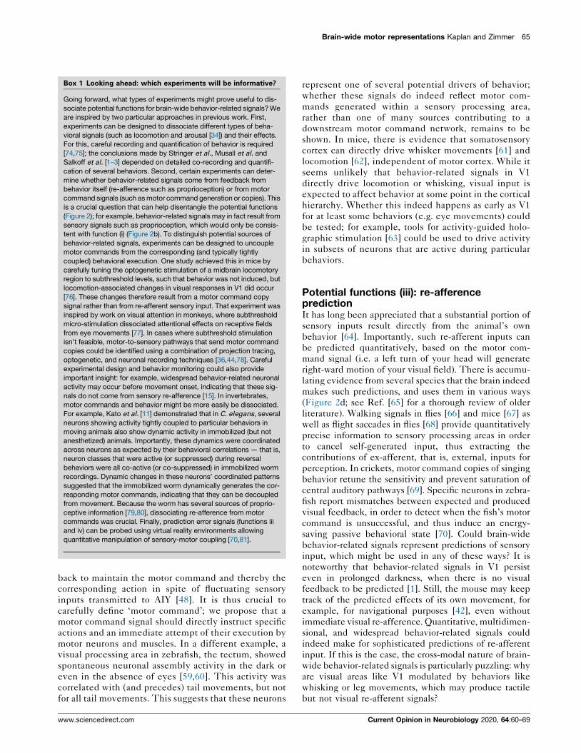
Figure 2
Sensory detection
Motor command generation
Sensorimotorprocessing
Behavior
Re-afferent sensory input
Ex-afferentsensory input Context-dependent
sensory processingDistributed motor
command generation
Re-afference prediction Predictive processing
Predictions of re-afferent inputs
Prediction errorsMotor commandgeneration
Multidimensionalbehavior signals
Multidimensionalsensory signals
(a)
(b) (c)
(d) (e)
Contextualcues
Current Opinion in Neurobiology
(a) A schematic summary of the findings reported by brain-wide studies [1–3,8,11]. Circles represent neuronal populations, which contain both
sensory signals of different types (e.g. modalities), represented by different blue shadings, and behavior-related signals of different types,
represented by different red shadings. In the intermediate step ‘Sensorimotor processing,’ sensory and behavior-related signals are mixed. The
studies reviewed here showed that behavior-related signals are widespread and dominate brain activity in worms, flies, and mice, even in primary
sensory areas. ‘Sensory detection’ may therefore refer only to sensory organs themselves, as downstream neuronal populations are dominated by
signals related to behavior. Similarly, it is not clear precisely where in the nervous system the line should be drawn between ‘sensorimotor
processing’ and ‘motor command generation’. Note that a significant portion of brain activity may consist of signals that cannot be explained by
either behavior or sensory input [86], which are not represented here. This schematic serves as the foundation for different models in panels
(b)–(e), which illustrate different potential functions for brain-wide behavior-related signals. (b) Model illustrating hypothesized function (i), which
suggests that brain-wide behavior-related signals are important for contextualizing sensory processing. These contextual behavior signals (orange
arrows) could originate from re-afferent sensory input (and therefore reach the nervous system via ‘sensory detection’) or from motor command
copies sent back from regions that generate motor commands. (c) Model illustrating hypothesized function (ii), which suggests that brain-wide
behavior-related signals reflect a distributed motor command network. In this model, motor commands are generated not by a small core
neuronal circuit, but rather by many neurons spread throughout most brain regions, potentially including ‘primary sensory’ regions. (d) Model
illustrating hypothesized function (iii), which suggests that brain-wide behavior-related signals are used to predict re-afferent sensory inputs
resulting from behavior. Such predictions arise from motor command copies and must therefore be transformed from motor to sensory
coordinates (arrow with red-blue gradient, indicating the transformation; see text). (e) Model illustrating hypothesized function (iv), in which
www.sciencedirect.com Current Opinion in Neurobiology 2020, 64:60–69
64 Systems neuroscience
also on the type of visual input (e.g. stimulus size) [41].
Combining wide-field calcium imaging with electrophys-
iology, [35] showed that V1 neurons are more strongly
coupled to local (i.e. intra-areal) versus distal circuits
during running versus rest periods. Higher-level naviga-
tional variables can also have a surprisingly major impact
on sensory processing: V1 neurons’ visual responses were
extremely sensitive to the animal’s subjective encoding of
its position [42]. This was also true in higher visual areas,
and was enhanced during active navigation [43]. In audi-
tory cortex, a variety of movements produced similar
subthreshold membrane potential changes [44]. In flies,
dopamine neurons’ activities depend upon both sensory
input and behavioral state (flailing or still), suggesting that
the latter might provide a reinforcement signal [45,46]. In
worms, behavioral responses to mechanosensory stimuli
were transiently inhibited during turn events [47]; more-
over, thermosensory responses of the AIY interneuron,
unlike those of the upstream sensory neuron AFD, were
restricted to episodes when animals crawled forward [48].
Both behaviors correspond to distinct brain-wide neuro-
nal activity states [11]. Behavioral state could therefore
affect sensory processing, and thus behavioral responses,
in many different cell-type-specific, circuit-specific and
stimulus-specific ways.
Potential functions (ii): distributed motorcommand generationCould brain-wide behavior-related signals actually be
involved in directly generating motor commands
(Figure 2c)? This hypothesis is currently less developed
and more speculative. We propose that brain-wide behav-
ior-related signals might be driven neither by re-afference
(i.e. self-generated sensory input) nor motor command
copies generated in specific motor areas, but might rather
be causal components of distributed motor command
generation networks. In this framework, behavior is gen-
erated by brain-wide networks that include neurons in
primary sensory areas; sensory input is then able to
impinge upon and influence behavior generation at
multiple points within this network. This is consistent
with observations in C. elegans [11]. Remarkably, many
C. elegans neurons that showed activity tightly correlated
with specific behaviors in moving worms continued show-
ing vigorous, coordinated dynamics in fully immobilized
worms. Moreover, these dynamics showed strong
between-neuron correlations, such that neurons active
during the same behavior in moving worms were co-
active in immobilized worm brain-wide recordings [11].
These coordinated dynamics were therefore interpreted
as reflecting motor command signals: the worm was
attempting to move forward, backward, or turn, even
brain-wide behavior-related signals represent internal models and their pred
model. Unlike function (iii), predictive processing proposes that neuronal pro
such that projections between brain regions carry mostly prediction and pre
exclusive.
Current Opinion in Neurobiology 2020, 64:60–69
though it could not execute those behaviors [11,27].
These findings ruled out a role for re-afferent sensory
input in the generation of the worm’s brain-wide behav-
ior-related dynamics.
If brain-wide behavior-related dynamics in C. elegans are
not re-afferent responses, do they correspond to brain-
wide motor command networks? Or do they instead
reflect copies of motor commands generated by a small
network, then broadcast (‘copied’) to the rest of the brain?
While there is evidence for motor command copies in
worms [48,49], Kato et al. [11] ruled out that brain-wide
behavior-related signals result from one particular
‘command neuron,’ AVA. AVA had previously been con-
sidered the most crucial neuron for reversal command
generation: consistently, AVA-killed or AVA-inhibited
animals showed near-complete loss of reversal behaviors
[11,50–54], and AVA stimulation reliably triggered rever-
sals [26,55,56]. However, many neurons (along with AVA)
showed activity tightly correlated with reversals in mov-
ing worms [11,25,26,57], suggesting that reversal motor
commands could be generated by distributed neuronal
population activity. Indeed, in AVA-inhibited animals,
brain-wide dynamics including reversal motor commands
persisted in the absence of reversal behaviors [11]: in
immobilized animals, motor command signals resembled
those in AVA-intact animals, with distinct, coordinated
neuronal population activity patterns; in moving animals,
reversal-active neurons continued showing activity, coin-
cident with the worm stopping rather than reversing.
These results confirmed that while AVA is required for
reversal behavior execution (likely due to its major output
to reversal-active motor neurons), it is not required for
reversal motor command generation [11]. Therefore, if
brain-wide reversal-related signals are motor command
copies, they are not emanating from AVA.
Thus far, behavior-related signals in C. elegans appear
tightly correlated with behavioral execution or the
corresponding motor command [11,23], and many of
these neurons have reliable behavioral effects when
manipulated [55,56,58]. Analogous to the observations
in mice, behavioral representations in worms extended
to interneurons previously implicated in primary sen-
sory processing [11]. In such a recurrent network, the
distinction between motor command generation versus
copy may be diffused and thus difficult or impossible to
disentangle. This is because both the neurons that
generate motor commands as well as those that receive
motor command copies should both causally influence
behavior [40,48,49,58]. For example, one recent study
indicates that a motor command copy sent to AIY feeds
ictions, used for comparison with sensory input to update the internal
cessing consists primarily of internal model generation and updating,
diction error signals. Note that the models in (b)–(e) are not mutually
www.sciencedirect.com
Brain-wide motor representations Kaplan and Zimmer 65
Box 1 Looking ahead: which experiments will be informative?
Going forward, what types of experiments might prove useful to dis-
sociate potential functions for brain-wide behavior-related signals? We
are inspired by two particular approaches in previous work. First,
experiments can be designed to dissociate different types of beha-
vioral signals (such as locomotion and arousal [34]) and their effects.
For this, careful recording and quantification of behavior is required
[74,75]; the conclusions made by Stringer et al., Musall et al. and
Salkoff et al. [1–3] depended on detailed co-recording and quantifi-
cation of several behaviors. Second, certain experiments can deter-
mine whether behavior-related signals come from feedback from
behavior itself (re-afference such as proprioception) or from motor
command signals (such as motor command generation or copies). This
is a crucial question that can help disentangle the potential functions
(Figure 2); for example, behavior-related signals may in fact result from
sensory signals such as proprioception, which would only be consis-
tent with function (i) (Figure 2b). To distinguish potential sources of
behavior-related signals, experiments can be designed to uncouple
motor commands from the corresponding (and typically tightly
coupled) behavioral execution. One study achieved this in mice by
carefully tuning the optogenetic stimulation of a midbrain locomotory
region to subthreshold levels, such that behavior was not induced, but
locomotion-associated changes in visual responses in V1 did occur
[76]. These changes therefore result from a motor command copy
signal rather than from re-afferent sensory input. That experiment was
inspired by work on visual attention in monkeys, where subthreshold
micro-stimulation dissociated attentional effects on receptive fields
from eye movements [77]. In cases where subthreshold stimulation
isn’t feasible, motor-to-sensory pathways that send motor command
copies could be identified using a combination of projection tracing,
optogenetic, and neuronal recording techniques [36,44,78]. Careful
experimental design and behavior monitoring could also provide
important insight: for example, widespread behavior-related neuronal
activity may occur before movement onset, indicating that these sig-
nals do not come from sensory re-afference [15]. In invertebrates,
motor commands and behavior might be more easily be dissociated.
For example, Kato et al. [11] demonstrated that in C. elegans, several
neurons showing activity tightly coupled to particular behaviors in
moving animals also show dynamic activity in immobilized (but not
anesthetized) animals. Importantly, these dynamics were coordinated
across neurons as expected by their behavioral correlations — that is,
neuron classes that were active (or suppressed) during reversal
behaviors were all co-active (or co-suppressed) in immobilized worm
recordings. Dynamic changes in these neurons’ coordinated patterns
suggested that the immobilized worm dynamically generates the cor-
responding motor commands, indicating that they can be decoupled
from movement. Because the worm has several sources of proprio-
ceptive information [79,80], dissociating re-afference from motor
commands was crucial. Finally, prediction error signals (functions iii
and iv) can be probed using virtual reality environments allowing
quantitative manipulation of sensory-motor coupling [70,81].
back to maintain the motor command and thereby the
corresponding action in spite of fluctuating sensory
inputs transmitted to AIY [48]. It is thus crucial to
carefully define ‘motor command’; we propose that a
motor command signal should directly instruct specific
actions and an immediate attempt of their execution by
motor neurons and muscles. In a different example, a
visual processing area in zebrafish, the tectum, showed
spontaneous neuronal assembly activity in the dark or
even in the absence of eyes [59,60]. This activity was
correlated with (and precedes) tail movements, but not
for all tail movements. This suggests that these neurons
www.sciencedirect.com
represent one of several potential drivers of behavior;
whether these signals do indeed reflect motor com-
mands generated within a sensory processing area,
rather than one of many sources contributing to a
downstream motor command network, remains to be
shown. In mice, there is evidence that somatosensory
cortex can directly drive whisker movements [61] and
locomotion [62], independent of motor cortex. While it
seems unlikely that behavior-related signals in V1
directly drive locomotion or whisking, visual input is
expected to affect behavior at some point in the cortical
hierarchy. Whether this indeed happens as early as V1
for at least some behaviors (e.g. eye movements) could
be tested; for example, tools for activity-guided holo-
graphic stimulation [63] could be used to drive activity
in subsets of neurons that are active during particular
behaviors.
Potential functions (iii): re-afferencepredictionIt has long been appreciated that a substantial portion of
sensory inputs result directly from the animal’s own
behavior [64]. Importantly, such re-afferent inputs can
be predicted quantitatively, based on the motor com-
mand signal (i.e. a left turn of your head will generate
right-ward motion of your visual field). There is accumu-
lating evidence from several species that the brain indeed
makes such predictions, and uses them in various ways
(Figure 2d; see Ref. [65] for a thorough review of older
literature). Walking signals in flies [66] and mice [67] as
well as flight saccades in flies [68] provide quantitatively
precise information to sensory processing areas in order
to cancel self-generated input, thus extracting the
contributions of ex-afferent, that is, external, inputs for
perception. In crickets, motor command copies of singing
behavior retune the sensitivity and prevent saturation of
central auditory pathways [69]. Specific neurons in zebra-
fish report mismatches between expected and produced
visual feedback, in order to detect when the fish’s motor
command is unsuccessful, and thus induce an energy-
saving passive behavioral state [70]. Could brain-wide
behavior-related signals represent predictions of sensory
input, which might be used in any of these ways? It is
noteworthy that behavior-related signals in V1 persist
even in prolonged darkness, when there is no visual
feedback to be predicted [1]. Still, the mouse may keep
track of the predicted effects of its own movement, for
example, for navigational purposes [42], even without
immediate visual re-afference. Quantitative, multidimen-
sional, and widespread behavior-related signals could
indeed make for sophisticated predictions of re-afferent
input. If this is the case, the cross-modal nature of brain-
wide behavior-related signals is particularly puzzling: why
are visual areas like V1 modulated by behaviors like
whisking or leg movements, which may produce tactile
but not visual re-afferent signals?
Current Opinion in Neurobiology 2020, 64:60–69
66 Systems neuroscience
Potential functions (iv): predictive processingThe brain may not only predict re-afferent input, but also
ex-afferent (i.e. non-self-generated) input [71,72]. In a
growing body of theoretical and experimental work
(recently summarized by Keller and Mrsic-Flogel [73]),
the mammalian cortex is proposed to make such large-
scale predictions, in the form of generative internal
models of the world, including body and environment.
Sensory input is then processed in the context of these
internal models — that is, sensory signals are used to
update the internal model when its predictions do not
agree with sensory inputs. The role of sensory cortex is
therefore to compare expected and actual sensory input,
according to the theory.
How does predictive processing differ from re-afferent
prediction (discussed in the previous section)? Keller
and Mrsic-Flogel [73] proposed that predictive proces-
sing is an alternative to the classical ‘representational
framework’ of cortical function, in which the brain
processes sensory signals into progressively more sophis-
ticated and higher-level representations of the outside
world. Re-afference prediction (Function (iii)), as well as
contextualization of sensory input (Function (i)), could
occur within such a representational framework: bottom-
up sensory input could be modulated in many ways
before being passed on for computations underlying
higher-order representations. In contrast, in a predictive
processing framework (function (iv), bottom-up input
will only be passed on to other brain regions if it is found
to disagree with the internal model [73]. In this case,
communication between brain regions consists primarily
of prediction or prediction error signals, and computa-
tions involve comparisons between those signals, or
internal model updating, rather than feature extraction
for increasingly specific representations [73].
If predictive processing is indeed a canonical computation,
brain-wide behavior-related signals could represent internal
model information (orpredictions fromthosemodels)broad-
cast to sensory areas to enable the comparison of expected
and actual sensory input. This requires a transformation of
motor commands from motor coordinates (e.g. specifying
head movement parameters) into sensory coordinates (e.g.
wide-field motion signals expected from such head move-
ments) [73]. The widespread signals recently reported in
worms and mice showed clear quantitative relationships to
behavioral variables; would such signals be so strongly corre-
lated with behavior if they are meant to be predictions in
sensory coordinates? Or are these the signals before transfor-
mation into sensory coordinates? Finally, Keller and Mrsic-
Flogel [73] discussed predictive processing as the role of the
cortex; however, motor command copies have been repeat-
edly demonstrated in invertebrates (see above section),
which also show brain-wide behavior-related signals. Could
re-afference prediction be an evolutionary precursor to gen-
erative models and predictive processing? While predictive
Current Opinion in Neurobiology 2020, 64:60–69
processing could be a unifying framework for cortical func-
tion, these questions could aid our understanding of brain-
wide behavior-related signals more generally.
ConclusionsThe preponderance of behavior-related neuronal activity
in both vertebrates and invertebrates suggests a funda-
mental role. Above, we described a few prominent
hypotheses and discussed experiments that could distin-
guish them (see Box 1 ‘Looking ahead’). We conclude
with three thoughts to encourage further discussion. First,
examining these data in C. elegans, we have wondered why
the brain would use so many neurons to seemingly
redundantly encode the same information. Referring to
the connectome, we observed that neurons showing sim-
ilar behavior-related signals nevertheless often have very
different connectivity, including both inputs and outputs.
These neurons could therefore serve as alternative chan-
nels for differentially mixing sensory information with
ongoing behavior signals, perhaps to ultimately affect
motor command generation (function ii) [27]. Indeed,
Stringer et al. [1] showed that such mixing of sensory
and motor information in individual neurons is ubiquitous
in V1. Second, the majority of neuronal activity record-
ings, especially those measuring brain-wide signals, are
performed in at least somewhat restrained settings (e.g.
head fixation in mice). It is important to consider how
these and other experimental settings, such as non-natu-
ralistic and open-loop stimuli, might affect the results
[82]. Several studies have indicated the potential impor-
tance of motor error signals [70,73]; the failure of head
movement motor commands in head-fixed animals could
massively engage such error detectors. Further, eye and
head movements are typically closely coupled in freely
moving mice [83]; disruption of this coupling during head
fixation could have a major effect on behavior and neuro-
nal activity. This brings us to our third and final point:
behavioral coordination, such as eye and head movement
coupling, is far from atypical. Rather, behaviors exhibit
complex multi-scale spatial and temporal organization
[75], for example, in the form of probabilistic sequences
[84] or hierarchies [85]. These interdependencies could
have structure that eludes the aforementioned unique
neuronal activity variance measures. Brain-wide behav-
ior-related signals should reflect the organizations and
interdependencies of behavior. Indeed, we found this to
be the case in C. elegans: (1) brain-wide dynamics relating
to various behaviors are tied together in specific
sequences, reflecting the animal’s action sequence [11],
and (2) interactions between global and local neuronal
dynamics implement a multi-scale behavioral hierarchy
[22]. Crucially, our understanding of the worm’s behavior
focused our attention on important neuronal activity
relationships that might otherwise have been overlooked.
We therefore suggest that better descriptions of the rules
governing animal behavior are likely to aid our under-
standing of the associated brain-wide neuronal activity
www.sciencedirect.com
Brain-wide motor representations Kaplan and Zimmer 67
patterns. These organizational rules likely also have a
major impact on the processing of sensory input, espe-
cially if they pervade neuronal activity patterns in sensory
processing circuits.
Conflict of interest statementNothing declared.
AcknowledgementsThe authors thank Anne Churchland, Simon Musall, Itamar Lev, andOriana Salazar Thula for critically reading and commenting on themanuscript. M.Z. is supported by the Simons Foundation (#543069) and theInternational Research Scholar Program by the Wellcome Trust andHoward Hughes Medical Institute (#208565/A/17/Z). The IMP is funded byBoehringer Ingelheim.
References
1. Stringer C, Pachitariu M, Steinmetz N, Reddy CB, Carandini M,Harris KD: Spontaneous behaviors drive multidimensional,brainwide activity. Science (New York, NY) 2019, 364:255-263.
2. Musall S, Kaufman MT, Juavinett AL, Gluf S, Churchland AK:Single-trial neural dynamics are dominated by richly variedmovements. Nat Neurosci 2019:1-16.
3. Salkoff DB, Zagha E, McCarthy E, McCormick DA: Movement andperformance explain widespread cortical activity in a visualdetection task. Cereb Cortex 2019, 94:891-897.
4. Jun JJ, Steinmetz NA, Siegle JH, Denman DJ, Bauza M,Barbarits B, Lee AK, Anastassiou CA, Andrei A, Aydn C et al.: Fullyintegrated silicon probes for high-density recording of neuralactivity. Nature 2017, 551:232-236.
5. Schro del T, Prevedel R, Aumayr K, Zimmer M, Vaziri A: Brain-wide3D imaging of neuronal activity in Caenorhabditis elegans withsculpted light. Nat Methods 2013, 10:1013-1020.
6. Venkatachalam V, Ji N, Wang X, Clark C, Mitchell JK, Klein M,Tabone CJ, Florman J, Ji H, Greenwood J et al.: Pan-neuronalimaging in roaming Caenorhabditis elegans. Proc Natl Acad SciU S A 2016, 113:E1082-1088.
7. Nguyen JP, Shipley FB, Linder AN, Plummer GS, Liu M, Setru SU,Shaevitz JW, Leifer AM: Whole-brain calcium imaging withcellular resolution in freely behaving Caenorhabditis elegans.Proc Natl Acad Sci U S A 2016, 113:E1074-1081.
8. Aimon S, Katsuki T, Jia T, Grosenick L, Broxton M, Deisseroth K,Sejnowski TJ, Greenspan RJ: Fast near-whole–brain imaging inadult Drosophila during responses to stimuli and behavior.PLoS Biol 2019, 17 e2006732-e2006731.
9. Ahrens MB, Li JM, Orger MB, Robson DN, Schier AF, Engert F,Portugues R: Brain-wide neuronal dynamics during motoradaptation in zebrafish. Nature 2012, 485:471-477.
10. Panier T, Romano S, Olive R, Pietri T, Sumbre G, Candelier R,Debregeas G: Fast functional imaging of multiple brain regionsin intact zebrafish larvae using selective plane illuminationmicroscopy. Front Neural Circuits 2013, 7:1-11.
11. Kato S, Kaplan HS, Schro del T, Skora S, Lindsay TH, Yemini E,Lockery S, Zimmer M: Global brain dynamics embed the motorcommand sequence of Caenorhabditis elegans. Cell 2015,163:1-50.
12. Fujiwara T, Chiappe E: Motor-Driven Modulation in Visual NeuralCircuits. Springer International Publishing; 2017:261-281.
13. Busse L, Cardin JA, Chiappe ME, Halassa MM, McGinley MJ,Yamashita T, Saleem AB: Sensation during active behaviors. JNeurosci 2017, 37:10826-10834.
14. Niell CM, Stryker MP: Modulation of visual responses by behavioralstate in mouse visual cortex. Neuron 2010, 65:472-479.
www.sciencedirect.com
15. Steinmetz NA, Zatka-Haas P, Carandini M, Harris KD: Distributedcoding of choice, action and engagement across the mousebrain. Nature 2019:1-35.
16. Seelig JD, Jayaraman V: Neural dynamics for landmarkorientation and angular path integration. Nature 2015,521:186-191.
17. Turner-Evans D, Wegener S, Rouault H, Franconville R, Wolff T,Seelig JD, Druckmann S, Jayaraman V: Angular velocityintegration in a fly heading circuit. eLife 2017, 6:e04577.
18. Green J, Adachi A, Shah KK, Hirokawa JD, Magani PS, Maimon G:A neural circuit architecture for angular integration inDrosophila. Nat Publishing Group 2017:1-19.
19. Poulter S, Hartley T, Lever C: The neurobiology of mammaliannavigation. Curr Biol 2018, 28:R1023-R1042.
20. Butler WN, Smith KS, van der Meer MAA, Taube JS: The head-direction signal plays a functional role as a neural compassduring navigation. Curr Biol 2017, 27:1259-1267.
21. Nichols ALA, Eichler T, Latham R, Zimmer M: A global brain stateunderlies C. elegans sleep behavior. Science (New York, NY)2017, 356:eaam6851.
22. Kaplan HS, Salazar Thula O, Khoss N, Zimmer M: Nestedneuronal dynamics orchestrate a behavioral hierarchy acrosstimescales. Neuron 2019:1-48.
23. Li Z, Liu J, Zheng M, Xu XZS: Encoding of both analog- anddigital-like behavioral outputs by one C. elegans interneuron.Cell 2014, 159:751-765.
24. Luo L, Wen Q, Ren J, Hendricks M, Gershow M, Qin Y,Greenwood J, Soucy ER, Klein M, Smith-Parker HK et al.:Dynamic encoding of perception, memory, and movement in aC. elegans chemotaxis circuit. Neuron 2014, 82:1115-1128.
25. Laurent P, Soltesz Z, Nelson GM, Chen C, Arellano-Carbajal F,Levy E, de Bono M: Decoding a neural circuit controlling globalanimal state in C. elegans. eLife 2015, 4.
26. Gordus A, Pokala N, Levy S, Flavell SW, Bargmann CI: Feedbackfrom network states generates variability in a probabilisticolfactory circuit. Cell 2015, 161:215-227.
27. Kaplan HS, Nichols ALA, Zimmer M: Sensorimotor integration inCaenorhabditis elegans: a reappraisal towards dynamic anddistributed computations. Philos Trans R Soc Lond Ser B Biol Sci2018, 373 20170371–20170312.
28. Dunn TW, Gebhardt C, Naumann EA, Riegler C, Ahrens MB,Engert F, Del Bene F: Neural circuits underlying visually evokedescapes in larval zebrafish. Neuron 2016:1-41.
29. Chen X, Mu Y, Hu Y, Kuan AT, Nikitchenko M, Randlett O,Chen AB, Gavornik JP, Sompolinsky H, Engert F et al.: Brain-wideorganization of neuronal activity and convergentsensorimotor transformations in larval zebrafish. Neuron 2018,100:1-21.
30. Raizen DM, Zimmerman JE, Maycock MH, Ta UD, You Y-J,Sundaram MV, Pack AI: Lethargus is a Caenorhabditis eleganssleep-like state. Nature 2008, 451:569-572.
31. Noudoost B, Chang MH, Steinmetz NA, Moore T: Top-downcontrol of visual attention. Curr Opin Neurobiol 2010,20:183-190.
32. Engel TA, Steinmetz NA, Gieselmann MA, Thiele A, Moore T,Boahen K: Selective modulation of cortical state during spatialattention. Science (New York, NY) 2016, 354:1140-1144.
33. Schroder S, Steinmetz NA, Krumin M, Pachitariu M, Rizzi M,Lagnado L, Harris KD, Carandini M: Retinal outputs depend onbehavioural state. bioRxiv 2019, 24:A41-23.
34. Vinck M, Batista-Brito R, Knoblich U, Cardin JA: Arousal andlocomotion make distinct contributions to cortical activitypatterns and visual encoding. Neuron 2015, 86:740-754.
35. Clancy KB, Orsolic I, Mrsic-Flogel TD: Locomotion-dependentremapping of distributed cortical networks. Nat Neurosci2019:1-13.
Current Opinion in Neurobiology 2020, 64:60–69
68 Systems neuroscience
36. Fu Y, Tucciarone JM, Espinosa JS, Sheng N, Darcy DP, Nicoll RA,Huang ZJ, Stryker MP: A cortical circuit for gain control bybehavioral state. Cell 2014, 156:1139-1152.
37. Yu AJ, Dayan P: Uncertainty, neuromodulation, and attention.Neuron 2005, 46:681-692.
38. Harrison TC, Pinto L, Brock JR, Dan Y: Calcium imaging of basalforebrain activity during innate and learned behaviors. FrontNeural Circuits 2016, 10:471-472.
39. Wachowiak M: All in a sniff: olfaction as a model for activesensing. Neuron 2011, 71:962-973.
40. Liu H, Yang W, Wu T, Duan F, Soucy E, Jin X, Zhang Y: Cholinergicsensorimotor integration regulates olfactory steering. Neuron2018, 97:390-405 e393.
41. Dipoppa M, Ranson A, Krumin M, Pachitariu M, Carandini M,Harris KD: Vision and locomotion shape the interactionsbetween neuron types in mouse visual cortex. Neuron2018:1-45.
42. Saleem AB, Diamanti EM, Fournier J, Harris KD, Carandini M:Coherent encoding of subjective spatial position in visualcortex and hippocampus. Nature 2018:1-18.
43. Diamanti EM, Reddy C, Schroder S, Harris KD, Saleem AB,Carandini M: Spatial encoding in the visual pathway arises incortex and depends on active navigation. bioRxiv 2019,562:832915.
44. Schneider DM, Nelson A, Mooney R: A synaptic and circuit basisfor corollary discharge in the auditory cortex. Nature 2014,513:189-194.
45. Cohn R, Morantte I, Ruta V: Coordinated andcompartmentalized neuromodulation shapes sensoryprocessing in Drosophila. Cell 2015, 163:1742-1755.
46. Berry JA, Cervantes-Sandoval I, Chakraborty M, Davis RL: Sleepfacilitates memory by blocking dopamine neuron-mediatedforgetting. Cell 2015, 161:1656-1667.
47. Liu M, Sharma AK, Shaevitz JW, Leifer AM: Temporal processingand context dependency in Caenorhabditis elegans responseto mechanosensation. eLife 2018, 7.
48. Ji N, Venkatachalam V, Rodgers H, Hung W, Kawano T, Clark CM,Lim M, Alkema MJ, Zhen M, Samuel ADT: Corollary dischargepromotes a sustained motor state in a neural circuit fornavigation. bioRxiv 2019, 11:338-344.
49. Hendricks M, Ha H, Maffey N, Zhang Y: Compartmentalizedcalcium dynamics in a C. elegans interneuron encode headmovement. Nature 2012, 487:99-103.
50. Chalfie M, Sulston JE, White JG, Southgate E, Thomson JN,Brenner S: The neural circuit for touch sensitivity inCaenorhabditis elegans. J Neurosci 1985, 5:956-964.
51. Wicks SR, Roehrig CJ, Rankin CH: A dynamic networksimulation of the nematode tap withdrawal circuit: predictionsconcerning synaptic function using behavioral criteria. JNeurosci 1996, 16:4017-4031.
52. Kawano T, Po MD, Gao S, Leung G, Ryu WS, Zhen M: Animbalancing act: gap junctions reduce the backward motorcircuit activity to bias C. elegans for forward locomotion.Neuron 2011, 72:572-586.
53. Roberts WM, Augustine SB, Lawton KJ, Lindsay TH, Thiele TR,Izquierdo EJ, Faumont S, Lindsay RA, Britton MC, Pokala N et al.: Astochasticneuronalmodelpredicts random searchbehaviors atmultiple spatial scales in C. elegans. eLife 2016, 5:489.
54. Pokala N, Pokala N, Liu Q, Liu Q, Gordus A, Gordus A, Bargmann CI,Bargmann CI: Inducible and titratable silencing ofCaenorhabditis elegans neurons in vivo with histamine-gatedchloride channels. Proc Natl Acad Sci U S A 2014, 111:2770-2775.
55. Shipley FB, Clark CM, Alkema MJ, Leifer AM: Simultaneousoptogenetic manipulation and calcium imaging in freelymoving C. elegans. Front Neural Circuits 2014, 8:28.
56. Schmitt C, Schultheis C, Pokala N, Husson SJ, Liewald JF,Bargmann CI, Gottschalk A: Specific expression of
Current Opinion in Neurobiology 2020, 64:60–69
channelrhodopsin-2 in single neurons of Caenorhabditiselegans. PLoS One 2012, 7:e43164.
57. Piggott BJ, Liu J, Feng Z, Wescott SA, Xu XZS: The neural circuitsand synaptic mechanisms underlying motor initiation in C.elegans. Cell 2011, 147:922-933.
58. Kocabas A, Shen C-H, Guo ZV, Ramanathan S: Controllinginterneuron activity in Caenorhabditis elegans to evokechemotactic behaviour. Nature 2012, 490:273-277.
59. Romano SA, Pietri T, Perez-Schuster V, Jouary A, Haudrechy M,Sumbre G: Spontaneous neuronal network dynamics revealcircuit’s functional adaptations for behavior. Neuron 2015, 85:1-48.
60. Pietri T, Romano SA, Perez-Schuster V, Boulanger-Weill J,Candat V, Sumbre G: The emergence of the spatial structure oftectal spontaneous activity is independent of visual inputs.Cell Rep 2017, 19:939-948.
61. Matyas F, Sreenivasan V, Marbach F, Wacongne C, Barsy B,Mateo C, Aronoff R, Petersen CCH: Motor control by sensorycortex. Science (New York, NY) 2010, 330:1240-1243.
62. Karadimas SK, Satkunendrarajah K, Laliberte AM, Ringuette D,Weisspapir I, Li L, Gosgnach S, Fehlings MG: Sensory corticalcontrol of movement. Nat Neurosci 2019:1-24.
63. Marshel JH, Kim YS, Machado TA, Quirin S, Benson B, Kadmon J,Raja C, Chibukhchyan A, Ramakrishnan C, Inoue M et al.: Corticallayer–specific critical dynamics triggering perception. Science(New York, NY) 2019:eaaw5202-eaaw5223.
64. von Holst E, Mittelstaedt H: Das Reafferenzprinzip.Naturwissenschaften 1950, 37:464-476.
65. Crapse TB, Sommer MA: Corollary discharge across the animalkingdom. Nat Rev Neurosci 2008, 9:587-600.
66. Fujiwara T, Cruz TL, Bohnslav JP, Chiappe ME: A faithful internalrepresentation of walking movements in the Drosophila visualsystem. Nat Neurosci 2016, 20:72-81.
67. Schneider DM, Sundararajan J, Mooney R: A cortical filter thatlearns to suppress the acoustic consequences of movement.Nature 2018, 561:1-17.
68. Kim AJ, Fenk LM, Lyu C, Maimon G: Quantitative predictionsorchestrate visual signaling in Drosophila. Cell 2017, 168:280-294 e212.
69. Poulet JFA, Hedwig B: Corollary discharge inhibition ofascending auditory neurons in the stridulating cricket. JNeurosci 2003, 23:4717-4725.
70. Mu Y, Bennett DV, Rubinov M, Narayan S, Yang C-T, Tanimoto M,Mensh BD, Looger LL, Ahrens MB: Glia accumulate evidencethat actions are futile and suppress unsuccessful behavior.Cell 2019, 178:27-43.e19.
71. Koster-Hale J, Saxe R: Theory of mind: a neural predictionproblem. Neuron 2013, 79:836-848.
72. Huang Y, Rao RPN: Predictive coding. Wiley Interdiscip RevCognit Sci 2011, 2:580-593.
73. Keller GB, Mrsic-Flogel TD: Predictive processing: a canonicalcortical computation. Neuron 2018, 100:424-435.
74. Krakauer JW, Ghazanfar AA, Gomez-Marin A, MacIver MA,Poeppel D: Neuroscience needs behavior: correcting areductionist bias. Neuron 2017, 93:480-490.
75. Datta SR, Anderson DJ, Branson K, Perona P, Leifer A:Computational neuroethology: a call to action. Neuron 2019,104:11-24.
76. Lee AM, Hoy JL, Bonci A, Wilbrecht L, Stryker MP, Niell CM:Identification of a brainstem circuit regulating visual corticalstate in parallel with locomotion. Neuron 2014, 83:455-466.
77. Armstrong KM, Fitzgerald JK, Moore T: Changes in visualreceptive fields with microstimulation of frontal cortex. Neuron2006, 50:791-798.
78. Saleem AB, Ayaz A, Jeffery KJ, Harris KD, Carandini M:Integration of visual motion and locomotion in mouse visualcortex. Nat Neurosci 2013, 16:1864-1869.
www.sciencedirect.com
Brain-wide motor representations Kaplan and Zimmer 69
79. Wen Q, Po MD, Hulme E, Chen S, Liu X, Kwok SW, Gershow M,Leifer AM, Butler V, Fang-Yen C et al.: Proprioceptive couplingwithin motor neurons drives C. elegans forward locomotion.Neuron 2012, 76:750-761.
80. Yeon J, Kim J, Kim D-Y, Kim H, Kim J, Du EJ, Kang K, Lim H-H,Moon D, Kim K: A sensory-motor neuron type mediatesproprioceptive coordination of steering in C. elegans via twoTRPC channels. PLoS Biol 2018, 16:e2004929.
81. Keller GB, Bonhoeffer T, Hubener M: Sensorimotor mismatchsignals in primary visual cortex of the behaving mouse. Neuron2012, 74:809-815.
82. Juavinett AL, Erlich JC, Churchland AK: Decision-makingbehaviors: weighing ethology, complexity, and sensorimotorcompatibility. Curr Opin Neurobiol 2017, 49:42-50.
www.sciencedirect.com
83. Meyer AF, Poort J, O’Keefe J, Sahani M, Linden JF: A head-mounted camera system integrates detailed behavioralmonitoring with multichannel electrophysiology in freelymoving mice. Neuron 2018, 100:46-60.e47.
84. Wiltschko AB, Johnson MJ, Iurilli G, Peterson RE, Katon JM,Pashkovski SL, Abraira VE, Adams RP, Datta SR: Mapping sub-second structure in mousebehavior. Neuron 2015,88:1121-1135.
85. Berman GJ, Bialek W, Shaevitz JW: Predictability and hierarchyin Drosophila behavior. Proc Natl Acad Sci U S A 2016,113:11943-11948.
86. Shimaoka D, Steinmetz NA, Harris KD, Carandini M: The impact ofbilateral ongoing activity on evoked responses in mousecortex. eLife 2019, 8:2323.
Current Opinion in Neurobiology 2020, 64:60–69