Embed Size (px)
Citation preview
1
المقرر الكود الفرقة المحاضرات نوع المادة العلمية بيانات التواصل
01003899211 PDF 9إلى 6من ك 105 دبلوم حيوي كيمياء حيوية اكلينيكية
Clinical biochemistry (510 ch)
for biochemistry diploma students
Lectures 6, 7 , 8 & 9
Chemistry of urine, glucose, ketone
bodies & lipid profile
2
3.2- General characteristics: (lecture 6)
3.2.2- Urine Appearance:
Fresh urine is clear to slightly hazy. Cloudy urine signals a possible abnormal
constituent, such as white blood cells (WBCs), RBCs or bacteria.On the other hand,
excretion of cloudy urine may not be abnormal because a change in urine pH can cause
precipitation, within the bladder, of normal urinary components. Alkaline urine may
appear cloudy because of phosphates but acidic urine may appear cloudy because of
urates.
Clinical Implications:
1. Pathologic urines are often turbid or cloudy.
2. Urine turbidity may result from urinary tract infections (UTls).
3. Urine may be cloudy because of the presence of red blood cells (RBCs), white
blood cells (WBCs), epithelial cells or bacteria.
Interfering Factors:
1. After ingestion of food, urates, carbonates, or phosphates may produce cloudiness in
normal urine on standing.
2. Semen or vaginal discharges mixed with urine are common causes of turbidity..
3. Fecal contamination causes turbidity especially in young aged childs.
4. Extraneous contamination (if any, eg, talcum, creams) can also cause turbidity.
5. "Greasy" cloudiness may be caused by large amounts of fat.
6. Often, normal urine develops a haze or turbidity after refrigeration or standing at room
temperature because of precipitation of crystals of urate.
3.2.3- Urine Colour:
The yellow colour of urine (amber yellow) is caused by the presence of the
pigment urochrome, a product of metabolism that under normal conditions is produced at
3
a constant rate.
Urine specimens may vary in colour from pale yellow to dark
amber. Variations in the yellow colour are related to the body's state
of hydration. The darker amber colour may be directly related to the
urine concentration.
Clinical Implications:
1. Almost colourless (straw-coloured) urine: Large fluid intake, Untreated diabetes
mellitus, Diabetes insipidus, Alcohol and caffeine ingestion and Diuretic therapy.
2. Orange-coloured (dark yellow) urine:
a- Concentrated urine caused if it is the first morning specimen, reduced fluid intake
or or fluid loss via fever.
b- Bilirubin (yellow foam when shaken)
c- Ingestion of large amounts of vitamin A.
d- Certain medications (e.g. nitrofurantoin).
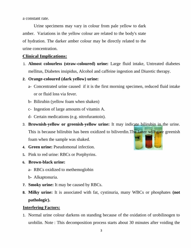
3. Brownish-yellow or greenish-yellow urine: It may indicate bilirubin in the urine.
This is because bilirubin has been oxidized to biliverdin.The latter will give greenish
foam when the sample was shaked.
4. Green urine: Pseudomonal infection.
5. Pink to red urine: RBCs or Porphyrins.
6. Brown-black urine:
a- RBCs oxidized to methemoglobin
b- Alkaptonuria.
7. Smoky urine: It may be caused by RBCs.
8. Milky urine: It is associated with fat, cystinuria, many WBCs or phosphates (not
pathologic).
Interfering Factors:
1. Normal urine colour darkens on standing because of the oxidation of urobilinogen to
urobilin. Note : This decomposition process starts about 30 minutes after voiding the
4
urine sample.
2. Some foods cause changes in urine colour:
a-Beets turn the urine red. b-Rhubarb can cause brown urine
3. Many drugs alter the colour of urine:
a- Cascara and senna laxatives in the presence of acid urine, they turn the urine in to
reddish brown; in the presence of alkaline urine, they turn the urine into red.
b- Bright-yellow colour in alkaline urine may be a result of riboflavin.
c- Urine that darkens on standing may indicate antiparkinsonian agents such as levodopa.
d- Black urine may be caused by cascara, ferrous salts e.g. sulfate, fumarate or gluconate
and metronidazole, nitrofurantoin, quinine for the same reason.
e- Blue urine may be caused by triamterene.
f- Blue-green urine may be caused by amitriptyline, methylene blue or mitoxantrone.
g- Red-pink urine may be caused by chloroxazone, daunorubicin, doxorubicin,
heparin, methyldopa and phenytoin.
3.2.4- Urine Odour:
Normal, freshly voided urine has a faint odour owing to the presence of volatile acids. It
is not generally offensive. Although not part of the routine UA, abnormal odours should
be noted.
Clinical Implications:
1. The urine of patients with diabetes mellitus may have a fruity (acetone) odour because
of ketosis (in addition to acetone 2 other ketone bodies; namely, acetoacetic acid and
β-hydroxy-butyric acid are found).
2. Bacteria result in foul-smelling urine due to its ability to split urea to form ammonia.
3. The urine of infants with an inherited disorder of amino acid metabolism known as
"maple syrup urine disease" smells like maple or burnt sugar.
4. Cystinuria and homocystinuria result in a sulfurous odour due to formation of….?
5. Tyrosinemia is characterized by a cabbage-like or "fishy" urine odour.
Interfering Factors:
5
a- Some foods, such as asparagus, produce characteristic urine odours.
b- Bacterial activity produces ammonia from the decomposition of urea, with its
characteristic pungent odour.
3.2.5- Urine pH:
The pH is an indicator of the renal tubules' ability to maintain normal hydrogen ion
concentration in the plasma and extracellular fluid. The pH of normal urine can vary
widely, from 4.6 to 7. The average pH value is about 6 (acidic).
Clinical Implications:
1. Acidic urine (pH <7.0) occurs in:
a- Metabolic acidosis, diabetic ketosis, diarrhea, starvation and uremia
b- Respiratory acidosis (carbon dioxide retention) c- Renal tuberculosis
2. Alkaline urine (pH> 7.0) occurs in:
a- Renal tubular acidosis and chronic renal failure
b- Metabolic acidosis (vomiting) c- Potassium depletion
d- Respiratory alkalosis involving hyperventilation ("blowing off" carbon dioxide).
Interfering Factors:
1. With prolonged standing, the pH of a urine specimen becomes alkaline. This is
because bacteria split urea (NH2 C=O NH2 and produce ammonia (NH4+).
2. Sodium bicarbonate, potassium citrate and acetazolamide may produce alkaline urine.
3. Urine becomes alkaline after eating because of excretion of stomach acid; this is
known as the "alkaline tide".
3.2.6- Specific gravity (SG):
Specific gravity is a measurement of the kidneys' ability to concentrate urine. It
compares the density of urine against the density of distilled water, which has an SG of
1.000. Because urine is a solution composed of minerals, salts and compounds dissolved
in water, therefore its SG is a measure of the density of the dissolved chemicals in the
specimenand must be higher than that of water.
6
Note : the number of particles present and the size of the particles influence SG.
Reference values:
a- Normal hydration and volume: 1.010 -1.025.
b- Concentrated urine: 1.025-1.030 or even more (+).
c- Diluted urine: 1.001-1.010.
d- Infant <2 years old: 1.001-1.018.
Clinical Implications:
1. SG values usually vary inversely with the amount of urine excreted (decreased urine
volume = increased SG). This relationship is not valid in certain conditions, including:
a- Diabetes-mellitus : increased urine volume but with increased SG….........Why?
b- Hypertension : normal volume but with decreased SG …………………..Why?
2. Hyposthenuria (low SG, 1.001-1.010) which occurs in:
a- Diabetes insipidus (low SG with large urine volume). It is caused by absence or
decrease of ADH (anti-diuretic hormones), a hormone that triggers kidney
absorption of water.
Note:Without ADH, the kidneys will produce excessive amounts of urine will not be
reabsorbed (sometimes 15 : 20 Litre/day).
b- Glomerulonephritis (kidney inflammation without infection).
c- Severe renal damage with disturbance in both concentrating and diluting abilities
of urine (isosthenuria). The SG is low (= 1.010) and fixed (Note: this fixed value
may vary little from one specimen to another).
3.Hypersthenuria (increased SG, 1.025-1.035) which occurs in the following:
Diabetes mellitus, nephrosis, excessive water loss (dehydration, fever, vomiting and
diarrhea), increased secretion of ADH and diuretic effects related to the stress of a
surgical procedure (What are the effects of adrenaline and nor-adrenaline on SG?)
Also, congestive heart failure and toxemia of pregnancy can also contribute to such
condition.
Interfering Factors:
7
a- Elevated readings may occur in the presence of moderate amounts of protein (1.0-
750 mg/dl) or with patients receiving intravenous albumin.
b- Diuretics and antibiotics cause high readings.
3.3- Chemical determinations of urine constituents:
3.3.1- Urine Blood [or Hemoglobin (Hb)]:
The presence of free hemoglobin in the urine is referred to as hemoglobinuria.
Hemoglobinuria can be related to conditions outside the urinary tract and occurs when
there is such extensive or rapid destruction (intravascular hemolysis) of circulating
erythrocytes. Hemoglobinuria may also occur because of lysis of RBCs in the urinary
tract.When intact RBCs are present in the urine (hematuria). Hematuriais most closely
related to disorders of the renal or genitourinary systems (trauma or damage to these
organs or systems).
The use of both a urine strips measurement and microscopic examination of urine
provide complete clinical evaluation of hemoglobinuria and hematuria. When urine
sediment is positive for occult blood but no RBCs are seen microscopically,
myoglobinuria can be suspected. Myoglobin can be distinguished from free hemoglobin
in the urine by chemical tests (How?).
Myoglobinuria is caused by excretion of myoglobin, a muscle protein, into the
urine because of:
a- Traumatic muscle injury e.g. in automobile accidents, football injuries or electric
shock.
b- A muscle disorder e.g. dystrophy.
Interfering Factors:
1- Drugs causing a positive result for blood or hemoglobin include:
a- Drugs toxic to the kidneys (eg, bacitracin and amphotericin)
b- Drugs that alter blood clotting (Warfarin, How?)
c- Drugs that cause hemolysis of RBCs (aspirin)
d- Drugs that may give a false-positive result (eg, bromides, copper, iodides and
8
oxidizing agents)
2- High doses of ascorbic acid or vitamin C may cause a false-negative result.
3- High SG or elevated protein reduces sensitivity.
4- Myoglobin produces a false-positive result.
5- Cleaning solution of the urine containers (Hypochlorites) causes false-positive
result.
6- Menstrual blood may contaminate the specimen and alter the result.
7- Prostatic infections may cause false-positive results.
3.3.2- Urine Protein (Albumin)
The presence of increased amounts of protein in the urine can be an important
indicator of renal disease. It may be the first sign of a serious problem and may appear
before any other clinical symptoms. However, other physiologic conditions (eg, exercise,
fever) can lead to increased protein excretion in urine. In addition, there are some renal
disorders in which proteinuria is absent.
In a healthy renal and urinary tract system, the urine contains no protein or only
trace amounts. These consist of albumin (one third of normal urine protein is albumin)
and globulins from the plasma. Because albumin is filtered more readily than the
globulins, it is usually abundant in pathologic conditions. Therefore, the term
albuminuria is often used synonymously with proteinuria.
Normally, the glomeruli prevent passage of protein from the blood to the
glomerular filtrate (What is role of chondroitin sulphate in such conditions?). Therefore,
the presence of protein in the urine is the single most important indication of renal
disease. If more than a trace of protein is found persistently in the urine, a quantitative
24-hour evaluation of protein excretion is necessary (How can the patient collect such
urine sample correctly?).
Interfering Factors for Qualitative Protein Test in urine:
1. Because of renal vasoconstriction, the presence of a functional, mild and transitory
proteinuria is associated with:
9
a- Strenuous exercise up to 300 mg/24 hours.
b- Severe emotional stress or seizures.
c- Cold baths or exposure to very cold temperature.
2- Increased protein in urine occurs in these benign states:
a- Fever and dehydration (salt depletion).
b- Non-immunoglobulin E food allergies.
c- Salicylate therapy
d- In the premenstrual period and immediately after delivery.
3- False or accidental proteinuria may occur because of a mixture of pus and RBCs in the
urinary tract related to infections, menstrual or vaginal discharge, mucus or semen
4- False-positive results can occur from incorrect use and interpretation of the colour
reagent strip test.
5- Alkaline, highly buffered urine can produce false-positive results on the dipstick test.
6- Very diluted urine may give a falsely low protein value.
3.3.2.1- Microalbuminuria
Microalbuminuria is an increase in urinary albumin that is below the detectable
range of the standard protein dipstick test. It is not a different chemical form of albumin.
Microalbuminuria occurs along before clinical proteinuria becomes evident
(Differentiate between proteinuria and microalbuminuria?).
This test allows for the routine detection of low concentrations of albumin in the
urine. This test has become a standard for the screening, monitoring and detection of
deteriorating renal function in diabetic patients. Studies have shown that diabetic patients
who progress to renal failure first excrete micro amounts of albumin and that, at this
stage, intervening treatment can reverse the proteinuria and thus prevent progression to
renal failure. This test is also used to monitor compliance of blood pressure control,
glucose and protein restriction. Its normal value is less than 30 mg /24 hours (24 hours
collection).
Clinical Implications:
10
Increased microalbuminuria is associated with:
1-Diabetes with early diabetic nephropathy. 2-Hypertension-heart disease
3-Generalized vascular diseases.
3.3.3- Urine Glucose (Glucosuria):
Glucose is present in glomerular filtrate and is reabsorbed by the proximal
convoluted tubule. If the blood glucose level exceeds the reabsorption capacity of the
tubules (180 mg%), glucose will appear in the urine. Tubular reabsorption of glucose is
by active transport in response to the body's need to maintain an adequate concentration
of glucose.
Note: The blood level at which tubular reabsorption stops is termed the renal threshold,
which for glucose is between 160 and 180 mg/dl.
Clinical Implications:
1. Increased glucose occurs in:
a- Diabetes mellitus
b- Endocrine disorders (thyrotoxicosis, Cushing's syndrome or acromegaly)
c- Liver and pancreatic disease.
d- Central nervous system disorders (brain injury or stroke)
e- Impaired tubular reabsorption
f- Pregnancy with possible latent diabetes (gestational diabetes).
2.Galactose: hereditary galactosuria (severe enzyme deficiency in infants; must be
treated promptly).
3. Pentose: certain drug therapies and rare hereditary conditions
4.Fructose: hereditary fructose intolerance or hepatic disorders
5.Lactose: pregnancy, lactation and lactose intolerance.
6. Xylose: excessive ingestion of fruit
Interfering Factors:
1. False-positive results:
a- Presence of non-sugar reducing substances such as ascorbic acid.
11
b-Testing after a heavy meal and testing soon after the administration of intravenous
glucose may all cause false-positive results.
c- Stress, excitement and myocardial infarction.
2. False-negative results may occur if urine is left to sit at room temperature for an
extended period, owing to the rapid glycolysis of glucose.
3.3.4- Urine Ketone Bodies (Acetone)
Ketones, which result from the metabolism of fatty acid (β- Oxidation of fatty
acids, followed by condensation of the produced active acetates) and fat. They consist
mainly of three substances ; namely, acetone, acetoacetic acid and β-hydroxybutyric
acid. The last two substances readily converted to acetone. This makes acetone is the
main substance being tested i.e. you can test for acetone only, instead of the other two.
However, some lab. specialists test only for acetoacetic acid.
In healthy persons, ketones are formed in the liver and are completely metabolized
sothat, only negligible amounts appear in the urine. However, when carbohydrate
metabolism is altered, an excessive amount of ketones is formed (acidosis) because fat
becomes the predominant body fuel instead of carbohydrates. Also, When fat is broken
down for energy, the body produces by-products called ketones (or ketone bodies) and
releases them into the urine. Large amounts of ketones in the urine may signal a
dangerous condition known as diabetic ketoacidosis. A diet low in sugars and starches
(carbohydrates), starvation, or prolonged vomiting may also cause the presence of
ketones in the urine.
The excess presence of ketones in the urine (ketonuria) is associated with diabetes
or altered carbohydrate metabolism. Testing for urine ketones in patients with diabetes
may provide the clue to early diagnosis of ketoacidosis and diabetic coma.
Ketonuria signals a need for caution, rather than crisis intervention, in either a
diabetic or a nondiabetic patient. However, this condition should not be taken lightly:
In the diabetic patient, ketone bodies in the urine suggest that the diabetes is not
adequately controlled and that adjustments of either the medication or the diet
12
should be made promptly.
In the nondiabetic patient, ketone bodies indicate a reduced carbohydrate
metabolism and excessive fat metabolism.
Positive urine ketones in a child younger than 2 years of age is a critical alert.
Interfering Factors:
1. Drugs that may cause a false-positive result include: Levodopa, Phenothiazines, Ether,
Insulin, Isopropyl alcohol, Metformin, Penicillamine, Phenazopyridine (Pyridium)
and Captopril.
2. False-negative results occur if urine stands too long, owing to loss of ketones into the
air.
3.3.5- Urine Nitrite (Nitrate reducing bacteria):
Some bacteria that cause a urinary tract infection (UTI) produce an enzyme that
converts urinary nitrates to nitrites. Thus, the presence of nitrites in urine indicates a UTI.
This test is a rapid, indirect method for detecting bacteria in the urine. Significant urinary
tract infection (UTI) may be present in a patient who does not experience any symptoms.
Common gram-negative organisms contain enzymes that reduce the nitrate in the urine to
nitrite.
Clinicians frequently request the urine nitrate test to screen high-risk patients:
pregnant women, school-aged children (especially girls), diabetic patients, elderly
patients and patients with a history of recurrent infections. The nitrate test can also be
used to evaluate the success of antibiotic therapy.
Clinical Implications:
1. Under the light microscope, the presence of >20 bacteria per high-power field (HPF)
may indicate a UTI. Untreated bacteriuria can lead to serious kidney disease.
2. The presence of a few bacteria suggests a UTI that cannot be confirmed or excluded
until more definitive studies, such as culture and sensitivity tests, are performed.
3. A positive nitrate test is a reliable indicator of significant bacteriuria and is a cue for
13
performing urine culture.
4. A negative result should never be interpreted as indicating absence of bacteriuria, for
the following reasons:
a- Some UTls are caused by organisms that do not convert nitrate to nitrite (eg,
staphylococci and streptococci).
b- Sufficient dietary nitrate may not be present for the nitrate-to-nitrite reaction to
occur.
Interfering Factors:
1. Azo dye metabolites and bilirubin can produce false-positive results.
2. Ascorbic acid can produce false-negative results.
3. False-positive results can be obtained if the urine sits too long at room temperature,
allowing contaminant bacteria to multiply.
4. High specific gravity will reduce the sensitivity.
3.3.6- Urine Bilirubin:
Bilirubin is formed in the reticuloendothelial cells of the spleen and bone marrow
because of the breakdown of hemoglobin; it is then transported to the liver. Urinary
bilirubin levels are increased to significant levels in the presence of any disease process
that increases the amount of conjugated bilirubin in the bloodstream. Elevated amounts
appear when the normal degradation cycle is disrupted by obstruction of the bile duct or
when the integrity of the I iver is damaged.
Urine bilirubin aids in the diagnosis and monitoring of treatment for hepatitis and
liver damage. Urine bilirubin is an early sign of hepatocellular disease or intrahepatic or
extrahepatic biliary obstruction. It should be a part of every UA because bilirubin often
appears in the urine before other signs of liver dysfunction (eg, jaundice or weakness)
become apparent.
Clinical Implications:
1. Even trace amounts of bilirubins are abnormal and are used for further investigation.
Normally, there is no detectable bilirubin in the urine.
14
2. Increased bilirubin occurs in:
a- Hepatitis and liver diseases caused by infections or exposure to toxic agents
b- Obstructive biliary tract disease
c- Septicemia
d- Liver or biliary tract tumors.
Interfering Factors:
1. Bilirubin rapidly decomposes when exposed to the light; therefore, urine should be
tested immediately.
2. High concentrations of ascorbic acid or nitrate cause decreased sensitivity.
3.3.7- Urine Urobilinogen:
Bilirubin, which is formed from the degradation of hemoglobin, is transformed
through the action of bacterial enzymes into urobilinogen after entering the intestines.
Some of the urobilinogen formed in the intestine is excreted as part of the feces, where it
is oxidized to urobilin; another portion is absorbed into the portal bloodstream and
carried to the liver, where it is metabolized and excreted in the bile. Traces of
urobilinogen in the blood that escape removal by the liver are carried to the kidneys and
excreted in the urine. This is the basis of the urine urobilinogen test. Unlike bilirubin,
urobilinogen is colourless.
Urine urobilinogen is one of the most sensitive tests available to determine
impaired liver function. Urinary urobilinogen is increased by any condition that causes an
increase in the production of bilirubin and by any disease that prevents the liver from
normally removing the reabsorbed urobilinogen from the portal circulation. An increased
urobilinogen level is one of the earliest signs of liver disease and hemolytic disorders.
Clinical Implications:
1. Urine urobilinogen is increased when there is:
a- Increased destruction of RBCs: Hemolytic anemias, Pernicious anemia
(megaloblastic) and Malaria.
15
b- Hemorrhage into tissues: Pulmonary infarction and Excessive bruising.
c- Hepatic damage: Biliary disease, Cirrhosis (viral and chemical) and Acute
hepatitis.
2. Urine urobilinogen is decreased or absent when normal amounts of bilirubin are not
excreted into the intestinal tract. This usually indicates partial or complete obstruction
of the bile ducts. The stool is pale in colour. Decreased urinary urobilinogen is
associated with Cholelithiasis, Severe inflammation of the biliary ducts or Cancer of
the head of the pancreas
3. During antibiotic therapy, suppression of normal gut flora may prevent the breakdown
of bilirubin to urobilinogen; therefore, urine levels will be decreased or absent
Interfering Factors:
1. Drugs that may affect urobilinogen levels include those that cause cholestasis and
those that reduce the bacterial flora in the gastrointestinal tract.
2. Peak excretion is known to occur from noon to 4:00 p.m. The amount of urobilinogen
in the urine is subject to diurnal variation.
3. Strongly alkaline urine shows a higher urobilinogen level, and strongly acidic urine
shows a lower urobilinogen level.
4. Drugs that may cause hemolysis with increased urobilinogen.
5. If the urine is highly coloured, the strip will be difficult to read.
3.4Microscopic examination of urine sediment
In this test, urine is spun in a centrifuge so the solid materials (sediment) settle out. The
sediment is spread on a slide and examined under a microscope.
In health, the urine contains small numbers of cells and other formed elements from the
entire genitourinary tract. Urinary sediment provides information useful for both
diagnosis and prognosis. It provides a direct sampling or urinary tract morphology.
16
3.4- Procedure for Microscopic Urine Examination: (lecture 7)
1. Collect a random urine specimen, then the specimenmust be transported to the
laboratory as soon as possible.
2. Urinary sediment is microscopically examined under both the low-power field (LPF)
and the high-power field (HPF). Low power is used to find and count casts; RBCs,
WBCs and bacteria show up and are counted under high power. Amounts present are
defined in the following terms: few, moderate, packed and packed solid; or 1+,2+,3+
and 4+. Crystals and other elements are also noted.
3. Microscopic results should be correlated with the physical and chemical findings
to ensure the accuracy of the report.
3.4.1- Urine Crystals:
A variety of crystals may appear in the urine. They can be identified by their
specific appearance and solubility characteristics. Crystals in the urine may present no
symptoms or they may be associated with the formation of urinary tract calculi and give
rise to clinical manifestations associated with partial or complete obstruction of urine
flow. A number of in vivo and in vitro factors influence the types and numbers of urinary
crystals in a given sample.
In vivo factors include:
a- Concentration and solubility of crystallogenic substances contained in the specimen.
b- Urine pH.
c- Excretion of diagnostic and therapeutic agents.
In vitro factors include:
a- Temperature (solubility decreases with temperature).
b- Evaporation (increases solute concentration).
c- Urine pH (changes with standing and bacterial overgrowth).
17
The most common crystals are:
Magnesium, ammonium phosphate and triple phosphate:Usually appear as colourless,
3-dimensional, prism-like crystals.
Bilirubin: Bilirubin crystals tend to precipitate onto other formed elements in the
urine. In the top picture, fine needlelike crystals have formed on an underlying cell.
This is the most common appearance of bilirubin crystals. In the lower two pictures,
cylindrical bilirubin crystals have formed in association with droplets of fat, resulting
in a "flashlight" appearance. This form is less commonly
Calcium Carbonate: Calcium carbonate crystals usually appear as large yellow-brown
or colourless spheroids with radial striations. They can also be seen as smaller crystals
with round, ovoid or dumbbell shapes.
Calcium Oxalate Dihydrate: Calcium oxalate dihydrate crystals typically are seen as
colourless squares whose corners are connected by intersecting lines (resembling an
envelope). They can occur in urine of any pH. The crystals vary in size from quite
large to very small. In some cases, large numbers of tiny oxalates may appear as
amorphous unless examined at high magnification.
Uric acid crystals: They exhibit extreme pleomorphism in size and in shape. They
appear readily in acid urine allowed to stand at room temperature.
Cystine: Cystine crystals are flat colourless plates and have acharacteristichexagonal
shape with equal or unequal sides. They often aggregate in layers. Their formation is
favored in acidic urine.
Interfering Factors:
1. Refrigerated urine will precipitate out many crystals because the solubility properties
of the compound are altered.
2. Urine left standing at room temperature will also cause precipitation of crystals or the
dissolving of the crystals.
3. Radiographic dye can cause crystals in improperly hydrated patients. These resemble
uric acid crystals and can be suspected in specimens that have an abnormally high

18
specific gravity (>1.030).
3.4.2- Urinary Casts
Some types of kidney diseases can cause plugs of material called casts. These
casts can then get flushed out into the urine. Casts can be made of different types of
material, such as red or white blood cells, waxy or fatty substances, or protein. The type
of cast can provide clues about the type of kidney disease that may be present.
Casts are cylindrical structures composed mainly of mucoprotein, which is secreted
by epithelial cells lining the loops of Henle, the distal tubules and the collecting ducts.
The factors responsible for the precipitation of this mucoprotein are not fully understood,
but may relate to the concentration and pH of urine in these areas. Casts may form in the
presence or absence of cells in the tubular lumen. If cells (epithelial cells, WBC) are
present as a cast forms, they may adhere to, and subsequently be surrounded by, the
fibrillar protein network.
The appearance of a cast observed in a urine sediment depends largely upon the
length of time it remained in situ in the tubules prior to being shed into the urine. A cast
recognizable as "cellular", for example, was shed shortly after it was formed. A waxy
cast, in contrast, was retained longer in the tubular system prior to being released.
General Interpretation of casts:
Casts are quantified for reporting as the number seen per low power field (10x
objective) and classified as to type (e.g., waxy casts, 5-10/LPF). Casts in urine from
normal individuals are few or none.
1. An absence of casts does not rule out renal disease. Casts may be absent or very few
in cases of chronic, progressive, generalized nephritis. Furthermore, casts are unstable
in urine and are prone to dissolution with time, especially in dilute and/or alkaline
urine.
2. Although the presence of numerous casts is solid evidence of generalized (usually
acute) renal disease, it is not a reliable indicator of prognosis.
Types of casts :
19
Hyaline Casts: are formed in the absence of cells in the tubularlumen. They have a
smooth texture and a refractive index very close to that of the surrounding fluid.
Reduced lighting is essential to see hyaline casts. Lower the substage condenser.
When present in low numbers (01/LPF) in concentrated urine of otherwise normal
patients, hyaline casts are not always indicative of clinically significant renal disease.
Cellular Casts: the most common result when disease processes such as ischemia,
infarction, or nephrotoxicity cause degeneration and necrosis of tubular epithelial
cells. The presence of these casts indicates acute tubular injury but does not indicate
the extent or reversibility of the injury. A common scenario is the patient with
decreased renal perfusion and oliguria secondary to severe dehydration
Granular Casts: Granular casts, as the name implies, have a textured appearance which
ranges from fine to coarse in character.
Fatty Casts: Fatty casts are identified by the presence of refractile lipid droplets. The
background matrix of the cast may be hyaline or granular in nature. They often seen in
urines in which free lipid droplets are present as well. Pictured here is a fatty cast with
a hyaline matrix.
Waxy Casts: Waxy casts have a smooth consistency but are more refractile and
therefore easier to see compared to hyaline casts. They commonly have squared off
ends, as if brittle and easily broken. Waxy casts indicate tubular injury of a more
chronic nature than granular or cellular casts and are always of pathologic
significance.
3.4.3-Leukocytes (white blood cells [WBCs]) in the urine
WBCs or leukocyte esterase detects leukocytes in the urine. The presence of
WBCs in the urine may indicate a UTI.
3.4.4- Red or white blood cells:
Normally blood cells are not found in urine. Inflammation, disease, or injury to the
kidneys, ureters, bladder, or urethra can cause blood in urine. Strenuous exercise (such as
running a marathon) can also cause blood in urine. White blood cells are often a sign of
20
infection, cancer, or kidney disease.
3.4.5- Infectious Agents in Urine Sediment:
Infectious agents of various classes can be observed in urine sediments. In most
cases, their significance can be properly assessed only in light of the clinical signs,
method of collection, post-collection interval, and other findings in the urinalysis.
Types of infectious agents:
Candida:Yeasts in unstained urine sediments are round to oval in shape, colourless,
and may have obvious budding. They often represent contaminants, and are especially
suspect if the sample is voided and/or old. In other circumstances, however, their
significance should not be discounted. The pictures shows pseudohyphae formation by
the yeasts, which were identified on culture as Candida albicans.
Bacteria: can be identified in unstained urine sediments when present in sufficient
numbers. Rod-shaped bacteria and chains of cocci are often readily identifiable. If
there is any doubt about the presence of bacteria, a Gramstained smear of urine
sediment should be examined. The lower panel at the right shows a neutrophil
containing phagocytized bacteria. Notice that the nucleus in this cell is round; nuclei
tend to become round as neutrophils age in urine
Fungi: Fungal hyphae in urine sediment most commonly represent over growth of
contaminants in such urine samples if their analyses were delayed. If seen in a fresh
sample, fungal infection of the kidneys and/or bladder should be suspected (Fungal
culture can be recommended).
3.4.6- Cells in Urine Sediment :
Urine is a hostile environment for cells since they encounter abnormal osmotic
pressures, pH changes and exposure to toxic metabolites. For these reasons, post-
collection delay of examination should be minimized. If delay is unavoidable,
refrigeration will slow degeneration of cells.
For routine purposes, cells are examined as unstained wet-mounts of sedimented
urine. Under some circumstances, air-dried smears are prepared and stained with
21
hematologic stains. Red blood cells and leukocytes are quantified as cells/HPF (High
Power Field - 40x objective). Other cell types are usually subjectively listed as "few,
moderate, or many".
Types of cells:
Red Blood Cells: The appearance RBCs in urine depends largely on the concentration
of the specimen and the length of time the red cells have been exposed. Fresh red cells
tend to have a red or yellow colour (lower panel). Prolonged exposure results in a pale
or colourless appearance as hemoglobin may be lost from the cells (upper panel). In
fresh samples with S.G. of 1.010-1.020, RBC may retain the normal disc shape
(upper panel). In more concentrated urines (>1.025), red cells tend to shrink and
appear as small, crenated cells (lower panel). In more dilute samples, they tend to
swell. At urine S.G. <1.008 and/or highly alkaline pH, red cell lysis is likely. Lysed
red cells appear as very faint "ghosts", or may be virtually invisible.
Red blood cells up to 5/HPF are commonly accepted as normal. Increased red
cells in urine is termed hematuria, which can be due to hemorrhage, inflammation,
necrosis, trauma or neoplasia somewhere along the urinary tract. The method of
collection must be considered because catheterization can induce hemorrhage.
White Blood Cells: WBCs in unstained urine sediments typically appear as round,
granular cells which are1.5-2.0 times the diameter of RBC. The details of nuclear
shape often are difficult to discern, especially if the specimen is not fresh. WBC in
urine are most commonly neutrophils. Staining of air-dried sediment smears with a
hematologic stain sometimes is useful for more specific identification. Like
erythrocytes, WBC may lyse in very dilute or highly alkaline urine. WBC up to 5/HPF
are commonly accepted as normal. Greater numbers (pyuria) generally indicate the
presence of an inflammatory process somewhere along the course of the urinary tract.
Pyuria (?) often is caused by urinary tract infection, and many times bacteria can be
seen in the sediments. Depending on clinical signs, pyuria may be an indication for
culture of urine even if no bacteria are seen.
22
Squames epithelial cells: Squamous epithelial cells are the largest cells which can be
present in normal urine samples. They are thin, flat cells, usually with an angular or
irregular outline and a small round nucleus. They may be present as single cells or in
variably-sized clusters. Those shown in the upper panel are unstained; that in the
lower panel was prepared using Sedi-Stain. Their main significance is as an indicator
of such contam ination.
Neoplastic Cells: Neoplastic cells may be seen in urine sediments of patients with
tumors of the urinary tract. The pictures shown are from a case of transitional cell
carcinoma in the bladder. Though the presence of neoplastic cells may be suspected
on examination of unstained wet-mounts (upper panel), evaluation of air-dried
sediment smears or cytocentrifuge preps stained with hematologic stains (lower panel)
is necessary for confirmation.
3.4.7 Contaminants in Urine Sediment :
Extraneous contaminating materials of many types can make their way into urine
specimens. Striving for optimal collection and transport of specimens will help maximize
useful results and minimize confusing findings.
Types of cells:
Overgrowth of Microbes: Specimens mailed to laboratories without refrigeration or
preservatives are subject to overgrowth of microbes, whether contaminants or
pathogens. They can multiply when analysis is delayed. This often clouds the
interpretation of both sediment examination and culture results. Refrigeration is
usedfor preserving a specimen.
Fibers: Cotton, plant and paper fibers may be confused for parasite larvae or urinary
casts. Care in sample collection and handling will minimize the presence of such
material.
Starch Granules: powder on surgical and exam gloves. They are variable in size,
round to polygonal in shape, colourless and usually have a circular or V-shaped "dot"
in the center.
23
Important notes :
1- Urine test may be done:
a- To screen for a disease or infection of the urinary tract. Symptoms that may lead
to a urine test include discolored or foul-smelling urine, pain during urination,
difficulty urinating, flank pain, blood in the urine (hematuria), or fever.
b- To monitor the treatment of certain conditions such as diabetes, kidney stones, a
urinary tract infection (UTI), hypertension, or some types of kidney or liver
disease.
c- As part of a routine examination.
2- Precautions before urine sampling :
a- Do not eat foods that can discolor the urine. Do not exercise strenuously before a
urine sample is taken.
b- For females, if you are menstruating or within a few days of starting your
menstrual period, your doctor may want to postpone the urine test, depending on
the suspected problem.
c- certain medications can discolor the urine, your doctor may instruct you to stop
taking the medications prior to the test. Medications that can change the color of
urine must be stoped or taken into consideration during interpretation of the test
result. Be sure to tell your doctor if you are taking diuretics, which may also affect
the test results.
24
Blood Glucose: (lecture 8)
Test Overview:
Glucose comes from carbohydrate foods. It is the main source of energy used
by the body. Insulin is a hormone that helps your body use and control the amount of
glucose in your blood. Insulin is produced in the pancreas and released into the blood
when the amount of glucose in the blood rises.
Normally, your blood glucose levels increase slightly after you eat. This
increase causes your pancreas to release insulin so that your blood glucose levels do
not get too high. Blood glucose levels that remain high over time can damage your
eyes, kidneys, nerves, and blood vessels.
Several different types of blood glucose tests are used:
Fasting blood sugar (FBS) measures blood glucose after you have not eaten for
at least 8 hours. It often is the first test done to check for diabetes.
2-hour postprandial blood sugar (2-hour PC) measures blood glucose exactly
2 hours after you eat a meal.
Random blood sugar (RBS) measures blood glucose regardless of when you
last ate. Several random measurements may be taken throughout the day.
Random testing is useful because glucose levels in healthy people do not vary
widely throughout the day. Blood glucose levels that vary widely may indicate a
problem. This test is also called a casual blood glucose test.
Oral glucose tolerance test is used to diagnose diabetes that occurs during
pregnancy (gestational diabetes). An oral glucose tolerance test is a series of
blood glucose measurements taken after you drink a sweet liquid that contains
glucose. This test is not recommended for diagnosing diabetes in a person who is
not pregnant. For more information, see the medical test Gestational Diabetes.

25
Blood glucose tests are done to:
i. Check for diabetes.
ii. Monitor treatment of diabetes.
iii. Check for diabetes that occurs during pregnancy (gestational diabetes).
iv. Determine if an abnormally low blood sugar level (hypoglycemia) is present.
Note:
1- A test to measure blood levels of a protein called C-peptide (this peptide cleaved
from pro-insulin during synthesizing insulin in its mature (active) form may be
done along with a blood glucose test to determine the cause of hypoglycemia.
2- Fasting blood sugar (FBS): For a fasting blood sugar test, do not eat or drink
anything other than water for at least 8 hours before the blood sample is
taken.
3- If you have diabetes, you may be asked to wait until you have had your blood
tested before taking your morning dose of insulin or diabetes medication.
4- 2-hour postprandial blood sugar (2-hour PC): For a 2-hour postprandial test, eat a
meal exactly 2 hours before the blood sample is taken. A home blood sugar test
is the most common way to check 2-hour postprandial blood sugar levels.
5- Random blood sugar (RBS): No special preparation is required before having a
random blood sugar test.
Normal Blood glucose
Fasting blood glucose: 70–99 mg/dL or less than 5.5 mmol/L
2 hours after eating (postprandial): 70–145 mg/dL (less than 7.9 mmol/L)
Random (casual): 70–125 mg/dL (less than 7.0 mmol/L)
High values:
26
The American Diabetes Association (ADA) criteria for diagnosing diabetes are
met when any of the following results have been repeated on at least two different
days:
o A fasting blood glucose level is 126 mg/dL (7.0 mmol/L) or higher.
o A 2-hour oral glucose tolerance test result is 200 mg/dL (11.1 mmol/L) or
higher. For more information, see the medical test Oral Glucose Tolerance Test.
o Symptoms of diabetes are present and a random blood glucose test is 200 mg/dL
(11.1 mmol/L) or higher. Symptoms of diabetes include increased thirst and
frequent urination (especially at night), unexplained increase in appetite,
unexplained weight loss, fatigue, erection problems, blurred vision, and tingling
or numbness in the hands or feet.
If your fasting blood glucose level is between 100 mg/dL (5.5 mmol/L) and 126
mg/dL (7.0 mmol/L), you are considered to have prediabetes (impaired fasting
glucose), and you have an increased chance of getting diabetes.
Other conditions that can cause high blood glucose levels include severe stress,
heart attack, stroke, Cushing's syndrome, medications such as corticosteroids,
cancers, or excess production of growth hormone (acromegaly).
Low values:
A fasting glucose level below 40 mg/dL (2.2 mmol/L) in women or below 50 mg/dL
(2.8 mmol/L) in men that is accompanied by symptoms of hypoglycemia may mean
you have an insulinoma, a tumor that produces abnormally high amounts of insulin.
Low glucose levels also may be caused by:
i. Addison's disease (Adrenal cortex insufficiency).
ii. Decreased thyroid hormone levels (hypothyroidism).
iii. A tumor in the pituitary gland.
iv. Liver disease, such as cirrhosis.
v. Kidney failure.
27
vi. Malnutrition or an eating disorder, such as anorexia.
vii. Medications used to treat diabetes.
Reasons you may not be able to have the test or why the results may not be
helpful include:
Eating or drinking less than 8 hours before a fasting blood test or less than 2
hours before a 2-hour postprandial test.
Illness or emotional stress (due to increased levels of adrenaline and nor-
adrenalin),
Treatment with corticosteroids
Notes:
Other tests are needed to accurately diagnose diabetes. A blood glucose test may
not identify some people with pre-diabetes or early diabetes. Many experts recommend
using a glucose tolerance test if the result of a fasting blood glucose test is between 100
mg/dL (5.5 mmol/L) and 126 mg/dL (7.0 mmol/L). This range is above the normal range
but below the range that indicates diabetes.
Glucose levels in urine also can be measured. Many people with diabetes have
glucose in their urine. However, the level in the blood must be very high before glucose
can be detected in the urine (Renal threshold is a limiting factor). For this reason, tests
for glucose in urine are not used to diagnose or monitor diabetes.
A glycohemoglobin test can help monitor the long-term control of blood glucose
levels in people with diabetes. This test is the preferred method of monitoring
long-term control of blood sugar levels (during the last 120 days Glycohemoglobin
(GHb) or HbA1c ).
An oral glucose tolerance test may be done with a blood glucose test to confirm a
diagnosis of diabetes. An oral glucose tolerance test is most commonly done to
screen pregnant women for gestational diabetes.
28
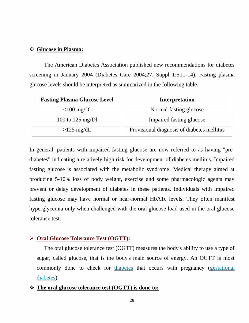
Glucose in Plasma:
The American Diabetes Association published new recommendations for diabetes
screening in January 2004 (Diabetes Care 2004;27, Suppl 1:S11-14). Fasting plasma
glucose levels should be interpreted as summarized in the following table.
Fasting Plasma Glucose Level Interpretation
<100 mg/Dl Normal fasting glucose
100 to 125 mg/Dl Impaired fasting glucose
>125 mg/dL Provisional diagnosis of diabetes mellitus
In general, patients with impaired fasting glucose are now referred to as having "pre-
diabetes" indicating a relatively high risk for development of diabetes mellitus. Impaired
fasting glucose is associated with the metabolic syndrome. Medical therapy aimed at
producing 5-10% loss of body weight, exercise and some pharmacologic agents may
prevent or delay development of diabetes in these patients. Individuals with impaired
fasting glucose may have normal or near-normal HbA1c levels. They often manifest
hyperglycemia only when challenged with the oral glucose load used in the oral glucose
tolerance test.
Oral Glucose Tolerance Test (OGTT):
The oral glucose tolerance test (OGTT) measures the body's ability to use a type of
sugar, called glucose, that is the body's main source of energy. An OGTT is most
commonly done to check for diabetes that occurs with pregnancy (gestational
diabetes).
The oral glucose tolerance test (OGTT) is done to:
29
1. Check pregnant women for gestational diabetes. When done for this purpose, the
test is called a glucose challenge screening test, and it is usually done during the
24th to the 28
th week of pregnancy. You have an increased chance of developing
gestational diabetes if you:
o Have had gestational diabetes during a previous pregnancy.
o Have previously given birth to a baby who weighed more than 8.8 lb (4 kg).
o Are younger than age 25 and were overweight before getting pregnant.
2. Confirm the presence of gestational diabetes if other blood glucose measurements
are high.
3. To screen women who have polycystic ovary syndrome (PCOS) for diabetes.
To prepare for the glucose tolerance diagnostic test:
1. Eat a balanced diet that contains at least 150 to 200 grams of carbohydrate per
day for 3 days before the test. Fruits, breads, cereals, grains, rice, crackers, and
starchy vegetables such as potatoes, beans, and corn are good sources of
carbohydrate.
2. Do not eat, drink, smoke, or exercise strenuously for at least 8 hours before
your first blood sample is taken.
3. The glucose tolerance diagnostic test may take up to 4 hours. Since activity can
interfere with test results, you will be asked to sit quietly during the entire test.
Do not eat during the test. You may drink only water during this time.
On the day of testing, the following steps will be done:
A blood sample will be collected when you arrive (fasting blood glucose value).
It provides a baseline for comparing other glucose values.
For the standard glucose tolerance test, you will drink 75 g to 100 g for
pregnant women.
Blood samples will be collected at timed intervals of 30 minutes and 1, 2, and to
more than 3 hours after you drink the glucose.
30
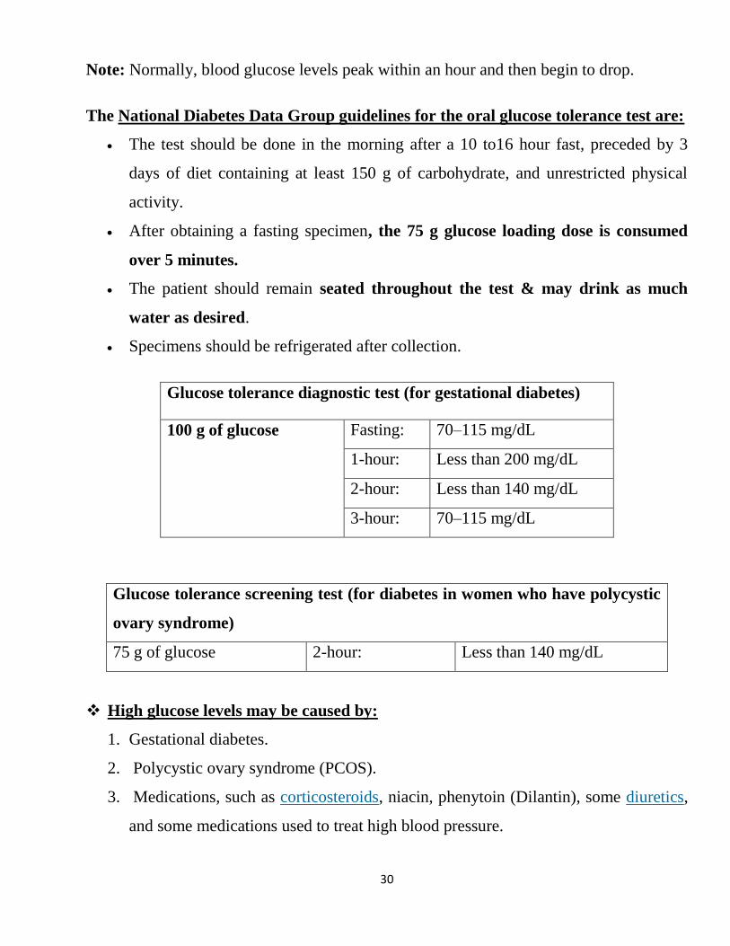
Note: Normally, blood glucose levels peak within an hour and then begin to drop.
The National Diabetes Data Group guidelines for the oral glucose tolerance test are:
The test should be done in the morning after a 10 to16 hour fast, preceded by 3
days of diet containing at least 150 g of carbohydrate, and unrestricted physical
activity.
After obtaining a fasting specimen, the 75 g glucose loading dose is consumed
over 5 minutes.
The patient should remain seated throughout the test & may drink as much
water as desired.
Specimens should be refrigerated after collection.
Glucose tolerance diagnostic test (for gestational diabetes)
100 g of glucose Fasting: 70–115 mg/dL
1-hour: Less than 200 mg/dL
2-hour: Less than 140 mg/dL
3-hour: 70–115 mg/dL
Glucose tolerance screening test (for diabetes in women who have polycystic
ovary syndrome)
75 g of glucose 2-hour: Less than 140 mg/dL
High glucose levels may be caused by:
1. Gestational diabetes.
2. Polycystic ovary syndrome (PCOS).
3. Medications, such as corticosteroids, niacin, phenytoin (Dilantin), some diuretics,
and some medications used to treat high blood pressure.
31
4. Severe stress. *-Large amounts of the hormone cortisol in the blood (Cushing's
syndrome).
5. Inherited diseases, such as cystic fibrosis, pheochromocytoma, or
hemochromatosis.
6. Overproduction of growth hormone(acromegaly).
Low glucose levels may be caused by:
1. Medications that used to treat diabetes.
2. A condition that prevents the intestines from absorbing nutrients from food,
such as celiac disease.
3. Decreased production of the hormones cortisol and aldosterone (Addison's
disease).
4. Problems with the thyroid gland (hypothyroidism) or an underactive pituitary
gland.
5. A tumor of the pancreas (insulinoma).
6. Inflammation and scarring of the liver (cirrhosis).
Reasons you may not be able to have the test or why the results may not be
helpful include:
1. Medications, such as corticosteroids, diuretics, seizure medications, birth
control pills, nonsteroidal anti-inflammatory drugs (NSAIDs), and some
medications used to treat high blood pressure.
2. Recent surgery, heart attack, or childbirth.
3. A low-carbohydrate diet.
4. Vomiting during the test.
5. Emotional stress.
6. Fever and infection.
32
Notes:
Glucose tolerance test screening by age 30 is recommend for all women who have
polycystic ovary syndrome. For more information, see the topic Polycystic Ovary
Syndrome (PCOS).
.Hemoglobin A1C (Glycosylated haemoglobin):
HbA1c refers to a minor population of HbA that has been modified by attachment
of glucose to the terminal amino acid of the beta globin chain.
The rate of formation of HbA1c is directly proportional to the plasma glucose
concentration. Since erythrocytes are freely permeable to glucose, HbA1c levels
provide a glycemic history during the average erythrocyte lifespan, which is
approximately 120 days.
HbA1c levels can be used not only to assess long-term glycemia, but also to
predict risk of developing chronic complications.
Baseline HbA1c levels are strongly related to the incidence and/or progression of
retinopathy, gross proteinuria, and loss of tactile sensation or temperature
sensitivity.
The Diabetes Control and Complications Trial (DCCT), which was completed in
1993, demonstrated that the risks for development and progression of the chronic
complications of type 1 diabetes are closely related to the degree of glycemic
control, as measured by serial HbA1c determinations.
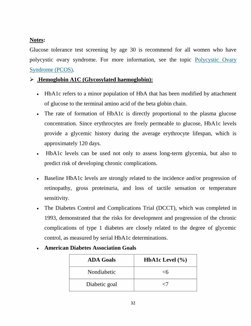
American Diabetes Association Goals
ADA Goals HbA1c Level (%)
Nondiabetic <6
Diabetic goal <7
33
Action suggested >8
Optimal frequency of HbA1c testing has not been well established. Monthly
HbA1c levels have been recommended for pregnant women with diabetes, but little
scientific data is available to support this recommendation.
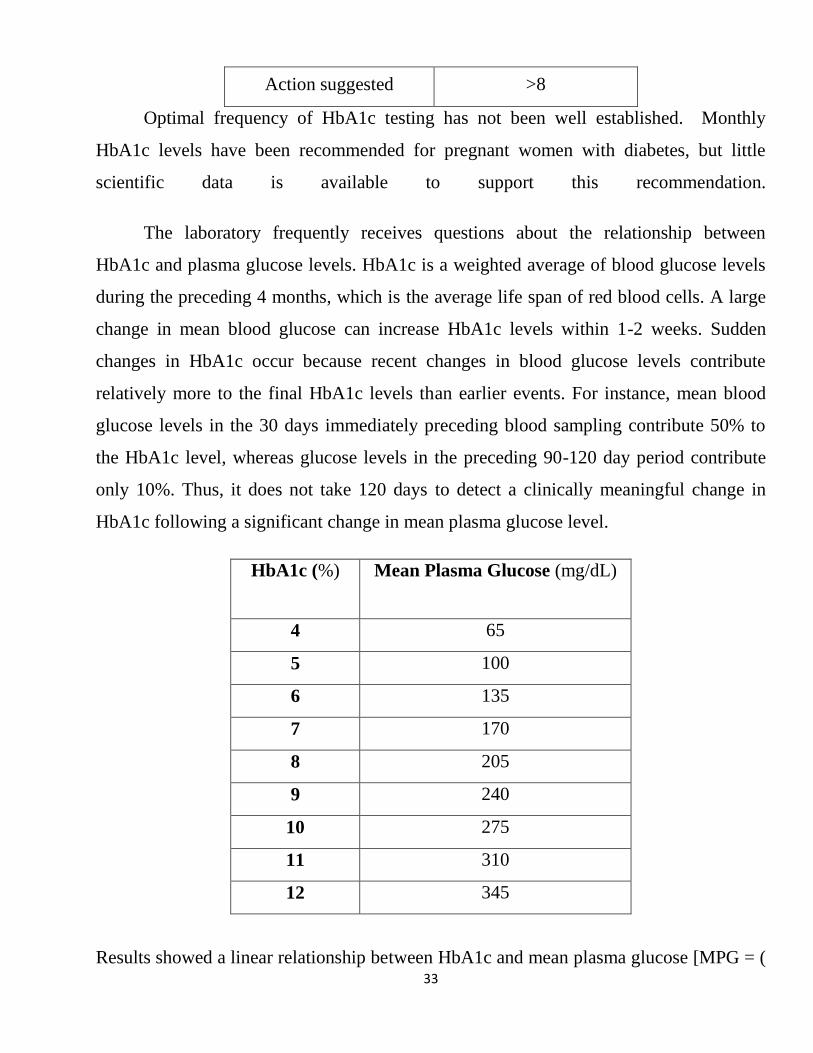
The laboratory frequently receives questions about the relationship between
HbA1c and plasma glucose levels. HbA1c is a weighted average of blood glucose levels
during the preceding 4 months, which is the average life span of red blood cells. A large
change in mean blood glucose can increase HbA1c levels within 1-2 weeks. Sudden
changes in HbA1c occur because recent changes in blood glucose levels contribute
relatively more to the final HbA1c levels than earlier events. For instance, mean blood
glucose levels in the 30 days immediately preceding blood sampling contribute 50% to
the HbA1c level, whereas glucose levels in the preceding 90-120 day period contribute
only 10%. Thus, it does not take 120 days to detect a clinically meaningful change in
HbA1c following a significant change in mean plasma glucose level.
HbA1c (%) Mean Plasma Glucose (mg/dL)
4 65
5 100
6 135
7 170
8 205
9 240
10 275
11 310
12 345
Results showed a linear relationship between HbA1c and mean plasma glucose [MPG = (
34
35.6 x HbA1c ) - 77] with a Pearson correlation coefficient (r) of 0.82. Each 1% change
in HbA1c represents a change of approximately 35 mg/dl in plasma glucose. It is
important to realize that this data is based on overall averages and may vary slightly in
individual patients.
HbA1c for Diagnosis of Diabetes:
HbA1c is more reproducible, informative, and convenient. In this setting, HbA1c
levels of 7% or greater were indicative of diabetes.
Hemoglobin A1c as a CV Risk Factor in Non-diabetic Individuals:
Macrovascular disease is the most important cause of mortality and morbidity in
individuals with type 2 diabetes. Even when adjusted for conventional risk factors,
diabetic individuals still exhibit a two to four fold increased risk of cardiovascular
disease in comparison to nondiabetic people. Therefore, hyperglycemia is strongly
suspected of promoting atherogenesis. Excess glucose is transformed into
advanced glycation endproducts (AGEs) that not only make blood vessels inelastic
and stenotic but also activates chronic inflammation.
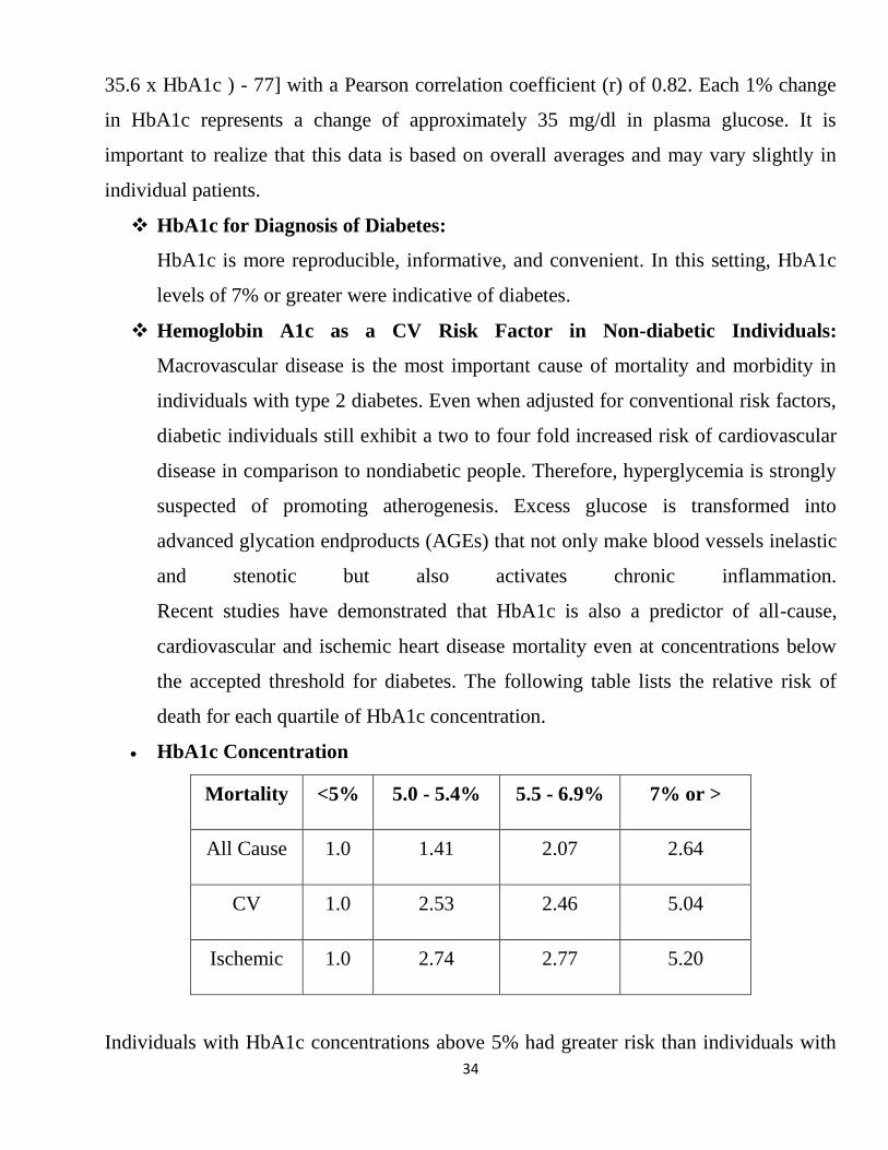
Recent studies have demonstrated that HbA1c is also a predictor of all-cause,
cardiovascular and ischemic heart disease mortality even at concentrations below
the accepted threshold for diabetes. The following table lists the relative risk of
death for each quartile of HbA1c concentration.
HbA1c Concentration
Mortality <5% 5.0 - 5.4% 5.5 - 6.9% 7% or >
All Cause 1.0 1.41 2.07 2.64
CV 1.0 2.53 2.46 5.04
Ischemic 1.0 2.74 2.77 5.20
Individuals with HbA1c concentrations above 5% had greater risk than individuals with
35
concentrations below 5%. Approximately 25% of population had HbA1c levels below
5% and 70% of the population had levels between 5 and 6.9%. HbA1c appears to
resemble blood pressure and cholesterol in terms of its continuous relationship with
cardiovascular risk. Two recent studies in the Annals of Internal Medicine have also
validated that HbA1c is a progressive risk factor for CV disease in individuals with and
without diabetes.
36
Ketones bodies: (lecture 9)
A Ketone test checks for ketones bodies in your blood or urine. Ketones are
substances that are made when the body breaks down fat for energy. Normally, your body
gets the energy it needs from carbohydrate in your diet. However, stored fat is broken
down and ketones are made if your diet does not contain enough carbohydrate to supply
the body with glucose for energy or if your body cannot use blood such sugar properly.
A ketone blood test is the most accurate method. It is recommended for all people
with diabetes whenever symptoms of illness, such as nausea, vomiting, or abdominal pain
are present. It is used to diagnose diabetic ketoacidosis when symptoms of high blood
sugar are present. A ketone urine test is the most commonly used method of measuring
ketones. However, it is less accurate than a blood test.
It may be done to:
Monitor a person on a very low carbohydrate diet.
Monitor a pregnant woman who has diabetes or has developed gestational diabetes.
Results:
Ketones
Normal: There are no ketones in your blood or urine.
Abnormal: Ketones are present in your blood or urine.
Urine test:
If either the test strip or the urine changes color when the tablet is dropped into the
sample, ketones are present in your urine sample. The test results are read as negative
to 1+ to 4+ or small to large.
You may have ketones in your urine if you:
1. Have poorly controlled diabetes or diabetic ketoacidosis.
2. Are on a very low carbohydrate diet.
37
3. Are starving or have an eating disorder, including disorders that result in poor
nutrition such as anorexia nervosa or bulimia, alcoholism, or poisoning from
drinking rubbing alcohol (isopropanol).
4. Have not eaten (fasted) for 18 hours or longer.
5. Are pregnant. However, a moderate amount of ketones in a pregnant woman
may harm the fetus and may be an indication of gestational diabetes.
Reasons why the results may not be helpful include:
1. When vitamin C (ascorbic acid) is taken in large amounts, the test result will be
affected.
2. Dehydration.
3. A high-fat diet.
4. Pregnancy.
5. If you are feeling very stressed.
Cholesterol and Triglycerides Tests:
Test Overview:
Cholesterol and triglyceride tests are blood tests that measure the total amount of fatty
substances (cholesterol and triglycerides) in the blood. Cholesterol travels through the
blood attached to a protein. This cholesterol-protein package is called a lipoprotein.
Lipoprotein analysis (lipoprotein profile or lipid profile) measures blood levels of total
cholesterol, LDL cholesterol, HDL cholesterol, and triglycerides.
Cholesterol. The body uses cholesterol to help build cells and produce
hormones. Too much cholesterol in the blood can build up along the inside of
the artery walls, forming what is known as plaque. Large amounts of plaque
increase your chances of having a heart attack or stroke.
HDL (high-density lipoprotein) cholesterol helps remove fat from the body
by binding with it in the bloodstream and carrying it back to the liver for
38
disposal. It is sometimes called “good” cholesterol. A high level of HDL
cholesterol may lower your chances of developing heart disease or stroke.
LDL (low-density lipoprotein) cholesterol carries mostly fat and only a
small amount of protein from the liver to other parts of the body. It is
sometimes called "bad cholesterol." A high LDL cholesterol level may
increase your chances of developing heart disease.
VLDL: (very low-density lipoprotein) cholesterol contains very little
protein. The main purpose of VLDL is to distribute the triglyceride produced
by your liver. A high VLDL cholesterol level can cause the buildup of
cholesterol in your arteries and increases your risk of heart disease and
stroke.
Triglycerides are a type of fat the body uses to store energy. Only small
amounts are found in the blood. Having a high triglyceride level along with a
high LDL cholesterol may increase your chances of having heart disease
more than having only a high LDL cholesterol level.
Some medical experts recommend routine cholesterol and triglyceride testing to screen
for problems that affect the way cholesterol is produced, used, carried in the blood, or
disposed of by the body. Others may choose to routinely measure only total cholesterol
and HDL levels.
Cholesterol and triglyceride testing is done:
1. As part of a routine physical exam to screen for a lipid disorder.
2. To check your response to medicines used to treat lipid disorders.
3. To help determine your chances of having of heart disease.
4. If you have unusual symptoms, such as yellow fatty deposits in the skin
(xanthomatosis)?.
39
Preparation for the test:
1. Do not eat or drink anything except water for 9 to 12 hours before having your
blood drawn.
2. Do not eat high-fat foods the night before the test. *- Do not exercise
strenuously before the test.
Results :
Cholesterol and triglyceride tests are blood tests that measure the total amount of fatty
substances (cholesterol and triglycerides) in the blood. Cholesterol and triglyceride
levels vary according to your age and sex.
40
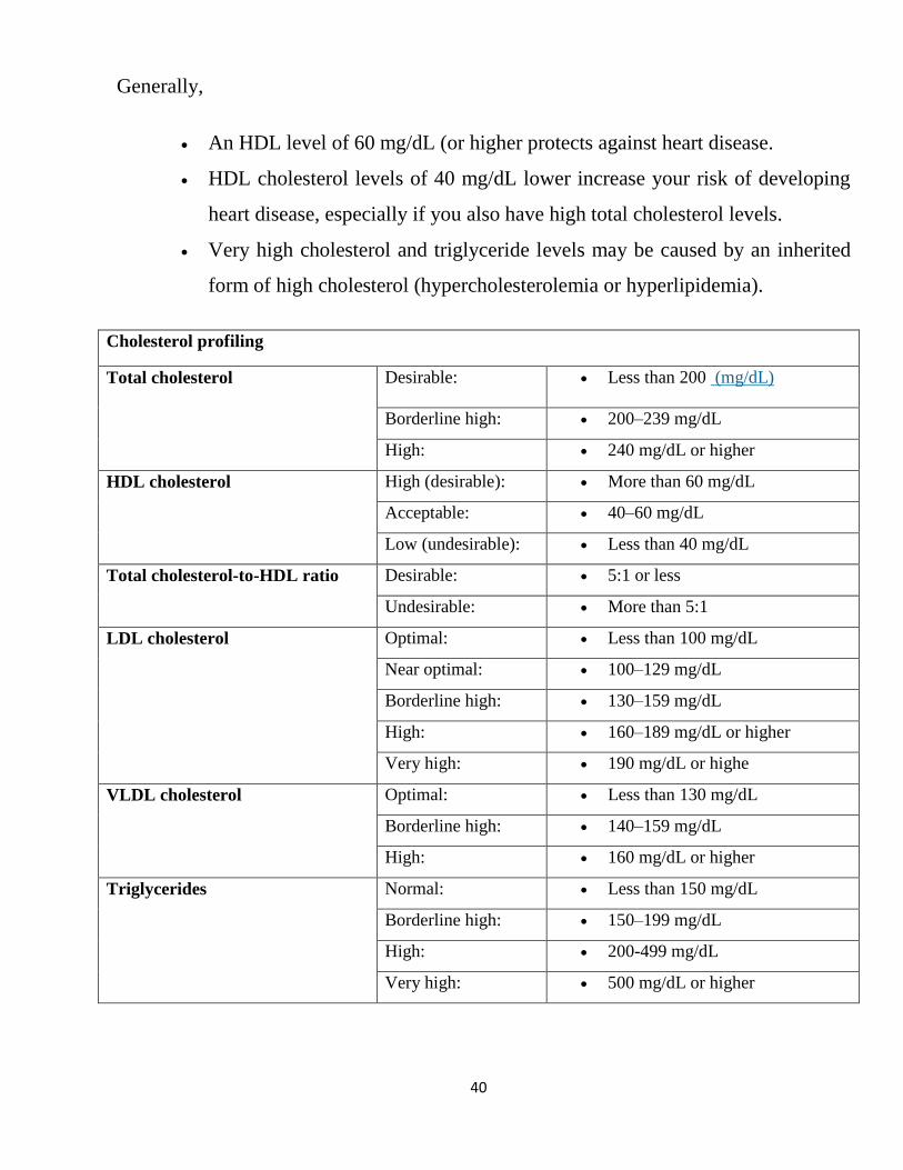
Generally,
An HDL level of 60 mg/dL (or higher protects against heart disease.
HDL cholesterol levels of 40 mg/dL lower increase your risk of developing
heart disease, especially if you also have high total cholesterol levels.
Very high cholesterol and triglyceride levels may be caused by an inherited
form of high cholesterol (hypercholesterolemia or hyperlipidemia).
Cholesterol profiling
Total cholesterol Desirable: Less than 200 (mg/dL)
Borderline high: 200–239 mg/dL
High: 240 mg/dL or higher
HDL cholesterol High (desirable): More than 60 mg/dL
Acceptable: 40–60 mg/dL
Low (undesirable): Less than 40 mg/dL
Total cholesterol-to-HDL ratio Desirable: 5:1 or less
Undesirable: More than 5:1
LDL cholesterol Optimal: Less than 100 mg/dL
Near optimal: 100–129 mg/dL
Borderline high: 130–159 mg/dL
High: 160–189 mg/dL or higher
Very high: 190 mg/dL or highe
VLDL cholesterol Optimal: Less than 130 mg/dL
Borderline high: 140–159 mg/dL
High: 160 mg/dL or higher
Triglycerides Normal: Less than 150 mg/dL
Borderline high: 150–199 mg/dL
High: 200-499 mg/dL
Very high: 500 mg/dL or higher