Embed Size (px)
Citation preview

Clinical Anatomy of theClinical Anatomy of thePleural Cavity & Pleural Cavity & MediastinumMediastinum
Lawrence M. Witmer, PhDLawrence M. Witmer, PhDDepartment of Biomedical SciencesCollege of Osteopathic MedicineOhio UniversityAthens, Ohio [email protected]
Handout download:http://www.oucom.ohiou.edu/dbms-witmer/gs-rpac.htm
23 Nov 2004

Pleura and Pleural CavityPleura and Pleural Cavity
From Moore & Dalley 1999
Pleura• Mesothelial lining of
each hemithorax• Derived from
embryonic coelomiclining
• Visceral pleura: lung• Parietal pleura: wall
• Costal• Diaphragmatic• Mediastinal• Cervical
Pleural Cavity• Potential space between visceral & parietal pleura• Capillary layer of serous fluid produced by mesothelium
• Reduces friction• Surface tension provides cohesion between lung and thoracic wall

endothoracicfascia
From Healey & Hodge 1990
Pleural sac and recessesPleural sac and recesses

Pleural Diseases & Signs 1: Pleural EffusionPleural Diseases & Signs 1: Pleural Effusion
From Daffner 1993From Daffner 1993
Right-sided pleural effusionRight-sided pleural effusion
• Accumulation of fluid in the pleural space• Transudative vs. exudative effusion• Empyema as potential sequelae to exudative effusion

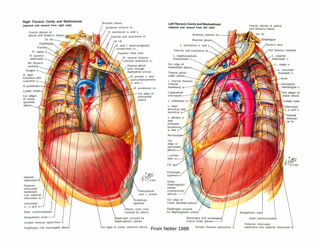
Pleural Diseases & Signs 2: HemothoraxPleural Diseases & Signs 2: Hemothorax
From Netter 1988From Netter 1988
• Intrathoracic bleeding (e.g., trauma)• Numerous sources of potential bleeds• Large hemothorax: hypovolemic shock,
restricted ipsilateral ventilationcontralateral mediastinal shift
• Clotting may not be tooproblematic (exceptfor catheters)

Pleural Diseases & Signs 3: ChylothoraxPleural Diseases & Signs 3: Chylothorax
From Netter 1988From Netter 1988
From Moore & Dalley 1999
• Leakage of lymph• Usually a result of surgical
trauma during mediast. proc.• Traumatic vs nontraumatic • Traumatic: 2/3, unilateral• Nontraumatic: 1/3, bilateral,
assoc. with SVC thrombosis

Pleural Diseases & Signs 4: Malignant MesotheliomaPleural Diseases & Signs 4: Malignant Mesothelioma
From Netter 1988From Netter 1988
• Neoplasm of pleural serosa• Linked to asbestos exposure• Coalescence of pleural
plaques• May be restricted to parietal
pleura but can involve visceral pleura
• Can lead to contracture of allstructures in affected hemithorax

Pleural Diseases & Signs 5: PneumothoraxPleural Diseases & Signs 5: Pneumothorax
From Netter 1988From Netter 1988
• Presence of free air or gas in the pleural cavity
• Types of pneumothorax• Open pneumothorax• Spontaneous pneumothorax• Tension pneumothorax
• Collapse of ipsilateral lung due to pressure change & disruption of surface tension
• Potential for mediastinal shifts

Divisions of the Mediastinum
anatomicalanatomicalanatomicalsurgicalsurgicalsurgical
MediastinumMediastinum
from Schwartz et al., 1999
• Anterior mediastinum– thymus, fat, lymphatics
• Posterior mediastinum– descending aorta, esophagus,
azygos veins, autonomics, thoracic duct
• Middle mediastinum– heart, pericardium, aorta, trachea,
main bronchi, lymph nodes
Mediastinal lesions and their distribution in 102
patients
From Netter 1988From Netter 1988

MediastinalMediastinal MassesMasses
Anterior mediastinum: “four Ts”— Thymoma, Thyroid tumor, Terrible lymphoma, Teratoma
From Netter 1988From Netter 1988

Pancoast’s SyndromePancoast’s Syndrome
• Bronchogenic carcinoma in the apex of the lung
• Horner’s Syndrome: miosis, ptosis, enophthalmos, anhidrosis
• Lower brachial plexus injury (C8-T1): Klumpke’s palsy
• Paresthesia of the upper extremity due to compression of subclavian a. & v.
• Shoulder pain: due to involvement of upper ribs and intercostal nerves
• Respiratory effects
From Netter 1988From Netter 1988

ReferencesReferences
Daffner, R. H. 1993. Clinical Radiology, The Essentials. Williams & Wilkins, Baltimore.
Healey, J. E. Jr., and J. Hodge. 1990. Surgical Anatomy, 2nd Ed. Decker, Philadelphia.
Moore, K. L. and A. F. Dalley. 1999. Clinically Oriented Anatomy, 4th Ed. Lippincott, Williams & Wilkins, Baltimore.
Netter, F. H. 1988. The CIBA Collection of Medical Illustrations, Volume 7: Respiratory System. CIBA-Geigy, Summit.
Schwartz et al. (eds.), Principles of Surgery, 7th Ed., McGraw Hill, New York.