Embed Size (px)
Citation preview
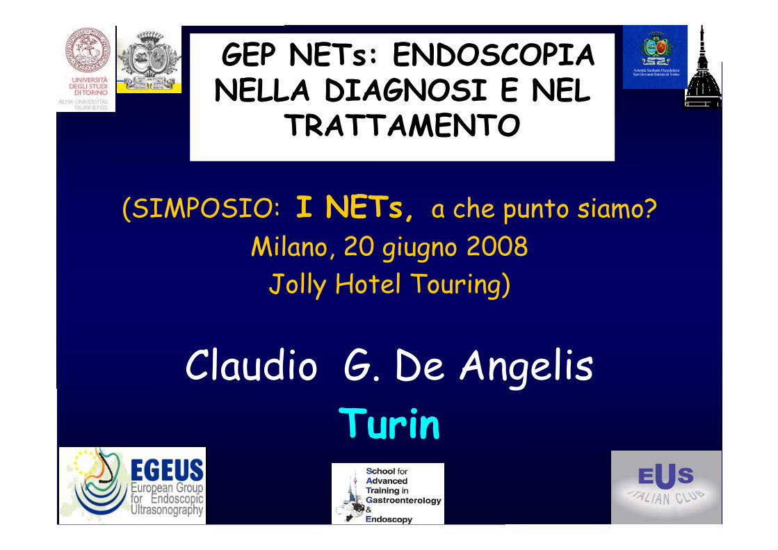
GEP NETs: ENDOSCOPIANELLA DIAGNOSI E NEL
TRATTAMENTO
(SIMPOSIO: I NETs, a che punto siamo?Milano, 20 giugno 2008
Jolly Hotel Touring)
Claudio G. De AngelisTurin

GEP (NEURO)-ENDOCRINE TUMORS:
“ A COMPLEX DILEMMA FORDIAGNOSTIC IMAGING”
• Small sizes: < 2 cm in 55-70% of insulinomas < 1 cm in 38% of gastrinomas < 1.5 cm (often) GI carcinoids (< 1 cm: 80% of rectal carcinoids)
• Profound site in the retroperitoneum, multiple andextrapancreatic locations
• Sometimes only submucosal location in the GItract (e.g. gastrinomas)

ERCP/ENDOSCOPY
EUS
IDUSPET
CT/hCT/MDR-CT
MRI + MRA + MRCP
DIGITAL ANGIOGRAPHY TUMOUR MARKERS
LAPAROSCOPY
US +/- THI +/-CD/PD-US +/-
CE-US
PET/CT
hCT/MDR-CT angiographyTECHNIQUES OF PRE-
OPERATIVEDIAGNOSIS/DETECTION AND
STAGING
OCTREOSCAN INTRAOPERATIVE PALPATION and/or US
SAIS/SAIC
SCINTIGRAphy

NEUROENDOCRINE PANCREATICTUMORS AND THE ENDOSCOPIST:
or “SEARCHING THE NEEDLE IN THE HAYSTACK”

THE ENDOSCOPIST’S SHOP
Enteroscopy
IDUS EUS
Endoscopic
Resection
FNA
FNI
Intra-operative
Endoscopy
ENDOSCOPY

WHAT YOU CAN ASK TOTHE ENDOSCOPIST ?
To identify/ detect the lesion(DIAGNOSIS ANDLOCALIZATION)
To stage the lesion(prognostic evaluation)(STAGING)
To treat the lesion (?)(THERAPY)

PANCREAS and NETs

ENDOSCOPY AND ENDOSONOGRAPHY INPRE-OPERATIVE DETECTION OF
PANCREATIC NETs• A correct pre-operative localization and staging
are MANDATORY in order to select the righttherapeutic options, optimize surgical treatment,reducing times and complexity of surgery:
• IMPROVING RESULTS AND OUTCOMES

PANCREATIC NETs: THE ROLE OFENDOSCOPIC TECHNIQUES
ERCP
Bile ducts
Ampulla
Pancreas*
Carcinoids (0.3%)Somatostatinomas (1.2%)
Somatostatinomas (9.3%)
Insulinomas (99%)Gastrinomas (33-79%)Carcinoids (0.46%)Glucagonomas (ca.100%)Vipomas (90%)Somatostatinomas (37.9%)PPomas (92%)Non-functioning (15-52%)
*Ogawa Y et al. Islet cell tumors ofthe pancreas: the diagnostic value ofERCP. Int J Pancreatol 6,1990

THE CHALLENGEOF EUS
•EUS is the most important of the manyinnovations that have occurred in GI endoscopyduring the last 25 yrs
•EUS has extended the range of possibilities forendoscopic diagnosis endowing the endoscopistwith the matchless ability to see within andbeyond the wall of the gut

ENDOSCOPIC ULTRASOUND (EUS )
THE BEST CURRENTLYAVAILABLE TECNIQUE FORIMAGING THE PANCREAS
HIGH RESOLUTION IMAGESOF THE MAIN PANCREATICDUCT AND SURROUNDINGPARENCHYMA
STRUCTURES AS SMALL AS2-3 MM CAN BEDISTINGUISHED
De Angelis C et al. Pancreatic cancer imaging: the new role of EUS.JOP J Pancreas (online) 2007;8 (1)

EUS FEATURES OF THE NETs OF THEPANCREAS
Echopattern as to the rest of the gland
Homogeneous 81%Hypoechoic 69%Hyperechoic 6%Isoechoic 6%Inhomogeneous 19%Cystic spaces 9%Calcifications 6%
Margins
Sharp 84%
Irregular/indistinct 16%
Hypoechoic border 6%


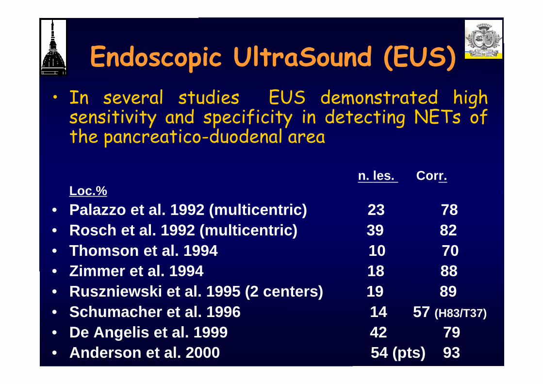
• In several studies EUS demonstrated highsensitivity and specificity in detecting NETs ofthe pancreatico-duodenal area
n. les. Corr.Loc.%
• Palazzo et al. 1992 (multicentric) 23 78• Rosch et al. 1992 (multicentric) 39 82• Thomson et al. 1994 10 70• Zimmer et al. 1994 18 88• Ruszniewski et al. 1995 (2 centers) 19 89• Schumacher et al. 1996 14 57 (H83/T37)
• De Angelis et al. 1999 42 79• Anderson et al. 2000 54 (pts) 93
Endoscopic UltraSound (EUS)

EUS: SUMMARY OFLITERATURE DATA
Medium/High(n° ofexams)
Fewcenters
0.05 –0.3
88 – 9557 – 100
CostsAvailabilityComplicationrate
%
Specificity
%
Sensitivity
%

“Endosonography in decision making andmanagement of gastrointestinal endocrine tumors” De Angelis C et al. Eur J Ultrasound 1999;10:139
42 lesions
Pancreas 23Duodenal wall 8Peripancreatic LN 10Paraduodenal solitary LN 1
7-35 mm < 20 mm: 83%< 15 mm: 67%

EUS AND PANCREATIC NETsPre-operative detection of NETs in the pancreas:comparison of EUS vs Other imaging techniques
Technique N. of pts Detection rate lesions %
EUS 19 20/23 86.7%US 19 4/23 17.4%CT 19 7/23 30.4%MRI 8 3/12 25%Angiography 11 4/15 26.6%SRS 9 2/13 15.4%
De Angelis C et al. 1999

CLINICAL IMPACT OF EUS ONDECISION-MAKING AND MANAGEMENTOF PATIENTS WITH PANCREATIC NETs
• All considered EUS alone gave us moreinfornation than all other imaging techniquestogether
• It changed treatment plans in 17/39 (44%)of pts with NETs
• No other procedure, even more invasive thanEUS, has been able to visualize the 3pancreatic tumors and the 5 duodenalgastrinomas that EUS could not detect
De Angelis C et al. 1999

• Using EUS as first-line method for thedetection of our NETs should have allowed asignificant costs saving in 15/23 (65.2%) ofpatients, avoiding both multiple and moreinvasive (like angiography in 50% of cases) andmore expensive (like SRS in 45% or MRI in32% of cases) diagnostic procedures
• Finally 6/39 patients (15.4%) did not undergoa major surgical intervention based on thenegative results of EUS examination
CLINICAL IMPACT OF EUS ONDECISION-MAKING AND MANAGEMENTOF PATIENTS WITH PANCREATIC NETs
De Angelis C et al. 1999

furthermore….
• EUS sensitivity was significantly reduced (30%) forthe NETs of the duodenal wall (gastrinomas)
• Intra-operative endoscopic transilluminationof the duodenum remains today the besttechnique for the detection of duodenal wallgastrinomas (sensitivity: 83%)
• L’EUS remains a highly operator-dependenttechnique
De Angelis C et al. 1999
CONCLUSIONS
• Notwithstanding these problems, EUS has imposeditself as an accurate method of preoperativedetection of pancreatic NETs and can be consideredthe imaging modality of first choice in this clinicalsetting.
• It is the single detection and staging technique moresensitive and should be used at an early stage in thediagnostic work up, if possibile straight after an USor a spiral CT to exclude hepatic metastases.
• EUS seems to be cost-effective: reducing costs,saving times and lowering morbidity due to moreinvasive tests
De Angelis C et al. 1999
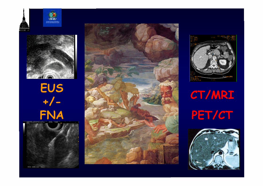
EUS+/-FNA
CT/MRI
PET/CT


Comparison of EUS and CT for the preoperativeevaluation of pancreatic cancer: a systematicr e v i e w .……………(DeWitt J et al. Clin Gastroenterol Hepatol 2006)
• Literature is heterogeneous in: study design,quality and results. Methodologic limitations thatpotentially affects results.
• Overall EUS is > to CT for detection of PC, for Tstaging and for vascular invasion of thesplenoportal confluence.
• The 2 tests appear to be equivalent for N staging,overall vascular invasion and assessment of tumorresectability.

…. however EUS can not define distantmetastatc disease, is still not universallyavailable and is to a high degree operatordependent
Spiral CT or multislice CT must be theinitial study of choice in pts with
suspected pancreatic tumors
De Angelis C et al. Pancreatic cancer imaging: the new role of EUS.JOP J Pancreas (online) 2007;8 (1)

INSULINOMA
Best Practice & Research Clinical Endocrinology & Metabolism 2005; 19:177–93

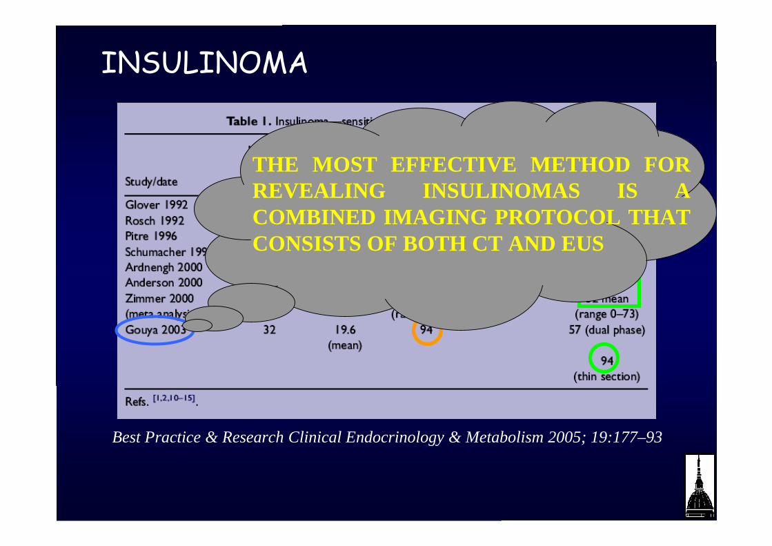
INSULINOMA
Best Practice & Research Clinical Endocrinology & Metabolism 2005; 19:177–93
THE MOST EFFECTIVE METHOD FORREVEALING INSULINOMAS IS ACOMBINED IMAGING PROTOCOL THATCONSISTS OF BOTH CT AND EUS

GASTRINOMA: problems
• the location: 50% extra-pancreatic
• lesions in the duodenal wall are smaller than the……………………….pancreatic ones (9.6 mm vs 28.7 mm)……………………...O Kisker et al. World J Surg 1998; 22: 651-7
• EUS sensitivity for pancreatic lesions: about 93%,it falls to 50% for extra-pancreatic lesions.……………….T Zimmer et al. Digestion 2000; 62: 45-50
• usefulness of intraoperative endoscopictransillumination (diagnostic improvement: + 20%)and duodenotomy (+15%)
Best Practice & Research Clinical Gastroenterology 2005; 19: 753–781

MEN-I
• many tumors are small (mean 1.1 cm)EJ Wamsteker et al. Gastrointest Endosc 2003; 58: 531-5
• very often tumors are multiple (mean 3.3tumors/pt)
• Screening with EUS in MEN-I asymptomaticpts can be recommended
EJ Wamsteker et al. Gastrointest Endosc 2003; 58: 531-5

MEN-I
• many tumors are small ( mean 1.1 cm ) EJ Wamsteker et al. Gastrointest Endosc 2003; 58: 531-5
• spesso i tumori sono multipli (media 3.3 tumori/p.te)
• raccomandato screening con EUS in p.ti asintomaticicon MEN-I. EJ Wamsteker et al. Gastrointest Endosc 2003; 58: 531-5
In 13 MEN I asymptomatic pts, anEUS follow up of 13 yrsdemonstrated the appearance ofpancreatic tumors in 11
Aggressive early surgicaltreatment may improve theprognosis for these pts.

MEN-I
However several papers subsequentlydemonstrated EUS effectiveness in
detecting and following smallpancreatic NETs in asymptomaticpatients with MEN I sindrome
Gauger PG et al. Br J Surg. 2003;90(6):748-54.Langer P et al. World J Surg. 2004;28(12):1317-22Hellman P et al. Br J Surg. 2005;92(12):1508-12.
Thomas-Marques L et al. Am J Gastroenterol. 2006;101(2):266-73.Kann PH et al. Endocr Relat Cancer. 2006;13(4):1195-202
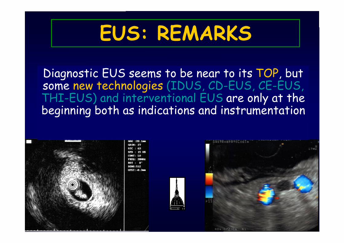
Diagnostic EUS seems to be near to its TOP, butsome new technologies (IDUS, CD-EUS, CE-EUS,THI-EUS) and interventional EUS are only at thebeginning both as indications and instrumentation
EUS: REMARKS

MINIPROBES
Initial data suggest that IDUS may improveevaluation by identifying PNTs within thepancreas unrecognized by other techniques.Gastrointest Endosc 2002; 55: 397-408

1. EUS-GUIDED BIOPSIES (EUS-FNA)
a) ↑ SPECIFICITY FOR THE DIAGNOSIS OFPANCREATIC CANCER AND LYMPH NODESINVOLVEMENT
b) “Usefulness of EUS-guided fine needle aspiration (EUS-FNA) in the diagnosis of functioning neuroendocrine tumors”
Ginès A et al. Gastrointest Endosc 2002;56:291
EUS-FNA safely provides cytologic confirmation withhigh accuracy in these patients.
2) COLOR-DOPPLER APPLICATION
ELECTRONIC INSTRUMENTS WITHLINEAR SCANNING ALLOW:

Gastrointest Endosc 2004;60: 378-84

EUS-FNA in the diagnosis of pancreatic NETs
• Other papers confirmed usefulness andeffectiveness of EUS-FNA in the diagnosis ofpancreatic NETs, both functioning and non-functioning.
• It is possible to reduce false positive resultsof only morphological EUS due to peri-andintra-pancreatic lymph nodes or splenosisnodules
Voss M et al. Gut. 2000;46(2):244-9Gu M et al.Diagn Cytopathol. 2005;32(4):204-10.Chang F et al. Cytopathology. 2006;17(1):10-7.Jani N et al.Gastrointest Endosc. 2008;67(1):44-50.

EUS-FNA in the diagnosis of pancreatic NETs
• EUS-FNA works better than CT-FNA
• Possibility of predicting biological behaviourand outcome of the NET applying molecularbiology techniques to the cell specimensobtained wth EUS-FNA .
Jhala D et al. Fine needle aspiration biopsy of the islet celltumor of pancreas: a comparison between computerized axialtomography and endoscopic ultrasound-guided fine needleaspiration biopsy. Ann Diagn Pathol. 2002;6(2):106-12.
Nodit L et al. Endoscopic ultrasound-guided fine needle aspiratemicrosatellite loss analysis and pancreatic endocrine tumoroutcome. Clin Gastroenterol Hepatol. 2006;4(12):1474-8.

Gastrointestinal Endoscopy 2002; 55:594-7
EUS allows identification of tiny lesionsdifficult to find by palpation during surgery
Zografos GN et al. Hormones (Athens). 2005;4(2):111-6.
WHEN THE RESULTS CAN ALTER PATIENTMANAGEMENT !!!
I.E.…
• Differential diagnosis between benign and malignant lesion
• When there is the suspicion that the pancreatic lesion visualized byEUS or other imaging modalities could be a peri- or intra-pancreaticlymph node or a splenosis nodule or another type of lesion amenable ofdifferent therapeutic approaches (lymphoma, metastasis etc)
• Patient or lesion not fit for surgery and there is indicationfor CT
• reluctance of the patient or the surgeon to perform a majorsurgical intervention, without a tissue diagnosis
WHEN DO WE NEED A TISSUE DIAGNOSIS ?WHEN DO WE NEED A TISSUE DIAGNOSIS ?

1) EUS-GUIDED BIOPSIES (EUS-FNA)
2) COLOR-DOPPLER application:
“Utility of Endoscopic Ultrasonography withColor Doppler Function for the diagnosisof islet cell tumor”
Ueno N. et al. AJG 1996
ELECTRONIC INSTRUMENTS WITHLINEAR SCANNING ALLOW:

EUS: NEW PROSPECTS• “Contrast-enhanced EUS” could improved thealready high accuracy of EUS in visualizing smallpancreatic NETs and in differential diagnosis of
pancreatic lesions