Embed Size (px)
Citation preview

Circulatory Systems III

Mammals & Birds

Mammals & Birds
Atrioventricular (AV) valves: located between atrias and ventricles and ensure one-way flow
◦ Right AV valve = tricuspid valve
◦ Left AV valve = bicuspid valve
Chordate tendinae: anchor valves to the papillary muscles and prevent them from opening backwards.

Mammals & Birds
Tricuspid Valve Bicuspid Valve

Mammals & Birds

Flow of Blood
Flow of Blood

Oxygenated or Deoxygenated?
Systemic Arteries?
◦ Heart to body tissues oxygenated blood
Systemic Veins?
◦ Body tissues to heart deoxygenated blood
Pulmonary Arteries?
◦ Heart to lungs deoxygenated blood
Pulmonary Veins?
◦ Lungs to heart oxygenated blood

The Cardiac Cycle
Cardiac Cycle:
Rhythmic Pumping of Heart
2 Phases of Cardiac Cycle =
1. Systole – contraction
2. Diastole – relaxation

The Cardiac Cycle
Fish Cardiac Cycle:
◦ Chambers contract in series

The Cardiac Cycle
Mammalian Cardiac Cycle:
◦ Coordinated contraction of atria and ventricles

The Cardiac Cycle

Cardiac Cycle
Mid Ventricular Diastole:
◦ Atria and ventricles are relaxed,
◦ AV valves are open,
◦ Semilunar valves are closed.
Mammals and birds:
◦ Blood returning to heart passes thru the atria and goes into the ventricles passively.
Fish and some amphibians:
◦ Ventricles fill primarily by contraction of the atrium.

Cardiac Cycle
Atrial Systole:
◦ Atria contract and additional blood gets pushed
into ventricles.
Blood is pumped into the ventricles until
they reach end-diastolic volume (EDV),
the max amount of blood in the ventricle.

Cardiac Cycle
Early Ventricular Systole:
◦ Ventricles contract.
◦ pressure cause AV valves to shut.
◦ Semilunar valves are closed.
Isovolumetric contraction:
◦ Blood is non-compressible, so pressure in the
chamber increases but volume does not.

Cardiac Cycle
Late Ventricular Systole:
◦ Pressure forces semilunar valves open.
◦ Blood flows out of the ventricles into arteries.
◦ Chordae tendinae prevent AV valves from
being forced open; preventing backflow.
Ventricle has reaches its end systolic
volume (ESV) or blood minimum.
Cardiac Cycle
Early Ventricular Diastole:
◦ Ventricles begin to relax, pressure drops.
◦ Pressure in ventricles drops below that of the arteries
◦ Backpressure forces semilunar valves shut.
Throughout ventricular systole, the atria have been in diastole filling with blood.
Pressure in filled atria exceeds pressure in relaxed ventricles and AV valves pop open.

Mammalian Cardiac Cycle
2 ventricles contract simultaneously
Left ventricle contracts much more
forcefully than the right ventricle and
develops a much higher pressure:
◦ Left Ventricle to body high resistance
◦ Right ventricle to lungs low resistance

Control of Contraction
Cardiomyocytes = myogenic
Produce spontaneous rhythmic
depolarizations that initiate contraction.
Electrically coupled via gap junctions:
◦ depolarization in one spreads to adjacent
cells, triggering coordinated contractions.

Control of Contraction

Control of Contraction

Control of Contraction
Pacemaker cells determine the
contraction rate for the entire heart.
In vertebrates these cells are located in
an area of the right atrium called the
Sinoatrial (SA) Node.

Control of Contraction
Pacemaker cells have unstable resting
potentials (pacemaker potential).
Resting potential drifts from -60mV until
it reaches threshold of -40mV.
At -40mV an action potential is initiated


Control of Contraction
Depolarization initiated in the pacemaker
cells can spread from cell to cell via
electronic current spread.
AP triggered in one cell spreads to
adjacent cells propagating the impulse
throughout the heart.

Control of Contraction
Cardiomyocytes have an extended
depolarization = plateau phase
Corresponds to the refractory period of
the cell in which an action potential
cannot fire.

Control of Contraction

Control of Contraction
Small mammals tend to have HRs and
plateau phases than larger mammals
whose hearts beat more slowly.

Impulse Conduction in Fish
Impulse conduction via gap junctions is
sufficient to provide coordinated
contraction of the chambers.
Signal travels from sinus venosus to the
atrium and then to the ventricle.
Contraction occurs in a series.

Mammalian Conducting Pathways
Contractile cells of the atrium and
ventricles do no form gap junctions with
each other.
Mammals utilize conduction pathways

Mammalian Conducting Pathways

Mammalian Conducting Pathways
SA node initiates the action potential ◦ Depolarization spreads rapidly via internodal
pathway through the walls of the atria.
Depolarization reaches atrioventricular (AV) node which communicates signal to the ventricle.
AV node causes signal delay ◦ allows atrium to finish contracting before
ventricles contract.

Mammalian Conducting Pathways
Signal travels from the AV node through
the bundles of his (“hiss”)
Electrical signal spreads into a network of
conducting pathways - purkinje fibers.
Signal spreads cell to cell via gap junctions
and ventricles contract.


Electrocardiogram (EKG)
Deflections = markers of electrical
activity of the heart
Electrocardiogram (EKG)
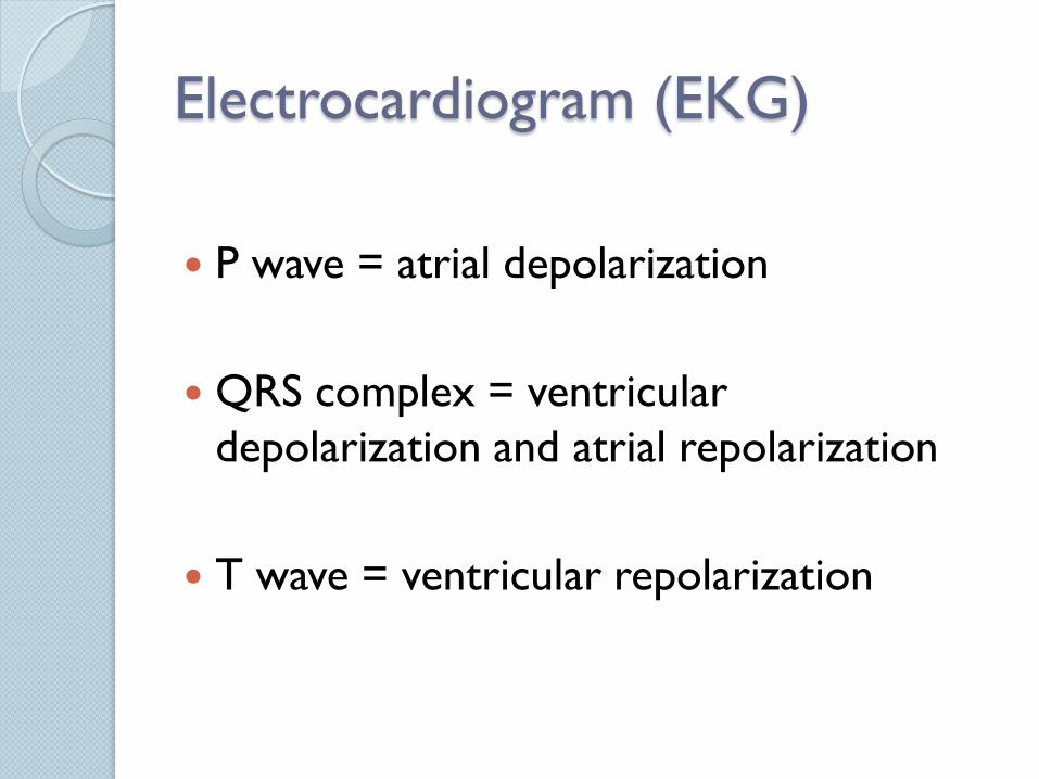
P wave = atrial depolarization
QRS complex = ventricular
depolarization and atrial repolarization
T wave = ventricular repolarization

Cardiac Output
Cardiac Output (CO) = the amount of
blood that the heart pumps per unit time.
CO = HR x SV
◦ Heart rate (HR) = beats per minute
◦ Stroke volume (SV) = amount of blood
pumped per beat

Cardiac Output
Animals can modulate CO by regulating
HR, SV, or both.
Decreasing HR = bradycardia
Increasing HR = tachycardia
Nervous and endocrine systems
modulate force of contraction (SV)

Frank-Starling Effect
When blood enters a ventricle, the increased volume causes it to stretch.
The more blood that enters the heart at the end of diastole (EDV), the greater the degree of stretch.
Frank-Starling Effect = autoregulation
as you stretch a cardiomyocyte the strength of contraction increases.

Frank-Starling Effect
Allows the heart to automatically
compensate for increases in the amount
of blood returning to the heart.