Embed Size (px)
Citation preview
1
Varicose VeinsVaricose Veins
Steven M. Dean, DO, FACP, RPVIAssociate Professor of Internal Medicine
Department of Internal MedicineDivision of Cardiovascular Medicine
The Ohio State University Wexner Medical Center
DefinitionDefinition
Chronic Venous Disease
Chronic Venous Disease
Definition: A spectrum of signs and symptoms that ranges from spider and varicose veins to chronic venous insufficiency.
Spider Veins or TelangiectasiasSpider Veins or Telangiectasias
• Non raised dilated intradermal veins/venules
• Typically < 1 mm in diameter
• Appear earlier than varicose veins
• Blue or Red
Photos courtesy of Dr. Eric Mowatt-Larssen
2
Reticular VeinsReticular Veins•• Dilated, nonDilated, non--
palpable palpable subcutaneous veinssubcutaneous veins
•• BlueBlue--greengreen
•• 11--3 mm3 mm
•• Sometimes coexist Sometimes coexist with and “feed” with and “feed” telangiectasiastelangiectasias
Photo courtesy of the American College of Phlebology/American Venous Forum (ACP/AVF)
Varicose VeinsVaricose Veins• Dilated, tortuous, palpable
subcutaneous veins > 3 mm (upright)
• Synonyms: varix, varices, varicosities
• Involve great and/or small saphenous veins (GSV/SSV) or any superficial vein tributaries
Photo courtesy of Dr. Mowatt-Larssen
PrevalencePrevalence
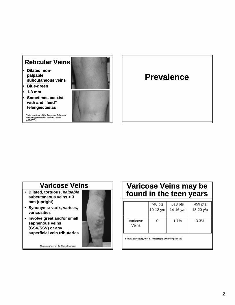
Varicose Veins may be found in the teen yearsVaricose Veins may be found in the teen years
740 pts
10-12 y/o
518 pts
14-16 y/o
459 pts
18-20 y/o
Varicose Veins
0 1.7% 3.3%
Schultz-Ehrenburg, U et al, Phlebologie. 1992 45(4):497-500
3
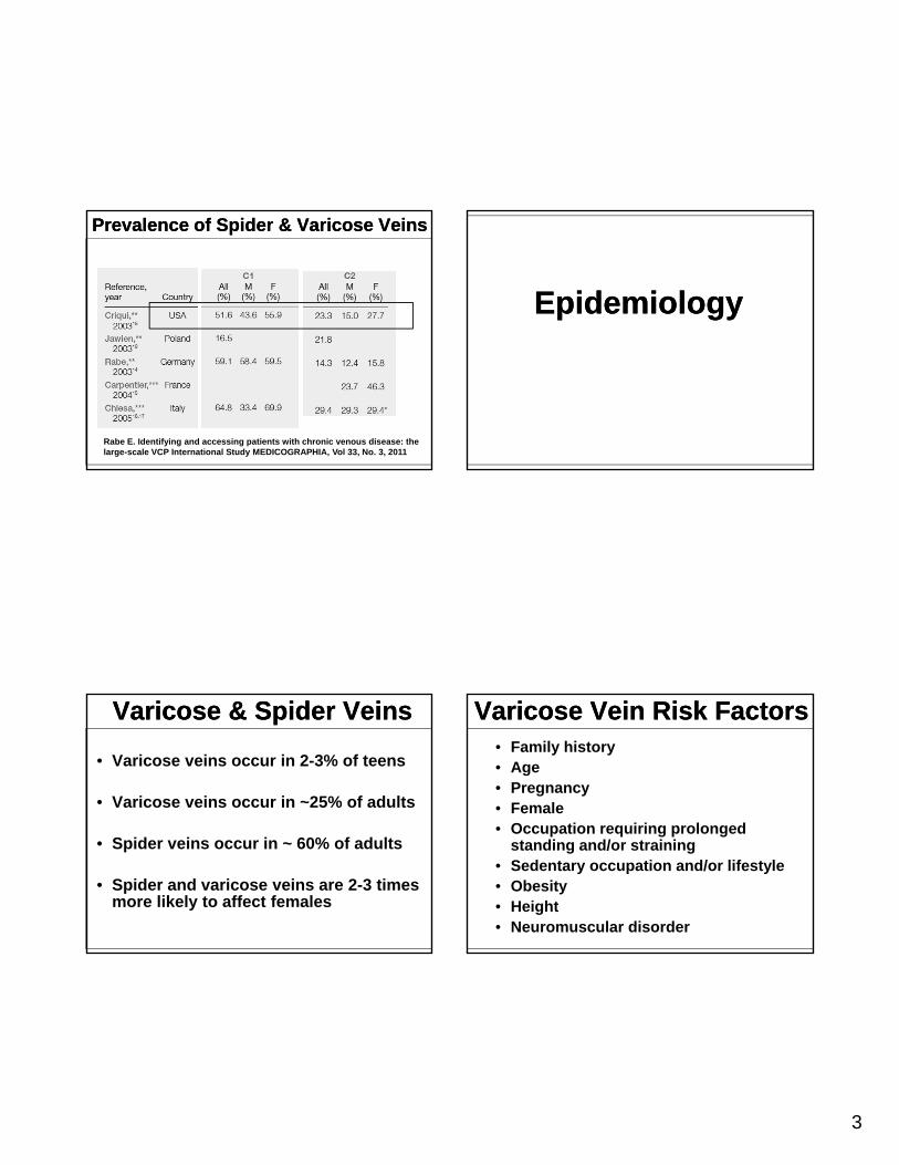
Prevalence of Spider & Varicose VeinsPrevalence of Spider & Varicose Veins
Rabe E. Identifying and accessing patients with chronic venous disease: the large-scale VCP International Study MEDICOGRAPHIA, Vol 33, No. 3, 2011
Varicose & Spider VeinsVaricose & Spider Veins
• Varicose veins occur in 2-3% of teens
• Varicose veins occur in ~25% of adults
• Spider veins occur in ~ 60% of adults
• Spider and varicose veins are 2-3 times more likely to affect females
EpidemiologyEpidemiology
Varicose Vein Risk FactorsVaricose Vein Risk Factors• Family history• Age• Pregnancy• Female
O ti i i l d• Occupation requiring prolonged standing and/or straining
• Sedentary occupation and/or lifestyle• Obesity• Height• Neuromuscular disorder
4
Varicose Veins are a Hereditary Disorder
Varicose Veins are a Hereditary Disorder
134 families examined
The risk of developing varicose veins:
• 89% if both parents had varicose veins
47% if t h d i i• 47% if one parent had varicose veins
• 20% of neither parent had varicose veins
Cornu-Thenard, A, J Dermatol Surg Oncol 1994 May; 20(5):318-26.
Autosomal dominant with incomplete penetrance
Autosomal dominant with incomplete penetrance
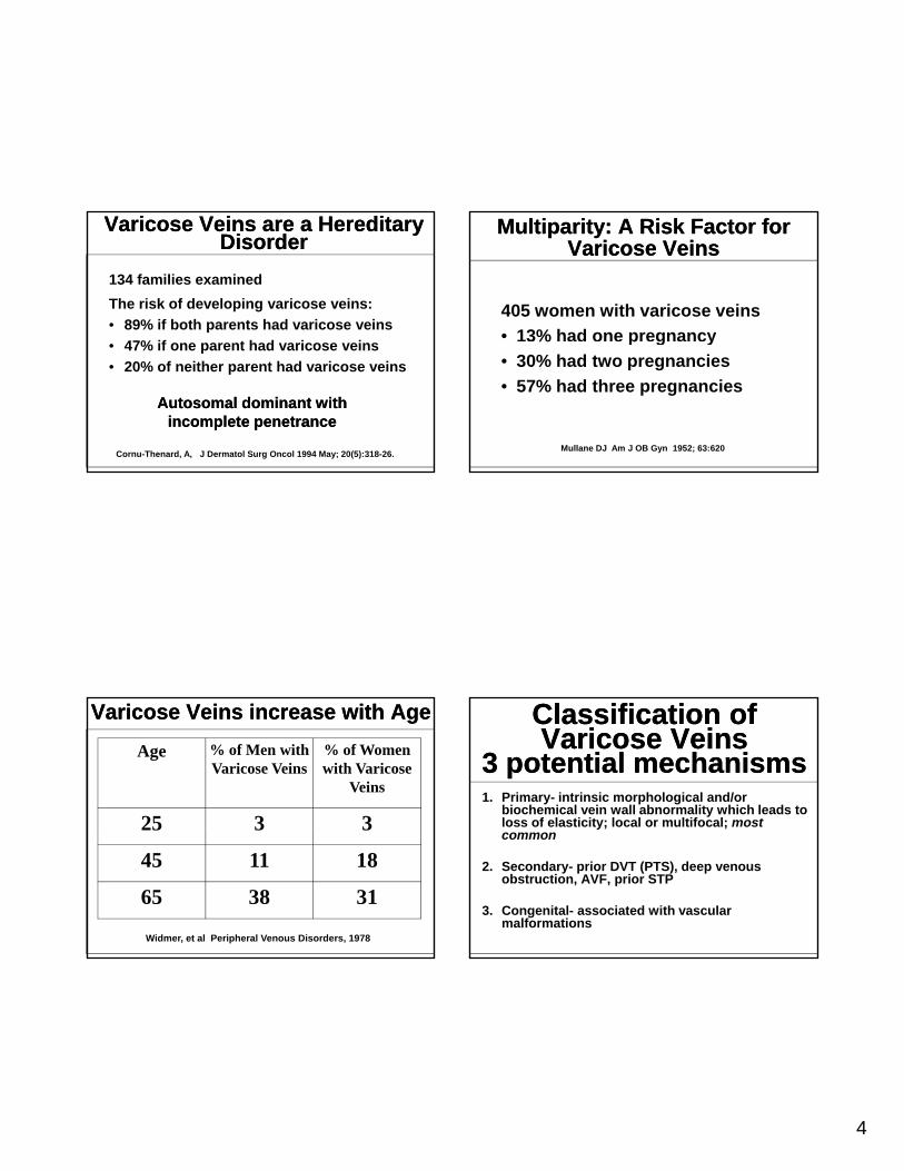
Varicose Veins increase with AgeVaricose Veins increase with Age
Age % of Men with Varicose Veins
% of Women with Varicose
Veins
25 3 325 3 3
45 11 18
65 38 31
Widmer, et al Peripheral Venous Disorders, 1978
Multiparity: A Risk Factor for Varicose Veins
Multiparity: A Risk Factor for Varicose Veins
405 women with varicose veins
• 13% had one pregnancy% p g y
• 30% had two pregnancies
• 57% had three pregnancies
Mullane DJ Am J OB Gyn 1952; 63:620
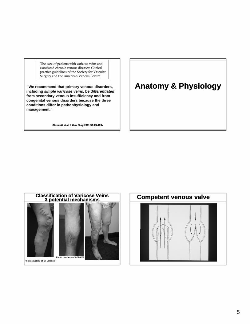
Classification of Varicose Veins
3 potential mechanisms
Classification of Varicose Veins
3 potential mechanisms1. Primary- intrinsic morphological and/or
biochemical vein wall abnormality which leads to loss of elasticity; local or multifocal; most y; ;common
2. Secondary- prior DVT (PTS), deep venous obstruction, AVF, prior STP
3. Congenital- associated with vascular malformations
5
“We recommend that primary venous disorders, including simple varicose veins, be differentiatedf d i ffi i d ffrom secondary venous insufficiency and from congenital venous disorders because the three conditions differ in pathophysiology and management.”
Gloviczki et al. J Vasc Surg 2011;53:2S-48S.Gloviczki et al. J Vasc Surg 2011;53:2S-48S.
Classification of Varicose Veins3 potential mechanisms
Classification of Varicose Veins3 potential mechanisms
Photo courtesy of Dr Larssen
Photo courtesy of ACP/AVF
Anatomy & PhysiologyAnatomy & Physiology
Competent venous valveCompetent venous valve
6
3 types of lower extremity VEINS3 types of lower extremity VEINS
Physiologic Blood Flow:
Superficial to Deep
“Up & In”
“Up & In”
Perforating VeinPerforating VeinPerforating VeinPerforating Vein
Veins
Bradbury & Ruckley. Atlas of Vascular Disease. 2nd edition 2003. Current Medicine, Inc
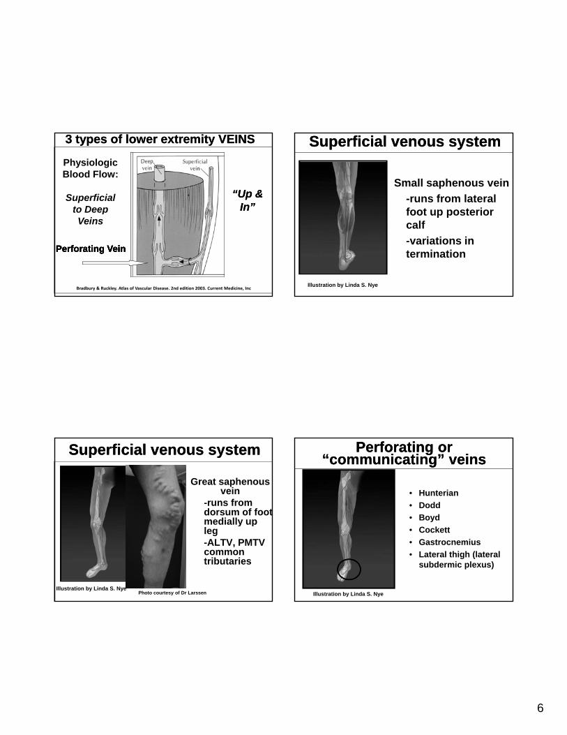
Superficial venous system Superficial venous system
Great saphenous vein
-runs from dorsum of foot medially upmedially up leg-ALTV, PMTV common tributaries
Illustration by Linda S. NyePhoto courtesy of Dr Larssen
Superficial venous systemSuperficial venous system
Small saphenous vein
-runs from lateral foot up posteriorfoot up posterior calf
-variations in termination
Illustration by Linda S. Nye
Perforating or “communicating” veins
Perforating or “communicating” veins
• Hunterian
• Dodd
• BoydBoyd
• Cockett
• Gastrocnemius
• Lateral thigh (lateral subdermic plexus)
Illustration by Linda S. Nye
7
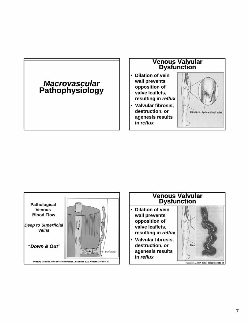
Macrovascular PathophysiologyMacrovascular
Pathophysiologyp y gyp y gy
Pathological Venous
Blood Flow
Deep to SuperficialVeins
“Down & Out”“Down & Out”
Bradbury & Ruckley. Atlas of Vascular Disease. 2nd edition 2003. Current Medicine, Inc
Venous Valvular Dysfunction
Venous Valvular Dysfunction
• Dilation of vein wall prevents opposition of valve leaflets,valve leaflets, resulting in reflux
• Valvular fibrosis, destruction, or agenesis results in reflux
Venous Valvular Dysfunction
Venous Valvular Dysfunction
• Dilation of vein wall prevents opposition of valve leaflets,valve leaflets, resulting in reflux
• Valvular fibrosis, destruction, or agenesis results in reflux
Hamden. JAMA 2012. 308(24): 2612-21
8
HistoryHistory
HistoryHistory• History of problem: onset,
pregnancies, prior DVT, immobilization
• Associated symptoms and relationship to heat, menses, exercise and compression
• Current medications
• Family history
• Previous treatment and result
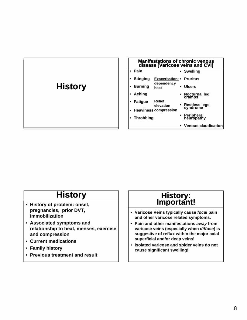
Manifestations of chronic venous disease [Varicose veins and CVI] Manifestations of chronic venous disease [Varicose veins and CVI]
• Pain
• Stinging
• Burning
Aching
• Swelling
• Pruritus
• Ulcers
Noct rnal leg
Exacerbation: dependencyheat
• Aching
• Fatigue
• Heaviness
• Throbbing
• Nocturnal leg cramps
• Restless legs syndrome
• Peripheral neuropathy
• Venous claudication
Relief: elevation compression
History: Important!
History: Important!
• Varicose Veins typically cause focal pain and other varicose related symptoms.
• Pain and other manifestations away from varicose veins (especially when diffuse) is suggestive of reflux within the major axial superficial and/or deep veins!
• Isolated varicose and spider veins do not cause significant swelling!
9
Physical Examination
Physical ExaminationExaminationExamination
Gloviczki et al. J Vasc Surg 2011;53:2S-48S.Gloviczki et al. J Vasc Surg 2011;53:2S-48S.
Examine patient in the standing position!
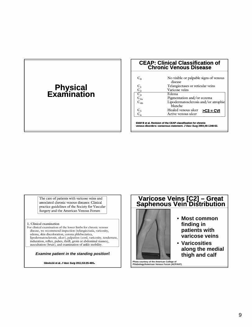
CEAP: Clinical Classification of Chronic Venous Disease
CEAP: Clinical Classification of Chronic Venous Disease
Eklöf B et al. Revision of the CEAP classification for chronicvenous disorders: consensus statement. J Vasc Surg 2004;40:1248-52.Eklöf B et al. Revision of the CEAP classification for chronicvenous disorders: consensus statement. J Vasc Surg 2004;40:1248-52.
>C3 = CVI>C3 = CVI
Varicose Veins [C2] – Great Saphenous Vein DistributionVaricose Veins [C2] – Great
Saphenous Vein Distribution
• Most common finding in patients withpatients with varicose veins
• Varicosities along the medial thigh and calf
Photo courtesy of the American College of Phlebology/American Venous Forum (ACP/AVF)
10
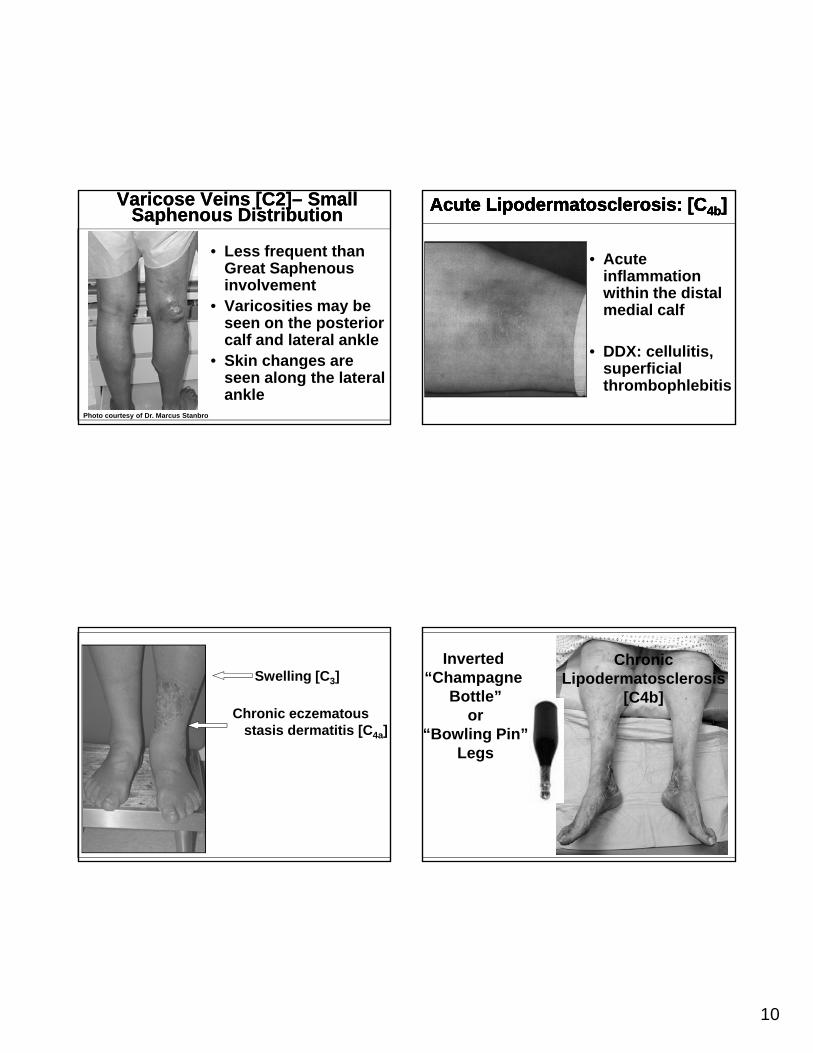
Varicose Veins [C2]– Small Saphenous Distribution
Varicose Veins [C2]– Small Saphenous Distribution
• Less frequent than Great Saphenous involvement
• Varicosities may be• Varicosities may be seen on the posterior calf and lateral ankle
• Skin changes are seen along the lateral ankle
Photo courtesy of Dr. Marcus Stanbro
Swelling [C3]
Chronic eczematousstasis dermatitis [C4a]
• Acute inflammation within the distal medial calf
Acute Lipodermatosclerosis: [Acute Lipodermatosclerosis: [CC4b4b] ] Acute Lipodermatosclerosis: [Acute Lipodermatosclerosis: [CC4b4b] ]
medial calf
• DDX: cellulitis, superficial thrombophlebitis
Inverted “Champagne
Bottle”or
“Bowling Pin”
Chronic Lipodermatosclerosis
[C4b]
Legs
11
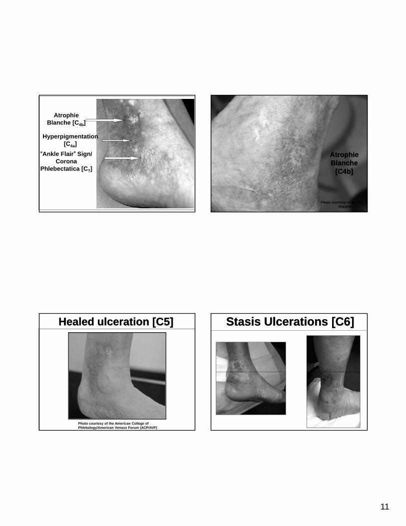
AtrophieBlanche [C4b]
“Ankle Flair” Sign/
Hyperpigmentation[C4a]
Ankle Flair Sign/Corona
Phlebectatica [C?]
Healed ulceration [C5]Healed ulceration [C5]
Photo courtesy of the American College of Phlebology/American Venous Forum (ACP/AVF)
AtrophieAtrophieAtrophie Blanche
[C4b]
Atrophie Blanche
[C4b]
Photo courtesy of Dr. MarcusStanbro
Stasis Ulcerations [C6]Stasis Ulcerations [C6]
12
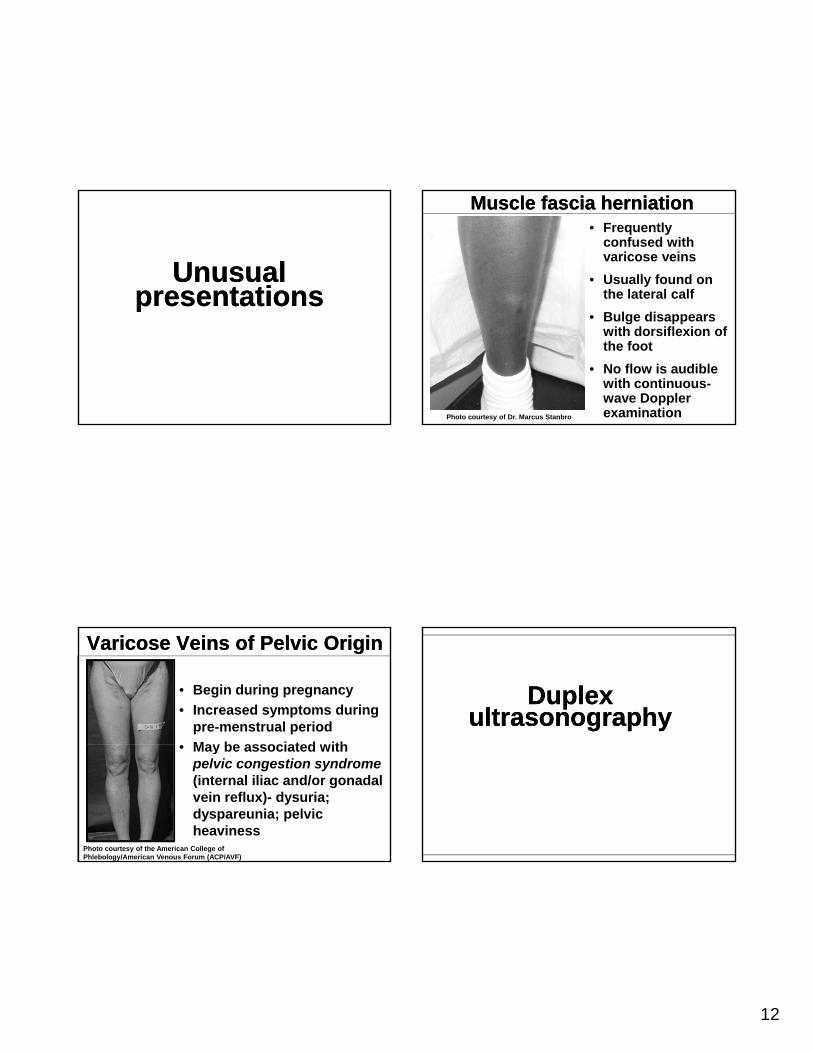
Unusual presentations
Unusual presentationspp
Varicose Veins of Pelvic OriginVaricose Veins of Pelvic Origin
• Begin during pregnancy
• Increased symptoms during pre-menstrual period
May be associated with• May be associated with pelvic congestion syndrome (internal iliac and/or gonadal vein reflux)- dysuria; dyspareunia; pelvic heaviness
Photo courtesy of the American College of Phlebology/American Venous Forum (ACP/AVF)
Muscle fascia herniationMuscle fascia herniation• Frequently
confused with varicose veins
• Usually found on the lateral calf
• Bulge disappears with dorsiflexion of the foot
• No flow is audible with continuous-wave Doppler examinationPhoto courtesy of Dr. Marcus Stanbro
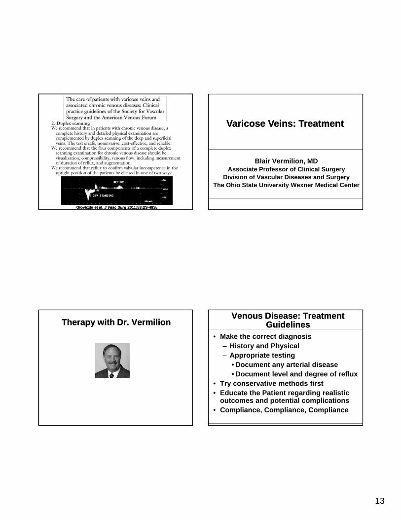
Duplex ultrasonography
Duplex ultrasonography
13
Gloviczki et al. J Vasc Surg 2011;53:2S-48S.Gloviczki et al. J Vasc Surg 2011;53:2S-48S.
Therapy with Dr. VermilionTherapy with Dr. Vermilion
Varicose Veins: TreatmentVaricose Veins: Treatment
Blair Vermilion, MDAssociate Professor of Clinical Surgery
Division of Vascular Diseases and SurgeryThe Ohio State University Wexner Medical Center
Venous Disease: Treatment Guidelines
Venous Disease: Treatment Guidelines
• Make the correct diagnosis– History and Physical– Appropriate testing
• Document any arterial diseasey• Document level and degree of reflux
• Try conservative methods first• Educate the Patient regarding realistic
outcomes and potential complications• Compliance, Compliance, Compliance
14
Venous Disease: Treatment OptionsVenous Disease: Treatment Options
• Compression Therapy
• Sclerotherapy
• Surgery
– Thermal ablation (Laser or Radio (Frequency)
– Phlebectomy
– “Stripping”
– SFJ Ligation
• Combination of any and all of the above
Venous Disease: Compression Therapy
Venous Disease: Compression Therapy
• Indications for Compression Therapy – Chronic Venous Insufficiency– Venous Ulcers, Dermatitis,– Post Sclerotherapy or Surgery– Superficial Phlebitis– DVT ( with anticoagulation)– Post Phlebitic Syndrome
Venous Disease: Compression Therapy
Venous Disease: Compression Therapy
• Contraindications for Compression Therapy
– Diminished Arterial Flow (<70 mm Hg )
– Acute DVT without sufficient collateralsAcute DVT without sufficient collaterals
– Severe CHF
– Undefined, non-venous Ulcers
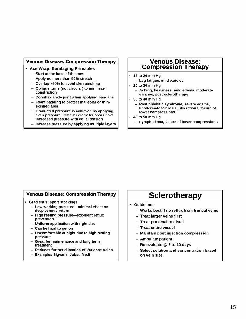
Venous Disease: Compression Therapy
Venous Disease: Compression Therapy
• Unna’s Boot
– Calamine lotion and zinc oxide
– High working pressure
– Low resting pressure
– Can be worn at night
– Use for Dermatitis, Ulcers
– Can be changed once/week
15
Venous Disease: Compression TherapyVenous Disease: Compression Therapy
• Ace Wrap: Bandaging Principles– Start at the base of the toes– Apply no more than 50% stretch– Overlap ~50% to avoid skin pinching – Oblique turns (not circular) to minimize
constrictionconstriction– Dorsiflex ankle joint when applying bandage– Foam padding to protect malleolar or thin-
skinned area – Graduated pressure is achieved by applying
even pressure. Smaller diameter areas have increased pressure with equal tension
– Increase pressure by applying multiple layers
Venous Disease: Compression TherapyVenous Disease: Compression Therapy
• Gradient support stockings– Low working pressure—minimal effect on
deep venous return– High resting pressure—excellent reflux
prevention– Uniform application with right sizeg– Can be hard to get on– Uncomfortable at night due to high resting
pressure– Great for maintenance and long term
treatment– Reduces further dilatation of Varicose Veins– Examples Sigvaris, Jobst, Medi
Venous Disease: Compression Therapy
Venous Disease: Compression Therapy
• 15 to 20 mm Hg– Leg fatigue, mild varicies
• 20 to 30 mm Hg– Aching, heaviness, mild edema, moderate
i i t l thvaricies, post sclerotherapy• 30 to 40 mm Hg
– Post phlebitic syndrome, severe edema, lipodermatosclerosis, ulcerations, failure of lower compressions
• 40 to 50 mm Hg – Lymphedema, failure of lower compressions
SclerotherapySclerotherapy• Guidelines
– Works best if no reflux from truncal veins
– Treat larger veins first
– Treat proximal to distal
– Treat entire vessel
– Maintain post injection compression
– Ambulate patient
– Re-evaluate @ 7 to 10 days
– Select solution and concentration based on vein size
16
Venous Disease: Sclerotherapy
Venous Disease: Sclerotherapy
• Complications of Sclerotherapy– Vasovagal Attack– Allergic reaction
Skin necrosis– Skin necrosis– Venous thrombosis– Arterial Injection/injury– Nerve Injection/injury– Skin Discoloration (Hyperpigmentation)– Telangiectatic matting
Venous Disease: Sclerotherapy
Venous Disease: Sclerotherapy
• Contraindications to Sclerotherapy of Varicose Veins– Bedridden Patient
Severe Arterial Disease– Severe Arterial Disease– Hypercoagulable state– Pregnancy– Morbid Obesity– Poor tolerance of compression hose– Allergies to the agents used
SclerotherapySclerotherapy• Mechanism:
– Solution causes irreversible chemical damage to the endothelial cell layercell layer
– Size of vein and flow in vein are variable therefore results are variable
– Results in “zones” of injury
Types of SclerosantsTypes of Sclerosants• Detergents:1. Sodium Morrhuate: Fatty acid extract from Cod
liver oil. Can cause extensive necrosis and possible anaphylaxis
2. Ethanolamine Oleate: synthetic and has high viscosity
3 S t d h l th ti FFA li bl d f3. Sotradechol: synthetic FFA, reliable and safe; tends to cause hyperpigmentation in higher concentrations
4. Polidocanol: synthetic FFA; not FDA approved; very safe; rare anaphylaxis and minimal hyperpigmentation
5. Glycerin: very weak and very viscous; rarely causes hyperpigmentation, necrosis or matting
17
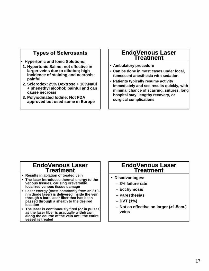
Types of SclerosantsTypes of Sclerosants
• Hypertonic and Ionic Solutions:1. Hypertonic Saline: not effective in
larger veins due to dilution; high incidence of staining and necrosis; painfulpainful
2. Sclerodex: 25% Dextrose + 10%NaCl + phenethyl alcohol; painful and can cause necrosis
3. Polyiodinated Iodine: Not FDA approved but used some in Europe
EndoVenous Laser Treatment
EndoVenous Laser Treatment
• Results in ablation of treated vein• The laser introduces thermal energy to the
venous tissues, causing irreversible localized venous tissue damage
• Laser energy (most commonly from an 810-Laser energy (most commonly from an 810nm diode laser) is delivered inside the vein through a bare laser fiber that has been passed through a sheath to the desired location
• The laser is continuously fired (or in pulses) as the laser fiber is gradually withdrawn along the course of the vein until the entire vessel is treated
• Ambulatory procedure
• Can be done in most cases under local, tumescent anesthesia with sedation
EndoVenous Laser Treatment
EndoVenous Laser Treatment
• Patients typically resume activity immediately and see results quickly, with minimal chance of scarring, sutures, long hospital stay, lengthy recovery, or surgical complications
• Disadvantages:
– 3% failure rate
– Ecchymosis
EndoVenous Laser Treatment
EndoVenous Laser Treatment
– Paresthesias
– DVT (1%)
– Not as effective on larger (>1.5cm.) veins
18
•Safety Issues
•Lasers emit beams of non-ionizing ti l di ti
EndoVenous Laser Treatment
EndoVenous Laser Treatment
optical radiation
– Eye Hazards: retina/ corneal
– Skin Hazards
– Fire Hazards
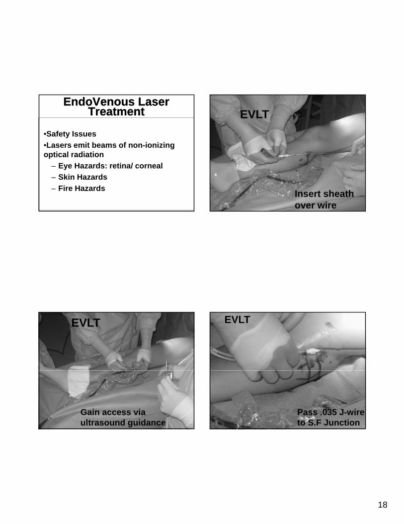
EVLT
Gain access via ultrasound guidance
EVLT
Insert sheath over wire
EVLT
Pass .035 J-wire to S.F Junction
19
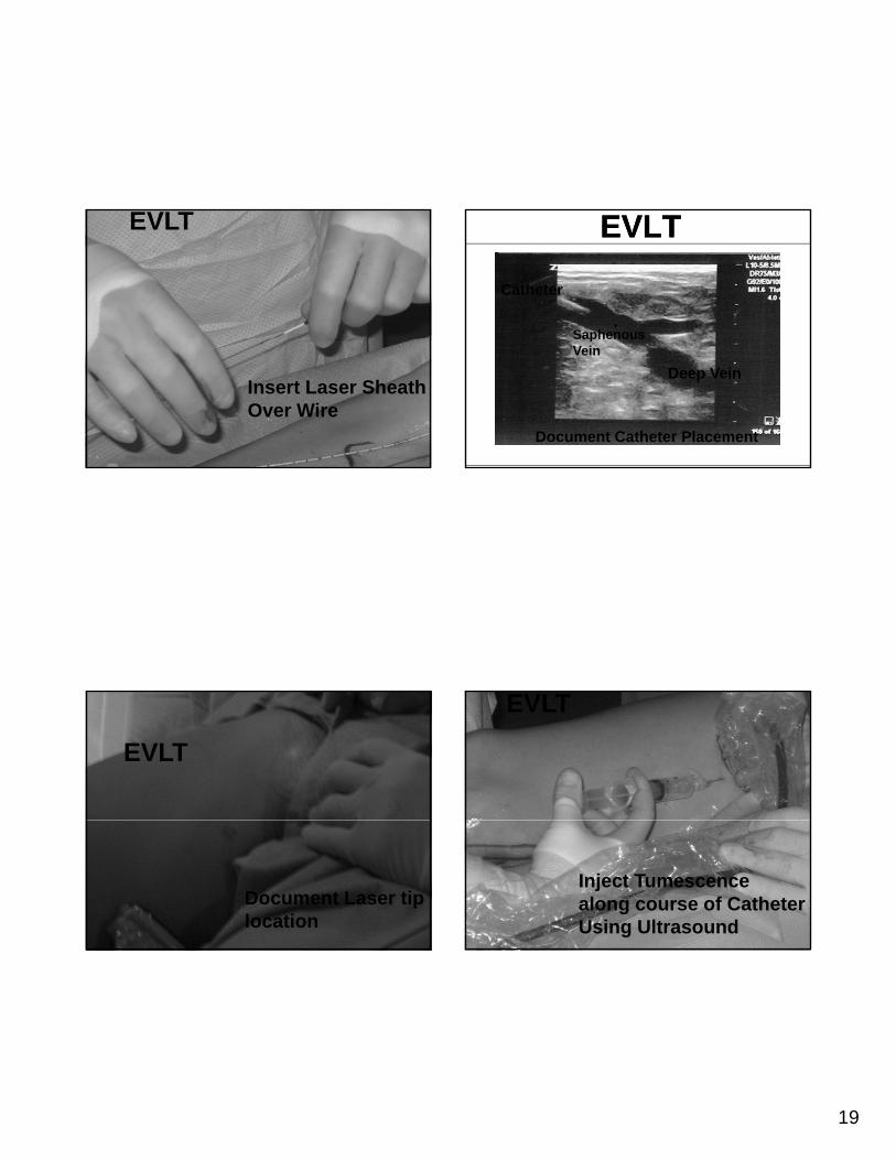
EVLT
Insert Laser Sheath Over Wire
EVLT
Document Laser tip location
EVLTEVLT
Saphenous
Catheter
Document Catheter Placement
Deep Vein
Saphenous Vein
EVLT
Inject Tumescence along course of Catheter Using Ultrasound
20
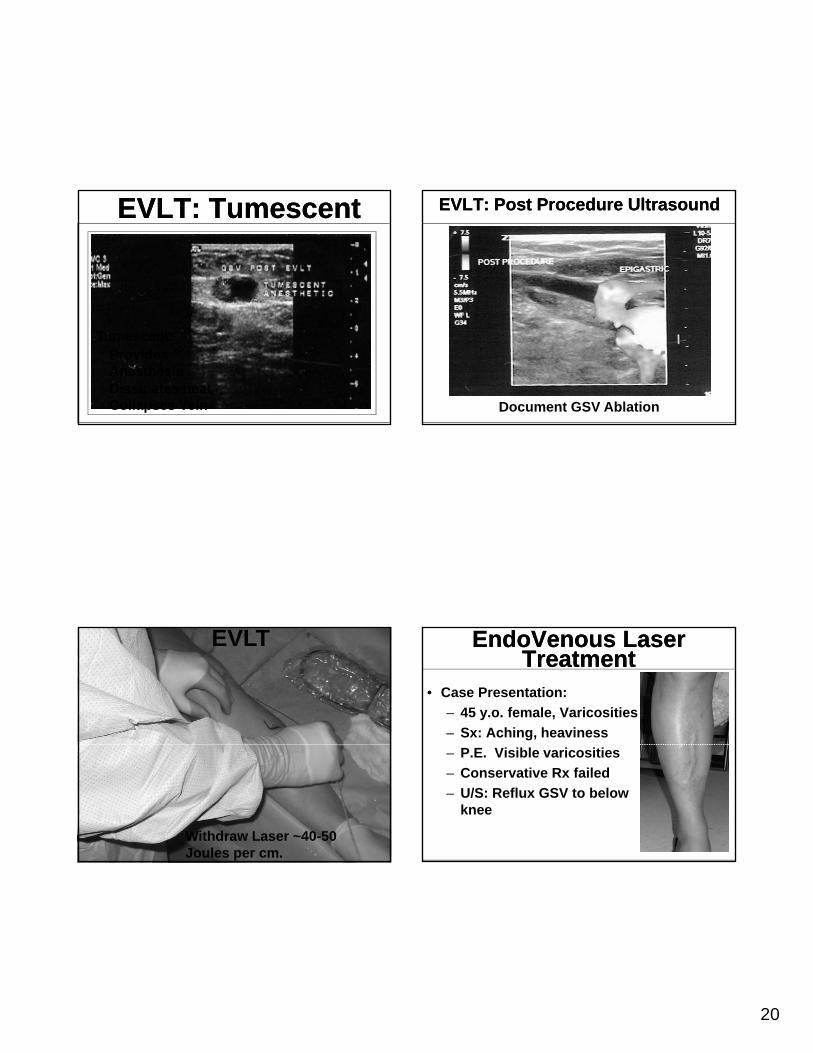
EVLT: TumescentEVLT: Tumescent
Tumescent:Provides AnesthesiaDissipates heatCollapses Vein
EVLT
Withdraw Laser ~40-50 Joules per cm.
EVLT: Post Procedure Ultrasound EVLT: Post Procedure Ultrasound
Document GSV Ablation
EndoVenous Laser Treatment
EndoVenous Laser Treatment
• Case Presentation:
– 45 y.o. female, Varicosities
– Sx: Aching, heaviness
– P.E. Visible varicosities
– Conservative Rx failed
– U/S: Reflux GSV to below knee
21
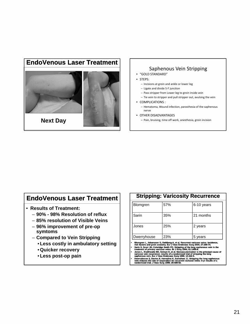
EndoVenous Laser TreatmentEndoVenous Laser Treatment
Next Day
EndoVenous Laser TreatmentEndoVenous Laser Treatment
• Results of Treatment:– 90% - 98% Resolution of reflux– 85% resolution of Visible Veins– 96% improvement of pre-op
symtoms– Compared to Vein Stripping
• Less costly in ambulatory setting• Quicker recovery• Less post-op pain
Saphenous Vein Stripping• “GOLD STANDARD”
• STEPS:
– Incisions at groin and ankle or lower leg
– Ligate and divide S‐F junction
– Pass stripper from Lower leg to groin inside vein
– Tie vein to stripper and pull stripper out, avulsing the vein
• COMPLICATIONS :
– Hematoma, Wound infection, parasthesia of the saphenousnerve
• OTHER DISADVANTAGES
– Pain, bruising, time off work, anesthesia, groin incision
Stripping: Varicosity RecurrenceStripping: Varicosity Recurrence
Blomgren 57% 6-10 years
Sarin 35% 21 months
Jones 25% 2 years
Dwerryhouse 23% 5 years
BlomgrenBlomgren L, Johansson G, DahlbergL, Johansson G, Dahlberg--A, et al. Recurrent varicose veins: incidence, A, et al. Recurrent varicose veins: incidence, risk factors and groin anatomy. risk factors and groin anatomy. EurEur J J VascVasc EndovascEndovasc SurgSurg 2004; 27:2692004; 27:269--74.74.
SarinSarin S, S, ScurrScurr JH, Coleridge Smith PD. Stripping of the long JH, Coleridge Smith PD. Stripping of the long saphenoussaphenous vein in the vein in the treatment of primary varicose veins. Br J treatment of primary varicose veins. Br J SurgSurg 1994; 81:14551994; 81:1455--8.8.
Jones L, Braithwaite BD, Selwyn D, et al. Jones L, Braithwaite BD, Selwyn D, et al. NeovascularizationNeovascularization is the principal cause of is the principal cause of varicose vein recurrence: results of a randomized trial of stripping the long varicose vein recurrence: results of a randomized trial of stripping the long saphenoussaphenous vein. vein. EurEur J J VascVasc EndovascEndovasc SurgSurg 1996; 12:4421996; 12:442--5.5.
DwerryhouseDwerryhouse S, Davies B, Harradine K, S, Davies B, Harradine K, EarnshawEarnshaw JJ. Stripping the long JJ. Stripping the long saphenoussaphenousvein reduces the rate of reoperation for recurrent varicose veins: 5vein reduces the rate of reoperation for recurrent varicose veins: 5--yr results of a yr results of a randomized trial. J randomized trial. J VascVasc SurgSurg 1999; 29:5891999; 29:589--92.92.
22
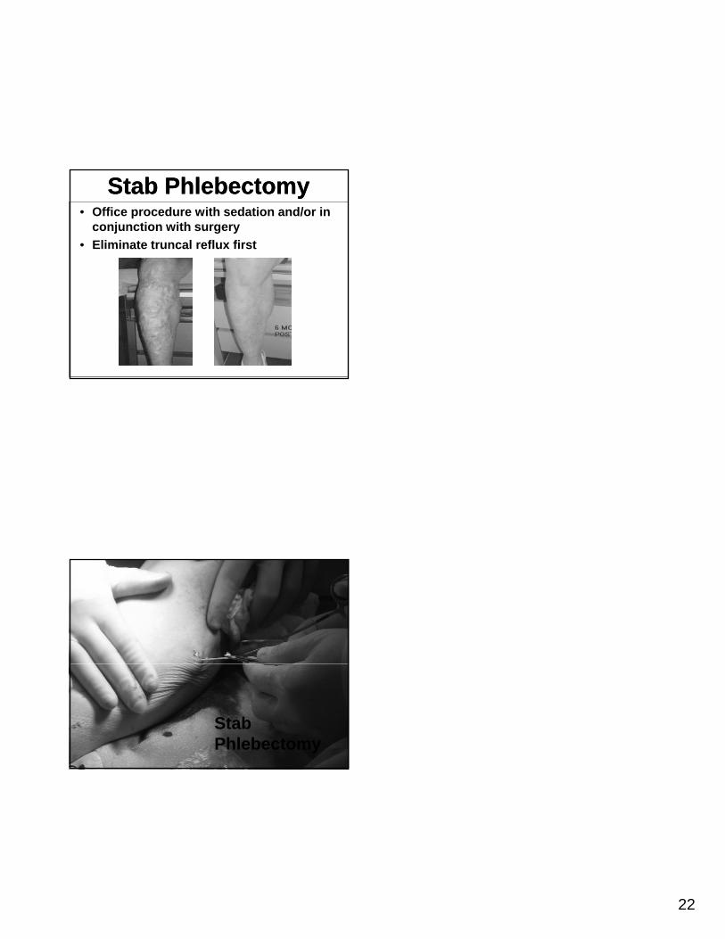
Stab PhlebectomyStab Phlebectomy• Office procedure with sedation and/or in
conjunction with surgery
• Eliminate truncal reflux first
Stab Phlebectomy