Embed Size (px)
Citation preview
JOURNAL OF CELLULAR PHYSIOLOGY 201:470–482 (2004)
Chondrocyte Protein With a Poly-Proline Region(CHPPR) Is a Novel Mitochondrial Protein
and Promotes Mitochondrial Fission
LAURA TONACHINI,1 MASSIMILIANO MONTICONE,1,2 CLAUDIA PURI,3 CARLO TACCHETTI,3
PAOLO PINTON,4 ROSARIO RIZZUTO,4 RANIERI CANCEDDA,1,2
SARA TAVELLA,2 AND PATRIZIO CASTAGNOLA1*1Istituto Nazionale per la Ricerca sul Cancro, Genova, Italy
2Dipartimento Oncologia, Biologia e Genetica, Universita di Genova, Italy3Dipartimento di Medicina Sperimentale, Sezione di Anatomia,
e Centro di Oncologia Cellulare e Ultrastrutturale IFOM, Genoa, Italy4Dip. di Medicina Sperimentale e Diagnostica,
Sezione di Patologia Generale, Universita di Ferrara, Italy
We have recently identified a chondrocyte protein with a poly-proline region,referred to as CHPPR, and showed that this protein is expressed intracellularly inchick embryo chondrocytes. Conventional fluorescence and confocal localizationof CHPPR shows that CHPPR is sorted to mitochondria. Furthermore, immunoe-lectron microscopy of CHPPR transfected cells demonstrates that this protein ismostly associated with the mitochondrial inner membranes. Careful analysis ofCHPPR expressing cells reveals, instead of the regular mitochondrial tubularnetwork, the presence of a number of small spheroid mitochondria. Here we showthat the domain responsible for network–spheroid transition spans amino acidresidues 182–309 including the poly-proline region. Functional analyses ofmitochondrial activity rule out the possibility of mitochondrial damage in CHPPRtransfected cells. Since cartilage expresses high levels of CHPPR mRNA whencompared to other tissues and because CHPPR is associated with late stages ofchondrocyte differentiation, we have investigated mitochondrial morphology inhypertrophic chondrocytes byMitoTracker Orange labeling. Confocal microscopyshows that these cells have spheroid mitochondria. Our data demonstrate thatCHPPR is able to promote mitochondrial fission with a sequence specificmechanism suggesting that this event may be relevant to late stage of chondrocytedifferentiation. J. Cell. Physiol. 201: 470–482, 2004. � 2004 Wiley-Liss, Inc.
We have recently characterized a cDNA encoding theprotein chondrocyte protein with a poly-proline region(CHPPR) (Tonachini et al., 2002). This cDNA wasisolated by a subtractive cDNA library screen ofhypertrophic and proliferating chick chondrocytes. Weshowed that CHPPR mRNA is more abundant in chickembryo cartilage than in several other tissues and thatin vitro and in vivo CHPPR gene expression increaseswith chondrocyte maturation (Tonachini et al., 2002).We also showed that in vitro parathyroid hormonepeptide [PTH (1–34)] enhances the accumulation ofCHPPR mRNA in this cell type (Tonachini et al., 2002).The CHPPR amino acid sequence contains a character-istic proline-rich region with a proline–proline–leu-cine–proline (PPLP, single letter amino acid code) motif(Tonachini et al., 2002). No other structural or func-tional motifs or domains so far characterized andavailable in public databases were identified in thisprotein. By BLAST analysis we determined that thecDNA insert of the isolated clone is the avian counter-part of a human partial cDNA, named KIAA0009
(Nomura et al., 1994). Since to our knowledge neitherfurther characteristics nor expression data of KIAA0009have been published, we consider KIAA0009 as thehuman CHPPR.
We demonstrated by both Western blot and immuno-histochemistry analysis that CHPPR is localized intra-cellularly in chondrocytes (Tonachini et al., 2002). Here
� 2004 WILEY-LISS, INC.
Laura Tonachini and Massimiliano Monticone contributedequally to this work.
Contract grant sponsor: Consiglio Nazionale delle Ricerche, Rome(Italy) P.F. Biotecnologie; Contract grant number: 99.00524.PF49.
*Correspondence to: Patrizio Castagnola, Istituto Nazionale per laRicerca sul Cancro, Largo R. Benzi 10, 16132 Genova, Italy.E-mail: [email protected]
Received 27 June 2003; Accepted 11 February 2004
DOI: 10.1002/jcp.20126
we prove that CHPPR is a novel mitochondrial proteinable to promote mitochondrial fission. In particular, weshow that CHPPR transfected cells and culturedhypertrophic chondrocytes, expressing endogenousCHPPR, have spheroid mitochondria instead of atubular mitochondrial network like other cell types.We have also demonstrated that a specific domain,including the proline rich region, is responsible for themitochondrial fission activity of CHPPR. This effect isalso observed in cells, transfected with constructscontaining this domain, expressing low or comparablelevels of mRNA with respect to cells transfected withfission incompetent CHPPR-derived constructs. Wehave also investigated whether the fission processinduced by CHPPR affects mitochondrial activity show-ing that these spheroid mitochondria display generationof proton motive force and oxidation ability. However,how this protein affects mitochondrial morphology atthe molecular level is unknown so far and will be subjectof further investigation.
MATERIALS AND METHODSSequence analysis and constructs generation
To obtain the coding sequence (cds) of the mouseortholog of chicken CHPPR, BLAST analysis of mouseexpressed sequence tags (EST) database was perform-ed with the chick sequence at the National Center forBiotechnology Information (NCBI, Bethesda, MD)(Altschul et al., 1990); this screening identified anIMAGE cDNA clone 832381 as the putative mouseCHPPR ortholog. The complete nucleotide sequence ofthis clone was determined by automated sequencingusing a 377 Perkin Elmer sequencer. All sequencingreactions were performed by using the BigDye termi-nator cycle sequencing kit (Perkin Elmer, Wellesley,MA). For sequence analyses, The ‘‘Sequencher’’ (GeneCodes Corp., Ann Arbor, MI) and the ‘‘GeneWorks1’’(IntelliGenetics Inc., Campbell, CA) software packageswere used. BLAST analysis demonstrated that thisIMAGE clone encodes the mouse CHPPR and itssequence has been submitted to GenBank; its accessionnumber is AF354708. The Mitoprot II protocol was used(Claros and Vincens, 1996) to assess probability ofmitochondrial sorting.
The CHPPR myc-his tagged expression construct wasgenerated by cloning the PCR generated cds fragment ofchick CHPPR in frame with the 50 end of the myc epitopecds of the pcDNA 3.1 myc-his vector (Invitrogen, SanDiego, CA). A control Actin myc-his tagged constructwas generated by cloning the PCR generated cdsfragment of Actin sequence with GeneBank accessionnumber NM_009606 in frame with the 50 end of themyc epitope cds of the pcDNA 3.1 myc-his vector. TheCHPPR flag-tagged expression construct was generatedby cloning, in the pcDNA3.1 vector, the PCR generatedcds fragment for the flag epitope (DYKDDDDK, singleletter amino acid code) in frame with the 30 end of chickCHPPR; a stop codon was inserted immediately after thecodon specifying the C terminal K residue. The chickCHPPR–RFP (red fluorescent protein) chimera con-struct was generated by subcloning the chick CHPPRcds in frame with the 50 end of the Ds2Red cds ofthe pDs2Red-N1 vector (Clontech, San Diego, CA). Toobtain a doxycycline inducible chick CHPPR–RFP the
cds encoding the protein chimera was subcloned inthe pTRE2 vector. CHPPR–GFP (green fluorescentprotein) chimeric construct were generated by subclon-ing, in the pcDNA3.1 vector, the entire CHPPR cds or itsspecific fragments in frame with a modified GFP cycle 3mutant.
All constructs were sequenced in order to rule out anyaccidental mutation introduced by PCR amplification.
RNA isolation and Northern blot analysis
For Northern blot analysis, total RNA was extractedfrom cell cultures by using TRIzol reagent (Invitrogen)according to the manufacture’s protocol. Total RNAswere electrophoresed through 1% agarose gels in thepresence of formaldehyde and blotted onto Hybond Nmembranes (Amersham, Piscataway, NJ). Blot pre-hybridizations were performed at 658C for 30 min in333 mM NaH2PO4 pH 7.2, 6.66% sodium dodecylsulphate, and 250 mg/ml denatured salmon spermDNA. Blot hybridization was performed at 658C for18 h in the same solution containing 106 cpm/ml ofdenatured and labeled chick CHPPR cds probe spanningamino acid residues 1–62. After hybridization, the blotswere washed twice at 658C for 15 min in 0.2% sodiumdodecyl sulphate, 2� SSPE and twice at 658C for 15 minin 0.2% sodium dodecyl sulphate, 0.2� SSPE. Digitalimages of blots were acquired using the Cyclone phos-phoimager (Packard instruments, Meridien, CT).
Cell culture and transfection
Chondrocyte cell culture methods are extensivelydescribed elsewhere (Altschul et al., 1990). Briefly, stage29–30 chick embryo tibiae were removed, cleaned,washed in phosphate buffered saline (PBS), pH 7.2,and digested for 15 min at 378C with 400 U/mlcollagenase I and 0.25% trypsin. After sedimentation,the supernatant, containing tissue debris and perichon-drium, was discarded and the pellet was digested foradditional 45 min in the above dissociation buffersupplemented with 1,000 U/ml of collagenase II. Freshlydissociated chondrocytes were plated onto culturedishes. These cells were maintained in Coon’s modifiedF12 medium with 10% FCS, supplemented with 5 mMglutamine, and cultured in adhesion for 3 weeks.During this time, chondrocytes dedifferentiate to fibro-blast-like cells expressing type I collagen (Castagnolaet al., 1988). Dedifferentiated cells were then trans-ferred by 0.25% trypsin (Gybco BRL, San Diego, CA)digestion (in phosphate buffered saline) into 1% agarosecoated petri dishes and cultured in suspension in a F12medium with 10% FCS and with 5 mM Glutamine.COS7 and HeLa cells were cultured in Dulbecco’smodified Eagle’s medium supplemented with 10% FCSand with 5 mM Glutamine.
For transient transfection, cells were plated on poly-L-lysine-coated glass coverslips 16 h before transfection.Expression constructs were introduced into the cellsusing the Effectene transfection reagent (Qiagen,Hilden, Germany) according to the manufacturer’sinstructions.
To induce the expression of CHPPR–RFP chimeracloned into pTRE2, transfection was performed underserum-free conditions (Quarto et al., 1997) and 48 h laterthe medium was supplemented with Doxycycline.
CHPPR PROMOTES MITOCHONDRIAL FISSION 471
Microscopic analysis of living cultured cells wasperformed at 378C in a buffer containing NaCl130 mM; KCl 5 mM; MgSO4 1.2 mM; KH2PO4 1.2 mM;CaCl2 1.2 mM; glucose 1.1 mg/ml; HEPES 25 mM pH 7.4.
Mitochondrial labeling
COS7 cells were labeled with 100 nM MitoTrackerOrange CM-H2TMROS (Molecular Probes, Eugene,OR) following the manufacturer’s instructions. Beforelabeling, hypertrophic chondrocytes were incubated in1 mg/ml Hyaluronidase (Sigma, Saint Louis, MO) inphosphate buffered saline (PBS) for 10 min at 378C.Subsequently, trypsin/EDTA (Sigma) was added andcells were incubated for another 10 min at 378C.Enzymes were neutralized by addition of completemedium. The chondrocytes were centrifuged and resus-pended in medium containing 100 nM MitoTrackerOrange CM-H2TMROS (Molecular probes) accordingto manufacturer’s instructions. Cells were fixed andpermeabilized as described in the immunolocalizationsection.
Immunolocalization
Cells were fixed in 3% paraformaldehyde in PBS, pH7.6 containing 2% sucrose for 5 min at room tempera-ture. After rinsing in PBS, cells were permeabilized in asolution containing 20 mM HEPES pH 7.4, 300 mMsucrose, 50 mM NaCl, 3 mM MgCl2, and 0.5% Triton X-100 for 5 min at 08C. Nuclei were stained with Hoechst33258 in PBS for 5 min at room temperature. Non-specific binding was prevented by incubation with pureGoat serum for 30 min at 08C. Slides were incubatedwith purified 10 mg/ml IgG1 of the anti c-myc (Evanet al., 1985) 9E10 monoclonal antibody (DevelopmentHybridoma Bank), in PBS supplemented with 10% goatserum (antibody dilution buffer) for 2 h at 08C. Afterextensive rinsing in PBS, slides were incubated for30 min with FITC-conjugated goat anti-mouse antibodyin antibody dilution buffer. Nuclei were stained withHoechst 33258.
Fluorescence analysis
Routine observations were performed with a ZeissAxiophot microscope equipped with a 100� Pan-Neo-fluar objective and with the following filter sets: 10, 15,and 02 to detect GFP/FITC (fluorescein iso-thiocyanate),RFP, and Hoechst 33258 respectively. Fluorescenceimages were recorded using a CCD camera (C5810Hamamatsu). Image merging, composition, and print-ing were performed using Adobe Photoshop 5.0.
To acquire confocal images, samples were subjected tomicroscopy using a BioRad-MRC 1024 confocal micro-scope (60� objective, argon laser, sequential excitationat 468 and 580 nm). Digital images were acquired andprocessed using the BioRad Laser Pix software.
Immunoelectron microscopy
Pellets of COS7 cells, transfected with myc- or flag-tagged CHPPR, were fixed in 2% paraformaldehyde and0.2% glutharaldehyde in PBS, embedded in 10% gelatin,infused with 2.3 M sucrose, and frozen in liquidnitrogen. Ultrathin cryo-sections were obtained with aReichert-Jung Ultracut E with FC4E cryoattachmentand collected on copper–formvar–carbon coated grids.
Immunogold localization on ultrathin cryo-sections wasperformed as previously described (Slot and Geuze,1984; Schiaffino et al., 1999). In particular, sectionswere immunostained with a rabbit anti-myc antibody(Santa Cruz, Santa Cruz, CA), or with a mouse anti-flagmonoclonal antibody (Clone M2, Sigma) followed by 10-nm Protein A-gold. Sections were examined with ZeissEM 10 and 902 electron microscopes.
Aequorin measurements
For aequorin measurements, HeLa cells (seeded onto13 mm coverslips) were co-transfected with 3 mgpcDNA3 (control sample) and 1 mg mitochondrial matrixtargeted Aequorin (mtAEQmut)/VR1012 or cytoplasmictargeted Aequorin (cytAEQ) (Montero et al., 2000). Forthe CHPPR sample, cells were transfected with 3 mgCHPPR–myc and 1 mg mtAEQmut/VR1012 or cytoplas-mic targeted Aequorin (cytAEQ) using the calciumphosphate technique. Thirty-six hours after transfec-tion, the coverslips with the cells were incubated with5 mM coelenterazine for 2 h. All aequorin measurementswere carried out in KRB (Krebs–Ringer modified buffer:125 mM NaCl, 5 mM KCl, 1 mM MgSO4, 1 mMNa2HPO4, 5.5 mM glucose, 20 mM NaHCO3, 2 mM L-glutamine, 1 mM CaCl2, and 20 mM HEPES, pH 7.4 at378C) in a 5% CO2 atmosphere, and then transferred tothe perfusion chamber.
One hundred micromolar histamine (Sigma-Aldrich,Saint Louis, MO) was added to the same medium. Inorder to obtain uncoupled mitochondria parallel controlexperiments were performed in the presence of 5 mMcarbonyl cyanide m-chlorophenylhydrazone (CCCP).The aequorin experiments were terminated by lysingthe cells with 100 mM digitonin (Sigma-Aldrich) in ahypotonic Ca2þ-rich solution (10 mM CaCl2 in H2O),thus discharging the remaining aequorin pool. The lightsignal was collected in a purpose-built luminometer andcalibrated into [Ca2þ] values as previously described(Chiesa et al., 2001).
Mitochondria membrane potential measurement
These measures were performed using 10 nM tetra-methylrhodamine, methyl ester, perchlorate (TMRM)on a Zeiss confocal microscope (LSM 510). The signalwas collected as total emission >570 nm. To distinguishcontrol from the CHPPR–myc expressing cells thesewere cotransfected with a construct expressing GFP.Green emission was collected as emission from the range>505–535 nm.
RESULTSComputer analysis prediction of CHPPR
subcellular localization
To examine the subcellular localization of CHPPR wesubjected the chick CHPPR amino acid sequence(Fig. 1A) to several subcellular protein localizationprediction protocols available through the World WideWeb. Some of these protocols suggested possible mito-chondrial localization for both the chick CHPPR and itshuman ortholog previously named KIAA0009. To con-firm this, we also analyzed the mouse CHPPR ortholog.We explored the mouse EST database and found that theIMAGE clone 832381 was a putative mouse CHPPRortholog. We determined its entire nucleotide sequence
472 TONACHINI ET AL.
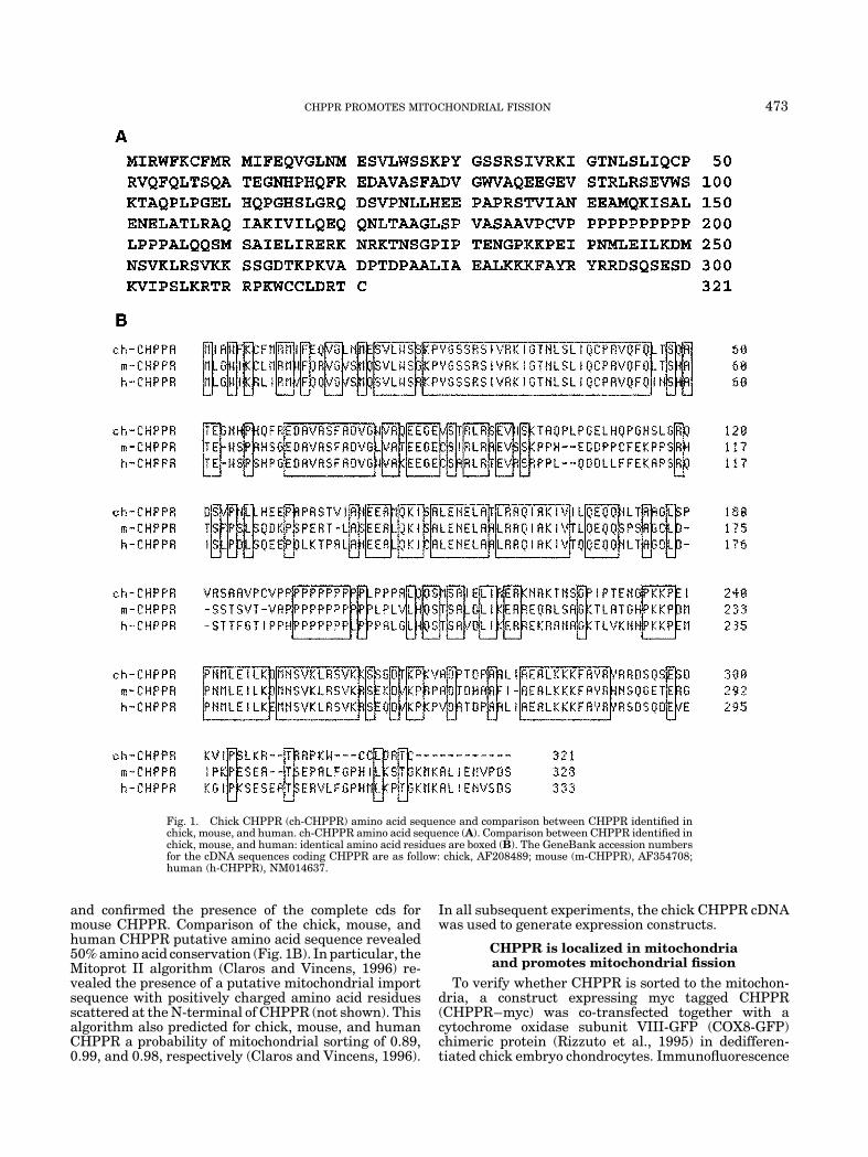
and confirmed the presence of the complete cds formouse CHPPR. Comparison of the chick, mouse, andhuman CHPPR putative amino acid sequence revealed50% amino acid conservation (Fig. 1B). In particular, theMitoprot II algorithm (Claros and Vincens, 1996) re-vealed the presence of a putative mitochondrial importsequence with positively charged amino acid residuesscattered at the N-terminal of CHPPR (not shown). Thisalgorithm also predicted for chick, mouse, and humanCHPPR a probability of mitochondrial sorting of 0.89,0.99, and 0.98, respectively (Claros and Vincens, 1996).
In all subsequent experiments, the chick CHPPR cDNAwas used to generate expression constructs.
CHPPR is localized in mitochondriaand promotes mitochondrial fission
To verify whether CHPPR is sorted to the mitochon-dria, a construct expressing myc tagged CHPPR(CHPPR–myc) was co-transfected together with acytochrome oxidase subunit VIII-GFP (COX8-GFP)chimeric protein (Rizzuto et al., 1995) in dedifferen-tiated chick embryo chondrocytes. Immunofluorescence
Fig. 1. Chick CHPPR (ch-CHPPR) amino acid sequence and comparison between CHPPR identified inchick, mouse, and human. ch-CHPPR amino acid sequence (A). Comparison between CHPPR identified inchick, mouse, and human: identical amino acid residues are boxed (B). The GeneBank accession numbersfor the cDNA sequences coding CHPPR are as follow: chick, AF208489; mouse (m-CHPPR), AF354708;human (h-CHPPR), NM014637.
CHPPR PROMOTES MITOCHONDRIAL FISSION 473
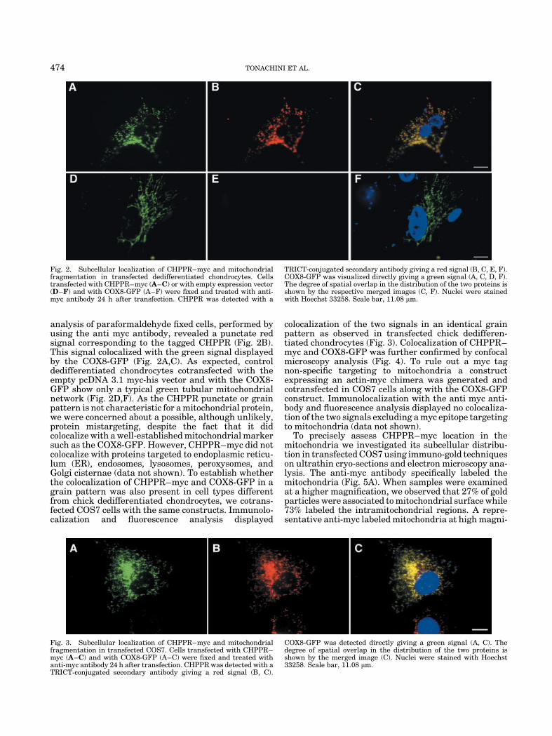
analysis of paraformaldehyde fixed cells, performed byusing the anti myc antibody, revealed a punctate redsignal corresponding to the tagged CHPPR (Fig. 2B).This signal colocalized with the green signal displayedby the COX8-GFP (Fig. 2A,C). As expected, controldedifferentiated chondrocytes cotransfected with theempty pcDNA 3.1 myc-his vector and with the COX8-GFP show only a typical green tubular mitochondrialnetwork (Fig. 2D,F). As the CHPPR punctate or grainpattern is not characteristic for a mitochondrial protein,we were concerned about a possible, although unlikely,protein mistargeting, despite the fact that it didcolocalize with a well-established mitochondrial markersuch as the COX8-GFP. However, CHPPR–myc did notcolocalize with proteins targeted to endoplasmic reticu-lum (ER), endosomes, lysosomes, peroxysomes, andGolgi cisternae (data not shown). To establish whetherthe colocalization of CHPPR–myc and COX8-GFP in agrain pattern was also present in cell types differentfrom chick dedifferentiated chondrocytes, we cotrans-fected COS7 cells with the same constructs. Immunolo-calization and fluorescence analysis displayed
colocalization of the two signals in an identical grainpattern as observed in transfected chick dedifferen-tiated chondrocytes (Fig. 3). Colocalization of CHPPR–myc and COX8-GFP was further confirmed by confocalmicroscopy analysis (Fig. 4). To rule out a myc tagnon-specific targeting to mitochondria a constructexpressing an actin-myc chimera was generated andcotransfected in COS7 cells along with the COX8-GFPconstruct. Immunolocalization with the anti myc anti-body and fluorescence analysis displayed no colocaliza-tion of the two signals excluding a myc epitope targetingto mitochondria (data not shown).
To precisely assess CHPPR–myc location in themitochondria we investigated its subcellular distribu-tion in transfected COS7 using immuno-gold techniqueson ultrathin cryo-sections and electron microscopy ana-lysis. The anti-myc antibody specifically labeled themitochondria (Fig. 5A). When samples were examinedat a higher magnification, we observed that 27% of goldparticles were associated to mitochondrial surface while73% labeled the intramitochondrial regions. A repre-sentative anti-myc labeled mitochondria at high magni-
Fig. 2. Subcellular localization of CHPPR–myc and mitochondrialfragmentation in transfected dedifferentiated chondrocytes. Cellstransfected with CHPPR–myc (A–C) or with empty expression vector(D–F) and with COX8-GFP (A–F) were fixed and treated with anti-myc antibody 24 h after transfection. CHPPR was detected with a
TRICT-conjugated secondary antibody giving a red signal (B, C, E, F).COX8-GFP was visualized directly giving a green signal (A, C, D, F).The degree of spatial overlap in the distribution of the two proteins isshown by the respective merged images (C, F). Nuclei were stainedwith Hoechst 33258. Scale bar, 11.08 mm.
Fig. 3. Subcellular localization of CHPPR–myc and mitochondrialfragmentation in transfected COS7. Cells transfected with CHPPR–myc (A–C) and with COX8-GFP (A–C) were fixed and treated withanti-myc antibody 24 h after transfection. CHPPR was detected with aTRICT-conjugated secondary antibody giving a red signal (B, C).
COX8-GFP was detected directly giving a green signal (A, C). Thedegree of spatial overlap in the distribution of the two proteins isshown by the merged image (C). Nuclei were stained with Hoechst33258. Scale bar, 11.08 mm.
474 TONACHINI ET AL.
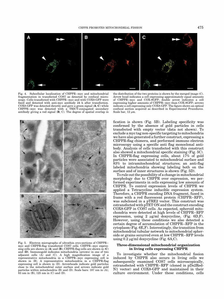
fication is shown (Fig. 5B). Labeling specificity wasconfirmed by the absence of gold particles in cellstransfected with empty vector (data not shown). Toexclude a myc tag non-specific targeting to mitochondriawe have also generated a further construct, expressing aCHPPR-flag chimera, and performed immuno electronmicroscopy using a specific anti flag monoclonal anti-body. Analysis of cells transfected with this constructalso showed a mitochondrial specific staining (Fig. 5C).In CHPPR-flag expressing cells, about 17% of goldparticles were associated to mitochondrial surface and83% to intramitochondrial structures; an anti-flaglabeled mitochondria showing labeling both on thesurface and of inner structures is shown (Fig. 5D).
To rule out the possibility of a change in mitochondrialmorphology due to CHPPR over expression, we per-formed experiments in cells expressing low amounts ofCHPPR. To control expression levels of CHPPR weapplied a Tetracycline inducible expression system.Therefore, a CHPPR encoding DNA fragment, fused inframe with a red fluorescent protein (CHPPR–RFP),was subcloned in a pTRE2 vector. This construct wascotransfected with pTET ON and the construct encodingCOX8-GFP in COS7 cells. As expected, spheroid mito-chondria were detected at high levels of CHPPR–RFPexpression, using 2 mg/ml doxycycline, (Fig. 6D,F).However, using these conditions we also detected acertain degree of accumulation of CHPPR–RFP in thecytoplasm (Fig. 6E,F). Interestingly, the transition frommitochondrial tubular network to mitochondrial spher-oids or grains occurred even at low CHPPR–RFP levelsusing 0.2 mg/ml doxycycline (Fig. 6A,C).
Three-dimensional mitochondrial organizationin living cells expressing CHPPR
To investigate whether the mitochondrial fissioninduced by CHPPR also occurs in living cells wesubsequently examined COS7 cells microscopically,cotransfected with CHPPR–RFP (cloned in pDs2Red-N1 vector) and COX8-GFP and maintained in theirculture environment. Under these conditions, cells
Fig. 4. Subcellular localization of CHPPR–myc and mitochondrialfragmentation in transfected COS7 as detected by confocal micro-scopy. Cells transfected with CHPPR–myc and with COX8-GFP werefixed and detected with anti-myc antibody 24 h after transfection.COX8-GFP was detected directly and gave a green signal (A, C) whileCHPPR–myc was detected with a TRICT-conjugated secondaryantibody giving a red signal (B, C). The degree of spatial overlap in
the distribution of the two proteins is shown by the merged image (C).Arrow head indicates a cell expressing approximately equal amountsof CHPPR–myc and COX-8GFP; double arrow indicates a cellexpressing higher amounts of CHPPR–myc than COX-8GFP; arrowsindicate a cell expressing only COX8-GFP. The figure shows an opticalconfocal section acquired as described in Experimental Procedures.Scale bar, 12 mm.
Fig. 5. Electron micrographs of ultrathin cryo-sections of CHPPR–myc and CHPPR-flag transfected COS7 cells. CHPPR–myc expres-sing cells are shown in (A) and (B); CHPPR-flag cells are shown in (C)and (D). Immunogold indicates mitochondria (arrows) in one of twoadjacent cells (A) and (C). A high magnification image of arepresentative mitochondria in a CHPPR–myc expressing cell isshown in (B). A representative mitochondria in a CHPPR-flagexpressing cell is shown in (D). Arrowheads indicate gold particlesclose to the mitochondrial outer surface and arrows indicate goldparticles within mitochondria (B) and (D). Scale bars: 187 nm in (A);83 nm in (B); 125 nm in (C) and (D).
CHPPR PROMOTES MITOCHONDRIAL FISSION 475
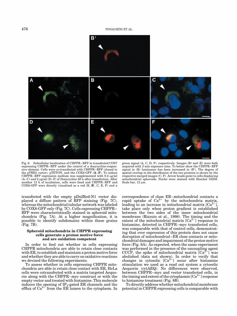
transfected with the empty pDs2Red-N1 vector dis-played a diffuse pattern of RFP staining (Fig. 7C),whereas the mitochondrial tubular network was labeledby COX8-GFP only (Fig. 7C). Cells expressing CHPPR–RFP were characteristically stained in spheroid mito-chondria (Fig. 7A). At a higher magnification, it ispossible to identify subdomains within these grains(Fig. 7B).
Spheroid mitochondria in CHPPR expressingcells generate a proton motive force
and are oxidation competent
In order to find out whether in cells expressingCHPPR mitochondria are able to retain close contactswith ER, to establish and maintain a proton motive forceand whether they are able to carry on oxidative reactionswe devised the following experiments.
To assess whether in cells expressing CHPPR mito-chondria are able to retain close contact with ER, HeLacells were cotransfected with a matrix targeted Aequo-rin along with the CHPPR–myc construct or with theempty vector and treated with histamine. This moleculeinduces the opening of IP3-gated ER channels and theefflux of Ca2þ from the ER lumen to the cytoplasm. In
correspondence of close ER–mitochondrial contacts arapid uptake of Ca2þ by the mitochondria matrix,leading to an increase in mitochondrial matrix [Ca2þ],take place only when proton gradient is establishedbetween the two sides of the inner mitochondrialmembrane (Rizzuto et al., 1998). The timing and theextent of the mitochondrial matrix [Ca2þ] response tohistamine, detected in CHPPR–myc transfected cells,was comparable with that of control cells, demonstrat-ing that over expression of this protein does not causedisruption of mitochondrial–ER close contacts or mito-chondrial damages and impairment of the proton motiveforce (Fig. 8A). As expected, when the same experimentwas performed in the presence of the uncoupling agentCCCP, the spike of mitochondrial matrix [Ca2þ] wasabolished (data not shown). In order to verify thatchanges in cytosolic [Ca2þ] occur after histaminestimulation we used as a read out system a cytosolicAequorin (cytAEQ). No differences were observed,between CHPPR–myc and vector transfected cells, inthe timing and extent of the cytoplasmic [Ca2þ] responseto histamine treatment (Fig. 8B).
To directly address whether mitochondrial membranepotential in CHPPR expressing cells is comparable with
Fig. 6. Subcellular localization of CHPPR–RFP in transfected COS7expressing CHPPR–RFP under the control of a doxycycline-respon-sive element. Cells were co-transfected with CHPPR–RFP (cloned inthe pTRE2 vector), pTETON, and the COX8-GFP (A–F). To induceCHPPR–RFP expression medium was supplemented with 0.2 mg/ml(A–C) and 2 mg/ml (D–F) of Doxycycline 48 h after transfection. Afteranother 12 h of incubation, cells were fixed and CHPPR–RFP andCOX8-GFP were directly visualized as a red (B, B0, C, E, F) and a
green signal (A, C, D, F), respectively. Images (B) and (E) were bothacquired with 2 min exposure time. To better show the CHPPR–RFPsignal in (B) luminance has been increased in (B0). The degree ofspatial overlap in the distribution of the two proteins is shown by therespective merged images (C, F). Arrow heads point to cells displayingmitochondrial spheroids. Nuclei were stained with Hoechst 33258.Scale bar, 12 mm.
476 TONACHINI ET AL.
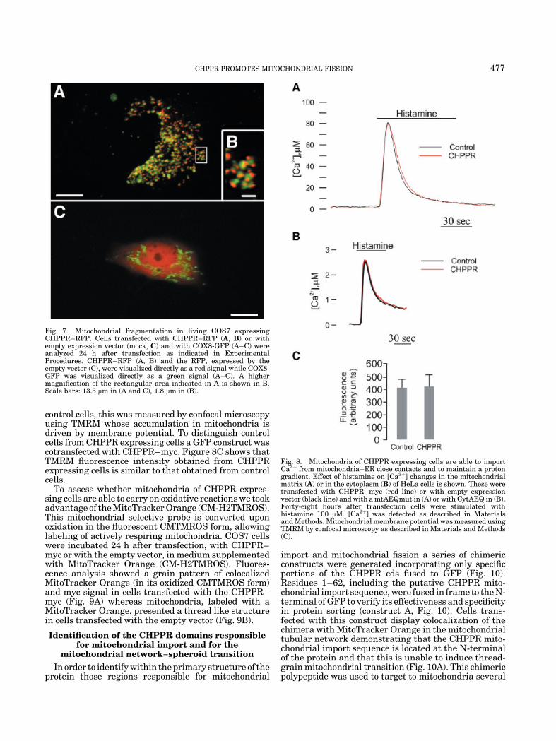
control cells, this was measured by confocal microscopyusing TMRM whose accumulation in mitochondria isdriven by membrane potential. To distinguish controlcells from CHPPR expressing cells a GFP construct wascotransfected with CHPPR–myc. Figure 8C shows thatTMRM fluorescence intensity obtained from CHPPRexpressing cells is similar to that obtained from controlcells.
To assess whether mitochondria of CHPPR expres-sing cells are able to carry on oxidative reactions we tookadvantage of the MitoTracker Orange (CM-H2TMROS).This mitochondrial selective probe is converted uponoxidation in the fluorescent CMTMROS form, allowinglabeling of actively respiring mitochondria. COS7 cellswere incubated 24 h after transfection, with CHPPR–myc or with the empty vector, in medium supplementedwith MitoTracker Orange (CM-H2TMROS). Fluores-cence analysis showed a grain pattern of colocalizedMitoTracker Orange (in its oxidized CMTMROS form)and myc signal in cells transfected with the CHPPR–myc (Fig. 9A) whereas mitochondria, labeled with aMitoTracker Orange, presented a thread like structurein cells transfected with the empty vector (Fig. 9B).
Identification of the CHPPR domains responsiblefor mitochondrial import and for the
mitochondrial network–spheroid transition
In order to identify within the primary structure of theprotein those regions responsible for mitochondrial
import and mitochondrial fission a series of chimericconstructs were generated incorporating only specificportions of the CHPPR cds fused to GFP (Fig. 10).Residues 1–62, including the putative CHPPR mito-chondrial import sequence, were fused in frame to the N-terminal of GFP to verify its effectiveness and specificityin protein sorting (construct A, Fig. 10). Cells trans-fected with this construct display colocalization of thechimera with MitoTracker Orange in the mitochondrialtubular network demonstrating that the CHPPR mito-chondrial import sequence is located at the N-terminalof the protein and that this is unable to induce thread-grain mitochondrial transition (Fig. 10A). This chimericpolypeptide was used to target to mitochondria several
Fig. 7. Mitochondrial fragmentation in living COS7 expressingCHPPR–RFP. Cells transfected with CHPPR–RFP (A, B) or withempty expression vector (mock, C) and with COX8-GFP (A–C) wereanalyzed 24 h after transfection as indicated in ExperimentalProcedures. CHPPR–RFP (A, B) and the RFP, expressed by theempty vector (C), were visualized directly as a red signal while COX8-GFP was visualized directly as a green signal (A–C). A highermagnification of the rectangular area indicated in A is shown in B.Scale bars: 13.5 mm in (A and C), 1.8 mm in (B).
Fig. 8. Mitochondria of CHPPR expressing cells are able to importCa2þ from mitochondria–ER close contacts and to maintain a protongradient. Effect of histamine on [Ca2þ] changes in the mitochondrialmatrix (A) or in the cytoplasm (B) of HeLa cells is shown. These weretransfected with CHPPR–myc (red line) or with empty expressionvector (black line) and with a mtAEQmut in (A) or with CytAEQ in (B).Forty-eight hours after transfection cells were stimulated withhistamine 100 mM. [Ca2þ] was detected as described in Materialsand Methods. Mitochondrial membrane potential was measured usingTMRM by confocal microscopy as described in Materials and Methods(C).
CHPPR PROMOTES MITOCHONDRIAL FISSION 477
portions of the CHPPR protein in order to explore theirability to induce fission (Fig. 10).
As a first step to locate the region responsible formitochondrial fragmentation, two constructs were gen-erated: construct B, bearing the amino terminal 1–188residues fused to the N-terminal of a GFP (Fig. 10) andconstruct C, coding for the 1–62 CHPPR–GFP fused tothe N terminal of the CHPPR region spanning aminoacid residues 182–321 (Fig. 10). Expression of constructB showed that although the chimera localized in
mitochondria it was unable to induce fission (Fig. 10B)whereas construct C was both localized in mitochondriaand able to induce a thread–grain transition (Fig. 10C).To verify whether this region, spanning amino acidresidues 182–321, required mitochondrial sorting to beeffective in fission, we generated the construct D:identical to construct C but lacking the mitochondrialimporting sequence (Fig. 10). This chimera was bothunable to localize in mitochondria, as expected, andunable to disrupt the mitochondrial tubular network(Fig. 10D). Within the region spanning amino acidresidues 182–321 we identified two subregions to befurther assayed for their fission ability: the first onecontaining the region spanning amino acid residues182–212, including the poly-proline region, and thesecond one spanning residues 206–321, C-terminal tothe poly-proline region (Fig. 10). Two distinct constructswere made fusing these two regions to the C-terminal of1–62 CHPPR–GFP: constructs E and F (Fig. 10).Although both chimerae were sorted to mitochondria,as shown by colocalization with Mito Tracker Orange,none of them was able to promote mitochondrial fission(Fig. 10E,F). A further construct generated fusing theregion spanning amino acid residues 251–321 to the C-terminal of 1–62 CHPPR–GFP was also unable toinduce a thread-grain transition (not shown). Fromthese experiments, we concluded first, that both thepoly-proline and the region located to its C-terminalside, are necessary to promote fission but not sufficient,if expressed separately, and second, that the fissiondomain begins with the poly-proline region at N-terminal and extends for some residues toward the C-terminal of CHPPR. In order to restrict the C-terminalboundary of this domain, two further constructs weremade, fusing to the C-terminal of 1–62 CHPPR–GFP,the regions spanning from residue 182 to residues 257(not shown) and 309, construct G (Fig. 10). Among thesetwo constructs, only construct G was able to inducemitochondrial fission (Fig. 10G). This result was alsoconfirmed by confocal microscopy (Fig. 11A–C). Takentogether these experiments demonstrated that theCHPPR domain necessary and sufficient to promote amitochondrial thread-grain transition is within resi-dues 182–309.
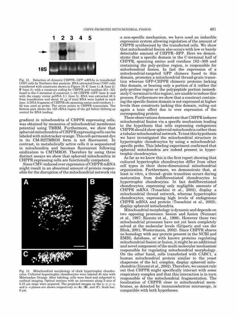
To verify whether high chimeric transcript levelscorrelated with fission ability of specific constructs, aNorthern blot was performed with RNA extracted fromtransfected COS7 cells. This analysis demonstrated thatendogenous CHPPR mRNA is undetectable in cellstransfected with the empty vector (Fig. 12). Northernanalysis also showed that the fission competent con-struct C (Fig. 10) expresses comparable, or even lowerlevel of chimeric mRNA, when compared to three con-structs incompetent for fission (Fig. 12). These fission
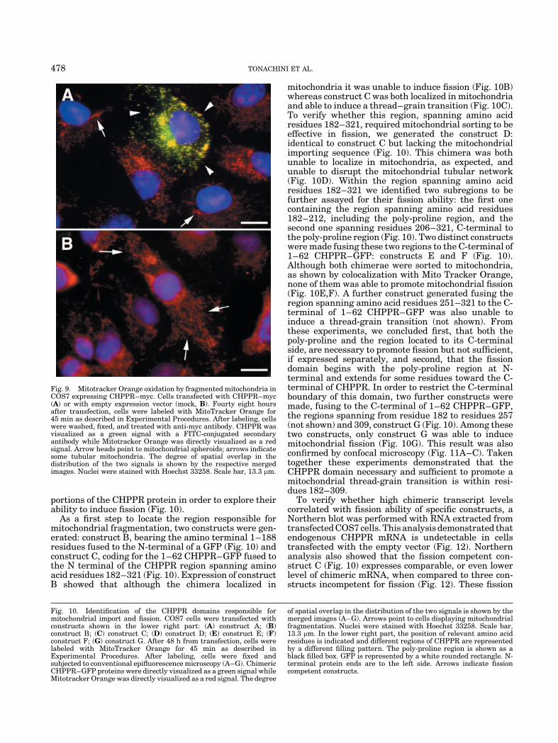
Fig. 9. Mitotracker Orange oxidation by fragmented mitochondria inCOS7 expressing CHPPR–myc. Cells transfected with CHPPR–myc(A) or with empty expression vector (mock, B). Fourty eight hoursafter transfection, cells were labeled with MitoTracker Orange for45 min as described in Experimental Procedures. After labeling, cellswere washed, fixed, and treated with anti-myc antibody. CHPPR wasvisualized as a green signal with a FITC-conjugated secondaryantibody while Mitotracker Orange was directly visualized as a redsignal. Arrow heads point to mitochondrial spheroids; arrows indicatesome tubular mitochondria. The degree of spatial overlap in thedistribution of the two signals is shown by the respective mergedimages. Nuclei were stained with Hoechst 33258. Scale bar, 13.3 mm.
Fig. 10. Identification of the CHPPR domains responsible formitochondrial import and fission. COS7 cells were transfected withconstructs shown in the lower right part: (A) construct A; (B)construct B; (C) construct C; (D) construct D; (E) construct E; (F)construct F; (G) construct G. After 48 h from transfection, cells werelabeled with MitoTracker Orange for 45 min as described inExperimental Procedures. After labeling, cells were fixed andsubjected to conventional epifluorescence microscopy (A–G). ChimericCHPPR–GFP proteins were directly visualized as a green signal whileMitotracker Orange was directly visualized as a red signal. The degree
of spatial overlap in the distribution of the two signals is shown by themerged images (A–G). Arrows point to cells displaying mitochondrialfragmentation. Nuclei were stained with Hoechst 33258. Scale bar,13.3 mm. In the lower right part, the position of relevant amino acidresidues is indicated and different regions of CHPPR are representedby a different filling pattern. The poly-proline region is shown as ablack filled box. GFP is represented by a white rounded rectangle. N-terminal protein ends are to the left side. Arrows indicate fissioncompetent constructs.
478 TONACHINI ET AL.
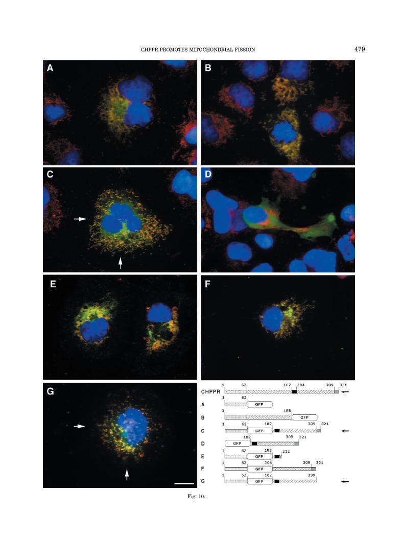
Fig. 10.
CHPPR PROMOTES MITOCHONDRIAL FISSION 479
incompetent constructs were: CHPPR 251–321 aminoacid residues fused to the C-terminal of 162 CHPPR–GFP (not shown) and constructs A and F (Fig. 10).
Three-dimensional mitochondrial organizationin hypertrophic chondrocytes
Since embryonic chick cartilage and hypertrophicchondrocytes express CHPPR (Tonachini et al., 2002),we expected to find spheroid mitochondria in these cells.Therefore we investigated the three-dimensional mito-chondrial structure in hypertrophic chondrocytes byusing MitoTracker Orange (CM-H2TMROS). Confocalimaging of these samples, showed that the fluorescentsignal generated by the oxidized form of MitoTracker(CMTMROS) was distributed in a grain pattern withinthese cells, indicating spheroid morphology of mitochon-dria (Fig. 13).
DISCUSSION
We have recently identified a chick cDNA encoding a321 amino acid protein referred to as chondrocyteprotein with a poly-proline region (CHPPR), which isexpressed in hypertrophic chondrocytes, in developinghypertrophic cartilage and to a lower extent in otherchick embryonic tissues (Tonachini et al., 2002). In thisstudy, we report that chick, mouse, and human CHPPRoverall share 50% sequence identity. A region ofCHPPR, spanning amino acid residues 1–62, fused tothe N-terminal of GFP is able to localize the chimericprotein in mitochondria demonstrating that this regioncontains a mitochondrial import sequence. Further-more, experiments of co-expression of CHPPR fused to amyc epitope (CHPPR–myc) and the mitochondrialmarker COX8-GFP (Rizzuto et al., 1995) demonstratethat the entire CHPPR is sorted to mitochondria notonly in dedifferentiated chick embryo chondrocytes butalso in other cell types such as COS7, HeLa, and 293Tcells. Since COS7 cells are much easier to obtain and totransfect, this cell line was used during most subsequentexperiments.
Immunoelectron microscopy shows that CHPPR ispresent in close proximity to the mitochondrial innerand, at a much lesser extent, close to the outer mem-branes regardless of the epitope used to tag the protein.In addition, these experiments show that labeledmitochondria do not display ultra-structural altera-tions. However, it should be noticed that labeling on themitochondrial surface might reveal proteins caughtduring the translocation process.
The most striking observation in CHPPR transfectedcells is the presence of spheroid mitochondria instead ofthe tubular mitochondrial network as detected in con-trol non-transfected cells. Furthermore as this mito-chondrial fission also occurs in chicken cells, transfectedwith chick CHPPR, a toxic effect due to the expression ofa species-unrelated protein is excluded. Since CHPPRmitochondrial localization and this change in the three-dimensional mitochondrial organization are also ob-served in living cells, we rule out the possibility thatthese phenomena are due to a fixation artifact.
To verify whether CHPPR expression and the orga-nelle spheroidal shape are compatible with normalmitochondrial activities we have investigated the abilityof transfected cells to generate a proton gradient and tooxidize an appropriate substrate. Since it has beenshown that after histamine stimulation and in thepresence of a proton gradient, ER released Ca2þ istransported in the mitochondrial matrix (Rizzuto et al.,1998) we measured [Ca2þ] modulation in this compart-ment of CHPPR transfected cells. This experimentshows that the cell behavior was not significantlydifferent from control cells transfected with an emptyvector. Therefore, CHPPR expression does not affectproton gradient and Ca2þ transport in the mitochondrialmatrix and close ER–mitochondrial contacts areretained when spheroid mitochondria are present inthe cell instead of the thread network. These contactswere shown to be determinants of mitochondrial Ca2þ
responses (Lawrie et al., 1996). An independent anddirect evidence confirming the existence of a proton
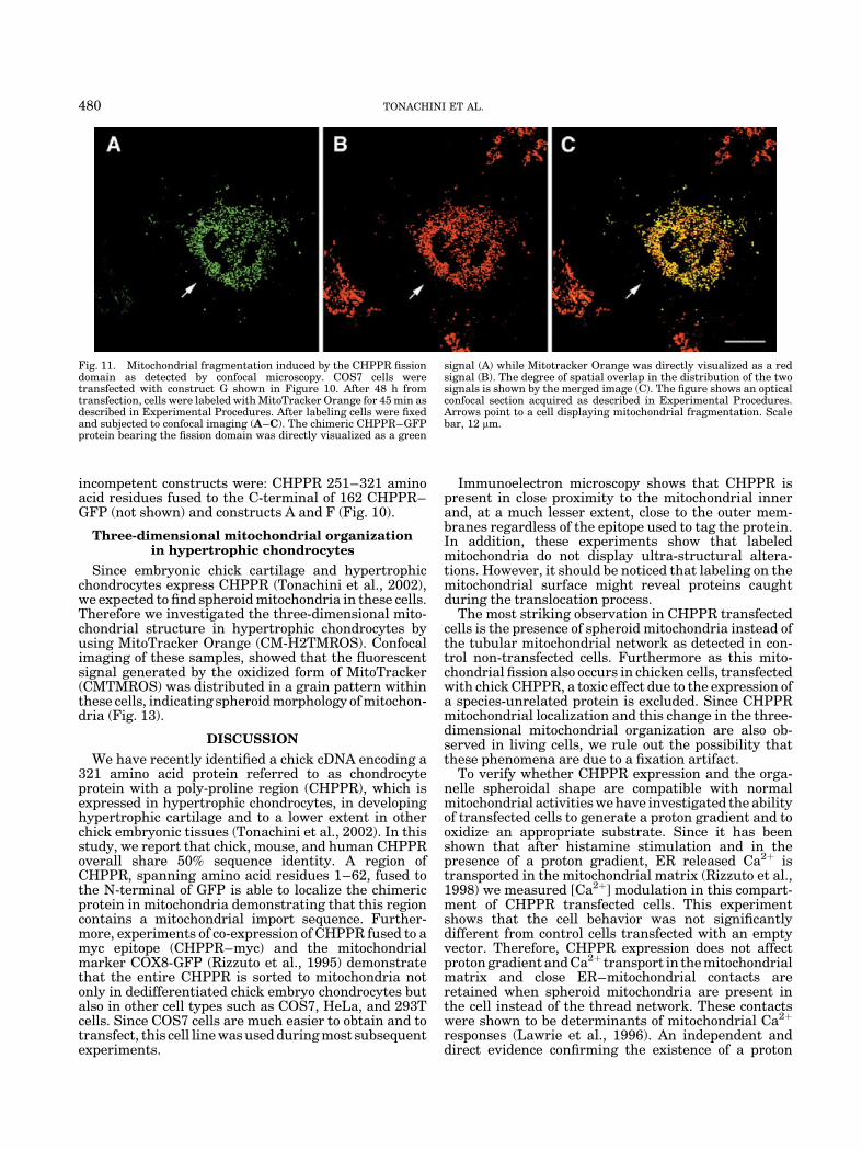
Fig. 11. Mitochondrial fragmentation induced by the CHPPR fissiondomain as detected by confocal microscopy. COS7 cells weretransfected with construct G shown in Figure 10. After 48 h fromtransfection, cells were labeled with MitoTracker Orange for 45 min asdescribed in Experimental Procedures. After labeling cells were fixedand subjected to confocal imaging (A–C). The chimeric CHPPR–GFPprotein bearing the fission domain was directly visualized as a green
signal (A) while Mitotracker Orange was directly visualized as a redsignal (B). The degree of spatial overlap in the distribution of the twosignals is shown by the merged image (C). The figure shows an opticalconfocal section acquired as described in Experimental Procedures.Arrows point to a cell displaying mitochondrial fragmentation. Scalebar, 12 mm.
480 TONACHINI ET AL.
gradient in mitochondria of CHPPR expressing cells,was obtained by measures of mitochondrial membranepotential using TMRM. Furthermore, we show thatspheroid mitochondria of CHPPR expressing cells can belabeled with mitotracker orange. This cell-permeant dyein its CM-H2TMROS form is not fluorescent. Incontrast, in metabolically active cells it is sequesteredin mitochondria and becomes fluorescent followingoxidization to CMTMROS. Therefore by using threedistinct assays we show that spheroid mitochondria inCHPPR expressing cells are functionally competent.
Since CMV-induced over expression of CHPPR mRNAmight result in an abnormal amount of protein respon-sible for the disruption of the mitochondrial network via
a non-specific mechanism, we have used an inducibleexpression system allowing regulation of the amount ofCHPPR synthesized by the transfected cells. We showthat mitochondrial fission also occurs with low or barelydetectable amount of CHPPR–RFP. Here we demon-strate that a specific domain in the C-terminal half ofCHPPR, spanning amino acid residues 182–309 andcontaining the poly-proline region, is responsible formitochondrial fission. In fact the expression of amitochondrial-targeted GFP chimera fused to thisdomain, promotes a mitochondrial thread-grain transi-tion whereas GFP-CHPPR chimeric proteins lackingthis domain, or bearing only a portion of it (either thepoly-proline region or the polypeptide portion immedi-ately C-terminal to this region), are unable to induce thisprocess. Furthermore we show that a construct contain-ing the specific fission domain is not expressed at higherlevels than constructs lacking this domain, ruling outagain a toxic effect due to over expression of thecorresponding protein.
These observations demonstrate that CHPPR inducesmitochondrial fission via a specific mechanism leadingto the hypothesis that cells expressing endogenousCHPPR should show spheroid mitochondria rather thana tubular mitochondrial network. To test this hypothesiswe have investigated the mitochondrial structure inhypertrophic chondrocytes by using a mitochondrial-specific probe. This labeling experiment confirmed thatspheroid mitochondria are indeed present in hyper-trophic chondrocytes.
As far as we know this is the first report showing thatcultured hypertrophic chondrocytes differ from othercell types in their three-dimensional mitochondrialorganization. Furthermore, we demonstrate that, atleast in vitro, a thread–grain transition occurs duringmaturation from dedifferentiated chondrocytes tohypertrophic chondrocytes. In fact dedifferentiatedchondrocytes, expressing only negligible amounts ofCHPPR mRNA (Tonachini et al., 2002), display amitochondrial thread network, whereas hypertrophicchondrocytes, expressing high levels of endogenousCHPPR mRNA and protein (Tonachini et al., 2002),display spheroid mitochondria.
Mitochondrial morphology is dynamic and depends ontwo opposing processes: fission and fusion (Nunnariet al., 1997; Rizzuto et al., 1998). However these twomitochondrial processes have not yet been completelysolved at the molecular levels (Griparic and van derBliek, 2001; Westermann, 2002). Since CHPPR sharesno homology with any protein present in the NCBI andEMBL database, or with known proteins regulatingmitochondrial fission or fusion, it might be an additionaland novel component of the multi molecular mechanismresponsible for regulating mitochondrial morphology.On the other hand, cells transfected with CABC1, ahuman mitochondrial protein similar to the yeastchaperone of the bc1 complex, display spheroid mito-chondria (Iiizumi et al., 2002). Therefore, we cannot ruleout that CHPPR might specifically interact with somerespiratory complex and that this interaction is in turnresponsible of the mitochondrial fragmentation. Thelocalization of CHPPR close to mitochondrial mem-branes, as detected by immunoelectron microscopy, iscompatible with both hypotheses.
Fig. 12. Detection of chimeric CHPPR–GFP mRNAs in transfectedCOS7 cells by Northern blot analysis. RNA extracted from COS7 cellstransfected with constructs shown in Figure 10: C (lane 1); A (lane 2);F (lane 3); with a construct coding for CHPPR acid residues 251–321fused to the C-terminal of construct 1–62 CHPPR–GFP (lane 4) andwith the empty vector pcDNA 3.1 (lane 5). RNA was extracted 48 hfrom transfection and about 10 mg of total RNA were loaded in eachlane. A DNA fragment of CHPPR cds spanning amino acid residues 1–62 was used as probe. The arrow points to CHPPR transcripts. TheBottom part shows the 18S rRNA region of the gel before blotting ascontrol for RNA loading.
Fig. 13. Mitochondrial morphology of chick hypertrophic chondro-cytes. Cultured hypertrophic chondrocytes were labeled 45 min withMitotracker Orange. After labeling, cells were fixed and subjected toconfocal imaging. Optical sections with an increment along Z-axis of0.15 mm steps were acquired. The projected images on the x–y; y–z,and x–z planes are shown respectively in (A), (B), and (C). Scale bar,6 mm.
CHPPR PROMOTES MITOCHONDRIAL FISSION 481
Lack of homology of CHPPR with Yeast and Droso-phila proteins suggests that CHPPR rather than havinga key function in the mitochondrial fission machinerymight play a facilitative/regulative role or that itsfunction is carried out by other non-vertebrate proteinsthat might share only a structural homology. It shouldbe pointed out that the only module/motif that we havebeen able to identify in CHPPR, using availableinformatics tools, is the poly-proline region includedin the fission domain and necessary for its function.Poly-proline regions have been demonstrated to beresponsible for establishing protein–protein interaction(Williamson, 1994; Kay et al., 2000). Therefore, if afunctional non-vertebrate CHPPR homologue exists, itis likely to share this module.
In conclusion, our study shows that CHPPR is amitochondrial protein containing a specific domain ableto induce, albeit by an unknown mechanism, mitochon-drial fission without loss of functional activity of theseorganelles.
ACKNOWLEDGMENTS
We thank Laura Amabile for technical assistance,Marina Fabbi, and Annemiek Beverdam for criticalreading of the manuscript and constructive discussions.
LITERATURE CITED
Altschul SF, Gish W, et al. 1990. Basic local alignment search tool.J Mol Biol 215:403–410.
Castagnola P, Dozin B, et al. 1988. Changes in the expression ofcollagen genes show two stages in chondrocyte differentiationin vitro. J Cell Biol 106:461–467.
Chiesa A, Rapizzi E, et al. 2001. Recombinant aequorin and greenfluorescent protein as valuable tools in the study of cell signaling.Biochem J 355:1–12.
Claros MG, Vincens P. 1996. Computational method to predictmitochondrially imported proteins and their targeting sequences.Eur J Biochem 241:779–786.
Evan GI, Lewis GK, et al. 1985. Isolation of monoclonal antibodiesspecific for human c-myc proto-oncogene product. Mol Cell Biol 5:3610–3616.
Griparic L, van der Bliek AM. 2001. The many shapes of mitochondrialmembranes. Traffic 2:235–244.
Iiizumi M, Hirofumi A, et al. 2002. Isolation of a novel gene, CABC1,encoding a mitochondrial protein that is highly homologous to yeastactivity of bc1 complex. Cancer Res 62:1246–1250.
Kay BK, Williamson MP, et al. 2000. The importance of being proline:The interaction of proline-rich motifs in signaling proteins withtheir cognate domains. Faseb J 14:231–241.
Lawrie AM, Rizzuto R, et al. 1996. A role for calcium influx in theregulation of mitochondrial calcium in endothelial cells. J BiolChem 271:10753–10759.
Montero M, Alonso MT, et al. 2000. Chromaffin-cell stimulationtriggers fast millimolar mitochondrial C2þ transients that modulatesecretion. Nat Cell Biol 2:57–61.
Nomura N, Miyajima N, et al. 1994. Prediction of the coding sequencesof unidentified human genes. I. The coding sequences of 40 newgenes (KIAA0001–KIAA0040) deduced by analysis of randomlysampled cDNA clones from human immature myeloid cell line KG-1.DNA Res 1:27–35.
Nunnari J, Marshall WF, et al. 1997. Mitochondrial transmissionduring mating in Saccaromyces cerevisiae is determined bymitochondrial fusion and fission and the intramitochondrialsegregation of mitochondrial DNA. Mol Biol Cell 8:1233–1242.
Quarto R, Campanile G, et al. 1997. Modulation of commitment,proliferation, and differentiation of chondrogenic cells in definedculture medium. Endocrinology 138:4966–4976.
Rizzuto R, Brini M, et al. 1995. Chimeric green fluorescent protein as atool for visualizing subcellular organelles in living cells. Curr Biol5:635–642.
Rizzuto R, Pinton P, et al. 1998. Close contacts with the endoplasmicreticulum as determinants of mitochondrial Ca2þ responses. Science280:1763–1766.
Schiaffino MV, d’Addio M, et al. 1999. Ocular albinism: Evidence for adefect in an intracellular signal transduction system. Nat Genet23:108–112.
Slot JW, Geuze HJ. 1984. Gold markers for single and doubleimmunolabeling of ultrathin cryosections. In: Polak JM, VarndellIM, editors. Immunolabeling for electron microscopy. Amsterdam,NL: Elsevier. pp 129–142.
Tonachini L, Monticone M, et al. 2002. Chondrocyte protein with apoly-proline region (CHPPR) is a novel protein expressed bychondrocytes in vitro and in vivo. Biochim Biophys Acta 1577:421–429.
Westermann B. 2002. Merging mitochondria matters: Cellular roleand molecular machinery of mitochondral fusion. EMBO Rep 3:527–531.
Williamson MP. 1994. The structure and function of proline-richregions in proteins. Biochem J 297:249–260.
482 TONACHINI ET AL.