Embed Size (px)
DESCRIPTION
Chemical Coordination in Humans
Citation preview
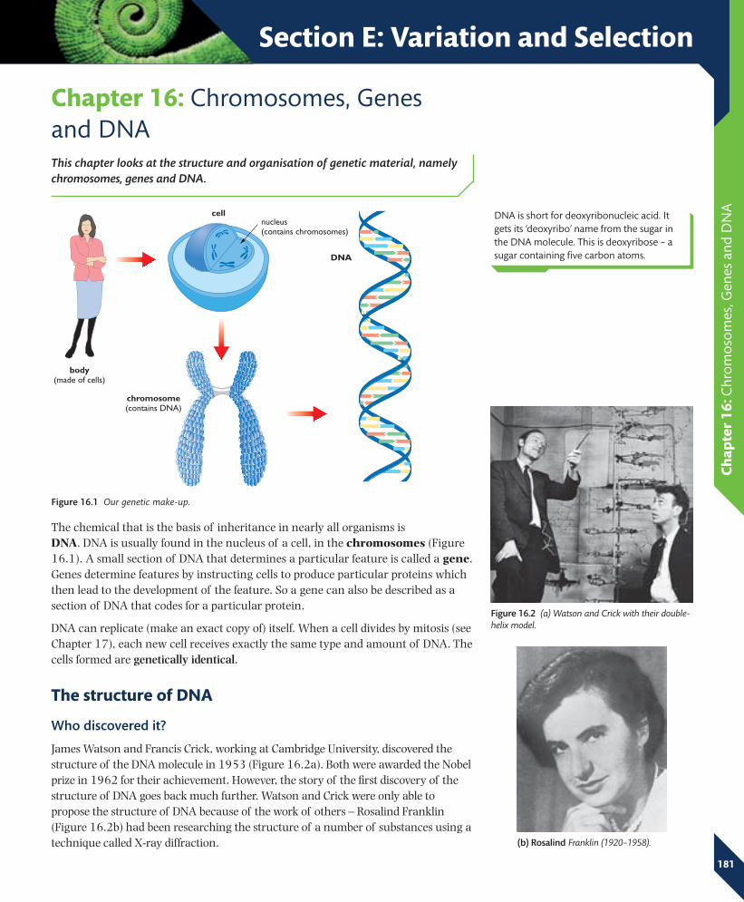
Fig_1601_A
body(made of cells)
cell
chromosome (contains DNA)
DNA
nucleus(contains chromosomes)
Section E: Variation and Selection
Ch
apte
r 16
: Chr
om
oso
mes
, Gen
es a
nd D
NA
Chapter 16: Chromosomes, Genes and DNAThis chapter looks at the structure and organisation of genetic material, namely chromosomes, genes and DNA.
The chemical that is the basis of inheritance in nearly all organisms is DNA. DNA is usually found in the nucleus of a cell, in the chromosomes (Figure 16.1). A small section of DNA that determines a particular feature is called a gene. Genes determine features by instructing cells to produce particular proteins which then lead to the development of the feature. So a gene can also be described as a section of DNA that codes for a particular protein.
DNA can replicate (make an exact copy of) itself. When a cell divides by mitosis (see Chapter 17), each new cell receives exactly the same type and amount of DNA. The cells formed are genetically identical.
The structure of DNA
Who discovered it?
James Watson and Francis Crick, working at Cambridge University, discovered the structure of the DNA molecule in 1953 (Figure 16.2a). Both were awarded the Nobel prize in 1962 for their achievement. However, the story of the first discovery of the structure of DNA goes back much further. Watson and Crick were only able to propose the structure of DNA because of the work of others – Rosalind Franklin (Figure 16.2b) had been researching the structure of a number of substances using a technique called X-ray diffraction.
DNA is short for deoxyribonucleic acid. It gets its ‘deoxyribo’ name from the sugar in the DNA molecule. This is deoxyribose – a sugar containing five carbon atoms.
Figure 16.2 (a) Watson and Crick with their double-helix model.
(b) Rosalind Franklin (1920–1958).
Figure 16.1 Our genetic make-up.
181
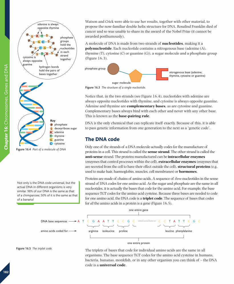
Fig_1604_A
phosphatedeoxyribose sugar
Key
guaninecytosine
adeninethymine
P
AT
S
GC
phosphate groups hold the nucleotides in each strand together
hydrogen bonds hold the pairs of bases together
cytosine is always opposite guanine
adenine is always opposite thymine
P
P
P
P
P
P
P
P
A
A
T
T
S
S
S
S
S
S
S
S
G
G
C
C
one entire gene
A T C G A A T T C C G C C C C C C C T A T T C G C
arginine
DNA base sequences
amino acids coded for isoleucine proline leucine phenylalanine
one entire protein
Fig_1605_A
Fig_1603_A
nitrogenous base (adenine, thymine, cytosine or guanine)
sugar molecule
phosphate group
Ch
apte
r 16
: Chr
om
oso
mes
, Gen
es a
nd D
NA
182
Watson and Crick were able to use her results, together with other material, to propose the now-familiar double helix structure for DNA. Rosalind Franklin died of cancer and so was unable to share in the award of the Nobel Prize (it cannot be awarded posthumously).
A molecule of DNA is made from two strands of nucleotides, making it a polynucleotide. Each nucleotide contains a nitrogenous base (adenine (A), thymine (T), cytosine (C) or guanine (G)), a sugar molecule and a phosphate group (Figure 16.3).
Notice that, in the two strands (see Figure 16.4), nucleotides with adenine are always opposite nucleotides with thymine, and cytosine is always opposite guanine. Adenine and thymine are complementary bases, as are cytosine and guanine. Complementary bases always bind with each other and never with any other base. This is known as the base-pairing rule.
DNA is the only chemical that can replicate itself exactly. Because of this, it is able to pass genetic information from one generation to the next as a ‘genetic code’.
The DNA codeOnly one of the strands of a DNA molecule actually codes for the manufacture of proteins in a cell. This strand is called the sense strand. The other strand is called the anti-sense strand. The proteins manufactured can be intracellular enzymes (enzymes that control processes within the cell), extracellular enzymes (enzymes that are secreted from the cell to have their effect outside the cell), structural proteins (e.g. used to make hair, haemoglobin, muscles, cell membranes) or hormones.
Proteins are made of chains of amino acids. A sequence of three nucleotides in the sense strand of DNA codes for one amino acid. As the sugar and phosphate are the same in all nucleotides, it is actually the bases that code for the amino acid. For example, the base sequence TGT codes for the amino acid cysteine. Because three bases are needed to code for one amino acid, the DNA code is a triplet code. The sequence of bases that codes for all the amino acids in a protein is a gene (Figure 16.5).
Figure 16.3 The structure of a single nucleotide.
Figure 16.4 Part of a molecule of DNA
Not only is the DNA code universal, but the actual DNA in different organisms is very similar. 98% of our DNA is the same as that of a chimpanzee; 50% of it is the same as that of a banana!
Figure 16.5 The triplet code. The triplets of bases that code for individual amino acids are the same in all organisms. The base sequence TGT codes for the amino acid cysteine in humans, bacteria, bananas, monkfish, or in any other organism you can think of – the DNA code is a universal code.
T
A
G
C
T
A
C
A
T
G
A
T
C
A
T
G
A
T
T
A
C
A
T
G
T
A
G
C
T
A
A
T
G
A
T
C
T
A
C
A
T
G
A
T
G
A
T
C
T
A
C
A
T
G
A
T
G
A
T
C
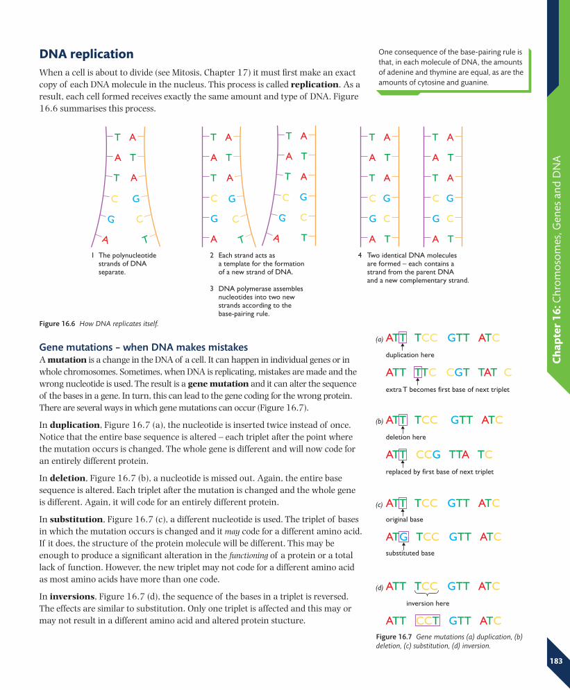
The polynucleotidestrands of DNAseparate.
Each strand acts asa template for the formationof a new strand of DNA.
DNA polymerase assemblesnucleotides into two newstrands according to thebase-pairing rule.
Two identical DNA moleculesare formed – each contains astrand from the parent DNAand a new complementary strand.
1 2 4
3
Fig_1607_A
Fig_1608_A
ATT TCC GTT ATCoriginal base
ATG TCC GTT ATCsubstituted base
ATT TCC GTT ATCdeletion here
ATT CCG TTA TCreplaced by first base of next triplet
ATT TCC GTT ATCduplication here
ATT TTC CGT TAT Cextra T becomes first base of next triplet
ATT TCC GTT ATC inversion here
ATT CCT GTT ATC
(a)
(b)
(c)
(d)
Ch
apte
r 16
: Chr
om
oso
mes
, Gen
es a
nd D
NA
183
DNA replicationWhen a cell is about to divide (see Mitosis, Chapter 17) it must first make an exact copy of each DNA molecule in the nucleus. This process is called replication. As a result, each cell formed receives exactly the same amount and type of DNA. Figure 16.6 summarises this process.
Gene mutations – when DNA makes mistakes A mutation is a change in the DNA of a cell. It can happen in individual genes or in whole chromosomes. Sometimes, when DNA is replicating, mistakes are made and the wrong nucleotide is used. The result is a gene mutation and it can alter the sequence of the bases in a gene. In turn, this can lead to the gene coding for the wrong protein. There are several ways in which gene mutations can occur (Figure 16.7).
In duplication, Figure 16.7 (a), the nucleotide is inserted twice instead of once. Notice that the entire base sequence is altered – each triplet after the point where the mutation occurs is changed. The whole gene is different and will now code for an entirely different protein.
In deletion, Figure 16.7 (b), a nucleotide is missed out. Again, the entire base sequence is altered. Each triplet after the mutation is changed and the whole gene is different. Again, it will code for an entirely different protein.
In substitution, Figure 16.7 (c), a different nucleotide is used. The triplet of bases in which the mutation occurs is changed and it may code for a different amino acid. If it does, the structure of the protein molecule will be different. This may be enough to produce a significant alteration in the functioning of a protein or a total lack of function. However, the new triplet may not code for a different amino acid as most amino acids have more than one code.
In inversions, Figure 16.7 (d), the sequence of the bases in a triplet is reversed. The effects are similar to substitution. Only one triplet is affected and this may or may not result in a different amino acid and altered protein stucture.
One consequence of the base-pairing rule is that, in each molecule of DNA, the amounts of adenine and thymine are equal, as are the amounts of cytosine and guanine.
Figure 16.6 How DNA replicates itself.
Figure 16.7 Gene mutations (a) duplication, (b) deletion, (c) substitution, (d) inversion.
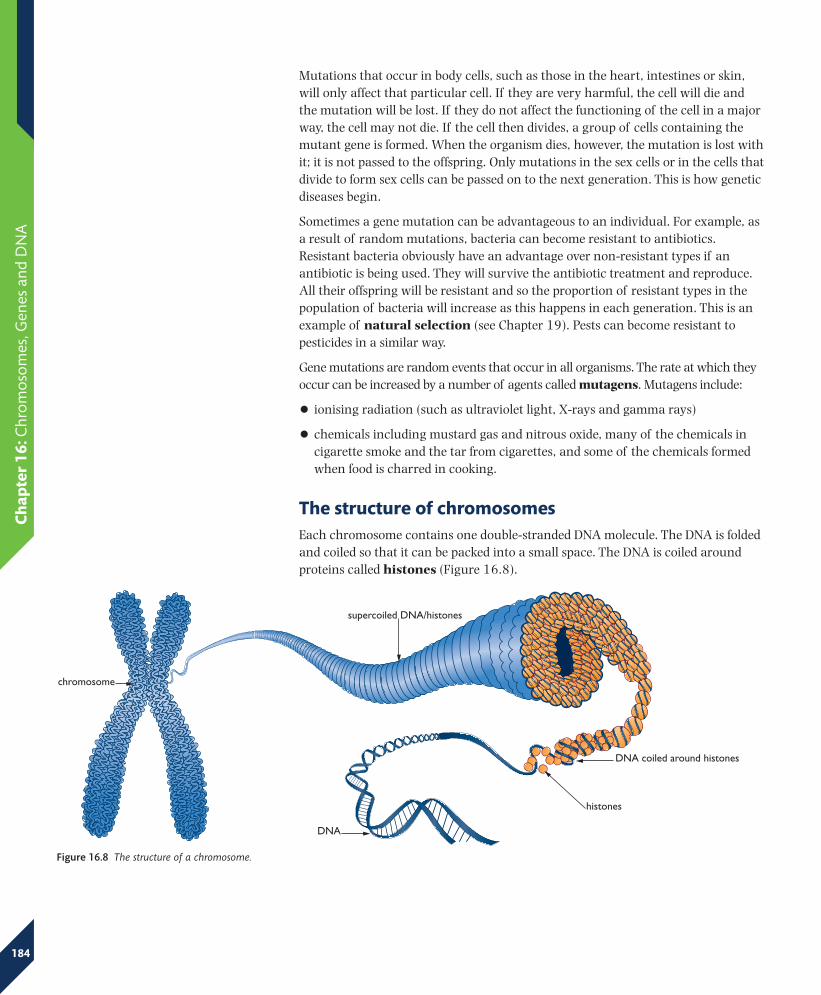
Fig_1610_A
chromosome
supercoiled DNA/histones
DNA coiled around histones
histones
DNA
Ch
apte
r 16
: Chr
om
oso
mes
, Gen
es a
nd D
NA
184
Mutations that occur in body cells, such as those in the heart, intestines or skin, will only affect that particular cell. If they are very harmful, the cell will die and the mutation will be lost. If they do not affect the functioning of the cell in a major way, the cell may not die. If the cell then divides, a group of cells containing the mutant gene is formed. When the organism dies, however, the mutation is lost with it; it is not passed to the offspring. Only mutations in the sex cells or in the cells that divide to form sex cells can be passed on to the next generation. This is how genetic diseases begin.
Sometimes a gene mutation can be advantageous to an individual. For example, as a result of random mutations, bacteria can become resistant to antibiotics. Resistant bacteria obviously have an advantage over non-resistant types if an antibiotic is being used. They will survive the antibiotic treatment and reproduce. All their offspring will be resistant and so the proportion of resistant types in the population of bacteria will increase as this happens in each generation. This is an example of natural selection (see Chapter 19). Pests can become resistant to pesticides in a similar way.
Gene mutations are random events that occur in all organisms. The rate at which they occur can be increased by a number of agents called mutagens. Mutagens include:
• ionising radiation (such as ultraviolet light, X-rays and gamma rays)
• chemicals including mustard gas and nitrous oxide, many of the chemicals in cigarette smoke and the tar from cigarettes, and some of the chemicals formed when food is charred in cooking.
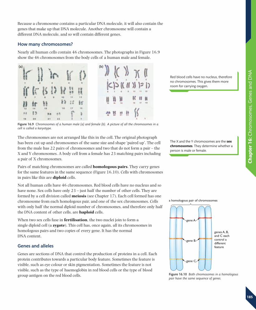
The structure of chromosomes Each chromosome contains one double-stranded DNA molecule. The DNA is folded and coiled so that it can be packed into a small space. The DNA is coiled around proteins called histones (Figure 16.8).
Figure 16.8 The structure of a chromosome.
genes A, B,and C eachcontrol adifferentfeature
Fig_1612_A
gene A
gene B
gene C
a homologous pair of chromosomes
(a) (b)
Ch
apte
r 16
: Chr
om
oso
mes
, Gen
es a
nd D
NA
185
Because a chromosome contains a particular DNA molecule, it will also contain the genes that make up that DNA molecule. Another chromosome will contain a different DNA molecule, and so will contain different genes.
How many chromosomes?
Nearly all human cells contain 46 chromosomes. The photographs in Figure 16.9 show the 46 chromosomes from the body cells of a human male and female.
The chromosomes are not arranged like this in the cell. The original photograph has been cut up and chromosomes of the same size and shape ‘paired up’. The cell from the male has 22 pairs of chromosomes and two that do not form a pair – the X and Y chromosomes. A body cell from a female has 23 matching pairs including a pair of X chromosomes.
Pairs of matching chromosomes are called homologous pairs. They carry genes for the same features in the same sequence (Figure 16.10). Cells with chromosomes in pairs like this are diploid cells.
Not all human cells have 46 chromosomes. Red blood cells have no nucleus and so have none. Sex cells have only 23 – just half the number of other cells. They are formed by a cell division called meiosis (see Chapter 17). Each cell formed has one chromosome from each homologous pair, and one of the sex chromosomes. Cells with only half the normal diploid number of chromosomes, and therefore only half the DNA content of other cells, are haploid cells.
When two sex cells fuse in fertilisation, the two nuclei join to form a single diploid cell (a zygote). This cell has, once again, all its chromosomes in homologous pairs and two copies of every gene. It has the normal DNA content.
Genes and alleles
Genes are sections of DNA that control the production of proteins in a cell. Each protein contributes towards a particular body feature. Sometimes the feature is visible, such as eye colour or skin pigmentation. Sometimes the feature is not visible, such as the type of haemoglobin in red blood cells or the type of blood group antigen on the red blood cells.
Figure 16.9 Chromosomes of a human male (a) and female (b). A picture of all the chromosomes in a cell is called a karyotype.
Red blood cells have no nucleus, therefore no chromosomes. This gives them more room for carrying oxygen.
The X and the Y chromosomes are the sex chromosomes. They determine whether a person is male or female.
Figure 16.10 Both chromosomes in a homologous pair have the same sequence of genes.
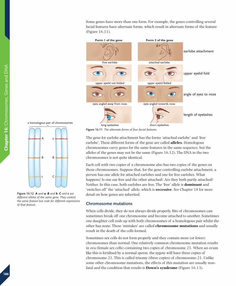
Fig_1614_A
A
B
c
a
B
C
a homologous pair of chromosomes
earlobe attachment
upper eyelid fold
angle of eyes to nose
length of eyelashes
free earlobe
eyes angled away from nose eyes angled towards nose
long eyelashes short eyelashes
attached earlobe
upper eyelid not folded upper eyelid folded
Fig_1613_A
Form 1 of the gene Form 2 of the gene
Ch
apte
r 16
: Chr
om
oso
mes
, Gen
es a
nd D
NA
186
Some genes have more than one form. For example, the genes controlling several facial features have alternate forms, which result in alternate forms of the feature (Figure 16.11).
Figure 16.12 A and a, B and b, C and c are different alleles of the same gene. They control the same feature but code for different expressions of that feature.
The gene for earlobe attachment has the forms ‘attached earlobe’ and ‘free earlobe’. These different forms of the gene are called alleles. Homologous chromosomes carry genes for the same features in the same sequence, but the alleles of the genes may not be the same (Figure 16.12). The DNA in the two chromosomes is not quite identical.
Each cell with two copies of a chromosome also has two copies of the genes on those chromosomes. Suppose that, for the gene controlling earlobe attachment, a person has one allele for attached earlobes and one for free earlobes. What happens? Is one ear free and the other attached? Are they both partly attached? Neither. In this case, both earlobes are free. The ‘free’ allele is dominant and ‘switches off ’ the ‘attached’ allele, which is recessive. See Chapter 18 for more detail on how genes are inherited.
Chromosome mutations
When cells divide, they do not always divide properly. Bits of chromosomes can sometimes break off one chromosome and become attached to another. Sometimes one daughter cell ends up with both chromosomes of a homologous pair whilst the other has none. These ‘mistakes’ are called chromosome mutations and usually result in the death of the cells formed.
Sometimes sex cells do not form properly and they contain more (or fewer) chromosomes than normal. One relatively common chromosome mutation results in ova (female sex cells) containing two copies of chromosome 21. When an ovum like this is fertilised by a normal sperm, the zygote will have three copies of chromosome 21. This is called trisomy (three copies) of chromosome 21. Unlike some other chromosome mutations, the effects of this mutation are usually non-fatal and the condition that results is Down’s syndrome (Figure 16.13).
Figure 16.11 The alternate forms of four facial features.
Ch
apte
r 16
: Chr
om
oso
mes
, Gen
es a
nd D
NA
187
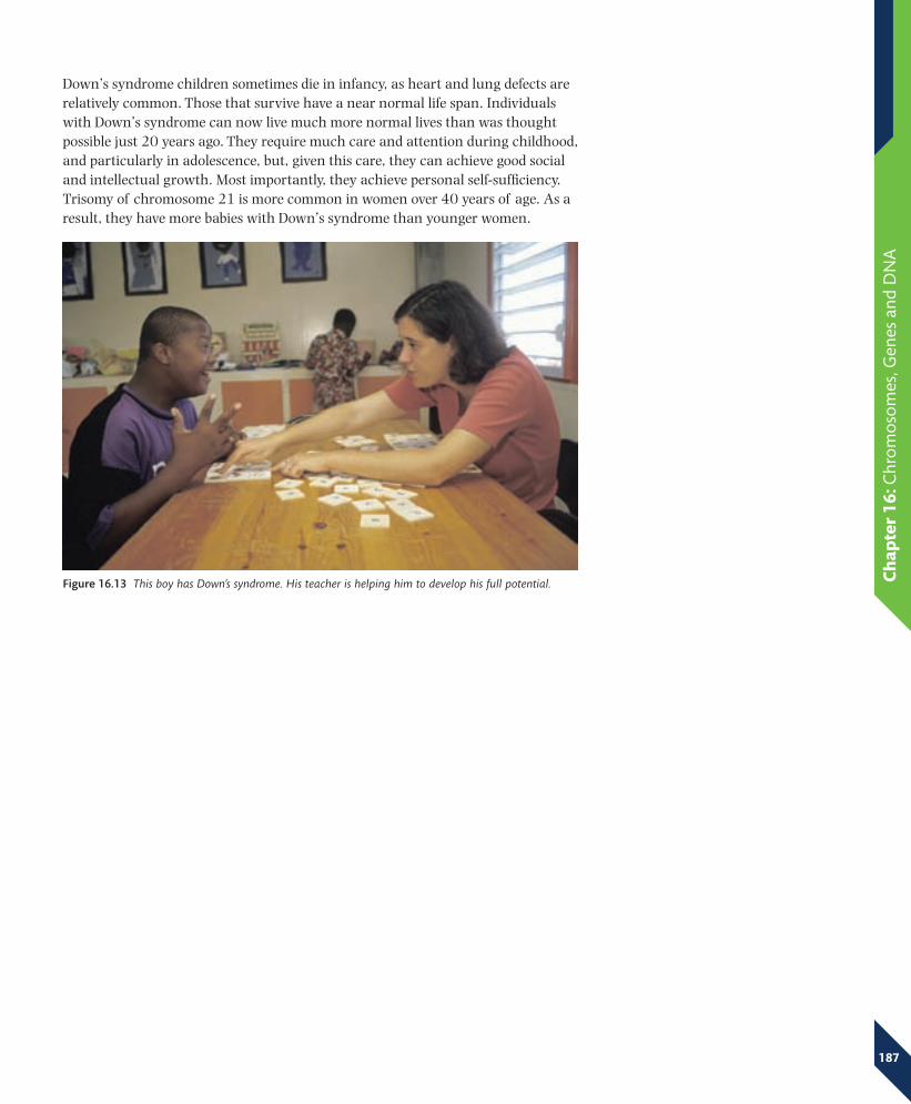
Down’s syndrome children sometimes die in infancy, as heart and lung defects are relatively common. Those that survive have a near normal life span. Individuals with Down’s syndrome can now live much more normal lives than was thought possible just 20 years ago. They require much care and attention during childhood, and particularly in adolescence, but, given this care, they can achieve good social and intellectual growth. Most importantly, they achieve personal self-sufficiency. Trisomy of chromosome 21 is more common in women over 40 years of age. As a result, they have more babies with Down’s syndrome than younger women.
Figure 16.13 This boy has Down’s syndrome. His teacher is helping him to develop his full potential.
Fig_1616_A
A
G
T
T
C
A B C DE
Ch
apte
r 16
: Chr
om
oso
mes
, Gen
es a
nd D
NA
188
End of Chapter Checklist
You should now be able to:
recall that the nucleus of a cell contains chromosomes on which genes are located✓✓
understand that a gene is a section of a molecule of DNA✓✓
describe the structure of a DNA molecule✓✓
understand, in outline, how DNA acts as a genetic code✓✓
understand, in outline, how DNA is replicated✓✓
recall that in human cells the diploid number of chromosomes is 46 and the haploid number is 23✓✓
understand the meaning of alleles of a gene (see also Chapter 18)✓✓
recall that mutation is a rare, random change in genetic material that can be inherited✓✓
understand that many mutations are harmful but some are neutral and a few are beneficial (see ✓✓also Chapter 19)
understand how resistance to antibiotics can increase in bacterial populations✓✓
understand that the incidence of mutations can be increased by mutagens such as ionising ✓✓radiation and some chemicals.
2 a) What is:
i) a gene
ii) an allele?
b) Describe the structure of a chromosome.
c) How are the chromosomes in a woman’s skin cells:
i) similar to
ii) different from those in a man’s skin cells?
3 DNA is the only molecule capable of replicating itself. Sometimes mutations occur during replication.
a) Describe how DNA replicates itself.
b) Explain how a single gene mutation can lead to the formation of a protein in which:
i) many of the amino acids are different from those coded for by the non-mutated gene
ii) only one amino acid is different from those coded for by the non-mutated gene.
QuestionsMore questions on DNA can be found at the end of Section E on page 226.
1 The diagram represents part of a molecule of DNA.
a) Name the parts labelled A, B, C, D and E.
b) What parts did James Watson, Frances Crick and Rosalind Franklin play in discovering the structure of DNA?
c) Use the diagram to explain the base-pairing rule.
Fig_DQ_03_A
20 30 40 500
100
200
0.01
0.02
300
Key number of Down’s syndrome babies born Down’s syndrome births as a percentage of all births
num
ber
of D
own’
s sy
ndro
me
babi
es b
orn
per
year
Dow
n’s
synd
rom
e bi
rths
as a
per
cent
age
of a
ll bi
rths
mother’s age (years)
Ch
apte
r 16
: Chr
om
oso
mes
, Gen
es a
nd D
NA
189
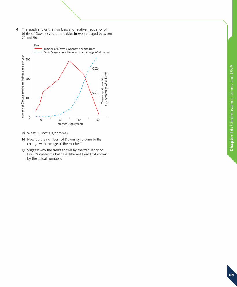
4 The graph shows the numbers and relative frequency of births of Down’s syndrome babies in women aged between 20 and 50.
a) What is Down’s syndrome?
b) How do the numbers of Down’s syndrome births change with the age of the mother?
c) Suggest why the trend shown by the frequency of Down’s syndrome births is different from that shown by the actual numbers.