Embed Size (px)
Citation preview
Isolation and characterization of
exosomes from bovine milk
Master’s Thesis
University of Turku
Department of Biochemistry
Biochemistry
11/19
Santeri Kankaanpää
The originality of this thesis has been checked in accordance with the University of
Turku quality assurance system using the Turnitin OriginalityCheck service.
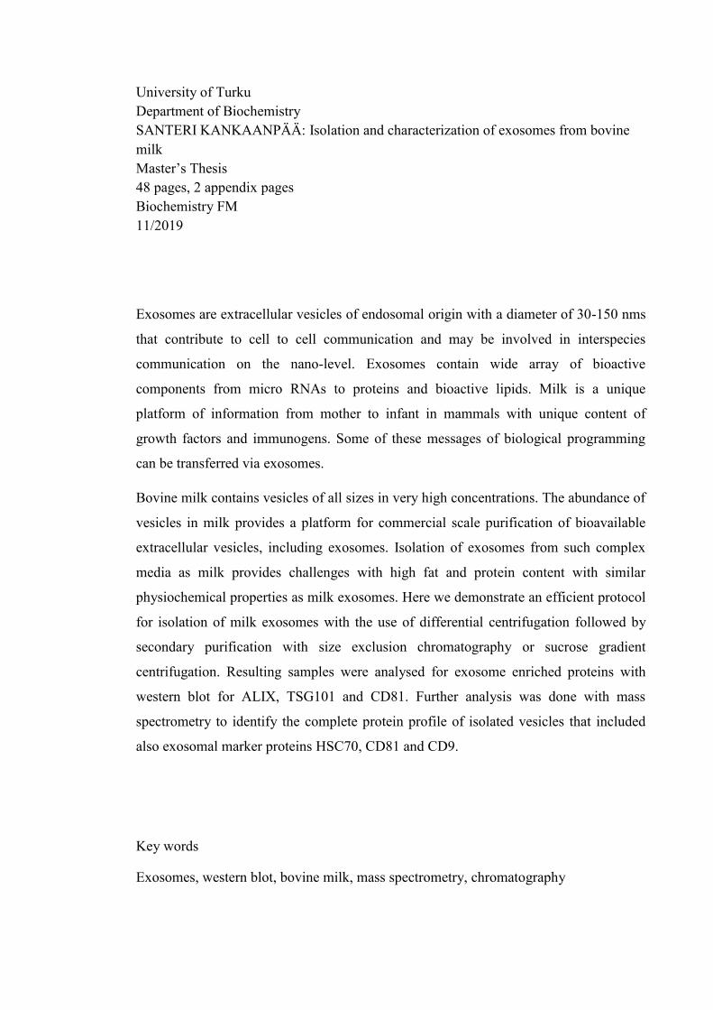
University of Turku
Department of Biochemistry
SANTERI KANKAANPÄÄ: Isolation and characterization of exosomes from bovine
milk
Master’s Thesis
48 pages, 2 appendix pages
Biochemistry FM
11/2019
Exosomes are extracellular vesicles of endosomal origin with a diameter of 30-150 nms
that contribute to cell to cell communication and may be involved in interspecies
communication on the nano-level. Exosomes contain wide array of bioactive
components from micro RNAs to proteins and bioactive lipids. Milk is a unique
platform of information from mother to infant in mammals with unique content of
growth factors and immunogens. Some of these messages of biological programming
can be transferred via exosomes.
Bovine milk contains vesicles of all sizes in very high concentrations. The abundance of
vesicles in milk provides a platform for commercial scale purification of bioavailable
extracellular vesicles, including exosomes. Isolation of exosomes from such complex
media as milk provides challenges with high fat and protein content with similar
physiochemical properties as milk exosomes. Here we demonstrate an efficient protocol
for isolation of milk exosomes with the use of differential centrifugation followed by
secondary purification with size exclusion chromatography or sucrose gradient
centrifugation. Resulting samples were analysed for exosome enriched proteins with
western blot for ALIX, TSG101 and CD81. Further analysis was done with mass
spectrometry to identify the complete protein profile of isolated vesicles that included
also exosomal marker proteins HSC70, CD81 and CD9.
Key words
Exosomes, western blot, bovine milk, mass spectrometry, chromatography
1
Table of contents
1. Introduction ............................................................................................................................... 2
1.1 Biogenesis of multivesicular bodies ................................................................................... 3
1.2 Protein composition and sorting of cargo ........................................................................... 5
1.3 Membrane lipid profile ....................................................................................................... 8
1.4 Isolation of exosomes ......................................................................................................... 8
1.5 Effects on cells .................................................................................................................. 12
1.6 Exosomes from bovine milk ............................................................................................. 13
2. Materials and methods ............................................................................................................ 15
2.1 Sample collection .............................................................................................................. 15
2.2 Isolation of exosomes ....................................................................................................... 15
2.3. Validation of exosome isolation methods ........................................................................ 18
2.3.1. Electrophoresis .......................................................................................................... 18
2.3.2. Western blot .............................................................................................................. 19
2.3.3. Nanoparticle tracking analysis .................................................................................. 19
2.3.4. Transmission electron microscopy ............................................................................ 20
2.3.5. Mass spectrometry and bioinformatics ..................................................................... 20
3. Results ..................................................................................................................................... 21
3.1. Sample pretreatments and storage .................................................................................... 21
3.2. Isolation of exosomes and validation of isolation methods ............................................. 22
3.2.1 Separation of exosomes from other milk components with differential centrifugation
............................................................................................................................................ 22
3.2.2 Removal of soluble proteins with size exclusion chromatography ............................ 23
3.2.3 Visualization of protein profiles of exosome isolates ................................................ 27
3.2.4 Size and morphology of isolated vesicles .................................................................. 28
3.3. Identification of exosome enriched markers .................................................................... 33
3.4. Proteome and functional analysis of exosomal proteins .................................................. 35
4. Discussion ............................................................................................................................... 41
4.1 Isolation and morphology of vesicles ............................................................................... 41
4.2 Identification of exosome enriched markers ..................................................................... 43
4.3 Further analysis and use of isolation protocols ................................................................. 45
4.4 Acknowledgements ........................................................................................................... 45
5. References ............................................................................................................................... 46
6. Appendix ................................................................................................................................. 53
2
1. Introduction
Exosomes are extracellular vesicles (EV) with a diameter of 30-150 nms that form in
multivesicular bodies (MVB), a certain type of late endosome with multiple inner
vesicles that form by inwards budding of the endosome membrane. Exosomes were first
characterized by Bin-Tao et al. when they visualized the secretion of ferritin receptors in
EVs in the maturation of sheep reticulocytes in vitro utilizing 125I- labelled anti
transferrin receptor antibodies. (Bin-Tao Pan, Kathy Teng et al. 1985). This was thought
to be a garbage disposal system for the cells to remove no longer needed components. It
was afterwards discovered that exosomes are important part of the cell to cell
communication with various functions such as transporting RNA and various proteins
from cell to another along with bioactive lipids in their membranes (Sjöstrand, Ekström
et al. 2007, Michel Record, Kevin Carayon et al. 2013).
Exosomes are present in multicellular organisms in all bodily fluids such as, blood
(Caby, Lankar et al. 2005), urine (Trairak Pisitkun, Rong-Fong Shen et al. 2004) and
breast milk (Admyre, Johansson et al. 2007) and the secretion of exosomes has been
characterized from several different cell types as stated in recent review articles
(Simpson, Jensen et al. 2008, Simpson, Lim et al. 2009, Sedgwick, D'Souza‐Schorey
2018). Exosomes are not spontaneously budded vesicles from the cell membrane but
precisely secreted from MVBs when MVBs fuse with the cell membrane. However the
differentiation between the two vesicle groups is often difficult, since both groups can
contain similar membrane lipid profile, surface proteins and can contain similar
intravesicular cargoes.
Formation of exosomes is conserved in mammals and plants and homologous proteins
are found in archaea to induce membrane budding.(Brooke L. Deatherage, Brad T.
Cookson 2012, Micali, Neumann et al. 2011, Regente, Pinedo et al. 2012) Bacteria have
been shown to secrete membrane enclosed vesicles with yet unknown mechanisms.
(Brooke L. Deatherage, Brad T. Cookson 2012) This universality of exosome mediated
cell signalling could be a platform for interspecies communication with highly
conserved and sophisticated mechanisms. The system includes similar mechanisms that
some viruses (such as HIV) utilize in infections. (Guillaume Van Niel, Gisela D'angelo
et al. 2018) Exosomes or exosome-like vesicles have been also isolated and
characterized from numerous plants such as Arabidopsis, rice, carrot, and several citric
3
fruits. Most of plant exosome studies are considering MVBs and exosomes as a
pathogen defence mechanism and as a form of unconventional protein secretion
(Hansen, Nielsen 2017).
The protein composition of exosomes varies along with the cells of which the vesicles
are secreted from (Smith, Lee et al. 2015) and the influence of exosomes to the recipient
cells is widely unknown. This variability in the molecular composition may provide
insight to possible disease related and systemic biomarkers and could lead to invention
of new diagnostic tools. Variability also provides the possibly to use exosomes as
bioengineered vesicles to transport molecules to specific tissues with high specificity
and biocompatibility. (Suresh Mathivanan, Justin W. E. Lim et al. 2010) This novel
concept of engineered biological nanovesicles is a promising possibility as a transport
system for delicate and unstable components to cells.
1.1 Biogenesis of multivesicular bodies
Exosomes form inside endosomes during inwards budding of the endosome membrane
thus creating a multivesicular body. This budding can happen two separate ways;
independently and dependently of endosomal sorting complex required for transport
(ESCRT). Dependent mechanisms require cargo sorting and clustering by ESCRT
complexes 0 and I which lead to budding of specific vesicles by ESCRT II and scission
of that vesicle by ESCRT III. Independent mechanism for vesicle budding is facilitated
by spontaneous or syntenin guided clustering of cargo which is followed by cytoplasmic
sorting and association with endosome membrane bound proteins. The resulting cluster
is then spontaneously released by vesicle budding of membrane that is enriched with
ceramides. (Guillaume Van Niel, Gisela D'angelo et al. 2018)
There are 5 ESCRT complexes each with specific protein composition and function.
Complexes are ESCRT-0, -I, -II, -III and VPS4 complex (Vacuolar protein sorting).
(Schmidt, Teis 2012) The following ESCRT proteins are described as named in yeast
for simplicity, respective mammalian proteins can be seen in review by Hurley (Hurley
2015). ESCRT 0 is composed of heterodimer of proteins Vps27 and Hse1 that associate
on flat clathrin-coated domains or phosphatidylinositol-3-phosphate (PI(3)P) rich
membrane and recruit ubiquitinated (Ub) proteins by five Ub binding domains
(Schmidt, Teis 2012, Hurley 2015, Hurley, Emr 2006, Hanson, Cashikar 2012). This
localization is done by FYVE domain in the complex that has specific zinc finger
4
domain that recognizes PI3P and LIEF or LIEL peptide sequence that binds to clathrin β
-propeller domains. (Hurley, Emr 2006) The 0-complex recruits ESCRT I complex by
binding directly to amino terminus of Vps23 (TSG101, tumour susceptibility gene 101,
in humans) by carboxyl terminal P(T/S)AP motif. 0-complex is not present in plants
which therefore utilize a different mechanism for starting of MVB pathway. (Schmidt,
Teis 2012)
ESCRT-I is a hetero tetrameric protein complex with one copy of each Vps23, Vps28,
Vps37 and Mvb12. Multiple binding domains are located on the stalk like structure of
Vps23 and Vps37, among these are the PTAP motifs of HIV-1 gag proteins. On the
other end is the linker for the ESCRT-II complex that binds to Vps28 carboxyl terminal
helical domain via GLUE (Gram-like ubiquitin) domain in Vps36. (Hurley, Hanson
2010) ESCRT-I complex along with –II is able to induce the inward budding of the
membrane. (Schmidt, Teis 2012)
ESCRT-II is another tetrameric protein complex with 2 subunits of Vps25 and one of
Vps22 and Vps36. The proteins form a Y-shape structure that has extending binding
sites for ESCRT-I domain and for ESCRT-III subunit Vps20. The interaction with
complexes I and III is characterized by specific protein motives that bind specific
sequences in each counterpart. GLUE domain is responsible for binding to PI3I and
ubiquitin. (Hurley, Hanson 2010, Hurley, Emr 2006)
Final ESCRT complex III consists of multiple subunits of Vps proteins. Vps2, Vps20,
Vps24 and Snf7 are mandatory for complex function in yeast. (Hurley, Emr 2006)
These core proteins are accompanied with accessory proteins Did2 Vps60 and Ist1 in
yeast and 9 more proteins in humans. (Hurley, Hanson 2010) Vps20 protein is
myristoylated and the myristylation could link the complex to the membrane, however
during isolation the complex is found to be cytosolic and not membrane associated.
(Hurley, Emr 2006) The ESCRT III complex is highly stable once associated to a
membrane and will remain intact until Vps4 hydrolyzes ATP to disassembly the
complex. (Hurley, Emr 2006) Vps4 complex is inactive in monomer or dimer form and
recruitment by separate complex drives the assembly to ESCRT-III complex interaction.
The interaction between complexes is most likely done by microtubule-interacting and
transport-motifs. (Schmidt, Teis 2012).
5
The functionality and interaction of ESCRT machineries is not completely understood
and analysis of different subunits by knockout mutants can lead to cell death. Yeast
models are most used for studies but present similar difficulties as higher organism
models. Initial budding is started by ESCRT I and II complexes in unilamellar vesicle
studies in vitro. These complexes localize to the neck of the budding membrane. Similar
effect can be achieved my ESCRT III complex proteins Snf7 and Vps20 but in much
higher concentrations; 15 nM for I and II and 600 nM for Snf7. (Hurley, Hanson 2010)
This curved surface provides a platform along with ESCRT II complex for ESCRT III
complex formation and scission of membrane bud. In yeast Snf7 is only mandatory
protein for membrane bud scission with other proteins provide the interaction platform
for the attachment of possible ESCRT III fiber-structure. (Schmidt, Teis 2012, Hurley,
Hanson 2010, Hanson, Cashikar 2012) ESCRT machinery is heavily involved in many
other membrane associated functions such as cytokinesis (ESCRT III, Vps4 and ESCRT
I along with ALIX), viral budding (same as in cytokinesis), membrane lesion repair and
nerve cell proliferation among others. (Hurley, Hanson 2010)
1.2 Protein composition and sorting of cargo
Protein composition of EVs varies one vesicle to another with major differences
between different sources of EVs. This great variance between vesicles provides
challenges and equal possibilities in analysis of EVs. Liquid biopsies are a rising trend
with more sensitive and non-invasive analytical methods to screen for disease and
systemic condition biomarkers. Exosomes could provide the next level of information
with the unique protein, nucleic acid and lipid profiles that could be connected to the
systemic state of patient. However, much more research is needed to confirm unique
biomarkers and validate the source of such biomarkers to the cells in distress or afflicted
by some condition.
Proteins can be targeted to exosomes via signal sequences such as myristylation tag and
phosphatidylinositol-(4,5)-bisphosphate-binding domain. These simple plasma
membrane anchors can be used to target cytosolic proteins or bioengineered proteins to
bind into exosome membranes. (Shen, Wu et al. 2011) Monoubiquitinylation, multiple
monouniquitinylations and farnesylation of proteins targets proteins for endosomes to
form multivesicular bodies as stated in recent review by Raposo et al., Llorente et al.
and Piper and Katzmann (Hessvik, Llorente 2018, Guillaume Van Niel, Gisela D'angelo
6
et al. 2018, Piper, Katzmann 2007). These signal sequences along with higher level of
oligomerisation of the membrane proteins seem to support the sequestering of proteins
to the extracellular microvesicles. These common features still do not explain the
intravesicular proteins (excluding ubiquitinylation), since the formerly mentioned
sequences guide proteins to membranes of exosomes. Term piggyback was used to
describe the co-localisation of a random protein along with vesicle cargo protein in a
recent review article. (Yang, J., Gould 2013) ESCRT machinery contains proteins with
catalytic activities that cleave ubiquitin from the proteins transported to the lumen of
vesicles inside MVBs and therefore multiple proteins with no apparent signal sequence
can be effectively localized inside exosomes with the removed ubiquitin tag. (Hurley,
Emr 2006)
Protein sorting is controlled by ESCRT complexes I and III along with chaperone
protein, heat shock cognate 70, to transport proteins to multivesicular bodies in the late
endosomes. Process was described as microautophagy for the similarities with regular
autophagy by Sahu et al. Heat shock cognate interacts with other cytosolic proteins with
electrostatic interactions and propagates their transport into late endosomes. These
interactions are more efficient and targeted with cytosolic proteins containing KFERQ-
sequences.(Sahu, Kaushik et al. 2011) With the important role of protein sorting and
transporting of heat shock cognate 70, it can be used as a enriched exosomal protein
marker to use in western blot.
ESCRT machinery influences the molecular composition of exosomes in mammals. The
downregulation of ESCRT machinery via short hairpin RNAs transported via
lentivectors that resulted in changes in the number of secreted exosomes as well as in
the surface protein profile. Downregulation of ALIX resulted in an increase of MHC II
(Major histocompatibility complex II) on the exosome membrane and effected in the
CD63 and HSC70 protein quantity on the membrane. TSG101 downregulation on the
other hand decreased the total exosome secretion in HeLa cells. (Colombo, Moita et al.
2013) Influencing the ESCRT machinery via lentivectors could provide a route for
engineering exosomes to overexpress certain proteins or using the ESCRT pathways to
transport specific molecules to exosomes. For engineering to be safe and efficient
fundamental understanding of the exosome formation is needed and further studies need
to be done.
7
Release of exosomes is balanced with the option of lysosomal degradation of MVBs.
Determination of which pathway MVBs take is regulated to some extent by ISGylation,
a similar modification than ubiquitylation, of TSG101 (among other proteins) targets
the MVB for lysosomal degradation. (Carolina Villarroya-beltri, Francesc Baixauli et al.
2016) Transport of MVBs to plasma membrane is dependant with the interaction of
cytosolic structure proteins actin and microtubules, actin interacting protein cortactin
and Rab GTPases. (Hessvik, Llorente 2018) Release of exosomes requires membrane
fusion with MVB and plasma membrane. However the actual mechanism of membrane
fusion of MVB and cell membrane to release exosomes is still unknown and separate
protein machinery has been characterized for different cells and exosomes carrying
different cargo. (Raposo, Stoorvogel 2013) SNARE proteins are included in the release
of exosomes from the multivesicular bodies by the fusion of membranes upon vesicle
release. Interaction with SNAREs and VAMP7 (vesicle associated membrane protein 7)
and the R-SNARE protein YKT6 have been shown to be important on exosome release,
with inhibition of SNARE-VAMP7 interaction or YKT6 expression leading to decrease
in exosome release. (Hessvik, Llorente 2018)
There are no universal or entirely specific markers for exosomes to use for
quantification and to use to isolate exosomes. The commonly used markers for
exosomes CD9, CD81, CD63, ALIX, TSG101 and FLOT1 are also present in
microvesicles and can be included in apoptotic bodies and other secreted vesicles
(Guillaume Van Niel, Gisela D'angelo et al. 2018). However these proteins seem to be
enriched in exosomes or in exosome like vesicles to a varying degree. It is not yet clear
if the expression of these markers is completely cell, exosome or function related and
could these differences be used as a mean to distinguish specific vesicle populations.
With the limitations of the characterization in mind, the enrichment of these markers is
still a way to show the presence of extracellular vesicles in the sample. With even better
methods for exosome isolation in development the future goal is to characterize the
possible subpopulations of exosomes depending on their size, density, cargo,
morphology, function or some completely different yet unknown factor and analyse the
vesicles on the subpopulation or even single vesicle level using high sensitivity
analytics.
8
1.3 Membrane lipid profile
Lipid profile of EVs can differ from one vesicle to another. Some differences can be
seen in the lipid content of the membranes compared to plasma membrane with higher
cholesterol and ceramide content in the exosome developing MVBs. (Mobius, Ohno-
Iwashita et al. 2002, Trajkovic, Hsu et al. 2008) The overall lipid composition differs
from the parental cells as seen in review article by Record et al. (Michel Record, Kevin
Carayon et al. 2013). Inhibition of ceramide production by sphingomyelinase inhibitor
leads to clear drop in the formation of exosomes when examining HeLa derived
exosomes (Trajkovic, Hsu et al. 2008). This did not completely inhibit the exosome
secretion which indicates ESCRT independent mechanism, which does not need
ceramide for function. Lysobisphosphatidic acid (LBPA) is shown to be another
important lipid for forming multivesicular bodies. Localization of LBPA among other
lipids is explained in article (Kobayashi, Gu et al. 1998) with enrichment of LBPA from
1.5 % to over 15 % in the late endosomes.
Interaction with a small glycoprotein saposin (acronym for Sphingolipid Activator
PrO(S)teINs) C is crucial to regulate forming of MVBs and fusion of the membranes at
least in human and mice fibroblasts. Interaction with saposin C and LBPA in artificially
generated unilamellar vesicles leads to increase of vesicle size from 200 nm up to 1500
nm and the growth is suspected come from spontaneous fusion of vesicles mediated by
saposin C. Similar reaction was not observed with other saposins. This indicates the
direct interaction with saposin C and LBPA. (Chu, Witte et al. 2005) Considering
exosomes, more interesting is the effect of saposin C deficiency to the number of MVBs
within a cell. The number of MVBs was over 4 times larger in saposin C deficient cells
and incubation with saposin C reverted the number of MVBs back to normal levels.
(Chu, Witte et al. 2005) This information could be utilized in production of exosomes to
increase the number of MVBs within a cell which could lead to increased number of
exosomes secreted by the cell by bioengineering saposin C deficient cells.
1.4 Isolation of exosomes
Previously the isolation of exosomes is based on differential centrifugation with various
speeds to separate exosome sized particles from media. Centrifugation is done with or
without gradient (sucrose or iodixanol) with exosome density ranging from 1.13-1.19
g/ml or as high as 1.21 g/ml (Michel Record, Kevin Carayon et al. 2013).
9
Centrifugation is time consuming and difficult to upscale due to the high g-forces
ranging from 70 000 xg up to 340 000 xg (Kristine Blans, Maria S. Hansen et al. 2017)
needed for exosome isolation. Many different isolation methods have been tested and
compared (Shtam, Samsonov et al. 2018) such as the use of commercial kits
(YAMADA, INOSHIMA et al. 2012) or the use of mild chemical treatments (Brighton
E. Maburutse, Mi-ri Park et al. 2017). Different isolation methods have been more or
less successful and the protocol used needs to suit the starting media to isolate the
exosomes from.
The classical differential centrifugation method is also suitable for isolation of plant-
based exosomes from various materials (Xiao, Feng et al. 2018). Multitude of plant
based exosome studies are done with the method starting with homogenisation of the
sample using a blender or from juice samples (Raimondo, Naselli et al. 2015, Ju, Mu et
al. 2013, Wang, Ren et al. 2015). The disrupted cells will produce vesicles of all sizes
and densities and this is made even more complex with samples containing chloroplasts
that can be in the same size ranges as the exosomes. These blender-based or juice-based
isolation protocols should be considered to contain exosome-like particles or
nanovesicles and not be described as exosomes and the following studies using the
isolated exosome-like particles need to be presented for the exosome-like particles and
not for exosomes.
Differential centrifugation will not produce pure exosome isolates, since aqueous
solution will always contain different size particles that will sediment along with the
exosomes in the centrifugation process. The purification should be continued with a
density gradient, filtration or with size exclusion chromatography to seclude protein
aggregates, membrane particles of different origin and soluble proteins from the
exosome isolate. The purity of the final isolate needs to suit the following analytics; as
an example the exosome sample with high soluble protein content used in cell culture
related studies can distort the results with the measured response of the cells being from
the soluble proteins rather than from the exosomes itself. Hence the studies need to have
a valid negative control to pin point the actual molecule responsible for the effect.
The commercial kits are widely based on the polymeric precipitation of exosomes with
poly-ethylene glycol or similar polymer (assumption due to disclosed product
specifications). Even though the kits don’t need high speed centrifugations to isolate the
10
exosomes, the overall yield is not comparable to the high speed centrifugation methods
as seen with bovine milk (Masaharu Somiya, Yusuke Yoshioka et al. 2018) and human
plasma (Stranska, Gysbrechts et al. 2018). The difference in the efficiency may also be
due the wider array of pelleted vesicles by the centrifugation rather than the limitations
of the precipitation chemicals. The precipitation efficiency was improved with the use
of positively charged protein protamine with the exosomes charge is negative and
dependent of the origin cell. Combined use of PEG and protamine yielded higher
exosome concentrations in the final isolate than ultracentrifugation-based method from
serum, saliva and cell culture media. (Deregibus, Figliolini et al. 2016)
Comparison between published methods is often difficult and the lack of complete
protocol and used instruments may hinder the ability to replicate the results with the
given protocol. Differences in centrifugation based methods are heavily influenced by
rotor along with K factor, therefore centrifuge specifications must be normalized when
replicating different protocols with different instruments. (Cvjetkovic, Lötvall et al.
2014) It is not enough to report just the used g-forces along with centrifugation time in
the protocol, since the differences between the rotors will greatly influence the run time
and the efficiency of pelleting. However, even with the equation of the different
equipment used in isolation the complete yield of exosomes is essentially sample related
and can be influenced by the source of exosomes. Therefore, the isolation procedure
needs to suit the medium of which the exosomes are to be isolated. This is even more
complicated and crucial when isolating exosomes from complex media, such as milk
which is seen comparing results from bottom-up and top-down centrifugation methods
by Zonneveld et al. (Zonneveld, Brisson et al. 2014) The physical properties of medium
of which exosomes are going to be isolated need to be taken into consideration. More
dense and viscous medium will demand longer centrifugation times or can be even
unsuitable for isolation of vesicles by centrifugation. Online calculators such as
http://vesicles.niifhm.ru/index.php?do=1 and
https://www.beckman.com/centrifuges/rotors/calculator can be utilized to determine
specific run parameters for centrifugation based isolation of exosomes.
Size exclusion chromatography has successfully been utilized to purify exosomes from
bovine and human milk without the need for pelleting with high speed centrifugation.
The isolated exosomes were pure from the most abundant milk-protein casein which is
troublesome to prevent from contaminating the final exosome isolate. (Kristine Blans,
11
Maria S. Hansen et al. 2017) Size exclusion chromatography could be scaled up for
bigger sample volumes, but slow flow rates and relatively delicate instruments require
time and capable operating personnel for use. The combined use of size exclusion
chromatography with tangential flow filtrations is shown to produce highly pure
extracellular vesicle isolates from cell culture mediums (Corso, Mäger et al. 2017,
Nordin, Lee et al. 2015).
Flow cytometric isolation has successfully been utilized; however, the heterogeneity of
the exosome membrane proteins demands a wide array of immunolabels and the final
isolate may be more homogenic than the actual exosome population in the original
sample. (Wiklander, Bostancioglu et al. 2018, Suresh Mathivanan, Justin W. E. Lim et
al. 2010) Isolation with the immunolabels could potentially be utilized for the
differentiation of separate vesicle populations with distinctive protein expression. This
could be even coupled with the engineering of exosomes to express some membrane
protein that is not commonly found in these vesicles and used to isolate the new
engineered vesicles from the sample. Tangential flow filtration is one of the
commercially available services for isolating exosomes and is used to produce ready
exosomes to use as internal standards (Lonza). Even though most exosome isolation
protocols are based on ultracentrifugation the effects of high speed centrifugation on
exosome structures have not been analysed. Immense g-forces could affect the elastic
membranes of exosomes and possibly lead to fusion of membranes. Influence of the
isolation and storage procedure can be seen with the cup-shaped morphology in the
electron microscopy caused by the collapsing and distortion of membranes.
Whichever method is to be used, the results should be reported with the complete
protocol to pair the results with the protocol. The isolated vesicles should be free of
known contaminants such as protein aggregates and other vesicles or the results from
the following analyses should take these contaminants account for. Negative controls
should be carried out in all analytical methods to assess the purity of sample and
therefore also define the effectiveness of the used method. Multiple separate online
platforms have been established for publishing EV-related results such as evpedia.info,
vesiclepedia (microvesicles.org) Exocarta (exocarta.org) and EV-Track (evtrack.org).
The proteomic analyses of these databases are filled with contaminating proteins and
comparison has become somewhat troublesome. The international society of
extracellular vesicles (ISEV) provides free information for researchers interested in
12
extracellular vesicles. The massive open online course “MOOC” gives a basic
understanding of everything EV-related. ISEV provides also standardized requirements
for EV related publications for quality control. These standards are taken further with
EV-Tracks EV-metric which is a 9 point assessment for providing quality control for
the results. This checklist makes sure that the researchers remember to consider all the
possible controls along with the conducted experiments.
1.5 Effects on cells
Exosomes derived from bovine milk have been analysed for use as a therapeutic agent
along with exosomal loading with therapeutic compounds. Exosomes showed high
chemical stability with tissue specific localization within subject rats depending on the
exosome exposure method. Treated rats and cell cultures showed clear indications of
anti-inflammatory responses from exosomes and higher responses from exosome loaded
drugs compared to free drug molecules and PBS (phosphate buffered saline) control.
(Munagala, Aqil et al. 2015) With similar affects bovine milk derived EVs reduced
arthritis symptoms in mouse models, when treated with oral administration of EVs. The
intake of EVs was visualized by flow cytometry and confocal microscopy. Both
macrophages and ileum cells took EVs in and the intake was reduced greatly when
lowering the incubation temperature to 4 ˚C. (Arntz, Pieters et al. 2015)
Exosomes isolated from bovine milk activate MAP- kinase pathway in IEC-6 cells. The
cell culture was starved prior to exosome treatment, which may influence the observed
change. However the kinase pathway activation was dose dependent and was greater in
cells grown in exosome depleted media. (Siran Yu, Zhehao Zhao et al. 2017) This
activation indicates the proliferative effects of exosomes to the recipient cells.
Exosomes from bovine milk are shown to contain proteins that are related to immune
response and growth. With major differences in protein composition between mature
and colostrum milk, it is clear that exosomes are precisely produced with specific cargo
to transport for the recipients. In this case the calf gets a probable immune response
activating surge of exosomes from the colostrum milk which then turns to growth
inducing signal later post-partum. (Samuel, Chisanga et al. 2017)
Isolated exosomes have been shown to withstand simulated human digestive system and
ingested by intestinal epithelial cells in vitro. This suggests the possibility of transport
of proteins and nucleic acids from different sources via gastro intestinal cells. (Wolf,
13
Baier et al. 2015, Benmoussa, A., Lee et al. 2016, Mu, Zhuang et al. 2014). Exogenous
RNA is already shown affect gene expression across species. MIR168a, abundant
miRNA in rice, is active and binds to human and mouse LDLRAP1 mRNA and inhibits
the expression of the respective protein. (Lin Zhang, Dongxia Hou et al. 2012)
The RNA in milk is shown to be stable under degrading conditions (Izumi, Kosaka et al.
2012) and are shown to be affect gene expression in mononuclear cells, HEK-293 and
mouse liver cells (Baier, Nguyen et al. 2014). There are only few publications on the
topic of food derived RNAs influencing gene expression directly and the subject is still
highly controversial. Intake of exosomes is mediated via endocytosis (Kusuma, Manca
et al. 2016, Wolf, Baier et al. 2015) and endocytosis is influenced heavily by the protein
and glycoprotein content of the exosome membrane. With glycosomal competition and
when the membrane proteins are treated with a protease the transport rate of exosomes
is lower. (Kusuma, Manca et al. 2016) The intake of bovine milk exosomes show no
cytotoxic effects as seen with mouse macrophage cells and no signs of systemic
anaphylaxis was measured after 35 days of exosome treatment. (Masaharu Somiya,
Yusuke Yoshioka et al. 2018)
1.6 Exosomes from bovine milk
Bovine milk exosomes are an interesting target for research as humans with western
diets consume milk products and are therefore subjected to high quantities of exosomes
from them. Milk exosomes are shown to be resistant to digestion and also bioavailable
in different species of which derived from. (Manca, Upadhyaya et al. 2018) Recent
review article combined interesting factors present in bovine milk exosomes with
potential health risks in mind. The miRNA contents of exosomes were linked to
multiple chronic conditions and higher risks of cancer. (Bodo C Melnik, Gerd Schmitz
2019) Indications of dangers of milk consumption could be explained with digestion
resistant exosomes being the causative factor in development of chronic conditions.
Therefore basic knowledge and a complete understanding of exosomes in bovine milk
should be collected to determine the safety and influence of bovine milk for the
consumers.
Unique protein signatures have been found with differences between mature and
colostrum milk samples (Samuel, Chisanga et al. 2017, Yang, M., Song et al. 2017).
Separate fractions isolated by centrifugation also are shown to have different protein
14
signatures as described by Benmoussa et al.(Benmoussa, Abderrahim, Gotti et al. 2019)
Along with the protein contents, RNA and lipid profiles of exosomes have been
characterized. Online database exocarta is designed for the characterization of exosome
protein, RNA and lipid contents. This database covers 9 different species and numerous
cell lines along with complex mediums of which the exosomes are isolated from
(http://www.exocarta.org/).
Molecular complexity of bovine milk provides challenges for the isolation of exosomes.
High fat and protein content with similar precipitation properties and density of
exosomes can lead to contamination of exosome isolate with the caseins and lipid
micelles (Masaharu Somiya, Yusuke Yoshioka et al. 2018). Filtration of the whey can
separate larger particles from the isolate, but casein aggregates with smaller than 0,22
µm diameter will still pass the filter and be present in the final isolate. Caseins will
spontaneously form micelles when the environment favours their formation with
interactions between monomeric K- and B-caseins. More effective protocol could allow
isolation from large quantities of abundant source of exosomes, such as bovine milk.
With easier and more efficient isolation methods exosomes could be utilized for
manufacturing pharmaceutical and nutraceutical products. The acetic acid isolation
method was successfully used previously by Somiya et al. and effective SEC-based
isolation method was utilized by Blans et al. (Masaharu Somiya, Yusuke Yoshioka et al.
2018, Kristine Blans, Maria S. Hansen et al. 2017) The bioavailability of exosomes
provides a possible use of exosomes as trafficking specific cargo, such as drugs or
polyphenols, to cells has been tested with promising results (Munagala, Aqil et al. 2015,
Wang, Wang et al. 2014).
The objective of this study is to set up effective isolation protocol for isolation of
exosomes from different bovine milk samples and characterize the proteins from the
isolated exosomes. Identification of the isolated EVs as exosomes will be done by
scanning electron microscopy, gel electrophoresis and western blot analysis with CanX,
CD63, CD81, TSG101 and ALIX antibodies. These are the antibodies widely used for
characterization of exosomes, even though there are major differences between with the
proteins expressed on the exosome membrane from different exosome sources (Smith,
Lee et al. 2015, Wiklander, Bostancioglu et al. 2018). This range of identification
covers the minimum basis of exosome characterization described by International
Society for Extracellular Vesicles in 2014 (Lötvall, Hill et al. 2014).
15
Isolated exosomes are quantified and characterized by nanoparticle tracking analysis
and with electron microscopy with negative staining using uranyl-acetate and/or with
immunolabelling using anti-CD63 with secondary antibody (donkey anti-goat) labelled
with 10 nm gold particles. The NTA and EM will be done as paid service from
FIMM/EV-Core at University of Helsinki.
After the isolation of exosomes proteins are digested with trypsin and analysed with LC
MS/MS (quadrupole/time of flight) mass spectrometry. The differences in bovine milk
sample exosomes will be analysed between different isolation procedures to examine
the differences between the isolation methods.
2. Materials and methods
2.1 Sample collection
Milk from healthy Nordic Red dairy cows was obtained from Luke Minkiö research
cow house (Jokioinen, Finland). Cold tank milk was collected into a glass bottle and
kept cold +4 - +8 ˚C prior to analysis. Excess milk after removal of fat and intact cells
was aliquoted to 40 ml fractions and frozen -80 ˚C for later use.
2.2 Isolation of exosomes
Differential centrifugation was applied to pellet the exosomes. Isolation was conducted
with and without acetic acid (AA) and followed by two additional purification steps;
size exclusion chromatography (SEC) and sucrose gradient centrifugation. Precipitation
with ammonium sulphate was examined as a possible isolation method, but was ruled
out due to difficulty to track solution density and therefore the fraction which should
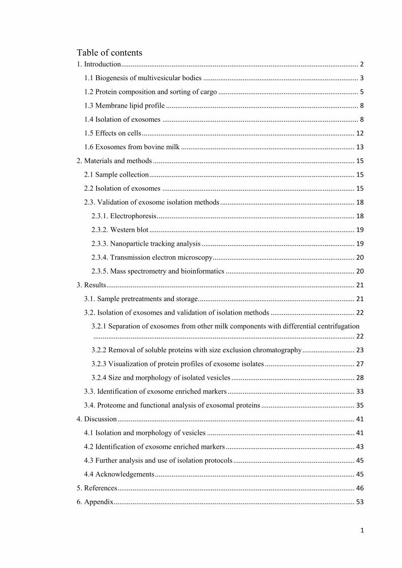
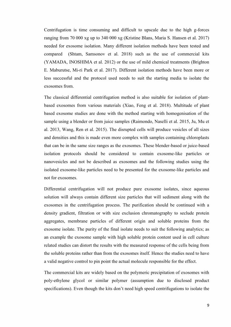
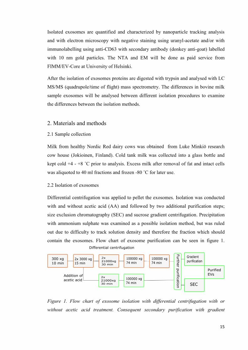
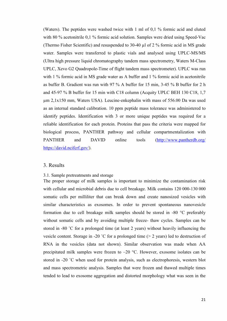
contain the exosomes. Flow chart of exosome purification can be seen in figure 1.
Figure 1. Flow chart of exosome isolation with differential centrifugation with or
without acetic acid treatment. Consequent secondary purification with gradient
16
purification or size exclusion chromatography was conducted for both isolation
methods to remove impurities from exosome isolates.
Fresh whole milk was centrifuged 300 xg 10 min +4 ˚C (Beckman coulter, JA-14 or JA-
17, J2-21M) to pellet intact cells to prevent further cell breakage and contamination
with cell debris. Separated middle layer was always used in the consecutive steps. Layer
was centrifuged twice 3000 xg 15 min +4 ˚C (Beckman coulter, JA-17) to pellet cell
debris, caseins and remaining fat on top. Resulting defatted milk was again centrifuged
twice 21500 xg +4 ˚C 30 min (JA-17 Beckman Coulter) to further pellet caseins.
Resulting clearer whey was ultracentrifuged 74 min (Beckman coulter, Type 50.2 Ti,
L8-80) to pellet remaining caseins. Resulting supernatant was ultracentrifuged again as
before to pellet the exosomes. Precipitated pellet was resuspended in 200 µl of PBS.
To further precipitate caseins and milk peptones the fraction after 2x 3000 xg
centrifugation was adjusted to pH 3.8 with glacial acetic acid (Sigma Aldrich, MO,
USA). The AA-treated sample was then centrifuged similarly as non-treated milk except
the AA exosomes were pelleted already during the first ultracentrifugation. The
resulting pellet was again resuspended in PBS and frozen until further analysis.
Isolation is done similarly as Caby et al. and Masaharu et al. (Caby, Lankar et al. 2005,
Masaharu Somiya, Yusuke Yoshioka et al. 2018)
Ammonium sulphate precipitation was tested for exosome isolation. Ammonium
sulphate was added to milk to form a saturated solution. The solution was stored over
night at +4 ˚C to precipitate proteins. Resulting solution was centrifuged 3000 xg 15
min +4 ˚C. The top layer was extracted and centrifuged again to separate the fat layer.
The following clear solution along with the cloudy top layer was subjected to the
following 21500 xg centrifugation steps as with the acetic acid treated and non-treated
milk. The top cloudy layer was separated and diluted to ¼ with PBS and
ultracentrifuged 40000 xg 10 min. Clear bottom layer was diluted and ultracentrifuged
as the top layer. The resulting top and bottom layer were again separated and diluted ¼
with PBS and ultracentrifuged 74 min with type 50.2 Ti rotor. Resulting pellets were
suspended to 200 µl of PBS. Ammonium sulphate precipitation could be utilized to
isolate exosomes from milk with adjustment to the density of the solution as well as the
precipitation proteins. This method needs to be optimized to accurately isolate exosome
fraction. Ammonium sulphate could therefore be effective combination of similar
17
effects as acid precipitation Size exclusion chromatography using ÄKTA Basic protein
purification system with two Superose™ 6 10/300 columns (GE Healthcare, Sweden)
was applied to remove remaining soluble proteins from the final exosome isolate and to
recover the exosomes from the void volume as the smaller soluble proteins are trapped
in the pores of the column. PBS (137 mM NaCl (Merck, Germany), 2,7 mM KCl
(Merck), 10 mM Na2HPO4 (Merck), 1,8 mM KH2PO4(Merck)) with 2 M urea (Thermo
Fisher, MA, USA) was used as running buffer. Urea was used in the running buffer to
dissociate the casein micelles to prevent the elution in the void volume peak.SEC was
performed with and without ethylenediaminetetraacetic acid (EDTA (Sigma Aldrich,
MO, USA)). 20 mM EDTA (Sigma Aldrich) was used to chelate bivalent cations from
the solution in order to prevent cation related casein-casein interaction to dissociate
casein micelles to prevent them eluting at the same time with the exosome void volume
peak. and separates the caseins from each other. EDTA has been previously utilized by
Blans et al. (Kristine Blans, Maria S. Hansen et al. 2017). Exosome pellets for SEC
were first suspended in 200 µl of PBS, filtered through 0,22 µm syringe filters and
adjusted to 600 µl with PBS to contain 1 M urea in the final sample. Solid urea was
weighed into Eppendorf tube and the sample was filtrated on top of the solid urea.
Samples were then loaded via 500 µl loop to ÄKTA. To identify proteins from the
samples, bovine milk standards were used in parallel with the samples. The bovine milk
standard was prepared using -, -, and Κ -casein, lactoferrin and lactalbumin in 5-10
mg/ml concentrations. All proteins for the standard mixture were manufactured by
Sigma Aldrich. In first runs the milk standard proteins were run as a mixture to allow
protein interaction and feasible micelle formation. In first runs samples and standard
were separated with urea in the running buffer. In later run the proteins were solubilised
separately in 10 mg/ml concentrations and run without the addition of urea in the
running buffer to identify individiual proteins.
Gradient centrifugation was done with stepwise sucrose gradient in PBS based on a
method by Luthe (Luthe 1983). The sucrose gradient was prepared step-wise by
pipetting and freezing (-80 ˚C) 2 ml fractions of sucrose with decreasing densities to
SW 41 Ti ultra-clear poly propylene centrifuge tubes. Fractions were as follows 45, 30,
20, 10 and 0 % w/v of sucrose in PBS. 200 µl of exosome isolate was pipetted on top of
the thawed gradient tube and centrifuged 16 h, 100000 xg at + 20 ˚C. Exosomes were
retrieved from the 30 % fraction with density of 1.127 g/ml and from the border of 45 %
18
fraction with density of 1.203 g/ml. Resulting fraction was then diluted to 22 ml of PBS
and ultracentrifuged with type 50.2 Ti rotor for 90 minutes to pellet the gradient purified
exosomes. Resulting pellet was resuspended in PBS and frozen -20 C until further
analysis.
2.3. Validation of exosome isolation methods
2.3.1. Electrophoresis
Proteins from each isolation process were separated and visualized with 12 % w/v
sodium dodecyl sulphate SDS poly acrylamide gel electrophoresis (SDS-PAGE). The
SDS-PAGE with separation gel (0,192 M Tris-HCl pH 8,8 (Sigma Aldrich), 3,35 M
urea (Sigma Aldrich), 12 % w/v acrylamide (19:1 Acrylamide/Bis-acrylamide-30 %
solution, Bio-Rad Laboratories, CA, USA), 0,22 % w/v sodium dodecyl sulphate (SDS,
Merck), 0,025 % w/v ammonium persulfate (APS, Amersham Biosciences, UK), 0,04
% v/v tetraethyl methyl diamine (TEMED, GE Healthcare)) and stacking gel (0,1834
M Tris-HCl pH 6,8, 7,5 M urea, 6 % w/v acryl amide, 0,17 % w/v SDS, 0,007 % w/v
APS and 0,007 % v/v TEMED) was prepared with standard protocol. Sample buffer of
138 mM Tris-HCl (pH 6,8), 6 M Urea, 4,3 % (w/v) SDS, 22 % (v/v) glycerol,
bromophenol blue (Merck) as indicator and 5 % (v/v) 2-mercapthoethanol (Sigma
Aldrich) as reducing agent was mixed 1:1 with samples and incubated 30 min in 40 ˚C.
Electrophoresis was run with GPS 601 power unit (GE Healthcare) and mini-
PROTEAN tetra cell electrophoresis unit (Bio-Rad) with 10-15 mA current and 100-
150 V voltage with SDS running buffer (50 mM Tris, 384 mM glycine (Bio-Rad), 6,9
mM SDS). Gels were fixed with 50 % methanol (VWR) 5 % acetic acid (Sigma
Aldrich) for 1 h and stained 1 h with Pro Q Emerald 300 (Invitrogen, CA, USA)
glycoprotein stain and afterwards overnight with Sypro Ruby protein gel stain (BioRad
Laboratories, CA, USA). Gels were destained with 10 % methanol 7 % acetic acid and
washed with milli-Q water prior of imaging with ChemiDoc™ MP scan camera and
ImageLab™ software (BioRad). Protein concentration was measured with DC™ protein
assay (BioRad) or with Pierce BCA protein assay (Thermo Fischer Scientific) using
microplate procedure and HIDEX spectrophotometer (HIDEX, Finland). Bovine serum
albumin (Thermo Fischer Scientific) was used as a standard in the measurements.
19
2.3.2. Western blot
For detection of exosome enriched marker proteins SDS PAGE –separated proteins
were transferred from gel onto a Immobilon-FL-PDVF membrane using Bio-Rad semi-
dry blotting device, current of 1 mA/cm3 and voltage maximum of 25 V, and TG as
transfer buffer (Bio-Rad; 25 mM Tris, 192 mM glycine, pH 8,3 + 20 % methanol). Tris-
buffered saline (TBS; 18 mM Tris, 0,5 M NaCl pH 7.5) was used as a washing and
blocking buffer with the addition of 5 % (w/v) fish gelatine in the latter (Thermo Fisher
Scientific). After transfer each blot was blocked with 5% blocking buffer for 1 h under
shaking at room temperature and then incubated with primary antibodies anti-CanX (ST
John laboratories, USA, STJ140017), anti-CD63 (ST John laboratories, STJ140029),
anti-Alix (Cell Signal, Netherlands, E6P9B Rabbit mAb), anti-CD81 (Santa Cruz
biotechnology, USA Texas, sc-166029 mouse monoclonal) and anti-TSG101 (Nordic
Biosite, ABB-709, rabbit polyclonal) diluted 1:5000-10000 in 1% (w/v) fish gelatine in
TBS-T (TBS with 1 % (v/v) Tween-20 (Sigma Aldrich) overnight at +4 ˚C with
agitation. After removing the primary antibody solution blots were washed 3x5 min
with TBS at room temperature with agitation and then incubated for 2 h at room
temperature with corresponding secondary antibody (mouse anti-goat (Santa Cruz
biotechnologies, sc-2354), anti-Rabbit HRP conjugated (Agrisera AB 10668) or anti-
mouse (Santa Cruz biotechnologies, sc-516102) diluted 1:20000 in 1 % 1% (w/v) fish
gelatine in TBS-T. StrepTactin™-HRP conjugate (1:50000) was added for detection of
Western C protein standard (BioRad). For detection blots were incubated with
SuperBright-ECL chemiluminesence reagent (Agrisera, Sweden) according
manufacturer’s protocol and the images were obtained using Bio-Rad ChemiDoc MP
image scan camera and Imagelab software.
2.3.3. Nanoparticle tracking analysis
Nanoparticle tracking analysis (NTA) was applied to characterize isolated nanoparticles.
NTA is based on the Brownian motion and light scattering properties of particles in
solution and it produces high resolution estimates of nanoparticle size-distribution and
concentration(https://www.malvernpanalytical.com/en/products/product-
range/nanosight-range/nanosight-lm10) . The analysis does not separate nanovesicles
form other nano-sized particles. The NTA analysis service was provided by EV Core
service laboratory in Helsinki. In short the samples were analysed on Nanosight
20
LM14C nanoparticle tracking instrument in 6 replicates and dilutions of 1:20 – 1:20000.
Resulting particle concentrations and sizes were averaged and charted with standard
deviance visible on the graphs.
2.3.4. Transmission electron microscopy
Transmission electron microscopy (TEM) was applied to detect, characterize and
visualize the isolated vesicles. The imaging was done at EV Core service laboratory in
Helsinki according to Puhka et al 2017. (Puhka, Nordberg et al. 2017). In short, the
samples were fixed with paraformaldehyde (Electron microscopy sciences, Hatfield,
PA) in 0,1M NaPO4 buffer (pH 7.0), stained with 2,0 % neutral uranyl acetate,
embedded in methyl cellulose uranyl acetate mixture (1,8/0,4%) and imaged with
transmission electron microscopy using Jeol JEM-1400 (Jeol Ltd., Tokyo, Japan)
operating in 80 kV. Images were taken with Gatan Orius SC 1000B CCD-camera
(Gatan Inc., USA).
2.3.5. Mass spectrometry and bioinformatics
For detection of exosome enriched proteins selected exosome isolates were subjected to
in-solution trypsin digestion. 100 µg of exosome protein was loaded to 3 kDa spin
columns (Nanosep, Pall Co. USA) and wash buffer (8 M urea in 10 mM Tris-HCl pH 8)
was added to reach at least 6 M urea concentration. Resulting solution was centrifuged
until almost dry at 10000 xg at room temperature with Eppendorf minispin plus tabletop
centrifuge. Sample was washed with 100 µl of wash buffer and spun dry. Proteins were
reduced with 50 µl of 10 mM DTT (Sigma Aldrich) in wash buffer and again spun dry.
Reduced proteins were alkylated with 27 mM iodoacetamide (Thermo Scientific) in
wash buffer and mixed gently by vortexing (1000 rpm) for 1 min and spun dry. Sample
was washed twice with wash buffer and the filter unit was transferred to a clean
Eppendorf tube. Trypsin (Sequencing grade modified trypsin, Promega, WI, USA) was
added in 1:25 relation to the sample protein quantity and the samples were digested in
37 C overnight. Resulting peptides were spun through the filter and filter was washed
with 50 µl of 0,5 M NaCl.
Formic acid (Merck) was added to the resulting peptide samples to 1 % concentration to
form solution < pH 3 to assure the peptide binding to C-18 columns. Solution was
loaded to activated (activated with 100 % methanol and 80 % acetonitrile 0,1 % formic
acid solutions) and balanced (0,1 % formic acid) SepPak C-18 vacuum columns
21
(Waters). The peptides were washed twice with 1 ml of 0,1 % formic acid and eluted
with 80 % acetonitrile 0,1 % formic acid solution. Samples were dried using Speed-Vac
(Thermo Fisher Scientific) and resuspended to 30-40 µl of 2 % formic acid in MS grade
water. Samples were transferred to plastic vials and analysed using UPLC-MS/MS
(Ultra high pressure liquid chromatography tandem mass spectrometry, Waters M-Class
UPLC, Xevo G2 Quadropole-Time of flight tandem mass spectrometer). UPLC was run
with 1 % formic acid in MS grade water as A buffer and 1 % formic acid in acetonitrile
as buffer B. Gradient was run with 97 % A buffer for 15 min, 3-45 % B buffer for 2 h
and 45-97 % B buffer for 15 min with C18 column (Acquity UPLC BEH 130 C18, 1,7
µm 2,1x150 mm, Waters USA). Leucine-enkephalin with mass of 556.00 Da was used
as an internal standard calibration. 10 ppm peptide mass tolerance was administered to
identify peptides. Identification with 3 or more unique peptides was required for a
reliable identification for each protein. Proteins that pass the criteria were mapped for
biological process, PANTHER pathway and cellular compartmentalization with
PANTHER and DAVID online tools (http://www.pantherdb.org/
https://david.ncifcrf.gov/).
3. Results
3.1. Sample pretreatments and storage
The proper storage of milk samples is important to minimize the contamination risk
with cellular and microbial debris due to cell breakage. Milk contains 120 000-130 000
somatic cells per milliliter that can break down and create nanosized vesicles with
similar characteristics as exosomes. In order to prevent spontaneous nanovesicle
formation due to cell breakage milk samples should be stored in -80 °C preferably
without somatic cells and by avoiding multiple freeze- thaw cycles. Samples can be
stored in -80 ˚C for a prolonged time (at least 2 years) without heavily influencing the
vesicle content. Storage in -20 ˚C for a prolonged time (> 2 years) led to destruction of
RNA in the vesicles (data not shown). Similar observation was made when AA
precipitated milk samples were frozen to –20 °C. However, exosome isolates can be
stored in -20 ˚C when used for protein analysis, such as electrophoresis, western blot
and mass spectrometric analysis. Samples that were frozen and thawed multiple times
tended to lead to exosome aggregation and distorted morphology what was seen in the
22
electron microscopy (Figure 6). For nucleic acid isolation and analysis exosome isolates
should be stored in -80 ˚C preferably in aliquots suitable for single extraction.
3.2. Isolation of exosomes and validation of isolation methods
3.2.1 Separation of exosomes from other milk components with differential
centrifugation
Milk is a complex mixture of water, emulsified fat, casein micelles, soluble proteins,
lactose and minerals. Separation of exosomes from such a complex solution is a
challenge especially due to the high concentration of milk caseins and soluble whey
proteins. Establishing efficient methods for exosome isolation, it is crucial to identify
vesicle proteome without contamination from common milk proteins. Differential
centrifugation (DC) based applications were compared and validated for exosome
isolation from milk as shown in figure 1. Separation of clear whey, that is relatively
free of caseins, is mandatory for the pelleting of exosomes in the ultracentrifugation
steps. Caseins will form a tight gel pellet in the tube if not removed previously by
centrifugation or other means. This may completely prevent the pelleting of exosomes
or lead to very large casein pellet with exosome content inside of matrix of caseins. This
was seen with identification of exosome enriched proteins from the solubilised casein
pellet with western blot (data not shown). To prevent the formation of casein pellet in
the ultracentrifugation protein precipitation with low pH was utilized successfully in
AA exosome to remove majority of highly abundant caseins from the sample. The
comparison of DC and AA-DC suggests that AA-treatment improves protein removal
from the sample (figure 2, 3 and 4). However, SEC shows soluble proteins still present
in the AA exosome samples and further purification is suggested that acetic acid
combined with differential centrifugation alone is not adequate to remove contaminating
milk proteins. Precipitation of caseins with acetic acid will not break the exosomes
since exosomes are shown to withstand acidic conditions as in digestive system and
enzymatic treatments (Wolf, Baier et al. 2015, Benmoussa, A., Lee et al. 2016, Mu,
Zhuang et al. 2014, Izumi, Kosaka et al. 2012).
Major issues with the sample purity; mainly casein and other soluble protein
contamination were further resolved with the additional purification with gradient
centrifugation or SEC as seen in figures 2 and 4.
23
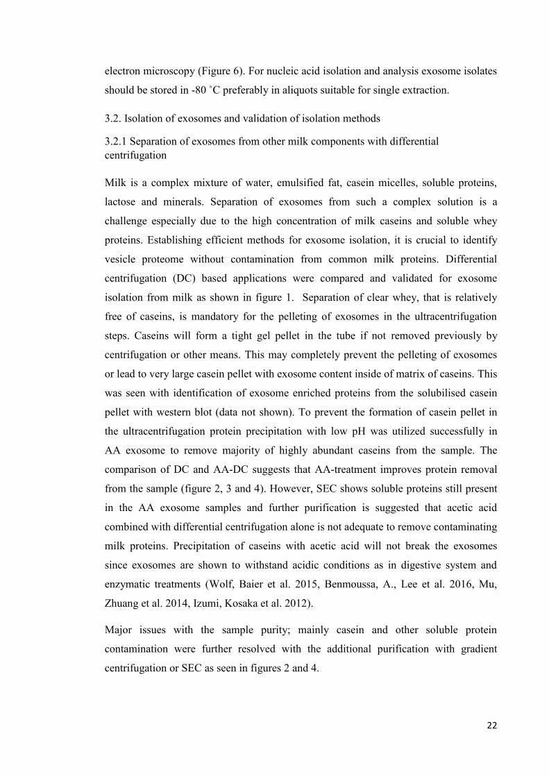
Density gradient centrifugation was utilized successfully to remove some of the
common milk proteins. However with the dilution of sucrose fractions that happened
during the preparation of gradient the tubes lacked a sufficiently dense fraction to float
the exosomes on top of therefore isolation of denser than 1.2 g/ml fractions were co-
isolated with the exosome fraction. Resulting densities of fractions can be seen in table
1. Exosomes were recovered from 30 -40 % fractions.
Fraction/ml Brix Refraction Density g/cm3 % of sucrose
1 0.6/0.7 1.3339 1.0023 0.6
2 2.1/2.2 1.3360 1.0082 2
3 5.3/5.3 1.3407 1.0209 5
4 9.6/9.8 1.3473 1.0388 10
5 15.1/15.1 1.3558 1.0615 15
6 19.7/19.8 1.3633 1.0816 20
7 24.9/25.0 1.3723 1.1036 25
8 30.3/30.1 1.3814 1.1301 30
9 36.0/36.0 1.3903 1.1513 35
10 40.6/40.7 1.3999 1.1764 40
Table 1.Sucrose density recovered from the gradient centrifugation fractions. Densities
recovered from table from United States department of agriculture
(greenwoodassociates.com) using the Brix index. Resulting exosome fractions are
recovered from 30-40 % sucrose fractions.
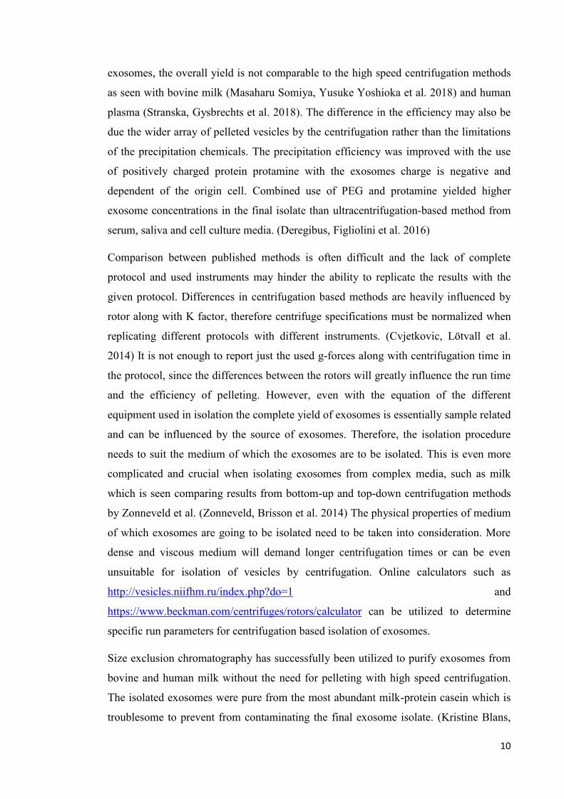
3.2.2 Removal of soluble proteins with size exclusion chromatography
To further purify isolated exosomes different conditions of size exclusion
chromatography were compared. First, column selection along with running buffers
with different urea concentrations were evaluated with whey exosome samples. Lower
urea concentration along with Superdex™ 200 column (GE Healtcare) resulted in
casein aggregates eluting along with the void volume peak fractions containing
exosomes. Second, the feasibility to remove protein contaminants with EDTA was
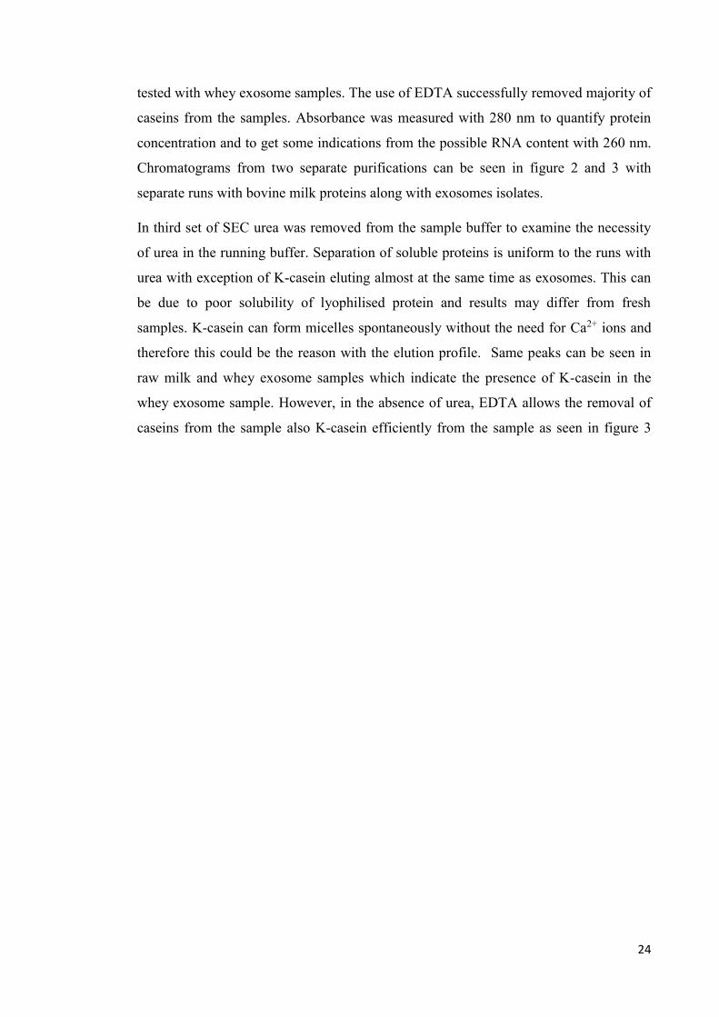
24
tested with whey exosome samples. The use of EDTA successfully removed majority of
caseins from the samples. Absorbance was measured with 280 nm to quantify protein
concentration and to get some indications from the possible RNA content with 260 nm.
Chromatograms from two separate purifications can be seen in figure 2 and 3 with
separate runs with bovine milk proteins along with exosomes isolates.
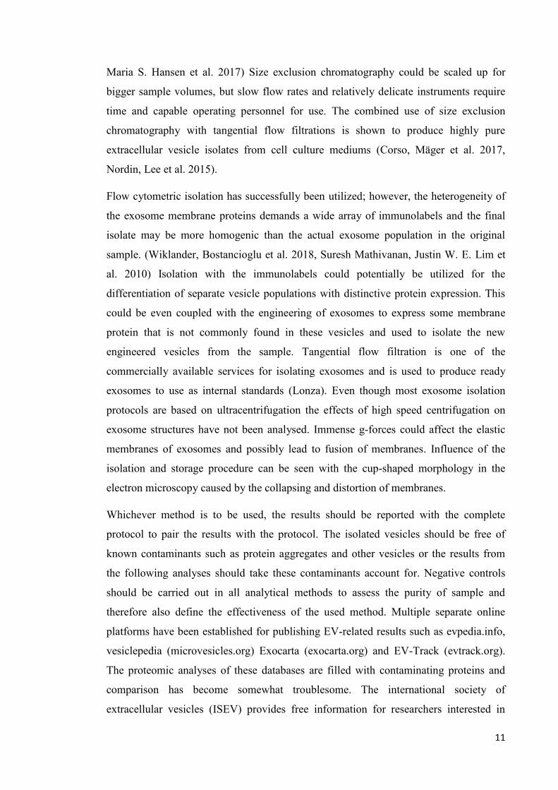
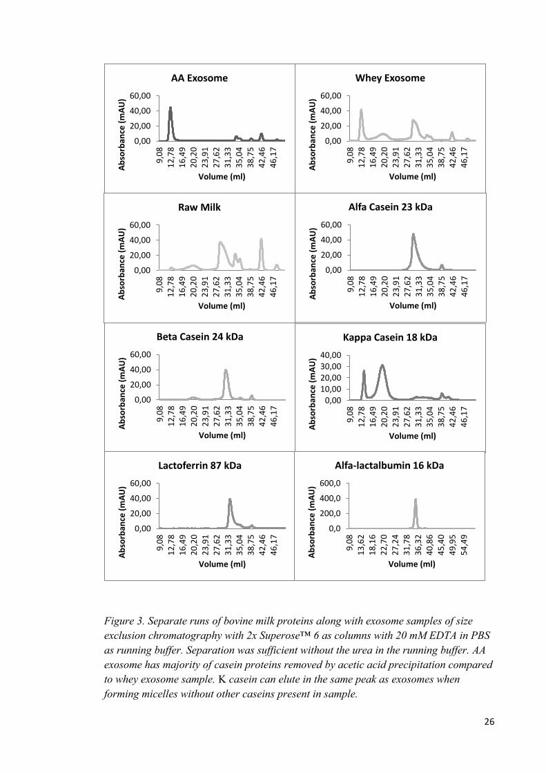
In third set of SEC urea was removed from the sample buffer to examine the necessity
of urea in the running buffer. Separation of soluble proteins is uniform to the runs with
urea with exception of K-casein eluting almost at the same time as exosomes. This can
be due to poor solubility of lyophilised protein and results may differ from fresh
samples. K-casein can form micelles spontaneously without the need for Ca2+ ions and
therefore this could be the reason with the elution profile. Same peaks can be seen in
raw milk and whey exosome samples which indicate the presence of K-casein in the
whey exosome sample. However, in the absence of urea, EDTA allows the removal of
caseins from the sample also K-casein efficiently from the sample as seen in figure 3
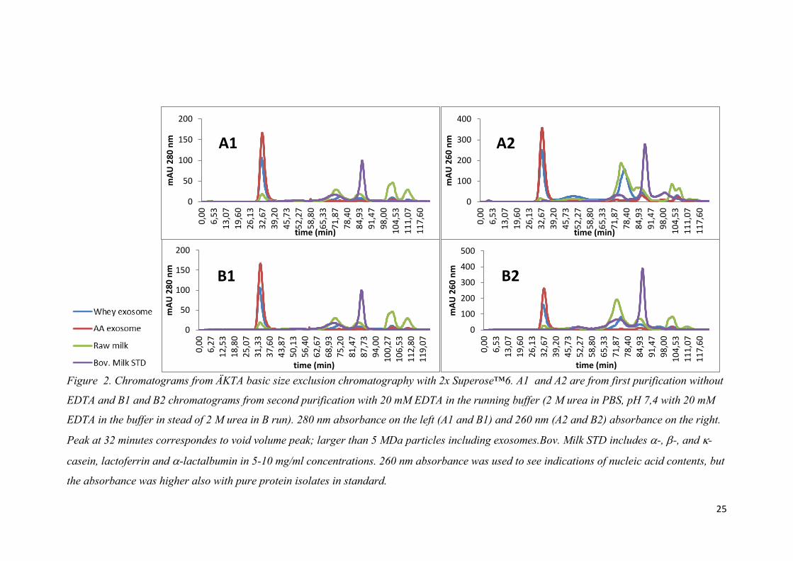
25
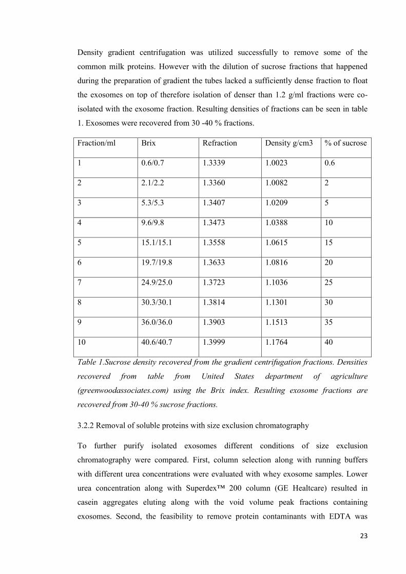
Figure 2. Chromatograms from ÄKTA basic size exclusion chromatography with 2x Superose™6. A1 and A2 are from first purification without
EDTA and B1 and B2 chromatograms from second purification with 20 mM EDTA in the running buffer (2 M urea in PBS, pH 7,4 with 20 mM
EDTA in the buffer in stead of 2 M urea in B run). 280 nm absorbance on the left (A1 and B1) and 260 nm (A2 and B2) absorbance on the right.
Peak at 32 minutes correspondes to void volume peak; larger than 5 MDa particles including exosomes.Bov. Milk STD includes -, -, and -
casein, lactoferrin and -lactalbumin in 5-10 mg/ml concentrations. 260 nm absorbance was used to see indications of nucleic acid contents, but
the absorbance was higher also with pure protein isolates in standard.
0
100
200
300
400
0,0
0
6,5
3
13
,07
19
,60
26
,13
32
,67
39
,20
45
,73
52
,27
58
,80
65
,33
71
,87
78
,40
84
,93
91
,47
98
,00
10
4,5
3
11
1,0
7
11
7,6
0
mA
U 2
60
nm
time (min)
A2
0
50
100
150
200
0,0
0
6,5
3
13
,07
19
,60
26
,13
32
,67
39
,20
45
,73
52
,27
58
,80
65
,33
71
,87
78
,40
84
,93
91
,47
98
,00
10
4,5
3
11
1,0
7
11
7,6
0
mA
U 2
80
nm
time (min)
A1
0
50
100
150
200
0,0
0
6,2
7
12
,53
18
,80
25
,07
31
,33
37
,60
43
,87
50
,13
56
,40
62
,67
68
,93
75
,20
81
,47
87
,73
94
,00
10
0,2
7
10
6,5
3
11
2,8
0
11
9,0
7
mA
U 2
80
nm
time (min)
B1
0
100
200
300
400
500
0,0
0
6,5
3
13
,07
19
,60
26
,13
32
,67
39
,20
45
,73
52
,27
58
,80
65
,33
71
,87
78
,40
84
,93
91
,47
98
,00
10
4,5
3
11
1,0
7
11
7,6
0
mA
U 2
60
nm
time (min)
B2
26
Figure 3. Separate runs of bovine milk proteins along with exosome samples of size
exclusion chromatography with 2x Superose™ 6 as columns with 20 mM EDTA in PBS
as running buffer. Separation was sufficient without the urea in the running buffer. AA
exosome has majority of casein proteins removed by acetic acid precipitation compared
to whey exosome sample. Κ casein can elute in the same peak as exosomes when
forming micelles without other caseins present in sample.
0,00
20,00
40,00
60,00
9,0
81
2,7
81
6,4
92
0,2
02
3,9
12
7,6
23
1,3
33
5,0
43
8,7
54
2,4
64
6,1
7
Ab
sorb
ance
(m
AU
)
Volume (ml)
AA Exosome
0,00
20,00
40,00
60,00
9,0
81
2,7
81
6,4
92
0,2
02
3,9
12
7,6
23
1,3
33
5,0
43
8,7
54
2,4
64
6,1
7
Ab
sorb
ance
(m
AU
)
Volume (ml)
Whey Exosome
0,00
20,00
40,00
60,00
9,0
81
2,7
81
6,4
92
0,2
02
3,9
12
7,6
23
1,3
33
5,0
43
8,7
54
2,4
64
6,1
7
Ab
sorb
ance
(m
AU
)
Volume (ml)
Raw Milk
0,00
20,00
40,00
60,00
9,0
81
2,7
81
6,4
92
0,2
02
3,9
12
7,6
23
1,3
33
5,0
43
8,7
54
2,4
64
6,1
7
Ab
sorb
ance
(m
AU
)Volume (ml)
Alfa Casein 23 kDa
0,00
20,00
40,00
60,00
9,0
81
2,7
81
6,4
92
0,2
02
3,9
12
7,6
23
1,3
33
5,0
43
8,7
54
2,4
64
6,1
7
Ab
sorb
ance
(m
AU
)
Volume (ml)
Beta Casein 24 kDa
0,0010,0020,0030,0040,00
9,0
81
2,7
81
6,4
92
0,2
02
3,9
12
7,6
23
1,3
33
5,0
43
8,7
54
2,4
64
6,1
7
Ab
sorb
ance
(m
AU
)
Volume (ml)
Kappa Casein 18 kDa
0,00
20,00
40,00
60,00
9,0
81
2,7
81
6,4
92
0,2
02
3,9
12
7,6
23
1,3
33
5,0
43
8,7
54
2,4
64
6,1
7
Ab
sorb
ance
(m
AU
)
Volume (ml)
Lactoferrin 87 kDa
0,0
200,0
400,0
600,0
9,0
8
13
,62
18
,16
22
,70
27
,24
31
,78
36
,32
40
,86
45
,40
49
,95
54
,49
Ab
sorb
ance
(m
AU
)
Volume (ml)
Alfa-lactalbumin 16 kDa
27
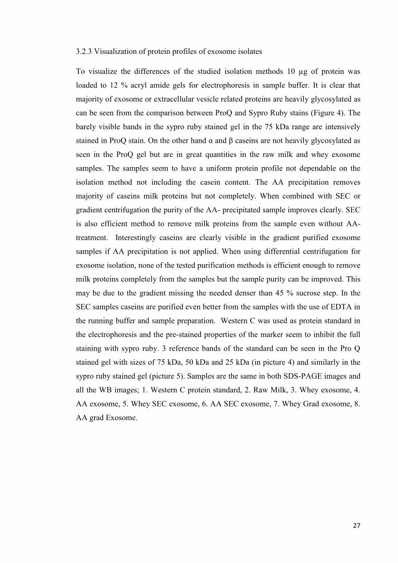
3.2.3 Visualization of protein profiles of exosome isolates
To visualize the differences of the studied isolation methods 10 µg of protein was
loaded to 12 % acryl amide gels for electrophoresis in sample buffer. It is clear that
majority of exosome or extracellular vesicle related proteins are heavily glycosylated as
can be seen from the comparison between ProQ and Sypro Ruby stains (Figure 4). The
barely visible bands in the sypro ruby stained gel in the 75 kDa range are intensively
stained in ProQ stain. On the other hand α and β caseins are not heavily glycosylated as
seen in the ProQ gel but are in great quantities in the raw milk and whey exosome
samples. The samples seem to have a uniform protein profile not dependable on the
isolation method not including the casein content. The AA precipitation removes
majority of caseins milk proteins but not completely. When combined with SEC or
gradient centrifugation the purity of the AA- precipitated sample improves clearly. SEC
is also efficient method to remove milk proteins from the sample even without AA-
treatment. Interestingly caseins are clearly visible in the gradient purified exosome
samples if AA precipitation is not applied. When using differential centrifugation for
exosome isolation, none of the tested purification methods is efficient enough to remove
milk proteins completely from the samples but the sample purity can be improved. This
may be due to the gradient missing the needed denser than 45 % sucrose step. In the
SEC samples caseins are purified even better from the samples with the use of EDTA in
the running buffer and sample preparation. Western C was used as protein standard in
the electrophoresis and the pre-stained properties of the marker seem to inhibit the full
staining with sypro ruby. 3 reference bands of the standard can be seen in the Pro Q
stained gel with sizes of 75 kDa, 50 kDa and 25 kDa (in picture 4) and similarly in the
sypro ruby stained gel (picture 5). Samples are the same in both SDS-PAGE images and
all the WB images; 1. Western C protein standard, 2. Raw Milk, 3. Whey exosome, 4.
AA exosome, 5. Whey SEC exosome, 6. AA SEC exosome, 7. Whey Grad exosome, 8.
AA grad Exosome.
28
Figure 4. Milk exosome samples separated on 12 % (w/v) acryl amide gel stained by
first Pro Q Emerald 300 glycoprotein stain(upper picture) and second with Sypro Ruby
gel stain (lower picture). Majority of exosome fraction proteins were seen in the 75-150
kDa range with high level of glycosylation. Caseins are present in all samples in the 25
kDa range, with majority of caseins present in raw milk (2) and UC exosome (3). 1.
Western C protein standard, 2. Raw Milk, 3. Whey exosome, 4. AA exosome, 5. Whey
SEC exosome, 6. AA SEC exosome, 7. Whey Grad exosome, 8. AA Grad Exosome.
3.2.4 Size and morphology of isolated vesicles
To validate the success of exosome isolation the size and morphology of isolated
vesicles were analysed with NTA and TEM. Exosome samples from bovine milk
29
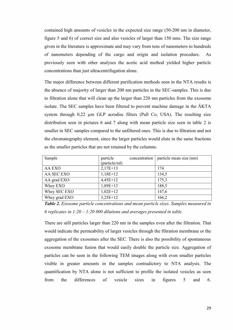
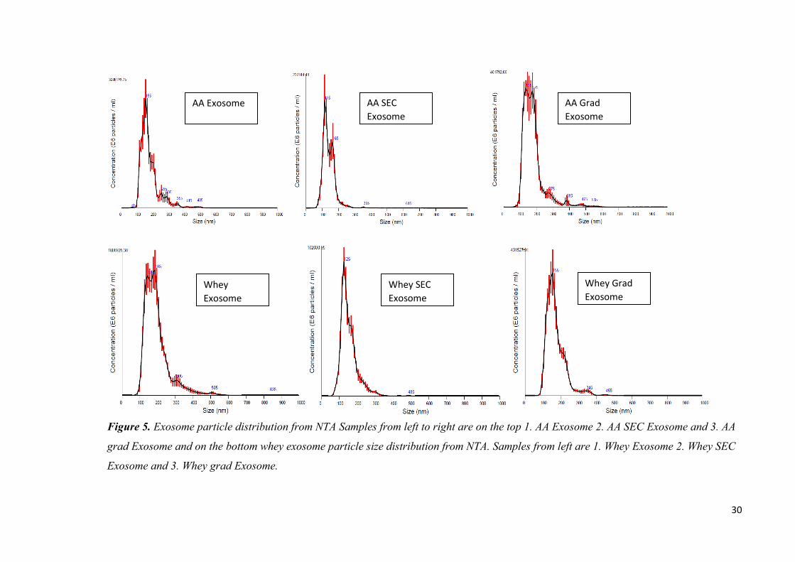
contained high amounts of vesicles in the expected size range (50-200 nm in diameter,
figure 5 and 6) of correct size and also vesicles of larger than 150 nms. The size range
given in the literature is approximate and may vary from tens of nanometers to hundreds
of nanometers depending of the cargo and origin and isolation procedure. As
previously seen with other analyses the acetic acid method yielded higher particle
concentrations than just ultracentrifugation alone.
The major difference between different purification methods seen in the NTA results is
the absence of majority of larger than 200 nm particles in the SEC-samples. This is due
to filtration alone that will clean up the larger than 220 nm particles from the exosome
isolate. The SEC samples have been filtered to prevent machine damage in the ÄKTA
system through 0,22 µm GLP acrodisc filters (Pall Co, USA). The resulting size
distribution seen in pictures 6 and 7 along with mean particle size seen in table 2 is
smaller in SEC samples compared to the unfiltered ones. This is due to filtration and not
the chromatography element, since the larger particles would elute in the same fractions
as the smaller particles that are not retained by the columns.
Sample particle concentration
(particle/ml)
particle mean size (nm)
AA EXO 2,17E+13 174
AA SEC EXO 1,18E+12 134,5
AA grad EXO 4,45E+12 175,3
Whey EXO 1,89E+13 188,5
Whey SEC EXO 1,02E+12 147,6
Whey grad EXO 3,25E+12 166,2
Table 2. Exosome particle concentrations and mean particle sizes. Samples measured in
6 replicates in 1:20 – 1:20 000 dilutions and averages presented in table.
There are still particles larger than 220 nm in the samples even after the filtration. That
would indicate the permeability of larger vesicles through the filtration membrane or the
aggregation of the exosomes after the SEC. There is also the possibility of spontaneous
exosome membrane fusion that would easily double the particle size. Aggregation of
particles can be seen in the following TEM images along with even smaller particles
visible in greater amounts in the samples contradictory to NTA analysis. The
quantification by NTA alone is not sufficient to profile the isolated vesicles as seen
from the differences of vesicle sizes in figures 5 and 6.
30
Figure 5. Exosome particle distribution from NTA Samples from left to right are on the top 1. AA Exosome 2. AA SEC Exosome and 3. AA
grad Exosome and on the bottom whey exosome particle size distribution from NTA. Samples from left are 1. Whey Exosome 2. Whey SEC
Exosome and 3. Whey grad Exosome.
AA Exosome AA SEC
Exosome
AA Grad
Exosome
Whey
Exosome
Whey SEC
Exosome
Whey Grad
Exosome
31
Images from transmission electron microscopy show vesicles of different shapes and
sizes. Smallest vesicles present in all samples range from 30 nm:s up that still present
vesicular morphology with distinctive lipid bilayer characteristics. Vesicles of smaller
size seem to be more prone to aggregate as seen from the clear groups and chains
formed by the small vesicles (figure 6). The small sized vesicles may represent
phospholipid micelles, or fat droplets commonly dispersed in milk. Largest particles
present in the samples range up to 500 nm that could be plasma membrane-derived
microvesicles or milk fat globules uniquely found in milk. Phospholipid trilayer
surrounded milk fat globules contain fat that crystallizes in low temperatures leading to
rigid bridge like structures inside vesicle as seen in figure 7B. However the differential
centrifugation excludes majority of the bigger sized vesicles out from the final isolate
and the seen larger vesicles can be result of membrane fusion during the isolation and
storage of samples. Larger vesicle with inner smaller vesicles can be seen in the
figure7B. Isolated exosomes present both cup-shape (figure 7D) and spherical
morphology in all the samples with more structural distress seen in the SEC (figure 7E).
This may be due to the 2 M urea concentration causing vesicle breakage due to osmotic
shock. Casein can be seen in the images with higher electron density in the calcium
phosphate bound in the protein. These micelles are visible in darker shades of gray as
also seen by Léonil et al. (Marchin, Puteaux et al. 2007). The casein micelles can
overlap the exosome structures indicating that the caseins are in interaction with the
exosomes or encased to spontaneously formed membrane vesicles from disrupted cell
membranes or milk fat globules.
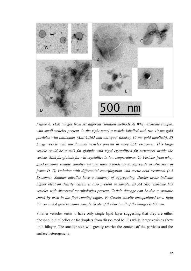
32
Figure 6. TEM images from six different isolation methods A) Whey exosome sample,
with small vesicles present. In the right panel a vesicle labelled with two 10 nm gold
particles with antibodies (Anti-CD63 and anti-goat (donkey 10 nm gold labelled)). B)
Large vesicle with intraluminal vesicles present in whey SEC exosomes. This large
vesicle could be a milk fat globule with rigid crystallized fat structures inside the
vesicle. Milk fat globule fat will crystallize in low temperatures. C) Vesicles from whey
grad exosome sample. Smaller vesicles have a tendency to aggregate as also seen in
frame D. D) Isolation with differential centrifugation with acetic acid treatment (AA
Exosome). Smaller micelles have a tendency of aggregating. Darker areas indicate
higher electron density; casein is also present in sample. E) AA SEC exosome has
vesicles with distressed morphologies present. Vesicle damage can be due to osmotic
shock by urea in the first running buffer. F) Casein micelle encapsulated by a lipid
bilayer in AA grad exosome sample. Scale of the bar in all of the images is 500 nm.
Smaller vesicles seem to have only single lipid layer suggesting that they are either
phospholipid micelles or fat droplets from dissociated MFGs while larger vesicles show
lipid bilayer. The smaller size will greatly restrict the content of the particles and the
surface heterogeneity.
33
Antibody labelling was applied to separate exosomes from other particles present in
samples. With the used CD63 it seems that CD63 is more prominent in smaller vesicles
with more antibodies are bound to smaller (<100 nm) vesicles than to larger vesicles.
However this may be due poor antibody specificity as seen in western blot analysis or
problems with ratio of antibody and vesicles. The antibody labelling affected only a
small fraction of vesicles with majority of gold labels binding to single antibodies per
vesicle. Gold particles used in the secondary antibody are only present in interaction
with membrane related particles and not with soluble proteins, such as high electron
density containing casein for as example. However similar distribution of CD63 to
smaller sized vesicles was seen in HeLa cell derived exosomes. (Colombo, Moita et al.
2013) Resulting CD63 labelled images can be seen in appendix 1 along with wide scale
images of different isolation methods and in frame A of figure 6. Limitations of SEC
purification can be seen in figure 6 frame B, with very large vesicle still present in
sample after the SEC. This is due to the inability of the columns to separate larger than
5 MDa particles from each other.
3.3. Identification of exosome enriched markers
Milk samples were analysed with several different primary antibodies generally used in
exosome studies for exosome protein marker identification. Successful identification of
exosome enriched proteins was done with anti-Alix, anti-TSG101 and to some extent
anti-CD81 (figure 7) and therefore identifying the isolated vesicles as exosomes. The
tested CanX, CD63 and β -casein antibodies bound to almost all proteins present in the
samples are could not therefore be utilised for the characterization of specific proteins.
This was accompanied with absence of the correct sized bands with the CD63 and β -
casein antibody (data not shown).
34
Figure 7. Western blot of milk exosomes with anti-Alix, anti-TSG101 and anti-CD81
antibodies. Top 3 bands correspond to Alix protein with size of 100-80 kDa. Anti-TSG
regocnizes two bands in samples 4, 7 and 8. CD81 binds only to samples 3 and 4 with
sufficient effiency for the chemiluminesence signal to be detected under the used
parameters. Different location of the band is due to heavy casein concentration pushing
CD81 band out of locus in sample 3. Casein concentration can be seen in figure 4.
Samples are 1. Western C protein standard, 2. Raw Milk, 3. Whey exosome, 4. AA
exosome, 5. Whey SEC exosome, 6. AA SEC exosome, 7. Whey Grad exosome, 8. AA
Grad Exosome
Alix is 95 kDa protein by peptide sequence and the corresponding band with few others
are clearly visible in all but AA-SEC exosome samples. Similar protein bands are
presented on the Cell Signalling web site for anti-alix-reactivity
(https://www.cellsignal.com/products/primary-antibodies/alix-e6p9b-rabbit-mab/92880?site-
search-type=Products).
With TSG101 the quantity of bound antibody was much lower compared to anti-Alix
resulting in lower intensity in the signal. Clear bands are only visible in AA exosome
and gradient exosome samples with some faint bands in whey exosome and whey SEC
35
exosome samples. The intensity of the protein standard masks the more faint bands of
the milk samples.
As seen with TSG101 the intensity of CD81 is lower than anti-Alix. The highest
intensity of CD81 band is seen in the acetic acid exosome sample. SEC samples show
no bands and really faint band is seen in gradient purified exosome samples. The
different migration of CD81 bands is due to excessively high casein content still present
in whey exosome sample as seen in figure 4. However the quantification of proteins via
WB is only semi-quantitative and without specific protein standards to use in parallel
the analysis should be done only qualitatively. Full western blot images are shown in
appendix 2 with clear unspecific binding seen with all antibodies.
3.4. Proteome and functional analysis of exosomal proteins
63 proteins were identified in 5 separate mass spectrometry runs with different samples
containing bovine milk exosomes. The identification was done with 3 or more unique
peptides for all proteins identified. Resulting protein list was analyzed with DAVID and
Panther online tools and 34 of 59 identified and gene mapped proteins were
compartmentalized in extracellular exosomes as seen in table GOTERM. 50 of
identified proteins were found to have molecular function (figure 8) and 20 of those
proteins were described with catalytic activity. 23 of total 63 identified proteins were
related to localization of proteins as seen in figure 9. On top of the resulting protein
identifications there were also six uncharacterized proteins G5E5T5, G5E513,
G3N0V0, G3N2D8, G5E5H5 and F1MCF8 present in the samples.
36
Figure 8. Proteins identified from exosomes mapped for their biological processes by
PANTHER gene ontology.
The cellular compartmentalization of identified proteins localizes the proteins mainly to
extracellular exosomes, plasma membrane, extracellular space and to focal adhesion.
List of 7 most abundant hits of GOTERMS can be seen in table 3. This could indicate
the pathway for exosome trafficking from cell to another and a possible messaging
system for the target cell. With the amount of proteins identified the probability of
random localization of analyzed proteins is relatively high in large categories such as
plasma membrane, cytosol and extracellular space. This can be seen with the Benjamini
corrected P-value of 2,1e-2 that is still statistically significant (p-value < 0,05) but the
value is much higher than with the other GOTERMs. However the localization for term
focal adhesion and extracellular exosomes are certainly specific for these proteins. This
still does not rule out the possibility of falsely categorized proteins to the extracellular
exosomes GOTERM. With mass spectrometer with higher sensitivity the list of
identified proteins could be vastly improved and larger dataset would lead to better
localization of proteins for their GOTERMS and biological processes.
12 %
9 %
23 %
23 %
8 %
1 %
7 %
5 %
3 % 8 %
1 %
Biological process annotationcellular component organization orbiogenesis (GO:0071840) 12cellular process (GO:0009987) 9
localization (GO:0051179) 23
biological regulation (GO:0065007) 24
response to stimulus (GO:0050896) 8
signaling (GO:0023052) 1
developmental process (GO:0032502) 7
multicellular organismal process(GO:0032501) 5biological adhesion (GO:0022610) 3
metabolic process (GO:0008152) 8
immune system process (GO:0002376) 1
37
Category Term Gene count Benjamini corrected
p value
GOTERM_CC_DIRECT extracellular
exosome 34
3.0E-14
GOTERM_CC_DIRECT plasma membrane 17 2.1E-2
GOTERM_CC_DIRECT extracellular space 16 1.4E-5
GOTERM_CC_DIRECT focal adhesion 15 1.1E-10
GOTERM_CC_DIRECT cytosol 15 1.1E-4
GOTERM_CC_DIRECT membrane 13 5.8E-4
GOTERM_CC_DIRECT extracellular region 11 1.1E-4
Table 3. Enriched cellular compartments for proteins identified from exosomes with
their respective gene count and corrected p-value.
Figure 9. 50 of identified proteins found to have molecular function, mapped with
PANTHER.
Entry name Proteins Gene names
A0A140T897_BOVIN Serum albumin ALB
A0A140T8A9_BOVIN Κ -casein CSN3
LALBA_BOVIN
Alpha-lactalbumin (Lactose synthase B protein)
(allergen Bos d 4)
LALBA
ALACTA
CASA1_BOVIN
Alpha-S1-casein (allergen Bos d 8) [Cleaved
into: Antioxidant peptide] CSN1S1
40 %
6 %
4 %
40 %
10 %
Molecular function
binding (GO:0005488)
structural moleculeactivity (GO:0005198)
molecular functionregulator (GO:0098772)
catalytic activity(GO:0003824)
transporter activity(GO:0005215)
38
CASA2_BOVIN
Alpha-S2-casein [Cleaved into: Casocidin-1
(Casocidin-I)] CSN1S2
CASB_BOVIN
β -casein [Cleaved into: Casoparan; Antioxidant
peptide; Casohypotensin] CSN2
CASK_BOVIN
Κ -casein [Cleaved into: Casoxin-C; Casoxin-6;
Casoxin-A; Casoxin-B; Casoplatelin]
CSN3 CSN10
CSNK
LACB_BOVIN β -lactoglobulin (β -LG) (allergen Bos d 5) LGB
APOA1_BOVIN
Apolipoprotein A-I (Apo-AI) (ApoA-I)
(Apolipoprotein A1) [Cleaved into:
Proapolipoprotein A-I (ProapoA-I);
apolipoprotein A-I] APOA1
TRFL_BOVIN
Lactotransferrin (Lactoferrin) (EC 3.4.21.-)
[Cleaved into: Lactoferricin-B (Lfcin-B)] LTF
B2D1N9_BOVIN
ATP-binding cassette sub-family G member 2
(Breast cancer resistance protein) ABCG2
F1MCF8_BOVIN Uncharacterized protein LOC112441499
F1MHI1_BOVIN Perilipin PLIN2
F1MUT3_BOVIN Xanthine dehydrogenase/oxidase XDH
F1N726_BOVIN Glycoprotein 2 GP2
G3MYH4_BOVIN Tetraspanin CD81
G3N0V0_BOVIN Uncharacterized protein
G3N2D8_BOVIN Uncharacterized protein GGT1
G5E513_BOVIN Uncharacterized protein
G5E5H5_BOVIN Uncharacterized protein
G5E5H7_BOVIN Uncharacterized protein PAEP
G5E5T5_BOVIN Uncharacterized protein
NB5R3_BOVIN
NADH-cytochrome b5 reductase 3 (B5R)
(Cytochrome b5 reductase) (EC 1.6.2.2)
(Diaphorase-1) [Cleaved into: NADH-
cytochrome b5 reductase 3 membrane-bound
form; NADH-cytochrome b5 reductase 3
soluble form] CYB5R3 DIA1
UBB_BOVIN Polyubiquitin-B [Cleaved into: Ubiquitin] UBB
FABPH_BOVIN
Fatty acid-binding protein, heart (Fatty acid-
binding protein 3) (Heart-type fatty acid-binding
protein) (H-FABP) (Mammary-derived growth
inhibitor) (MDGI) FABP3
GBB2_BOVIN
Guanine nucleotide-binding protein
G(I)/G(S)/G(T) subunit β -2 (G protein subunit
β -2) (Transducin β chain 2) GNB2
39
ENPP3_BOVIN
Ectonucleotide
pyrophosphatase/phosphodiesterase family
member 3 (E-NPP 3) (Phosphodiesterase I β)
(PD-I β) (Phosphodiesterase I/nucleotide
pyrophosphatase 3) (CD antigen CD203c)
[Includes: Alkaline phosphodiesterase I (EC
3.1.4.1); Nucleotide pyrophosphatase (NPPase)
(EC 3.6.1.9) (Nucleotide diphosphatase)] ENPP3
BT1A1_BOVIN Butyrophilin subfamily 1 member A1 (BT) BTN1A1 BTN
HSP7C_BOVIN
Heat shock cognate 71 kDa protein (Heat shock
70 kDa protein 8) HSPA8 HSC70
CD36_BOVIN
Platelet glycoprotein 4 (Glycoprotein IIIb)
(GPIIIB) (PAS IV) (PAS-4) (Platelet
glycoprotein IV) (GPIV) (CD antigen CD36) CD36 PAS4
CD9_BOVIN CD9 antigen (CD antigen CD9) CD9
EZRI_BOVIN Ezrin (Cytovillin) (Villin-2) (p81) EZR VIL2
GDIB_BOVIN
Rab GDP dissociation inhibitor β (Rab GDI β)
(Guanosine diphosphate dissociation inhibitor 2)
(GDI-2)
GDI2
RABGDIB
ACTB_BOVIN
Actin, cytoplasmic 1 (β -actin) [Cleaved into:
Actin, cytoplasmic 1, N-terminally processed] ACTB
RAP1B_BOVIN
Ras-related protein Rap-1b (GTP-binding
protein smg p21B) RAP1B
RHOA_BOVIN Transforming protein RhoA (Gb) (p21)
RHOA ARHA
RHO12
GBB1_BOVIN
Guanine nucleotide-binding protein
G(I)/G(S)/G(T) subunit β -1 (Transducin β
chain 1) GNB1
PPIA_BOVIN
Peptidyl-prolyl cis-trans isomerase A (PPIase
A) (EC 5.2.1.8) (Cyclophilin A) (Cyclosporin
A-binding protein) (Rotamase A) [Cleaved into:
Peptidyl-prolyl cis-trans isomerase A, N-
terminally processed] PPIA
RAC1_BOVIN
Ras-related C3 botulinum toxin substrate 1
(p21-Rac1) RAC1
EF1A1_BOVIN
Elongation factor 1-alpha 1 (EF-1-alpha-1)
(Elongation factor Tu) (EF-Tu) (Eukaryotic
elongation factor 1 A-1) (eEF1A-1)
EEF1A1
EEF1A EF1A
GLCM1_BOVIN
Glycosylation-dependent cell adhesion molecule
1 (GlyCAM-1) (28 kDa milk glycoprotein PP3)
(Lactophorin) (Proteose-peptone component 3)
(PP3) GLYCAM1
XDH_BOVIN
Xanthine dehydrogenase/oxidase [Includes:
Xanthine dehydrogenase (XD) (EC 1.17.1.4);
Xanthine oxidase (XO) (EC 1.17.3.2) (Xanthine
oxidoreductase) (XOR)] XDH
40
PIGR_BOVIN
Polymeric immunoglobulin receptor (PIgR)
(Poly-Ig receptor) [Cleaved into: Secretory
component] PIGR
ANXA5_BOVIN
Annexin A5 (Anchorin CII) (Annexin V)
(Annexin-5) (Calphobindin I) (CBP-I)
(Endonexin II) (Lipocortin V) (Placental
anticoagulant protein 4) (Placental anticoagulant
protein I) (PAP-I) (Thromboplastin inhibitor)
(Vascular anticoagulant-alpha) (VAC-alpha) ANXA5 ANX5
ARF1_BOVIN ADP-ribosylation factor 1 ARF1
5NTD_BOVIN
5'-nucleotidase (5'-NT) (EC 3.1.3.5) (Ecto-5'-
nucleotidase) (CD antigen CD73)
NT5E NT5
NTE
RAB18_BOVIN Ras-related protein Rab-18 RAB18
CIB1_BOVIN
Calcium and integrin-binding protein 1
(Calmyrin) CIB1
RHOC_BOVIN Rho-related GTP-binding protein RhoC RHOC ARHC
NPT2B_BOVIN
Sodium-dependent phosphate transport protein
2B (Sodium-phosphate transport protein 2B)
(Na(+)-dependent phosphate cotransporter 2B)
(Sodium/phosphate cotransporter 2B) (Na(+)/Pi
cotransporter 2B) (NaPi-2b) (Solute carrier
family 34 member 2) SLC34A2
RAB1B_BOVIN Ras-related protein Rab-1B RAB1B
CDC42_BOVIN Cell division control protein 42 homolog CDC42
RB11B_BOVIN Ras-related protein Rab-11B RAB11B
Q3SYR8_BOVIN
Immunoglobulin J chain (Joining chain of
multimeric IgA and IgM) JCHAIN IGJ
RAB7A_BOVIN Ras-related protein Rab-7a RAB7A RAB7
CD81_BOVIN CD81 antigen (CD antigen CD81) CD81
ABCG2_BOVIN
ATP-binding cassette sub-family G member 2
(Urate exporter) (CD antigen CD338) ABCG2
MCP_BOVIN Membrane cofactor protein (CD antigen CD46) CD46 MCP
MUC15_BOVIN
Mucin-15 (MUC-15) (Component II)
(Glycoprotein 4) (Glycoprotein C) (PAS3)
(PASIII) MUC15
MUC1_BOVIN
Mucin-1 (MUC-1) (CD antigen CD227)
[Cleaved into: Mucin-1 subunit alpha (MUC1-
NT) (MUC1-alpha); Mucin-1 subunit β (MUC1-
β) (MUC1-CT)] MUC1
MFGM_BOVIN
Lactadherin (BP47) (Components 15/16)
(MFGM) (MGP57/53) (Milk fat globule-EGF
factor 8) (MFG-E8) (PAS-6/PAS-7
glycoprotein) (SED1) (Sperm surface protein
SP47) MFGE8
PLIN2_BOVIN
Perilipin-2 (Adipophilin) (Adipose
differentiation-related protein) (ADRP)
PLIN2 ADFP
ADRP
41
IDHC_BOVIN
Isocitrate dehydrogenase [NADP] cytoplasmic
(IDH) (EC 1.1.1.42) (Cytosolic NADP-
isocitrate dehydrogenase) (IDP) (NADP(+)-
specific ICDH) (Oxalosuccinate decarboxylase) IDH1 ICDH
Table 4. Proteins identified with mass spectrometry. Proteins stained red are common
milk proteins that are present in samples and not specific for exosomes.
4. Discussion
4.1 Isolation and morphology of vesicles
The molecular complexity of milk provides certain challenges in nanovesicle related
work. Milk fat droplets can form with same density as exosomes and therefore will be
located in the same fractions as exosomes in gradient centrifugation. These particles are
accompanied with casein micelles or aggregates that spontaneously form in milk even
after casein precipitation. These micelles can range from the size of single casein
protein from 25 kDa to 60 - 200 nms of which normal casein micelles form (Fox,
McSweeney 2003) or up to gel or cheese like complexes as done in the isolation steps.
The casein micelles are highly stable and not affected by addition of urea. The used 20
mM EDTA concentration is relative to the amount of colloidal Ca2+ ions in casein
micelles in bovine milk. (Fox, McSweeney 2003) Despite the efforts to remove majority
of the soluble proteins there are still common soluble milk proteins present in the
samples even after additional purification with SEC or gradient purification.
In SEC the urea concentration was assumed to be safe to not cause osmotic shock to
exosomes, but from the TEM images can be seen the flaky and disrupted morphology in
the SEC-samples. This is most likely from the osmotic shock caused by addition of urea
accompanied with some shear stress from the chromatography column. EDTA was
utilized in 20 mM concentration to dissipate formed casein aggregates to allow the
separation with size as previously done by Blans et al. (Kristine Blans, Maria S. Hansen
et al. 2017)
The separation of the protein fractions could be increased with stronger running buffers
that could include SDS, EDTA, mercaptoethanol or DTT. However the SDS in the
running buffer would lead to exosome membrane dissipation and loss of sample. The
better resolution between the proteins is not mandatory since exosomes are recovered in
42
the void volume peak. However the difference between exosome peak and second peak
needs to be clear for efficient purification of exosomes. This could be problematic with
the pure K-casein close to the void volume peak but this seems to be different with
isolated protein and a raw milk sample.
SEC provided a novel in-house identification of particles from the samples. With no
NTA or other similar device available to identify vesicle size distribution the ÄKTA
chromatography system with SEC columns provided a novel way of identifying
particles from the samples in the void volume peak of the elution. This was due to the
separation capacity of the columns and therefore provided a UV-based identification for
larger than 5 MDa particles from samples.
The clear differences between the glycosylation of milk exosome proteins can be seen
in the comparison between pro Q and sypro ruby stained protein gels. Glycosylation is
crucial for function of exosomes and the loss of glycosylation will inhibit the
functionality of exosomes. Use of acetic acid does not seem to have an effect on the
glycosylation of the exosome proteins, since the intensities of proteins are comparable
to the intensities of exosomes isolated by centrifugation alone. In this study we did not
have the means to study the functional effects of the acetic acid treatments and therefore
suggesting milder isolation protocol with differential centrifugation along with SEC to
remove soluble proteins. This method could be utilised to isolate purer and possibly
more native-like vesicles from milk. Glycosylated proteins are clearly enriched in all
isolated samples compared to raw milk sample, with all samples containing 10 µg of
protein loaded to the gel. The pro Q glycoprotein stain will also stain proteins with very
high protein concentration and the staining of casein bands may be due to that
nonspecific staining to some extent. K-casein with size of 18 kDa will be visible on the
pro Q stained gels as K-casein can be glycosylated up to nine amino acid residues and
can be described as a hairy protein due to the high level of glycosylation.
Nanoparticle tracking analysis and transmission electron microscopy are two
recommended analytical methods for characterization of extracellular vesicles
(evtrack.org). The limits of NTA accuracy can be seen comparing the results to the
TEM images, since there are clearly smaller than 100 nm particles visible in all samples
despite NTA showing basically no particles of this size. TEM images however confirm
the aggregation of vesicles in groups and chain like formations especially in the smaller
43
sized exosomes. NTA is based on Brownian motion and light scattering of the particles
and the resulting particle aggregates will have the characteristics of larger particles
while still being built of smaller vesicles. This interaction is understandable since some
of the identified proteins in isolated samples are related to cell adhesion and cellular
component organization which could mediate the interaction of vesicles. The size
differences between our set of samples compared to vesicles described in literature can
be also due to vesicle swelling or shrinking in different solutions and different steps of
storage conditions. As previously described casein is present in almost all samples as
seen in TEM images. The interaction with the casein along with formation of chain like
structures could explain the median size of vesicles being larger than 150 nm in all but
SEC samples. The quantification of vesicles from the TEM images is not quantitative
since the micrograms produced are just certain levels of the sample, not the complete
3D image.
4.2 Identification of exosome enriched markers
Antibodies used in western blot showed some levels of nonspecific binding but some
migration differences could be due to the different glycosylation of identified proteins.
Pro Q stained gel will provide challenges for identifying the correct bands on the
membrane. This is accompanied by the lack of available protein standards to use in
parallel to identify and quantify the correct protein bands in WB. Such protein standards
could include CD81, CD63 or other exosome marker proteins to identify specific bands
from the samples. Sample protein and standard marker concentrations need to be
optimized, since the really intense bands of standard can mask the signal from the faint
bands in the samples. Glycosylation also greatly influences the mobility of proteins in
the electrophoresis and proteins can be located in different location than based on the
protein size alone.
Still even without entirely specific antibodies, successful identification of exosomal
marker proteins was done for some of the samples. Anti-ALIX antibody worked with
the highest sensitivity and identification was successful from all but AA-SEC sample.
Samples also contained exosomal marker proteins that were identified in mass
spectrometry such as HSC70, CD81 and CD9 that could be utilized also in western blot
to identify the vesicles as exosomes or to be coupled to some immuno-based isolation
methods with only limiting factor being the proteins need to be membrane proteins to be
44
utilized in isolation. Mass spectrometry results also contain interesting uncharacterized
proteins among with the enriched exosomal marker proteins. From the six
uncharacterized proteins G5E5H5 has no described functionality and closest identified
protein with only 78,5 % protein identity is UPF0472 protein C16orf72 (chromosome
16 open reading frame 72) from sperm whale, that has no described function.
Homologous protein expressed in Zebrafish is related to cellular response to xenobiotic
stimulus (uniprot entry A2BE76). The protein could be therefore related to antibodies as
with the 4 other uncharacterized proteins.
The uncharacterized protein G3N2D8 is encoded by GG1 gene which is related to
glutathione recycling and amino acid metabolism by similarity and gene ontology.
Remaining 4 uncharacterized proteins are related to IgM antibodies according to
BLAST results of amino acid sequences. It comes as no surprise that from the large
quantity of immunogens present in milk fraction is also present in exosome fraction.
Our data suggests that the immunogens should be localized in the vesicles and not
soluble free proteins, since these would be discarded in the isolation method, first in the
differential centrifugation step and second in the SEC to remove the remaining soluble
proteins. This could be also one of the passageways for the mother to deliver specific
immunogens to the infant as a bioavailable immuno-programming.
The amount of identified proteins is low due to the lack of systems sensitivity as
previously Reinhardt et al identified over 2000 proteins related to bovine milk
exosomes. (Reinhardt, Lippolis et al. 2012) The proteins identified in our experiments
were also present in the most abundant proteins identified in analysis by Reinhardt et al.
Contradictory to paper by Benmoussa et al we also identified xanthine hydrogenase in
100 000 xg isolated pellet compared to enrichment to fractions that they isolated by
lower speed centrifugations. Some of our protein identifications could be thefore
derived from co-isolated milk fat globules or fragmented milk fat globules with
majority of xanthine hydrogenase is localized in milk fat globules. (Benmoussa,
Abderrahim, Gotti et al. 2019) Due to the limitations of the used mass spectrometry
system results are pooled from all the different isolation methods and not represented as
separate lists. Major differences were found in the abundance of common milk protein
related peptides between the isolation methods.
45
4.3 Further analysis and use of isolation protocols
As stated before the isolation procedure needs to suit the following experiments. The
differences seen in the protein profile, vesicle concentration and morphology can
influence greatly the suitability of the samples for different analyses. Retaining the
glycosylation of exosome membrane proteins is crucial for working with cell or tissue
models as an example (Kusuma, Manca et al. 2016). Keeping the limitations of different
isolation procedures in mind it is mandatory to work with as pure exosome samples as
possible to rule out false positive results. Discovering the complete proteome could
enable the separation to separate subpopulations by their protein content as described
previously by Kowal et al. (Joanna Kowal, Guillaume Arras et al. 2016) This set of
proteome date could also be utilized to discard unwanted impurities in the isolation
procedure. For further experiments with mass spectrometry combinatory purification
with both SEC and gradient centrifugation could be utilized to exclude majority of
unwanted impurities. Even further purified or sub-populations could be isolated with
anti-body based methods to isolate specific vesicle populations for further analysis. This
may result in loss of sample and therefore can be difficult to utilize for use in cell
culture studies or feeding experiments. For these studies it would be more suitable to
have a soluble protein mixture as a reference for the negative control utilized in the
studies. However depending on the downstream analysis; metabolome analysis, RNA-
sequencing or quantitative polymerase chain reaction analysis the experiments have to
be highly controlled to identify the actual influence of the exosomes and their contents
rather than the effect of cells being exposed to a stimulus.
4.4 Acknowledgements
I’d like to thank Natural Resources Institute Finland for providing me with a subject and
materials for my master’s thesis. Also I’d like to thank Markus Nurmi and Sirja Viitala
for guidance and help with my lab work and data analysis and Sari Mäkinen with
guidance on the final report.
46
5. References
ADMYRE, C., JOHANSSON, S.M., QAZI, K.R., FILEN, J., LAHESMAA, R.,
NORMAN, M., NEVE, E.P.A., SCHEYNIUS, A. and GABRIELSSON, S., 2007.
Exosomes with Immune Modulatory Features Are Present in Human Breast Milk. The
Journal of Immunology, 179(3), pp. 1969-1978.
ARNTZ, O.J., PIETERS, B.C., OLIVEIRA, M.C., BROEREN, M.G.A., BENNINK,
M.B., VRIES, M.D., LENT, P L E M VAN, KOENDERS, M.I., BERG, W B VAN
DEN, KRAAN, P M VAN DER and LOO, F A J VAN DE, 2015. Oral administration
of bovine milk derived extracellular vesicles attenuates arthritis in two mouse models.
Molecular Nutrition & Food Research (Formerly Nahrung/Food), 59(9), pp. 1701-
1712.
BAIER, S.R., NGUYEN, C., XIE, F., WOOD, J.R. and ZEMPLENI, J., 2014.
MicroRNAs are absorbed in biologically meaningful amounts from nutritionally
relevant doses of cow milk and affect gene expression in peripheral blood mononuclear
cells, HEK-293 kidney cell cultures, and mouse livers. The Journal of nutrition,
144(10), pp. 1495-1500.
BENMOUSSA, A., LEE, C.H.C., LAFFONT, B., SAVARD, P., LAUGIER, J.,
BOILARD, E., GILBERT, C., FLISS, I. and PROVOST, P., 2016. Commercial Dairy
Cow Milk microRNAs Resist Digestion under Simulated Gastrointestinal Tract
Conditions. The Journal of Nutrition, 146(11), pp. 2206-2215.
BENMOUSSA, A., GOTTI, C., BOURASSA, S., GILBERT, C. and PROVOST, P.,
2019. Identification of protein markers for extracellular vesicle (EV) subsets in cow's
milk. Journal of Proteomics, 192, pp. 78-88.
BIN-TAO PAN, KATHY TENG, CHOAN WU, MOHAMMED ADAM and ROSE M.
JOHNSTONE, 1985. Electron Microscopic Evidence for Externalization of the
Transferrin Receptor in Vesicular Form in Sheep Reticulocytes. The Journal of Cell
Biology, 101(3), pp. 942-948.
BODO C MELNIK and GERD SCHMITZ, 2019. Exosomes of pasteurized milk:
potential pathogens of Western diseases. Journal of Translational Medicine, 17(1), pp.
3-33.
BRIGHTON E. MABURUTSE, MI-RI PARK, SANGNAM OH and YOUNGHOON
KIM, 2017. Evaluation and Characterization of Milk-derived Microvescicle Isolated
from Bovine Colostrum. 한국축산식품학회지, 37(5), pp. 654.
BROOKE L. DEATHERAGE and BRAD T. COOKSON, 2012. Membrane Vesicle
Release in Bacteria, Eukaryotes, and Archaea: a Conserved yet Underappreciated
Aspect of Microbial Life. Infection and Immunity, 80(6), pp. 1948-1957.
47
CABY, M., LANKAR, D., VINCENDEAU-SCHERRER, C., RAPOSO, G. and
BONNEROT, C., 2005. Exosomal-like vesicles are present in human blood plasma.
International Immunology, 17(7), pp. 879-887.
CAROLINA VILLARROYA-BELTRI, FRANCESC BAIXAULI, MARÍA
MITTELBRUNN, IRENE FERNÁNDEZ-DELGADO, DANIEL TORRALBA, OLGA
MORENO-GONZALO, SARA BALDANTA, CARLOS ENRICH, SUSANA
GUERRA and FRANCISCO SÁNCHEZ-MADRID, 2016. ISGylation controls
exosome secretion by promoting lysosomal degradation of MVB proteins. Nature
Communications, 7(1), pp. 13588.
CHU, Z., WITTE, D.P. and QI, X., 2005. Saposin C–LBPA interaction in late-
endosomes/lysosomes. Experimental Cell Research, 303(2), pp. 300-307.
COLOMBO, M., MOITA, C., VAN NIEL, G., KOWAL, J., VIGNERON, J.,
BENAROCH, P., MANEL, N., MOITA, L.F., THÉRY, C. and RAPOSO, G., 2013.
Analysis of ESCRT functions in exosome biogenesis, composition and secretion
highlights the heterogeneity of extracellular vesicles. Journal of cell science, 126(Pt
24), pp. 5553-5565.
CORSO, G., MÄGER, I., LEE, Y., GÖRGENS, A., BULTEMA, J., GIEBEL, B.,
WOOD, M.J.A., NORDIN, J.Z. and ANDALOUSSI, S.E., 2017. Reproducible and
scalable purification of extracellular vesicles using combined bind-elute and size
exclusion chromatography. Scientific Reports, 7(1), pp. 1.
CVJETKOVIC, A., LÖTVALL, J. and LÄSSER, C., 2014. The influence of rotor type
and centrifugation time on the yield and purity of extracellular vesicles. Journal of
extracellular vesicles, 3(1), pp. 23111-11.
DEREGIBUS, M.C., FIGLIOLINI, F., D'ANTICO, S., MANZINI, P.M., PASQUINO,
C., DE LENA, M., TETTA, C., BRIZZI, M.F. and CAMUSSI, G., 2016. Charge-based
precipitation of extracellular vesicles. International Journal of Molecular Medicine,
38(5), pp. 1359-1366.
FOX, P.,F. and MCSWEENEY, P.,L.H., 2003. Advanced Dairy Chemistry—1 Proteins:
Part A / Part B.
GUILLAUME VAN NIEL, GISELA D'ANGELO and GRAÇA RAPOSO, 2018.
Shedding light on the cell biology of extracellular vesicles. Nature Reviews. Molecular
Cell Biology, 19(4), pp. 213-228.
HANSEN, L.L. and NIELSEN, M.E., 2017. Plant exosomes: using an unconventional
exit to prevent pathogen entry? Journal of experimental botany, 69(1), pp. 59-68.
HANSON, P.I. and CASHIKAR, A., 2012. Multivesicular Body Morphogenesis.
Annual Review of Cell and Developmental Biology, 28(1), pp. 337-362.
HESSVIK, N. and LLORENTE, A., 2018. Current knowledge on exosome biogenesis
and release. Cellular and Molecular Life Sciences, 75(2), pp. 193-208.
48
HURLEY, J.H., 2015. ESCRTs are everywhere. The EMBO Journal, 34(19), pp. 2398-
2407.
HURLEY, J.H. and EMR, S.D., 2006. THE ESCRT COMPLEXES: Structure and
Mechanism of a Membrane-Trafficking Network. Annual review of biophysics and
biomolecular structure, 35(1), pp. 277-298.
HURLEY, J.H. and HANSON, P.I., 2010. Membrane budding and scission by the
ESCRT machinery: it's all in the neck. Nature Reviews Molecular Cell Biology, 11(8),
pp. 556-566.
IZUMI, H., KOSAKA, N., SHIMIZU, T., SEKINE, K., OCHIYA, T. and TAKASE,
M., 2012. Bovine milk contains microRNA and messenger RNA that are stable under
degradative conditions. Journal of dairy science, 95(9), pp. 4831.
JOANNA KOWAL, GUILLAUME ARRAS, MARINA COLOMBO, MABEL
JOUVE, JAKOB PAUL MORATH, BJARKE PRIMDAL-BENGTSON, FLORENT
DINGLI, DAMARYS LOEW, MERCEDES TKACH and CLOTILDE THÉRY, 2016.
Proteomic comparison defines novel markers to characterize heterogeneous populations
of extracellular vesicle subtypes. Proceedings of the National Academy of Sciences,
113(8), pp. E977.
JU, S., MU, J., DOKLAND, T., ZHUANG, X., WANG, Q., WANG, B., JIANG, H.,
XIANG, X., DENG, Z., ZHANG, L., ZHANG, H., ROTH, M., WELTI, R., MOBLEY,
J., JUN, Y. and MILLER, D., 2013. Grape Exosome-like Nanoparticles Induce
Intestinal Stem Cells and Protect Mice From DSS-Induced Colitis. Molecular Therapy,
21(7), pp. 1345-1357.
KOBAYASHI, T., GU, F. and GRUENBERG, J., 1998. Lipids, lipid domains and
lipid–protein interactions in endocytic membrane traffic. Seminars in Cell and
Developmental Biology, 9(5), pp. 517-526.
KRISTINE BLANS, MARIA S. HANSEN, LAILA V. SØRENSEN, MICHAEL L.
HVAM, KENNETH A. HOWARD, ARNE MÖLLER, LARS WIKING, LOTTE B.
LARSEN and JAN T. RASMUSSEN, 2017. Pellet-free isolation of human and bovine
milk extracellular vesicles by size-exclusion chromatography.
KUSUMA, R.J., MANCA, S., FRIEMEL, T., SUKREET, S., NGUYEN, C. and
ZEMPLENI, J., 2016. Human vascular endothelial cells transport foreign exosomes
from cow's milk by endocytosis. American journal of physiology. Cell physiology,
310(10), pp. C800-C807.
LIN ZHANG, DONGXIA HOU, XI CHEN, DONGHAI LI, LINGYUN ZHU, YUJING
ZHANG, JING LI, ZHEN BIAN, XIANGYING LIANG, XING CAI, YUAN YIN,
CHENG WANG, TIANFU ZHANG, DIHAN ZHU, DIANMU ZHANG, JIE XU, QUN
CHEN, YI BA, JING LIU, QIANG WANG, JIANQUN CHEN, JIN WANG, MENG
WANG, QIPENG ZHANG, JUNFENG ZHANG, KE ZEN and CHEN-YU ZHANG,
2012. Exogenous plant MIR168a specifically targets mammalian LDLRAP1: evidence
of cross-kingdom regulation by microRNA. Cell Research, 22(1), pp. 107-126.
49
LÖTVALL, J., HILL, A.F., HOCHBERG, F., BUZÁS, E.I., DI VIZIO, D.,
GARDINER, C., GHO, Y.S., KUROCHKIN, I.V., MATHIVANAN, S.,
QUESENBERRY, P., SAHOO, S., TAHARA, H., WAUBEN, M.H., WITWER, K.W.
and THÉRY, C., 2014. Minimal experimental requirements for definition of
extracellular vesicles and their functions : a position statement from the International
Society for Extracellular Vesicles. Journal of Extracellular Vesicles, 3(1), pp. 26913-6.
LUTHE, D.S., 1983. A simple technique for the preparation and storage of sucrose
gradients. Analytical Biochemistry, 135(1), pp. 230-232.
MANCA, S., UPADHYAYA, B., MUTAI, E., DESAULNIERS, A.T., CEDERBERG,
R.A., WHITE, B.R. and ZEMPLENI, J., 2018. Milk exosomes are bioavailable and
distinct microRNA cargos have unique tissue distribution patterns. Scientific reports,
8(1), pp. 11321-11.
MARCHIN, S., PUTEAUX, J., PIGNON, F. and LÉONIL, J., 2007. Effects of the
environmental factors on the casein micelle structure studied by cryo transmission
electron microscopy and small-angle x-ray scattering/ultrasmall-angle x-ray scattering.
Journal of Chemical Physics, 126, pp. 1-10.
MASAHARU SOMIYA, YUSUKE YOSHIOKA and TAKAHIRO OCHIYA, 2018.
Biocompatibility of highly purified bovine milk- derived extracellular vesicles.
MICALI, C.O., NEUMANN, U., GRUNEWALD, D., PANSTRUGA, R. and
O'CONNELL, R., 2011. Biogenesis of a specialized plant–fungal interface during host
cell internalization of Golovinomyces orontii haustoria. Cellular Microbiology, 13(2),
pp. 210-226.
MICHEL RECORD, KEVIN CARAYON, MARC POIROT and SANDRINE
SILVENTE-POIROT, 2013. Exosomes as new vesicular lipid transporters involved in
cell–cell
communication and various pathophysiologies. Biochimica et biophysica acta, .
MOBIUS, W., OHNO-IWASHITA, Y., VAN DONSELAAR, E.G., OORSCHOT,
V.M.J., SHIMADA, Y., FUJIMOTO, T., HEIJNEN, H.F.G., GEUZE, H.J. and SLOT,
J.W., 2002. Immunoelectron Microscopic Localization of Cholesterol Using
Biotinylated and Non-cytolytic Perfringolysin O. Journal of Histochemistry and
Cytochemistry, 50(1), pp. 43-55.
MU, J., ZHUANG, X., WANG, Q., JIANG, H., DENG, Z., WANG, B., ZHANG, L.,
KAKAR, S., JUN, Y., MILLER, D. and ZHANG, H., 2014. Interspecies
communication between plant and mouse gut host cells through edible plant derived
exosome‐like nanoparticles. Molecular Nutrition & Food Research, 58(7), pp. 1561-
1573.
MUNAGALA, R., AQIL, F., JEYABALAN, J. and GUPTA, R.C., 2015. Bovine milk-
derived exosomes for drug delivery. Cancer Letters, 371(1), pp. 48-61.
NORDIN, J., LEE, Y., VADER, P., MAGER, I., JOHANSSON, H., HEUSERMANN,
W., WIKLANDER, O., HALLBRINK, M., SEOW, Y., BULTEMA, J., GILTHORPE,
50
J., DAVIES, T., FAIRCHILD, P., GABRIELSSON, S., MEISNER-KOBER, N.,
LEHTIO, J., SMITH, C., WOOD, M. and ANDALOUSSI, S., 2015. Ultrafiltration with
size-exclusion liquid chromatography for high yield isolation of extracellular vesicles
preserving intact biophysical and functional properties. Nanomedicine :
nanotechnology, biology, and medicine, 11(4), pp. 879.
PIPER, R.C. and KATZMANN, D.J., 2007. Biogenesis and Function of Multivesicular
Bodies. Annual review of cell and developmental biology, 23(1), pp. 519-547.
PUHKA, M., NORDBERG, M., VALKONEN, S., RANNIKKO, A., KALLIONIEMI,
O., SILJANDER, P. and AF HÄLLSTRÖM, T.M., 2017. KeepEX, a simple dilution
protocol for improving extracellular vesicle yields from urine. European Journal of
Pharmaceutical Sciences, 98, pp. 30-39.
RAIMONDO, S., NASELLI, F., FONTANA, S., MONTELEONE, F., LO DICO, A.,
SAIEVA, L., ZITO, G., FLUGY, A., MANNO, M., DI BELLA, M.A., DE LEO, G. and
ALESSANDRO, R., 2015. Citrus limon-derived nanovesicles inhibit cancer cell
proliferation and suppress CML xenograft growth by inducing TRAIL-mediated cell
death. Oncotarget, 6(23), pp. 19514.
RAPOSO, G. and STOORVOGEL, W.|., 2013. Extracellular vesicles: Exosomes,
microvesicles, and friends. Journal of Cell Biology, 200(4), pp. 373-383.
REGENTE, M., PINEDO, M., ELIZALDE, M. and DE LA CANAL, L., 2012.
Apoplastic exosome-like vesicles: A new way of protein secretion in plants? Plant
Signaling & Behavior, 7(5), pp. 544-546.
REINHARDT, T.A., LIPPOLIS, J.D., NONNECKE, B.J. and SACCO, R.E., 2012.
Bovine milk exosome proteome. Journal of Proteomics, 75(5), pp. 1486-1492.
SAHU, R., KAUSHIK, S., CLEMENT, C., CANNIZZO, E., SCHARF, B., FOLLENZI,
A., POTOLICCHIO, I., NIEVES, E., CUERVO, A. and SANTAMBROGIO, L., 2011.
Microautophagy of Cytosolic Proteins by Late Endosomes. Developmental Cell, 20(3),
pp. 405-406.
SAMUEL, M., CHISANGA, D., LIEM, M., KEERTHIKUMAR, S., ANAND, S.,
ANG, C., ADDA, C.G., VERSTEEGEN, E., JOIS, M. and MATHIVANAN, S., 2017.
Bovine milk-derived exosomes from colostrum are enriched with proteins implicated in
immune response and growth. Scientific Reports, 7(1), pp. 1-10.
SCHMIDT, O. and TEIS, D., 2012. The ESCRT machinery. Current Biology, 22(4), pp.
R116-R120.
SEDGWICK, A.E. and D'SOUZA‐SCHOREY, C., 2018. The biology of extracellular
microvesicles. Traffic, 19(5), pp. 319-327.
SHEN, B., WU, N., YANG, J. and GOULD, S.J., 2011. Protein targeting to
exosomes/microvesicles by plasma membrane anchors. The Journal of biological
chemistry, 286(16), pp. 14383-14395.
51
SHTAM, T., SAMSONOV, R., VOLNITSKIY, A., KAMYSHINSKY, R., VERLOV,
N., KNIAZEVA, M., KOROBKINA, E., OREHOV, A., VASILIEV, A., KONEVEGA,
A. and MALEK, A., 2018. Isolation of Extracellular Microvesicles from Cell Culture
Medium: Comparative Evaluation of Methods. Biochemistry (Moscow), Supplement
Series B: Biomedical Chemistry, 12(2), pp. 167-175.
SIMPSON, R.J., JENSEN, S.S. and LIM, J.W.E., 2008. Proteomic profiling of
exosomes: current perspectives. Proteomics, 8(19), pp. 4083-4099.
SIMPSON, R.J., LIM, J.W., MORITZ, R.L. and MATHIVANAN, S., 2009. Exosomes:
proteomic insights and diagnostic potential. Expert review of proteomics, 6(3), pp. 267-
283.
SIRAN YU, ZHEHAO ZHAO, PING LI and LIMING SUN, 2017, Mar 2,.
Fermentation Results in Quantitative Changes in Milk-Derived Exosomes and Different
Effects on Cell Growth and Survival. Food Weekly News, 188. ISSN 1944-1754.
SJÖSTRAND, M., EKSTRÖM, K., LEE, J.J., VALADI, H., BOSSIOS, A. and
LÖTVALL, J.O., 2007. Exosome-mediated transfer of mRNAs and microRNAs is a
novel mechanism of genetic exchange between cells. Nature Cell Biology, 9(6), pp.
654-659.
SMITH, Z.J., LEE, C., ROJALIN, T., CARNEY, R.P., HAZARI, S., KNUDSON, A.,
LAM, K., SAARI, H., IBAÑEZ, E.L., VIITALA, T., LAAKSONEN, T.,
YLIPERTTULA, M. and WACHSMANN-HOGIU, S., 2015. Single exosome study
reveals subpopulations distributed among cell lines with variability related to membrane
content. Journal of extracellular vesicles, 4(1), pp. 28533-15.
STRANSKA, R., GYSBRECHTS, L., WOUTERS, J., VERMEERSCH, P., BLOCH,
K., DIERICKX, D., ANDREI, G. and SNOECK, R., 2018. Comparison of membrane
affinity-based method with size-exclusion chromatography for isolation of exosome-
like vesicles from human plasma. Journal of translational medicine, 16(1), pp. 1-9.
SURESH MATHIVANAN, JUSTIN W. E. LIM, BOW J. TAURO, HONG JI,
ROBERT L. MORITZ and RICHARD J. SIMPSON, 2010. Proteomics Analysis of A33
Immunoaffinity-purified Exosomes Released from the Human Colon Tumor Cell Line
LIM1215 Reveals a Tissue-specific Protein Signature. Molecular & Cellular
Proteomics, 9(2), pp. 197-208.
TRAIRAK PISITKUN, RONG-FONG SHEN, MARK A. KNEPPER and WILLIAM S.
SLY, 2004. Identification and Proteomic Profiling of Exosomes in Human Urine.
Proceedings of the National Academy of Sciences of the United States of America,
101(36), pp. 13368-13373.
TRAJKOVIC, K., HSU, C., CHIANTIA, S., RAJENDRAN, L., WENZEL, D.,
WIELAND, F., SCHWILLE, P., BRUGGER, B. and SIMONS, M., 2008. Ceramide
Triggers Budding of Exosome Vesicles into Multivesicular Endosomes. Science,
319(5867), pp. 1244-1247.

52
WANG, Q., REN, Y., MU, J., EGILMEZ, N.K., ZHUANG, X., DENG, Z., ZHANG,
L., YAN, J., MILLER, D. and ZHANG, H., 2015. Grapefruit-Derived Nanovectors Use
an Activated Leukocyte Trafficking Pathway to Deliver Therapeutic Agents to
Inflammatory Tumor Sites. Cancer research, 75(12), pp. 2520-2529.
WANG, Q., WANG, B., ZHUANG, X., DENG, Z., JIANG, H., MU, J., XIANG, X.,
GUO, H., ZHANG, H., ZHANG, L., DRYDEN, G., YAN, J. and MILLER, D., 2014.
Targeted Drug Delivery to Intestinal Macrophages by Bioactive Nanovesicles Released
from Grapefruit. Molecular Therapy, 22(3), pp. 522-534.
WIKLANDER, O.P.B., BOSTANCIOGLU, R.B., WELSH, J.A., ZICKLER, A.M.,
MURKE, F., CORSO, G., FELLDIN, U., HAGEY, D.W., EVERTSSON, B., LIANG,
X., GUSTAFSSON, M.O., MOHAMMAD, D.K., WIEK, C., HANENBERG, H.,
BREMER, M., GUPTA, D., BJÖRNSTEDT, M., GIEBEL, B., NORDIN, J.Z., JONES,
J.C., EL ANDALOUSSI, S. and GÖRGENS, A., 2018. Systematic Methodological
Evaluation of a Multiplex Bead-Based Flow Cytometry Assay for Detection of
Extracellular Vesicle Surface Signatures. Frontiers in immunology, 9, pp. 1326.
WOLF, T., BAIER, S.R. and ZEMPLENI, J., 2015. The Intestinal Transport of Bovine
Milk Exosomes Is Mediated by Endocytosis in Human Colon Carcinoma Caco-2 Cells
and Rat Small Intestinal IEC-6 Cells. The Journal of nutrition, 145(10), pp. 2201-2206.
XIAO, J., FENG, S., WANG, X., LONG, K., LUO, Y., WANG, Y., MA, J., TANG, Q.,
JIN, L., LI, X. and LI, M., 2018. Identification of exosome-like nanoparticle-derived
microRNAs from 11 edible fruits and vegetables. PeerJ, 6, pp. e5186.
YAMADA, T., INOSHIMA, Y., MATSUDA, T. and ISHIGURO, N., 2012.
Comparison of Methods for Isolating Exosomes from Bovine Milk. Journal of
Veterinary Medical Science, 74(11), pp. 1523-1525.
YANG, J. and GOULD, S.J., 2013. The cis-acting signals that target proteins to
exosomes and microvesicles. Biochemical Society transactions, 41(1), pp. 277.
YANG, M., SONG, D., CAO, X., WU, R., LIU, B., YE, W., WU, J. and YUE, X.,
2017. Comparative proteomic analysis of milk-derived exosomes in human and bovine
colostrum and mature milk samples by iTRAQ-coupled LC-MS/MS. Food Research
International, 92, pp. 17-25.
ZONNEVELD, M.I., BRISSON, A.R., VAN HERWIJNEN, MARTIJN J C, TAN, S.,
VAN DE LEST, CHRIS H A, REDEGELD, F.A., GARSSEN, J., WAUBEN, M.H.M.
and NOLTE-'T HOEN, ESTHER N M, 2014. Recovery of extracellular vesicles from
human breast milk is influenced by sample collection and vesicle isolation procedures.
Journal of extracellular vesicles, 3(1), pp. 24215-12.
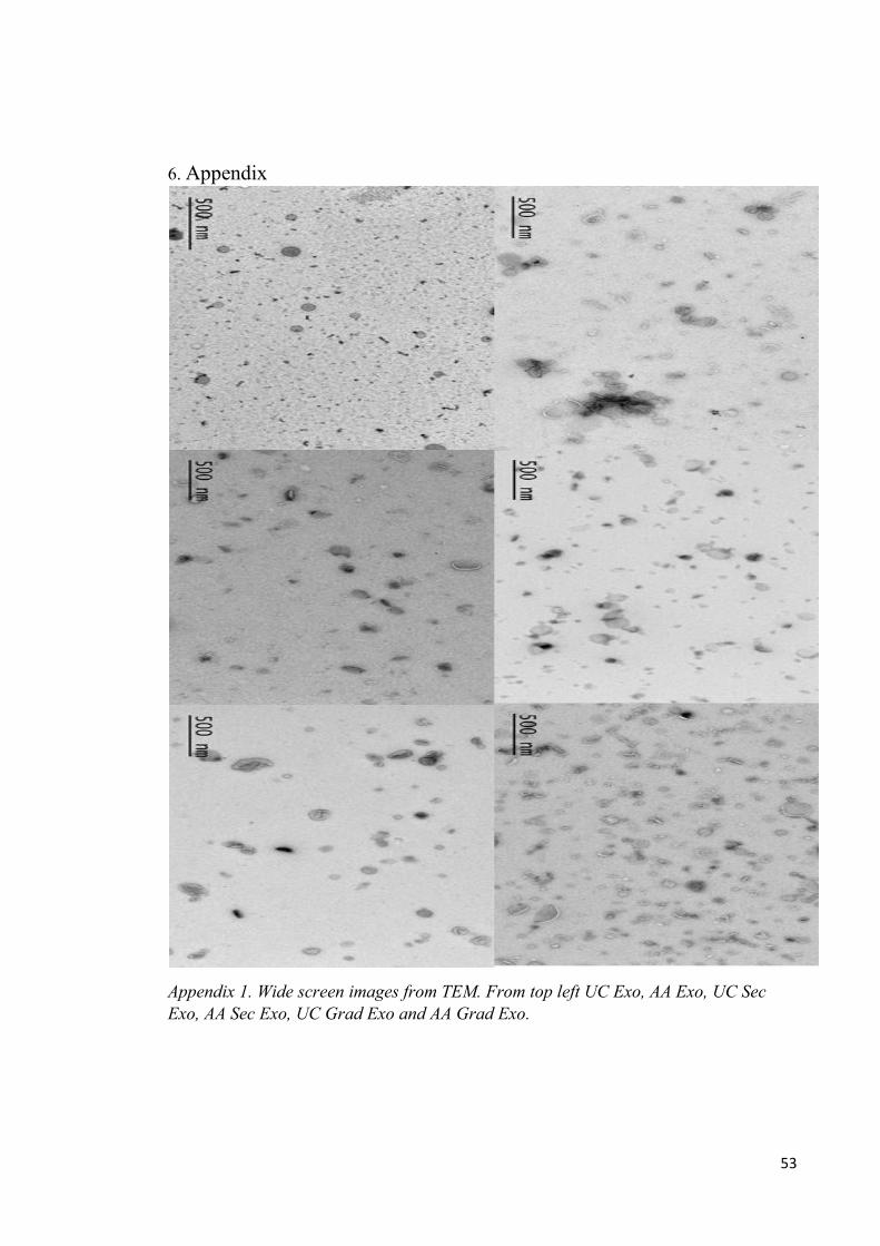
53
6. Appendix
Appendix 1. Wide screen images from TEM. From top left UC Exo, AA Exo, UC Sec
Exo, AA Sec Exo, UC Grad Exo and AA Grad Exo.
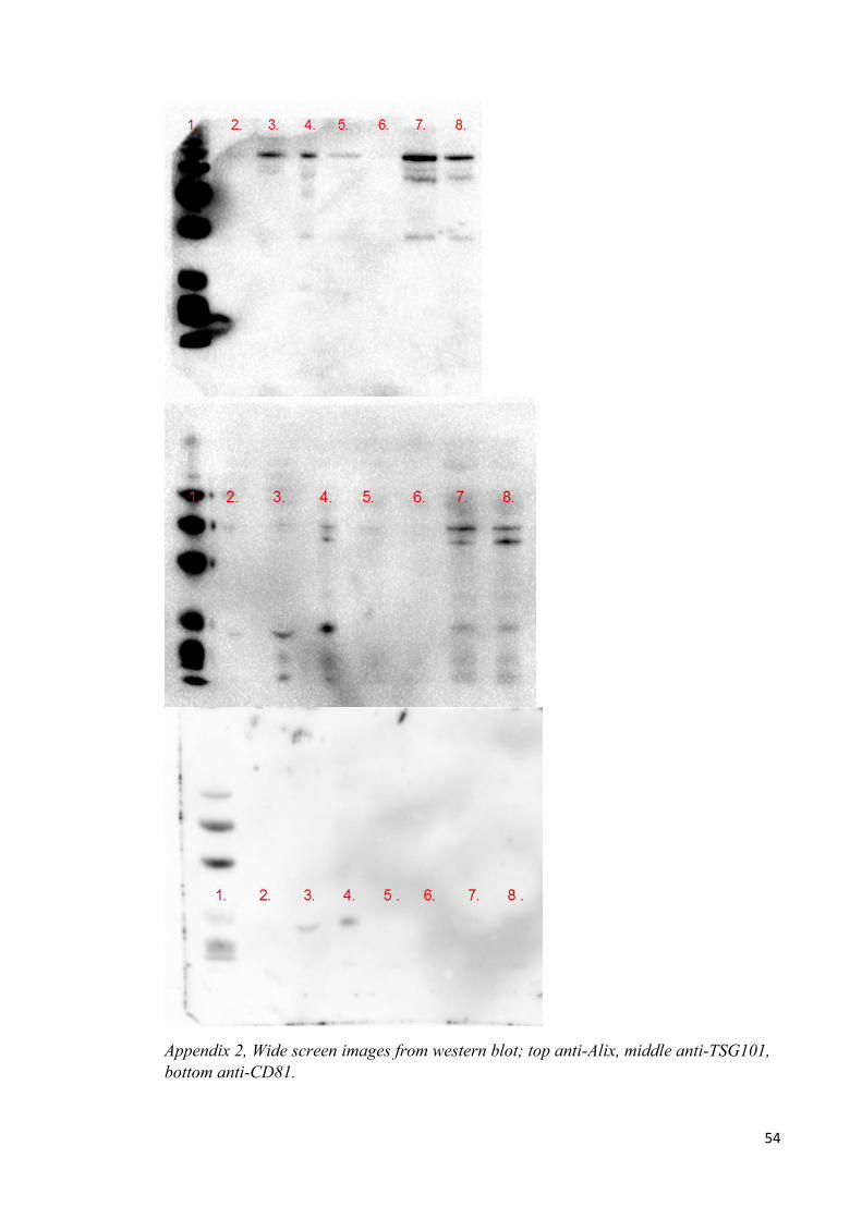
54
Appendix 2, Wide screen images from western blot; top anti-Alix, middle anti-TSG101,
bottom anti-CD81.
![The Role of Exosomes in Bone Remodeling: …downloads.hindawi.com/journals/dm/2019/9417914.pdfregulation [35]. 3.2. Exosomes from Osteoblasts. Ample data suggest that exosomes shed](https://img.pdfslide.us/doc/110x75/5f03c0c07e708231d40a9922/the-role-of-exosomes-in-bone-remodeling-regulation-35-32-exosomes-from-osteoblasts.jpg)