Embed Size (px)
Citation preview
1Mosby items and derived items © 2011, 2006 by Mosby, Inc., an affiliate of Elsevier Inc.
Chapter 8
Other Important Tests and
Procedures
2Mosby items and derived items © 2011, 2006 by Mosby, Inc., an affiliate of Elsevier Inc.
Introduction
Additional important diagnostic studies
include: Sputum examination
Skin tests
Endoscopic examination
Lung biopsy
Thoracentesis
Hematology, blood chemistry, and electrolyte tests

3Mosby items and derived items © 2011, 2006 by Mosby, Inc., an affiliate of Elsevier Inc.
Sputum Examination
4Mosby items and derived items © 2011, 2006 by Mosby, Inc., an affiliate of Elsevier Inc.
Culture and Sensitivity Study
For a culture and sensitivity study, a single
sputum sample is collected in a sterile
container.
This test is performed to diagnose bacterial
infection, select an antibiotic, and evaluate
the effectiveness of antibiotic therapy.
The turnaround time for this test is 48 to 72
hours.
5Mosby items and derived items © 2011, 2006 by Mosby, Inc., an affiliate of Elsevier Inc.
Gram Staining
The Gram staining of sputum is performed to
classify bacteria into gram-negative and
gram-positive types.
The results of the Gram stain tests guide
therapy until the culture and sensitivity results
are obtained.
6Mosby items and derived items © 2011, 2006 by Mosby, Inc., an affiliate of Elsevier Inc.
The Acid-Fast Smear and Culture
The acid-fast smear and culture is performed
to determine the presence of acid-fast bacilli
(e.g., Mycobacterium tuberculosis).
Three early morning sputum samples are
tested.
7Mosby items and derived items © 2011, 2006 by Mosby, Inc., an affiliate of Elsevier Inc.
Cytology
Cytology entails the collection of a single
sputum sample in a special container with
fixative solution.
The sample is evaluated under a microscope
for the presence of abnormal cells that may
indicate a malignant condition.

8Mosby items and derived items © 2011, 2006 by Mosby, Inc., an affiliate of Elsevier Inc.
Box 8-1. Common Organisms Associated With Respiratory Disorders
9Mosby items and derived items © 2011, 2006 by Mosby, Inc., an affiliate of Elsevier Inc.
Skin Test
Skin tests are commonly performed to
evaluate allergic reactions or exposure to
tuberculous bacilli or fungi.
Skin tests entail the intradermal injection of
an antigen.
10Mosby items and derived items © 2011, 2006 by Mosby, Inc., an affiliate of Elsevier Inc.
Skin Test (Cont’d)
Positive test result Exposure to antigen
Negative test result No exposure to antigen

11Mosby items and derived items © 2011, 2006 by Mosby, Inc., an affiliate of Elsevier Inc.
Endoscopic Examinations

12Mosby items and derived items © 2011, 2006 by Mosby, Inc., an affiliate of Elsevier Inc.
Bronchoscopy
The bronchoscope is a flexible fiberoptic
bronchoscope that allows direct visualization
of the upper airways. Nose
Oral cavity and pharynx
Larynx
Vocal cords
Subglottic area
Trachea, bronchi, lobar bronchi, and segmental
bronchi

13Mosby items and derived items © 2011, 2006 by Mosby, Inc., an affiliate of Elsevier Inc.
Bronchoscopy (Cont’d)
Diagnostic bronchoscopy Abnormal x-rays
Persistent atelectasis
Excessive bronchial secretions
Therapeutic bronchoscopy Suctioning of excessive secretions
Removal of foreign bodies
Selective lavage
14Mosby items and derived items © 2011, 2006 by Mosby, Inc., an affiliate of Elsevier Inc.
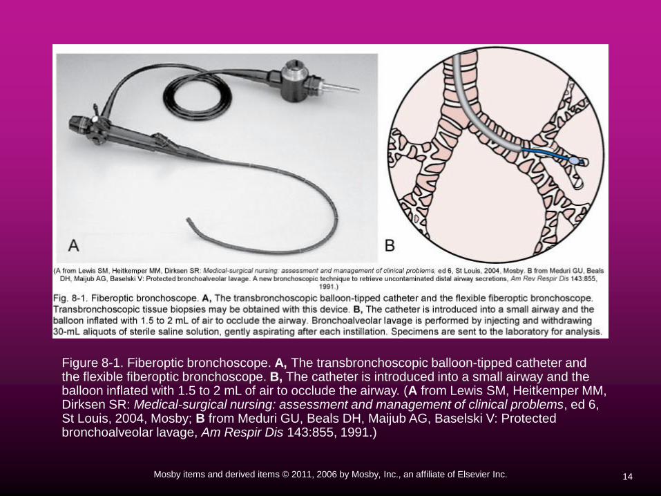
Figure 8-1. Fiberoptic bronchoscope. A, The transbronchoscopic balloon-tipped catheter andthe flexible fiberoptic bronchoscope. B, The catheter is introduced into a small airway and the balloon inflated with 1.5 to 2 mL of air to occlude the airway. (A from Lewis SM, Heitkemper MM, Dirksen SR: Medical-surgical nursing: assessment and management of clinical problems, ed 6,St Louis, 2004, Mosby; B from Meduri GU, Beals DH, Maijub AG, Baselski V: Protected bronchoalveolar lavage, Am Respir Dis 143:855, 1991.)

15Mosby items and derived items © 2011, 2006 by Mosby, Inc., an affiliate of Elsevier Inc.
Mediastinoscopy
A mediastinoscopy is the insertion of a scope
through a small incision in the suprasternal
notch; the scope is then advanced into the
mediastinum.
The test is used to inspect and biopsy lymph
nodes in the mediastinal area.
16Mosby items and derived items © 2011, 2006 by Mosby, Inc., an affiliate of Elsevier Inc.
Lung Biopsy
A lung biopsy specimen can be obtained by
means of a transbronchial needle biopsy or
an open-lung biopsy.
A transbronchial lung biopsy entails passing
forceps or a needle through a bronchoscope
to obtain a specimen.
17Mosby items and derived items © 2011, 2006 by Mosby, Inc., an affiliate of Elsevier Inc.
Lung Biopsy (Cont’d)
An open lung biopsy involves surgery to
remove a sample of lung tissue.
An incision is made over the area of the lung
from which the tissue sample is to be
collected.

18Mosby items and derived items © 2011, 2006 by Mosby, Inc., an affiliate of Elsevier Inc.
Figure 8-2. Transbronchial needle biopsy. The diagram shows a transbronchial biopsy needle penetrating the bronchial wall and entering a mass of subcarinal lymph nodes or tumor. (Redrawn from DuBois RM, Clarke SW: Fiberoptic bronchoscopy in diagnosis and management, Orlando, 1987, Grune and Stratton.)
Insert Figure 8-2 here

19Mosby items and derived items © 2011, 2006 by Mosby, Inc., an affiliate of Elsevier Inc.
Video-Assisted Thoracoscopy
(VATS)
Insertion of thoracoscope through the
chest wall
Results displayed on a video monitor
Helpful in the diagnosis of: Tuberculosis
Mesothelioma
Metastatic cancer

20Mosby items and derived items © 2011, 2006 by Mosby, Inc., an affiliate of Elsevier Inc.
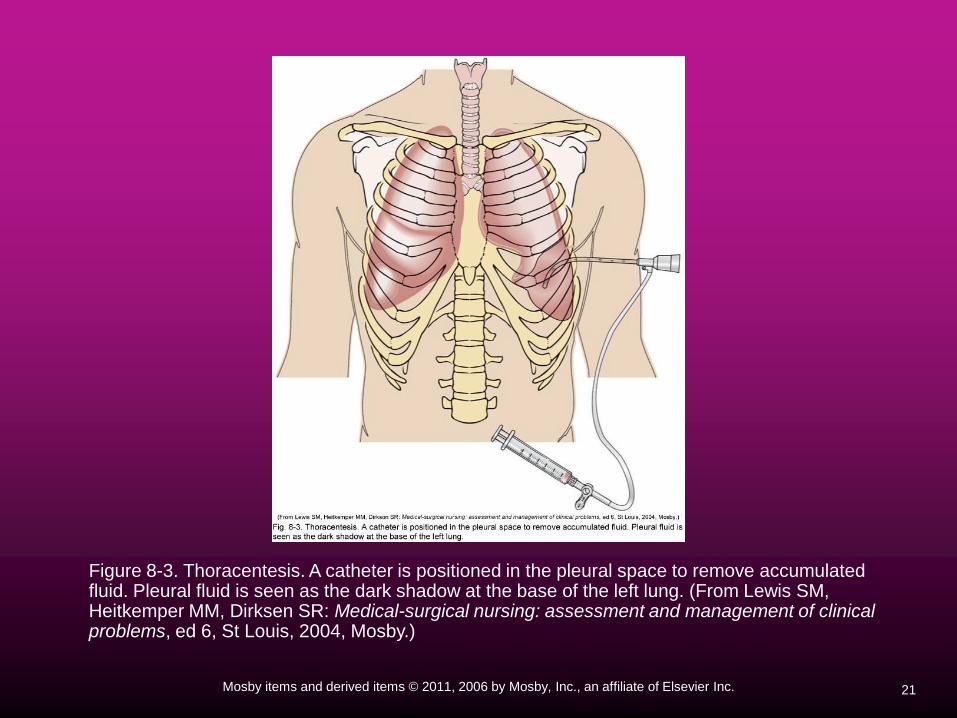
Thoracentesis
Thoracentesis is a procedure in which excess
fluid accumulation (pleural effusion) between
the chest cavity and lungs (pleural space) is
aspirated through a needle inserted through
the chest wall.
A diagnostic thoracentesis may be performed
to identify the cause of a pleural effusion.
21Mosby items and derived items © 2011, 2006 by Mosby, Inc., an affiliate of Elsevier Inc.
Figure 8-3. Thoracentesis. A catheter is positioned in the pleural space to remove accumulated fluid. Pleural fluid is seen as the dark shadow at the base of the left lung. (From Lewis SM, Heitkemper MM, Dirksen SR: Medical-surgical nursing: assessment and management of clinical problems, ed 6, St Louis, 2004, Mosby.)
22Mosby items and derived items © 2011, 2006 by Mosby, Inc., an affiliate of Elsevier Inc.
Pleurodesis
Pleurodesis is performed to prevent the
recurrence of a pneumothorax or pleural
effusion.
A pleurodesis is achieved by injecting any
number of agents (called sclerosing agents or
sclerosants) into the pleural space through a
chest tube.
Common sclerosant chemicals include a
slurry of talc, bleomycin, nitrogen mustard,
doxycycline, povidone iodine, and quinacrine.
23Mosby items and derived items © 2011, 2006 by Mosby, Inc., an affiliate of Elsevier Inc.
Pleurodesis (Cont’d)
The instilled sclerosing agents cause irritation
and inflammation (pleuritis) between the
parietal and the visceral layers of the pleura.
This action causes the pleurae to stick
together and thereby prevents subsequent
gas or fluid accumulation.
24Mosby items and derived items © 2011, 2006 by Mosby, Inc., an affiliate of Elsevier Inc.
Pleurodesis Risks
Infection
Bleeding
Acute respiratory distress syndrome
Collapsed lung (pneumothorax), and
respiratory failure
25Mosby items and derived items © 2011, 2006 by Mosby, Inc., an affiliate of Elsevier Inc.
Pleurodesis Risks (Cont’d)
Complications may be specific for each
sclerosant. Talc and doxycycline can cause fever and pain.
Quinacrine can cause low blood pressure, fever,
and hallucination.
Bleomycin can cause fever, pain, and nausea.

26Mosby items and derived items © 2011, 2006 by Mosby, Inc., an affiliate of Elsevier Inc.
Hematology, Blood Chemistry,
and Electrolyte Findings
27Mosby items and derived items © 2011, 2006 by Mosby, Inc., an affiliate of Elsevier Inc.
Hematology
The most frequent laboratory hematologic
test is the complete blood count (CBC).
The CBC provides important information
about the patient’s diagnosis, prognosis,
response to treatment, and recovery.
28Mosby items and derived items © 2011, 2006 by Mosby, Inc., an affiliate of Elsevier Inc.
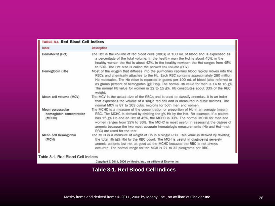
Table 8-1. Red Blood Cell Indices

29Mosby items and derived items © 2011, 2006 by Mosby, Inc., an affiliate of Elsevier Inc.
Table 8-1. Red Blood Cell Indices
30Mosby items and derived items © 2011, 2006 by Mosby, Inc., an affiliate of Elsevier Inc.
White Blood Cell Count (WBC)
The major functions of the WBCs
(leukocytes) are to: 1. Fight against infection
2. Defend the body by phagocytosis against foreign
organisms
3. Produce (or at least transport and distribute)
antibodies in the immune response
31Mosby items and derived items © 2011, 2006 by Mosby, Inc., an affiliate of Elsevier Inc.
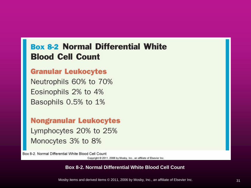
Box 8-2. Normal Differential White Blood Cell Count

32Mosby items and derived items © 2011, 2006 by Mosby, Inc., an affiliate of Elsevier Inc.
Table 8-2. Common Causes of WBC Increase
33Mosby items and derived items © 2011, 2006 by Mosby, Inc., an affiliate of Elsevier Inc.
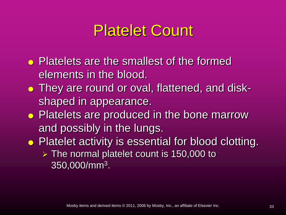
Platelet Count
Platelets are the smallest of the formed
elements in the blood.
They are round or oval, flattened, and disk-
shaped in appearance.
Platelets are produced in the bone marrow
and possibly in the lungs.
Platelet activity is essential for blood clotting. The normal platelet count is 150,000 to
350,000/mm3.
34Mosby items and derived items © 2011, 2006 by Mosby, Inc., an affiliate of Elsevier Inc.
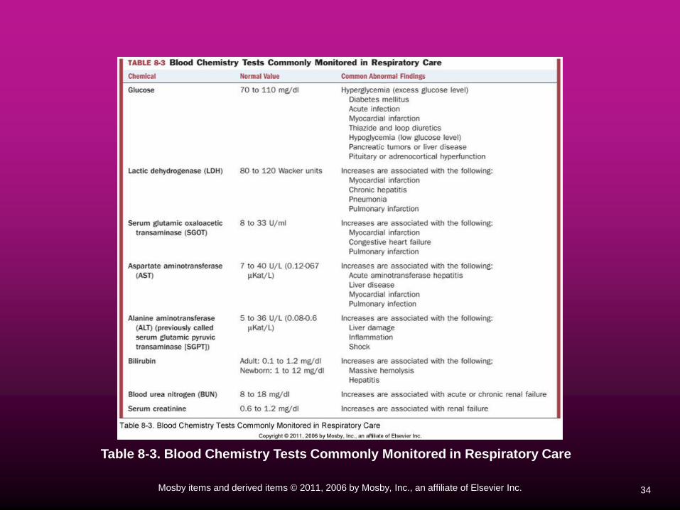
Table 8-3. Blood Chemistry Tests Commonly Monitored in Respiratory Care
35Mosby items and derived items © 2011, 2006 by Mosby, Inc., an affiliate of Elsevier Inc.
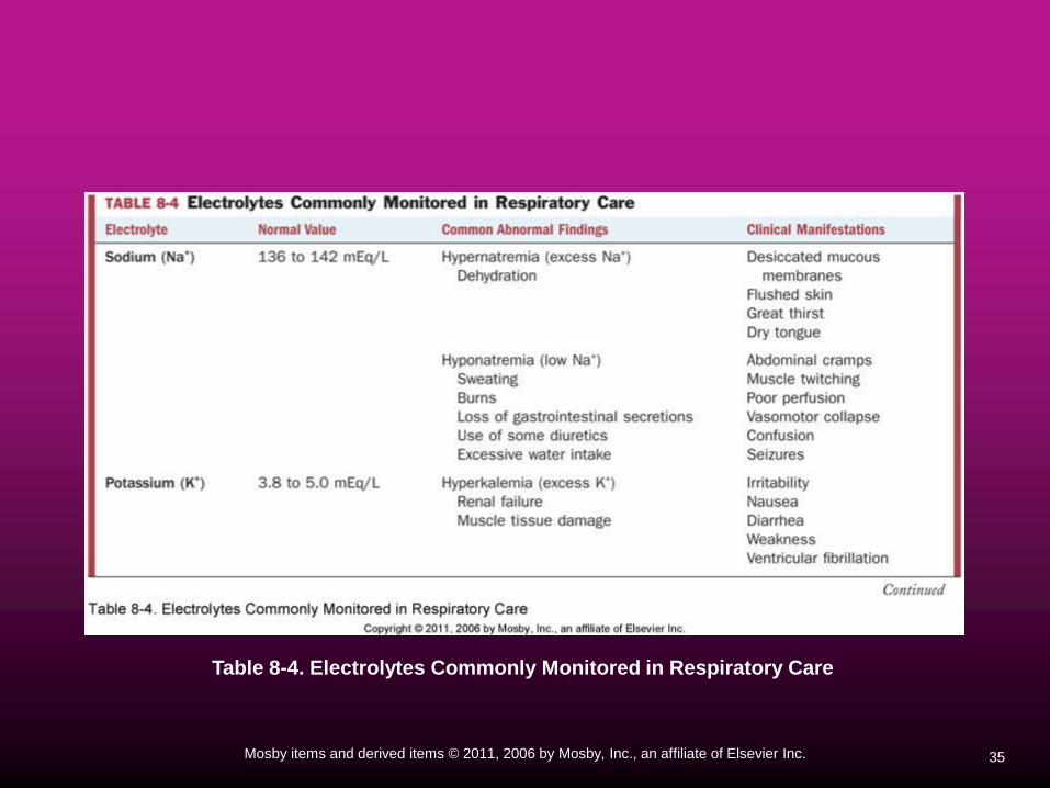
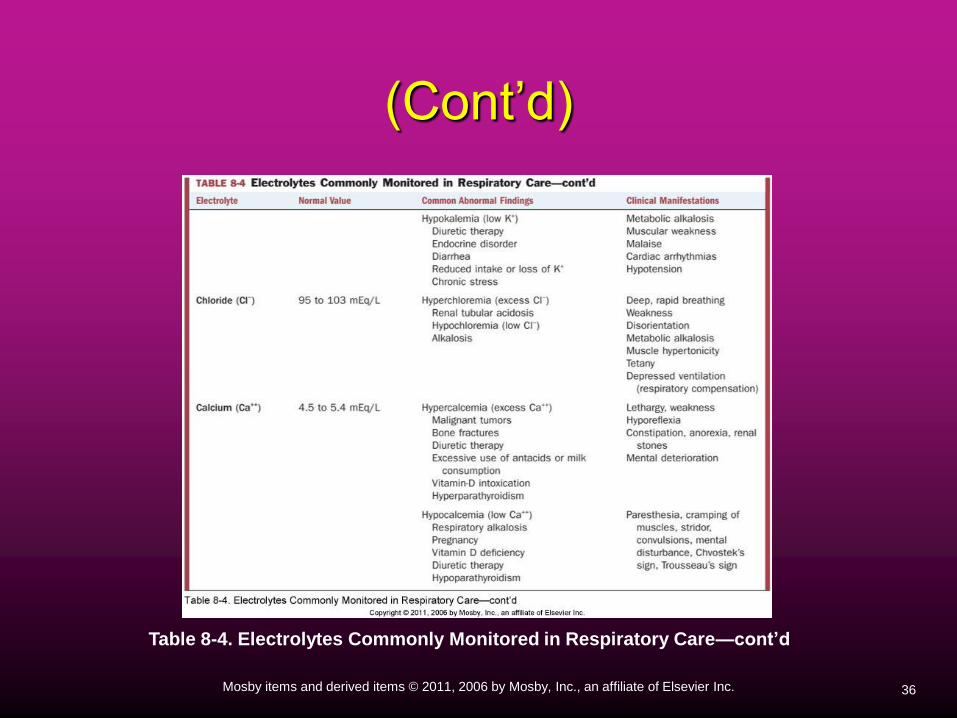
Table 8-4. Electrolytes Commonly Monitored in Respiratory Care
36Mosby items and derived items © 2011, 2006 by Mosby, Inc., an affiliate of Elsevier Inc.
(Cont’d)
Table 8-4. Electrolytes Commonly Monitored in Respiratory Care—cont’d