Embed Size (px)
Citation preview

Chapter 23: The Elbow
Jennifer Doherty-Restrepo, MS, LAT, ATC
Academic Program Director, Entry-Level ATEP
Florida International University
Acute Care and Injury Prevention

Anatomy of the ElbowReview


Functional Anatomy Elbow ROM = flexion, extension, pronation and
supination 145 degrees of flexion 90 degrees of supination and pronation
Stable joint: protection from overuse and traumatic injuries Bony limitations, ligamentous support, and muscular
stability at the elbow help to Carrying angle due to distal projection of humerus
Normal in females is 10-15 degrees, males 5 degrees Critical link in kinetic chain of upper extremity

Assessment of the Elbow: History Past history Mechanism of injury When does it hurt? Where does it hurt? Motions that increase
pain? Motions that decrease
pain?
Type of, quality of, duration of, pain?
Sounds or feelings? How long were you
disabled? Swelling? Previous treatments?

Deformities and swelling? Carrying angle
Cubitus valgus vs. Cubitus varus Flexion and extension
Cubitus recurvatum Elbow at 45 degrees
Isosceles triangle formed by the olecranon and epicondyles
Observations

Palpation: Bony and Soft Tissue Humerus Medial and lateral epicondyles Olecranon process Radial head Radius Ulna Medial and lateral collateral
ligaments Annular ligament
Biceps brachii Brachialis Brachioradialis Pronator teres Triceps Supinator Wrist flexors Wrist extensors

Pulse Assessed at brachial artery and radial artery
Skin sensation Determine presence of nerve root compression or
irritation in cervical or shoulder region Tinel’s sign
Ulnar nerve test Tap on ulnar nerve in ulnar groove Positive test = numbness/tingling along the
forearm and hand
Special Tests: Circulatory and Neurological Function

Tested after hyperextension of elbow Athlete position
Elbow is flexed to 45 degrees Wrist is fully flexed and extended
Positive test = pain in elbow joint If joint pain is severe, sprain or fracture should be
suspected Joint pain may indicate chronic injury as well
Special Tests: Capsular Injury

Valgus/Varus Stress Test Assess injury to the medial and lateral collateral
ligaments, respectively Positive test = joint laxity or complaint of pain
Special Tests: Ligament Injury

Medial Epicondylitis Test Athlete position
Elbow flexed to 45 degrees Resist wrist flexion
Positive test = pain at medial epicondyle Lateral Epicondylitis Test
Athlete position Elbow flexed to 45 degrees Resist wrist extension
Positive test = pain at lateral epicondyle
Special Tests: Muscle Injury
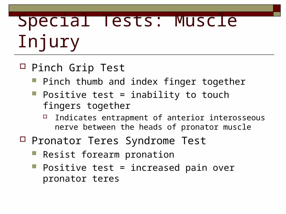
Pinch Grip Test Pinch thumb and index finger together Positive test = inability to touch fingers together
Indicates entrapment of anterior interosseous nerve between the heads of pronator muscle
Pronator Teres Syndrome Test Resist forearm pronation Positive test = increased pain over pronator teres
Special Tests: Muscle Injury

Special Tests: Functional Evaluation
Evaluate AROM, PROM and RROM Flexion Extension Pronation Supination
Positive test = pain and weakness

Elbow Injuries Subject to injury due to…
Broad range of motion Weak lateral bone structure Exposure of soft tissue
Many sports place excessive stress on joint Locking motion of some activities Use of implements Throwing motion

MOI = direct blow or repetitive blows Vulnerable area due to lack of padding
Signs and Symptoms Rapid swelling due to irritation of bursa or
synovial membrane Management
PRICE immediately, for at least 24 hours If severe, refer for X-ray to rule out fracture
Elbow Injuries: Contusion

Elbow Injuries: Olecranon Bursitis MOI = direct blow
Superficial location makes it extremely susceptible to injury
Signs and Symptoms Pain, swelling, and point
tenderness Swelling will appear almost
spontaneously without the pain and heat

Management Acute: compression for at
least 1 hour Chronic: requires superficial
therapy primarily involving compression
If swelling fails to resolve, aspiration may be necessary
May be padded to return to competition

MOI = overstretching or too forceful a contraction Falling on outstretched arm Repeated microtears may cause chronic injury
Rupture of distal biceps is most common in UE Signs and Symptoms
Pain with AROM and RROM Point tenderness in muscle, tendon, or lower part
of muscle belly
Elbow Injuries: Muscle Strains
Management PRICE Sling in severe cases Follow-up treatment
Cryotherapy, ultrasound, ROM and PRE exercises If severe loss of function, refer for X-ray to rule
out avulsion or epiphyseal fracture
Elbow Injuries: Muscle Strains

MOI = valgus force from repetitive trauma Secondary injuries may include…
Ulnar nerve inflammation Wrist flexor tendinitis Overuse flexor/pronator strain Ligamentous sprains Elbow flexion contractures Joint instability
Elbow Injuries: Ulnar Collateral Ligament Injuries

Signs and Symptoms Pain along medial aspect of elbow Point tenderness over UCL Associated paresthesia
Positive Tinel’s sign Positive Valgus Stress Test
Possible end-point laxity X-ray may show evidence of…
Hypertrophy of humeral condyle and/or posteromedial aspect of olecranon; and osteophytes
Calcification within the UCL Loose bodies in posterior compartment
Management Conservative treatment
PRICE and NSAID’s
ROM and PRE exercises as pain decreases Analysis of the throwing motion (if applicable) Surgical intervention may be necessary
Tommy John Procedure Throwing athlete may be able to return to activity
approximately 22-26 weeks post surgery
MOI = repetitive microtrauma to insertion of wrist extensor muscles Tendinosis may result
Degeneration of tendon without inflammation
Signs and Symptoms Aching pain at lateral epicondyle after activity Decreased elbow ROM Pain with AOM and RROM wrist extension Pain and weakness in wrist and hand develop
Elbow Injuries: Lateral Epicondylitis (Tennis Elbow)

Management PRICE NSAID’s and analgesics Mobilization and stretching in pain free ranges
ROM and PRE exercises as pain decreases
Deep friction massage Hand grasping while in supination
Avoid pronation motions
Use of neoprene sleeve Mechanics and skills training in order to avoid recurrence of
injury

MOI = repeated forceful wrist flexion and extreme valgus torque on the elbow May involve pronator teres, flexor carpi radialis,
flexor carpi ulnaris, and palmaris longus tendons Can be associated with ulnar nerve neuropathy
Signs and Symptoms Pain with AOM and RROM wrist extension
Pain with wrist flexion as well in severe injuries
Point tenderness and mild swelling at medial epicondyle
Elbow Injuries: Medial Epicondylitis

Management PRICE NSAID's and analgesics Sling in severe cases
Severe cases may require splinting and complete rest for 7-10 days
Cryotherapy, Ultrasound Curvilinear brace
Below elbow to reduce stress at the elbow joint

MOI = Repetitive microtrauma Injurious movements include elbow rotation and
extension Excessive valgus stresses causes compression of the radial
head, which adds shearing forces at the radiocapitular joint Impairment of blood supply may result, which causes
degeneration of articular cartilage creating loose bodies
Panner’s disease Occurs in children (age <10) Osteochondrosis of capitellum due to localized avascular
necrosis
Elbow Injuries: Osteochondritis Dissecans

Signs and Symptoms Sudden pain at radiohumeral joint Swelling, creptitus Decreased ROM (full extension)
ROM usually returns in a few days Grating with pronation and supination
Locking of the joint X-ray
May show flattening and crater of capitulum May show loose bodies in joint

Management Activity restriction for 6-12 weeks NSAID’s Splint and cast applied in severe cases of
deterioration If repeated locking of the elbow joint occurs,
loose bodies are removed surgically

MOI = repetitive microtraumas that occur from throwing motion (Not due to the type of pitch)
Linked to: Accelerated apophyseal growth and delayed medial
epicondyle epiphysis growth Traction apophysitis with possible fragmentation of
medial epicondylar apophysis Avulsion fracture at medial epicondyle or radial head Osteochondrosis of humeral capitellum Non-union stress fracture of olecranon epiphysis
Elbow Injuries: Little League Elbow

Signs and Symptoms Onset is slow Slight flexion contracture Tight anterior joint capsule Weakness in triceps “Locking” or “Catching” sensation Decreased ROM
Especially forearm pronation and supination

Management PRICE NSAID’s and analgesics Stop throwing until…
Pain resolved Full ROM is regained
Gentle ROM exercises Gently triceps strengthening exercises Analysis of throwing motion
MOI = narrowing of cubital canal or irregularity of cubital tunnel Pronounced cubital valgus may cause deep
friction contributing to injury Ulnar nerve injury may result
Ulnar nerve subluxation or dislocation Traction of ulnar nerve from valgus force Ulnar nerve compression from ligaments
Elbow Injuries: Cubital Tunnel Syndrome

Signs and Symptoms Pain on medial aspect of elbow
Pain may be referred proximally or distally
Point tenderness in cubital tunnel Pain with hyperflexion Intermittent paresthesia in 4th and 5th fingers

Management Rest, immobilization for 2 weeks NSAID’s Splinting, surgical decompression or
transposition of subluxating nerve may be necessary
Avoid hyperflexion and valgus stresses

Elbow Injuries: Dislocation MOI = fall on outstretched hand with elbow
extended or severe twist while elbow flexed High incidence in sports Dislocation may be posterior, anterior, or lateral
Signs and Symptoms Swelling, severe pain, disability Median and radial nerves may be compromised Blood vessels may be compromised Often a radial head fracture is involved


Management Pack with ice and apply sling immediately Refer for reduction immediately Following reduction…
Immobilize in elbow flexion for 3 weeks PRE exercises for grip and shoulder strenthening
Following immobilization… Heat and PROM exercises to regain full ROM ROM and PRE exercises should be initiated by athlete
Exercises that are too strenuous should be avoided before complete healing due to high probability of developing myositis ossificans
Forced stretching should be avoided

MOI = fall on flexed elbow or direct blow May occur in one or more of bones in elbow joint Fall on outstretched hand may fracture the humerus
above condyles or between condyles Condylar fracture may result in gunstock deformity
Direct blow may fracture olecranon or radial head Signs and Symptoms
May not result in visual deformity Hemorrhaging, swelling, muscle spasm
Elbow Injuries: Fractures
Management Monitor neurovascular status Non-surgical treatment
Appropriate for stable fractures Immobilize with cast or removable splint for 6-8
weeks Surgical treatment
Used to stabilize unstable fractures in adults ROM exercises initiated early to prevent frozen elbow

MOI = impaired circulation or ischemia Associate with humeral supracondylar fractures, which
compromises the brachial artery and inhibits circulation to forearm
May be loss of motor and sensory function Classic case involves median nerve
Edema further impairs circulation via condition called compartment syndrome Muscle necrosis may occur with irreversible muscle damage
after 4-6 hours, which may lead to secondary fibrosis and calcification
Elbow Injuries: Volkmann’s Contracture

Signs and Symptoms Pain in forearm which increases with PROM
finger extension Cessation of brachial and radial pulses Coldness in arm Decreased ROM
Management Monitor neurovascular integrity

Rehabilitation of the Elbow General Body Conditioning
Must maintain pre-injury CV and LE strength fitness levels
Flexibility Restoring ROM is critical in elbow rehab Variety of approaches can be used as long as they
do not “force” the joint

Joint Mobilizations Loss of proper arthrokinematics following
immobilization is expected Joint mobilization and traction
Very useful to increase mobility Useful to decrease pain Restores accessory motions

Strengthening Achieved through low-resistance, high-repetition exercises
Must be pain free Shoulder and hand grip exercises Isometrics can be used while immobilized PNF and isokinetics are useful in early and intermediate
stages of rehabilitation PRE exercises with tubing, weights, or manual resistance Closed kinetic chain activities
Assist in both static and dynamic stability to the elbow Proprioceptive training should also incorporated

Functional Progressions Will enhance healing and performance
PNF, swimming, pulley machines, and rubber tubing Simulate sports activities
Should include steps Warm-up Gradual build up to activity, becoming increasingly
more difficult Return to Activity
ROM must be WNL Strength should be restored without pain