Embed Size (px)
Citation preview
Chapter 21: The Thigh, Hip, Groin, and Pelvis
Jennifer Doherty-Restrepo, MS, LAT, ATCAcademic Program Director, Entry-Level ATEP
Florida International UniversityAcute Care and Injury Prevention
Anatomy of the Thigh
Review
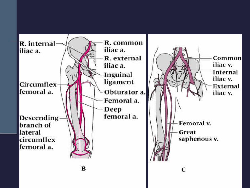
Nerve and Blood Supply
Tibial and common peroneal nerves Arise from the sacral plexus to form the largest
nerve in the body, the sciatic nerve The main arteries of the thigh include:
Deep circumflex, deep femoral, and femoral The two main veins of the thigh include:
Great saphenous and femoral
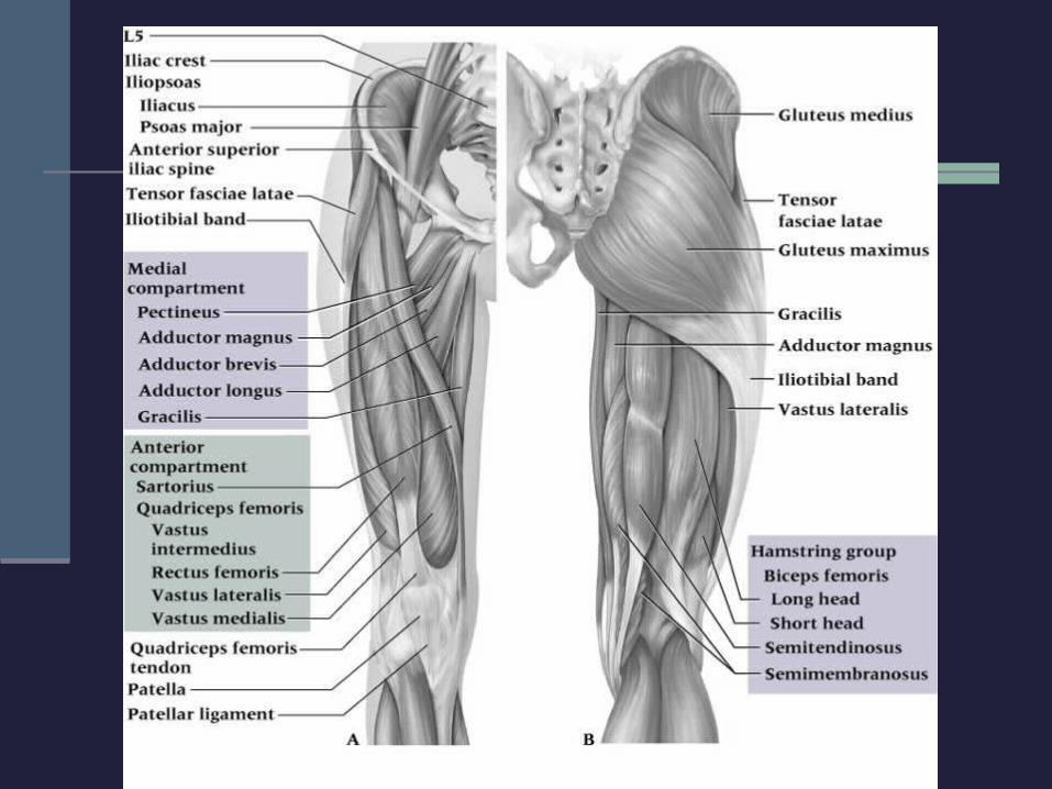
Muscles
Fascia lata femoris Deep fascia that surrounds thigh musculature Thick anteriorly, laterally, and posteriorly Thin on the medial side
IT-band Attachment site for the tensor fascia lata and
gluteus maximum
Quadriceps
Insertion at proximal patella via common tendon Pre-patellar tendon
Rectus femoris = bi-articulate muscle Only quad muscle that also crosses the hip Extends knee and flexes the hip
Important: distinguish between knee extensors and hip flexors Injury evaluation Treatment and rehabilitation programs
Cross the knee joint posteriorly All hamstrings, except the short of head of the
biceps femoris, are bi-articulate Crosses the hip joint as well Forces dependent upon position of both knee and hip
Important: distinguish between knee flexors and hip extensors Injury evaluation Treatment and rehabilitation programs
Hamstrings
Assessment of the Thigh
History Onset (sudden or slow?) Previous history? Mechanism of injury? Pain description, intensity, quality, duration, type, and
location? Observation
Symmetry? Size, deformity, swelling, discoloration? Skin color and texture? Is the athlete in obvious pain? Is the athlete willing to move the thigh?
Palpation: Bony Tissue
Medial and lateral femoral condyles Greater trochanter Lesser trochanter Anterior superior iliac spine (ASIS)
Palpation: Soft Tissue
Sartorius Rectus femoris Vastus lateralis Vastus medialis Vastus intermedius Semimembranosus Semitendinosus Biceps femoris
Adductor brevis, longus, and magnus
Gracilis Sartorius Pectineus Iliotibial Band (IT-band) Gluteus medius Tensor fasciae latae
Not performed if a fracture is suspected!!! Passive knee flexion
Normal = full, pain-free ROM Injury = swelling or spasm restricting ROM
Active knee extension Muscle strain = strong and painful ROM 3rd degree strain or partial rupture = weak and pain
free ROM Resistive knee extension
Nerve injury = muscle weakness against an isometric resistance
Special Tests
Prevention of Thigh Injuries
Maximum strength Endurance Flexibility In collision sports, thigh guards are
mandatory to prevent injuries
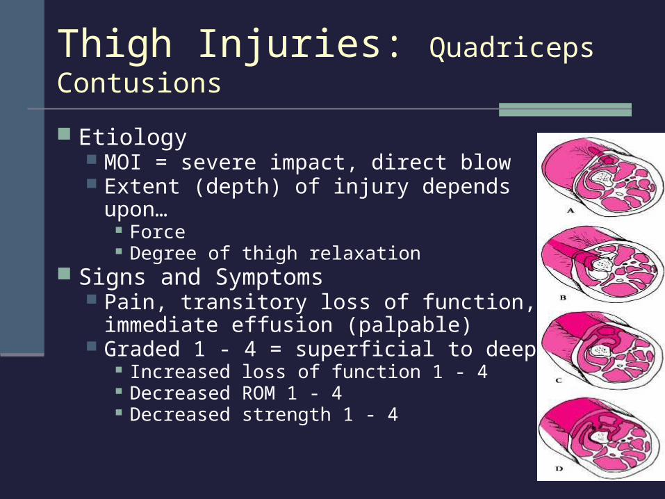
Thigh Injuries: Quadriceps Contusions
Etiology MOI = severe impact, direct blow Extent (depth) of injury depends upon…
Force Degree of thigh relaxation
Signs and Symptoms Pain, transitory loss of function,
immediate effusion (palpable) Graded 1 - 4 = superficial to deep
Increased loss of function 1 - 4 Decreased ROM 1 - 4 Decreased strength 1 - 4

Management RICE NSAID’s and analgesics Crutches, if indicated Aspiration of hematoma Ice post exercise or re-injury Follow-up care
ROM exercises PRE in pain-free ROM
Modalities Heat Massage Ultrasound to prevent
myositis ossificans
Thigh Injuries: Quadriceps Contusions
Etiology Formation of ectopic bone MOI = repeated blunt trauma May be the result of improper thigh contusion
treatment (too aggressive) Signs and Symptoms
X-ray shows Ca++ deposit 2 - 6 weeks post injury Pain, weakness, swelling, tissue tension, point
tenderness, and decreased ROM Management
Treatment must be conservative May require surgical removal
Thigh Injuries: Myositis Ossificans Traumatica
Etiology MOI = over-stretching or too forceful contraction
Signs and Symptoms Pain, point tenderness, spasm, loss of function,
and ecchymosis Superficial strain results in fewer S&S than
deeper strain Complete tear results in deformity
Athlete displays little disability and discomfort
Thigh Injuries: Quadriceps Muscle Strain
Management RICE NSAID’s and analgesics Manage swelling
Compression, crutches Stretching PRE strengthening exercises Neoprene sleeve for added support
Thigh Injuries: Quadriceps Muscle Strain
Etiology: multiple theories of injury Hamstrings and quadriceps contract together Change from hip extender to knee flexor Fatigue Posture Leg length discrepancy Lack of flexibility Strength imbalances
Thigh Injuries: Hamstring Muscle Strains
Thigh Injuries: Hamstring Muscle Strains
Signs and Symptoms Pain in muscle belly
or point of attachment
Capillary hemorrhage
Ecchymosis
Grade 1 Pain with movement Point tenderness <20% of fibers torn
Grade 2 Partial tear
<70% of fibers torn Sharp snap or tear Severe pain Loss of function
Grade 3 Rupture of tendinous or
muscular tissue >70% muscle fiber tearing
Severe hemorrhage Disability Edema Loss of function Ecchymosis Palpable mass or gap
Thigh Injuries: Hamstring Muscle Strains
Management RICE, NSAID’s and analgesics Modalities PRE exercises When soreness is
eliminated, focus on eccentrics strengthening
Recovery may require months to a full year
Scaring increases risk of injury recurrence of
Grade I Do not resume full
activity until complete function restored
Grade 2 and 3 Should treat
conservatively Gradual return to
stretching and strengthening in later stages of healing
Etiology Fracture in middle third of femoral shaft MOI = great deal of force
Signs and Symptoms Pain, swelling, deformity, muscle guarding Leg with fx positioned in hip adduction and ER Leg with fx may appear shorter
Management Medical emergency! Treat for shock, splint, refer Analgesics and ice
Thigh Injuries: Acute Femoral Fractures
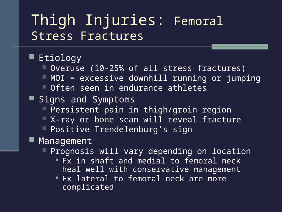
Etiology Overuse (10-25% of all stress fractures) MOI = excessive downhill running or jumping Often seen in endurance athletes
Signs and Symptoms Persistent pain in thigh/groin region X-ray or bone scan will reveal fracture Positive Trendelenburg’s sign
Management Prognosis will vary depending on location
Fx in shaft and medial to femoral neck heal well with conservative management
Fx lateral to femoral neck are more complicated
Thigh Injuries: Femoral Stress Fractures
Anatomy of the Hip, Groin, and Pelvic Region
Review
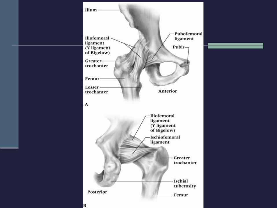

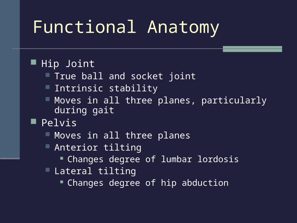
Functional Anatomy
Hip Joint True ball and socket joint Intrinsic stability Moves in all three planes, particularly during gait
Pelvis Moves in all three planes Anterior tilting
Changes degree of lumbar lordosis Lateral tilting
Changes degree of hip abduction
Assessment of the Hip and Pelvis
Injuries to the hip or pelvis cause major disability in the lower limbs, trunk, or both
Low back may also become involved History
Onset (sudden or slow?) Previous history? Mechanism of injury? Pain description, intensity, quality, duration,
type, and location?
Observation Symmetry - hips, pelvis tilt (anterior/posterior)
Lordosis or flat back Lower limb alignment
Knees, patella, feet Pelvic landmarks
ASIS, PSIS, iliac crest Standing on one leg
Pubic symphysis pain or drop to one side Ambulation
Assessment of the Hip and Pelvis
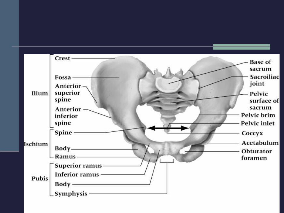
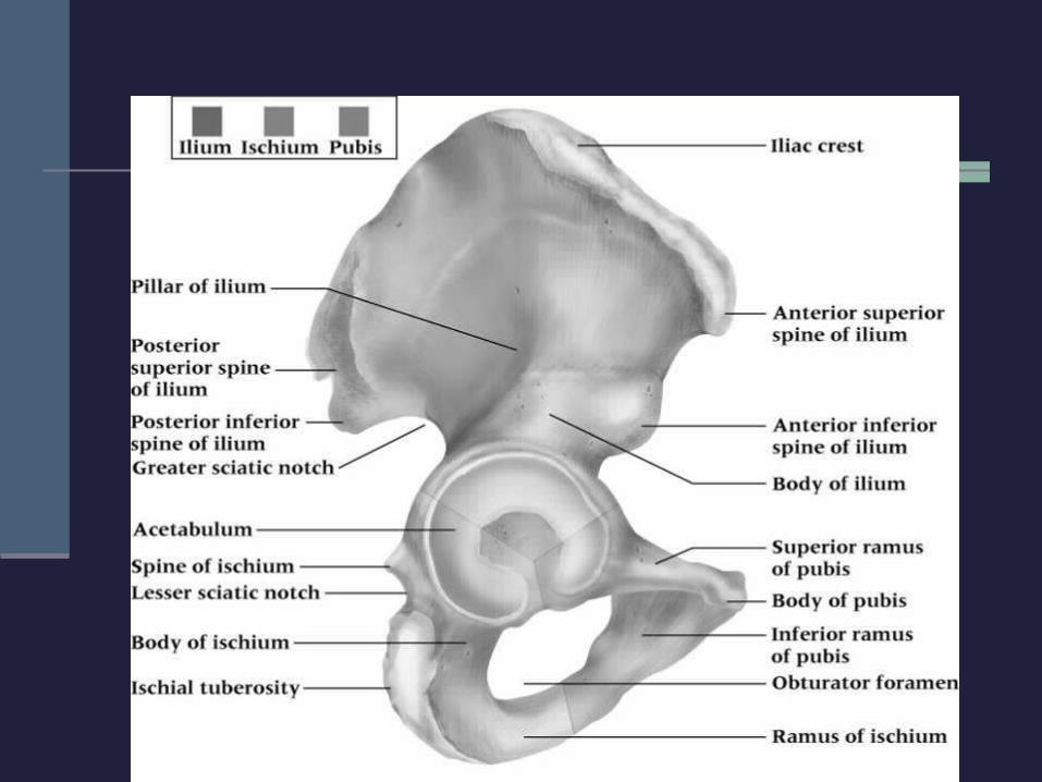
Palpation: Bony Tissue
Iliac crest Anterior superior iliac
spine (ASIS) Anterior inferior iliac
spin (AIIS) Posterior superior iliac
spine (PSIS)
Pubic symphysis Ischial tuberosity Greater trochanter Femoral neck Poster inferior iliac
spine (PIIS)
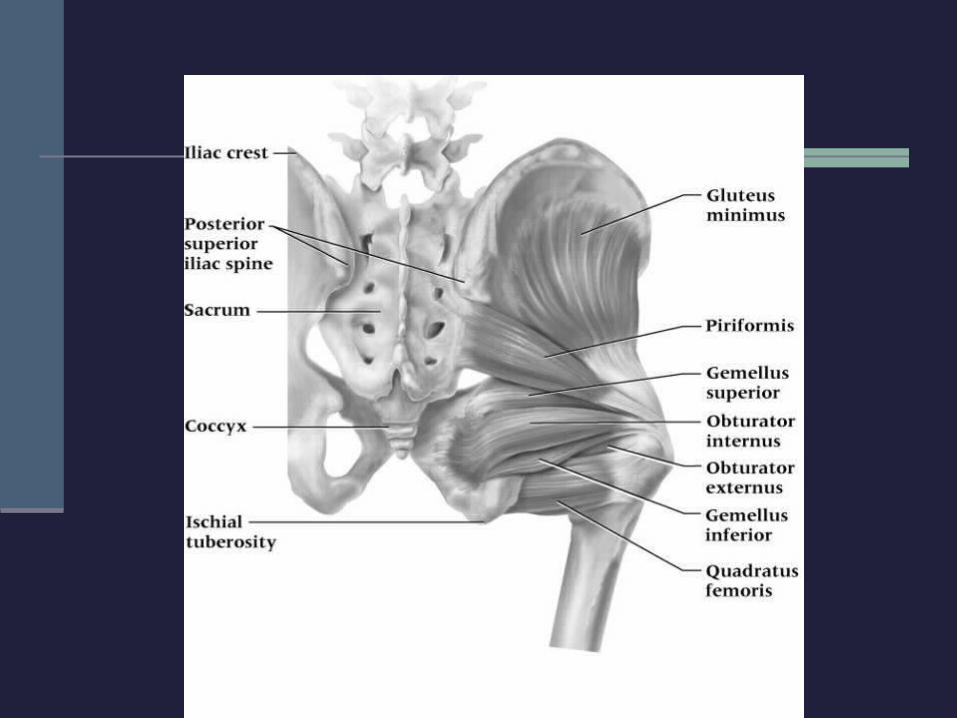
Palpation: Soft Tissue
Rectus femoris Sartorius Iliopsoas Inguinal ligament Gracilis Adductor magnus,
longus & brevis Pectineus
Gluteus maximus, medius & minimus
Piriformis Hamstrings Tensor fasciae latae Iliotibial Band
Major regions of concern are the groin, femoral triangle, sciatic nerve, and lymph
nodes
Special Tests
Functional Evaluation PROM, AROM, RROM Hip adduction and abduction Hip flexion and extension Hip internal and external rotation
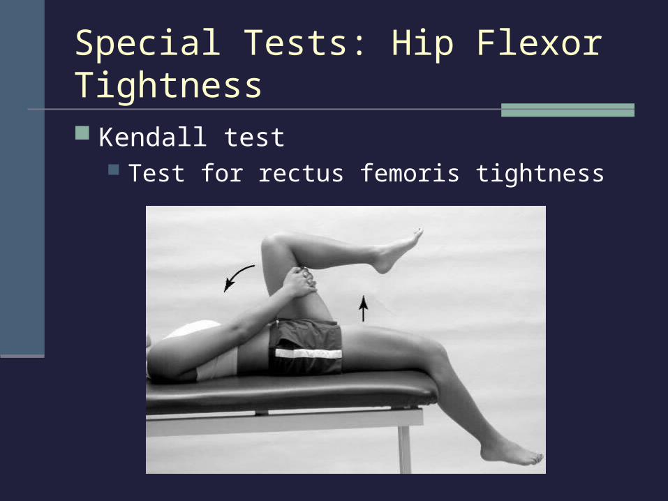
Special Tests: Hip Flexor Tightness
Kendall test Test for rectus femoris tightness
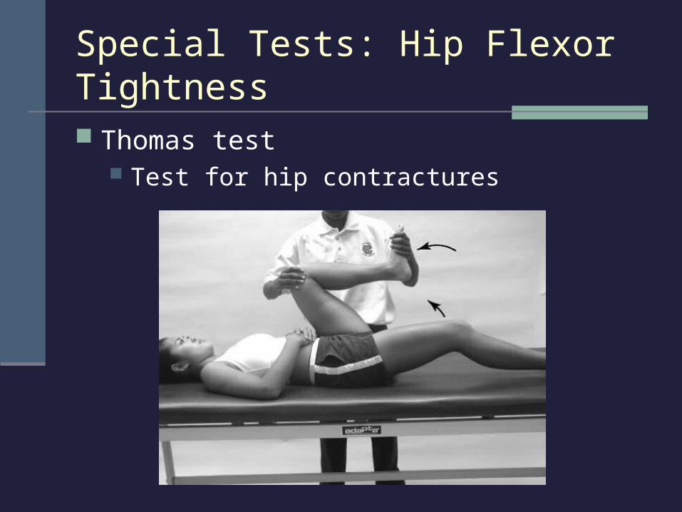
Special Tests: Hip Flexor Tightness
Thomas test Test for hip contractures
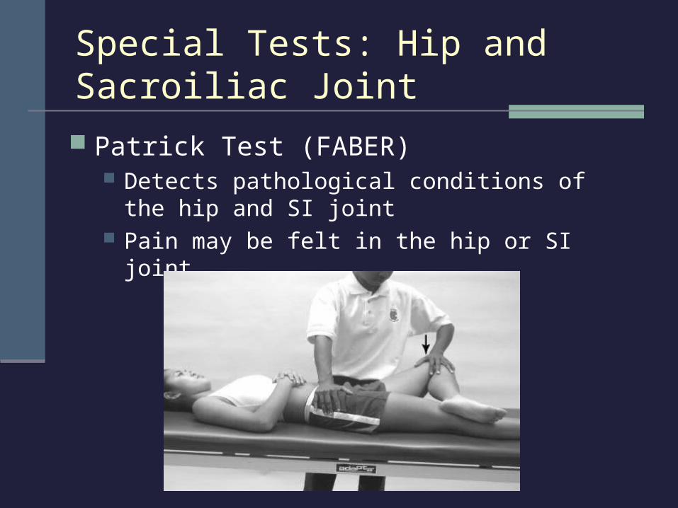
Special Tests: Hip and Sacroiliac Joint
Patrick Test (FABER) Detects pathological conditions of the hip and SI
joint Pain may be felt in the hip or SI joint
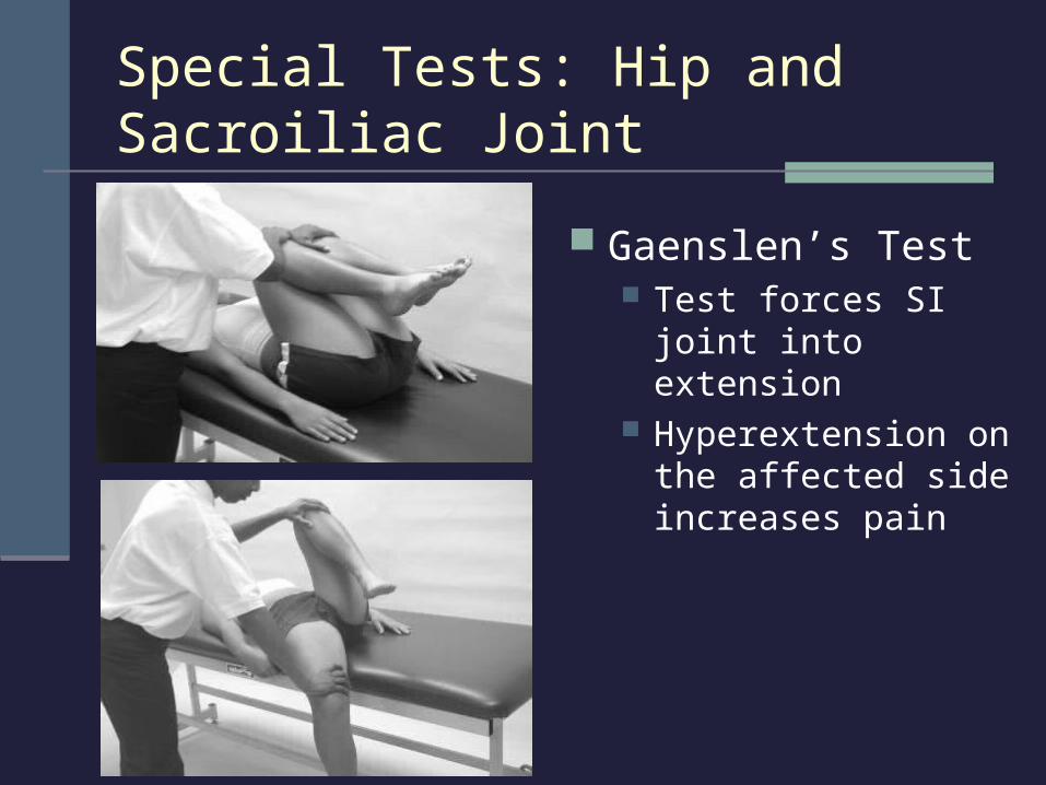
Special Tests: Hip and Sacroiliac Joint
Gaenslen’s Test Test forces SI joint into
extension Hyperextension on the
affected side increases pain
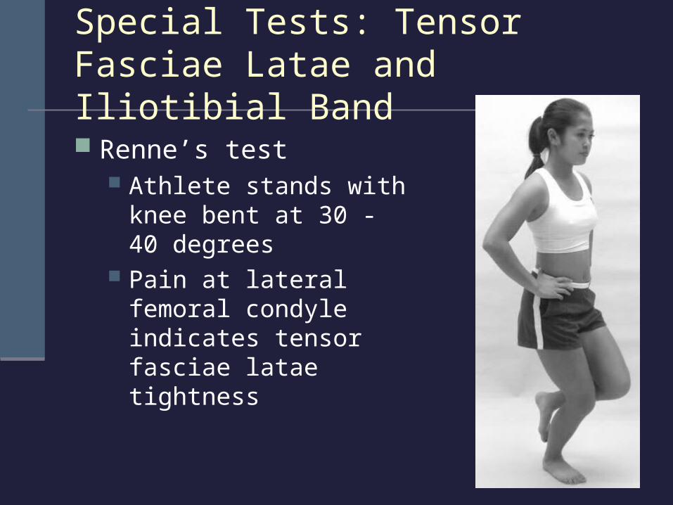
Special Tests: Tensor Fasciae Latae and Iliotibial Band
Renne’s test Athlete stands with knee
bent at 30 - 40 degrees Pain at lateral femoral
condyle indicates tensor fasciae latae tightness
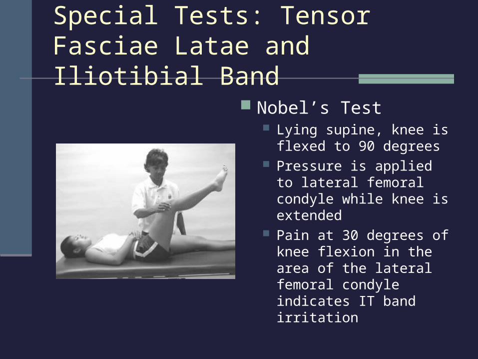
Special Tests: Tensor Fasciae Latae and Iliotibial Band
Nobel’s Test Lying supine, knee is
flexed to 90 degrees Pressure is applied to
lateral femoral condyle while knee is extended
Pain at 30 degrees of knee flexion in the area of the lateral femoral condyle indicates IT band irritation
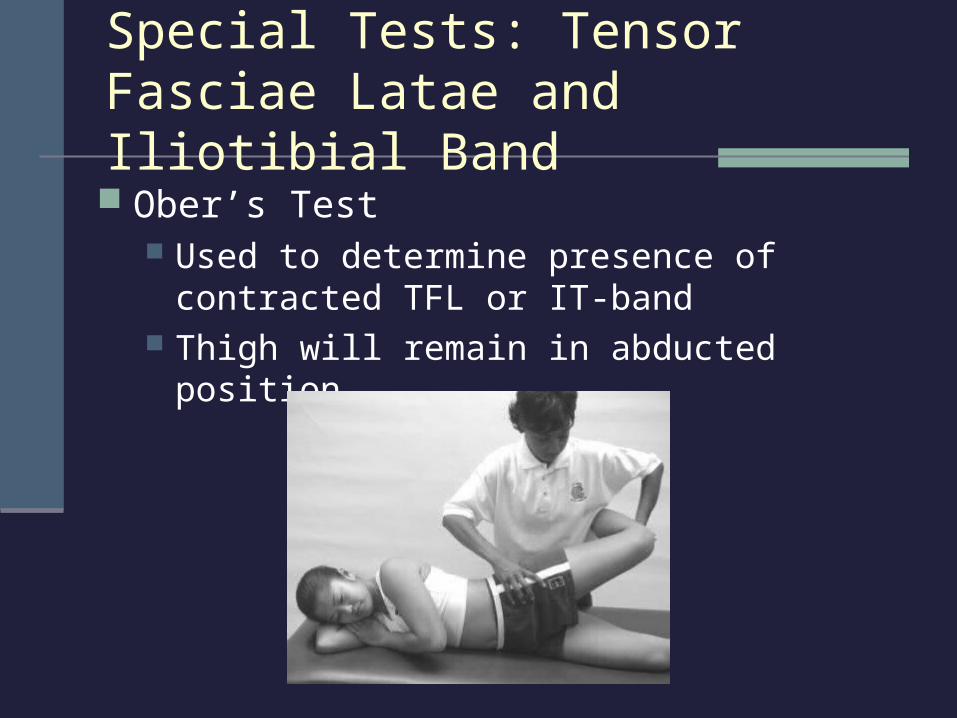
Ober’s Test Used to determine presence of
contracted TFL or IT-band Thigh will remain in abducted position
Special Tests: Tensor Fasciae Latae and Iliotibial Band
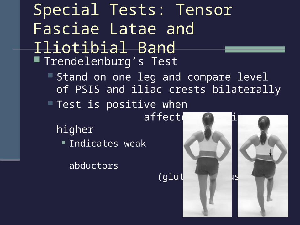
Special Tests: Tensor Fasciae Latae and Iliotibial Band Trendelenburg’s Test
Stand on one leg and compare level of PSIS and iliac crests bilaterally
Test is positive when affected side is higher
Indicates weak hip abductors (gluteus medius)
Special Tests: Piriformis
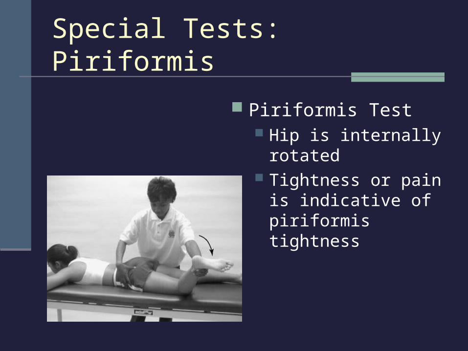
Piriformis Test Hip is internally rotated Tightness or pain is
indicative of piriformis tightness
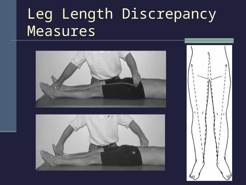
True or anatomical Shortening may be equal throughout limb or
localized in femur or lower leg Measure from ASIS to medial malleolus
Apparent or functional May result due to lateral pelvic tilt, flexion, or
adduction deformity Measure from umbilicus to medial malleolus
Special Tests: Leg Length Discrepancy
Leg Length Discrepancy Measures
Hip and Groin Injuries
Groin Strain Etiology
Injury usually occurs to the adductor longus MOI = running, jumping, or twisting with hip
external rotation; over-stretching; or too forceful contraction
Signs and Symptoms Sudden twinge or tearing during movement Pain, weakness, and internal hemorrhaging
Groin Strain (continued) Management
RICE NSAID’s and analgesics Rest is critical Modalities
Daily whirlpool and cryotherapy Ultrasound
Delay exercise until pain free Restore normal ROM and strength Provide support with elastic wrap
Hip and Groin Injuries
Trochanteric Bursitis Etiology
Inflammation of bursa at greater trochanter Insertion site for gluteus medius and where IT-band
passes over the greater trochanter
Signs and Symptoms Lateral hip pain that may radiate down the leg Point tenderness over greater trochanter IT-band and TFL tests should be performed
Hip and Groin Injuries
Trochanteric Bursitis (continued Management
RICE NSAID’s and analgesics ROM and PRE exercises for hip abductors
and external rotators Phonophoresis Evaluate biomechanics and Q-angle Runners should avoid inclined surfaces
Hip and Groin Injuries
Sprains of the Hip Joint Etiology
Unusual movement exceeding normal ROM MOI = force from opponent/object, or, trunk
forced over planted foot in opposite direction Signs and Symptoms
Pain, which increases with hip rotation Inability to circumduct hip Similar S&S to stress fracture
Hip and Groin Injuries
Sprains of the Hip Joint (continued) Management
RICE NSAID’s and analgesics Depending on severity, crutches may be
required ROM and PRE are delayed until hip is pain-free X-rays or MRI should be performed to rule out
a possible fracture
Hip and Groin Injuries
Dislocated Hip Etiology
Result of traumatic force directed along the long axis of the femur
Posterior dislocation more common Hip flexed, adducted, and internally rotated Knee flexed
Rarely occurs in sport Signs and Symptoms
Flexed, adducted, and internally rotated hip Palpation reveals displaced femoral head Medical emergency
Compications include soft tissue damage, neurological damage, and possible fracture
Hip and Groin Injuries
Dislocated Hip (continued) Management
Immediate medical care Blood and nerve supply may be compromised
Contractures may further complicate reduction 2 weeks immobilization Crutch use for at least one month
Hip and Groin Injuries

Avascular Necrosis Etiology
Temporary or permanent loss of blood supply to the proximal femur
MOI = traumatic conditions (ie: hip dislocation) or non-traumatic conditions (ie: steroids, blood coagulation disorders)
Signs and Symptoms Possibly no S&S in early stages
Develop over the course of months to a year Joint pain with weight bearing, progressing to pain at rest Limited ROM Osteoarthritis may develop
Hip and Groin Injuries
Avascular Necrosis (continued) Management
Must be referred for X-ray, MRI, or CT scan Most cases will ultimately require surgery
Conservative treatment Non-weight bearing;ROM exercises; e-stim for bone
growth; medication to treat pain Limit necrosis
Reduce fatty substances, which react with corticosteroids Limit blood clotting in the presence of clotting disorders
Hip and Groin Injuries
Hip Problems in the Young Athlete
Legg Calve’-Perthes Disease (Coxa Plana) Etiology
Avascular necrosis of the femoral head in child ages 4-10
MOI = trauma (accounts for 25% of cases) Signs and Symptoms
Pain in groin Referred pain to the abdomen or knee
Limping may exhibit limited ROM
Hip Problems in the Young Athlete
Legg Calve’-Perthes Disease (continued) Management
Bed rest to alleviate synovitis Brace to avoid direct weight bearing With early treatment, the femoral head may
re-ossify and revascularize Complications
If not treated early, will result in ill-shaping May develop into osteoarthritis in later life
Slipped Capital Femoral Epiphysis Etiology
Found mostly in tall boys between ages 10-17 May be growth hormone related MOI = trauma (accounts for 25% of cases)
25% of cases are seen in both hips Femoral head slippage on X-ray appears in
posterior and inferior direction
Hip Problems in the Young Athlete
Slipped Capital Femoral Epiphysis (continued)
Signs and Symptoms Pain in groin that progresses over weeks or months Hip and knee pain during passive and active motion
Limitations of hip abduction, flexion, and medial rotation Limp
Management Minor slippage
Rest and non-weight bearing may prevent further slippage Major slippage results in displacement
Requires surgery If condition goes undetected or if surgery fails, severe
problems will result
Hip Problems in the Young Athlete
The Snapping Hip Phenomenon Etiology
Common in young female dancers, gymnasts, and hurdlers
MOI = repetitive movement that leads to muscle imbalance
Related to narrow pelvis, increased hip abduction, and limited lateral rotation
Hip stability is compromised
Hip Problems in the Young Athlete
The Snapping Hip Phenomenon (continued) Signs and Symptoms
Pain while balancing on one leg Possible inflammation
Management ROM exercises to increase flexibility
Flexion and lateral rotation Cryotherapy and ultrasound may be utilized
PRE exercises to strengthen weak muscles
Hip Problems in the Young Athlete
Contusion (hip pointer) Etiology
Contusion of iliac crest or abdominal musculature
MOI = direct blow Signs and Symptoms
Pain, spasm, and transitory paralysis Decreased ROM due to pain
Rotation of trunk, thigh/hip flexion
Pelvic Injuries
Contusion (hip pointer) continued Management
RICE for at least 48 hours NSAID’s, Bed rest 1 - 2 days Referral must be made for X-ray Modailities
Ice massage, ultrasound, occasionally steroid injection
Recovery lasts 1 - 3 weeks
Pelvic Injuries
Osteitis Pubis Etiology
Often seen in distance runners MOI = repetitive stress
Signs and Symptoms Chronic pain and inflammation of groin Point tenderness on pubic tubercle Pain with running, sit-ups, and squats
Management Rest, NSAID’s, and gradual return to activity
Pelvic Injuries
Athletic Pubalgia Etiology
Chronic pubic region pain MOI = repetitive stress to pubic symphysis from
kicking, twisting, or cutting Signs and Symptoms
No presence of hernia Chronic pain during exertion Sharp and burning pain that radiates into
adductors and testicles
Pelvic Injuries
Athletic Pubalgia (continued) Signs and Symptoms (continued)
Point tenderness on pubic tubercle Increased pain with resisted hip flexion, internal
rotation, abdominal contraction, and hip adduction Management
Conservative treatment (rarely effective): rest, ROM exercises, and PRE exercises
Aggressive treatment: cortisone injection or surgical tightening of pelvic wall
Pelvic Injuries
Stress Fractures Etiology
Seen in distance runners – more common in women than men
MOI = repetitive cyclical forces from ground reaction forces
Common sites include inferior pubic ramus, femoral neck, and subtrochanteric area of the femur
Signs and Symptoms Groin pain
Aching sensation in thigh that increases with activity and decreases with rest
Standing on one leg may be impossible Deep palpation results in point tenderness
Pelvic Injuries
Stress Fractures (continued) Management
Rest for 2 - 5 months Crutch walking
Especially for ischium and pubis stress fractures X-rays are usually normal for 6 -10 weeks,
therefore a bone scan will be required to detect the stress fracture
Swimming can be used to maintain CV fitness Breast stroke should be avoided
Pelvic Injuries
Avulsion Fractures and Apophysitis Etiology
Common sites include ischial tuberosity, AIIS, and ASIS
MOI = sudden accelerations and decelerations Signs and Symptoms
Sudden localized pain Limited ROM Pain, swelling, point tenderness Muscle testing increases pain
Pelvic Injuries
Avulsion Fractures and Apophysitis (continued)
Management X-ray required for diagnosis RICE, NSAID’s, crutch “toe-touch” walking ROM exercises PRE exercises
When 80 degrees of ROM have been regained Return to play when full ROM and strength are
restored
Pelvic Injuries
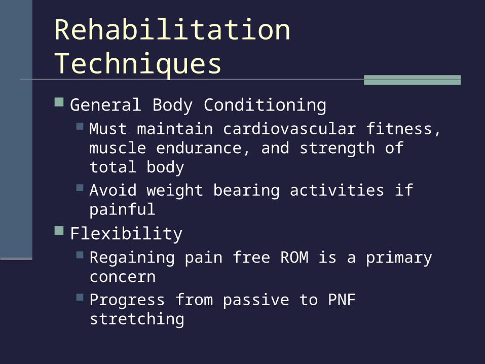
Rehabilitation Techniques
General Body Conditioning Must maintain cardiovascular fitness, muscle
endurance, and strength of total body Avoid weight bearing activities if painful
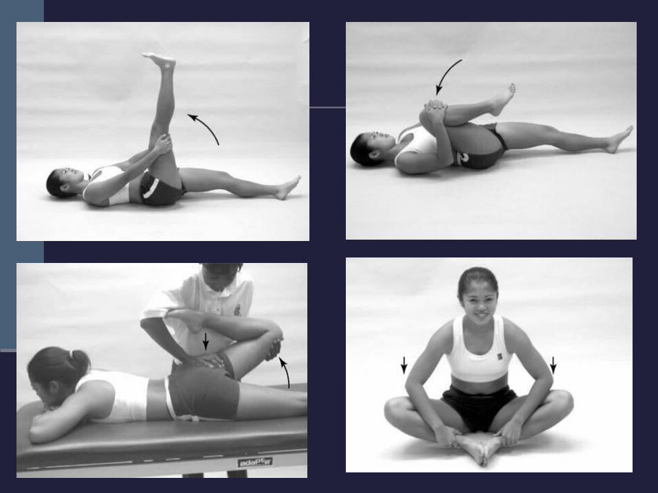
Flexibility Regaining pain free ROM is a primary concern Progress from passive to PNF stretching
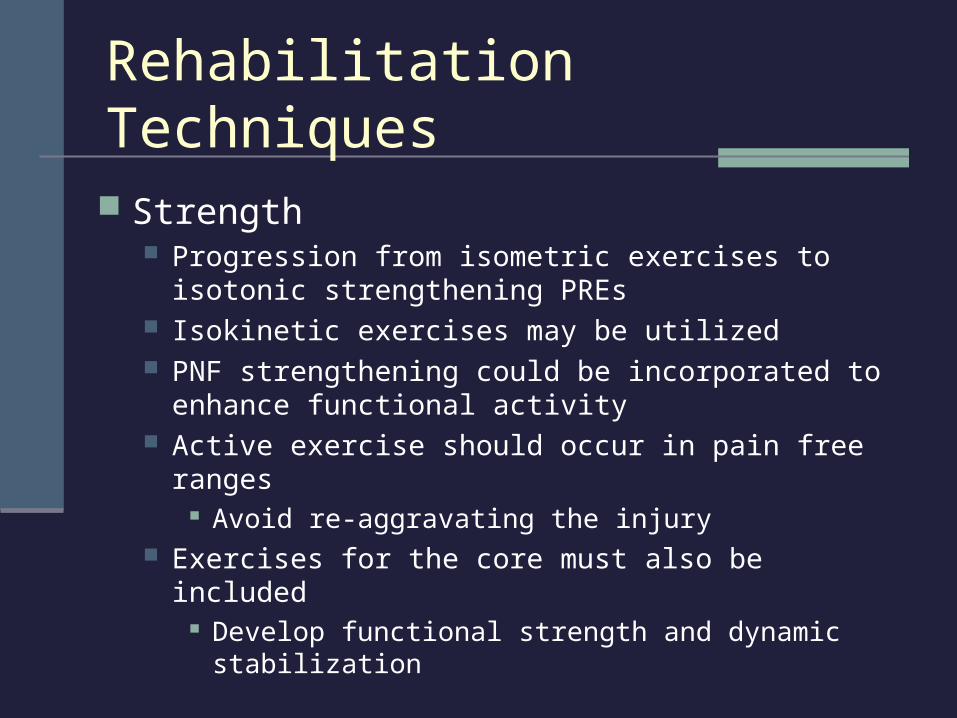
Rehabilitation Techniques
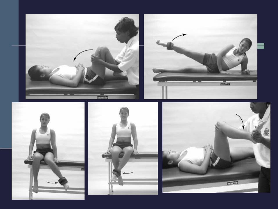
Strength Progression from isometric exercises to isotonic
strengthening PREs Isokinetic exercises may be utilized PNF strengthening could be incorporated to enhance
functional activity Active exercise should occur in pain free ranges
Avoid re-aggravating the injury Exercises for the core must also be included
Develop functional strength and dynamic stabilization
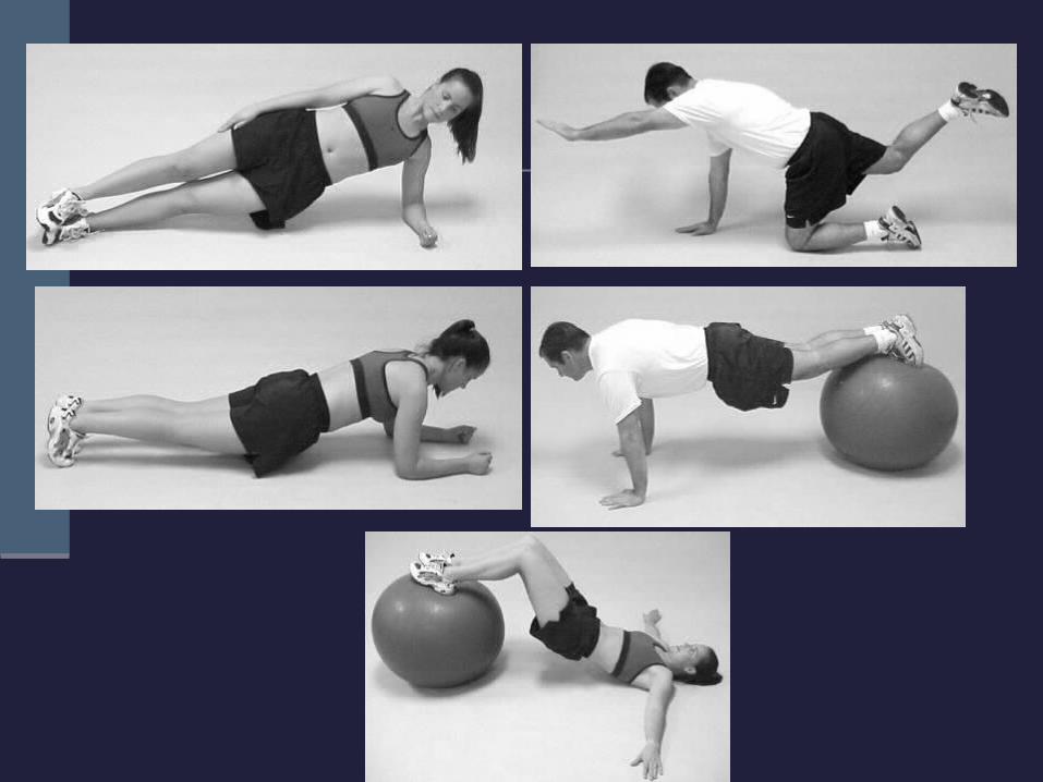
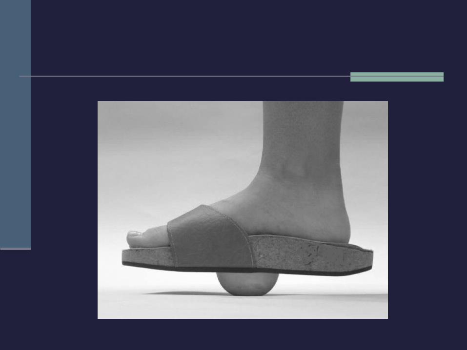
Rehabilitation Techniques
Neuromuscular Control Established through postural alignment and stability
strength As neuromuscular control is enhanced, the ability of
the kinetic chain to maintain appropriate forces and dynamic stabilization increases
Focus on balance and closed kinetic chain activities
Functional Progression and Return to Activity Begin in pool, non-weight bearing Progression of walking, to jogging, to running,
and to more difficult agility tasks Before returning to play, athlete should
demonstrate pain free function, full ROM, strength, balance, and agility