Embed Size (px)
Citation preview

Chapter 22 The Chest and Abdomen

© 2010 Delmar, Cengage Learning2© 2011 Delmar, Cengage Learning
Objectives Upon completion of this chapter, you should
be able to: Describe the anatomy of the thoracic cavity Describe the structures and functions of the
organs of respiration Explain the breathing and respiratory process

© 2010 Delmar, Cengage Learning3© 2011 Delmar, Cengage Learning
Objectives (cont’d.) Upon completion of this chapter, you should
be able to (cont’d.): Discuss the significance of chest and
abdominal injuries List and describe the various injuries associated
with the thoracic cavity List and describe the various injuries associated
with the abdominal cavity

© 2010 Delmar, Cengage Learning4© 2011 Delmar, Cengage Learning
Ribs & Sternum Sternum
(“breastbone”)—3 parts Manubrium Body Xiphoid process
Ribs—12 pairs 7 true 3 false ribs—do not
connect to sternum directly
2 floating—no connection to sternum at all

© 2010 Delmar, Cengage Learning5© 2011 Delmar, Cengage Learning
The Respiratory System Obtains oxygen for use by body cells Eliminates carbon dioxide produced in cellular
respiration

© 2010 Delmar, Cengage Learning6© 2011 Delmar, Cengage Learning
The Respiratory System (cont’d.) Air moves into the lungs through passageways:
Nasal cavity Pharynx (throat) Larynx (voice box) Trachea (wind pipe) Bronchi (branches of trachea) Bronchioles Alveoli

© 2010 Delmar, Cengage Learning7© 2011 Delmar, Cengage Learning
Respiration Process by which body supplies cells and tissues with
oxygen for metabolism and relieves them of carbon dioxide External respiration
Exchange of oxygen and carbon dioxide between lungs and outside environment—breathing
Internal respiration Exchange of carbon dioxide and oxygen between cells and
lymph, plus oxidative process of energy in cells (cellular respiration)
© 2010 Delmar, Cengage Learning8© 2011 Delmar, Cengage Learning
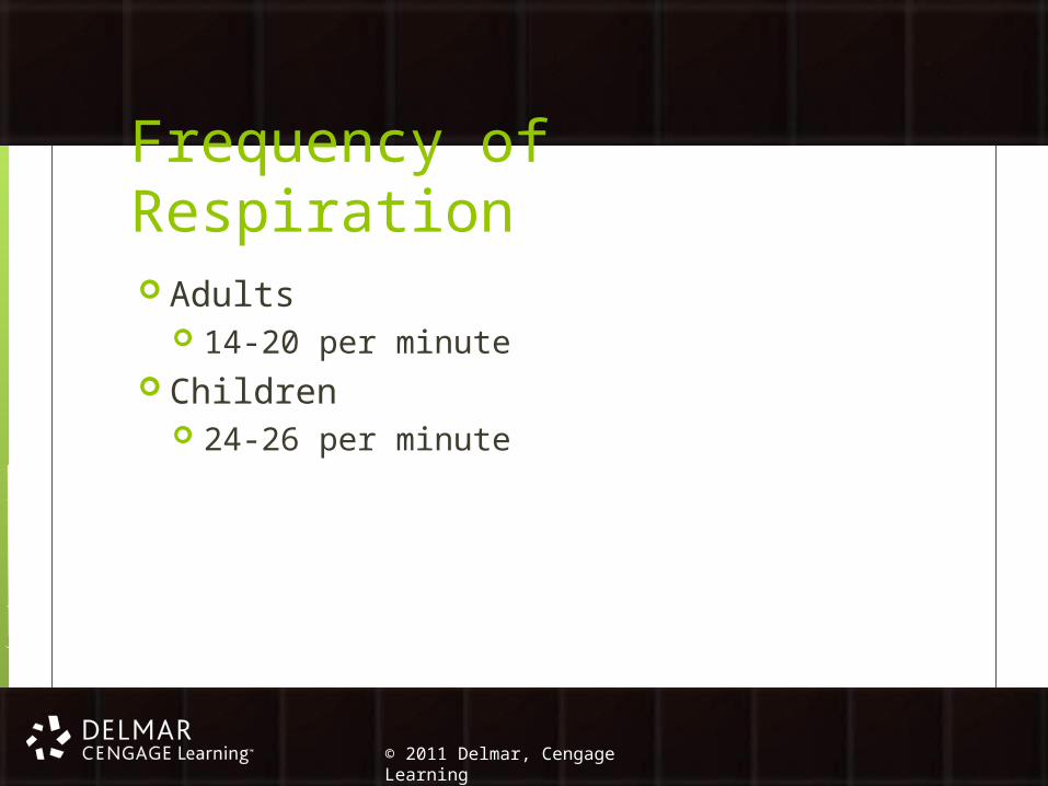
Frequency of Respiration Adults
14-20 per minute Children
24-26 per minute

© 2010 Delmar, Cengage Learning9© 2011 Delmar, Cengage Learning
Control of Breathing Rate of breathing is controlled by neural
(nervous) and chemical factors Same goal but function independently Chemical control of respiration depends on
carbon dioxide level in the blood Chemoreceptors in carotid arteries and aorta
are sensitive to blood oxygen levels

© 2010 Delmar, Cengage Learning10© 2011 Delmar, Cengage Learning
Lung Capacity and Volume Factors:
Tidal volume Inspiratory reserve
volume Expiratory reserve
volume Vital lung capacity Residual volume Functional residual
capacity Total lung capacity
Use a spirometer—measures volume & flow of air

© 2010 Delmar, Cengage Learning11© 2011 Delmar, Cengage Learning
Disorders of the Respiratory System Asthma
Muscles around airways tighten and airway lining swells and gets clogged with thick mucus Symptoms: coughing, wheezing, dyspnea (difficulty
in breathing), and chest tightness Treatment: varies
Exercise-Induced Asthma (EIA) Increased physical activity causes narrowing of
airway
© 2010 Delmar, Cengage Learning12© 2011 Delmar, Cengage Learning
Chest (Thorax) Injuries Rib contusions
Caused by a forceful blow to the ribcage that bruises intercostal muscle
S/S: point tender, pain when breathing Treatment: removal from activity, ice
Rib fractures Break in bony structure of thorax Most often the result of a direct blow to the
ribcage

© 2010 Delmar, Cengage Learning13© 2011 Delmar, Cengage Learning
Chest (Thorax) Injuries (cont’d.) Chest contusions
Bruising over central area of chest Results from a compressive, forceful blow to the
body Myocardial contusion and aortic rupture
Occurs if force applied to sternum is great enough to compress the heart against the spine
Emergency!
© 2010 Delmar, Cengage Learning14© 2011 Delmar, Cengage Learning
Chest (Thorax) Injuries (cont’d.) Sudden death syndrome
Usually caused by some form of heart disease Pneumothorax
Occurs when air enters thoracic cavity between the chest wall and lung Sucking chest wound Spontaneous pneumothorax Tension pneumothorax

© 2010 Delmar, Cengage Learning15© 2011 Delmar, Cengage Learning
Chest (Thorax) Injuries (cont’d.) Hemopneumothorax
Can occur with both open and closed chest injuries Often accompanies a pneumothorax
Blood accumulates in pleural space between chest wall and lung
Pulmonary contusion Bruise on lung caused by a direct blow

© 2010 Delmar, Cengage Learning16© 2011 Delmar, Cengage Learning
Chest (Thorax) Injuries (cont’d.) Blows to the solar plexus
“Having the wind knocked out” Hyperventilation
Breathing at a rate faster than required for proper exchange of oxygen and carbon dioxide
Side stitches Occur during vigorous exercises
Usually with novice exercisers

© 2010 Delmar, Cengage Learning17© 2011 Delmar, Cengage Learning
Injury Prevention for the Chest Begins with proper equipment and education
Good, well-maintained, equipment that fits properly will reduce chance of injury
At risk athletes should wear additional protection
Education and use of proper techniques can also minimize risk of trauma

© 2010 Delmar, Cengage Learning18© 2011 Delmar, Cengage Learning
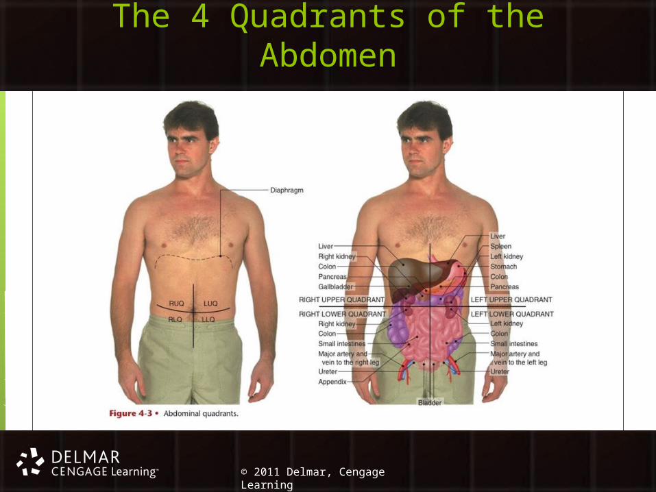
The Abdominopelvic Cavity One large cavity, with no separation between the
abdomen and pelvis Abdominal cavity contains: stomach, liver, gallbladder,
pancreas, spleen, small intestine, appendix, and part of the large intestines Kidneys are close to but behind abdominal cavity
Pelvic cavity contains: urinary bladder, reproductive organs, rectum, remainder of large intestine, and appendix

© 2010 Delmar, Cengage Learning19© 2011 Delmar, Cengage Learning
Protection of the Abdominal Organs Abdominal area is vulnerable to injury
Muscular abdominal wall is most commonly involved
Injury to contents of abdominal cavity are infrequent Musculature of abdominal wall provides adequate
protection from most injuries Serious injuries to the intra-abdominal contents
occur and can be life threatening
© 2010 Delmar, Cengage Learning20© 2011 Delmar, Cengage Learning
The 4 Quadrants of the Abdomen

© 2010 Delmar, Cengage Learning21© 2011 Delmar, Cengage Learning
Organs of the Abdominopelvic Cavity
Stomach Small intestine Pancreas Liver Gallbladder Urinary bladder
Large intestine Colon Cecum Appendix
Kidneys

© 2010 Delmar, Cengage Learning22© 2011 Delmar, Cengage Learning
Abdominal Injuries Kidney contusion
Uncommon in athletics Occurs with a violent blow to upper posterior
abdominal wall Liver contusion
Uncommon but probable life-threatening injury Occurs with a hard blow to right side of ribcage
© 2010 Delmar, Cengage Learning23© 2011 Delmar, Cengage Learning
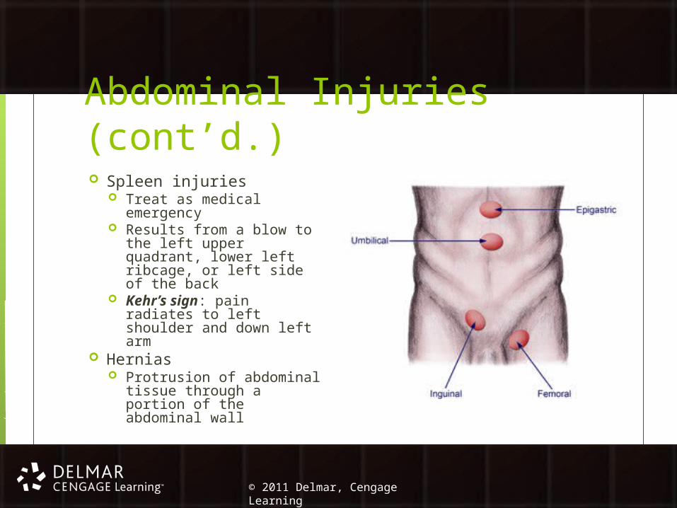
Abdominal Injuries (cont’d.) Spleen injuries
Treat as medical emergency
Results from a blow to the left upper quadrant, lower left ribcage, or left side of the back
Kehr’s sign: pain radiates to left shoulder and down left arm
Hernias Protrusion of abdominal
tissue through a portion of the abdominal wall

© 2010 Delmar, Cengage Learning24© 2011 Delmar, Cengage Learning
Conclusion The chest and abdomen contain the body’s
vital organs Organs in the chest are protected by the
ribcage Chest contains the heart and lungs Abdomen contains kidneys, liver, spleen,
stomach, urinary bladder, intestines, among others

© 2010 Delmar, Cengage Learning25© 2011 Delmar, Cengage Learning
Conclusion (cont’d.) Chest and abdominal injuries are uncommon
in athletics, but do occur Most internal organs are very vascular and can
bleed profusely if injured Proper recognition and treatment of these
injuries are vital to the health and well-being of the athlete