Embed Size (px)
Citation preview

Chapter 12Tetramisole and Levamisole SuppressNeuronal Activity Independentlyfrom Their Inhibitory Action on TissueNon-specific Alkaline Phosphatasein Mouse Cortex
Lionel G. Nowak, Benoît Rosay, Dávid Czégé and Caroline Fonta
Abstract Tissue non-specific alkaline phosphatase (TNAP) may be involved in thesynthesis of GABA and adenosine, which are themain inhibitory neurotransmitters incortex. We explored this putative TNAP function through electrophysiologicalrecording (local field potential) in slices of mouse somatosensory cortex maintainedin vitro. We used tetramisole, a well documented TNAP inhibitor, to block TNAPactivity. We expected that inhibiting TNAP with tetramisole would lead to anincrease of neuronal response amplitude, owing to a diminished availability ofGABA and/or adenosine. Instead, we found that tetramisole reduced neuronalresponse amplitude in a dose-dependent manner. Tetramisole also decreased axonalconduction velocity. Levamisole had identical effects. Several control experimentsdemonstrated that these actions of tetramisole were independent from this compoundacting on TNAP. In particular, tetramisole effects were not stereo-specific and theywere not mimicked by another inhibitor of TNAP, MLS-0038949. The decrease ofaxonal conduction velocity and preliminary intracellular data suggest that tetramisoleblocks voltage-dependent sodium channels. Our results imply that levamisole ortetramisole should not be used with the sole purpose of inhibiting TNAP in livingexcitable cells as it will also block all processes that are activity-dependent. Our data
L.G. Nowak (&) � B. Rosay � C. Fonta (&)Centre de Recherche Cerveau et Cognition (CerCo), Université de Toulouse UPS;CNRS UMR 5549, Toulouse, Francee-mail: [email protected]
C. Fontae-mail: [email protected]
L.G. NowakCNRS, Cerco, Toulouse, France
D. CzégéDepartment of Physiology and Neurobiology, Eötvös Loránd University, Budapest, Hungary
© Springer Science+Business Media Dordrecht 2015C. Fonta and L. Négyessy (eds.), Neuronal Tissue-Nonspecific AlkalinePhosphatase (TNAP), Subcellular Biochemistry 76,DOI 10.1007/978-94-017-7197-9_12
239

and a review of the literature indicate that tetramisole may have at least four differenttargets in the nervous system. We discuss these results with respect to the neuro-logical side effects that were observed when levamisole and tetramisole were used formedical purposes, and that may recur nowadays due to the recent use of levamisoleand tetramisole as cocaine adulterants.
Keywords Vitamin B6 � Pyridoxal � Ectonucleotidase � Epilepsy � Inflammatoryleukoencephalopathy
12.1 Introduction
Alkaline phosphatases (APs) are a group of enzymes that release inorganic phos-phate from phosphomonoesters. APs are ecto-enzymes, thus located on the extra-cellular side of the cells. Four types of APs have been identified in human. Threehave restricted regional distributions and have been named accordingly as intestinalAP, germinal AP and placental AP. The fourth type, the tissue non-specific alkalinephosphatase (TNAP), has been found in all mammalian genera examined. As itsname implies, TNAP shows a widespread distribution, being present in varioustissues such as blood vessels, bone and cartilage, kidney, liver, lung, etc. (e.g.,Borgers 1973; de Bernard et al. 1986; Morris et al. 1992; Hoshi et al. 1997).
In human, mutations of the TNAP gene lead to hypophosphatasia (Rathbun1948), a rare disease characterized by low level of plasmatic AP and by abnormalbone mineralization (reviewed in: Fraser 1957; Millán 2006; Mornet 2007; Whyte2010; Taketani et al. 2014; see also Chaps. 1 and 2 in this book). Importantly, themost severe forms of hypophosphatasia (perinatal and, to a lesser extent, infantilehypophosphatasia) are often associated with epileptic seizures (e.g., Rathbun 1948;Fraser 1957; Béthenod et al. 1967; Baumgartner-Sigl et al. 2007; Balasubramaniamet al. 2010; Taketani et al. 2014; see also Chaps. 14 and 15 in this book). In themouse, inactivation of the TNAP gene produces an animal model of severe humanhypophosphatasia, with bone mineralization defect and epileptic seizures as well(Waymire et al. 1995; Narisawa et al. 1997; see also Chap. 3).
Occurrence of epileptic seizures in TNAP KO mice and in the severe forms ofhuman hypophosphatasia suggests that TNAP plays a fundamental role in thecontrol of nerve cell activity—although indirect effects cannot be ruled out. Thisimplies that TNAP should be present in the nervous tissue. Indeed, although earlystudies (Landow et al. 1942; Bourne 1943) did not report AP activity at the neu-ronal level in mammals—except in the spinal cord and medulla –, later studiesdemonstrated significant AP activity in multiple brain structures of various mam-malian species. The pharmacological profile of this neuronal AP activity suggestedit resulted from TNAP activity (Fonta et al. 2004; Langer et al. 2008) and it hasrecently been formally ascribed to TNAP gene expression (Ermonval et al. 2009;Brun-Heath et al. 2011).
240 L.G. Nowak et al.
AP activity in the brain is not homogenous; some structures show strong activity—hypothalamus, superior colliculus, subtancia nigra, spinal cord for example—while other—hippocampus, cerebellum, caudate nucleus, putamen for example—show much weaker activity (Shimizu 1950; Nandy and Bourne 1963; Friede 1966;Sugimura and Mizutani 1979; Langer et al. 2008; Brun-Heath et al. 2011; Streetet al. 2013). AP activity is also observed in the cerebral cortex. In primate its levelis particularly high in layer 4 of primary visual, auditory and somatosensory cor-tices (Friede 1966; Fonta and Imbert 2002; Fonta et al. 2004, 2005; Négyessy et al.2011). In area 17 of primates, the thalamorecipient layers 4Cα and 4Cβ areexquisitely delineated by TNAP activity (Fonta and Imbert 2002; Fonta et al. 2004).In visual cortex, AP activity depends on neuronal activity, as revealed by mon-ocular deprivation experiments (Fonta et al. 2004). TNAP is also highly expressedin the prefrontal cortex of primate (Fonta et al. 2004) and is found preferentially inlayer 5 in non-sensory cortices of non-human primates (Friede 1966) and in human(Négyessy et al. 2011). AP activity is less patterned in rodent cortex but cleardifferences between cortical areas are nevertheless noticeable. For example, layer Iaof the piriform cortex and layer 4 of the somatosensory cortex show stronger APactivity than neighboring layers and surrounding cortical areas (Fonta et al. 2004;Langer et al. 2008; Brun-Heath et al. 2011).
In both cortical and subcortical structures, the suggestion that AP is located onsynapses (Nandy and Bourne 1963) has been confirmed by electron microscopystudies, which revealed AP in the synaptic clefts of both excitatory and inhibitorysynapses (Sugimura and Mizutani 1979; Mori and Nagano 1985; Fonta et al. 2004,2005; Hanics et al. 2012; see also Chap. 5, this book). Strong AP activity has alsobeen revealed on the nodes of Ranvier of myelinated axons (Pinner et al. 1964;Mori and Nagano 1985; Fonta et al. 2005; Hanics et al. 2012; see Chap. 5).Altogether, these data suggest that TNAP may be involved in the control of actionpotential propagation in axons as well as in the control of synaptic transmission.
In this study we used an electrophysiological approach with the aim to determinewhether and how TNAP controls neuronal activity in the cerebral cortex, in par-ticular through its putative involvement in the synthesis of two major neurotrans-mitters, GABA and adenosine. In cortex, both GABA and adenosine are inhibitoryneurotransmitters: GABA acts postsynaptically through GABAA and GABAB
receptors and presynaptically through GABAB receptors (e.g., Howe et al. 1987a, b;Connors et al. 1988; Deisz and Prince 1989; McCormick 1989). Adenosine mostlyacts presynaptically through adenosine A1 receptors (e.g., Collins and Anson 1985;Fontanez and Porter 2006).
GABA is synthesized from glutamate by the glutamic acid decarboxylase(GAD) (e.g., Martin and Rimvall 1993). GAD uses the active form of vitamin B6,pyridoxal phosphate (PLP), as cofactor.1 PLP cannot cross membranes; only the
1Note that GAD is one among multiple PLP-dependent enzymes. Approximately 60 genesencoding for enzymes using PLP as cofactor have been identified in mammals (Percudani andPeracchi 2009). In addition to GAD, some of these enzymes are directly involved in the synthesisof other neurotransmitters such as dopamine and serotonin (Ermonval et al. 2009).
12 TNAP and Tetramisole, an Electrophysiological Approach 241

nonphosphorylated form, pyridoxal (PL), can diffuse passively through membranes(e.g., Rifkin et al. 1972; Mehansho and Henderson 1980; see also Chap. 11, thisbook). TNAP plays a key role at this level as it is responsible for the dephospho-rylation of extracellular PLP. In support for the involvement of TNAP in this pro-cess, the PLP concentration is markedly elevated in the serum of hypophosphatasicpatients (Whyte et al. 1985, 1988). Likewise, PLP concentration in the serum ofTNAP KO mice appears to be 20 time higher than in wild type mice (Waymire et al.1995). In parallel, GABA levels appear to be strongly reduced in the brain ofTNAP KO mice (Waymire et al. 1995; Fonta et al. 2012), confirming the importanceof TNAP in the control of GABA synthesis. Yet the presence of AP on excitatorysynapses suggests that its role is not solely limited to the control of GABA synthesis.
Adenosine synthesis in the extracellular space results from the degradation ofATP. ATP is released by neurons and glial cells (Fields and Burnstock 2006;Abbracchio et al. 2009; Butt 2011). ATP and intermediate nucleotides are rapidlyhydrolyzed by a variety of ectonucleotidases. Four different ectonucleotidase familieshave been identified in the brain (e.g., Zimmermann et al. 2012). The first threefamilies are quite specific: ectonucleoside triphosphate diphosphohydrolase degradesATP in ADP and ADP in AMP; ectonucleotide pyrophosphatase/phosphodiesterasedegrades ATP in AMP; and ecto-5′-nucleotidase degrades AMP in adenosine. Incontrast, TNAP shows broader substrate specificity and is capable of degrading ATPto ADP, ADP to AMP, and AMP to adenosine. The ectonucleotidase activity ofTNAP has been demonstrated in various tissues such as bone (e.g., Ciancaglini et al.2010; Simão et al. 2013) and airways (Picher et al. 2003). Supports in favor of a roleof TNAP in adenosine synthesis in the brain come from a cultured neuronal cell linestudy (Ohkubo et al. 2000) and from a recent spinal cord study (Street et al. 2013; seeChap. 13). Yet another study failed to reveal a significant role for TNAP in extra-cellular adenosine synthesis in the hippocampus (Zhang et al. 2012).
In order to examine the role of TNAP in the control of inhibitory synaptictransmission, we performed electrophysiological experiments on slices of mousesomatosensory cortex maintained in vitro. We used tetramisole to inhibit TNAPactivity. Tetramisole is the racemic mixture of two stereoisomers: a levorotatoryenantiomer, “levamisole”, and a dextrorotary enantiomer, “dexamisole”. Studies inthe seventies showed that levamisole is highly effective at inhibiting TNAP (VanBelle 1972, 1976a, b) while dexamisole has no significant effect on this enzyme(Van Belle 1972, 1976b; Borgers 1973). Levamisole and tetramisole have sincebeen used as TNAP inhibitors in a countless number of studies.
Our hypothesis was that suppressing TNAP activity with tetramisole wouldresult in an increase of neuronal response amplitude owing to a reduction of pre-and/or postsynaptic inhibition mediated by GABA and/or adenosine. The results weobtained were opposite to this expectation: tetramisole, at concentration that fullysuppress TNAP activity in biochemical assays (1–5 mM), also strongly suppressedneuronal activity. Levamisole had a similar effect. This suppression largely resultedfrom a reduction of action potential transmission along the axons. These resultsmight have been interpreted as revealing a hitherto non-described control of axonal
242 L.G. Nowak et al.
excitability by TNAP present on the nodes of Ranvier, yet several control experi-ments revealed that neuronal activity suppression by tetramisole and levamisolewas not due to TNAP inhibition by tetramisole and levamisole. Instead, we proposethat tetramisole and levamisole block voltage-dependent sodium channels.
Our results therefore indicate that, in cortex, TNAP is not the unique target oftetramisole and levamisole. In addition, other neuronal processes are likely to beaffected by levamisole and tetramisole: indeed, studies showed that, in theperipheral nervous system of mammals, levamisole and tetramisole may alsointerfere with adrenergic and cholinergic synaptic transmission.
Outside the nervous system, studies suggested that tetramisole and levamisolepossess “immunostimulating” properties. For this reason, tetramisole and levam-isole have received numerous clinical and pharmaceutical applications in a varietyof diseases. Nevertheless, multiple side effects have been reported, includingdevastating neurological adverse effects such as epilepsy and inflammatory leu-koencephalopathy, that have led to discontinue chronic tetramisole and levamisoleusage in most countries. Yet these side effects have recently regained the attentionof public health specialists because most of the illegal cocaine on the marketnowadays appears to be adulterated with levamisole or tetramisole. We discuss thepossible links between the neurological side effects of tetramisole and levamisoleand the different targets of these compounds in the CNS.
12.2 Methods
12.2.1 Brain Slice Preparation
All procedures were conducted in accordance with the guidelines from the FrenchMinistry of Agriculture (décret 87/848) and from the European Community(directive 86/609) and was approved by the local ethical committee(MP/06/79/11/12, comité d’éthique Midi-Pyrénées pour l’expérimentation ani-male). Adult (>2 month-old) wild type female mice were used for these experi-ments. Mouse was anesthetized with isoflurane. Once deeply anesthetized, themouse was killed by decapitation. The scalp was removed, the skull was drilled, theupper part of the skull was lifted off, and the whole brain was carefully removed.These operations were performed in cold (3–4 °C) modified artificial cerebrospinalfluid (mACSF), whose composition was (in mM): NaCl 124, NaHCO3 26, KCl 3.5,MgSO4 1, MgCl2 9, NaH2PO4 1.25, and glucose 10. Note that Ca++ was omittedwhile Mg++ concentration was 10 mM. The rationale for using such a modifiedACSF has been presented elsewhere (Nowak and Bullier 1996). The mACSF wasoxygenated for 1 h before the beginning of the surgery with a mixture of 95 % O2
and 5 % CO2. Four hundred-micrometer-thick coronal brain slices were cut on avibratome (752 M vibroslice, Campden Instrument, UK), whose chamber was filledwith cold oxygenated mACSF. Once obtained, the slices were kept at room
12 TNAP and Tetramisole, an Electrophysiological Approach 243
temperature for at least one hour in a storage chamber filled with an in vivo-like(Brumberg et al. 2000; Sanchez-Vives and McCormick 2000) artificial cerebro-spinal fluid (ACSF) of the following composition (in mM): NaCl 124, NaHCO3 26,KCl 3.5, MgSO4 1, NaH2PO4 1.25, CaCl2 1.2, and glucose 10. This ACSF wascontinuously bubbled with a 95 % O2–5 % CO2 mixture (pH 7.4). Recordings wereperformed in a submersion type chamber (Scientific System Design, Mississauga,Ontario, Canada) where the temperature was thermostatically held at 33–34 °C.The ACSF was gravity fed at a flow rate of 2.5–3.75 ml/min.
12.2.2 Recording and Stimulation
Neuronal signals examined in this study consisted mostly in local field potentials(LFPs) recorded in layer 4 and in the supragranular layers of the whisker, trunk andhindlimb representations of the primary somatosensory cortex (S1). Intracellularrecordings were also attempted in these regions. In both cases, neuronal responseswere evoked by extracellular electrical stimulation applied in the white matter or atthe white matter-layer 6 border. Some extracellular recordings were also performedin the corpus callosum. In this later case, stimulation was applied in the corpuscallosum too.
Tungsten-in-glass microelectrodes, with glass removed from the tip over a lengthof 45–110 µm, were used for extracellular electrical stimulation. Electrical stimu-lation was delivered through a stimulation isolation unit (A365 Stimulus Isolated,WPI) and consisted in monopolar, cathodal pulses of 0.2 or 0.3 ms duration. Pulseswere delivered either in isolation at a frequency of 0.5 Hz, or as pair of pulses withan interstimulus interval of 20 ms, the pairs being repeated every 10 s (0.1 Hz).Stimulation intensity was between 50 and 180 µA.
LFPs were recorded through tungsten-in-glass microelectrodes and glass mic-ropipettes. Glass micropipettes were pulled on a P97 Flaming Brown micropipettepuller from 1.2 mm OD medium walled capillaries with filament (GC120F, HarvardApparatus). The micropipette tip was broken to a 15–20 µm diameter opening andfilled with ACSF (resistance 2–7 MΩ). The signal was amplified with anAxoClamp 2B amplifier (Axon Instrument, Foster City, CA) and further amplifiedwith a Neurolog post-amplifier (final gain: × 1000). The signal was low-pass fil-tered at 10 kHz. Signals recorded through tungsten-in-glass microelectrodes (5–15 µm exposed tip) were amplified (×1000) and bandpass filtered (0.1 Hz–10 kHz)with the Neurolog recording system. Micropipettes for “sharp” intracellularrecording were pulled on the P97 Flaming Brown micropipette puller from 1.2 mmOD medium walled capillaries with filament and filled with K-Acetate 3 M (DCresistance: 60–100 ΜΩ). The AxoClamp 2B was used for amplification(gain: × 10). The signal was low pass filtered (10 kHz). Signals were digitized witha 1401plus interface (CED, Cambridge, UK) with a digitization rate of 20 kHz.
244 L.G. Nowak et al.
12.2.3 Sample
The present study is based on a total of 27 successful experiments. By “experiment”we designate an ensemble of data obtained with one pair of stimulation andrecording sites in one slice. Given the duration of the protocols used in this study(pharmacological manipulation requiring controls, several concentrations of variouspharmacological compounds and recoveries), there was usually only one experi-ment per slice and per day, and by extension one experiment per mouse (19experiments). More rarely two experiments were performed the same day on twodistinct brain slices obtained from the same mouse, with one of the two experimentsbeing dedicated to corpus callosum recording (8 experiments total).
12.2.4 Analysis
Signals were analyzed offline using spike2 software (CED) and custom scripts. Thebulk of the data are issued from extracellular LFP recording evoked by electricalstimulation. The first pass of the LFP analysis consisted in averaging 6 or 30consecutive sweeps, depending on stimulation frequency (0.1 or 0.5 Hz). Thisresulted in series of averaged LFPs for each consecutive minute of recording(Fig. 12.1d). These time series allowed following the time-course of the drugeffects. They were visualized using Origin software (OriginLab Corporation, USA).
The averaged LFPs obtained in the gray matter (23 experiments) consisted in fastand slow components. The slow component, or field PSP (noted “fPSP” in Figures),reflects postsynaptic responses. Depending on where the recording electrode waslocated with respect to current sources and sinks (e.g., Mitzdorf 1985), the slowcomponent appeared either as a slow positivity (Figs. 12.1e, 12.6a and 12.7a) or as aslow negativity (Fig. 12.5a). The fast components—always negative—correspond topopulation spikes, that is, to the synchronous discharge of action potentials by apopulation of neurons located in the vicinity of the recording electrode. Two types ofpopulation spikes were identified in our experiments: antidromic and orthodromic.Antidromic population spikes (indicated by “APS” in Figures) result from the(unnatural) retrograde propagation of action potentials elicited in the axons at thestimulation site toward the cell body, where spike firing is initiated without interca-lated synapses. Orthodromic population spikes (“OPS” in Figures) are elicited whenthe (regular) propagation of action potentials along the axons results in postsynapticresponses that are large enough to trigger action potentials in the postsynaptic neurons(Fig. 12.1e, h). Since they are initiated by excitatory synaptic inputs, their latency islonger than that of antidromic responses. In addition, they disappear when synaptictransmission is suppressed. We systematically tested whether the population spikeswe were recording fromwere orthodromic and/or antidromic by suppressing synapticresponses with ACSF solutions without Ca++ and containing either 6–10 mM Mg++
or 1.2–2 mM Mn++. Antidromic population spikes were present in all but one of the
12 TNAP and Tetramisole, an Electrophysiological Approach 245

23 experiments involving gray matter recording, whereas measurable orthodromicresponses occurred in 16 of these experiments.
The amplitude of antidromic population spikes was measured as the differencebetween the peak of the population spike and the pre-stimulus baseline. The latencycorresponds to the latency of the peak of the population spikes relative to stimulusonset. As orthodromic population spike followed antidromic responses and wereeventually partially merged with them, their amplitude was measured as the dif-ference between the peak and the positivity corresponding to the repolarization ofthe preceding antidromic population spike. The amplitude of the slow componentwas measured relative to pre-stimulus baseline. When the orthodromic responseshowed both a slow component and a population spike, the amplitude of theresponse usually corresponds to that of the population spike. Population data in textcorresponds to the mean ± SEM.
12.2.5 Chemicals
Tetramisole (0.1–5 mM), levamisole (0.5 and 1 mM), adenosine (100 µm), ATP(1 mM) and pyridoxal phosphate (10 µM) were dissolved in ACSF. MLS-0038949(10 µM final concentration) was first dissolved in DMSO (final DMSO concen-tration: 0.1 % in ACSF). All chemicals were purchased from Sigma exceptMLS-0038949 purchased from Merck. When several concentrations of tetramisoleor levamisole were used, their order of application was randomized from oneexperiment to the next.
12.3 Results
12.3.1 Tetramisole Reduces Both Orthodromicand Antidromic Responses in a Dose-DependentManner
Figure 12.1a–c illustrates the effects of levamisole (1 mM) on an antidromic popu-lation spike. Each trace corresponds to the average of 30 sweeps, representing 1 minof recording in control ACSF (Fig. 12.1a), 1 min of recording after 20 min ofperfusion with ACSF containing 1 mM tetramisole (Fig. 12.1b) and 1 min ofrecording after 20 min of reperfusion with regular ACSF (recovery, Fig. 12.1c). Theresponse was identified as antidromic as it persisted when Ca2+ was replaced byMn2+
2 mM (not shown). The white matter stimulation did not elicit a measurable ortho-dromic response in this experiment. In the presence of tetramisole (1mM, Fig. 12.1b),the response amplitude was decreased to 49 % of the amplitude measured in controlwhile the peak latency was increased from 2.6 to 3.5 ms. The amplitude and latencyreturned to near control values after tetramisole washout (Fig. 12.1c).
246 L.G. Nowak et al.
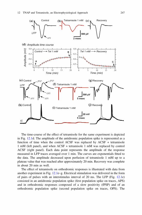
The time-course of the effect of tetramisole for the same experiment is depictedin Fig. 12.1d. The amplitude of the antidromic population spike is represented as afunction of time when the control ACSF was replaced by ACSF + tetramisole1 mM (left panel), and when ACSF + tetramisole 1 mM was replaced by controlACSF (right panel). Each data point represents the amplitude of the responsemeasured in LFP traces averaged over 1 min. The curves are exponentials fitted tothe data. The amplitude decreased upon perfusion of tetramisole 1 mM up to aplateau value that was reached after approximately 20 min. Recovery was completein about 20 min as well.
The effect of tetramisole on orthodromic responses is illustrated with data fromanother experiment in Fig. 12.1e–g. Electrical stimulation was delivered in the formof pairs of pulses with an interstimulus interval of 20 ms. The LFP (Fig. 12.1e)consisted in an antidromic population spike (first population spike on traces, APS)and in orthodromic responses composed of a slow positivity (fPSP) and of anorthodromic population spike (second population spike on traces, OPS). The
(a) (b) (c)
(d)
(e)(f)
(g)
(h)(i)
(j)
12 TNAP and Tetramisole, an Electrophysiological Approach 247
orthodromic responses were more prominent after the second stimulation as a resultof paired-pulse facilitation. In the presence of tetramisole (1 mM), the orthodromicresponses were completely suppressed while the antidromic response was reducedto 45 % (first APS) or 46 % (second APS) of the control amplitude (Fig. 12.1f). Theeffect of tetramisole was reversible (Fig. 12.1g).
One intracellular recording was performed simultaneously with the extracellularLFP recording of Fig. 12.1e–g. In control ACSF, the intracellularly recordedresponse (Fig. 12.1h) consisted in an excitatory postsynaptic potential (EPSP) of22 mV amplitude. The EPSP eventually triggered orthodromic action potentialsafter the first stimulation, and systematically did so after the second stimulation(Fig. 12.1h). The slow EPSP and fast action potentials offer a mirror image of theorthodromic responses recorded extracellularly (Fig. 12.1e). The postsynaptic
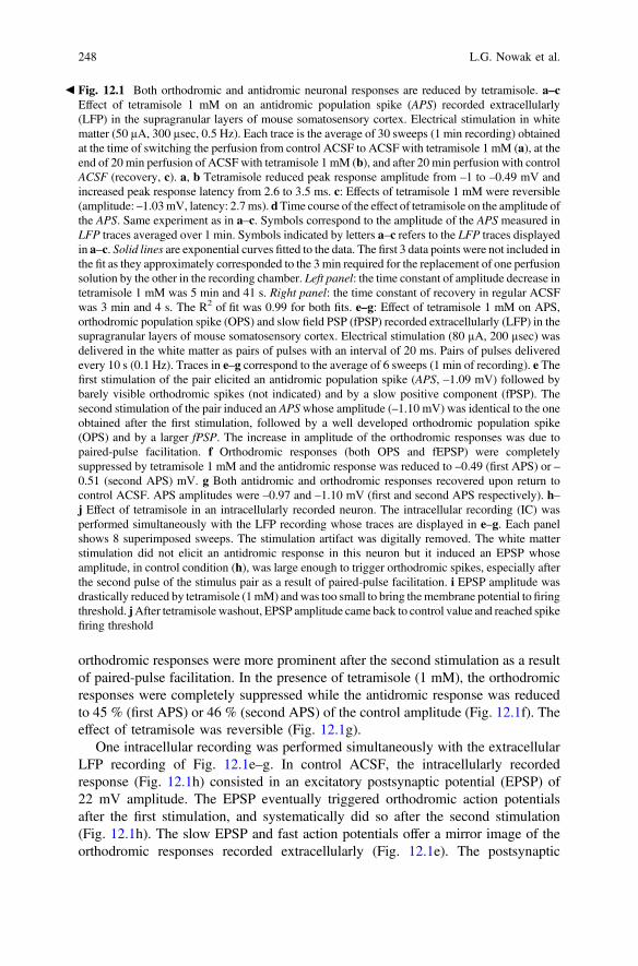
b Fig. 12.1 Both orthodromic and antidromic neuronal responses are reduced by tetramisole. a–cEffect of tetramisole 1 mM on an antidromic population spike (APS) recorded extracellularly(LFP) in the supragranular layers of mouse somatosensory cortex. Electrical stimulation in whitematter (50 µA, 300 µsec, 0.5 Hz). Each trace is the average of 30 sweeps (1 min recording) obtainedat the time of switching the perfusion from control ACSF to ACSF with tetramisole 1 mM (a), at theend of 20 min perfusion of ACSF with tetramisole 1 mM (b), and after 20 min perfusion with controlACSF (recovery, c). a, b Tetramisole reduced peak response amplitude from –1 to –0.49 mV andincreased peak response latency from 2.6 to 3.5 ms. c: Effects of tetramisole 1 mM were reversible(amplitude: –1.03mV, latency: 2.7ms). dTime course of the effect of tetramisole on the amplitude ofthe APS. Same experiment as in a–c. Symbols correspond to the amplitude of the APS measured inLFP traces averaged over 1 min. Symbols indicated by letters a–c refers to the LFP traces displayedin a–c. Solid lines are exponential curves fitted to the data. The first 3 data points were not included inthe fit as they approximately corresponded to the 3 min required for the replacement of one perfusionsolution by the other in the recording chamber. Left panel: the time constant of amplitude decrease intetramisole 1 mM was 5 min and 41 s. Right panel: the time constant of recovery in regular ACSFwas 3 min and 4 s. The R2 of fit was 0.99 for both fits. e–g: Effect of tetramisole 1 mM on APS,orthodromic population spike (OPS) and slow field PSP (fPSP) recorded extracellularly (LFP) in thesupragranular layers of mouse somatosensory cortex. Electrical stimulation (80 µA, 200 µsec) wasdelivered in the white matter as pairs of pulses with an interval of 20 ms. Pairs of pulses deliveredevery 10 s (0.1 Hz). Traces in e–g correspond to the average of 6 sweeps (1 min of recording). e Thefirst stimulation of the pair elicited an antidromic population spike (APS, –1.09 mV) followed bybarely visible orthodromic spikes (not indicated) and by a slow positive component (fPSP). Thesecond stimulation of the pair induced an APSwhose amplitude (–1.10 mV) was identical to the oneobtained after the first stimulation, followed by a well developed orthodromic population spike(OPS) and by a larger fPSP. The increase in amplitude of the orthodromic responses was due topaired-pulse facilitation. f Orthodromic responses (both OPS and fEPSP) were completelysuppressed by tetramisole 1 mM and the antidromic response was reduced to –0.49 (first APS) or –0.51 (second APS) mV. g Both antidromic and orthodromic responses recovered upon return tocontrol ACSF. APS amplitudes were –0.97 and –1.10 mV (first and second APS respectively). h–j Effect of tetramisole in an intracellularly recorded neuron. The intracellular recording (IC) wasperformed simultaneously with the LFP recording whose traces are displayed in e–g. Each panelshows 8 superimposed sweeps. The stimulation artifact was digitally removed. The white matterstimulation did not elicit an antidromic response in this neuron but it induced an EPSP whoseamplitude, in control condition (h), was large enough to trigger orthodromic spikes, especially afterthe second pulse of the stimulus pair as a result of paired-pulse facilitation. i EPSP amplitude wasdrastically reduced by tetramisole (1mM) andwas too small to bring themembrane potential to firingthreshold. jAfter tetramisole washout, EPSP amplitude came back to control value and reached spikefiring threshold
248 L.G. Nowak et al.
response was nearly completely suppressed by tetramisole 1 mM (Fig. 12.1i) andthe peak of the remnant depolarization (4 mV) remained far below action potentialthreshold. Both responses recovered upon return to control ACSF (Fig. 12.1j).Since the resting membrane potential did not depolarize in the presence of te-tramisole, the reduction of ortho- and antidromic response amplitude cannot beattributed to a depolarization block.
Figure 12.2a represents the effect of different concentrations of tetramisole onthe antidromic population spike amplitude at the population level. Responseamplitudes were normalized by the control response amplitude and are expressed aspercentage of the control response amplitude. The reduction of the antidromicresponse amplitude was dose-dependent (ANOVA, P < 0.0001) and reversible. Incomparison to control and recovery, response amplitude was significantly reducedwith tetramisole at 0.5, 1, 2 and 5 mM (PLSD Fischer test, P < 0.0001 for allcomparisons). Response suppression was nearly complete with 2 mM tetramisole(4.4 % ± 0.04 of control response amplitude).
Figure 12.2C shows the dose-response relationship for antidromic responsereduction fitted with Hill’s equation:
R ¼ 100� 100� Cn
ICn50 þ Cn
where R represent the normalized response amplitude (% of control), C the te-tramisole concentration, IC50 the concentration producing half the maximal effect,and n the Hill coefficient determining the slope of the curve. The IC50 returned bythe fit was 0.73 ± 0.08 (SE) mM and the Hill coefficient was 3.04 ± 0.38.
Tetramisole also reduced the amplitude of the orthodromic responses in adose-dependent manner (ANOVA, P < 0.0001, Fig. 12.2b). The amplitude oforthodromic response was significantly less than control and recovery with te-tramisole at 0.5 and 1 mM (PLSD Fischer test, P < 0.0001 for all comparisons).2
Comparing Fig. 12.2a, b, it can be seen that, with tetramisole at 0.5 and 1 mM,orthodromic responses were significantly more reduced than antidromic responses.With 0.5 mM the antidromic response amplitude represented 73 ± 4 % of thecontrol whereas the orthodromic response was reduced to 35 ± 12 % of the control(P = 0.01, paired t-test); likewise, with 1 mM tetramisole the antidromic responseamplitude (42 ± 4 % of control) was less reduced that the orthodromic response(9 ± 6 % of control) (P = 0.0002). The stronger effect of tetramisole on orthodromicresponse amplitude is confirmed by the Hill’s equation fitted to the dose-responserelationship, which returned an IC50 of 0.41 ± 0.15 mM (Fig. 12.2d). The Hillcoefficient was 2.97 ± 2.27.
2Orthodromic responses could not be tested with 2 mM tetramisole: among the 4 experiments inwhich we used this concentration, 2 showed no measurable orthodromic response in the controlcondition, while in the other 2 experiments the orthodromic response obtained in the controlcondition failed to recover.
12 TNAP and Tetramisole, an Electrophysiological Approach 249

(a)
(b)
(c) (d)
250 L.G. Nowak et al.

12.3.2 Tetramisole Increases Antidromic Response Latency
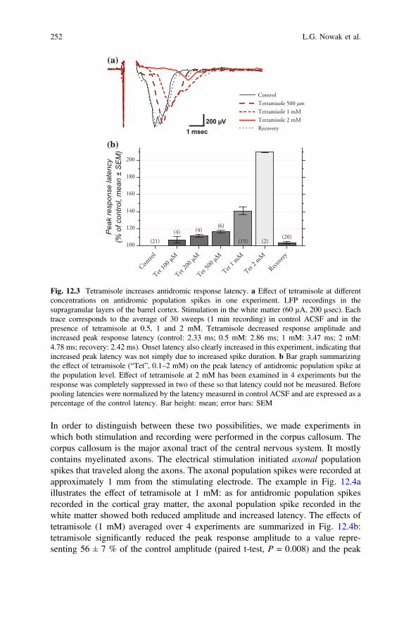
In addition to decreasing their amplitudes, tetramisole also increased the latency ofthe antidromic population spikes. This is illustrated with one example on Fig. 12.3a.In this experiment we applied 3 different tetramisole concentrations. It can be seenthat both the onset latency and the latency to peak of the antidromic responseincreased with increasing concentration of tetramisole. Increase in latency is evenvisible with a tetramisole concentration of 0.5 mM although this concentration hada marginal effect on response amplitude in this experiment.
For population level analysis we examined and quantified the effect of te-tramisole on the latency to peak of the antidromic population spikes. As shown inFig. 12.3b, tetramisole had a clear, dose-dependent effect on peak latency(ANOVA, P < 0.0001). Relative to control (100 %), the latency was significantlyincreased (+11 ± 1.5 %) with 200 µM tetramisole (PLSD Fischer test, P = 0.03),although this concentration had no significant effect on response amplitude(Fig. 12.2a). Increases in latency were also significant with 0.5 mM (+17 ± 2 %,P = 0.0004), 1 mM (+41 ± 5 %, P < 0.0001) and 2 mM tetramisole; with 2 mMtetramisole, the latency was more than doubled (+110 ± 0.3 %, P < 0.0001). Thus,tetramisole not only reduced the amplitude of the responses, but also increased theirlatencies.
12.3.3 Tetramisole Reduces Amplitude and Increases Latencyof Axonal Population Spikes
The increase of antidromic response latency could have two origins. The first is thattetramisole affected the somatic membrane potential, leading to an impairment ofthe coupling between the axon and the cell body. The second is that the increase inresponse latency originated from an action of tetramisole on the axons themselves.
b Fig. 12.2 Reduction of antidromic and orthodromic response amplitude by tetramisole isdose-dependent. a–b Bar graphs illustrating effect of various tetramisole (“Tet” on x-axis)concentrations (100 µM–5 mM) on antidromic (a) and orthodromic (b) response amplitude. Beforepooling amplitudes were normalized by the control amplitudes and are expressed as a percentageof control response amplitude. Bar height represents the mean and error bars represent the SEM.Note near complete suppression of antidromic responses with tetramisole at 2 mM, and oforthodromic responses with tetramisole at 1 mM. In a and b the total number of testedconcentrations is larger than the number of controls and recoveries because in some experimentsseveral tetramisole concentrations were successively tested between one control and one recovery.For the discrepancy in the number of controls and recoveries in a and b see legend of Fig. 12.5. c–d Dose-response relationships for antidromic (c) and orthodromic (d) responses. The dots showmean normalized amplitudes, as in a–b, but the error bars represent 1 SD. The continuous linescorrespond to the Hill equation (see Results) fitted to the data. The fits were weighted by thevariance. The R2 of the fits was 0.99 for both antidromic and orthodromic responses. The freeparameters values (IC50 and n) are displayed in the figure
12 TNAP and Tetramisole, an Electrophysiological Approach 251
In order to distinguish between these two possibilities, we made experiments inwhich both stimulation and recording were performed in the corpus callosum. Thecorpus callosum is the major axonal tract of the central nervous system. It mostlycontains myelinated axons. The electrical stimulation initiated axonal populationspikes that traveled along the axons. The axonal population spikes were recorded atapproximately 1 mm from the stimulating electrode. The example in Fig. 12.4aillustrates the effect of tetramisole at 1 mM: as for antidromic population spikesrecorded in the cortical gray matter, the axonal population spike recorded in thewhite matter showed both reduced amplitude and increased latency. The effects oftetramisole (1 mM) averaged over 4 experiments are summarized in Fig. 12.4b:tetramisole significantly reduced the peak response amplitude to a value repre-senting 56 ± 7 % of the control amplitude (paired t-test, P = 0.008) and the peak
(a)
(b)
Fig. 12.3 Tetramisole increases antidromic response latency. a Effect of tetramisole at differentconcentrations on antidromic population spikes in one experiment. LFP recordings in thesupragranular layers of the barrel cortex. Stimulation in the white matter (60 µA, 200 µsec). Eachtrace corresponds to the average of 30 sweeps (1 min recording) in control ACSF and in thepresence of tetramisole at 0.5, 1 and 2 mM. Tetramisole decreased response amplitude andincreased peak response latency (control: 2.33 ms; 0.5 mM: 2.86 ms; 1 mM: 3.47 ms; 2 mM:4.78 ms; recovery: 2.42 ms). Onset latency also clearly increased in this experiment, indicating thatincreased peak latency was not simply due to increased spike duration. b Bar graph summarizingthe effect of tetramisole (“Tet”, 0.1–2 mM) on the peak latency of antidromic population spike atthe population level. Effect of tetramisole at 2 mM has been examined in 4 experiments but theresponse was completely suppressed in two of these so that latency could not be measured. Beforepooling latencies were normalized by the latency measured in control ACSF and are expressed as apercentage of the control latency. Bar height: mean; error bars: SEM
252 L.G. Nowak et al.

latency was significantly increased by +43 ± 10 % relative to the control latency(P = 0.02, Fig. 12.4b). These data indicate that action potential transmission alongthe axons, hence axonal conduction velocity, was altered by tetramisole.
12.3.4 Effect of Tetramisole on Antidromic Population SpikesIs not Compensated by Pyridoxal
The data we have presented thus far suggest that orthodromic and antidromicresponse suppression by tetramisole results in large part from an impaired trans-mission of action potentials along the axons. Action potential electrogenesis inmyelinated axons takes place at the node of Ranvier. Coincidentally, TNAP appearsto be highly concentrated at the nodes of Ranvier. Since one of the main substrate ofTNAP is PLP, we hypothesized that tetramisole, by preventing extracellular PLP toPL conversion, perturbed an (unknown to us) intracellular PL/PLP-dependentmechanism involved in action potential initiation and/or propagation. To test thispossibility, we recorded antidromic population spikes and compared the effect oftetramisole 1 mM with the effect of tetramisole 1 mM to which we added PL,expecting that exogenous PL would compensate for the inhibition of TNAP by
(a) (b)
Fig. 12.4 Tetramisole reduces axonal population spike amplitude and reduces axonal conductionvelocity. For these experiments both stimulation and recording were performed in the corpuscallosum. a Example of the effect of tetramisole (1 mM) on axonal population spike amplitude andlatency. Each trace corresponds to the average of 6 sweeps (1 min recording). Stimulation in themidline of the corpus callosum; recording approximately 1 mm lateral to the midline, below motorcortex. The amplitude of the axonal population spike was reduced in tetramisole 1 mM (control: –0.33 mV; tetramisole 1 mM: –0.14 mV; recovery: –0.32 mV). The peak latency was increased intetramisole (control: 2.25 ms; tetramisole 1 mM: 3.54 ms; recovery: 2.10 ms), implying reducedconduction velocity in the axons. b Effects of tetramisole 1 mM (“Tet 1 mM”) on the amplitudeand latency of axonal population spikes, group data. Amplitudes and latencies normalized bycontrol values. The squares represent the mean peak latency and the circles represent the meanamplitude; the error bars represent the SEM
12 TNAP and Tetramisole, an Electrophysiological Approach 253

tetramisole. We used a PL concentration of 10 µM, a value much larger than theconcentration measured in the CSF (≤0.1 µM: Spector 1978, van der Ham et al.2012). An example is presented in Fig. 12.5a. The effect of tetramisole alone isidentical to that illustrated previously: a reduction of the antidromic populationspike amplitude associated with an increased latency. Yet the trace obtained withtetramisole 1 mM + PL 10 µM is nearly identical to that obtained with tetramisole1 mM alone, indicating that PL did not palliate the action of tetramisole. Whenexamined at the population level (Fig. 12.5b), the mean population spike amplitudemeasured in the presence of tetramisole did not differ significantly from that
(a)
(b)
Fig. 12.5 Extracellular PL does not compensate for the effect of tetramisole. a Comparison of theeffects of tetramisole at 1 mM and tetramisole 1 mM + pyridoxal 10 µM in one experiment.Recording in the supragranular layers of the somatosensory cortex, stimulation in the white matter.Each trace is the average of 30 sweeps (1 min recording) in control ACSF (control and recovery),in ASCF + tetramisole 1 mM, and in ACSF + tetramisole 1 mM + PL 10 µm. Tetramisole reducedthe antidromic population spike amplitude and increased its latency, and completely suppressedthe slow postsynaptic response (fPSP). PL did not compensate for the effect of tetramisole. b Bargraph summarizing the effect of tetramisole 1 mM or tetramisole 1 mM + PL 10 µM on antidromicpopulation spike amplitude. As previously data were normalized by control response amplitudeand are expressed as a percentage of control response amplitude. Bar height: mean; error bars:SEM. The discrepancy between the number of controls and recoveries comes from one experimentin which the order of drug application and tests was: control ACSF → tetramisole 1 mM→ tetramisole 1 mM + PL 10 µM → tetramisole 1 mM. Response amplitude during the first andsecond application of tetramisole 1 mM were perfectly identical, and therefore fitted with ourstationarity criteria although recovery was not tested in regular ACSF in this experiment
254 L.G. Nowak et al.
measured in the presence of tetramisole + PL (paired t-test, P = 0.11, n = 4experiments). On the other hand, both were significantly less than the controlresponse amplitude (tetramisole 1 mM alone: 45 ± 9 % of control, P = 0.008; te-tramisole 1 mM + PL 10 µM: 42 ± 8 % of control, P = 0.005). In short, thedecreased amplitude and increased latency of the antidromic responses cannot beexplained by the alteration of a PL/PLP-dependent mechanism consecutive toTNAP inhibition by tetramisole.
12.3.5 Effect of Tetramisole Is not Compensatedby Adenosine and Is not Mimicked by ATP
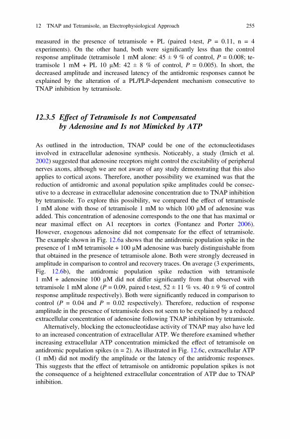
As outlined in the introduction, TNAP could be one of the ectonucleotidasesinvolved in extracellular adenosine synthesis. Noticeably, a study (Irnich et al.2002) suggested that adenosine receptors might control the excitability of peripheralnerves axons, although we are not aware of any study demonstrating that this alsoapplies to cortical axons. Therefore, another possibility we examined was that thereduction of antidromic and axonal population spike amplitudes could be consec-utive to a decrease in extracellular adenosine concentration due to TNAP inhibitionby tetramisole. To explore this possibility, we compared the effect of tetramisole1 mM alone with those of tetramisole 1 mM to which 100 µM of adenosine wasadded. This concentration of adenosine corresponds to the one that has maximal ornear maximal effect on A1 receptors in cortex (Fontanez and Porter 2006).However, exogenous adenosine did not compensate for the effect of tetramisole.The example shown in Fig. 12.6a shows that the antidromic population spike in thepresence of 1 mM tetramisole + 100 µM adenosine was barely distinguishable fromthat obtained in the presence of tetramisole alone. Both were strongly decreased inamplitude in comparison to control and recovery traces. On average (3 experiments,Fig. 12.6b), the antidromic population spike reduction with tetramisole1 mM + adenosine 100 µM did not differ significantly from that observed withtetramisole 1 mM alone (P = 0.09, paired t-test, 52 ± 11 % vs. 40 ± 9 % of controlresponse amplitude respectively). Both were significantly reduced in comparison tocontrol (P = 0.04 and P = 0.02 respectively). Therefore, reduction of responseamplitude in the presence of tetramisole does not seem to be explained by a reducedextracellular concentration of adenosine following TNAP inhibition by tetramisole.
Alternatively, blocking the ectonucleotidase activity of TNAP may also have ledto an increased concentration of extracellular ATP. We therefore examined whetherincreasing extracellular ATP concentration mimicked the effect of tetramisole onantidromic population spikes (n = 2). As illustrated in Fig. 12.6c, extracellular ATP(1 mM) did not modify the amplitude or the latency of the antidromic responses.This suggests that the effect of tetramisole on antidromic population spikes is notthe consequence of a heightened extracellular concentration of ATP due to TNAPinhibition.
12 TNAP and Tetramisole, an Electrophysiological Approach 255
(a)
(b)
(c)
Fig. 12.6 High extracellular adenosine concentration does not compensate for the effect oftetramisole and the effect of tetramisole is not mimicked by high extracellular ATP concentration.a Comparison of the effects of tetramisole at 1 mM and tetramisole 1 mM + adenosine 100 µM.Recording in the supragranular layers of the somatosensory cortex, stimulation (100 µA, 300 µs) atthe white matter-layer 6 border. Each trace is the average of 30 sweeps (1 min recording) in controlACSF (control and recovery), in ASCF + tetramisole 1 mM, and in ACSF + tetramisole1 mM + adenosine 100 µm. As previously, tetramisole reduced the amplitude and increased thelatency of the APS and eliminated the fPSP. Adenosine did not reverse the effect of tetramisole.b Bar graph summarizing the effect of tetramisole 1 mM or tetramisole 1 mM + adenosine(Ade) 100 µM on antidromic population spike amplitude. Amplitudes normalized by controlvalues and expressed as a percentage of control response amplitude. Bar height: mean; error bars:SEM. c ATP (1 mM) does not mimic the effect of tetramisole. Recording in the supragranularlayers of the somatosensory cortex, stimulation (100 µA, 300 µsec) in the white matter. Each traceis the average of 30 sweeps (1 min recording) in control ACSF (control and recovery) and inASCF + ATP 1 mM
256 L.G. Nowak et al.
12.3.6 The Effect of Tetramisole Is not Stereo-Specific
Although they constitute two of the identified metabolites of TNAP, exogenouslyapplied adenosine or PL did not compensate for the effect of tetramisole on neu-ronal response amplitude. In addition, the IC50 for neuronal response suppressionby tetramisole (Fig. 12.2c, d) was much higher than that expected given the IC50
values reported for TNAP inhibition by levamisole or tetramisole in biochemicalassays on cell extracts or cell cultures (Van Belle 1972, 1976a, 1976b; Goldsteinet al. 1980; Anagnostou et al. 1996; Calhau et al. 2000; Picher et al. 2003;Sergienko and Millán 2010; Debray et al. 2013): the IC50 that has been reported inthese studies is between 10 and 70 µM. In addition, the slope of the Hill equationfitted to our data was close to 3, while a Hill coefficient value close to 1 has beenreported for the inhibition of TNAP by levamisole (Suzuki et al. 1994). Altogether,these negative results and discrepancies led us to suspect that tetramisole may haveacted on a target other than TNAP.
Studies showed that levamisole (the levorotatory enantiomer of tetramisole), butnot dexamisole (the dextrorotary enantiomer of tetramisole), is effective at inhib-iting TNAP (Van Belle 1972, 1976b; Borgers 1973). If inhibition of TNAP activitywas responsible for the effects we observed, then it should also be stereo-specific. Itfollows that the effect observed with a given concentration of tetramisole should bemimicked by levamisole at half that concentration. We therefore compared theeffect of levamisole and tetramisole on antidromic response amplitude in 7experiments. In each of these experiments 1 or 2 different concentrations of lev-amisole (0.5 or 1 mM) and identical concentrations of tetramisole were used. Theprediction was that levamisole at 0.5 mM should have the same effect as tetramisoleat 1 mM.
The results we obtained did not fit with this prediction. As illustrated inFig. 12.7a, levamisole at 0.5 and 1 mM reduced both antidromic and orthodromicresponse amplitude and increased response latency, in a way comparable to te-tramisole. Summary data in Fig. 12.7b show a highly significant reduction ofantidromic response amplitude by both tetramisole and levamisole at 0.5 and 1 mM(ANOVA, P < 0.0001; PLSD Fisher test: P < 0.0001 for levamisole at 0.5 and1 mM compared to control). However, the reduction of antidromic population spikeamplitude by levamisole at 0.5 mM (67 ± 5 % of control response amplitude) wasnot significantly different (P = 0.9) from that of tetramisole at 0.5 mM (68 ± 3 % ofcontrol response amplitude). Likewise, the effect of levamisole at 1 mM did notdiffer significantly (P = 0.3) from that of tetramisole at 1 mM (46 ± 6 % and40 ± 6 % of control response amplitude, respectively). On the other hand, levam-isole at 0.5 mM was significantly less potent than tetramisole at 1 mM (P < 0.0001).These results show that the effect of tetramisole is not stereo-specific. This stronglysuggests that the reduction of response amplitude induced by tetramisole was notmediated by TNAP.
12 TNAP and Tetramisole, an Electrophysiological Approach 257

12.3.7 The Effect of Tetramisole Is not Mimickedby MLS-0038949, Another TNAP Inhibitor
An additional proof that tetramisole reduced neuronal response amplitude indepen-dently from TNAP inhibition was obtained by using another inhibitor of TNAP. Thisinhibitor—MLS-0038949—has been recently isolated and shows a very high
(a)
(b)
Fig. 12.7 Comparison of the effectiveness of tetramisole and levamisole on antidromic populationspike amplitude reduction indicates lack of stereo-specificity in the action of tetramisole. a Exampleof the effects of levamisole at 0.5 and 1 mM. Recording in the supragranular layers of the barrelcortex, stimulation (65 µA, 200 µs) at the white matter-layer 6 border. Each trace is the average of 6sweeps (1 min recording) in control ACSF (control and recovery) and in ASCF containing either 0.5or 1 mM levamisole. As with tetramisole, levamisole reduced the amplitude and increased thelatency of the antidromic population spike (APS). As with tetramisole, levamisole also suppressedthe slow postsynaptic response (fPSP). b group data. “Tet” and “Lev” on x-axis refer to tetramisoleand levamisole, respectively. Bar height represents the mean percent change in APS amplitude, errorbars represent the SEM. Before averaging the amplitudes were normalized by the amplitude obtainedin the control condition. The data presented here come from 7 experiments in which at least oneconcentration of levamisole and one concentration of tetramisole were tested in the same experiment.The total number of controls is 14 because control measurements were performed before bothlevamisole and tetramisole applications. One recovery not tested. Total number of measurements inlevamisole and tetramisole sums to 18 because several concentrations of tetramisole or levamisolewere eventually tested between one control and one recovery. The mean amplitude obtained intetramisole 0.5 mM does not differ significantly from that obtained in levamisole 0.5 mM, andsimilarly when comparing tetramisole and levamisole at 1 mM
258 L.G. Nowak et al.
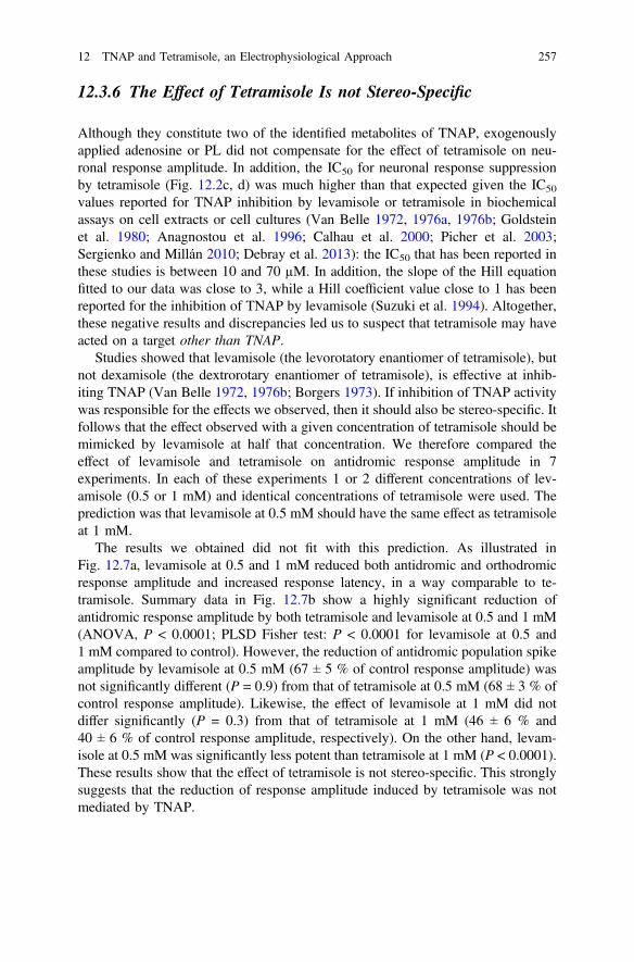
specificity for TNAP (Dahl et al. 2009). In Dahl et al. (2009) study, the IC50 forMLS-0038949 was about 0.2 µM and a complete inactivation of TNAP activity wasachieved with 10 µM of MLS-0038949. In control histochemical experiments, wefound that MLS-0038949 at 10 µM largely inhibited TNAP activity in mouse brainslices as assessed by histochemisty (NBT-BCIPmethod, as in Fonta and Imbert 2002)(not illustrated). We therefore examined the effect of MLS-0038949 at 10 µM in 3experiments. Results of one of these experiments are presented in Fig. 12.8. SinceMLS-0038949 was dissolved in DMSO to a final concentration of 0.1 % DMSO inACSF (Methods), we first checked that DMSO 0.1 % per se had no effect on theresponse; the response obtained in ACSF + DMSO 0.1 % (Fig. 12.8, dashed line) isindistinguishable from that obtained in regular ACSF. We also checked that te-tramisole had its usual action on the antidromic population spike; here again te-tramisole (500 µM in this experiment) reduced the response amplitude and increasedthe peak response latency (Fig. 12.8, short dash). On the other hand, the antidromicpopulation spikes was completely unaffected by MLS-0038949 at 10 µM (Fig. 12.8,thick solid line). The two other experiments failed to show any change on antidromicresponse amplitude and latency as well. These results confirm that the effects oftetramisole and levamisole on antidromic responses were not due to these compoundsinhibiting TNAP activity.
12.3.8 Tetramisole Modifies Action Potential Shapeand DV/Dt
In order to untangle how tetramisole reduced antidromic population spike ampli-tude, we next sought to examine its action on action potential electrogenesis using
Fig. 12.8 Antidromic population spikes are not affected by MLS-0038949, another TNAPinhibitor. In this experiment, recording was performed in the supragranular layers of the barrelcortex and stimulation (100 µA, 200 µs) was applied in the white matter. Each trace is the averageof 6 sweeps (1 min recording). Since MLS-0038949 was dissolved in DMSO, two controls wereperformed, one in regular ACSF and the other in ACSF + DMSO 0.1 %. DMSO had no effect theantidromic population spike. As in previous experiments, tetramisole (here at 0.5 mM) reduced theamplitude of the antidromic population spike and increased its latency. In contrast, MLS-0038949(10 µM) had no effect on the amplitude and latency of the antidromic population spike
12 TNAP and Tetramisole, an Electrophysiological Approach 259
intracellular recording in somatosensory cortex neurons. Although several cellshave been recorded, only one has been held for long enough to obtain a controlbaseline, to examine the effect of tetramisole (1 mM), and to achieve a completerecovery. The results presented here are therefore very preliminary.
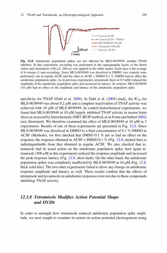
We observed that tetramisole (1 mM) modified the shape of the action potentials.This is illustrated in Fig. 12.9a, where each trace corresponds to the average of 30action potential traces; these action potentials were induced by intracellular currentinjection; the mean firing rate induced by these current injections was around 40spikes/sec in the three conditions. In comparison to the control trace, the actionpotential in the presence of 1 mM tetramisole showed a decrease in amplitudetogether with an increased duration. This effect of tetramisole was reversible.
We next computed the first derivative of the action potential (dV/dt). Thepositive peak in the dV/dt corresponds to the maximum rate-of-rise of the actionpotential. The maximum rate-of-rise, in turn, is a measure of the inward currentunderlying the rise of the action potential (Hodgkin and Katz 1949). This inwardcurrent is largely, if not entirely, determined by the voltage-dependent sodiumconductance (Hodgkin and Katz 1949; Cohen and Strichartz 1977; Hondeghem1978; Carter and Bean 2009). It can be seen in Fig. 12.9b that tetramisole (1 mM)reduced the maximum rate of rise of the action potential to half the control value.This effect was completely reversible. This result suggests that voltage-dependentsodium channels were partially blocked by tetramisole at 1 mM.
(a) (b)
Fig. 12.9 Tetramisole prolongs action potential duration and reduces action potential rate-of-rise.a Action potentials were induced by intracellular current injection in an intracellularly recordedneuron (same cell as in Fig. 12.1h–j). Each trace corresponds to the average of 30 action potentialsfor each condition. Note reduced amplitude and prolonged duration in tetramisole 1 mM. b Eachtrace corresponds to the temporal derivative (dV/dt) of the action potential traces shown in a. Themaximum rate of rise of the action potential (positive peak in the dV/dt) was reduced to 48 % ofthe control value in tetramisole 1 mM (control: 316 V/s; tetramisole 1 mM: 152 V/s; recovery:302 V/s). These data suggest that the voltage-dependent sodium channels responsible for actionpotential upstroke were partially blocked by tetramisole 1 mM
260 L.G. Nowak et al.
12.4 Discussion
Our study disclosed a new action of tetramisole and levamisole on neuronal signaltransmission, which appears to be independent of these compounds acting onTNAP. We showed that tetramisole and levamisole reduced both antidromic andorthodromic response amplitude in mouse somatosensory cortex in adose-dependent manner. This is a hitherto undocumented effect of tetramisole andlevamisole. Yet our control experiments showed that these effects of tetramisolewere not explained by inhibition of TNAP. Instead, the decrease of axonal con-duction velocity and preliminary intracellular data suggest that tetramisole andlevamisole may block voltage-dependent sodium channels. These results implythat, in addition to inhibiting TNAP, levamisole or tetramisole at concentration ≥1or ≥2 mM completely inhibit all orthodromic and antidromic neuronal activity,respectively, and are therefore likely to suppress all processes that areactivity-dependent in living brain cells, such as, among others, axonal growth,myelination or synaptic plasticity.
12.4.1 Suppression of Neuronal Activity by Tetramisoleand Levamisole is not Due to TNAP Inhibition
Neuronal responses, both orthodromic and antidromic, were reduced by tetramisoleand levamisole (Figs. 12.1, 12.2 and 12.7). These effects are unlikely to be due toTNAP inhibition, for several reasons:
First, the dose-response relationships (Fig. 12.2) appear to be very different fromthose reported for TNAP inhibition by tetramisole or levamisole. The IC50 wereport here were 730 µM for antidromic response and 410 µM for orthodromicresponses. Both values are much larger than those reported for TNAP inhibition bylevamisole in biochemical assays on cell extracts or on cells cultures—in the rangeof 10–70 µM (Van Belle 1972, 1976a, b; Goldstein et al. 1980; Anagnostou et al.1996; Picher et al. 2003; Sergienko and Millán 2010; Debray et al. 2013), includingbrain cell extracts (IC50 = 44 µM in Calhau et al. 2000). In addition the exponent ofthe Hill equation fitted to our data was close to 3, whereas TNAP inhibition has aHill coefficient close to 1 (Suzuki et al. 1994).
Second, neuronal activity block was not rescued by the application of exogenousadenosine or PL (Figs. 12.5 and 12.6), although these were the metabolites whoseextracellular concentrations were presumed to be reduced by TNAP inhibition inour experimental conditions (see Introduction).
Third, in our experiments equivalent concentrations of levamisole and tetrami-sole reduced neuronal responses by the same amount (Fig. 12.7). Given that te-tramisole contains both levamisole and dexamisole in identical proportions, thisimplies that dexamisole had the same potency at inhibiting neuronal responses aslevamisole. This further rules out TNAP inhibition as being responsible for the
12 TNAP and Tetramisole, an Electrophysiological Approach 261

effects reported here, given that only levamisole if effective at inhibiting TNAP(Van Belle 1972, 1976b; Borgers 1973).
Fourth and finally, blockage of neuronal activity by levamisole and tetramisolewere not mimicked by MLS-0038949, a new and highly selective inhibitor ofTNAP (Dahl et al. 2009) (Fig. 12.8).
Our results should not be interpreted as meaning that tetramisole or levamisoledid not inhibit TNAP in our experimental conditions. Indeed, we performed controlhistochemical experiments (same method as Fonta and Imbert 2002) using brainslices prepared as for the electrophysiological experiments. These experimentsconfirmed that AP activity was effectively inhibited by tetramisole (not illustrated).Therefore, TNAP inhibition likely took place in our electrophysiological experi-ments but its consequences on synaptic transmission, if they occurred, were maskedby the suppression of neuronal activity by tetramisole and levamisole.
12.4.2 Tetramisole and Levamisole May Suppress NeuronalActivity by Blocking Voltage-Dependent SodiumChannels
If TNAP inhibition was not responsible for the decrease in neuronal responseamplitude produced by levamisole and tetramisole, then what other targets couldexplain the effects we observed? Two observations suggest that voltage-dependentsodium channels were blocked by tetramisole and levamisole.
First, with concentrations of tetramisole that did not completely suppress theresponses, we observed that the latency of the antidromic population spikesincreased (Fig. 12.3). The same effect was noticed when recording from axons inthe corpus callosum (Fig. 12.4). These results indicate a slowing down of actionpotential propagation along the axons. Slowing down of action potential propa-gation speed in axons is not observed with compounds that blockvoltage-dependent potassium channels (Bostock et al. 1981; Fox and Ruan 1989),but it is typically observed with compounds that block voltage-dependent sodiumchannels such as TTX (Pinto et al. 2008), phenytoin (Le Quesne et al. 1976; Marcuset al. 1981) and several local anesthetics such as procaine (Franz and Perry 1974)and lidocaine (Raymond 1992; Yokota et al. 1994; De Col et al. 2008).Interestingly, a pharmacological study reported that levamisole possesses localanesthetic properties with a potency representing half that of lidocaine(Onuaguluchi and Igbo 1987); the ED50 obtained in this study was quite close to theIC50 we report here for the reduction of antidromic population spike amplitude.
Second, studies showed that the compounds that reduce axonal conductionvelocity also reduce the peak height of the action potential dV/dt (TTX: Kao andWalker 1982; phenytoin: Selzer 1979, Hershkowitz and Ayala 1981; procaine:Ibusuki et al. 1998; lidocaine: Schwarz and Puil 1998). Since the peak height in thedV/dt is a measure of the sodium current underlying action potential electrogenesis,
262 L.G. Nowak et al.

the reduction of the dV/dtmax observed in the present study (Fig. 12.9) furthersupports the possibility that tetramisole blocks voltage-dependent sodium channels,although additional studies are required for a definitive confirmation. It is alsopresently unclear whether tetramisole directly blocks voltage-dependent sodiumchannels, or whether this effect is secondary to tetramisole acting on intracellularsignaling pathways that control sodium channel gating properties.
Complete suppression of antidromic responses required a tetramisole concen-tration of about 2 mM whereas orthodromic responses were nearly completelysuppressed with 1 mM tetramisole. Likewise, the IC50 for the two types ofresponses differed by a factor close to 2. We can only speculate on the origin of thisdiscrepancy. The postsynaptic response is proportional to the amount of neuro-transmitter released, which is determined by calcium influx inside the presynapticterminals, which is itself the result of the activation of high thresholdvoltage-dependent calcium channels by sodium spikes in the axon terminals.Reduction of sodium spike amplitude may then results in a decreased neurotrans-mitter release but, to fit with our data, a nonlinearity in the presynaptic spikeamplitude to postsynaptic response transform would be required. Alternatively,high threshold voltage-dependent calcium channels, which show some structuralhomologies with voltage-dependent sodium channels (e.g., Zakon 2012), may alsobe blocked by tetramisole.
12.4.3 Other Targets of Tetramisole and Levamisolein the Nervous System
It has long been shown that tetramisole and levamisole inhibit TNAP. The resultswe report here further suggest that tetramisole and levamisole may also blockvoltage-dependent sodium channels. In addition to these two targets, tetramisoleand levamisole have been reported to have other actions in the nervous system. Weshall first review studies that examined the effect of tetramisole and levamisoleusing electrophysiological approaches. These studies have been very few andconcentrated mostly on nematode neuromuscular junction and mammalianperipheral nervous system.
12.4.3.1 Tetramisole, Levamisole and Acetylcholine Receptors
Tetramisole has originally been isolated for its anthelmintic properties (Thienpontet al. 1966; Raeymaekers et al. 1966). Initially based on screening tests in chicken,tetramisole was proven to be efficient against both intestinal and pulmonary nem-atode infections in a dozen mammalian species including human and tiger.Levamisole was shown to be the effective compound while dexamisole has noanthelmintic properties (Bullock et al. 1968). The action of tetramisole and
12 TNAP and Tetramisole, an Electrophysiological Approach 263
levamisole consists in a paralysis of the nematodes (Thienpont et al. 1966; Aceveset al. 1970; Atchison et al. 1992). Nematode paralysis results from a maintaineddepolarization of their muscle cells (Aceves et al. 1970; Harrow and Gration 1985;Atchison et al. 1992). Muscle cell depolarization is the consequence of levamisoleacting as an agonist on neuromuscular acetylcholine (ACh) receptors of the nem-atode (Lewis et al. 1980; Harrow and Gration 1985; Robertson and Martin 1993).
Fortunately in mammals levamisole does not appear to be an agonist of theneuromuscular ACh receptor (Atchison et al. 1992; Rayes et al. 2004). Instead, onthe basis of their pharmacological profiles (Lewis et al. 1980) and of genetichomologies (Fleming et al. 1997), it has been proposed that the neuromuscular AChreceptors that are activated by levamisole in the nematodes are homologous toganglionic ACh receptors of mammals.
Nevertheless, the effect of levamisole on the ganglionic ACh receptors ofmammals (α3β2 and α3β4 receptors) is unlike that observed in nematodes(Levandoski et al. 2003): levamisole applied alone has virtually no effect andtherefore does not behave as an agonist of the ganglionic ACh receptors. On theother hand, levamisole modulates the response to exogenously applied ACh. Yetthis modulation appears to be complex, as it depends on both levamisole and AChconcentrations. Thus the modulation exerted by levamisole can be either facilitatoryor inhibitory.
Beside autonomic ganglia, the α3β2 and α3β4 receptors can be found in differentbrain regions but not in cortex (Perry et al. 2002). A large fraction of the nicotinicreceptors found in cortex corresponds to the α4β2 subunits composition, for which,to our knowledge, levamisole action has not been examined. The α7* is anotherACh receptor family that is largely represented in cortex (Paterson and Nordberg2000) but levamisole does not seem to have any effect on this receptor family(Bartos et al. 2006).
12.4.3.2 Inhibition of Noradrenaline Reuptake by Dexamisole,Levamisole and Tetramisole in Peripheral Nervous System
In addition to cholinergic transmission, noradrenergic transmission in the peripheralnervous system also appears to be affected by tetramisole and levamisole(Vanhoutte et al. 1977; Pires et al. 1979; Gulati et al. 1985). In the various prep-arations studied (heart, smooth muscle, vas deferens), levamisole or tetramisole hadno effect when applied alone and therefore do not appear to be agonists of nor-adrenaline (NA) receptors. Nonetheless, levamisole and tetramisole produced anenhancement of the response to endogenously released or to exogenously appliedNA (Vanhoutte et al. 1977; Pires et al. 1979; Gulati et al. 1985). Responseenhancement appeared to result from an inhibition of NA reuptake. The concen-trations of levamisole or tetramisole required for near complete NA uptake inhi-bition were ≤40 µM. Dexamisole was more potent than levamisole (Vanhoutteet al. 1977).
264 L.G. Nowak et al.
The potency of levamisole represented half that of cocaine (Pires et al. 1979) andwhen NA uptake was already blocked by cocaine, levamisole had no further effect(Gulati et al. 1985), suggesting that levamisole and cocaine were affecting andcompeting for the same uptake mechanism. Several NA uptake inhibitors, such ascocaine and amphetamines, also inhibit dopamine uptake. Whether this is also thecase for levamisole is not known to us but this could provide an explanation as towhy illegal cocaine is now so often adulterated with levamisole.
In summary, tetramisole and levamisole may have—at least—4 different effectsin the nervous system of mammals:
• Inhibition of TNAP, that may have multiple consequences resulting frominterference with numerous PLP-dependent enzymes, including GABA syn-thesizing enzymes, or from modifications of the extracellular concentrations ofATP and adenosine.
• Modulation of responses mediated by ganglionic ACh receptors.• Blockage of NA uptake mechanism.• Blockage of voltage-dependent sodium channels, as reported in the present
study.
In addition to these 4 targets, studies suggested that levamisole and tetramisolemay affect additional targets in the nervous system, although these are less firmlyestablished. Hence it has been proposed that tetramisole and levamisole could inhibitacetylcholinesterase (Eyre 1970) and monoamine oxidase (Vanhoutte et al. 1977).One study suggested that levamisole interferes with opiate receptors (Spector et al.1998) but the levamisole doses that have been used in this study have been shown tobe deadly in other studies (Mohammad et al. 2006; Rehni and Singh 2010).
12.4.4 Side Effects of Tetramisole and Levamisole Therapies
Levamisole and tetramisole received multiple medical applications: first as ananthelmintic, later on as an immunostimulant for the treatment of a number ofdiseases.
When used as an anthelmintic, levamisole and tetramisole are used in a singledose and, unless overdosing (Joly et al. 1998), this regimen has not been reported tohave serious side effects.3 On the other hand, serious side effects have been noticedwith the chronic (several weeks or months) use of levamisole.
The chronic use of levamisole was largely based on studies reporting that te-tramisole and levamisole possess immunostimulating properties (e.g., Renoux andRenoux 1972; Brugmans et al. 1973; Pabst and Crawford 1975; Spreafico et al. 1975;
3Adverse reactions after single levamisole or tetramisole doses have been consistently reported inveterinary medicine, and were usually attributed to their actions on ganglionic ACh receptors(reviewed in Hsu 1980).
12 TNAP and Tetramisole, an Electrophysiological Approach 265
Renoux et al. 1976; Hadden et al. 1977). Levamisole has thus been used to boost theimmune system of patients suffering diseases that were supposed to be associatedwith, or that received treatments leading to, decreased immunity. Hence levamisolehas been used for the treatment of rheumatoid arthritis (Schuermans 1975), ofpediatric nephrotic syndrome (Tanphaichitr et al. 1980; Niaudet et al. 1984;Mongeauet al. 1988; British Association for Paediatric Nephrology 1991) and against variousskin infections (reviewed in Hadden et al. 1977). Nevertheless, more recent studieshave somehow questioned the efficacy of levamisole as an immunostimulant (e.g.:Toivanen et al. 1981; Webster et al. 1982; Aymard et al. 1984; Schiller et al. 1991;Ahmed et al. 1996). Likewise, studies questioned the efficacy of levamisole in thetreatment of rheumatoid arthritis (Dinai and Pras 1975) or skin infections (Chang andFiumara 1978; Seidlin and Straus 1984; Sanchez 2000).
Levamisole has also been used as an immunostimulant in cancer therapy. In thiscontext, levamisole, when used alone, proved to be of limited efficacy (e.g.,Toivanen et al. 1981; Treurniet-Donker et al. 1987; Arnaud et al. 1989; Barth andMorton 1995; Moertel et al. 1995) and has even been reported to be worse thanplacebo (e.g., Chlebowski et al. 1994). On the other hand, quite favorable outcomeshave been reported when levamisole was combined with 5-fluorouracil in coloncarcinoma treatment (e.g., Moertel et al. 1995).4
Thus, with few exceptions (colon carcinoma and pediatric nephrotic syndrometreatment), chronic levamisole treatments proved to be of limited efficacy. Moreproblematical, it appeared that chronic levamisole treatment was associated withmultiple side effects, some relatively mild and short-lived such as nausea anddiarrhea, other much more severe and eventually life threatening such as dermatitis,cutaneous necrotizing vasculitis, leukopenia and agranulocytosis (e.g., Ruuskanenet al. 1976; Parkinson et al. 1977; MacFarlane and Bacon 1978; Scheinberg et al.1978; Chang and Fiumara 1978; Toivanen et al. 1981; Niaudet et al. 1984; Moertelet al. 1990; reviewed in Symoens et al. 1978; Larocque and Hoffman 2012; Leeet al. 2012).
Chronic levamisole treatment also led to serious neurological side effects, inparticular epileptic seizures and inflammatory leukoencephalopathy, that we shalldiscuss below.
Beforehand, we shall remind that, although levamisole and tetramisole haveseveral putative targets in the CNS, the involvement of these targets in pathogenicprocesses depends on their sensitivity to levamisole, hence to the concentration oflevamisole in the tissues. With respect to this issue, it is worth mentioning that theplasmatic levamisole concentration is <5 µM in most individuals receiving single,therapeutically relevant levamisole doses (Woestenborghs et al. 1981; Luyckx et al.
4In the context of cancer therapy, levamisole has also been reported to possess antiproliferativeaction in vitro (Kovach et al. 1992; Artwohl et al. 2000). However, the levamisole concentrationsrequired to achieve significant effects in vitro appear to be much higher than the plasmaticconcentration of levamisole in clinical therapy. Levamisole concentration in a range similar to theplasmatic concentration measured in patients fails to prevent the proliferation of cancer cell linesin vitro (Grem and Allegra 1989; Wiebke et al. 2003).
266 L.G. Nowak et al.
1983; Kouassi et al. 1986; Gwilt et al. 2000; Hess et al. 2014), although higherplasmatic concentration (up to 8 µM in Luyckx et al. 1983) have been observed in aminority of individuals. One study further suggested that levamisole may accu-mulate following repeated daily doses (Reid et al. 1998). In any event, plasmaticlevamisole concentration was probably not much higher than 10−15 µM even in themost intensive cures. This rules out the new action of levamisole that we describehere since reduction of neuronal response amplitude and reduction of axonalconduction velocity required tetramisole concentration >100−200 µM. On the otherhand, TNAP activity may be significantly affected by levamisole at around 10 µM.
12.4.4.1 Levamisole and Epileptic Seizures
Epileptic seizures have been observed, in all cases in children, during levamisoletherapy for the treatment of pediatric nephrotic syndrome (Ruuskanen et al. 1976;Prieur et al. 1978; Palcoux et al. 1994). Seizures occurred after a delay of a few daysor weeks after the beginning of the treatment. In Prieur et al. (1978) study, epilepticseizures were observed in 3/50 children. An action of levamisole on nicotinicreceptors has been evoked (Palcoux et al. 1994) but alternative mechanisms are quitepossible, in particular those involving TNAP inhibition by levamisole.
There are multiple types of epilepsy, each with its own etiology. Quite com-monly, impairments of inhibitory mechanisms are involved. In this respect, it iswell established that dysfunctions of both GABA- (reviewed in Cossart et al. 2005;Macdonald et al. 2010) and adenosine- (Pagonopoulou et al. 2006; Boison 2012)mediated signaling can lead to epilepsy. As outlined in Introduction, the synthesisof GABA involves TNAP through vitamin B6 metabolism, while the synthesis ofextracellular adenosine partially depends, in some structures, on the ectonucleoti-dase activity of TNAP. That TNAP deficiencies result in epileptic seizures is welldocumented: in human, epileptic seizures are often observed in perinatal andinfantile hypophosphatasia (e.g., Rathbun 1948; Fraser 1957; Béthenod et al. 1967;Baumgartner-Sigl et al. 2007; Balasubramaniam et al. 2010; Taketani et al. 2014;see also Chaps. 14 and 15) and TNAP KO mice, the murine model of severehypophosphatasia, also present with epileptic seizures (Waymire et al. 1995;Narisawa et al. 1997). In parallel these mice show decreased level of GABA(Waymire et al. 1995; Fonta et al. 2012). In these mice, seizure incidence wasreduced, though not definitely suppressed, by administration of PL (Waymire et al.1995; Narisawa et al. 2001). Altogether, these studies point toward a PLP metab-olism deficiency, and its consequence on, at least, GABA synthesis, as the origin ofepileptic seizures in TNAP KO mice. In addition to reduced GABA level, werecently observed that adenosine concentration is reduced in the brain of 7 day-oldTNAP+/- mice (Fonta et al. 2014). This suggests that reduced adenosine level mayalso participate in the generation of epileptic seizures in TNAP KO mice.
Since levamisole inhibits TNAP (among others), it is tempting do draw a parallelbetween the epileptogenic action of levamisole (Ruuskanen et al. 1976; Prieur et al.1978; Palcoux et al. 1994) and the epileptic phenotype that has been observed in
12 TNAP and Tetramisole, an Electrophysiological Approach 267
both TNAP KO mice and in severe forms of hypophosphatasia. It is thereforepossible that levamisole, by inhibiting TNAP, impairs the balance of excitation andinhibition by reducing the availability of GABA and/or adenosine, leading toepileptic seizures in susceptible individuals.5
12.4.4.2 Levamisole and Multifocal InflammatoryLeukoencephalopathy
The first cases of multifocal inflammatory leukoencephalopathy were reported in asmall proportion (<1 %) of patients treated for colon carcinoma with 5-fluorouraciland levamisole (Hook et al. 1992; Kimmel and Schutt 1993; Leichman et al. 1993;Neu 1993; Bozik and Gilbert 1994; Critchley et al. 1994; Fassas et al. 1994; Ferroiret al. 1994; additional references in Wu et al. 2006). The patients presented neuro-logical disorders such as diplopia, dysarthria, paresthesia, ataxia, short-term memoryimpairments and/or loss of consciousness. In all cases MRI examination revealedleukoencephalopathy with multiple white matter lesions. Histological examinationsperformed from biopsies typically revealed extensive myelin loss and gliosis, but noor only marginal axonal loss. Perivascular lymphocytes and macrophage infiltrationwere consistently observed, demonstrating an inflammatory process in the leuko-encephalopathy (Hook et al. 1992; Neu 1993; Fassas et al. 1994; Bozik and Gilbert1994; Chen et al. 1994; Savarese et al. 1996;Murray et al. 1997; Israel et al. 2000; Liuet al. 2006; Wu et al. 2006). It was initially unclear whether the lesions were due tolevamisole or to 5-fluorouracil but further studies and case reports indicated thatlevamisole alone could be responsible (Chen et al. 1994; Kimmel et al. 1995; Luciaet al. 1996; Liu et al. 2006; Wu et al. 2006). Years earlier, an experimental studyperformed in dogs had revealed that chronic levamisole intake resulted in mononu-clear cell infiltration in the brain parenchyma, eventually accompanied by demye-lination, in the vast majority of tested animals (Vandevelde et al. 1978).
The clinical features of the inflammatory multifocal leukoencephalopathyinduced by levamisole are reminiscent of those observed in multiple sclerosis. Thisis further supported by studies reporting interaction between multiple sclerosis andlevamisole: in a clinical trial, levamisole was used with the purpose to alleviate thesymptoms of multiple sclerosis, yet levamisole had the opposite effect: it worsenedthe clinical picture in 5 of the 7 patients (Dau et al. 1976); this led to the prematureinterruption of the trial. In studies using rat or mouse models of multiple sclerosis(experimental allergic encephalomyelitis, EAE), levamisole increased both theincidence and the severity of the disease, and inflammation and demyelination wereexacerbated in animals receiving levamisole in comparison to non-treated animals(Spreafico et al. 1975; Hertz and Deghenghi 1985; Lucchinetti et al. 1997).
5The epileptic seizures that have been observed in the course of chronic levamisole treatmentshave all been reported in children but not in adults. This offers another interesting parallel withvitamin B6 metabolism alteration: experimental studies showed that vitamin B6 deficiency resultsin epileptic crisis in young animals but not in the adults (Daniel et al. 1942; Guilarte 1989).
268 L.G. Nowak et al.

It is generally admitted that the etiology of multiple sclerosis involves anautoimmune component (for review: Goverman 2009; Cieślak et al. 2011; Stys2013). In particular, T cells are suspected to play a prominent role in multiplesclerosis. In parallel, it has been reported that the increase in cell-mediatedimmunity by levamisole largely rests on the recruitment and activation of T cells(e.g., Renoux et al. 1976). Thus the general belief was that levamisole-inducedmultifocal leukoencephalopathy was the consequence of the immunostimulatingaction of levamisole in patients with subclinical multiple sclerosis.
Yet, to our knowledge none of the studies that examined the etiology oflevamisole-induced multifocal leukoencephalopathy considered the possibility thatinhibition of TNAP by levamisole could be involved. A support for this possibilityis that TNAP is strategically located on Ranvier’s node (Pinner et al. 1964; Moriand Nagano 1985; Fonta et al. 2005; Hanics et al. 2012). Here we propose two, nonexclusive hypothetical mechanisms, that both involve TNAP inhibition inlevamisole-induced multifocal leukoencephalopathy.
The first mechanism concerns the inflammatory reaction observed inlevamisole-induced leukoencephalopathy and a possible role for TNAP in thisprocess through its ectonucleotidase activity. There is now ample evidence thatinflammation is tightly associated with purinergic signaling (reviewed in: Khakhand North 2006; Di Virgilio et al. 2009; Trautmann 2009; Cieślak et al. 2011;Sperlágh and Illes 2014). To make a long story short, it has been shown that rises inthe extracellular concentration of ATP is pro-inflammatory, whereas extracellularadenosine acts as an anti-inflammatory. It is also worth mentioning that purinergicsignaling is likely to control the severity of the deficits and the extent of the lesionsin EAE: the inactivation of both adenosine A1 and A2A receptors aggravatesinflammation and demyelination (Tsutsui et al. 2004; Yao et al. 2012) whileblockade or inactivation of ATP P2X7 receptors has the opposite effect (Matuteet al. 2007; Sharp et al. 2008).
ATP is released by axons, in proportion to action potential firing rate (reviewed inFields, 2011). In the extracellular space, ATP is rapidly degraded by ectonucleotid-ases and TNAP, through its strategic location on Ranvier’s node, may play an activerole in setting the ATP/adenosine balance at this location. Inhibition of TNAP bylevamisole may thus lead to an increased extracellular ATP concentration and/or adecreased extracellular adenosine concentration in the close vicinity of myelinatedstructures. This could trigger a series of reactions, in particular secretion of a varietyof pro-inflammatory cytokines, that would ultimately result in monocyte infiltrationandmyelin loss. This does not rule out the involvement of an autoimmune componentin levamisole-induced leukoencephalopathy. If this was the case, levamisole wouldrather amplify the autoimmune response. In this sense TNAP inhibition by levam-isole could be considered as “immunostimulating”. However this immunostimulationwould not be due to levamisole acting directly on immune cells (Schiller et al. 1991);instead, by altering the extracellular ATP/adenosine balance, levamisole may have anaction upstream from the immune system.
The second mechanism where TNAP could play a role concerns the remyeli-nation that may be impaired in levamisole-induced leukoencephalopathy. TNAP is
12 TNAP and Tetramisole, an Electrophysiological Approach 269
likely to intervene in myelin synthesis. This is supported by the observation thatTNAP is highly expressed on axons during development, that is, during myelina-tion (Fonta and Imbert 2002; Fonta et al. 2005) and by the fact that myelination isaltered in TNAP KO mice (Hanics et al. 2012). The way TNAP participates inmyelination may again involve its function as an ectonucleotidase.
Studies showed that myelination is in large part promoted by action potentialfiring in the axons (Demerens et al. 1996). Spike firing in the axons leads to ATPrelease (Fields 2011). Early stages of myelination, which consist in reducing oli-godendrocyte precursor cells proliferation and in their differentiation in matureoligodendrocytes, requires adenosine signaling (Stevens et al. 2002). On the otherhand, myelination by mature oligodendrocyte is promoted by extracellular ATP,which induces the release of a cytokine, leukemia inhibitory factor, by astrocytes(Ishibashi et al. 2006). Yet leukemia inhibitory factor has a dual action: it promotesmyelination at low concentration but inhibits it when present at high concentration.Here again, inhibition of TNAP by levamisole may alter the extracellularATP/adenosine balance, which would result in compromising (re)myelination: first,TNAP inhibition by levamisole may reduce the availability of adenosine required inthe early stages of myelination; secondly, by reducing ATP degradation, TNAPinhibition may lead to an excess of leukemia inhibitory factor in the later stages ofmyelination.6
12.4.4.3 Levamisole and Tetramisole as Cocaine Adulterants
Between the sixties and the nineties, tetramisole and levamisole have been used in aconsiderable number of medical treatments. Yet, as briefly summarized above,tetramisole and levamisole appeared to be of limited efficacy in most of theirapplications and, more problematically, their chronic use resulted in seriousside-effects in a non-negligible proportion of the patients. For these reasons, te-tramisole and levamisole have been discontinued in most countries at the turn of thecentury, or their uses have been drastically restricted.7
6In addition to its involvement as an ectonucleotidase, TNAP could also intervene in myelinationthrough vitamin B6 metabolism. In support for a role of PLP in myelination, it has been reportedthat vitamin B6 deficiency during development results in altered myelination (Lott et al. 1978;Kirksey et al. 1990). Indeed, PLP is a cofactor for two enzymes (serine palmitoyltransferase andsphingosine-1-phosphatelyase) involved in sphingolipid synthesis (Bourquin et al. 2011). Amongsphingolipids, sphingomyelin, as expected from its name, happens to be particularly abundant inmyelin. On the other hand, if myelination impairments observed in levamisole-induced multifocalleukoencephalopathy were consecutive to altered vitamin B6 metabolism, another PLP-dependentprocess, GABA synthesis, should also be impaired. This should result in occurrences of epilepticseizures in patients with levamisole-induced leukoencephalopathy, which, to our knowledge, havenot been reported in case studies.7In France for example, levamisole can still be used for the treatment of pediatric nephroticsyndrome, but under very restrictive conditions.
270 L.G. Nowak et al.
Yet, near the middle of the last decade, levamisole and tetramisole made anunexpected come-back as it turns out that they are now used as illegal cocaineadulterants (Morley et al. 2006; Fucci 2007; LeGatt 2009). Sixty to eighty percent ofthe cocaine seized around 2010 in North America or in Europe appeared to containlevamisole (Schneider and Meys 2011; Casale et al. 2012; Lahaie 2012), or, morerarely, tetramisole (Casale et al. 2012). The reasons why traffickers adopted suchadulterants have not been trumpeted far and wide. One possibility is that levamisolemay be degraded to aminorex, an amphetamine-like compound. Aminorex has beendetected in horses that were treated with levamisole (see Larocque and Hoffman2012) but aminorex is barely detectable in human after levamisole intake (Hess et al.2014). A more likely reason is that, as inhibitors of noradrenaline uptake, levamisoleand dexamisole may have cocaine-like action (see above).
As a result, the undesirable consequences that formerly were observed duringchronic levamisole treatment are now observed in a number of cocaine users(reviewed in: Buchanan and Lavonas 2012; Larocque and Hoffman 2012; Lee et al.2012). This includes thrombocytopenia, agranulocytosis and leukopenia, dermatitisand vasculitis. Occurrence of inflammatory multifocal leukoencephalopathy has alsobeen reported in illegal cocaine users; although not definitively demonstrated, lev-amisole was suspected to be responsible for these rare instances of leukoencepha-lopathy (Gilbert 2011; Blanc et al. 2012; González-Duarte and Williams 2013).
Levamisole and tetramisole side-effects, including those that affect the nervoussystem, might not be something of the past. Understanding the mechanismsresponsible for these side-effects may become crucially important in the near future.As we hypothesized above, TNAP may be a key player in these mechanisms, whichcertainly deserve further study.
Acknowledgment We would like to thank the SARA service and particularly Gregory Marsal foranimal care. We acknowledge Pr José Luis Millán for his useful comments on this work while itwas in progress. This work was supported by CNRS (PICS 4331), by Hypophosphatasie Europe(grant No. 88461) and by PHC Egide (Balaton 17341 UE).
Conflict of Interest The authors declare no conflict of interest.
References
Abbracchio MP, Burnstock G, Verkhratsky A, Zimmermann H (2009) Purinergic signalling in thenervous system: an overview. Trends Neurosci 32:19–29
Aceves J, Erlij D, Martínez-Marañón R (1970) The mechanism of the paralysing action oftetramisole on Ascaris somatic muscle. Br J Pharmacol 38:602–607
Ahmed FY, Leonard GA, A’Hern R, Taylor AE, Lorentzos A, Atkinson H, Moore J,Nicolson MC, Riches PG, Gore ME (1996) A randomised dose escalation study ofsubcutaneous interleukin 2 with and without levamisole in patients with metastatic renal cellcarcinoma or malignant melanoma. Br J Cancer 74:1109–1113
Anagnostou F, Plas C, Forest N (1996) Ecto-alkaline phosphatase considered aslevamisole-sensitive phosphohydrolase at physiological pH range during mineralization incultured fetal calvaria cells. J Cell Biochem 60:484–494
12 TNAP and Tetramisole, an Electrophysiological Approach 271
Arnaud JP, Buyse M, Nordlinger B, Martin F, Pector JC, Zeitoun P, Adloff A, Duez N (1989)Adjuvant therapy of poor prognosis colon cancer with levamisole: results of an EORTCdouble-blind randomized clinical trial. Br J Surg 76:284–289
Artwohl M, Hölzenbein T, Wagner L, Freudenthaler A, Waldhäusl W, Baumgartner-Parzer SM(2000) Levamisole induced apoptosis in cultured vascular endothelial cells. Br J Pharmacol131:1577–1583
Atchison WD, Geary TG, Manning B, VandeWaa EA, Thompson DP (1992) Comparativeneuromuscular blocking actions of levamisole and pyrantel-type anthelmintics on rat andgastrointestinal nematode somatic muscle. Toxicol Appl Pharmacol 112:133–143
Aymard JP, Janot C, Thibaut G, Bertrand F, Legras B, Lederlin P, Streiff F (1984) Levamisole inchronic lymphocytic leukaemia: a prospective study of 15 patients. Acta Haematol 71:316–321
Balasubramaniam S, Bowling F, Carpenter K, Earl J, Chaitow J, Pitt J, Mornet E, Sillence D,Ellaway C (2010) Perinatal hypophosphatasia presenting as neonatal epileptic encephalopathywith abnormal neurotransmitter metabolism secondary to reduced co-factor pyridoxal-5′-phosphate availability. J Inherit Metab Dis Suppl 3:S25–S33
Barth A, Morton DL (1995) The role of adjuvant therapy in melanoma management. Cancer 75(S2):726–734
Bartos M, Rayes D, Bouzat C (2006) Molecular determinants of pyrantel selectivity in nicotinicreceptors. Mol Pharmacol 70:1307–1318
Baumgartner-Sigl S, Haberlandt E, Mumm S, Scholl-Bürgi S, Sergi C, Ryan L, Ericson KL,Whyte MP, Högler W (2007) Pyridoxine-responsive seizures as the first symptom of infantilehypophosphatasia caused by two novel missense mutations (c.677T > C, p. M226T;c.1112C > T, p.T371I) of the tissue-nonspecific alkaline phosphatase gene. Bone 40:1655–1661
Béthenod M, Cotte MF, Collombel C, Fréderich A, Cotte J (1967) Hypophosphatasie à révélationnéo-natale. Amélioration osseuse. Encéphalopathie convulsivante fatale. Ann Pediatr (Paris)14:835–841
Blanc PD, Chin C, Lynch KL (2012) Multifocal inflammatory leukoencephalopathy associatedwith cocaine abuse: is levamisole responsible? Clin Toxicol 50:534–535
Boison D (2012) Adenosine dysfunction in epilepsy. Glia 60:1234–1243Borgers M (1973) The cytochemical application of new potent inhibitors of alkaline phosphatases.
J Histochem Cytochem 21:812–824Bostock H, Sears TA, Sherratt RM (1981) The effects of 4-aminopyryridine and tetraethylamo-
nium ions on normal and demyelinated mammalian nerve fibres. J Physiol 313:301–315Bourne G (1943) The distribution of alkaline phosphatase in various tissues. Quart J exp physiol
32:1–20Bourquin F, Capitani G, Grütter MG (2011) PLP-dependent enzymes as entry and exit gates of
sphingolipid metabolism. Protein Sci 20:1492–1508Bozik ME, Gilbert MR (1994) The role of levamisole in 5-fluorouracil-levamisole-associated
multifocal leukoencephalopathy. Ann Neurol 36:295British Association for Paediatric Nephrology (1991) Levamisole for corticosteroid-dependent
nephrotic syndrome in childhood. Lancet 337: 1555–1557Brugmans J, Schuermans V, De Cock W, Thienpont D, Janssen P, Verhaegen H, Van Nimmen L,
Louwagie AC, Stevens E (1973) Restoration of host defense mechanisms in man bylevamisole. Life Sci 13:1499–1504
Brumberg JC, Nowak LG, McCormick DA (2000) Ionic mechanisms underlying repetitivehigh-frequency burst firing in supragranular cortical neurons. J Neurosci 20:4829–4843
Brun-Heath I, Ermonval M, Chabrol E, Xiao J, Palkovits M, Lyck R, Miller F, Couraud PO,Mornet E, Fonta C (2011) Differential expression of the bone and the liver tissue non-specificalkaline phosphatase isoforms in brain tissues. Cell Tissue Res 343:521–536
Buchanan JA, Lavonas EJ (2012) Agranulocytosis and other consequences due to use of illicitcocaine contaminated with levamisole. Curr Opin Hematol 19:27–31
Bullock MW, Hand JJ, Waletzky E (1968) Resolution and racemization of dl-tetramisole,dl-6-phenyl-2,3,5,6-tetrahydroimidazo-[2,1-b]thiazole. J Med Chem 11:169–171
272 L.G. Nowak et al.
Butt AM (2011) ATP: a ubiquitous gliotransmitter integrating neuron-glial networks. Semin CellDev Biol 22:205–213
Calhau C, Martel F, Hipólito-Reis C, Azevedo I (2000) Differences between duodenal and jejunalrat alkaline phosphatase. Clin Biochem 33:571–577
Carter BC, Bean BP (2009) Sodium entry during action potentials of mammalian neurons:incomplete inactivation and reduced metabolic efficiency in fast-spiking neurons. Neuron64:898–909
Casale JF, Colley VL, Legatt DF (2012) Determination of phenyltetrahydroimidazothiazoleenantiomers (Levamisole/Dexamisole) in illicit cocaine seizures and in the urine of cocaineabusers via chiral capillary gas chromatography-flame-ionization detection: clinical andforensic perspectives. J Anal Toxicol 36:130–135
Chang TW, Fiumara N (1978) Treatment with levamisole of recurrent herpes genitalis. AntimicrobAgents Chemother 13:809–812
Chen TC, Hinton DR, Leichman L, Atkinson RD, Apuzzo ML, Couldwell WT (1994) Multifocalinflammatory leukoencephalopathy associated with levamisole and 5-fluorouracil: case report.Neurosurgery 35:1138–1142
Chlebowski RTL, Lillington L, Nystrom JS, Sayre J (1994) Late mortality and levamisole adjuvanttherapy in colorectal cancer. Br J Cancer 69:1094–1097
Ciancaglini P, Yadav MC, Simão AM, Narisawa S, Pizauro JM, Farquharson C, Hoylaerts MF,Millán JL (2010) Kinetic analysis of substrate utilization by native and TNAP-, NPP1-, orPHOSPHO1-deficient matrix vesicles. J Bone Miner Res 25:716–723
Cieślak M, Kukulski F, Komoszyński M (2011) Emerging role of extracellular nucleotides andadenosine in multiple sclerosis. Purinergic Signal 7:393–402
Cohen IS, Strichartz GR (1977) On the voltage-dependent action of tetrodotoxin. Biophys J17:275–279
Collins GG, Anson J (1985) Adenosine A1 receptors mediate the inhibitory effects of exogenousadenosine in the rat olfactory cortex slice. Neuropharmacology 24:1077–1084
Connors BW, Malenka RC, Silva LR (1988) Two inhibitory postsynaptic potentials, and GABAA
and GABAB receptor-mediated responses in neocortex of rat and cat. J Physiol 406:443–468Cossart R, Bernard C, Ben-Ari Y (2005) Multiple facets of GABAergic neurons and synapses:
multiple fates of GABA signalling in epilepsies. Trends Neurosci 28:108–115Critchley P, Abbott R, Madden FJ (1994) Multifocal inflammatory leukoencephalopathy
developing in a patient receiving 5-fluorouracil and levamisole. Clin Oncol (R Coll Radiol)6:406
Dahl R, Sergienko EA, Su Y, Mostofi YS, Yang L, Simao AM, Narisawa S, Brown B,Mangravita-Novo A, Vicchiarelli M, Smith LH, O’Neill WC, Millán JL, Cosford ND (2009)Discovery and validation of a series of aryl sulfonamides as selective inhibitors oftissue-nonspecific alkaline phosphatase (TNAP). J Med Chem 52:6919–6925
Daniel EP, Kline OD, Tolle CD (1942) A convulsive syndrome in young rats associated withpyridoxine deficiency. J Nutr 23:205–216
Dau PC, Johnson KP, Spitler LE (1976) The effect of levamisole on cellular immunity in multiplesclerosis. Clin Exp Immunol 26:302–309
de Bernard B, Bianco P, Bonucci E, Costantini M, Lunazzi GC, Martinuzzi P, Modricky C,Moro L, Panfili E, Pollesello P, Stagni N, Vittur F (1986) Biochemical and immunohisto-chemical evidence that in cartilage an alkaline phosphatase is a Ca2+-binding glycoprotein.J Cell Biol 103:1615–1623
De Col R, Messlinger K, Carr RW (2008) Conduction velocity is regulated by sodium channelinactivation in unmyelinated axons innervating the rat cranial meninges. J Physiol 586:1089–1103
Debray J, Chang L, Marquès S, Pellet-Rostaing S, Le Duy D, Mebarek S, Buchet R, Magne D,Popowycz F, Lemaire M (2013) Inhibitors of tissue-nonspecific alkaline phosphatase: design,synthesis, kinetics, biomineralization and cellular tests. Bioorg Med Chem 21:7981–7987
Deisz RA, Prince DA (1989) Frequency-dependent depression of inhibition in guinea-pigneocortex in vitro by GABAB receptor feed-back on GABA release. J Physiol 412:513–541
12 TNAP and Tetramisole, an Electrophysiological Approach 273

Demerens C, Stankoff B, Logak M, Anglade P, Allinquant B, Couraud F, Zalc B, Lubetzki C(1996) Induction of myelination in the central nervous system by electrical activity. Proc NatlAcad Sci USA 93:9887–9892
Di Virgilio F, Ceruti S, Bramanti P, Abbracchio MP (2009) Purinergic signalling in inflammationof the central nervous system. Trends Neurosci 32:79–87
Dinai Y, Pras M (1975) Levamisole in rheumatoid arthritis. Lancet 2(7934):556Ermonval M, Baudry A, Baychelier F, Pradines E, Pietri M, Oda K, Schneider B,
Mouillet-Richard S, Launay JM, Kellermann O (2009) The cellular prion protein interactswith the tissue non-specific alkaline phosphatase in membrane microdomains of bioaminergicneuronal cells. PLoS ONE 4:e6497
Eyre P (1970) Some pharmacodynamic effects of the nematocides: methyridine, tetramisole andpyrantel. J Pharm Pharmacol 22:26–36
Fassas AB, Gattani AM, Morgello S (1994) Cerebral demyelination with 5-fluorouracil andlevamisole. Cancer Invest 12:379–383
Ferroir JP, Fenelon G, Beaugerie L, Avenin Recoing D (1994) Leucoencéphalopathie multifocaleinflammatoire, complication de la chimiothérapie par 5 fluorouracil et levamisole. Rev Neurol(Paris) 150:471–474
Fields RD, Burnstock G (2006) Purinergic signalling in neuron-glia interactions. Nat Rev Neurosci7:423–436
Fields RD (2011) Nonsynaptic and nonvesicular ATP release from neurons and relevance toneuron-glia signaling. Semin Cell Dev Biol 22:214–219
Fleming JT, Squire MD, Barnes TM, Tornoe C, Matsuda K, Ahnn J, Fire A, Sulston JE,Barnard EA, Sattelle DB, Lewis JA (1997) Caenorhabditis elegans levamisole resistance geneslev-1, unc-29, and unc-38 encode functional nicotinic acetylcholine receptor subunits.J Neurosci 17:5843–5857
Fonta C, Imbert M (2002) Vascularization in the primate visual cortex during development. CerebCortex 12:199–211
Fonta C, Négyessy L, Renaud L, Barone P (2004) Areal and subcellular localization of theubiquitous alkaline phosphatase in the primate cerebral cortex: evidence for a role inneurotransmission. Cereb Cortex 14:595–609
Fonta C, Negyessy L, Renaud L, Barone P (2005) Postnatal development of alkaline phosphataseactivity correlates with the maturation of neurotransmission in the cerebral cortex. J CompNeurol 486:179–196
Fonta C, Négyessy L, Brun-Heath I, Ermonval M, Czege D, Nowak LG, Frances B, Xiao JS,Mornet E, Millán JL (2012) Bone TNAP in the brain: functions in neurotransmission. BullGroup Int Rech Sci Stomatol Odontol 51:27
Fonta C, Cruz T, Rosay B, Millán JL, Balayssac S, Gilard V, Malet-Martino M, Nowak LG (2014)A dual approach to explore the functions of the tissue nonspecific alkaline phosphatase (TNAP)in the brain. Ninth FENS Forum of Neuroscience, Milan, Italy. Abstract Number: FENS-1232
Fontanez DE, Porter JT (2006) Adenosine A1 receptors decrease thalamic excitation of inhibitoryand excitatory neurons in the barrel cortex. Neuroscience 137:1177–1184
Fox DA, Ruan DY (1989) Time- and frequency-dependent effects of potassium channel blockerson large and medium diameter optic tract axons. Brain Res 498:229–242
Franz DN, Perry RS (1974) Mechanisms for differential block among single myelinated andnon-myelinated axons by procaine. J Physiol 236:193–210
Fraser D (1957) Hypophosphatasia. Am J Med 22:730–746Friede RL (1966) A quantitative mapping of alkaline phosphatase in the brain of the rhesus
monkey. J Neurochem 13:197–203Fucci N (2007) Unusual adulterants in cocaine seized on Italian clandestine market. Forensic Sci
Int 172:e1Gilbert JW (2011) J Neuroimaging 21:e188Goldstein DJ, Rogers CE, Harris H (1980) Expression of alkaline phosphatase loci in mammalian
tissues. Proc Natl Acad Sci USA 77:2857–2860
274 L.G. Nowak et al.
González-Duarte A, Williams R (2013) Cocaine-induced recurrent leukoencephalopathy.Neuroradiol J 26:511–513
Goverman J (2009) Autoimmune T cell responses in the central nervous system. Nat Rev Immunol9:393–407
Grem JL, Allegra CJ (1989) Toxicity of levamisole and 5-fluorouracil in human colon carcinomacells. J Natl Cancer Inst 81:1413–1417
Guilarte TR (1989) Regional changes in the concentrations of glutamate, glycine, taurine, andGABA in the vitamin B-6 deficient developing rat brain: association with neonatal seizures.Neurochem Res 14:889–897
Gulati OD, Hemavathi KG, Joshi DP (1985) Interactions of levamisole with some autonomicdrugs on guinea-pig vas deferens. J Auton Pharmacol 5:19–23
Gwilt P, Tempero M, Kremer A, Connolly M, Ding C (2000) Pharmacokinetics of levamisole incancer patients treated with 5-fluorouracil. Cancer Chemother Pharmacol 45:247–251
Hadden JW, Lopez C, O’Reilly RJ, Hadden EM (1977) Levamisole and inosiplex: antiviral agentswith immunopotentiating action. Ann NY Acad Sci 284:139–152
Hanics J, Barna J, Xiao J, Millán JL, Fonta C, Négyessy L (2012) Ablation of TNAP functioncompromises myelination and synaptogenesis in the mouse brain. Cell Tissue Res 349:459–471
Harrow ID, Gration KAF (1985) Mode of action of the anthelmintics morantel, pyrantel andlevamisole on muscle cell membrane of the nematode Ascaris suum. Pestic Sci 16:662–672
Hershkowitz N, Ayala GF (1981) Effects of phenytoin on pyramidal neurons of the rathippocampus. Brain Res 208:487–492
Hertz F, Deghenghi R (1985) Effect of rat and beta-human interferons on hyperacute experimentalallergic encephalomyelitis in rats. Agents Actions 16:397–403
Hess C, Ritke N, Sydow K, Mehling LM, Ruehs H, Madea B, Musshoff F (2014) Determination oflevamisole, aminorex, and pemoline in plasma by means of liquid chromatography-massspectrometry and application to a pharmacokinetic study of levamisole. Drug Test Anal6:1049–1054
Hodgkin AL, Katz B (1949) The effect of sodium ions on the electrical activity of giant axon of thesquid. J Physiol 108:37–77
Hondeghem LM (1978) Validity of Vmax as a measure of the sodium current in cardiac andnervous tissues. Biophys J 23:147–152
Hook CC, Kimmel DW, Kvols LK, Scheithauer BW, Forsyth PA, Rubin J, Moertel CG,Rodriguez M (1992) Multifocal inflammatory leukoencephalopathy with 5-fluorouracil andlevamisole. Ann Neurol 3:262–267
Hoshi K, Amizuka N, Oda K, Ikehara Y, Ozawa H (1997) Immunolocalization of tissuenon-specific alkaline phosphatase in mice. Histochem Cell Biol 107:183–191
Howe JR, Sutor B, Zieglgänsberger W (1987a) Characteristics of long-duration inhibitorypostsynaptic potentials in rat neocortical neurons in vitro. Cell Mol Neurobiol 7:1–18
Howe JR, Sutor B, Zieglgänsberger W (1987b) Baclofen reduces postsynaptic potentials of ratcortical neurones by an action other than its hyperpolarizing action. J Physiol 384:539–569
Hsu WH (1980) Toxicity and drug interactions of levamisole. J Am Vet Med Assoc 176:1166–1169
Ibusuki S, Katsuki H, Takasaki M (1998) The effects of extracellular pH with and withoutbicarbonate on intracellular procaine concentrations and anesthetic effects in crayfish giantaxons. Anesthesiology 88:1549–1557
Irnich D, Tracey DJ, Polten J, Burgstahler R, Grafe P (2002) ATP stimulates peripheral axons inhuman, rat and mouse—differential involvement of A(2B) adenosine and P2X purinergicreceptors. Neuroscience 110:123–129
Ishibashi T, Dakin KA, Stevens B, Lee PR, Kozlov SV, Stewart CL, Fields RD (2006) Astrocytespromote myelination in response to electrical impulses. Neuron 49:823–832
Israel ZH, Lossos A, Barak V, Soffer D, Siegal T (2000) Multifocal demyelinative leukoenceph-alopathy associated with 5-fluorouracil and levamisole. Acta Oncol 39:117–120
12 TNAP and Tetramisole, an Electrophysiological Approach 275

Joly C, Palisse M, Ribbe D, De Calmes O, Genevey P (1998) Intoxication aiguë au levamisole “àdose de cheval”. La revue des SAMU 1998:237–240
Kao CY, Walker SE (1982) Active groups of saxitoxin and tetrodotoxin as deduced from actionsof saxitoxin analogues on frog muscle and squid axon. J Physiol 323:619–637
Khakh BS, North RA (2006) P2X receptors as cell-surface ATP sensors in health and disease.Nature 442:527–532
Kimmel DW, Schutt AJ (1993) Multifocal leukoencephalopathy: occurrence during 5-fluorouraciland levamisole therapy and resolution after discontinuation of chemotherapy. Mayo Clin Proc68:363–365
Kimmel DW, Wijdicks EF, Rodriguez M (1995) Multifocal inflammatory leukoencephalopathyassociated with levamisole therapy. Neurology 45:374–376
Kirksey A, Morré DM, Wasynczuk AZ (1990) Neuronal development in vitamin B6 deficiency.Ann NY Acad Sci 585:202–218
Kouassi E, Caillé G, Léry L, Larivière L, Vézina M (1986) Novel assay and pharmacokinetics oflevamisole and p-hydroxylevamisole in human plasma and urine. Biopharm Drug Dispos7:71–89
Kovach JS, Svingen PA, Schaid DJ (1992) Levamisole potentiation of fluorouracil antiproliferativeactivity mimicked by orthovanadate, an inhibitor of tyrosine phosphatase. J Natl Cancer Inst84:515–519
Lahaie E (2012) Quel est le produit qui circule? In: Pousset M (ed) Observatoire Français desDrogues et des Toxicomanes; cocaïne, données essentielles, pp 35–41
Landow H, Kabat EA, Newman W (1942) Distribution of alkaline phosphatase in normal and inneoplastic tissues of the nervous system. A histochemical study. Arch Neurol Psychiatry48:518–530
Langer D, Hammer K, Koszalka P, Schrader J, Robson S, Zimmermann H (2008) Distribution ofectonucleotidases in the rodent brain revisited. Cell Tissue Res 334:199–217
Larocque A, Hoffman RS (2012) Levamisole in cocaine: unexpected news from an oldacquaintance. Clin Toxicol 50:231–241
Le Quesne PM, Goldberg V, Vajda F (1976) Acute conduction velocity changes in guinea-pigsafter administration of diphenylhydantoin. J Neurol Neurosurg Psychiatry 39:995–1000
Lee KC, Ladizinski B, Federman DG (2012) Complications associated with use oflevamisole-contaminated cocaine: an emerging public health challenge. Mayo Clin Proc87:581–586
LeGatt DF (2009) New cocaine cutting agent poses greater risk to users. Clinical & ForensicToxicology News, Sep 2009
Leichman L, Brown T, Poplin B (1993) Symptomatic, radiologic and pathologic changes in thecentral nervous system (CNS) associated with 5-fluorouracil (5-FU) and levamisole(LEV) therapy. Proc Am Soc Clin Oncol 12:198
Levandoski MM, Piket B, Chang J (2003) The anthelmintic levamisole is an allosteric modulatorof human neuronal nicotinic acetylcholine receptors. Eur J Pharmacol 471:9–20
Lewis JA, Wu CH, Levine JH, Berg H (1980) Levamisole-resistant mutants of the nematodeCaenorhabditis elegans appear to lack pharmacological acetylcholine receptors. Neuroscience5:967–989
Liu HM, Hsieh WJ, Yang CC, Wu VC, Wu KD (2006) Leukoencephalopathy induced bylevamisole alone for the treatment of recurrent aphthous ulcers. Neurology 67:1065–1067
Lott IT, Coulombe T, Di Paolo RV, Richardson EP Jr, Levy HL (1978) Vitamin B6-dependentseizures: pathology and chemical findings in brain. Neurology 28:47–54
Lucchinetti CF, Kimmel DW, Pavelko K, Rodriguez M (1997) 5-Fluorouracil and levamisoleexacerbate demyelination in susceptible mice infected with Theiler’s virus. Exp Neurol147:123–129
Lucia P, Pocek M, Passacantando A, Sebastiani ML, De Martinis C (1996) Multifocalleucoencephalopathy induced by levamisole. Lancet 348:1450
276 L.G. Nowak et al.
Luyckx M, Cazin M, Brunet C, Cazin JC, Devulder B, Gosselin P, Adenis L, Cappelaere P,Granier AM, Demaille A (1983) Lymphocytes T and pharmacokinetics after a single oral doseof levamisole in healthy and cancer subjects. Methods Find Exp Clin Pharmacol 5:467–469
Macdonald RL, Kang JQ, Gallagher MJ (2010) Mutations in GABAA receptor subunits associatedwith genetic epilepsies. J Physiol 588:1861–1869
Macfarlane DG, Bacon PA (1978) Levamisole-induced vasculitis due to circulating immunecomplexes. Br Med J 1(6110):407–408
Marcus DJ, Swift TR, McDonald TF (1981) Acute effects of phenytoin on peripheral nervefunction in the rat. Muscle Nerve 4:48–50
Martin DL, Rimvall K (1993) Regulation of gamma-aminobutyric acid synthesis in the brain.J Neurochem 60:395–407
Matute C, Torre I, Pérez-Cerdá F, Pérez-Samartín A, Alberdi E, Etxebarria E, Arranz AM,Ravid R, Rodríguez-Antigüedad A, Sánchez-Gómez M, Domercq M (2007) P2X(7) receptorblockade prevents ATP excitotoxicity in oligodendrocytes and ameliorates experimentalautoimmune encephalomyelitis. J Neurosci 27:9525–9533
McCormick DA (1989) GABA as an inhibitory neurotransmitter in human cerebral cortex.J Neurophysiol 62:1018–1027
Mehansho H, Henderson LM (1980) Transport and accumulation of pyridoxine and pyridoxal byerythrocytes. J Biol Chem 255:11901–11907
Millán JL (2006). Mammalian alkaline phosphatases. From biology to applications in medicineand biotechnology. Wiley-VCH Verlag GmbH & Co, Weinheim, Germany
Mitzdorf U (1985) Current source-density method and application in cat cerebral cortex:investigation of evoked potentials and EEG phenomena. Physiol Rev 65:37–100
Moertel CG, Fleming TR, Macdonald JS, Haller DG, Laurie JA, Goodman PJ, Ungerleider JS,Emerson WA, Tormey DC, Glick JH, Veerder MH, Mailliard JA (1990) Levamisole andfluorouracil for adjuvant therapy of resected colon carcinoma. N Engl J Med 322:352–358
Moertel CG, Fleming TR, Macdonald JS, Haller DG, Laurie JA, Tangen CM, Ungerleider JS,Emerson WA, Tormey DC, Glick JH, Veeder MH, Mailliard JA (1995) Fluorouracil pluslevamisole as effective adjuvant therapy after resection of stage III colon carcinoma: a finalreport. Ann Int Med 122:321–326
Mohammad FK, Faris GA, Rhayma MS, Ahmed K (2006) Neurobehavioral effects of tetramisolein mice. Neurotoxicology 27:153–157
Mongeau JG, Robitaille PO, Roy F (1988) Clinical efficacy of levamisole in the treatment ofprimary nephrosis in children. Pediatr Nephrol 2:398–401
Mori S, Nagano M (1985) Electron microscopic cytochemistry of alkaline phosphatase in neuronsof rats. Arch Histol Jpn 48:389–397
Morley SR, Forrest ARW, Galloway JH (2006) Levamisole as a contaminant of illicit Cocaine. IntAssoc Forensic Toxicologists 44:6
Mornet E (2007) Hypophosphatasia. Orphanet J Rare Dis 2:40Morris DC, Masuhara K, Takaoka K, Ono K, Anderson HC (1992) Immunolocalization of alkaline
phosphatase in osteoblasts and matrix vesicles of human fetal bone. Bone Miner 19:287–298Murray CL, Ford WJ, Swenson KK, Heros D, Sperduto PW (1997) Multifocal inflammatory
leukoencephalopathy after fluorouracil and levamisole therapy for colon cancer. AJNR Am JNeuroradiol 18:1591–1592
Nandy K, Bourne GH (1963) Alkaline phosphatases in brain and spinal cord. Nature 200:1216–1217
Narisawa S, Fröhlander N, Millán JL (1997) Inactivation of two mouse alkaline phosphatase genesand establishment of a model of infantile hypophosphatasia. Dev Dyn 208:432–446
Narisawa S, Wennberg C, Millán JL (2001) Abnormal vitamin B6 metabolism in alkalinephosphatase knock-out mice causes multiple abnormalities, but not the impaired bonemineralization. J Pathol 193:125–133
Négyessy L, Xiao J, Kántor O, Kovács GG, Palkovits M, Dóczi TP, Renaud L, Baksa G, Glasz T,Ashaber M, Barone P, Fonta C (2011) Layer-specific activity of tissue non-specific alkalinephosphatase in the human neocortex. Neuroscience 172:406–418
12 TNAP and Tetramisole, an Electrophysiological Approach 277
Neu IS (1993) Multifocal inflammatory leukencephalopathy caused by adjuvant therapy with5-fluorouracil and levamisole after resection for an adenocarcinoma of the colon. Acta NeurolScand 87:70
Niaudet P, Drachman R, Gagnadoux MF, Broyer M (1984) Treatment of idiopathic nephroticsyndrome with levamisole. Acta Paediatr Scand 73:637–641
Nowak LG, Bullier J (1996) Spread of stimulating current in the cortical grey matter of rat visualcortex studied on a new in vitro slice preparation. J Neurosci Methods 67:237–248
Ohkubo S, Kimura J, Matsuoka I (2000) Ecto-alkaline phosphatase in NG108-15 cells : a keyenzyme mediating P1 antagonist-sensitive ATP response. Br J Pharmacol 131:1667–1672
Onuaguluchi G, Igbo IN (1987) Comparative local anaesthetic and antiarrhythmic actions oflevamisole hydrochloride and lignocaine hydrochloride. Arch Int Pharmacodyn Ther 289:278–289
Pabst HF, Crawford J (1975) L-tetramisole. Enhancement of human lymphocyte response toantigen. Clin Exp Immunol 21:468–473
Pagonopoulou O, Efthimiadou A, Asimakopoulos B, Nikolettos NK (2006) Modulatory role ofadenosine and its receptors in epilepsy: possible therapeutic approaches. Neurosci Res 56:14–20
Palcoux JB, Niaudet P, Goumy P (1994) Side effects of levamisole in children with nephrosis.Pediatr Nephrol 8:263–264
Parkinson DR, Cano PO, Jerry LM, Capek A, Shibata HR, Mansell PW, Lewis MG, Marquis G(1977) Complications of cancer immunotherapy with levamisole. Lancet 1(8022):1129–1132
Paterson D, Nordberg A (2000) Neuronal nicotinic receptors in the human brain. Prog Neurobiol61:75–111
Percudani R, Peracchi A (2009) The B6 database: a tool for the description and classification ofvitamin B6-dependent enzymatic activities and of the corresponding protein families. BMCBioinformatics 10:273
Perry DC, Xiao Y, Nguyen HN, Musachio JL, Dávila-García MI, Kellar KJ (2002) Measuringnicotinic receptors with characteristics of alpha4beta2, alpha3beta2 and alpha3beta4 subtypesin rat tissues by autoradiography. J Neurochem 82:468–481
Picher M, Burch LH, Hirsh AJ, Spychala J, Boucher RC (2003) Ecto 5’-nucleotidase andnonspecific alkaline phosphatase. Two AMP-hydrolyzing ectoenzymes with distinct roles inhuman airways. J Biol Chem 278:13468–13479
Pinner B, Davison JF, Campbell JB (1964) Alkaline Phosphatase in Peripheral Nerves. Science145:936–938
Pinto V, Derkach VA, Safronov BV (2008) Role of TTX-sensitive and TTX-resistant sodiumchannels in Adelta- and C-fiber conduction and synaptic transmission. J Neurophysiol 99:617–628
Pires JG, Futuro-Neto HA, Oliveira AM, Cabral AM (1979) The effect of levamisole on thecardiac responses to sympathomimetics and on the amine uptake process. J Pharm Pharmacol31:257–258
Prieur AM, Buriot D, Lefur JM, Griscelli C (1978) Possible toxicity of levamisole in children withrheumatoid arthritis. J Pediatr 93:304–305
Raeymaekers AH, Allewijn FT, Vandenberk J, Demoen PJ, Van Offenwert TT, Janssen PA (1966)Novel broad-spectrum anthelmintics. Tetramisole and related derivatives of 6-arylimidazo[2,1-b]thiazole. J Med Chem 9:545–551
Rathbun JC (1948) Hypophosphatasia; a new developmental anomaly. Am J Dis Child 75:822–831
Rayes D, De Rosa MJ, Bartos M, Bouzat C (2004) Molecular basis of the differential sensitivity ofnematode and mammalian muscle to the anthelmintic agent levamisole. J Biol Chem279:36372–36381
Raymond SA (1992) Subblocking concentrations of local anesthetics: effects on impulsegeneration and conduction in single myelinated sciatic nerve axons in frog. Anesth Analg75:906–921
278 L.G. Nowak et al.

Rehni AK, Singh TG (2010) Levamisole-induced reduction in seizure threshold: a possible role ofnicotinic acetylcholine receptor-mediated pathway. Naunyn Schmiedebergs Arch Pharmacol382:279–285
Reid JM, Kovach JS, O’Connell MJ, Bagniewski PG, Moertel CG (1998) Clinical andpharmacokinetic studies of high-dose levamisole in combination with 5-fluorouracil in patientswith advanced cancer. Cancer Chemother Pharmacol 41:477–484
Renoux G, Renoux M (1972) Antigenic competition and nonspecific immunity after a rickettsialinfection in mice: restoration of antibacterial immunity by phenyl-imidothiazole treatment.J Immunol 109:761–765
Renoux G, Renoux M, Teller MN, McMahon S, Guillaumin JM (1976) Potentiation of T-cellmediated immunity by levamisole. Clin Exp Immunol 25:288–296
Rifkin DB, Compans RW, Reich E (1972) A specific labeling procedure for proteins on the outersurface of membranes. J Biol Chem 247:6432–64327
Robertson SJ, Martin RJ (1993) Levamisole-activated single-channel currents from muscle of thenematode parasite Ascaris suum. Br J Pharmacol 108:170–178
Ruuskanen O, Remes M, Mäkela AL, Isomäki H, Toivanen A (1976) Levamisole andagranulocytosis. Lancet 308:958–959
Sanchez MR (2000) Miscellaneous treatments: thalidomide, potassium iodide, levamisole,clofazimine, colchicine, and D-penicillamine. Clin Dermatol 18:131–145
Sanchez-Vives MV, McCormick DA (2000) Cellular and network mechanisms of rhythmicrecurrent activity in neocortex. Nat Neurosci 3:1027–1034
Savarese DM, Gordon J, Smith TW, Litofsky NS, Licho R, Ragland R, Recht L (1996) Cerebraldemyelination syndrome in a patient treated with 5-fluorouracil and levamisole. The use ofthallium SPECT imaging to assist in noninvasive diagnosis—a case report. Cancer 77:387–394
Scheinberg MA, Bezerra JB, Almeida FA, Silveira LA (1978) Cutaneous necrotising vasculitisinduced by levamisole. Br Med J 1(6110):408
Schiller JH, Lindstrom M, Witt PL, Hank JA, Mahvi D, Wagner RJ, Sondel P, Borden EC (1991)Immunological effects of levamisole in vitro. J Immunother 10:297–306
Schneider S, Meys F (2011) Analysis of illicit cocaine and heroin samples seized in Luxembourgfrom 2005 to 2010. Forensic Sci Int 212:242–246
Schuermans Y (1975) Levamisole in rheumatoid arthritis. Lancet 1(7898):111Schwarz SK, Puil E (1998) Analgesic and sedative concentrations of lignocaine shunt tonic and
burst firing in thalamocortical neurones. Br J Pharmacol 124:1633–1642Seidlin M, Straus SE (1984) Treatment of mucocutaneous herpes simplex infections. Clin
Dermatol 2:100–116Selzer ME (1979) The effect of phenytoin on the action potential of a vertebrate spinal neuron.
Brain Res 171:511–521Sergienko EA, Millán JL (2010) High-throughput screening of tissue-nonspecific alkaline
phosphatase for identification of effectors with diverse modes of action. Nat Protoc 5:1431–1439
Sharp AJ, Polak PE, Simonini V, Lin SX, Richardson JC, Bongarzone ER, Feinstein DL (2008)P2x7 deficiency suppresses development of experimental autoimmune encephalomyelitis.J Neuroinflammation 5:33
Shimizu N (1950) Histochemical studies on the phosphatase of the nervous system. J CompNeurol 93:201–217
Simão AM, Bolean M, Hoylaerts MF, Millán JL, Ciancaglini P (2013) Effects of pH on theproduction of phosphate and pyrophosphate by matrix vesicles’ biomimetics. Calcif Tissue Int93:222–232
Spector R (1978) Vitamin B6 transport in the central nervous system: in vivo studies. J Neurochem30:881–887
Spector S, Munjal I, Schmidt DE (1998) Effects of the immunostimulant, levamisole, on opiatewithdrawal and levels of endogenous opiate alkaloids and monoamine neurotransmitters in ratbrain. Neuropsychopharmacology 19:417–427
12 TNAP and Tetramisole, an Electrophysiological Approach 279

Sperlágh B, Illes P (2014) P2X7 receptor: an emerging target in central nervous system diseases.Trends Pharmacol Sci 35:537–547
Spreafico F, Vecchi A, Mantovani A, Poggi A, Franchi G, Anaclerio A, Garattini S (1975)Characterization of the immunostimulants levamisole and tetramisole. Eur J Cancer 11:555–563
Stevens B, Porta S, Haak LL, Gallo V, Fields RD (2002) Adenosine: a neuron-glial transmitterpromoting myelination in the CNS in response to action potentials. Neuron 36:855–868
Street SE, Kramer NJ, Walsh PL, Taylor-Blake B, Yadav MC, King IF, Vihko P, Wightman RM,Millán JL, Zylka MJ (2013) Tissue-nonspecific alkaline phosphatase acts redundantly withPAP and NT5E to generate adenosine in the dorsal spinal cord. J Neurosci 33:11314–11322
Stys PK (2013) Pathoetiology of multiple sclerosis: are we barking up the wrong tree? F1000PrimeRep 5:20
Sugimura K, Mizutani A (1979) Histochemical and cytochemical studies of alkaline phosphataseactivity in the synapses of rat brain. Histochemistry 61:123–129
Suzuki K, Yoshimura Y, Hisada Y, Matsumoto A (1994) Sensitivity of intestinal alkalinephosphatase to L-homoarginine and its regulation by subunit-subunit interaction. Jpn JPharmacol 64:97–102
Symoens J, Veys E, Mielants M, Pinals R (1978) Adverse reactions to levamisole. Cancer TreatRep 62:1721–1730
Taketani T, Onigata K, Kobayashi H, Mushimoto Y, Fukuda S, Yamaguchi S (2014) Clinical andgenetic aspects of hypophosphatasia in Japanese patients. Arch Dis Child 99:211–215
Tanphaichitr P, Tanphaichitr D, Sureeratanan J, Chatasingh S (1980) Treatment of nephroticsyndrome with levamisole. J Pediatr 96:490–493
Thienpont D, Vanparijs OF, Raeymaekers AH, Vandenberk J, Demoen JA, Allewijn FT,Marsboom RP, Niemegeers CJ, Schellekens KH, Janssen PA (1966) Tetramisole (R 8299), anew, potent broad spectrum anthelmintic. Nature 209:1084–1086
Toivanen A, Lassila O, Nordman E (1981) Lack of effect of levamisole on the immune function inmelanoma patients. Cancer Immunol Immunother 10:191–195
Trautmann A (2009) Extracellular ATP in the immune system: more than just a “danger signal”.Sci Sig 2:pe6
Treurniet-Donker AD, Meischke-de Jongh ML, van Putten WL (1987) Levamisole as adjuvantimmunotherapy in breast cancer. Cancer 59:1590–1593
Tsutsui S, Schnermann J, Noorbakhsh F, Henry S, Yong VW, Winston BW, Warren K, Power C(2004) A1 adenosine receptor upregulation and activation attenuates neuroinflammation anddemyelination in a model of multiple sclerosis. J Neurosci 24:1521–1529
van Belle H (1972) Kinetics and inhibition of alkaline phosphatases from canine tissues. BiochimBiophys Acta 289:158–168
van Belle H (1976a) Kinetics and inhibition of rat and avian alkaline phosphatases. Gen Pharmacol7:53–58
van Belle H (1976b) Alkaline phosphatase. I. Kinetics and inhibition by levamisole of purifiedisoenzymes from humans. Clin Chem 22:972–976
van der Ham M, Albersen M, de Koning TJ, Visser G, Middendorp A, Bosma M, Verhoeven-DuifNM, de Sain-van der Velden MG (2012) Quantification of vitamin B6 vitamers in humancerebrospinal fluid by ultra performance liquid chromatography-tandem mass spectrometry.Anal Chim Acta 712:108–114
Vandevelde M, Boring JG, Hoff EJ, Gingerich DA (1978) The effect of levamisole on the caninecentral nervous system. J Neuropathol Exp Neurol 37:165–173
Vanhoutte PM, Van Nueten JM, Verbeuren TJ, Laduron PM (1977) Differential effects of theisomers of tetramisole on adrenergic neurotransmission in cutaneous veins of dog. J PharmacolExp Ther 200:127–140
Waymire KG, Mahuren JD, Jaje JM, Guilarte TR, Coburn SP, MacGregor GR (1995) Micelacking tissue non-specific alkaline phosphatase die from seizures due to defective metabolismof vitamin B-6. Nat Genet 11:45–51
280 L.G. Nowak et al.
Webster DJ, Whitehead RH, Richardson G, Hughes LE (1982) Levamisole: a double blindimmunological study. Anticancer Res 2:29–32
Whyte MP (2010) Physiological role of alkaline phosphatase explored in hypophosphatasia.Ann NY Acad Sci 1192:190–200
Whyte MP, Mahuren JD, Fedde KN, Cole FS, McCabe ER, Coburn SP (1988) Perinatalhypophosphatasia: tissue levels of vitamin B6 are unremarkable despite markedly increasedcirculating concentrations of pyridoxal-5′-phosphate. Evidence for an ectoenzyme role fortissue-nonspecific alkaline phosphatase. J Clin Invest 81:1234–1239
Whyte MP, Mahuren JD, Vrabel LA, Coburn SP (1985) Markedly increased circulatingpyridoxal-5′-phosphate levels in hypophosphatasia. Alkaline phosphatase acts in vitamin B6metabolism. J Clin Invest 76:752–756
Wiebke EA, Grieshop NA, Loehrer PJ, Eckert GJ, Sidner RA (2003) Antitumor effects of5-fluorouracil on human colon cancer cell lines: antagonism by levamisole. J Surg Res 111:63–69
Woestenborghs R, Michielsen L, Heykants J (1981) Determination of levamisole in plasma andanimal tissues by gas chromatography with thermionic specific detection. J Chromatogr224:25–32
Wu VC, Huang JW, Lien HC, Hsieh ST, Liu HM, Yang CC, Lin YH, Hwang JJ, Wu KD (2006)Levamisole-induced multifocal inflammatory leukoencephalopathy: clinical characteristics,outcome, and impact of treatment in 31 patients. Medicine 85:203–213
Yao SQ, Li ZZ, Huang QY, Li F, Wang ZW, Augusto E, He JC, Wang XT, Chen JF, Zheng RY(2012) Genetic inactivation of the adenosine A(2A) receptor exacerbates brain damage in micewith experimental autoimmune encephalomyelitis. J Neurochem 123:100–112
Yokota T, Saito Y, Miyatake T (1994) Conduction slowing without conduction block ofcompound muscle and nerve action potentials due to sodium channel block. J Neurol Sci124:220–224
Zakon HH (2012) Adaptive evolution of voltage-gated sodium channels: the first 800 millionyears. Proc Natl Acad Sci USA 109(Suppl 1):10619–10625
Zhang D, Xiong W, Chu S, Sun C, Albensi BC, Parkinson FE (2012) Inhibition of hippocampalsynaptic activity by ATP, hypoxia or oxygen-glucose deprivation does not require CD73.PLoS ONE 7:e39772
Zimmermann H, Zebisch M, Sträter N (2012) Cellular function and molecular structure ofecto-nucleotidases. Purinergic Signal 8:437–502
12 TNAP and Tetramisole, an Electrophysiological Approach 281