Embed Size (px)
Citation preview
AP Biology
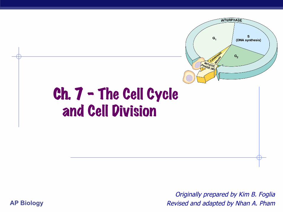
Ch. 7 – The Cell Cycle and Cell Division
Originally prepared by Kim B. Foglia Revised and adapted by Nhan A. Pham
AP Biology
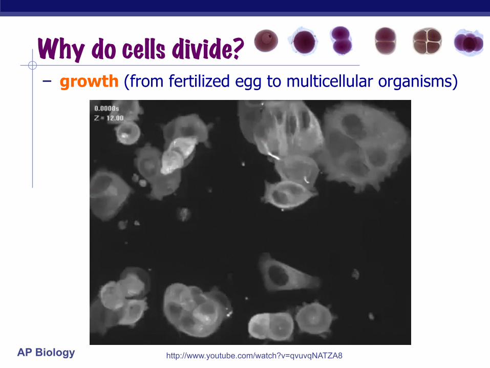
- growth (from fertilized egg to multicellular organisms)
Why do cells divide?
amoeba
http://www.youtube.com/watch?v=qvuvqNATZA8
AP Biology
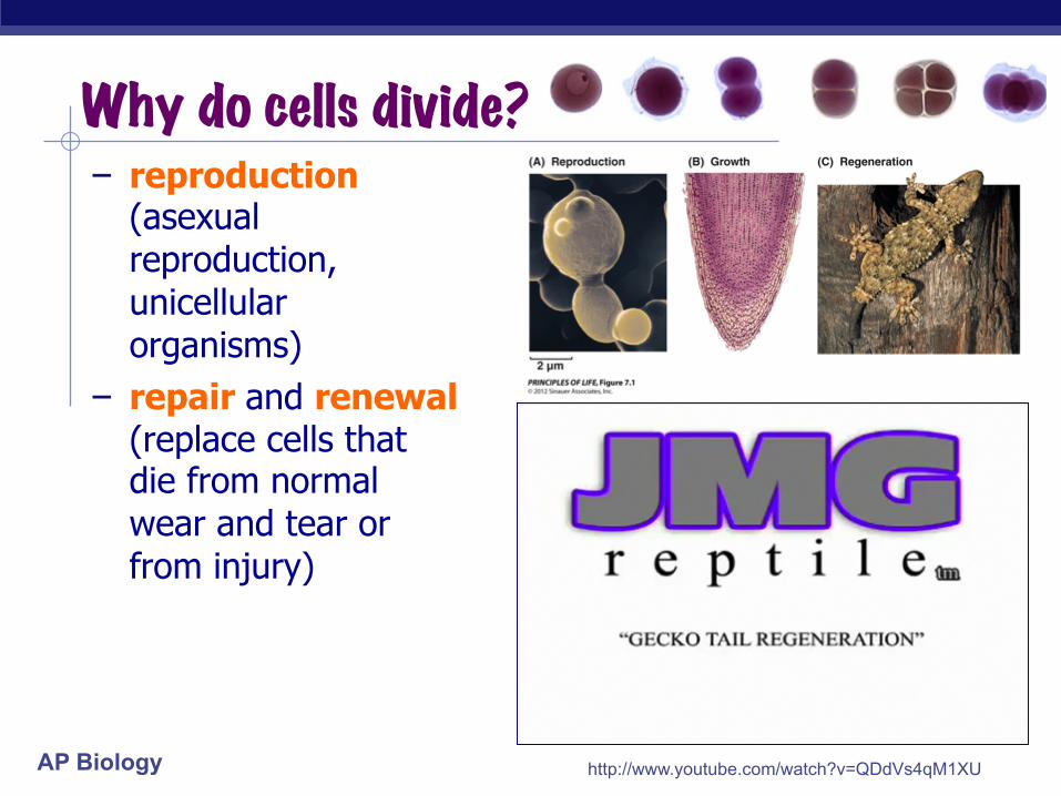
- reproduction (asexual reproduction, unicellular organisms)
- repair and renewal (replace cells that die from normal wear and tear or from injury)
Why do cells divide?
amoeba
http://www.youtube.com/watch?v=QDdVs4qM1XU

AP Biology
Getting the right stuff § What is passed on to daughter cells? - exact copy of chromosomes - organelles, cytoplasm, cell membrane, enzymes (in
cytokinesis)
chromosomes (stained orange) in kangaroo rat epithelial cell → notice cytoskeleton fibers

AP Biology
AP Biology
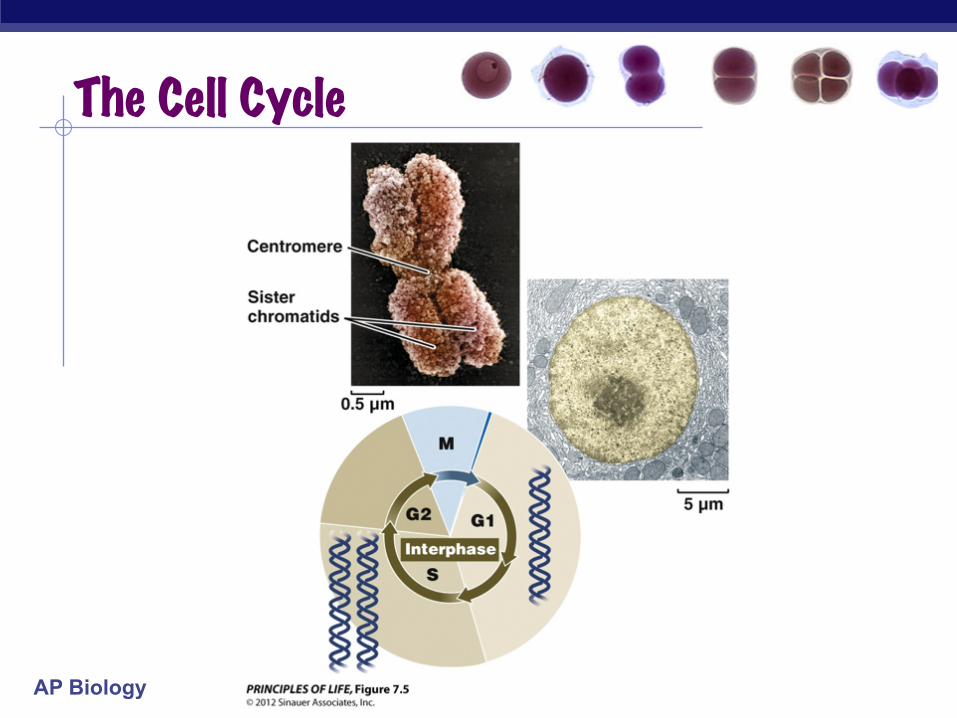
The Cell Cycle
AP Biology
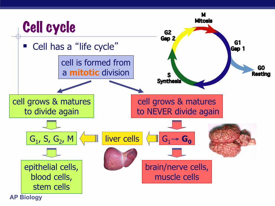
Cell cycle M
Mitosis
G1Gap 1
G0Resting
G2Gap 2
SSynthesis
§ Cell has a “life cycle”
cell is formed from a mitotic division
cell grows & matures to divide again
cell grows & matures to NEVER divide again
G1, S, G2, M G1→ G0
epithelial cells, blood cells, stem cells
liver cells
brain/nerve cells, muscle cells
AP Biology
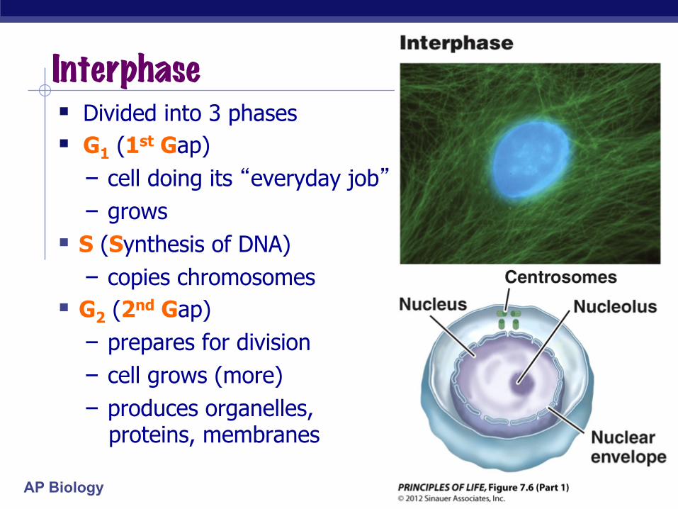
Interphase § Divided into 3 phases § G1 (1st Gap) - cell doing its “everyday job” - grows
§ S (Synthesis of DNA) - copies chromosomes
§ G2 (2nd Gap) - prepares for division - cell grows (more) - produces organelles,
proteins, membranes
AP Biology
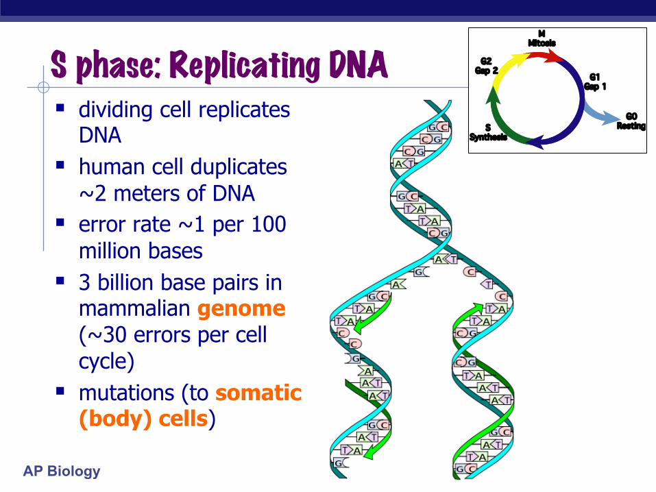
§ dividing cell replicates DNA
§ human cell duplicates ~2 meters of DNA
§ error rate ~1 per 100 million bases
§ 3 billion base pairs in mammalian genome (~30 errors per cell cycle)
§ mutations (to somatic (body) cells)
S phase: Replicating DNA M
Mitosis
G1Gap 1
G0Resting
G2Gap 2
SSynthesis

AP Biology
Organizing DNA § DNA is organized in
chromosomes
§ double helix DNA molecule
§ wrapped around histone proteins
§ chromatin is DNA-protein complex, organized into long thin fiber condensed further during mitosis
DNA
histones
chromatin
duplicated mitotic chromosome
ACTGGTCAGGCAATGTC
double stranded chromosome

AP Biology
Mitotic Chromosome § 2 sister chromatids § held together by adhesive
proteins at centromeres § contain identical
copies of original DNA
homologous chromosomes
homologous chromosomes
sister chromatids homologous = “same information” single-stranded double-stranded
AP Biology
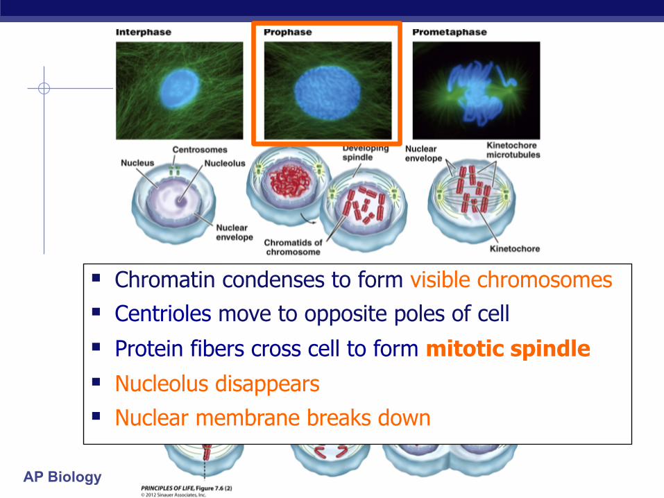
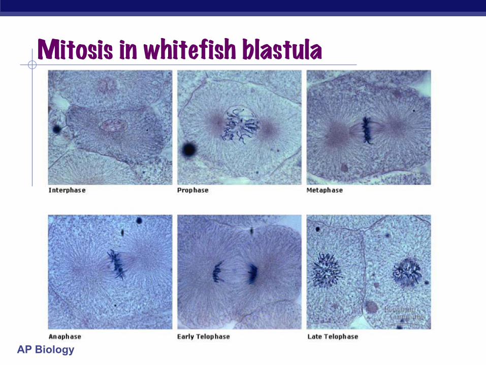
§ Chromatin condenses to form visible chromosomes § Centrioles move to opposite poles of cell
§ Protein fibers cross cell to form mitotic spindle
§ Nucleolus disappears
§ Nuclear membrane breaks down
AP Biology
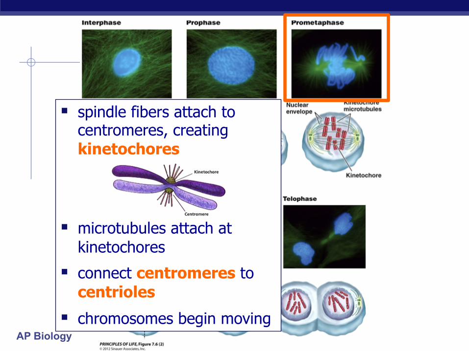
§ spindle fibers attach to centromeres, creating kinetochores
§ microtubules attach at kinetochores
§ connect centromeres to centrioles
§ chromosomes begin moving
AP Biology
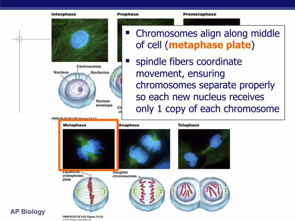
§ Chromosomes align along middle of cell (metaphase plate)
§ spindle fibers coordinate movement, ensuring chromosomes separate properly so each new nucleus receives only 1 copy of each chromosome

AP Biology
AP Biology
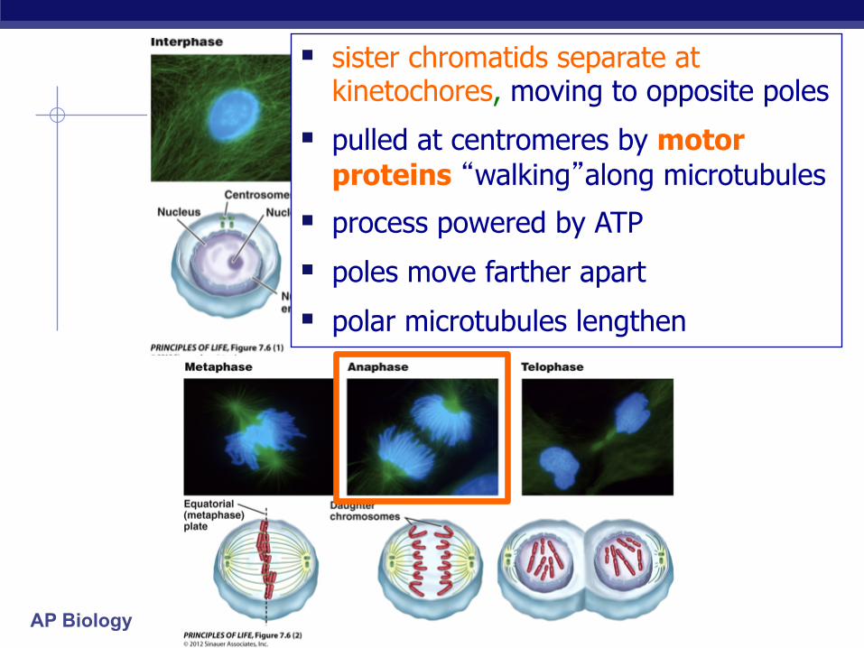
§ sister chromatids separate at kinetochores, moving to opposite poles
§ pulled at centromeres by motor proteins “walking”along microtubules
§ process powered by ATP § poles move farther apart
§ polar microtubules lengthen

AP Biology
Separation of Chromatids § In anaphase, proteins holding together sister chromatids
are inactivated § sister chromatids separate to become individual
chromosomes
2 chromosomes 1 chromosome 2 chromatids single-stranded
double-stranded
AP Biology
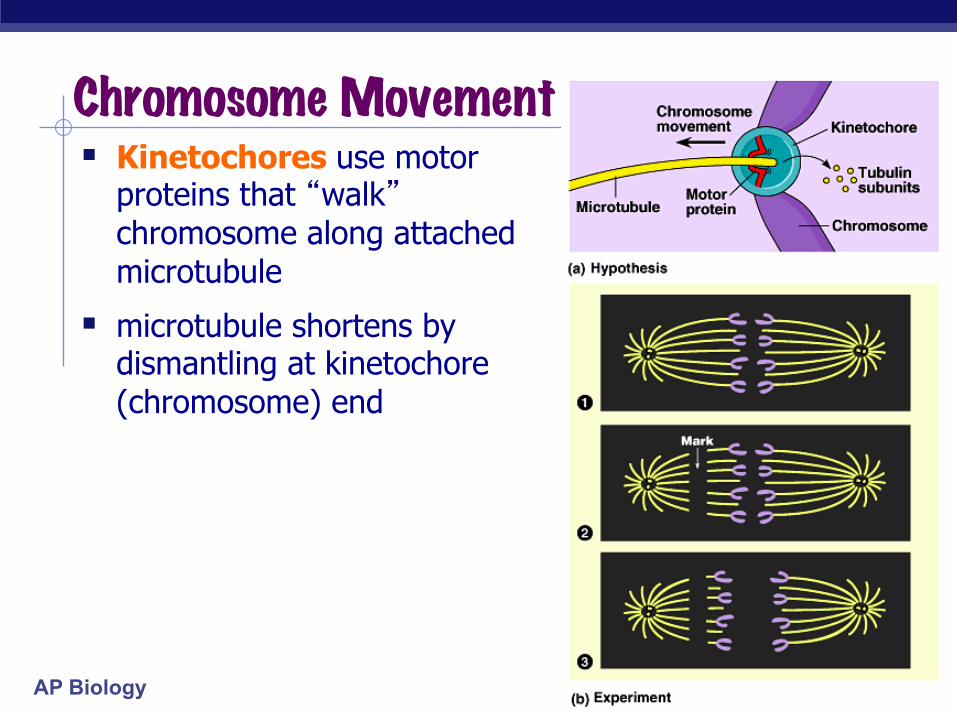
§ Kinetochores use motor proteins that “walk” chromosome along attached microtubule
§ microtubule shortens by dismantling at kinetochore (chromosome) end
Chromosome Movement
AP Biology
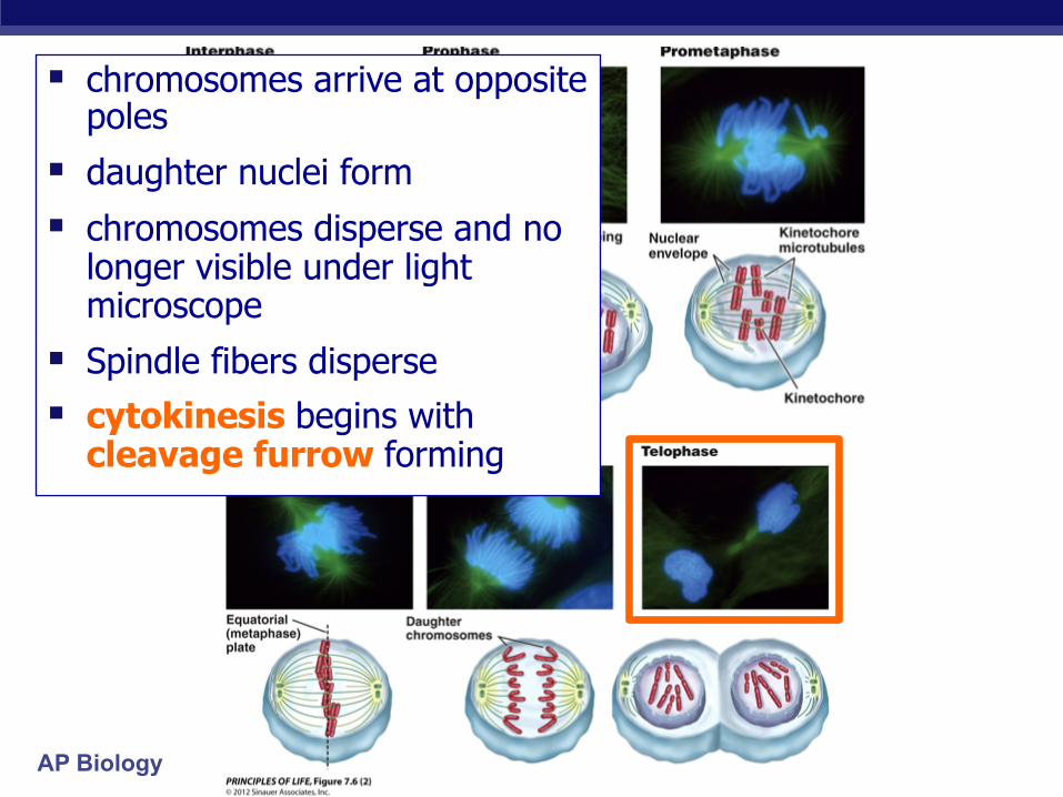
§ chromosomes arrive at opposite poles
§ daughter nuclei form
§ chromosomes disperse and no longer visible under light microscope
§ Spindle fibers disperse
§ cytokinesis begins with cleavage furrow forming
AP Biology
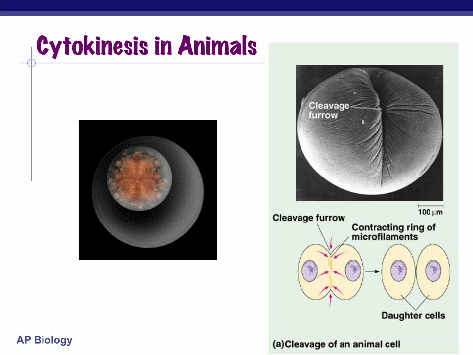
Cytokinesis in Animals
AP Biology
Mitosis in whitefish blastula

AP Biology
Mitosis in animal cells

AP Biology
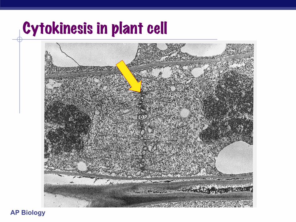
Cytokinesis in Plants § cell plate forms
§ Golgi-derived vesicles line up at equator
§ vesicles fuse to form 2 cell membranes
§ new cell wall laid down between membranes
§ new cell wall fuses with existing cell wall
AP Biology
Cytokinesis in plant cell

AP Biology
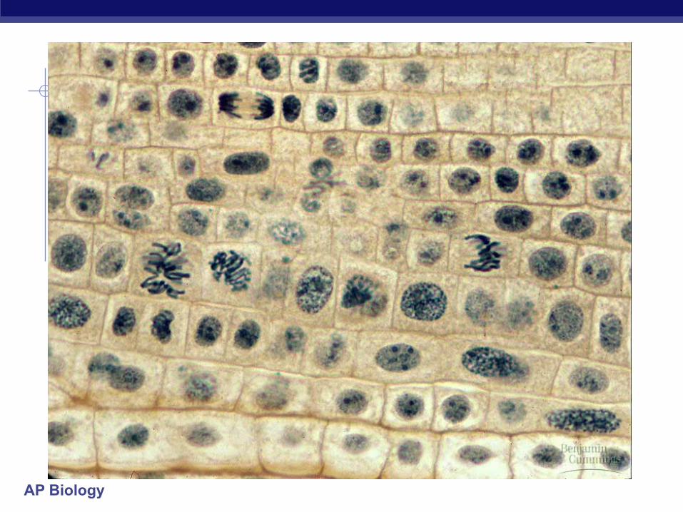
Mitosis in Plant Cell
AP Biology
AP Biology
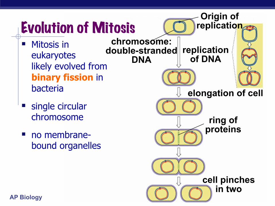
Origin of replication
chromosome: double-stranded
DNA replication
of DNA
elongation of cell
cell pinches in two
ring of proteins
Evolution of Mitosis § Mitosis in
eukaryotes likely evolved from binary fission in bacteria
§ single circular chromosome
§ no membrane-bound organelles
AP Biology
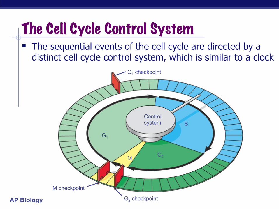
The Cell Cycle Control System § The sequential events of the cell cycle are directed by a
distinct cell cycle control system, which is similar to a clock
Control system
G2 checkpoint
M checkpoint
G1 checkpoint
G1
S
G2 M
AP Biology
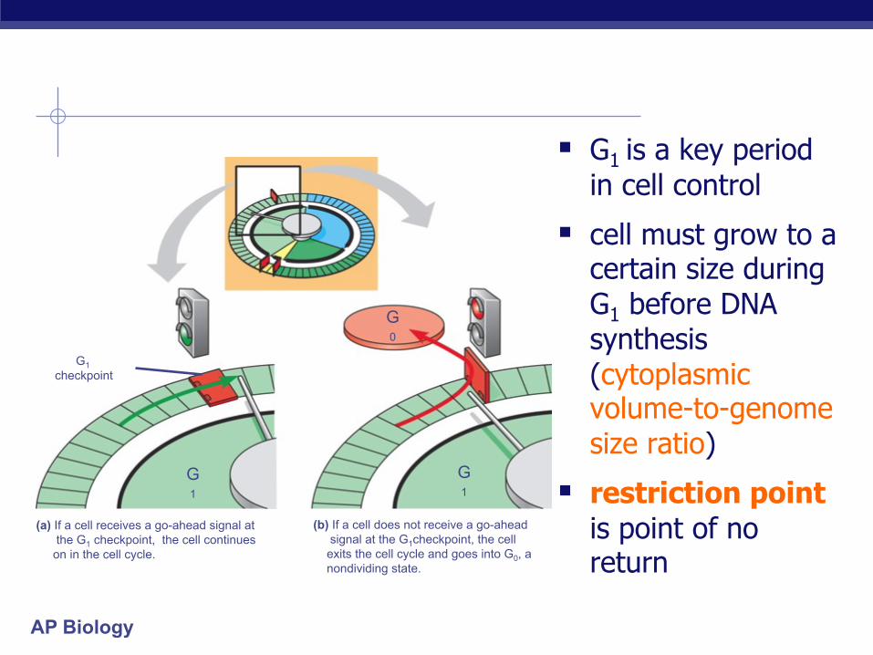
§ G1 is a key period
in cell control
§ cell must grow to a certain size during G1 before DNA synthesis (cytoplasmic volume-to-genome size ratio)
§ restriction point is point of no return
G1 checkpoint
G1
G1
G0
(a) If a cell receives a go-ahead signal at the G1 checkpoint, the cell continues on in the cell cycle.
(b) If a cell does not receive a go-ahead signal at the G1checkpoint, the cell exits the cell cycle and goes into G0, a nondividing state.
AP Biology
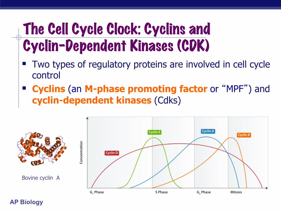
The Cell Cycle Clock: Cyclins and Cyclin-Dependent Kinases (CDK) § Two types of regulatory proteins are involved in cell cycle
control § Cyclins (an M-phase promoting factor or “MPF”) and
cyclin-dependent kinases (Cdks)
Bovine cyclin A
AP Biology
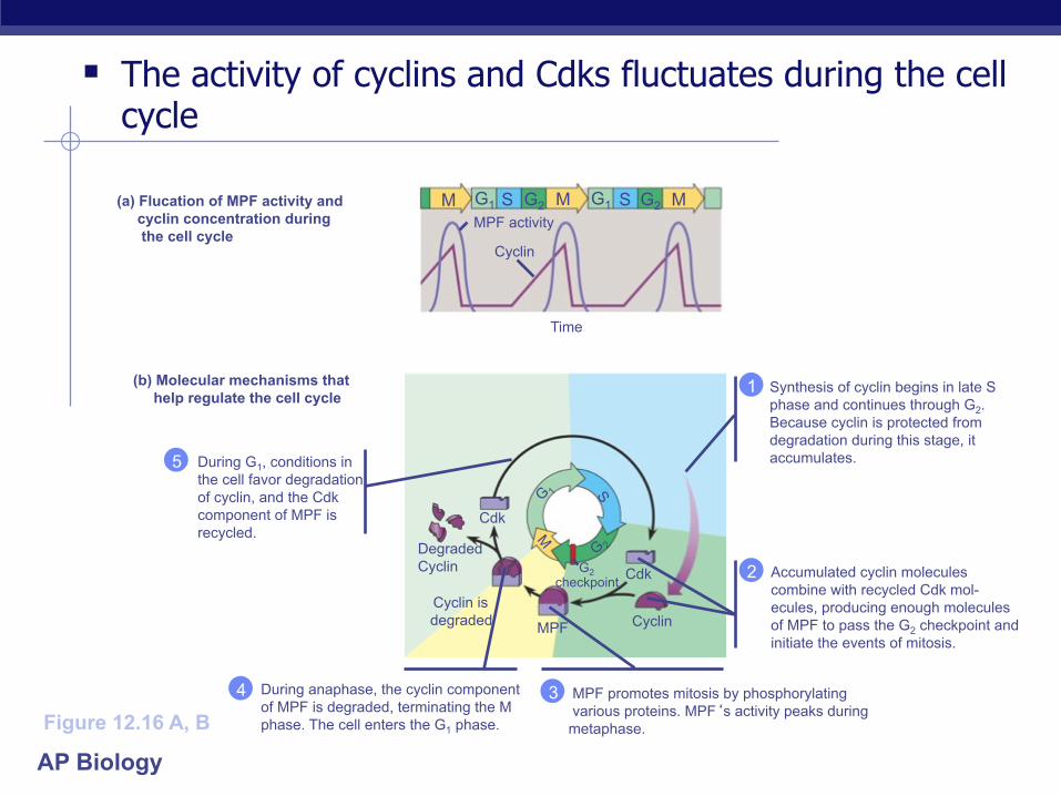
§ The activity of cyclins and Cdks fluctuates during the cell cycle
During G1, conditions in the cell favor degradation of cyclin, and the Cdk component of MPF is recycled.
5
During anaphase, the cyclin component of MPF is degraded, terminating the M phase. The cell enters the G1 phase.
4
Accumulated cyclin molecules combine with recycled Cdk mol- ecules, producing enough molecules of MPF to pass the G2 checkpoint and initiate the events of mitosis.
2
Synthesis of cyclin begins in late S phase and continues through G2. Because cyclin is protected from degradation during this stage, it accumulates.
1
Cdk
Cdk G2 checkpoint
Cyclin MPF
Cyclin is degraded
Degraded Cyclin
G1 G1 S G2 G2 S M M MPF activity
Cyclin
Time
(a) Flucation of MPF activity and cyclin concentration during the cell cycle
(b) Molecular mechanisms that help regulate the cell cycle
MPF promotes mitosis by phosphorylating various proteins. MPF‘s activity peaks during metaphase.
3 Figure 12.16 A, B
M

AP Biology
Growth Factors § Growth factors stimulate other cells to divide
EXPERIMENT
A sample of connective tissue was cut up into small pieces.
Enzymes were used to digest the extracellular matrix, resulting in a suspension of free fibroblast cells.
Cells were transferred to sterile culture vessels containing a basic growth medium consisting of glucose, amino acids, salts, and antibiotics (as a precaution against bacterial growth). PDGF was added to half the vessels. The culture vessels were incubated at 37°C.
3
2
1 Petri plate
Without PDGF
With PDGF
Scalpels

AP Biology
§ In density-dependent inhibition, crowded cells stop dividing
§ Most animal cells exhibit anchorage dependence (they must be attached to a substratum to divide)
Cells anchor to dish surface and divide (anchorage dependence).
When cells have formed a complete single layer, they stop dividing (density-dependent inhibition).
If some cells are scraped away, the remaining cells divide to fill the gap and then stop (density-dependent inhibition).
Normal mammalian cells. The availability of nutrients, growth factors, and a substratum for attachment limits cell density to a single layer.
(a)
25 µm

AP Biology
Cancer Cells § exhibit neither density-dependent inhibition nor
anchorage dependence
§ do not respond to control mechanisms
§ form tumors
http://www.youtube.com/watch?v=Y2kTEbyMvXA
AP Biology
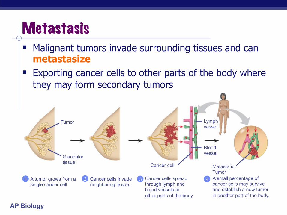
Metastasis § Malignant tumors invade surrounding tissues and can
metastasize § Exporting cancer cells to other parts of the body where
they may form secondary tumors
Cancer cells invade neighboring tissue.
2 A small percentage of cancer cells may survive and establish a new tumor in another part of the body.
4 Cancer cells spread through lymph and blood vessels to other parts of the body.
3 A tumor grows from a single cancer cell.
1
Tumor
Glandular tissue Cancer cell
Blood vessel
Lymph vessel
Metastatic Tumor