Embed Size (px)
Citation preview

Part ii – Neurological Disorders
Dr William P. Howlett2012
CHAPTER 16 INTRACRANIAL TUMOURS

Kilimanjaro Christian Medical Centre, Moshi, Kilimanjaro, Tanzania
BRIC 2012 University of Bergen PO Box 7800 NO-5020 Bergen Norway
NEUROLOGY IN AFRICAWilliam HowlettIllustrations: Ellinor Moldeklev Hoff, Department of Photos and Drawings, UiBCover: Tor Vegard TobiassenLayout: Christian Bakke, Division of Communication, University of BergenPrinted by Bodoni, Bergen, NorwayCopyright © 2012 William HowlettNEUROLOGY IN AFRICA is freely available to download at Bergen Open Research Archive (https://bora.uib.no) www.uib.no/cih/en/resources/neurology-in-africaISBN 978-82-7453-085-0
Notice/DisclaimerThis publication is intended to give accurate information with regard to the subject matter covered. However medical knowledge is constantly changing and information may alter. It is the responsibility of the practitioner to determine the best treatment for the patient and readers are therefore obliged to check and verify information contained within the book. This recommendation is most important with regard to drugs used, their dose, route and duration of administration, indications and contraindications and side effects. The author and the publisher waive any and all liability for damages, injury or death to persons or property incurred, directly or indirectly by this publication.
MILJØMERKET
241 Trykksak 699

CONTENTS
INTRACRANIAL TUMOURS 367EPIDEMIOLOGY � � � � � � � � � � � � � � � � � � � � � � � � � � � � � � � � � � � � � � � � � � � � � � � � � 367CLINICAL FEATURES � � � � � � � � � � � � � � � � � � � � � � � � � � � � � � � � � � � � � � � � � � � � � � 368MAIN SITES � � � � � � � � � � � � � � � � � � � � � � � � � � � � � � � � � � � � � � � � � � � � � � � � � � � � 369GLIOMA � � � � � � � � � � � � � � � � � � � � � � � � � � � � � � � � � � � � � � � � � � � � � � � � � � � � � � 370MENINGIOMA � � � � � � � � � � � � � � � � � � � � � � � � � � � � � � � � � � � � � � � � � � � � � � � � � � 371PITUITARY TUMOURS � � � � � � � � � � � � � � � � � � � � � � � � � � � � � � � � � � � � � � � � � � � � � 373METASTASES � � � � � � � � � � � � � � � � � � � � � � � � � � � � � � � � � � � � � � � � � � � � � � � � � � � 374OTHER TUMOURS � � � � � � � � � � � � � � � � � � � � � � � � � � � � � � � � � � � � � � � � � � � � � � � � 375MANAGEMENT OF TUMOURS � � � � � � � � � � � � � � � � � � � � � � � � � � � � � � � � � � � � � � � � 378
CHAPTER 16
INTRACRANIAL TUMOURSIntracranial tumours (ICT) include tumours arising from the brain or surrounding tissues. They are classified as either primary or secondary and as malignant or benign. Other classifications include tissue type, grade of malignancy and the main site affected. Primary intracranial tumours originate mostly from brain or meninges whereas secondary or metastatic tumours originate mostly elsewhere in the body. The most common primary tumours are gliomas accounting for between 30-50% of all adult intracranial tumours. Gliomas are malignant tumours which arise from glial support cells rather than neuronal cells and are termed astrocytoma, oligodendroglioma, ependymoma and pinealoma. The most common primary benign tumours are meningiomas. These arise from the meninges covering the brain and cranial nerves accounting for 20-30% of all adult intracranial tumours. Other benign intracranial tumours are pituitary adenoma, craniopharyngioma, colloid cyst and acoustic neuroma. The aim of this chapter is to present an overview of the main intracranial tumours. The student should aim to know the main types of tumour, their clinical presentation, diagnosis and management.
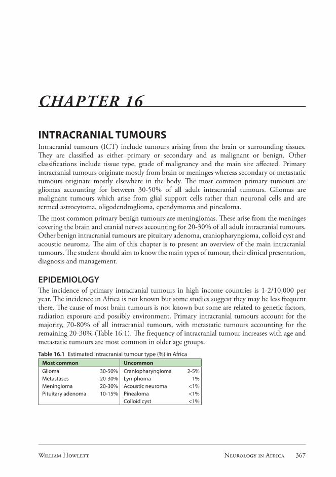
EPIDEMIOLOGYThe incidence of primary intracranial tumours in high income countries is 1-2/10,000 per year. The incidence in Africa is not known but some studies suggest they may be less frequent there. The cause of most brain tumours is not known but some are related to genetic factors, radiation exposure and possibly environment. Primary intracranial tumours account for the majority, 70-80% of all intracranial tumours, with metastatic tumours accounting for the remaining 20-30% (Table 16.1). The frequency of intracranial tumour increases with age and metastatic tumours are most common in older age groups.
Table 16.1 Estimated intracranial tumour type (%) in AfricaMost common UncommonGlioma 30-50%Metastases 20-30%Meningioma 20-30%Pituitary adenoma 10-15%
Craniopharyngioma 2-5%Lymphoma 1%Acoustic neuroma <1%Pinealoma <1%Colloid cyst <1%
William Howlett Neurology in Africa 367

CLINICAL FEATURESIntracranial tumours in Africa are characterised by late presentation with on average a 2 year delay before diagnosis and a high mortality of >80% at 1-2 years follow up. They can exist for long periods with no or few symptoms and when symptoms do occur the tumours may be advanced. Headache is the most common complaint though occurring only in <50% of cases. Pain is variable, ranging from being dull, low grade and intermittent to being severe, continuous, deep, nocturnal, present on waking and often associated with vomiting. Intracranial tumours have three recognizable main modes of clinical presentation (Table 16.2): (1) focal neurological deficits (FND), (2) seizures, and (3) raised intracranial pressure ( ICP). These can occur either alone or together depending on type, stage and site of tumour. The pathogenic mechanisms underlying these presentations include tumour mass effect, brain irritation and blocked CSF flow. Highly malignant or fast growing tumours tend to present with combinations of all three main modes of presentation occurring over weeks or months whereas low grade or slow growing tumours tend to present with isolated seizures and/or neurological deficits occurring over months or years. The age of the patient, speed of onset of symptoms and neurological findings all help to determine the site and the probable type of tumour.
Table 16.2 Main presenting clinical features of intracranial tumoursNeurological finding Symptoms/Signsfocal neurological deficits hemiparesis, dysphasia, visual loss, field defect, ataxia, cranial nerve palsiesraised intracranial pressure headaches vomiting, papilloedema, LOCseizures simple or complex partial, secondary GTC
Focal neurological deficitsFND are the most common neurological presentation of brain tumour. The type of FND is very variable and reflects the cell type, grade of malignancy and the site affected (Chapter 2). These include hemiparesis, dysphasia, visual loss, field defects, cognitive impairment, personality change, cranial nerve palsies and ataxia. The combination of ataxia and cranial nerve palsies occurs more frequently with tumours arising in the posterior fossa.
SeizuresSeizures are the presenting complaint in approximately a quarter of patients and occur as a complication in about another quarter (Chapter 4). Seizures arise mostly from tumours affecting the temporal lobes and occur most commonly in association with malignant tumours. The seizures are mostly generalised tonic clonic-type seizures with a focal origin.
Raised intracranial pressure ( ICP)The symptoms and signs of ICP are headache, vomiting, papilloedema and altered level of consciousness; these occur because of either mass effect or hydrocephalus. Hydrocephalus arises because of mechanical blockage to CSF flow through the ventricles. Headache is the most common symptom being typically severe in advanced tumours, often waking the person from sleep during the night or early morning and frequently associated with vomiting (Chapter 15). The site of the headache is mostly frontal in supratentorial tumours and occipital in posterior fossa tumours, however the site may not necessarily be localising. Mass lesions arising from above the tentorium, and affecting both the hemispheres frequently give rise to combinations of features of FNDs and ICP. The most common neurological deficits include hemiparesis and 3rd, 4th and 6th nerve palsies. These may be false localising signs, if they occur as a result of remote
Chapter 16 IntraCranIal tumours
Part ii – Neurological Disorders 368

compression at a site away from the tumour. Space occupying lesions (SOL) arising below the tentorium within the posterior fossa cause cranial nerve palsies, ataxia, and long track signs and raised ICP secondary to obstructive hydrocephalus. A history of visual disturbances and the presence of papilloedema are usually late clinical findings. Eventually, the expanding tumour results in herniation either through the tentorium or foramen magnum leading to death.
Key points
· most common malignant ICTs are gliomas & metastases · most common benign ICTs are meningiomas & pituitary adenomas · main presentations include progressive FNDs, seizures & ICP · other presentations are headaches, visual failure, cranial nerve signs & seizures · most patients presenting with headaches do not have brain tumours
MAIN SITESThe site and types of intracranial tumour determine the presenting symptoms and signs and the main ones are outlined below (Chapter 2).
Frontal lobeTumours involving the frontal lobe typically present late because the frontal lobe has a large silent area. Contralateral hemiparesis occurs if the motor strip is involved. Tumours involving the anterior frontal lobe may present with personality changes and a loss of initiative, inhibition and cognitive function. There may also be focal motor seizures, urinary incontinence and loss of smell. An expressive aphasia occurs if Broca’s area in the dominant hemisphere is involved.
Parietal lobeTumours of the parietal lobe result in difficulties or inability to recognise sensory and proprioceptive input from the opposite side of the body. This may show itself as tending to ignore the contralateral side visuospatially (hemineglect) or as difficulties recognizing familiar shapes, textures or numbers when placed in the opposite hand. If the dominant hemisphere is involved, there may be difficulties particularly with understanding speech, numbers, reading and writing and carrying out motor tasks (apraxia). Patients with non dominant hemisphere involvement may present with or develop hemineglect. Patients may also have a visual field defect involving the lower quadrant from the opposite side.
Temporal lobeTumours involving the dominant temporal lobe (usually left sided) may result in aphasia which is receptive in type and also memory impairment. Tumours on either side may result in recent onset temporal lobe seizures and visual field loss in the contralateral upper quadrant.
Occipital lobeTumours involving the occipital lobe present with visual disturbances, hallucinations, and a loss of vision from the opposite side of the body, a contralateral homonymous hemianopia.
Brain stem and cerebellumTumours of the brain stem present with a combination of ipsilateral cranial nerve palsies and cerebellar ataxia, and contralateral long tract signs. These may be associated with hydrocephalus
maIn sItes
William Howlett Neurology in Africa 369

and ICP depending on the tumour type and site. Tumours involving this area of the brain are more common in children.
GLIOMAGliomas account for up to half of all brain tumours and occur most commonly in older age groups >50-60 years. Glioma is the generic name for brain tumours of neuroepithelial cell origin. These are the support cells in the brain and the main ones are astrocytes and oligodendrocytes. Astrocytic tumours are separated histologically into grades I & II which are low grade, well differentiated and relatively benign and into grades III & IV which are high grade, poorly differentiated and highly malignant. Glioblastoma multiforme represents the most malignant stage of glioma. Oligodendrogliomas on the whole tend to be lower grade tumours characterized by a capsule and the presence of cysts and calcium with a good prognosis, but after years about one third may evolve into more malignant tumours. Other forms of glioma include ependymomas which are derived from cells which line the ventricles and choroid plexus. Medulloblastomas are gliomas of the cerebellum and the roof of the 4th ventricle occurring mostly in young children aged 4-8 years (Fig. 16.1).
Clinical featuresGliomas present clinically with increasing symptoms usually over weeks or months or years depending on the grade of malignancy. Presentations include headache and combinations of focal neurological deficit, seizures, and signs of ICP. Diagnosis is confirmed by neuroimaging, most commonly a CT of the head. It typically shows a unifocal enhancing mass with surrounding oedema and mass effect (Fig. 16.1).
ManagementManagement where there are full resources includes a combination of surgery, chemotherapy and radiotherapy. Surgery is indicated for biopsy to establish a tissue diagnosis and for partial tumour resection to relieve symptoms. Chemotherapy is used in high grade malignant gliomas and usually involves the alkylating drug temozolomide in combination with other drugs. However temozolomide is expensive, used mostly but not exclusively in younger patients and only available in some specialized oncology units. Radiation is indicated for most high grade gliomas but this is only palliative at this stage.
PrognosisThe prognosis in low grade gliomas is good with a median survival of 8-10 years. However in patients with higher grade malignancy the prognosis even with treatment is poor with survival of usually <12 months.
Key points
· gliomas account for almost half of all ICTs · presentations include ICP, FNDs & seizures over weeks & months · diagnosis is by neuroimaging, CT or MRI · management is mainly with combination of surgery, chemotherapy & radiotherapy · prognosis for high grade gliomas is poor
Chapter 16 IntraCranIal tumours
Part ii – Neurological Disorders 370

MENINGIOMAThese account for about 20-30% of all intracranial tumours. They occur mostly in the middle and older age groups, >50 years and more commonly in females 2:1. They arise from either the dural or arachnoid meninges, overlying the surface of the brain, cranial nerves, falx and tentorium and are nearly always benign. Less than 1% of meningiomas are malignant. Small meningiomas <2 cm are usually asymptomatic and tumours only become symptomatic when they reach a size sufficient to affect function. Most tumours arise on the convexities of the brain, as these are the largest surface areas.
Clinical featuresMeningiomas tend to present clinically with focal neurological signs that reflect the site and size of the tumour in much the same way as malignant tumours but over a much longer time course usually months or years. Focal seizures are an early symptom of tumours that overlie the cortex. ICP may also be a late feature of large meningiomas. Meningiomas affecting certain areas have specific modes of presentation. Parasagittal meningiomas present with spastic paraparesis, olfactory groove meningiomas with anosmia and papilloedema and sphenoid wing and frontal meningiomas with Foster-Kennedy syndrome: unilateral optic atrophy with contralateral papilloedema.
Diagnosis & managementDiagnosis is made by neuroimaging with CT or MRI scans. CT typically shows a meningioma as a uniformly homogenously enhancing extra axial mass which may be partially calcified or
CT (with contrast)
Right frontal glioma Medulloblastoma & hydrocephalus in a child
MRI T1 & T2 (enhanced)
Right sided glioma with midline extension/shift
Figure 16.1 Gliomas
menIngIoma
William Howlett Neurology in Africa 371
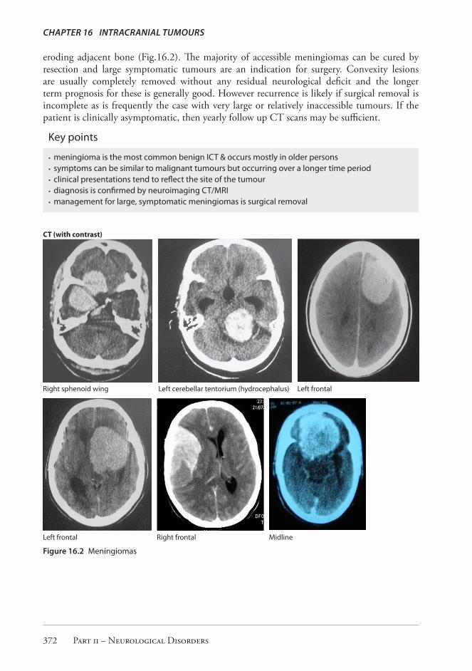
eroding adjacent bone (Fig.16.2). The majority of accessible meningiomas can be cured by resection and large symptomatic tumours are an indication for surgery. Convexity lesions are usually completely removed without any residual neurological deficit and the longer term prognosis for these is generally good. However recurrence is likely if surgical removal is incomplete as is frequently the case with very large or relatively inaccessible tumours. If the patient is clinically asymptomatic, then yearly follow up CT scans may be sufficient.
Key points
· meningioma is the most common benign ICT & occurs mostly in older persons · symptoms can be similar to malignant tumours but occurring over a longer time period · clinical presentations tend to reflect the site of the tumour · diagnosis is confirmed by neuroimaging CT/MRI · management for large, symptomatic meningiomas is surgical removal
CT (with contrast)
Right sphenoid wing Left cerebellar tentorium (hydrocephalus) Left frontal
Left frontal Right frontal Midline
Figure 16.2 Meningiomas
Chapter 16 IntraCranIal tumours
Part ii – Neurological Disorders 372

PITUITARY TUMOURSPituitary adenomas account for about a tenth of all intracranial tumours.
Clinical featuresThese are benign intracranial tumours occurring outside the brain which present with headache, local pressure and also endocrine effects. Expansion of the adenoma upwards leads to compression of the optic chiasm which results in a visual field defect, most commonly bitemporal hemianopia, initially involving the upper quadrants. The endocrine effects may vary from being clinically asymptomatic in non functioning adenomas, to prolactinomas causing amenorrhoea/galactorrhoea and to micro or macro adenomas secreting growth hormone in acromegaly or microadenoma secreting ACTH in Cushing’s disease. Infrequently, pituitary tumours undergo infarction and patients then present with sudden headache, vomiting and features of acute hypopituitarism. The differential diagnosis of pituitary tumours includes other mass lesions that may compress the optic chiasm including craniopharyngioma, meningioma and internal carotid aneurysm.
DiagnosisDiagnosis is confirmed by endocrine and imaging studies. Endocrine studies should measure prolactin, TSH (thyroid stimulating hormone), free T4, morning cortisol and growth hormone levels if available. A plain skull X-ray may show an enlarged pituitary fossa and neuroimaging with a CT or preferably an MRI may show a pituitary tumour (Fig.16.3).
Widened sella turcica
Skull X-ray (lateral view)
CT (with contrast) MRI T1
Pituitary tumour
Figure 16.3 Pituitary tumours
pItuItary tumours
William Howlett Neurology in Africa 373

ManagementManagement is either medical or surgical. Surgery via the transphenoidal route is usually the management of choice, if the tumour is intrasellar except in prolactin secreting adenomas which can often be managed medically with dopamine receptor agonists such as bromocriptine up to 20-40 mg daily. Growth hormone secreting tumours can also sometimes be managed medically as well with dopamine agonists or a somatostatin analogue (e.g. octreotide). Larger pituitary tumours extending above the sella may require a transfrontal craniotomy.
Key points
· pituitary tumours are benign and account for about 10% of ICTs · symptoms are caused by local pressure effects, hormone secretion & pituitary failure · CFs include headache, visual failure, visual field defect & endocrine disease · treatment involves hormone replacement, dopamine agonists & surgery
METASTASESThese account for about a quarter of all intracranial tumours and are usually the most common intracranial tumour in the older age groups >60 yrs. They arise mostly by haematogenous spread from cancers occurring outside the brain. The main sources are breast, lung, renal, gastrointestinal cancers and melanoma. Very rarely local metastases can arise within the brain; the usual source is a glioblastoma, the most malignant type of brain tumour. The history in metastatic brain tumour is usually short, involving day or weeks of progressive neurological symptoms of raised intracranial pressure, seizures and focal neurological deficits. Diagnosis is confirmed by neuroimaging usually a CT of the head. Metastases show as multiple ring or solid enhancing lesions anywhere in the brain with surrounding oedema and mass effect (Fig. 16.4). In about 10% of cases the metastases may be solitary.
Differential diagnosisThe differential diagnosis of intracranial tumour includes other causes of mass or space occupying lesions (SOL). In Africa these are mostly infections. The most common infectious causes are HIV related and include toxoplasma encephalitis and tuberculoma. Other infectious causes include pyogenic and parasitic infections. The main parasitic infections are hydatid cysts, cysticercosis and very occasionally schistosomiasis. While there may be some difficulty separating a tumour from these causes clinically, the history and signs of systemic illness and
CT (with contrast)
Multiple metastases
Figure 16.4 Metastases
Chapter 16 IntraCranIal tumours
Part ii – Neurological Disorders 374

typical neuroimaging findings should suggest an infectious origin. The non infectious causes are mostly vascular and include large aneurysms and arteriovenous malformations. These can usually be differentiated from metastatic brain tumours by their typical history, vascular risk factors and appearances on CT scan. A chronic subdural haematoma particularly in the elderly without a history of head trauma may sometimes mimic a brain tumour in clinical presentation and require a CT scan of the head to confirm the correct diagnosis. Other neurological disorders to be considered include hydrocephalus and benign intracranial hypertension.
InvestigationsBaseline haematological and biochemical investigations should be carried out in suspected cases. X-rays involving the chest and skull can be helpful. The chest X-ray may show evidence of metastases or a primary lung tumour. The skull X-ray may show enlargement of the pituitary fossa, erosion of bone or other signs of chronic raised intracranial pressure. A lumbar puncture is relatively contraindicated in patients with suspected intracranial tumours and possible ICP as any sudden decrease in intracranial pressure can result in death due to coning. Neuroimaging using computerized tomography (CT) or magnetic resonance imaging (MRI) is the method of choice in the detection of brain tumours. CT is now available in most specialised centres. In patients with a suspected brain tumour having neuroimaging, intravenous contrast is given unless there is a contraindication; a pattern of increased enhancement points to brain tumours.
ManagementManagement is mostly symptomatic and aimed mainly at palliation. Dexamethasone 4-6 mg 3-4 times daily temporarily reduces cerebral oedema and symptoms. Chemotherapy is usually not indicated and excision surgery is reserved for isolated single accessible metastases. Prognosis is poor anywhere in the world with most untreated patients surviving less than a month after diagnosis.
Key points
· metastases account for about 25% of all ICTs & occur mostly in older age groups · main sites of origin are breast, lung, kidney, GIT and melanoma · CFs are similar to gliomas but occurring over a faster time course · differential diagnosis includes brain abscess, parasitic cysts & vascular causes · diagnosis confirmed by (CT/MRI) & evidence of primary tumour elsewhere
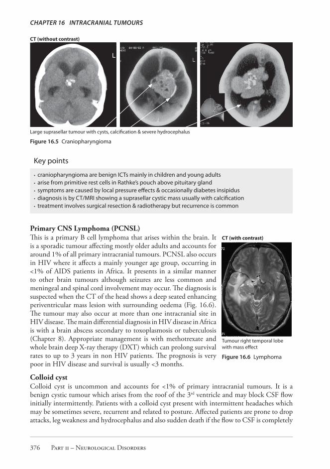
OTHER TUMOURSCraniopharyngiomaThese are uncommon benign tumours that arise from primitive cells in Rathke’s pouch which lies just above the pituitary gland and below the floor of the third ventricle. They represent 2-5% of all intracranial tumours in high income countries and mainly affect children but are also seen in usually younger adults. They present clinically with pressure effects including headache, visual field defect, and occasionally diabetes insipidus and hydrocephalus. Neuroimaging using CT or MRI is diagnostic showing a non enhancing suprasellar cystic mass usually with irregular calcification (Fig. 16.5). The tumours can be quite large at the time of clinical presentation. Management is surgical and by irradiation; however the outcome is often unfavourable because of a high likelihood of recurrence.
other tumours
William Howlett Neurology in Africa 375
Key points
· craniopharyngioma are benign ICTs mainly in children and young adults · arise from primitive rest cells in Rathke’s pouch above pituitary gland · symptoms are caused by local pressure effects & occasionally diabetes insipidus · diagnosis is by CT/MRI showing a suprasellar cystic mass usually with calcification · treatment involves surgical resection & radiotherapy but recurrence is common
Primary CNS Lymphoma (PCNSL)This is a primary B cell lymphoma that arises within the brain. It is a sporadic tumour affecting mostly older adults and accounts for around 1% of all primary intracranial tumours. PCNSL also occurs in HIV where it affects a mainly younger age group, occurring in <1% of AIDS patients in Africa. It presents in a similar manner to other brain tumours although seizures are less common and meningeal and spinal cord involvement may occur. The diagnosis is suspected when the CT of the head shows a deep seated enhancing periventricular mass lesion with surrounding oedema (Fig. 16.6). The tumour may also occur at more than one intracranial site in HIV disease. The main differential diagnosis in HIV disease in Africa is with a brain abscess secondary to toxoplasmosis or tuberculosis (Chapter 8). Appropriate management is with methotrexate and whole brain deep X-ray therapy (DXT) which can prolong survival rates to up to 3 years in non HIV patients. The prognosis is very poor in HIV disease and survival is usually <3 months.
Colloid cystColloid cyst is uncommon and accounts for <1% of primary intracranial tumours. It is a benign cystic tumour which arises from the roof of the 3rd ventricle and may block CSF flow initially intermittently. Patients with a colloid cyst present with intermittent headaches which may be sometimes severe, recurrent and related to posture. Affected patients are prone to drop attacks, leg weakness and hydrocephalus and also sudden death if the flow to CSF is completely
CT (without contrast)
Large suprasellar tumour with cysts, calcification & severe hydrocephalus
Figure 16.5 Craniopharyngioma
CT (with contrast)
Tumour right temporal lobe with mass effect
Figure 16.6 Lymphoma
Chapter 16 IntraCranIal tumours
Part ii – Neurological Disorders 376

blocked. CT may show a well demarcated rounded cyst arising within the 3rd ventricle with or without obstructive hydrocephalus (Fig. 16.7). The differential diagnosis includes other causes of intraventricular cysts including neurocysticercosis where cysts are usually multiple. Management is by surgical removal or destruction of the cyst.
Acoustic neuroma (Schwannoma)These are rare benign slow growing tumours that arise from Schwann cells of the vestibular nerve with an incidence rate of 1 per 100,000 in high income countries. The corresponding incidence reported in a black population in SA is reported to be very low, (0.01/100,000). They arise sporadically on one side but may occur bilaterally in neurofibromatosis type 2 when they usually occur before the age of 21 years (Chapter 18). The principal sites are the internal auditory canal (IAC) and the cerebellopontine angle (CPA). The distinguishing clinical features are unilateral deafness coupled with tinnitus, vertigo, ipsilateral unsteadiness and facial weakness and numbness. Hydrocephalus and raised ICP are a late complication. Diagnosis is by showing high frequency hearing loss either clinically or by audiograph and by demonstrating an expanding lesion in the IAC or in the CPA by either CT or MRI (Fig. 16.8). The main differential diagnosis is between a meningioma and epidermoid cyst. Management is by deep X-ray therapy and by surgical resection, though resection is technically difficult with significant morbidity.
PinealomaTumours may arise from the pineal gland and are uncommon. These are classified as germinomas, pinealomas, teratomas and gliomas. They are uncommon slow growing tumours and occur mostly in children and young adults <30 years. Patients present clinically with disorders of eye
MRI T1
Colloid cyst blocking foramen of Monro with dilatation of lateral ventricle
Figure 16.7 Colloid cyst
MRI T1 (with contrast) CT (with contrast)
Acoustic neuroma extending into right auditory canal
Acoustic neuroma (left sided)
Figure 16.8 Acoustic neuroma
other tumours
William Howlett Neurology in Africa 377

movement, mainly up gaze paralysis and symptoms of raised ICP. These symptoms are explained by the local effects of the tumour invading the upper midbrain and blocking the aqueduct of Sylvius causing hydrocephalus (Fig.16.9). The diagnosis is confirmed by neuroimaging, CT or MRI and management is neurosurgical.
MANAGEMENT OF TUMOURSThe management of intracranial tumours involves confirming the diagnosis histologically, grading the tumour when indicated and treating the patient medically and surgically.
MedicalGeneral measures are aimed at controlling symptoms including pain and anxiety and reducing intracranial pressure. These measures include treating the symptoms and ensuring adequate cerebral perfusion by maintaining hydration, oxygen saturation and blood pressure. The use of osmotic therapy can be effective in reducing acute elevations of intracranial pressure but is only effective for a few days. The drug of choice in ICP is mannitol 20% solution in boluses of 0.25-0.75 g/kg every 4-6 hours. Corticosteroids are useful for patients with cerebral oedema secondary to mass lesions. Dexamethasone is the most commonly used steroid in a dose 4-8 mg/po or iv/three to four times daily. This often provides significant symptomatic relief and can be maintained at the lowest dose that provides relief which is usually 4 mg twice daily. Opiates and anxiolytics may be necessary to control pain and anxiety.
SurgicalMeningiomas, pituitary tumours, acoustic neuromas are among the benign ICTs that can be successfully removed if not too advanced at presentation. Primary malignant brain tumours that are at or near the surface of the brain may be partially removed by debulking operations. The main aim is to palliate symptoms by removing as much tissue as is safely possible. Biopsy of the tumour is also important as the histological findings help to determine the type and grade of tumour and the subsequent management and prognosis. Patients presenting with resectable primary brain tumours should be referred to a national centre with neurosurgical facilities.
MRI T1 (with contrast) CT head
Pinealoma enhancing Pinealoma with calcification & hydrocephalus
Figure 16.9 Pinealoma
Chapter 16 IntraCranIal tumours
Part ii – Neurological Disorders 378

RadiotherapyMiddle grade gliomas, acoustic neuromas, incompletely removed pituitary adenomas and some other tumours are often radiosensitive. Tumours of childhood in the posterior fossa of the brain, medulloblastoma and lymphoma are sensitive to radiotherapy.
ChemotherapyChemotherapy is sometimes given as adjunctive treatment for some high grade gliomas but the response is usually poor.
AnticonvulsantsControl of epilepsy is an important part of the management of brain tumour. Phenytoin 3-400 mg daily is the drug of choice.
PrognosisThe majority of brain tumours are either malignant gliomas or metastases and carry a poor prognosis with few patients surviving longer than one year. However benign tumours such as meningiomas have a much better prognosis although they frequently present late in Africa.
Key points
· resources in Africa are limited for investigation & management of ICTs · if ICT suspected then refer to centre with scanning & neurosurgical facilities · symptomatic treatment of ICTs is very important · patient & family are helped by being informed of clinical diagnosis & prognosis · prognosis for most benign ICTs is good & for most malignant ICTs is poor
Selected referencesAdeloye A, Odeku EL. Metastatic neoplasms of the brain in Nigeria. Br J Cancer. 1969 Jun;23(2):340-8.Eyenga VC, Ngah JE, Atangana R, Etom E, Ngowe MN, Bassong Y, Oyono JL, et al. Central nervous
system tumours in Cameroon: histopathology and demography. Sante. 2008 Jan-Mar;18(1):39-42. French
Froman C, Lipschitz R. Demography of tumors of the central nervous system among the Bantu (African) population of the Transvaal, South Africa. J Neurosurg. 1970 Jun;32(6):660-4.
Gurney JG, Kadan-Lottick N. Brain and other central nervous system tumors: rates, trends, and epidemiology. Curr Opin Oncol. 2001 May;13(3):160-6.
Idowu OE, Apemiye RA. Delay in presentation and diagnosis of adult primary intracranial neoplasms in a tropical teaching hospital: a pilot study. Int J Surg. 2009 Aug;7(4):396-8.
Maier D, Doppler M, Gasser A, Zellner H, Dharsee J, Schmutzhard E, et al. Imaging-based disease pattern in a consecutive series of cranial CTs and MRIs in a rural and an urban Tanzanian hospital: a comparative, retrospective, neuroradiological analysis. Wien Klin Wochenschr. 2010 Oct;122 Suppl 3:40-6
McKinney PA. Brain tumours: incidence, survival, and aetiology. J Neurol Neurosurg Psychiatry. 2004 Jun;75 Suppl 2:ii12-7.
Mwang’ombe NJ, Ombachi RB. Brain tumours at the Kenyatta National Hospital, Nairobi. East Afr Med J. 2000 Aug;77(8):444-7.
Odeku EL, Osuntokun BO, Adeloye A, Williams AO. Tumors of the brain and its coverings. An African series. Int Surg. 1972 Oct;57(10):798-801.
management of tumours
William Howlett Neurology in Africa 379

Olasode BJ. A pathological review of intracranial tumours seen at the University College Hospital, Ibadan between 1980 and 1990. Niger Postgrad Med J. 2002 Mar;9(1):23-8.
Seedat RY, Claassen AJ, Mol DA. Incidence and management of acoustic neuromas in South Africa. Otol Neurotol. 2002 Nov;23(6):996-8.
Surawicz TS, McCarthy BJ, Kupelian V, Jukich PJ, Bruner JM, Davis FG. Descriptive epidemiology of primary brain and CNS tumors: results from the Central Brain Tumor Registry of the United States, 1990-1994. Neuro Oncol. 1999 Jan;1(1):14-25.
Yaari L, Paltiel O, Barchana M, Liphshiz I, Shoshan Y. Low incidence of brain tumors among Ethiopian immigrants in Israel. J Neurooncol. 2011 Jan;101(2):279-85.
Chapter 16 IntraCranIal tumours
Part ii – Neurological Disorders 380