Embed Size (px)
Citation preview
Accepted Manuscript
Title: Cervical flexion and extension includes anti-directional cervical joint
motion in healthy adults
Author: Xu Wang, René Lindstroem, Maciej Plocharski, Lasse Riis
Østergaaard, Thomas Graven-Nielsen
PII: S1529-9430(17)30491-6
DOI: http://dx.doi.org/doi: 10.1016/j.spinee.2017.07.170
Reference: SPINEE 57418
To appear in: The Spine Journal
Received date: 9-2-2017
Revised date: 31-5-2017
Accepted date: 17-7-2017
Please cite this article as: Xu Wang, René Lindstroem, Maciej Plocharski, Lasse Riis
Østergaaard, Thomas Graven-Nielsen, Cervical flexion and extension includes anti-directional
cervical joint motion in healthy adults, The Spine Journal (2017), http://dx.doi.org/doi:
10.1016/j.spinee.2017.07.170.
This is a PDF file of an unedited manuscript that has been accepted for publication. As a service
to our customers we are providing this early version of the manuscript. The manuscript will
undergo copyediting, typesetting, and review of the resulting proof before it is published in its
final form. Please note that during the production process errors may be discovered which could
affect the content, and all legal disclaimers that apply to the journal pertain.
1
Cervical Flexion and Extension Includes Anti-directional Cervical Joint Motion in
Healthy Adults
Xu Wang, MD1,3
, René Lindstroem, PhD1, Maciej Plocharski, MSc
2, Lasse Riis Østergaaard,
PhD2, Thomas Graven-Nielsen, DMSc, PhD
1
1 Center for Neuroplasticity and Pain (CNAP), SMI, Department of Health and Science
Technology, Faculty of Medicine, Aalborg University, Denmark 2
Medical Informatics Group, Department of Health and Science Technology, Faculty of
Medicine, Aalborg University, Denmark 3
Department of Orthopedics, the Second Hospital of Jilin University, Changchun, 130021,
People’s Republic of China
Corresponding author:
Professor Thomas Graven-Nielsen, DMSc, Ph.D.
Center for Neuroplasticity and Pain (CNAP)
SMI, Department of Health Science and Technology
Faculty of Medicine
Aalborg University
Fredrik Bajers Vej 7D-3
9220 Aalborg E, Denmark
Phone: +45 9940 9832
Fax: +45 9815 4008
E-mail: [email protected]
Original paper for: The Spine Journal
Running Title: Healthy cervical flexion and extension includes anti-directional motion
Ethical Approval: The Research Ethics Committee of the North Denmark Region
N20140004.
Device status: The device(s)/drug(s) is/are FDA-approved or approved by corresponding
national agency for this indication.
Acknowledgement
Page 1 of 24
Healthy cervical flexion and extension includes anti-directional motion
2
XW has been awarded a scholarship provided by the China Scholarship Council (File NO.
201306170027) to pursue his PhD study at Aalborg University. Center for Neuroplasticity and
Pain (CNAP) is supported by the Danish National Research Foundation (DNRF121).
Chiropractor Niels Peter Bak Carstens provided fluoroscopy facilities at Vejgaard
Kiropraktisk Klinik, Aalborg, Denmark.
Page 2 of 24
Healthy cervical flexion and extension includes anti-directional motion
3
ABSTRACT
Background context: Anti-directional cervical joint motion has previously been
demonstrated. However, quantitative studies of anti-directional and pro-directional cervical
flexion and extension motions have not been published.
Purpose: Quantitative assessment of directional and anti-directional cervical joint motion in
healthy subjects.
Study design: Observational study.
Patients sample: Eighteen healthy subjects.
Outcome measures: Anti-directional and pro-directional cervical flexion and extension
motion from each cervical joint in degrees.
Methods: Fluoroscopy videos of cervical flexion and extension motions (from neutral to end-
range) were acquired from 18 healthy subjects. The videos were divided into 10% epochs of
C0/C7 range of motion (ROM). The pro-directional and anti-directional motions in each 10%
epoch were extracted, and the ratios of anti-directional motions with respect to the pro-
directional motions (0% = no anti-directional movement) were calculated for joints and 10%
epochs. This study was funded by University $ 2,000.
Results: The flexion and extension ROM for C0/C7 were 51.9±9.3° and 57.2±12.2°. The
anti-directional motions of flexion and extension ROM constituted 42.8±9.7% and 41.2±8.2%
of the respective pro-directional movements. For flexion, the first three joints (C0/C1, C1/C2,
C2/C3) demonstrated larger ratios compared to the last three joints (C4/C5, C5/C6, C6/C7)
(P<0.03). For extension, C1/C2 and C2/C3 ratios were larger compared to C0/C1, C4/C5, and
Page 3 of 24
Healthy cervical flexion and extension includes anti-directional motion
4
C5/C6 (P<0.03). Comparisons between flexion and extension motions showed larger C0/C1
ratio but smaller C5/C6 and C6/C7 ratios in extension (P<0.05).
Conclusions: This is the first report of quantified anti-directional cervical flexion and
extension motion. The anti-directional motion is approximately 40% of the pro-directional
motion. The results document that large proportions of anti-directional cervical flexion and
extension motions were normal.
Key words. Spine, Anti-directional motion, Range of Motion, Cervical Vertebrae, Neck,
Fluoroscopy
Page 4 of 24
Healthy cervical flexion and extension includes anti-directional motion
5
INTRODUCTION
Cervical flexion and extension range of motion (ROM) are frequently assessed in healthy
subjects [1], whiplash patients [2], and patients after disc arthroplasty and fusion [3] as a
measure of cervical function. Reduced and absent cervical joint motion are diagnostic signs in
clinical and surgical assessment of the spine [4–6]. Cervical joint motion is an alternative
measure, which have been demonstrated more precise and clinical relevant for cervical
biomechanics and postoperative assessments compared to cervical ROM [5–8].
Flexion and extension joint motions are typically assumed linear and continuous [1,9].
However, joint motions opposite to the intended motion direction have pervious been reported
in healthy subjects [10–12]. Healthy anti-directional cervical joint motions have never been
quantified. Anti-directional joint motion was defined as motion opposite to the intended
motion direction (pro-directional motion). Cervical spine motion is often modelled as a
spring-like spine structure with linear joint motions where the deep cervical muscles stabilize
the spring-like spine, and the superficial muscles function as the prime movers [13,14].
Anatomically, the deep muscles provide precise motor control on individual cervical joint
movements, in contrast to the superficial muscles acting across multiple joints [15–17]. The
superficial muscles cannot flex or extend an individual joint without simultaneous activation
of the deep muscles. Thus, a motion strategy including joint specific anti-directional motions
requires more activity of the deep cervical muscles compared to a motion strategy of a spring-
like structure [13,14].
Recent studies do not support the linear and continuous pattern of joint motion during
cervical flexion and extension [11]. Craine et al. demonstrated that the lower cervical joints
may flex while the upper spine simultaneously extends, and vice-versa [12]. Brief anti-
Page 5 of 24
Healthy cervical flexion and extension includes anti-directional motion
6
directional motions of C6/C7 during flexion were accompanied by anti-directional upper
cervical motions (C0-C2) [18]. Anti-directional motion of atlas (C1) has been attributed to the
biconvex anatomy of the atlanto-axial articulation [19]. Anti-directional motions were also
demonstrated for C0/C1 and C7/T1 during cervical flexion and extension [10].
Cervical manipulation is a frequent and evident bases treatment of neck pain [20–23].
Hypo-mobility of cervical joints is the key element in motion assessments prior to cervical
manipulations [23] and hypo-mobility is also important in pre- and post-surgical assessments
[3]. Evidence for large amounts of healthy anti-directional motion questions the clinical
assumption that unidirectional hypo-mobility is a potential clinical problem.
Video-fluoroscopy has previously been reported reliable for in vivo investigation of
spine kinematics [4,6,24]. Thus, the aim of this study was to quantify anti-directional cervical
joint motion during neck flexion and extension by video-fluoroscopy in healthy subjects.
MATERIALS AND METHODS
Subjects
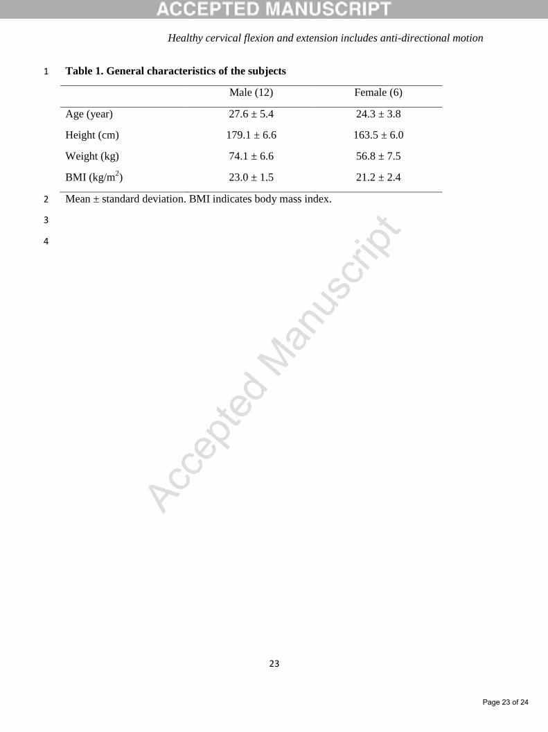
The study included eighteen participants (6 females) (Table 1). Subjects were excluded in
case of neck pain within the last 3 months, any neck disorders, cervical trauma, possible
pregnancy, rheumatoid arthritis or other inflammatory disorders. The participants were
between the age of 20 to 80 years old, and subjects were recruited from campus and through
public media. Subjects were paid approximate $ 22 per hour for their participation. All
subjects received oral information about the experiment and signed a written informed
consent. The study was conducted according to the Declaration of Helsinki and approved by
the local research ethics committee (N20140004).
Page 6 of 24
Healthy cervical flexion and extension includes anti-directional motion
7
Experimental procedures
Participants performed flexion and extension motions from neutral to end-range when sitting
in a chair with knees, hips and ankles at 90°. Shoulders, elbows and waist were fixed with
straps. For better visual tracking of the occiput subjects were asked to wear custom built
glasses with external markers (metal balls attached to steel wires). The motions of flexion and
extension were free and unrestricted. Subjects were instructed to visually follow a line on the
floor, wall, ceiling, and a cross at eye height (natural position). Compliance to flexion or
extension motion speed of 12 seconds was practiced before recording. Steady neutral and end-
range positions were recorded for 2 s. After the end-range recording subjects retuned to the
neutral position at their own pace. Two flexion motions (flexion 1 & 2) followed by two
extension motions (extension 1 & 2) were recorded and analyzed in this study
(Supplementary 1).
Fluoroscopic recordings
Fluoroscopy videos of the cervical spine were recorded from the left side with 25 frames per
second, with the source-to-subject (C7) distance of 76 cm (Philips BV Libra, 2006,
Netherland), with 45 KV, 208 mA, 6.0 ms X-ray pulses and the videos were digitalized
(Honestech VHS to DVD 3.0 SE). The average radiation exposure was estimated to be 0.48
mSv (PCXMC software, STUK, Helsinki, Finland).
Page 7 of 24
Healthy cervical flexion and extension includes anti-directional motion
8
Image analysis
Videos were divided into 10% epochs with respect to the C0/C7 ROM from neutral to end-
range positions. Two images on either end of the exact 10% C0/C7 epoch were selected for
linear interpolation. Neutral position, end-range position, and nine interpolated images yielded
cervical flexion or extension joint motion for the joints C0/C1 to C6/C7.
Images were manually marked on a high-resolution screen with 22 osseous points for
C1 to C7 and 4 external points for C0 (Supplementary 2). Occiput (C0) was marked with 2
anterior and 2 posterior external markers (steel balls). The centers of anterior and posterior
medullary cavities of atlas (C1) were marked. The inferior plate of axis (C2) was marked with
two points at the endplate. Likewise, there were two points at the superior endplate of C7. The
superior and inferior endplates of third to the sixth vertebra (C3-C6) were marked with four
points [1,25]. Joint motion was analyzed in a MATLAB-based program. The program
calculated a representative mid-plane of C0 to C7 and calculated the joint motion in degrees
between two adjacent midplanes [1,25]. The manual marking of vertebrae and the change in
joint motion were previously published as supplementary material [25]. Positive numbers
show the joint opens anteriorly, and negative numbers that the joint opens posteriorly and
zero for no change in joint opening. Investigator XW marked five images three times to test
intra-rater reliability (upright, mid-range flexion and extension, end-range flexions and
extension).
Page 8 of 24
Healthy cervical flexion and extension includes anti-directional motion
9
Motion analysis
Anti-directional joint motion was defined as opposite motion to the intended motion direction
(pro-directional). Joint motion in each of the ten epochs was calculated as the difference in
degrees between two adjacent 10% interpolated images.
Two flexions and two extensions yielded ten joint motion angles for each joint from
C0/C1 to C6/C7 and seventy joint motion angles for each flexion or extension. The two
repeated flexions and extensions were averaged into 70 joint motion angles before
calculations of anti- and pro-directional motions. Cervical actual range of C0/C7 motion was
the sum of the 70 joint motion angles. Cervical anti-directional C0/C7 motion was the sum of
negative numbers among the 70 joint motion angles. Cervical pro-directional C0/C7 motion
was the sum of positive numbers among the 70 joint motion angles. Likewise, pro-directional
or anti-directional joint motion angles within a particular joint across all epochs or within an
epoch across all joints were extracted. The ratio between anti-directional and pro-directional
motions was extracted (0% mean no anti-directional movement).
Statistical analysis
Normal distribution was tested with Shapiro Wilk test and a Q-Q plot. Ratios between anti-
directional and pro-directional joint motions were compared separately for flexion and
extension with one-way ANOVA. Ratios of 10% epochs were compared separately for
flexion and extension with one-way repeated measured ANOVA. Comparisons of ratios
between joints and movement type were performed by mixed-model ANOVA with joint
(C0/C1 to C6/C7) as between-subject factor and movement (flexion, extension) as within-
Page 9 of 24
Healthy cervical flexion and extension includes anti-directional motion
10
subject factor. Comparisons of the ratio between 10% epochs were tested with two-way
repeated measured ANOVA with epoch (from 10% to 100%) and movement (flexion,
extension) as within-subject factors. The assumption of sphericity was tested with Mauchly’s
test, if sphericity was not found a Greenhouse-Geisser correction was applied [26,27]. The
measurement errors assessed in five subjects were presented as mean (SD) and intra-class
correlation coefficient (ICC 3,1). Significant ANOVA factors or interactions were tested with
Tukey’s post-hoc test. Significance was set at P<0.05. Statistical analysis was performed in
SPSS (version 22, IBM).
This study was supported by University with $ 2,000.
RESULTS
The analysis included two times seventy 10% epochs from repeated flexion and extension
recordings from 18 subjects with a total of 5040 10% joint epochs. Low image quality of
C5/C6 and C6/C7 excluded two subjects from the analysis.
The intra-rater measurement errors and ICC of the five images were for the neutral
position 0.14 ± 0.49° and 0.998, for the mid-range epoch in flexion 0.42 ± 1.35° and 0.973,
for the mid-range epoch in extension 0.13 ± 1.19° and 0.989, for end-range flexion 0.01 ±
0.87° and 0.996, and for end-range extension 0.00 ± 0.90° and 0.990, respectively. The
measurement errors were normal distributed.
Cervical motion pattern
Page 10 of 24
Healthy cervical flexion and extension includes anti-directional motion
11
The cervical motion patterns were diverse and illustrated the scattered anti-directional motion
within the pro-directional motion. A representative motion pattern is illustrated in Fig. 1 & 2
where the maximal C2/C3 flexion ROM was reached in the 6th
epoch (Fig. 1) and
C0/C1moves anti-directional in flexion during extension with maximum anti-directional
ROM in the 8th
epoch (Fig 2).
Cervical flexion
The average cervical C0/C7 flexion ROM was 51.9 ± 9.3°. The total C0/C7 anti-directional
flexion was 39.9 ± 14.3°, and the total C0/C7 pro-directional flexion was 91.9 ± 16.3°. The
anti-directional movements constituted 42.8 ± 9.7% of the pro-directional movements. Thus,
the flexion motion consists of approximately 76.9% anti-directional motion of resultant
motion.
The upper cervical joints (C0/C1, C1/C2, C2/C3) showed higher ratios of anti-
directional motions compared with lower cervical joints (C4/C5, C5/C6, and C6/C7)
(ANOVA: F [6, 119] = 14.02; P<0.001). Post-hoc analysis showed that the ratio was larger
for C0/C1 compared with C3/C4, C4/C5, C5/C6, and C6/C7 (Fig. 3; Tukey: P<0.005), and for
C1/C2 compared with C4/C5, C5/C6, and C6/C7 (Fig. 3; Tukey: P<0.02), C2/C3 compared
with C4/C5, C5/C6, and C6/C7 (Fig. 3; Tukey: P<0.001), respectively.
The ratios of anti-directional motion were not different between 10% epochs except for
the 10th
epoch which showed larger anti-directional motion (Mauchly’s test X2(44) = 48.29,
P=0.35. RM-ANOVA: F [9, 153] = 13.3; P<0.002) and post-hoc analysis revealed that the
ratio was larger for the 10th
epoch compared to 2nd
to 9th
epochs (Fig. 4; Tukey: P<0.05).
Page 11 of 24
Healthy cervical flexion and extension includes anti-directional motion
12
Cervical extension
The cervical C0/C7 extension ROM was average 57.3 ± 12.1°, with an average of 40.2 ± 10.8°
anti-directional motion and 91.9 ± 16.3° pro-directional motion. The ratio between anti- and
pro-directional motions was 41.2 ± 8.2%. Thus, the ratio between anti-directional motion and
resultant motion was about 71.9%.
The joints C1/C2 and C2/C3 showed more anti-directional motions the ratios between
anti- and pro-directional motions (ANOVA: F [6, 119] = 10.09; P<0.001). Post-hoc analysis
indicated the ratio was larger for C1/C2 compared with C0/C1, C3/C4, C4/C5, and C5/C6
(Fig. 3; Tukey: P<0.001) and also larger for C2/C3 compared with C0/C1, C3/C4, C4/C5 and
C5/C6 (Fig. 3; Tukey: P<0.03).
The first and last 10% epoch showed more anti-directional motions (Mauchly’s test
X2(44) = 44.66, P=0.50. RM-ANOVA: F [9, 153] = 4.30; P<0.001). Post-hoc analysis showed
larger ratio of anti-directional motion for the 1st epoch compared with the 2
nd, 3
rd and 4
th
epochs (Fig. 4; Tukey: P<0.04) and larger ratios for the 10th
epoch compared with the 2nd
, 3rd
and 4th
epochs (Fig. 4; Tukey: P<0.03).
Comparison of flexion and extension
Comparing ratios of anti-directional joint motion of joints between flexion and extension
showed there was a significant interaction between movement and joints (Mixed ANOVA: F
[6, 119] = 8.14< 0.001). Following up the post hoc analysis showed the larger ratio of C0/C1
for flexion compared to extension (Fig. 3; Tukey: P<0.002) and C5/C6 and C6/C7 showed
larger ratio for extension compared to flexion (Fig. 3; Tukey: P<0.05).
Page 12 of 24
Healthy cervical flexion and extension includes anti-directional motion
13
Ratios of the 1st and last epochs were lager compared to other epochs. There was a
significant main effects of epochs on the ratios of anti-directional motion (Mauchly’s test
X2(44) = 31.00, P=0.94. RM-ANOVA: F [9, 153] = 12.22; P<0.001). Post hoc analysis
showed larger ratios for the 1st epoch compared to the 2
nd, 3
rd and 4
th epochs and also larger
ratios of the last epoch compared to epochs from the 2nd
to the 9th
(Fig. 4; Tukey: P<0.05).
However, there was no significant interaction or main effects of movement with Greenhouse-
Geisser corrections.
DISCUSSION
Anti-directional motions were frequent and scattered through healthy cervical flexion and
extension motion. This study quantifies the average anti-directional motion of cervical spine
to approximately 40 degrees in flexion and extension with a ratio between anti- and pro-
directional motions of approximately 40%.
Representative sample of motion pattern
Figure 1 & 2 show representative samples of cervical motion patterns found in this study. The
patterns show large variations in pro- and anti-directional motions, this variation was found
both within and between joints. The figures further show, that several joints reached the
maximum joint motion excursion before end-range of neck motion. This diversity in motion
patterns was not possible without the scattered anti-directional motion.
The method of image acquisition appears to influence anti-directional motions. Branney
et al [6]. showed a representative subject with control of head and neck motion with a pivot
Page 13 of 24
Healthy cervical flexion and extension includes anti-directional motion
14
mechanism, this subject showed smaller amounts of anti-directional motion in contrast to the
free and unrestricted motions applied in this study and in another study by Reinartz et al [11].
Cervical anti-directional motion
Anti-directional joint motions have previous been reported for cervical joints; however the
reports have not given quantified descriptions [6,10,12]. The unique anatomy of the upper
cervical spine (C0-C2) has previously explained why C1 flexes when the neck is extending,
and vice versa [19]. Without specifying the joints levels Craine et al. reported cases with
flexion of the upper part of the lower cervical spine, while the lower part of the lower cervical
spine extends, and vice versa [12]. The rational for healthy anti-directional motion is
unknown; however, factors which influence cervical ROM may also influence the proportions
of pro- and anti-directional motions. Multiple factors such as cervical anatomy [19], posture
[33], biomechanics [34], motor control [35], position sense [36] and cervical proprioception
[37] influence healthy cervical ROM.
Deep cervical muscles
The deep cervical muscles are the only muscles, which can control anti-directional motions
between adjacent joints. Thus, anti-directional motion may be associated with the muscle
activity of the deep cervical muscles. Pain decreases the muscle activity of the deep cervical
muscles and increases the muscle activity of superficial muscles [38], and pain [39], whiplash
[40] or age [41–43] also reduces cervical ROM. However, the association between cervical
pain and anti-directional motion is unknown.
Page 14 of 24
Healthy cervical flexion and extension includes anti-directional motion
15
Study limitations
The actual ROM of C0/C1to C6/C7 is similar to previously published studies [5,24,36,44].
However, sixteen of the subjects were between the age of 20 to 30 years and 6 of the 18
subjects were female. Larger studies of age and sex differences is necessary to clarify, how
much the proportion of anti-directional motion is influenced by age or sex. Previous studies
show that the cervical spine degenerates by age [29,45]. Women are reported to have larger
cervical ROMs compared with males [41]. Other demographic or anatomical characteristics
such as curvature, long, thin, short and fat neck may also influence the cervical ROM and the
proportion of anti-directional motions [46].
Manual analysis of video-fluoroscopy has several limitations, the largest confounder is
the measurement error; however, this analysis method is in agreement with other previous
studies showing good reliability by high ICCs [1,9,24]. Out of plane sagittal motion is another
confounder. Subjects were asked to follow a central line in order to reduce out of plane
motions. Straps applied to control upper thoracic spine movement may reduce freedom of
movements in the cervical spine. Upper thoracic spine and C7/T1 motion were not included in
the study.
Clinical implications
The concept of normal and healthy anti-directional cervical motions challenges the current
understanding of cervical motion in normative and diagnostic studies. An example is cervical
joint assessment from palpation in tactile tests, and the gold standard interpretation does not
include anti-directional motion. Pro-directional joint motion is recognized as normal, whereas
no motion or anti-directional motion is considered a potential neck problem [47–51].
Page 15 of 24
Healthy cervical flexion and extension includes anti-directional motion
16
However, healthy subjects demonstrate anti-directional motion and the present study suggests
changing the current concepts of healthy cervical biomechanics to include anti-directional
motions.
The present results also challenge the interpretation of healthy motion on cervical
flexion and extension roentgen images. The common perception is that each joint should
contribute to the resultant end-range motion. Flexion and extension roentgen images may not
reflect the motion before end-range. The study indicates that considerable pro- and anti-
directional flexion or extension joint motion may be present before end-range without any
signs of this motion at end-range. The common interpretation of flexion and extension
roentgen images is further challenged, when a small proportion of healthy joint moves
predominantly anti-directional and contributes with anti-directional motion to the resultant
end-range motion.
Conclusion
This is the first study to quantify anti-directional motions in flexion and extension. Anti-
directional motions were scattered in large amounts throughout cervical flexion and extension.
This study indicates that unidirectional hypo-mobility should have decreased value in clinical
motion assessment compared to the present day standard. A better understanding of anti-
directional motion may provide clinicians with better biomechanical information for diagnosis
and rehabilitation.
Page 16 of 24
Healthy cervical flexion and extension includes anti-directional motion
17
REFERENCES
[1] Frobin W, Leivseth G, Biggemann M, Brinckmann P. Sagittal plane segmental motion of the
cervical spine. A new precision measurement protocol and normal motion data of healthy
adults. Clin Biomech 2002;17:21–31. doi:10.1016/S0268-0033(01)00105-X.
[2] Stemper BD, Yoganandan N, Pintar F a. Gender dependent cervical spine segmental
kinematics during whiplash. J Biomech 2003;36:1281–9. doi:10.1016/S0021-9290(03)00159-3.
[3] Auerbach JD. Segmental Contribution Toward Total Cervical Range of Motion. Spine (Phila
Pa 1976) 2011;36:1593–9. doi:10.1097/BRS.0b013e31821cfd47.
[4] Ahmadi A, Maroufi N, Behtash H, Zekavat H, Parnianpour M. Kinematic analysis of dynamic
lumbar motion in patients with lumbar segmental instability using digital videofluoroscopy.
Eur Spine J 2009;18:1677–85. doi:10.1007/s00586-009-1147-x.
[5] Anderst WJ, Donaldson WF, Lee JY, Kang JD. Cervical Motion Segment Percent
Contributions to Flexion-Extension During Continuous Functional Movement in Control
Subjects and Arthrodesis Patients. Spine (Phila Pa 1976) 2013;38:E533–9.
doi:10.1097/BRS.0b013e318289378d.
[6] Branney J, Breen AC. Does inter-vertebral range of motion increase after spinal manipulation?
A prospective cohort study. Chiropr Man Therap 2014;22:24. doi:10.1186/s12998-014-0024-9.
[7] Dvorak J, Froehlich D, Penning L, Baumgartner H, Panjabi MM. Functional radiographic
diagnosis of the cervical spine: flexion/extension. Spine (Phila Pa 1976) 1988;13:748–55.
[8] Bogduk N, Amevo B, Pearcy M. A biological basis for instantaneous centres of rotation of the
vertebral column. Proc Inst Mech Eng H 1995;209:177–83.
[9] Wu SK, Kuo LC, Lan HCH, Tsai SW, Chen CL, Su FC. The quantitative measurements of the
intervertebral angulation and translation during cervical flexion and extension. Eur Spine J
2007;16:1435–44. doi:10.1007/s00586-007-0372-4.
[10] Anderst WJ, Donaldson WF, Lee JY, Kang JD. Cervical motion segment contributions to head
motion during flexion\extension, lateral bending and axial rotation. Spine J 2015;15:2538–43.
doi:10.1016/j.spinee.2015.08.042.
[11] Reinartz R, Platel B, Boselie T, Van Mameren H, Van Santbrink H, Romeny BTH. Cervical
vertebrae tracking in video-fluoroscopy using the normalized gradient field. Med. image
Comput. Comput. Interv., vol. 12, 2009, p. 524–31.
[12] Craine JG, Jenkins KA, Shull MG, Rist D. Cervical spine segmental motion and its relationship
to cervical spine facet angulation. Chesterfield, MO Logan Coll Chiropractic, c1993
1993;1:12–3.
[13] Ombregt L. Applied anatomy of the cervical spine. A Syst. Orthop. Med., Elsevier; 2013, p.
e1–12. doi:10.1016/B978-0-7020-3145-8.00060-0.
[14] Thambiah J, Philips GO, Philips GO, Nather A PG. Anatomy of the spine. Sci. Basis Tissue
Transplant., World Scientific Pub Co Pte Lt; 2001, p. 42–50.
[15] O’Leary S, Falla D, Elliott JM, Jull G. Muscle dysfunction in cervical spine pain: implications
for assessment and management. J Orthop Sports Phys Ther 2009;39:324–33.
doi:10.2519/jospt.2009.2872.
[16] Mayoux Benhamou M, Mayoux-Benhamou M, Revel M et al. Rôle postural du muscle long
Page 17 of 24
Healthy cervical flexion and extension includes anti-directional motion
18
du cou. Surg Radiol Anat 1994;16:367–71.
[17] Boyd-Clark LC, Briggs CA, Galea MP. Muscle spindle distribution, morphology, and density
in longus colli and multifidus muscles of the cervical spine. Spine (Phila Pa 1976)
2002;27:694–701.
[18] Van Mameren H, Drukker J, Sanches H, Beursgens J. Cervical spine motion in the sagittal
plane (I) range of motion of actually performed movements, an X-ray cinematographic study.
Eur J Morphol 1990;28:47–68.
[19] Swartz EE, Floyd RT, Cendoma M. Cervical spine functional anatomy and the biomechanics
of injury due to compressive loading. J Athl Train 2005;40:155–61.
[20] Tseng YL, Wang WTJ, Chen WY, Hou TJ, Chen TC, Lieu FK. Predictors for the immediate
responders to cervical manipulation in patients with neck pain. Man Ther 2006;11:306–15.
doi:10.1016/j.math.2005.08.009.
[21] Bronfort G, Evans R, Nelson B, Aker PD, Goldsmith CH, Vernon H. A randomized clinical
trial of exercise and spinal manipulation for patients with chronic neck pain. Spine (Phila Pa
1976) 2001;26:788–97. doi:10.1097/00007632-200104010-00020.
[22] Carlesso LC, Gross AR, Santaguida PL, Burnie S, Voth S, Sadi J. Adverse events associated
with the use of cervical manipulation and mobilization for the treatment of neck pain in adults:
A systematic review. Man Ther 2010;15:434–44. doi:10.1016/j.math.2010.02.006.
[23] Schneider M, Weinstein S, Chimes GP. Cervical Manipulation for Neck Pain. PM R
2012;4:606–12. doi:10.1016/j.pmrj.2012.07.003.
[24] Wu S-K, Kuo L-C, Lan H-CH, Tsai S-W, Su F-C. Segmental percentage contributions of
cervical spine during different motion ranges of flexion and extension. J Spinal Disord Tech
2010;23:278–84. doi:10.1097/BSD.0b013e3181a98d26.
[25] Wang X, Lindstroem R, Carstens NPB, Graven-Nielsen T. Cervical spine reposition errors
after cervical flexion and extension. BMC Musculoskelet Disord 2017;18:102.
doi:10.1186/s12891-017-1454-z.
[26] Kleinbaum DG, Kupper LL MK. Applied Regression Analysis and Other Multivariable
Methods. 2nd ed. Boston: PWS Publishing Co; 1988.
[27] Andy Field. Discovering statistics using IBM SPSS statistics. 2nd ed. London: SAGE
Publications Ltd; 2005.
[28] Boos N, Weissbach S, Rohrbach H, Weiler C, Spratt KF, Nerlich AG. Classification of age-
related changes in lumbar intervertebral discs. Spine (Phila Pa 1976) 2002;27:2631–44.
doi:10.1097/01.BRS.0000035304.27153.5B.
[29] Siemionow K, An H, Masuda K, Andersson G, Cs-Szabo G. The Effects of Age, Sex, Ethnicity,
and Spinal Level on the Rate of Intervertebral Disc Degeneration. Spine (Phila Pa 1976)
2011;36:1333–9. doi:10.1097/BRS.0b013e3181f2a177.
[30] Pickett GE, Rouleau JP, Duggal N. Kinematic analysis of the cervical spine following
implantation of an artificial cervical disc. Spine (Phila Pa 1976) 2005;30:1949–54.
doi:10.1097/01.brs.0000176320.82079.ce.
[31] Huang RC, Tropiano P, Marnay T, Girardi FP, Lim MR, Cammisa FP. Range of motion and
adjacent level degeneration after lumbar total disc replacement. Spine J 2006;6:242–7.
Page 18 of 24

Healthy cervical flexion and extension includes anti-directional motion
19
doi:10.1016/j.spinee.2005.04.013.
[32] Dvorak J, Antinnes JA, Panjabi M, Loustalot D, Bonomo M. Age and gender related normal
motion of the cervical spine. Spine (Phila Pa 1976) 1992;17:S393-8.
[33] Lee M-Y, Lee H-Y, Yong M-S. Characteristics of cervical position sense in subjects with
forward head posture. J Phys Ther Sci 2014;26:1741–3. doi:10.1589/jpts.26.1741.
[34] Anderst WJ, Donaldson WF, Lee JY, Kang JD. Cervical spine intervertebral kinematics with
respect to the head are different during flexion and extension motions. J Biomech 2013.
doi:10.1016/j.jbiomech.2013.03.004.
[35] Meisingset I, Stensdotter AK, Woodhouse A, Vasseljen O. Neck motion, motor control, pain
and disability: A longitudinal study of associations in neck pain patients in physiotherapy
treatment. Man Ther 2016;22:94–100. doi:10.1016/j.math.2015.10.013.
[36] Wibault J, Vaillant J, Vuillerme N, Dedering Å, Peolsson A. Using the cervical range of
motion (CROM) device to assess head repositioning accuracy in individuals with cervical
radiculopathy in comparison to neck- healthy individuals. Man Ther 2013;18:403–9.
doi:10.1016/j.math.2013.02.004.
[37] Artz NJ, Adams M a., Dolan P. Sensorimotor function of the cervical spine in healthy
volunteers. Clin Biomech 2015;30:260–8. doi:10.1016/j.clinbiomech.2015.01.005.
[38] Falla DL, Jull GA, Hodges PW. Patients with neck pain demonstrate reduced
electromyographic activity of the deep cervical flexor muscles during performance of the
craniocervical flexion test. Spine (Phila Pa 1976) 2004;29:2108–14.
[39] Lee H, Nicholson LL, Adams RD. Cervical range of motion associations with subclinical neck
pain. Spine (Phila Pa 1976) 2004;29:33–40. doi:10.1097/01.BRS.0000103944.10408.BA.
[40] Dall’Alba PT, Sterling MM, Treleaven JM, Edwards SL, Jull GA. Cervical range of motion
discriminates between asymptomatic persons and those with whiplash. Spine (Phila Pa 1976)
2001;26:2090–4.
[41] Castro WH, Sautmann A, Schilgen M, Sautmann M. Noninvasive three-dimensional analysis
of cervical spine motion in normal subjects in relation to age and sex. An experimental
examination. Spine (Phila Pa 1976) 2000;25:443–9.
[42] Sforza C, Grassi G, Fragnito N, Turci M, Ferrario V. Three-dimensional analysis of active head
and cervical spine range of motion: effect of age in healthy male subjects. Clin Biomech
(Bristol, Avon) 2002;17:611–4.
[43] Malmström E-M, Karlberg M, Fransson PA, Melander A, Magnusson M. Primary and coupled
cervical movements: the effect of age, gender, and body mass index. A 3-dimensional
movement analysis of a population without symptoms of neck disorders. Spine (Phila Pa 1976)
2006;31:E44-50.
[44] Schoen DC ed. Adult Orthopedic Nursing. 1st ed. Philadelphia: Lippincott Williams and
Wilkins; 2000.
[45] Weiler C, Schietzsch M, Kirchner T, Nerlich AG, Boos N, Wuertz K. Age-related changes in
human cervical, thoracal and lumbar intervertebral disc exhibit a strong intra-individual
correlation. Eur Spine J 2012;21:810–8. doi:10.1007/s00586-011-1922-3.
[46] Reynolds J, Marsh D, Koller H, Zenenr J, Bannister G. Cervical range of movement in relation
Page 19 of 24
Healthy cervical flexion and extension includes anti-directional motion
20
to neck dimension. Eur Spine J 2009;18:863–8. doi:10.1007/s00586-009-0894-z.
[47] Leach R PJ. Segmental dysfunction hypothesis: Joint and muscle pathology and facilitation. In:
Leach R E, editor. Chiropr. Theor. 4th ed., 2005, p. 137–206.
[48] C H. Three neurophysiologic theories on the chiropratic subluxation. Found. Chiropr.
Subluxation. 2nd ed., St. Louis: Elsevier Mosby; 2005, p. 296–303.
[49] Hoving JL, Gross AR, Gasner D, Kay T, Kennedy C, Hondras MA, et al. A critical appraisal of
review articles on the effectiveness of conservative treatment for neck pain. Spine (Phila Pa
1976) 2001;26:196–205.
[50] Cochran T, Irstam L, Nachemson A. Long-term anatomic and functional changes in patients
with adolescent idiopathic scoliosis treated by Harrington rod fusion. Spine (Phila Pa 1976)
1983;8:576–84.
[51] Fernández-de-las-Peñas C. Interaction between Trigger Points and Joint Hypomobility: A
Clinical Perspective. J Man Manip Ther 2009;17:74–7. doi:10.1179/106698109790824721.
Page 20 of 24

21
Figure 1. Show neck flexion from one representative male subject. Pro- and anti-directional 1
motion directions interchanged with occasional larger one-directional deviations. The 2
maximum flexion C2/C3 motion was reached in the 6th
epoch, the maximum motion was 3
4.05°. Maximum flexion motions of C2/C3, C3/C4, C4/C5 and C6/C7 were reached before 4
end-range. Thus, these joints move in anti-directional flexion towards end-range. 5
6
Figure 2. Show neck extension from the subject in figure 1. Likewise, pro- and anti-7
directional motion directions interchanged with larger one-directional deviation. The 8
maximum extension joint motion for C1/C2 and C5/C6 were reached before end-range, and 9
also noted that C0/C1 moves predominantly anti-directional with maximum anti-directional 10
motion in the 8th
epoch. 11
12
Figure 3. Mean (+SD) ratio between anti- and pro-directional joint motion for flexion (light 13
gray bars) and extension (dark gray bars). For flexion, C0/C1 was significantly larger (*, 14
Tukey: P<0.001) compared to C3/C4, C4/C5, C5/C6 and C6/C7 flexion, C1/C2 and C2/C3 15
were significantly larger (¤, Tukey: P<0.02) compared to C4/C5, C5/C6 and C6/C7 flexion. 16
For extension, C1/C2 and C2/C3 were significantly larger (†, Tukey P<0.03) compared to 17
C0/C1, C3/C4, C4/C5 and C5/C6 extension. 18
19
Figure 4. Mean (+SD) ratio between anti- and pro-directional motion in epochs for flexion 20
(light gray bars) and extension (dark gray bars). For flexion, the final epoch showed larger 21
ratio (Ω, Tukey: P<0.05) compared to 2nd
tough 9th
flexion epochs. For extension, the first and 22
Page 21 of 24

Healthy cervical flexion and extension includes anti-directional motion
22
final epochs shower larger ratio (&, Tukey: P<0.04) compared to 2nd
, 3rd
and 4th
extension 1
epochs. 2
3
Page 22 of 24
Healthy cervical flexion and extension includes anti-directional motion
23
Table 1. General characteristics of the subjects 1
Male (12) Female (6)
Age (year) 27.6 ± 5.4 24.3 ± 3.8
Height (cm) 179.1 ± 6.6 163.5 ± 6.0
Weight (kg) 74.1 ± 6.6 56.8 ± 7.5
BMI (kg/m2) 23.0 ± 1.5 21.2 ± 2.4
Mean ± standard deviation. BMI indicates body mass index. 2
3
4
Page 23 of 24
Healthy cervical flexion and extension includes anti-directional motion
24
Supplementary 1 illustrates the experimental procedures. This experiment includes two 1
repeated flexion and extension motions from neutral to end-range position then back to 2
neutral position, however, only motions from neutral to end-range position were recorded by 3
video fluoroscopy and analyzed in this study. The return motions from end-range to neutral 4
position were not recorded. RF: Recorded flexion NRR: Not recorded return RE: Recorded 5
extension. 6
7
Supplementary 2 illustrates marking points for identification of the joint angles. Four lead 8
balls served as external markers for the head (C0), two points on atlas (C1) were identified by 9
the central areas of the medullary cavities of the anterior and posterior arches. For 10
identification of C2 two points of the inferior vertebral plate were marked. The third to the 11
sixth cervical vertebrae (C3-C6) were identified with 4 points of the vertebral corners. The 12
seventh vertebra (C7) was identified with two points in proximity to the superior vertebral 13
plate. The joint angles were derived from the mid-planes calculated from the marking points. 14
Page 24 of 24


![CT Metrizamide Myelography of the Cervical Spine in ... junction with flexion and extension. Chronic atlan toaxial subluxation may develop and cause severe cord compression [4]. Narrowing](https://img.pdfslide.us/doc/110x75/5afabad07f8b9ad2208fa4fa/ct-metrizamide-myelography-of-the-cervical-spine-in-junction-with-flexion-and.jpg)