Embed Size (px)
Citation preview
1
ANGIOMA ALLIANCEAngioma.org
A PATIENT'SGUIDE
Cerebral Cavernous Angioma CavernomaCerebral Cavernous Malformation
20Q
UESTIONS
A N S W E R E D
2 3
WELCOMEYou received this booklet because your doctor diagnosed you or a loved one with at least one cavernous angioma in your brain or spinal cord. Your doctor may have used the terms cerebral cavernous malformation (CCM), cavernoma, or cavernous hemangioma for your diagnosis. These are all names for the same blood vessel abnormality. We will use the terms cavernous angioma and lesion here.Chances are this is the first time you’ve heard of a cavernous angioma. In this booklet, we’ll be offering information to help you make informed decisions about your care. You may also want to share this booklet with the important people in your life to help them gain an understanding of cavernous angiomas.There is no typical way in which cavernous angiomas affect people. The symptoms, the course of the illness, and its severity can be very different from person to person. This booklet offers general information and does not replace medical advice from your doctor. We base our expert information on clinical management guidelines created by the Angioma Alliance Scientific Advisory Board Clinical Experts Panel. The Angioma Alliance website at Angioma.org contains extensive additional information.
4 5
4
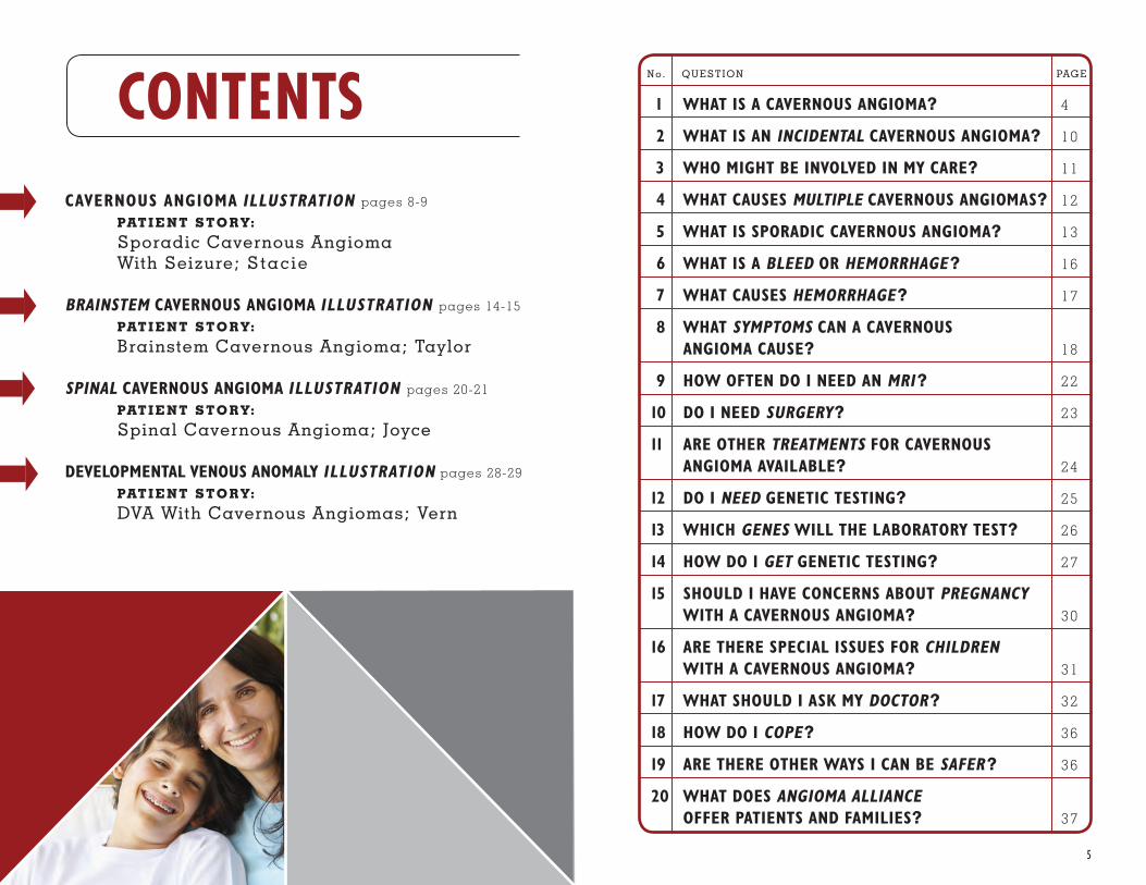
CONTENTSCAVERNOUS ANGIOMA ILLUSTRATION pages 8-9
PAT I E N T S T O R Y:
Sporadic Cavernous Angioma With Seizure; S tac ie BRAINSTEM CAVERNOUS ANGIOMA ILLUSTRATION pages 14-15
PAT I E N T S T O R Y:
Brainstem Cavernous Angioma; Taylor SPINAL CAVERNOUS ANGIOMA ILLUSTRATION pages 20-21
PAT I E N T S T O R Y:
Spinal Cavernous Angioma; Joyce DEVELOPMENTAL VENOUS ANOMALY ILLUSTRATION pages 28-29
PAT I E N T S T O R Y: DVA With Cavernous Angiomas; Vern
1 WHAT IS A CAVERNOUS ANGIOMA? 4
2 WHAT IS AN INCIDENTAL CAVERNOUS ANGIOMA? 10
3 WHO MIGHT BE INVOLVED IN MY CARE? 11
4 WHAT CAUSES MULTIPLE CAVERNOUS ANGIOMAS? 12
5 WHAT IS SPORADIC CAVERNOUS ANGIOMA? 13
6 WHAT IS A BLEED OR HEMORRHAGE?? 16
7 WHAT CAUSES HEMORRHAGE? 17
8 WHAT SYMPTOMS CAN A CAVERNOUS ANGIOMA CAUSE? 18
9 HOW OFTEN DO I NEED AN MRI? 22
10 DO I NEED SURGERY? 23
11 ARE OTHER TREATMENTS FOR CAVERNOUS ANGIOMA AVAILABLE? 24
12 DO I NEED GENETIC TESTING? 25
13 WHICH GENES WILL THE LABORATORY TEST? 26
14 HOW DO I GET GENETIC TESTING? 27
15 SHOULD I HAVE CONCERNS ABOUT PREGNANCY WITH A CAVERNOUS ANGIOMA? 30
16 ARE THERE SPECIAL ISSUES FOR CHILDREN WITH A CAVERNOUS ANGIOMA? 31
17 WHAT SHOULD I ASK MY DOCTOR? 32
18 HOW DO I COPE? 36
19 ARE THERE OTHER WAYS I CAN BE SAFER? 36
20 WHAT DOES ANGIOMA ALLIANCE OFFER PATIENTS AND FAMILIES? 37
No. QUESTION PAGE
6 7
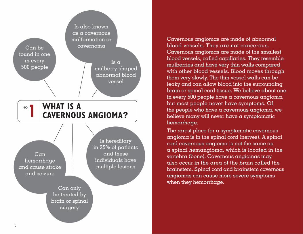
Can only be treated by
brain or spinal surgery
Is a mulberry-shaped abnormal blood
vessel
Is hereditary in 25% of patients
and these individuals have multiple lesions
Can be found in one
in every 500 people
Is also known as a cavernous malformation or
cavernoma
Can hemorrhage
and cause stroke and seizure
WHAT IS ACAVERNOUS ANGIOMA?
NO. 1
Cavernous angiomas are made of abnormal blood vessels. They are not cancerous. Cavernous angiomas are made of the smallest blood vessels, called capillaries. They resemble mulberries and have very thin walls compared with other blood vessels. Blood moves through them very slowly. The thin vessel walls can be leaky and can allow blood into the surrounding brain or spinal cord tissue. We believe about one in every 500 people have a cavernous angioma, but most people never have symptoms. Of the people who have a cavernous angioma, we believe many will never have a symptomatic hemorrhage. The rarest place for a symptomatic cavernous angioma is in the spinal cord (nerves). A spinal cord cavernous angioma is not the same as a spinal hemangioma, which is located in the vertebra (bone). Cavernous angiomas may also occur in the area of the brain called the brainstem. Spinal cord and brainstem cavernous angiomas can cause more severe symptoms when they hemorrhage.
8 9
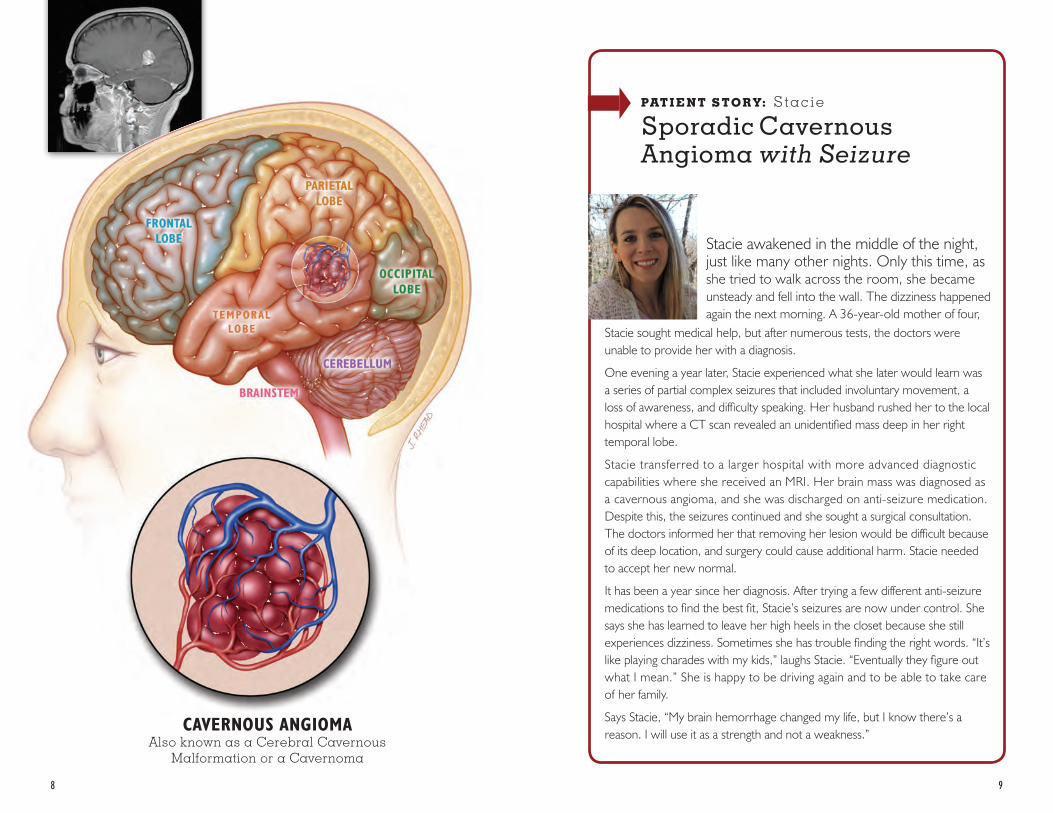
CAVERNOUS ANGIOMAAlso known as a Cerebral Cavernous
Malformation or a Cavernoma
PAT I E N T S T O R Y: Stac ie
Sporadic Cavernous Angioma with Seizure
Stacie awakened in the middle of the night, just like many other nights. Only this time, as she tried to walk across the room, she became unsteady and fell into the wall. The dizziness happened again the next morning. A 36-year-old mother of four,
Stacie sought medical help, but after numerous tests, the doctors were unable to provide her with a diagnosis.
One evening a year later, Stacie experienced what she later would learn was a series of partial complex seizures that included involuntary movement, a loss of awareness, and difficulty speaking. Her husband rushed her to the local hospital where a CT scan revealed an unidentified mass deep in her right temporal lobe.
Stacie transferred to a larger hospital with more advanced diagnostic capabilities where she received an MRI. Her brain mass was diagnosed as a cavernous angioma, and she was discharged on anti-seizure medication. Despite this, the seizures continued and she sought a surgical consultation. The doctors informed her that removing her lesion would be difficult because of its deep location, and surgery could cause additional harm. Stacie needed to accept her new normal.
It has been a year since her diagnosis. After trying a few different anti-seizure medications to find the best fit, Stacie’s seizures are now under control. She says she has learned to leave her high heels in the closet because she still experiences dizziness. Sometimes she has trouble finding the right words. “It’s like playing charades with my kids,” laughs Stacie. “Eventually they figure out what I mean.” She is happy to be driving again and to be able to take care of her family.
Says Stacie, “My brain hemorrhage changed my life, but I know there’s a reason. I will use it as a strength and not a weakness.”
10 11
Your doctor may have discov-ered your cavernous angioma when you had brain or spinal imaging for a reason not related to your cavernous angioma. For example, you may have received a CT scan or an MRI after a car accident or concus-sion. In this case, doctors will call your cavernous angioma an incidental finding. Research has shown that incidental cavernous angiomas that have not had a previous hemorrhage have a very small chance of
WHAT IS AN INCIDENTAL CAVERNOUS ANGIOMA?
NO. 2
ever becoming problematic. While you may want to follow the precautions listed in the Hemorrhage section of this book-let, you may not need repeat imaging unless instructed by your doctor or you develop symp-toms. However, many doctors and patients choose to have imaging, typically with MRI, to monitor the lesion. For example, your doctor may order imaging during pregnancy, after medication changes, if another illness is diagnosed, or at times of dramatic lifestyle change, which can include ex-treme or unusual exercise.
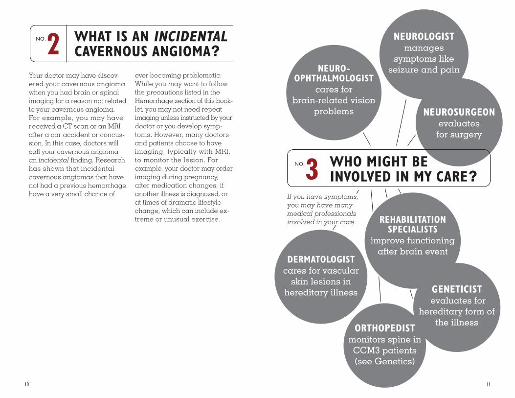
NEUROSURGEON evaluates for surgery
If you have symptoms, you may have many medical professionals involved in your care. REHABILITATION
SPECIALISTSimprove functioning
after brain event
ORTHOPEDISTmonitors spine in CCM3 patients (see Genetics)
NEURO- OPHTHALMOLOGIST
cares for brain-related vision
problems
GENETICIST evaluates for
hereditary form of the illness
DERMATOLOGIST cares for vascular
skin lesions in hereditary illness
WHO MIGHT BE INVOLVED IN MY CARE?
NO. 3
NEUROLOGISTmanages
symptoms like seizure and pain
12 13
Some people have more than one cavernous angioma. This can happen for several reasons:
• Most commonly, people with more than one cavernous angioma have a hereditary form of the illness. People with the genetic form of the illness typically develop additional cavernous angiomas over time.
• Some people may have a second kind of abnormal blood vessel called a developmental venous anomaly (DVA). Your doctor might also call this a venous malformation or venous angioma. This dilated blood vessel only rarely causes symptoms on its own. However, it may create conditions that make it more likely for cavernous angiomas to form. This is not hereditary.
• Brain or spinal radiation for cancer treatment can cause cavernous angiomas to form many years later. In people with the hereditary form, radiosurgery to treat a cavernous angioma may also cause more lesions to form. You can find more information about radiosurgery in the section on Other Treatments in this booklet.
WHAT CAUSES MULTIPLE CAVERNOUS ANGIOMAS?
NO. 4
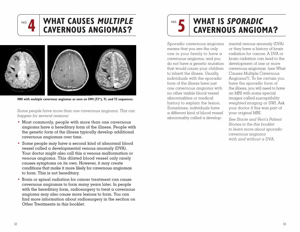
MRI with multiple cavernous angiomas as seen on SWI (T2*), T1, and T2 sequences.
Sporadic cavernous angioma means that you are the only one in your family to have a cavernous angioma, and you do not have a genetic mutation that would cause your children to inherit the illness. Usually, individuals with the sporadic form of the illness have just one cavernous angioma with no other visible blood vessel abnormalities or medical history to explain the lesion. Sometimes, individuals have a different kind of blood vessel abnormality called a develop-
WHAT IS SPORADICCAVERNOUS ANGIOMA?
NO. 5
mental venous anomaly (DVA) or they have a history of brain radiation for cancer. A DVA or brain radiation can lead to the development of one or more cavernous angiomas (see What Causes Multiple Cavernous Angioma?). To be certain you have the sporadic form of the illness, you will need to have an MRI with some special images called susceptibility weighted imaging or SWI. Ask your doctor if this was part of your original MRI.
See Stacie and Vern’s Patient Stories in the this booklet to learn more about sporadic cavernous angioma with and without a DVA.
14 15
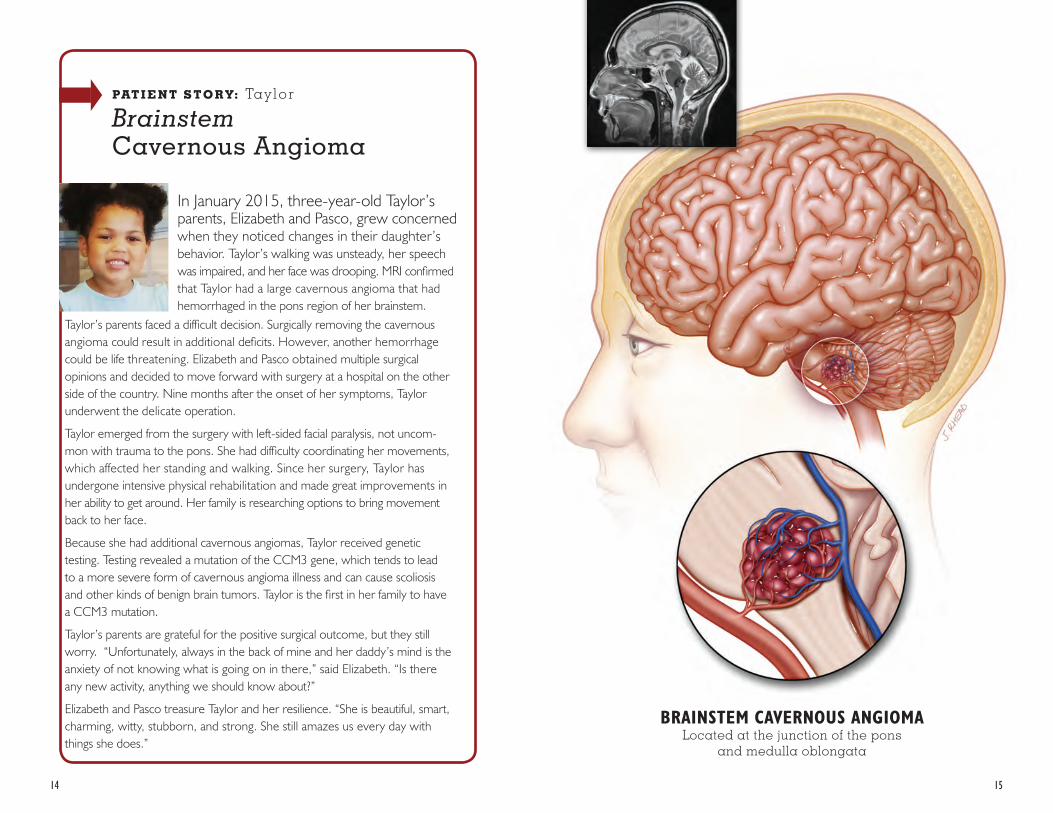
In January 2015, three-year-old Taylor’s parents, Elizabeth and Pasco, grew concerned when they noticed changes in their daughter’s behavior. Taylor’s walking was unsteady, her speech was impaired, and her face was drooping. MRI confirmed that Taylor had a large cavernous angioma that had hemorrhaged in the pons region of her brainstem.
Taylor’s parents faced a difficult decision. Surgically removing the cavernous angioma could result in additional deficits. However, another hemorrhage could be life threatening. Elizabeth and Pasco obtained multiple surgical opinions and decided to move forward with surgery at a hospital on the other side of the country. Nine months after the onset of her symptoms, Taylor underwent the delicate operation.
Taylor emerged from the surgery with left-sided facial paralysis, not uncom-mon with trauma to the pons. She had difficulty coordinating her movements, which affected her standing and walking. Since her surgery, Taylor has undergone intensive physical rehabilitation and made great improvements in her ability to get around. Her family is researching options to bring movement back to her face.
Because she had additional cavernous angiomas, Taylor received genetic testing. Testing revealed a mutation of the CCM3 gene, which tends to lead to a more severe form of cavernous angioma illness and can cause scoliosis and other kinds of benign brain tumors. Taylor is the first in her family to have a CCM3 mutation.
Taylor’s parents are grateful for the positive surgical outcome, but they still worry. “Unfortunately, always in the back of mine and her daddy’s mind is the anxiety of not knowing what is going on in there,” said Elizabeth. “Is there any new activity, anything we should know about?”
Elizabeth and Pasco treasure Taylor and her resilience. “She is beautiful, smart, charming, witty, stubborn, and strong. She still amazes us every day with things she does.”
PAT I E N T S T O R Y: Tay lor
Brainstem Cavernous Angioma
BRAINSTEM CAVERNOUS ANGIOMALocated at the junction of the pons
and medulla oblongata
16
All cavernous angiomas have some chronic oozing of blood in the area of the lesion. This is what gives their typical appearance on MRI. Oozing is different from the more significant “overt” hemorrhage or bleed. An overt hemorrhage is new bleeding in or around the cavernous angioma and is often associated with new symptoms. Symptomatic hemorrhage is the most serious complication of cav-ernous angioma and is the most common reason for surgery. The hemorrhage may cause new symptoms or an increase in symptoms. The specific symptoms a person experiences will depend on the location and size of the cavernous angioma and the amount of blood that has leaked outside the lesion. Some lesions will have slow oozes that produce mild or no symptoms. Eventually, the blood breaks down leaving behind an iron deposit called hemosiderin. Hemorrhage
WHAT IS A BLEED OR HEMORRHAGE ?
NO. 6
risk is greater for those with multiple cavernous angiomas, but it is impossible to predict which of the many lesions might bleed. Many lesions never hemorrhage. However, once a cavernous angioma has had one overt or symptomatic hem-orrhage, it is at significantly greater risk of bleeding again. In the first five years after a hemorrhage, the risk of another hemorrhage averages about 6% per year. This means within 5 years of an original hemor-rhage, 1 in 3 lesions will have a second hemorrhage. Most of these second hemorrhages will take place in the first two years after the original hemorrhage. The percentage of people who have a second symptomatic hemorrhage is even higher for those who have cavernous angiomas in the brainstem. For- tunately, at five years after a hemorrhage, the risk of another hemorrhage gradually returns to less than 2% per year, similar to the risk from a lesion that has never bled.
17
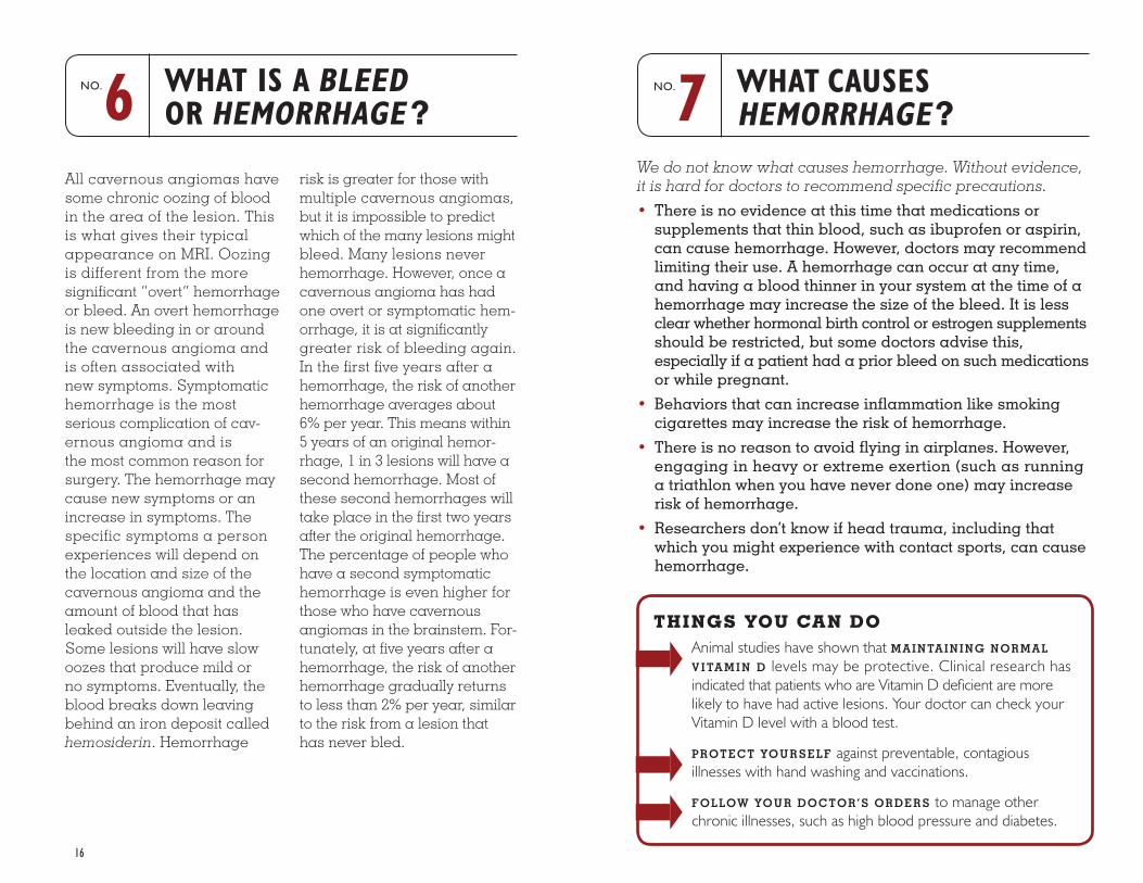
We do not know what causes hemorrhage. Without evidence, it is hard for doctors to recommend specific precautions.
• There is no evidence at this time that medications or supplements that thin blood, such as ibuprofen or aspirin, can cause hemorrhage. However, doctors may recommend limiting their use. A hemorrhage can occur at any time, and having a blood thinner in your system at the time of a hemorrhage may increase the size of the bleed. It is less clear whether hormonal birth control or estrogen supplements should be restricted, but some doctors advise this, especially if a patient had a prior bleed on such medications or while pregnant.
• Behaviors that can increase inflammation like smoking cigarettes may increase the risk of hemorrhage.
• There is no reason to avoid flying in airplanes. However, engaging in heavy or extreme exertion (such as running a triathlon when you have never done one) may increase risk of hemorrhage.
• Researchers don’t know if head trauma, including that which you might experience with contact sports, can cause hemorrhage.
WHAT CAUSES HEMORRHAGE?
NO. 7
THINGS YOU CAN DOAnimal studies have shown that MAINTAINING NORMAL
V I TA M I N D levels may be protective. Clinical research has indicated that patients who are Vitamin D deficient are more likely to have had active lesions. Your doctor can check your Vitamin D level with a blood test.
PROTECT YOURSELF against preventable, contagious illnesses with hand washing and vaccinations.
FOLLOW YOUR DOCTOR’S ORDERS to manage other chronic illnesses, such as high blood pressure and diabetes.
18 19
• Seizures are one of the most common symptoms of cav-ernous angioma. Seizures fall into two general groups: partial seizures that are local to one area of the brain and generalized seizures that involve both sides of the brain. All cavernous angioma seizures begin as partial seizures but some progress to generalized seizures. Neurologists use anti-epilepsy medications to control seizures. However, neurosurgeons have had good results in eliminating seizure with brain surgery if they are able to pinpoint which cavernous angioma is causing the seizures. Surgery is most successful when it occurs within two years of a first seizure. Brainstem and spinal cavernous angiomas do not cause seizures.
• We know people with cavernous angioma experience more frequent headache than other people. A headache does not necessarily mean a new hemorrhage. For the most part, we can’t distinguish a cavernous angioma headache from any other kind of headache. A headache unlike one you have ever experienced or headaches, particularly on the same side or general location as your lesion, may be related to your lesion. A headache may be related to your cavernous angioma if it is unlike one you have ever experienced or if it is in the general location of your lesion.
WHAT SYMPTOMS CAN A CAVERNOUS ANGIOMA CAUSE?
NO. 8
The symptoms of a cavernous angioma hemorrhage depend on its location and size.
Cavernous angiomas can cause attention, memory, social skills, mood, and learning problems, particularly if the lesions are in the frontal, parietal, or temporal lobes, or in the cerebellum, even without obvious bleeding. This is particularly true for individuals with many lesions throughout the brain.
• Cavernous angiomas in many parts of the brain and spinal cord can cause weakness or numbness in arms or legs. In some areas, such as the thalamus, they can also cause pain. A cavernous angioma in the brainstem can cause coordination problems called ataxia or can cause facial paralysis, usually on one side.
• Cavernous angiomas can cause vision problems. There are two kinds of vision problems: those caused by lesions in the occipital lobe of the brain, which affect how visual information is processed and those caused by lesions in the brainstem, which affect how the eyes work.
• A cavernous angioma can cause hearing problems, including loss of hearing and tinnitus, dizziness or nausea, particularly if it is located in or near the cerebellum.
• A cavernous angioma in the medulla, the lowest part of the brainstem, can cause spasms of the diaphragm, which resemble hiccups that don’t go away. More rarely, these can cause swallowing or even breathing problems.
• Cavernous angiomas in the spinal cord can cause prob-lems with bladder and bowel control.
• Cavernous angioma hemorrhages in the brain can cause fatigue. Individuals may complain of fatigue for months to years after a major hemorrhage or brain surgery.
• Spinal cord cavernous angiomas can cause numbness, weakness, paralysis, tingling, burning, or itching. The location and extent of the symptom depend on the level of the spine affected. Spinal cord lesions can also cause difficulty with bladder and bowel control.
20 21
“I had just put my left arm down on the kitchen table when I experienced this intense pain from the tips of my fingers to my elbow,” Joyce told the New York Times in a 2007 interview. “It felt like my whole arm was burning.”
It would be years before doctors accurately diagnosed Joyce with a cavernous angioma in her cervical spinal cord. By that time, the pain had spread to her right arm. Her surgeon told her the lesion was accessible, and Joyce had successful surgery in 2004. The surgery helped to resolve some of Joyce’s pain and to prevent any further escalation.
Joyce also has multiple cavernous angiomas in her brain. This is because of a hereditary genetic change known as the Common Hispanic Mutation. While anyone of any ethnicity can have a hereditary form of cavernous angioma illness, thousands of people who trace their heritage to the original Spanish settlers of New Mexico share this specific genetic mutation. A single ancestor connects them all, and many of these families have remained in New Mexico, including Joyce. No other place in the world has as many affected people.
Joyce has been active in advocacy and in raising awareness for the illness throughout New Mexico. In addition to the New York Times interview, Joyce was central in drafting legislation in New Mexico that created a mandate to educate more doctors. Joyce has presented at patient conferences and worked with media around the state to share her story and the genealogy she has researched. Joyce was happy to discover that neither of her children inherited the illness. In Joyce’s family, the Common Hispanic Mutation has stopped with her, though her work toward further research and a cure continues.
PAT I E N T S T O R Y: Joyce
SpinalCavernous Angioma
21
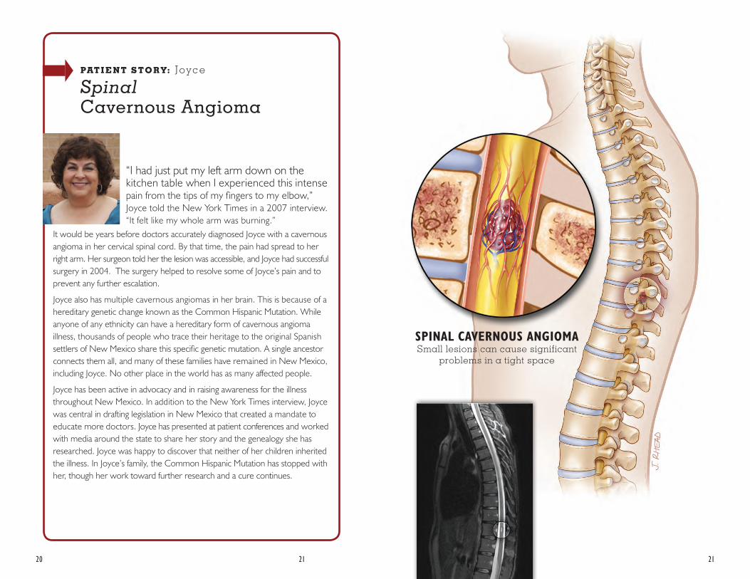
SPINAL CAVERNOUS ANGIOMASmall lesions can cause significant
problems in a tight space
22 23
You and your doctor will decide how often to repeat your MRI. Some doctors advise their patients to have repeat MRIs on a specific schedule. Others suggest waiting until there are additional symptoms Experts suggest more frequent imaging for those who may not be able to report symptoms, like young children or those with intellectual or communication problems. These individuals may also need sedation for MRI. You and your doctor will weigh the risk of sedation against the benefit of imaging.
CT scan is another kind of imaging that is sometimes used. CT scan is much faster than MRI, but the images are not as clear, and CT scan involves exposing a patient to radiation. You may have a CT scan in an emergency when MRI is not available. People with multiple cavernous angiomas should limit their exposure to CT scan as much as possible because it is not clear whether the radiation exposure can cause the development of more lesions.
A doctor may suggest a cerebral angiogram also known as a cerebral arteriogram. This pro-cedure allows the doctor to see the arteries and veins in your brain. A cavernous angioma is not visible on angiogram, but the test is done when another type of blood vessel lesion, called an arteriovenous malfor-mation, is suspected. If a cavernous angioma has a typical appearance on MRI, it does not require a cerebral angio-gram as part of routine care.
HOW OFTEN DO I NEED AN MRI ?
NO. 9
If your cavernous angioma is causing symptoms, you should have a very detailed discussion about surgical options with your neurosurgeon. The decision to have surgery always involves weighing risks and benefits. In general, experts recommend surgery if three criteria are met. First, the individual must have symptoms. Second, the cavern-ous angioma must have had at least two hemorrhages. Finally, removing the cavernous angioma will cause fewer defi-cits than another hemorrhage would. Sometimes, experts recommend surgery after just one hemorrhage. For example, surgery may be a good option for someone with epilepsy caused by a cavernous angioma even if they have only had one hemorrhage.
DO I NEED SURGERY? NO. 10
There are neurosurgeons who specialize in cerebrovascular (brain blood vessel) and skull base (brainstem) surgery. The experience level of a surgeon is the best predictor of a good surgical outcome. The American Academy of Neurological Surgeons offers a searchable directory of neurosurgeons by specialty. It is helpful to interview a number of surgeons, includ-ing asking them about their experience, and to consult with other patients through Angioma Alliance before making decisions.
24 25
Doctors have treated cavern-ous angioma with stereotactic radiosurgery, also known as gamma knife, linear accelerator, X Knife, Brainlab, or cyber knife, for many years, but its effectiveness is not clear. In stereotactic radiosurgery, focused radiation is directed at the cavernous angioma without opening the skull. Experts now recommend that radiosurgery be considered
ARE OTHER TREATMENTS FOR CAVERNOUS ANGIOMA AVAILABLE?
NO. 11
Researchers are working to find medications to treat cavernous angiomas. It may take more than one medication to treat every situation. Medications could be useful to:
• Stabilize the cavernous angioma so it does not hemorrhage. Patients could use medications even before a first hemorrhage. This is particularly true for people with hered-itary forms of the illness.
• Stabilize the cavernous angioma after a hemorrhage to reduce the risk of the lesion bleeding again. Remember, there is a high-risk time in the first years after a hemorrhage. During this time, it might make sense to use a stronger temporary medication.
• Slow or stop the development of more lesions in people who have multiple cavernous angiomas.
• Shrink or destroy existing cavernous angiomas. This is the ultimate goal.
only with individuals who have a single symptomatic lesion that is in an area of the brain where the risks of traditional surgery would be too high. Radio- surgery should not be used for treating cavernous angiomas that do not cause symptoms or for cavernous angioma in peo-ple with the hereditary form of the illness, as the radiation itself might trigger new cavernous angiomas to form.
As of this writing, prescription medications are being tested in clinical drug trials, but none have been FDA-approved for treatment of cavernous angioma. However, as mentioned above, experts recommend having your Vitamin D level checked to make sure it is in the normal range.
Most people with a cavernous angioma do not need genetic testing. We believe only 25% of individuals with a cavernous angioma have a hereditary form of the illness. If you have just one cavernous angioma, you need an MRI with special imaging called susceptibility-weighted imaging (SWI) to rule out the hereditary form. If you have mul- tiple cavernous angiomas but you also have a developmental venous anomaly (DVA), SWI will show whether your lesions cluster near the DVA or whether they are in other areas of your brain. If the cavernous angiomas are only around
DO I NEED GENETIC TESTING?
NO. 12
the DVA, you do not need test-ing. Having multiple cavernous angiomas clustered around a DVA is typical of the sporadic form of cavernous angioma. You can’t inherit or pass down the sporadic form of the illness.
People who have multiple cav- ernous angiomas that are not associated with a developmental venous anomaly should request genetic testing. Some-times people are the first in their family with a hereditary form. You do not need other affected family members to justify testing.
26 27
There are three known genes that, when mutated, can cause a hereditary form of the illness. Researchers have named them CCM1, CCM2, and CCM3. In the United States, individuals who trace their ancestry to the original settlers of New Mexico have passed a specific muta-tion of the CCM1 gene down from generation to generation since at least the early 1600s. Researchers call this the common Hispanic mutation. However, you do not need to be Hispanic to have the he-reditary form of the illness. Hereditary forms of cavernous angioma exist in every ethnic group everywhere in the world. Each child of a person with the hereditary form has a 50/50 chance of inheriting the illness. It does not skip
WHICH GENES WILL THE LABORATORY TEST?
NO. 13
generations. However, up to half of people with a hereditary form may have lesions but have no symptoms.
Individuals with a mutation of the CCM1 or CCM2 gene develop multiple cavernous angiomas and some develop cavernous malformations under the skin. Individuals with a mutation on the CCM3 gene may have additional features. About half of individuals with a mutation on the CCM3 gene have their first hemorrhage as children. They hemorrhage more often and develop new lesions more quickly, at a rate of 2-3 new lesions per year. They may have other physical issues such as scoliosis and benign brain tumors, most commonly meningioma. They are also likely to be the first in their family with the illness. Patients with a CCM3 mutation need additional monitoring and should contact Angioma Alliance to learn more about our special programs.
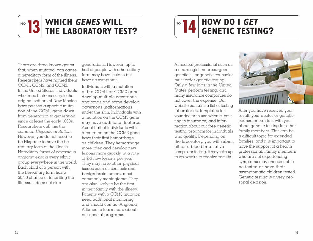
HOW DO I GET GENETIC TESTING?
NO. 14
A medical professional such as a neurologist, neurosurgeon, geneticist, or genetic counselor must order genetic testing. Only a few labs in the United States perform testing, and many insurance companies do not cover the expense. Our website contains a list of testing laboratories, templates for your doctor to use when submit-ting to insurance, and infor-mation about our free genetic testing program for individuals who qualify. Depending on the laboratory, you will submit either a blood or a saliva sample for testing. It may take up to six weeks to receive results.
After you have received your result, your doctor or genetic counselor can talk with you about genetic testing for other family members. This can be a difficult topic for extended families, and it is important to have the support of a health professional. Family members who are not experiencing symptoms may choose not to be tested or have their asymptomatic children tested. Genetic testing is a very per-sonal decision.
28 29
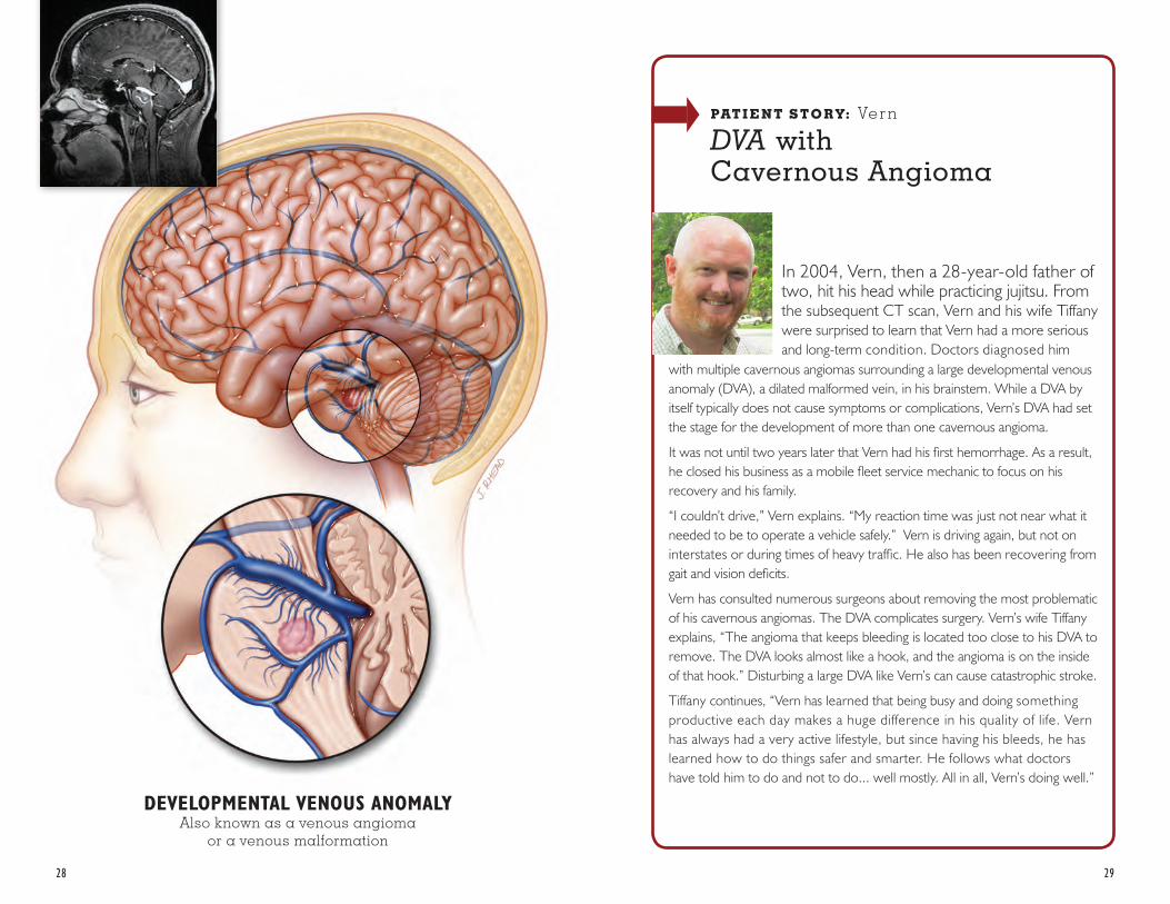
DEVELOPMENTAL VENOUS ANOMALYAlso known as a venous angioma
or a venous malformation
In 2004, Vern, then a 28-year-old father of two, hit his head while practicing jujitsu. From the subsequent CT scan, Vern and his wife Tiffany were surprised to learn that Vern had a more serious and long-term condition. Doctors diagnosed him
with multiple cavernous angiomas surrounding a large developmental venous anomaly (DVA), a dilated malformed vein, in his brainstem. While a DVA by itself typically does not cause symptoms or complications, Vern’s DVA had set the stage for the development of more than one cavernous angioma.
It was not until two years later that Vern had his first hemorrhage. As a result, he closed his business as a mobile fleet service mechanic to focus on his recovery and his family.
“I couldn’t drive,” Vern explains. “My reaction time was just not near what it needed to be to operate a vehicle safely.” Vern is driving again, but not on interstates or during times of heavy traffic. He also has been recovering from gait and vision deficits.
Vern has consulted numerous surgeons about removing the most problematic of his cavernous angiomas. The DVA complicates surgery. Vern’s wife Tiffany explains, “The angioma that keeps bleeding is located too close to his DVA to remove. The DVA looks almost like a hook, and the angioma is on the inside of that hook.” Disturbing a large DVA like Vern’s can cause catastrophic stroke.
Tiffany continues, “Vern has learned that being busy and doing something productive each day makes a huge difference in his quality of life. Vern has always had a very active lifestyle, but since having his bleeds, he has learned how to do things safer and smarter. He follows what doctors have told him to do and not to do... well mostly. All in all, Vern’s doing well.”
PAT I E N T S T O R Y: Vern
DVA withCavernous Angioma
30 31
At this time, experts believe women are no more at risk for hemorrhage during pregnancy than at any other time. But, a bleed during pregnancy can present challenges. Brain surgery to remove a cavernous angioma during pregnancy is very rare, but is undertaken in cases where a second hemorrhage would be life threat-ening. Women with cavernous angioma can have a vaginal delivery if they have not had a recent hemorrhage.
If your doctor suggests an MRI during your pregnancy, your MRI should not include
SHOULD I HAVE CONCERNS ABOUT PREGNANCY WITH A CAVERNOUS ANGIOMA?
NO. 15
gadolinium contrast. Women who have epilepsy and are on anti-epilepsy medications should talk with their doctors about the choice of medication and folate supplementation. Ideally, you should have this conversation before you become pregnant to prevent harm because some anti- epilepsy medication can increase the risk of birth defects when taken in the first weeks of pregnancy. However, having a seizure during pregnancy can cause harm that is far more serious to the fetus. It’s important to continue anti- epilepsy medication during pregnancy if you have had seizures and are pregnant.
There is the potential for many special issues with children, depending on their age at diagno-sis and their symptoms. Young children may not be able to tell you about symptoms, so it becomes important to schedule imaging regularly. On the other hand, it is also important to avoid overreacting so that children aren’t exposed to unnecessary medical procedures, radiation from CT scans, MRI contrast med- ication, and sedation. It is not always possible to strike a bal- ance, and every parent will err in both directions at one point or another. As a child grows older and parents become more experienced, managing this illness becomes easier.
Telling your child about his or her diagnosis can be emotional and can require a period of adjustment for your child. Try to explain in simple terms that are age appropriate. Hav-ing a pediatric mental health professional ready to help can
ARE THERE SPECIAL ISSUES FOR CHILDREN WITH A CAVERNOUS ANGIOMA?
NO. 16
make this easier. A mental health professional can also help if a child with hemorrhage or surgery-related deficits struggles with peer relationships, restrictions on activities, or academics. You will need to ex-plain your child’s illness many times to many professionals including school staff, as well as friends and other caregivers. Good support from other parents in the Angioma Alliance community or your local special needs community can be helpful for you.
32 33
WHAT SHOULD I ASK MY DOCTOR?
NO. 17
If you don’t already know the answers to these questions, you may want to ask these at your next appointment and write the answers here.
WHAT SIZE IS THE CAVERNOUS ANGIOMA?
HOW MANY CAVERNOUS ANGIOMAS DO I HAVE?
WHAT IS THE EXACT LOCATION OF THE CAVERNOUS ANGIOMA?
WHAT FUNCTIONS DOES THIS AREA OF THE BRAIN CONTROL?
DO THERE APPEAR TO BE ANY VENOUS OR OTHER MALFORMATIONS NEAR THE CAVERNOUS ANGIOMA?
DOES IT APPEAR TO HAVE BLED PREVIOUSLY?
IF I HAVE ANOTHER BLEED, WHAT SYMPTOMS WOULD YOU EXPECT?
IN YOUR OPINION, WHAT ARE THE CONDITIONS UNDER WHICH YOU RECOMMEND SURGERY TO REMOVE THIS CAVERNOUS ANGIOMA?
34 35
HOW OFTEN WILL I FOLLOW UP WITH YOU?
WHAT SYMPTOMS WOULD WARRANT A CALL TO YOU OR A TRIP TO THE ER?
HOW OFTEN WILL I HAVE FOLLOW UP TESTS (AND WHICH ONES)?
The Angioma Alliance website has additional questions if your doctor recommends surgery.
NOTES
36 37
HOW DO I COPE? NO. 18
ARE THERE OTHER WAYS I CAN BE SAFER?
NO. 19
Receiving a diagnosis of cavernous angioma in the brain or spinal cord for you or a loved one can be difficult and upsetting. No matter your level of medical involvement, you will require a period of adjustment to a “new normal” which will include a time of grieving for what has changed. A counselor or other mental health professional can help
Request a CD with your most recent MRI and a printed copy of the radiology report. This is good to have with you when you travel and in case of emer-gency. You may also want to wear a medical alert bracelet indicating you have a condi-
if you experience emotional difficulty that interferes with your ability to get through your days. Close friends and family may have difficulty under- standing, particularly if you don’t appear ill. The Angioma Alliance online community or one-on-one peer support can be helpful as you look for others who share your issues.
tion that can cause brain hem-orrhage. Finally, you can have your medical information in more detail on your cellphone. Some cellphones offer apps that emergency personnel can access even when your phone is locked.
Our mission is to inform, support, and empower those affected by cavernous angio-ma and drive research for better treatments and a cure.
PAT I E N T I N F O R M AT I O N : Angioma Alliance provides ex- tensive information on our website and in our newsletter. We announce new research findings as they are published. We host patient conferences with presentations by disease experts. You can view these any time on our YouTube channel.
PAT I E N T S U P P O R T : Angioma Alliance offers patient support through Facebook, facilitated video conferences, and through our local Community Alliances. At our patient confer-ences, we offer time for attend-ees to share their stories.
G E N E T I C T E S T I N G : Angioma Alliance offers free genetic testing to individuals with multiple cavernous angi-omas who can’t get coverage through their insurance.
O P P O R T U N I T I E S T O
PA R T I C I PAT E I N R E S E A R C H : Angioma Alliance has a patient registry where you can
WHAT DOES ANGIOMA ALLIANCE OFFER PATIENTS AND FAMILIES?
NO.20sign up to be notified about research studies, including clinical drug trials, that are recruiting. We also are collab-orators in multiple projects that are exploring the impact of the illness on patient’s quality of life.
C L I N I C A L C E N T E R S : Angioma Alliance has a growing network of recognized Clinical Centers that provide expert multi-disciplinary care.
AWA R E N E S S E V E N T S : We sponsor volunteer-organized awareness opportunities around the country including at major league sporting events and at community walks.
In addition to these patient and family activities, Angioma Alliance sponsors the annual International Angioma Alliance Scientific Meeting, which brings together researchers from around the world. We offer grants to researchers and provide consultations to assist with their clinical drug trial planning.
38 39
ANGIOMA ALLIANCE
A cavernous angioma diagnosis may seem overwhelming. A N G I O M A A L L I A N C E is an excellent resource for you and your family members to meet others and learn more. Even though the diagnosis is rare, our online patient and family communities are large and growing. We are committed to helping each other. Please visit us on our website at Angioma.org or on Facebook.
GOING FORWARD
ANGIOMA ALLIANCE520 W 21st St STE G2-411Norfolk, VA 23517-1950
EMAIL [email protected]
WEBSITE Angioma.org