Embed Size (px)
Citation preview

Consen
susSta
temen
t
Consensus Statement JOURNAL OF CLINICAL AND EXPERIMENTAL HEPATOLOGY
KeyRecAddgarE-mAbbendationonhtt
© 2
Portal Cavernoma Cholangiopathy: ConsensusStatement of a Working Party of the Indian National
Association for Study of the Liver
Radha K. Dhiman*, Vivek A. Saraswaty, Dominique C. Vallaz, Yogesh Chawla*, Arunanshu Beherax, Vibha Varmak,Swastik Agarwal*, Ajay Duseja*, Pankaj Puri{, Naveen Kalra#, Chittapuram S. Rameshbabu**, Vikram Bhatiayy,Malay Sharmazz, Manoj Kumaryy, Subhash Guptaxx,kk, Sunil Tanejaxx,kk, Leileshwar Kamanx, Showkat A. Zargar{{,
Samiran Nundyk, Shivaram P. Singh##, Subrat K. Acharya***, Jang B. Dilawariyyy
*Department of Hepatology, Postgraduate Institute of Medical Education and Research, Chandigarh 160012, India; yDepartment ofGastroenterology, Sanjay Gandhi Postgraduate Institute of Medical Sciences, Lucknow, India; zService d'H�epatologie, Hopital Beaujon,Clichy, France; xDepartment of Surgery, Postgraduate Institute of Medical Education and Research, Chandigarh 160012, India; kDepartmentof Surgical Gastroenterology and Liver Transplantation, Sir Ganga Ram Hospital, New Delhi, India; {Department of Gastroenterology, ArmyHospital (R&R), New Delhi, India; #Department of Radiodiagnosis, Postgraduate Institute of Medical Education and Research, Chandigarh160012, India; **Muzaffarnagar Medical College, Muzaffarnagar, India; yyInstitute of Liver and Biliary Sciences, New Delhi, India; zzDepartmentof Gastroenterology, Jaswant Rai Speciality Hospital, Meerut, India; xxDepartment of Surgical Gastroenterology & Liver Transplantation,Indraprastha Apollo Hospital, New Delhi, India; kkDepartment of Gastroenterology and Hepatology, Indraprastha Apollo Hospital, New Delhi,India; {{Department of Gastroenterology, Sher-i-Kashmir Institute of Medical Sciences, Srinagar, India; ##Department of Gastroenterology,S.C.B. Medical College, Cuttack, India; ***Department of Gastroenterology, All India Institute of Medical Sciences, New Delhi, India; and
yyyDepartment of Gastroenterology, Inver Clyde Royal Hospital, Greenock, UK
wordeived:ress fh 160ail: rreviaoscon; IN-cirrp://d
014
Portal cavernoma cholangiopathy (PCC) is defined as abnormalities in the extrahepatic biliary systemincluding the cystic duct and gallbladder with or without abnormalities in the 1st and 2nd generation biliaryducts in a patient with portal cavernoma. Presence of a portal cavernoma, typical cholangiographic changes onendoscopic or magnetic resonance cholangiography and the absence of other causes of these biliary changeslike bile duct injury, primary sclerosing cholangitis, cholangiocarcinoma etc are mandatory to arrive a diag-nosis. Compression by porto-portal collateral veins involving the paracholedochal and epicholedochal venousplexuses and cholecystic veins and ischemic insult due to deficient portal blood supply or prolonged compres-sion by collaterals bring about biliary changes. While the former are reversible after porto-systemic shunt sur-gery, the latter are not. Majority of the patients with PCC are asymptomatic and approximately 21% aresymptomatic. Symptoms in PCC could be in the form of long standing jaundice due to chronic cholestasis,or biliary pain with or without cholangitis due to biliary stones. Endoscopic retrograde cholangiographyhas no diagnostic role because it is invasive and is associated with risk of complications, hence it is reservedfor therapeutic procedures. Magnetic resonance cholangiography and portovenography is a noninvasive andcomprehensive imaging technique, and is the modality of choice for mapping of the biliary and vascular ab-normalities in these patients. PCC is a progressive condition and symptoms develop late in the course of portalhypertension only in patients with severe or advanced changes of cholangiopathy. Asymptomatic patients withPCC do not require any treatment. Treatment of symptomatic PCC can be approached in a phased manner,coping first with biliary clearance by nasobiliary or biliary stent placement for acute cholangitis andendoscopic biliary sphincterotomy for biliary stone removal; second, with portal decompression by creatingportosystemic shunt; and third, with persistent biliary obstruction by performing second-stage biliary drainagesurgery such as hepaticojejunostomy or choledochoduodenostomy. Patients with symptomatic PCC have goodprognosis after successful endoscopic biliary drainage and after successful shunt surgery. ( J CLIN EXP
HEPATOL 2014;4:S2–S14)
s: cholestasis, extrahepatic portal venous obstruction, gallbladder varices, obstructive jaundice, portal hypertensive biliopathy1.2.2014; Accepted: 2.2.2014or correspondence: Radha K. Dhiman, Professor, Department of Hepatology, Postgraduate Institute of Medical Education and Research, Chandi-012, India. Tel.: +91 172 2756337; fax: +91 172 [email protected]: CBD: common bile duct; CHD: common hepatic duct; CT: computed tomography; ERC: endoscopic retrograde cholangiography; EUS:pic ultrasound; EHPVO: extrahepatic portal venous obstruction; GRADE: Grading of Recommendations, Assessment, Development and Evalu-ASL: Indian National Association for Study of the Liver; MRC: magnetic resonance cholangiography; MRI: magnetic resonance imaging; NCPF:hotic portal fibrosis; PVT: portal vein thrombosis; PSS: portosystemic shunt; USG: ultrasound; UDCA: ursodeoxycholic acidx.doi.org/10.1016/j.jceh.2014.02.003
, INASL Journal of Clinical and Experimental Hepatology | February 2014 | Vol. 4 | No. S1 | S2–S14

JOURNAL OF CLINICAL AND EXPERIMENTAL HEPATOLOGY
Biliary changes seen in association with cavernoma-tous transformation of the portal vein have beendescribed by various authors since the first report
of symptomatic biliary obstruction due to collateral vesselsin a patient with extrahepatic portal venous obstruction(EHPVO) in 1944 by Fraser and Brown.1 Subsequentlybiliary changeswere alsodescribed in relation toother causesof portal hypertension like cirrhosis andnon-cirrhotic portalfibrosis (NCPF) albeit with a lower frequency than seen inEHPVO.2–4 Authors have used differing nomenclaturesand variable criteria to define these biliary changes.5 Dueto the lack of a standard definition and uniform inclusioncriteria in these studies, the clinical significance, natural his-tory and prognosis of these biliary changes remain poorlydefined and the management is not standardized. The In-dian National Association for Study of the Liver (INASL)set up aworking party in 2012 tofill these lacunae and guidecliniciansbyproposing a standarddefinition, uniform inclu-sion criteria for further research and a rational managementalgorithm. The final report of the INASLWorking Party waspresented at the annual meeting of the INASL on 23rdMarch 2013. This is the first-ever Consensus Statementdeveloped on this subject.
Conse
nsu
sStatemen
t
THE FOLLOWING QUESTIONS WEREADDRESSED BY THE WORKING PARTY
Definition and NomenclatureWhat is the most appropriate definition for the spectrumof biliary changes seen in association with portal hyper-tension? Of the many biliary changes described on chol-angiography, which ones have been consistentlyreproduced in the literature and are suitably sensitiveand specific for this condition? Is the mere presence ofportal hypertension of any etiology enough to producethese changes, or is the presence of a portal cavernomamandatory? Should portal hypertension due to cirrhosisand NCPF be considered as an etiology? What wouldbe most appropriate nomenclature based on the resultsof the above answers?
PathogenesisWhat is the relative contribution of mechanical compres-sion by collaterals and ischemia in the development ofbiliary changes?
Prevalence and Clinical FeaturesWhat is the prevalence of the set of biliary changes seen inportal hypertension? How many of these patients aresymptomatic? What are the common presentations? Howare patients with symptomatic disease different fromasymptomatic disease?
Journal of Clinical and Experimental Hepatology | February 2014 | Vol. 4 | N
DiagnosisAmong the large number of changes seen in the biliary treeamong patients with portal hypertension, which are char-acteristic diagnostic features on imaging? Which imagingmodality is best for the screening and confirmation ofthese changes?
Natural History and Prognosishat is the natural course of progression of biliary changesclinically and radiologically? How do these patients fare onfollow-up?
TreatmentWhat are the relative roles of endoscopic and surgical man-agement in this condition?
QUALITY OF EVIDENCE ON WHICH ARECOMMENDATION IS BASED
The Working Party adopted the Grading of Recommenda-tions, Assessment, Development and Evaluation (GRADE)system for evaluating evidence. The group assessed existingevidence and, based on its strength, ranked its recommen-dations as strong (1) or weak (2) and rated level of evidenceas high (A), moderate (B) or low (C).6
HISTORICAL PERSPECTIVE,NOMENCLATURE AND DEFINITION
Fraser and Brown first reported symptomatic biliaryobstruction due to collateral vessels in a patient withEHPVO in 1944.1 Only sporadic reports appeared overthe next few decades7–9 illustrating the rarity of thiscondition in the West. Though surgical hazards posed bycommon bile duct (CBD) varices were recognized in thelate 1970s,10 it was the seminal paper by Webb and Sher-lock11 that focussed attention on this condition. Choledo-chal varices were first demonstrated on cholangiography byWilliams in 1982,12 which was later confirmed by otherworkers.13,14 That collateral decompression byportosystemic shunt (PSS) surgery can lead to reversal ofbiliary obstruction was first reported in 1988,15 an observa-tion corroborated and qualified subsequently.16,17 The eraof endoscopic management in this condition began in1993 with the first report of endoscopic biliary stenting inthis condition.18Definition of biliary changes seen inportalhypertension has varied widely between studies. Sarinet al19 have defined portal biliopathy broadly as biliaryductal and gallbladder wall abnormalities seen in patientswith portal hypertension. While this definition is simpleit is not specific and includes biliary changes due to anyother coincidental disease in a patient who happens tohave portal hypertension also.19 The definition proposed
o. S1 | S2–S14 S3
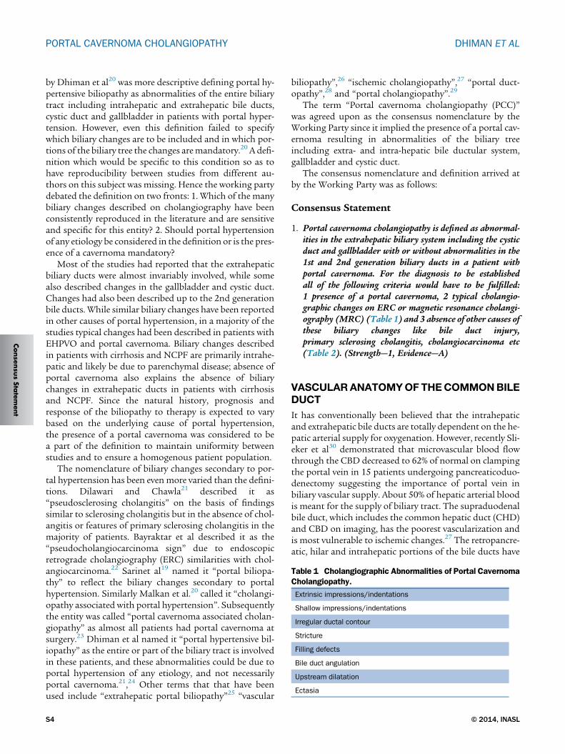
Table 1 Cholangiographic Abnormalities of Portal CavernomaCholangiopathy.
Extrinsic impressions/indentations
Shallow impressions/indentations
Irregular ductal contour
Stricture
Filling defects
Bile duct angulation
Upstream dilatation
Ectasia
PORTAL CAVERNOMA CHOLANGIOPATHY DHIMAN ET AL
Consen
susSta
temen
t
by Dhiman et al20 was more descriptive defining portal hy-pertensive biliopathy as abnormalities of the entire biliarytract including intrahepatic and extrahepatic bile ducts,cystic duct and gallbladder in patients with portal hyper-tension. However, even this definition failed to specifywhich biliary changes are to be included and in which por-tions of the biliary tree the changes aremandatory.20 A defi-nition which would be specific to this condition so as tohave reproducibility between studies from different au-thors on this subject was missing. Hence the working partydebated the definition on two fronts: 1. Which of the manybiliary changes described on cholangiography have beenconsistently reproduced in the literature and are sensitiveand specific for this entity? 2. Should portal hypertensionof any etiology be considered in the definition or is the pres-ence of a cavernoma mandatory?
Most of the studies had reported that the extrahepaticbiliary ducts were almost invariably involved, while somealso described changes in the gallbladder and cystic duct.Changes had also been described up to the 2nd generationbile ducts. While similar biliary changes have been reportedin other causes of portal hypertension, in a majority of thestudies typical changes had been described in patients withEHPVO and portal cavernoma. Biliary changes describedin patients with cirrhosis and NCPF are primarily intrahe-patic and likely be due to parenchymal disease; absence ofportal cavernoma also explains the absence of biliarychanges in extrahepatic ducts in patients with cirrhosisand NCPF. Since the natural history, prognosis andresponse of the biliopathy to therapy is expected to varybased on the underlying cause of portal hypertension,the presence of a portal cavernoma was considered to bea part of the definition to maintain uniformity betweenstudies and to ensure a homogenous patient population.
The nomenclature of biliary changes secondary to por-tal hypertension has been even more varied than the defini-tions. Dilawari and Chawla21 described it as“pseudosclerosing cholangitis” on the basis of findingssimilar to sclerosing cholangitis but in the absence of chol-angitis or features of primary sclerosing cholangitis in themajority of patients. Bayraktar et al described it as the“pseudocholangiocarcinoma sign” due to endoscopicretrograde cholangiography (ERC) similarities with chol-angiocarcinoma.22 Sarinet al19 named it “portal biliopa-thy” to reflect the biliary changes secondary to portalhypertension. Similarly Malkan et al.20 called it “cholangi-opathy associated with portal hypertension”. Subsequentlythe entity was called “portal cavernoma associated cholan-giopathy” as almost all patients had portal cavernoma atsurgery.23 Dhiman et al named it “portal hypertensive bil-iopathy” as the entire or part of the biliary tract is involvedin these patients, and these abnormalities could be due toportal hypertension of any etiology, and not necessarilyportal cavernoma.21,24 Other terms that that have beenused include “extrahepatic portal biliopathy”25 “vascular
S4
biliopathy”,26 “ischemic cholangiopathy”,27 “portal duct-opathy”,28 and “portal cholangiopathy”.29
The term “Portal cavernoma cholangiopathy (PCC)”was agreed upon as the consensus nomenclature by theWorking Party since it implied the presence of a portal cav-ernoma resulting in abnormalities of the biliary treeincluding extra- and intra-hepatic bile ductular system,gallbladder and cystic duct.
The consensus nomenclature and definition arrived atby the Working Party was as follows:
Consensus Statement
1. Portal cavernoma cholangiopathy is defined as abnormal-ities in the extrahepatic biliary system including the cysticduct and gallbladder with or without abnormalities in the1st and 2nd generation biliary ducts in a patient withportal cavernoma. For the diagnosis to be establishedall of the following criteria would have to be fulfilled:1 presence of a portal cavernoma, 2 typical cholangio-graphic changes on ERC or magnetic resonance cholangi-ography (MRC) (Table 1) and 3 absence of other causes ofthese biliary changes like bile duct injury,primary sclerosing cholangitis, cholangiocarcinoma etc(Table 2). (Strength—1, Evidence—A)
VASCULARANATOMYOF THECOMMONBILEDUCT
It has conventionally been believed that the intrahepaticand extrahepatic bile ducts are totally dependent on the he-patic arterial supply for oxygenation. However, recently Sli-eker et al30 demonstrated that microvascular blood flowthrough the CBD decreased to 62% of normal on clampingthe portal vein in 15 patients undergoing pancreaticoduo-denectomy suggesting the importance of portal vein inbiliary vascular supply. About 50% of hepatic arterial bloodis meant for the supply of biliary tract. The supraduodenalbile duct, which includes the common hepatic duct (CHD)and CBD on imaging, has the poorest vascularization andis most vulnerable to ischemic changes.27 The retropancre-atic, hilar and intrahepatic portions of the bile ducts have
© 2014, INASL

Table 2 Differential Diagnosis of Portal CavernomaCholangiopathy.
Primary sclerosing cholangitis
Bile duct neoplasms
Biliary tract surgery
Recurrent pyogenic cholangitis
Acquired immunodeficiency syndrome cholangiopathy
Biliary parasitosis
Choledocholithiasis
Congenital abnormalities of the biliary tract
Ischemic bile duct stricture
Toxic bile duct strictures
Strictures due to autoimmune and chronic pancreatitis
JOURNAL OF CLINICAL AND EXPERIMENTAL HEPATOLOGY
Conse
nsu
sStatemen
t
relatively copious arterial blood supply.31 Starting fromthe ampulla of Vater, the entire biliary system derives arte-rial blood supply from the branches of the celiac trunk.32
The arrangement of arterial and venous plexuses of CBDis similar. Epicholedochal plexus on the surface of theCBD is formed from the branches of the paracholedochalvessels. The paracholedochal and epicholedochal plexusesconnect with the intramural and subepithelial plexusesin the wall of the CBD via perforator vessels that pierceits wall.33
The venous drainage of the CBD is arranged in theform of two plexuses. The epicholedochal venous plexusof Saint is a fine reticular plexus on the surface of thebile ducts. The paracholedochal venous plexus of Petrenruns outside and parallel to the bile ducts. Two marginalveins known as 3 o'clock and 9 o'clock marginal veins areformed by the paracholedochal plexus, with, occasionally,an additional 6 o'clock marginal vein seen on posteriorsurface of CBD. Inferiorly these veins communicate withgastric veins, posterior superior pancreaticoduodenalvein, and gastrocolic trunk while superiorly the marginalveins join the hilar venous plexus to drain into adjacentportal vein branches or enter into the hepatic substance.Detailed arterial supply and venous drainage of the biliarysystem has been described in an article in this issue of theJournal.34
In EHPVO multiple porto-portal collateral veins withhepatopetal flow develop to bypass the obstructedsegment of the portal vein. These collaterals involve theparacholedochal and epicholedochal venous plexusesand cholecystic veins. The paracholedochal veins dilatefirst causing external compression of the CBD wallwith protrusion of dilated paracholedochal veins intothe thin and pliable wall of the CBD. The normallysmooth intraluminal surface of the CBD becomes irreg-ular due to the dilated epicholedochal venous collaterals.The pressure is transmitted via the perforators to thesub-epithelial plexus resulting in the development of
Journal of Clinical and Experimental Hepatology | February 2014 | Vol. 4 | N
subepithelial varices in the CBD wall which can causebleeding.35 Cavernoma formation has been demon-strated as early as within 6–20 days of acute portal veinthrombosis (PVT) with intrahepatic extension of the cav-ernoma seen in 76% of patients.36 Both porto-systemic(chiefly through left gastric vein) and porto-portal (peri-portal and pericholecystic venous channels) collateralsare seen, with all the vessels showing hepatopetal bloodflow.36
Consensus Statement
2. General pattern of arterial supply and venous drainage ofthe bile ducts is quite similar. Fine branches from the pos-terior superior pancreaticoduodenal, retroportal, gastro-duodenal, hepatic and cystic arteries form two plexusesto supply the bile ducts. The paracholedochal plexus, asright and left marginal arteries, runs along the marginsof the bile duct and the reticular epicholedochal plexuslies on the surface. The retropancreatic, hilar and intrahe-patic parts of biliary tract have copious supply, but thesupraduodenal bile duct has the poorest vascularizationand hence is susceptible to ischemic changes. (Strength—1,Evidence—A)
3. Two venous plexuses drain the biliary tract. The fine retic-ular epicholedochal venous plexus of Saint on the wall ofthe bile duct drains into the paracholedochal venous plexusof Petren (also called marginal veins or parabiliaryvenous system) which in turn is connected to the posteriorsuperior pancreaticoduodenal vein, gastrocolic trunk,right gastric vein, superior mesenteric vein inferiorlyand intrahepatic portal vein branches superiorly.(Strength—1, Evidence—A)
4. In conditions of porto-mesenteric venous obstruction, theseplexuses enlarge to form a portal cavernoma surroundingthe bile duct and bring about morphological changesobserved in portal cavernoma cholangiopathy.(Strength—1, Evidence—B)
PATHOGENESIS
Acute portal vein occlusion causes loss of portal blood flow,which is initially compensated by the hepatic artery bufferresponse, resulting in dilatation of the hepatic arterial sys-tem.37,38 Subsequently, rapid development of venouscollaterals and formation of varices as well as cavernomaoccur. These dilated venous collaterals exert externalpressure and protrude into the thin and pliable CBD andthe hepatic ducts (Figure 1).17,23,24,39 The dilatation of theplexus of Saint causes fine irregularities in the biliarytract while the dilatation of the plexus of Petren causesextrinsic compression over bile duct.17 The frequentinvolvement of the left hepatic duct in PCC could be dueto prominent collateral veins at the junction of the umbil-ical vein and left branch of the portal vein.40 Intra-ductal
o. S1 | S2–S14 S5

Figure 1 (A) Endoscopic cholangiography shows common bile duct stricture (arrows). (B) MR cholangiography confirms common bile duct strictureseen on endoscopic cholangiography (arrows). (C) MR venography shows that common bile duct stenosis is caused by pressure from a large collat-eral. Blood vessels appear in white in flow sensitive sequence (large arrows).
PORTAL CAVERNOMA CHOLANGIOPATHY DHIMAN ET AL
Consen
susSta
temen
t
collaterals may also contribute to the biliary abnormalitiesas evidenced by endoscopic ultrasound (EUS) demonstra-tion of paracholedochal varices which perforate the wallof the bile duct to lie in the subepithelial layer, causingbiliary irregularity.35,41 Evidence in favor of compressionas a cause of biliary changes comes from imagingevidence of collaterals impinging on the bile ducts at thesite of biliary irregularities17,23 and from the reversal ofbiliary changes after shunt surgery in some patients.42
However, not all biliary changes reverse after shunt sur-gery. This could be due to incomplete disappearance ofvarices after surgery, presence of a fibrous tumor-like-cavernoma causing persistent compression17,23,43 orirreversible irregularities due to ischemic changes withinthe bile duct secondary to deficient portal blood supplyor prolonged compression by collaterals. Also, not allbiliary strictures correlate with collaterals on imagingwith MRC coupled with MR venography. In a study byDhiman et al17 only 55% of dominant biliary strictureswere caused by compression from adjacent collateralswhereas no such relationship was seen in the remaining45%, which were presumed to have an ischemic origin.Decrease in biliary blood flow on portal vein clampinghas been described in humans by Seiker et al30 Portalbranch ligation in rabbits results in slight dilatation ofthe bile ducts of affected lobes.44 It is tempting to speculatethat development of biliary changes in PCC is due to twocomponents a)a reversible component, due to biliarycompression by engorged collaterals, that reverses aftershunt surgery and b) a fixed component that does notreverse after shunt surgery and is likely due to ischemicchanges in the bile duct, occurring either at the time of por-tal vein thrombosis or due to prolonged compression bycollaterals or by a solid, tumor-like ‘fibrotic’ cavernoma,containing connective tissue. Detailed pathogenesis ofPCC has been described in an article in this issue of theJournal.42
S6
Consensus Statement
5. Biliary changes seen in PCC may be due to compression bylarge collaterals or may be ischemic, due to portal veinthrombosis and/or prolonged biliary compression. Whilethe former are reversible after porto-systemic shunt sur-gery, the latter are not. Collateral compression andischemic injury, are not mutually exclusive in a patientwith PCC. (Strength—1, Evidence—A)
PREVALENCE AND CLINICALCHARACTERISTICS
Theprevalence of biliary changes on cholangiography rangesfrom 78 to 100% in most series of patients with EHPVO.However, symptoms are present in only 5–50% of patients(Table 3).45 Symptoms could be in the formof long standingjaundice due to chronic cholestasis, or biliary pain with orwithout cholangitis due to biliary stones.20 Jaundice is invari-ably present in patients with symptomatic PCC, andwas pre-sent at diagnosis in about 2/3rd of patients, while it was thesole manifestation of EHPVO (without preceding varicealbleeding) in 15% of patients managed surgically.46 Jaundiceis usually mild; mean serum bilirubin level was 2.7 mg/dL(range 0.7–16) in one study while only 15% of patients hada bilirubin value ofmore than 5mg/dL in another study.20,47
Half to 2/3rd of patients have experienced cholangitis, withnumber of episodes ranging from 1 to 25. Abdominal painis seen in about half of symptomatic PCC whereasawareness of splenic lump is reported in only a minority ofcases.46–48 Diagnosis of EHPVO antedates symptomaticPCC by 8–10 years. Age of the patient and duration ofEHPVO are chief risk factors for development ofsymptoms in PCC. Patients with symptomatic PCC wereon average 14 years older at (35 years versus 21 years age)and had been diagnosed with EHPVO for 7 years longer(11 years versus 4 years) compared to asymptomatic PCC.20
© 2014, INASL

Table 3 Frequency of Biliary Changes in Patients with Extra-hepatic Portal Venous Obstruction.
Study Year n Age meanand/or range
M/F Frequency of biliarychanges (%)
Symptomaticpatients (%)
Dilawari and Chawla21 1992 20 22 (13–38) 16/4 100 5
Sarin et al19 1992 20 9–32 16/4 90 15
Khuroo et al49 1993 21 14 � 8.8 13/8 81 38
Bayraktar et al22 1995 44 31.5 (9–67) 24/20 94 30
Malkan et al2 1999 20 23 12/8 85 10
Nagi et al50 2000 43 14–45 25/18 100 19
Condat et al23 2003 25 49.5 15/1 84 28
Sezgin et al51 2003 36 NA NA 94 10
Dhiman et al24 2006 53 24.5 (13–56) 36/17 100 24.5
Chevallier et al52 2006 10 43.5 (29–56) 5/5 90 50
Llop et al29 2011 67 47 (19–77) 41/26 78 21
Total [median (range)] 262 90 (78–100) 21 (5–50)
NA, not available.
JOURNAL OF CLINICAL AND EXPERIMENTAL HEPATOLOGY
Conse
nsu
sStatemen
t
Gallstones and CBD calculi are other major risk factors forthe development of symptoms. In one study 95% patientswith symptomatic PCC had dilated intrahepatic biliary rad-icles and 82% had dilated CBD on ultrasound. Gallstoneswere seen in 1/3rd and bile duct stones in 18% of symptom-atic patients.46 In another study, gallstones and CBD stoneswere seen in 54% and 23% of symptomatic PCC patientsrespectively compared to 0% and 2.5% respectively in asymp-tomatic PCC patients.20 Prevalence and clinical characteris-tics of PCC have been described in an article in this issue ofthe Journal.45
Consensus Statement
6. Majority of the patients with PCC are asymptomatic.(Strength—1, Evidence—A)
7. Approximately 21% (range 5–50%) of patients with PCCare symptomatic. (Strength—1, Evidence—A)
8. Risk factors for development of symptomatic PCC includeolder age of the patient, long duration of EHPVO, dilatedsegments of bile ducts, and the presence of gallstones andCBD stones (Strength—2, Evidence—C).
DIAGNOSIS
Endoscopic Retrograde CholangiographyERC has traditionally been the gold standard for diagnosisof PCC. However due to its invasive nature, risk of compli-cations and need for sequential imaging it is now being re-placed by non-invasive radiological modalities fordiagnosis with ERC being reserved for therapy. Standard-ized nomenclature (Table 1) and definitions of variousfindings on ERC findings in PCC, are proposed by theWorking Party in this issue of the Journal. Based oncurrently available literature reports rather than on
Journal of Clinical and Experimental Hepatology | February 2014 | Vol. 4 | N
rigorously validated data, this terminology is expected toprovide a framework for ensuring uniformity in futurestudies. Nomenclature and definitions of various findingson ERC findings in PCC have been described in an articlein this issue of the Journal.53 Briefly, nomenclature anddefinitions of ERC findings are as follows:
i) Extrinsic impressions/indentations: smooth thumb-like im-pressions on the bile duct, with a nodular contour. Theindentation is more than one-quarter of the width of theopacified duct. Impressions may be multiple.
ii) Shallow impressions/indentation(s): smooth non-contiguous impressions on the bile duct, less thanone-quarter of ductal diameter.
iii) Irregular ductal contour: fine-wavy, irregular contour ofthe bile duct walls due to contiguous shallow indenta-tions, less than one-quarter of the ductal diameter.
iv) Stenosis: variable length narrowing of the ductal lumen,in reference to well opacified downstream ductsegment. Stenoses might be associated with upstreamdilatation and may be due to extrinsic compressionby collaterals or intrinsic narrowing or stricturingdue to mural fibrosis. Strictured bile duct segmentsshould offer some resistance to passage of anadequately inflated extraction balloon across it andshould produce a waist on non-compliant balloons in-flated within the narrowed segment. Stenoses and stric-tures may be divided into ‘mild to moderate’ or‘severe’depending on whether the narrowed segment is > or <two-third of the diameter of adjacent normal segment.
v) Upstream dilatation: proximal dilatation can be similarlyclassified as ‘mild to moderate’ or ‘severe’, depending onwhether the dilated segment is between 1.5–2 or > 2diameter of the adjacent normal duct, respectively.
vi) Filling defects: round, oval, or elongated defects in thecholangiographic image, with contrast on three or all
o. S1 | S2–S14 S7
PORTAL CAVERNOMA CHOLANGIOPATHY DHIMAN ET AL
Consen
susSta
temen
t
sides. Filling defects can represent stones, prolapsingintra-luminal varices, or clots.
vii) Bile duct angulation: it is proposed that an angle of#145� between lower and upper CBD be consideredas significant
viii) Ectasia: dilated segment of biliary tree without anyevident downstream obstruction
Cholangiographic findings in PCCwere first classified byChandra et al4 who proposed a system based on ERC whichhasbeenapplied toMRCaswell, basedon thedistributionoflesions.Type I is involvementof extrahepatic ducts only, typeII is involvement of intrahepatic bile ducts only, type IIIa isextrahepatic bile duct with unilateral intrahepatic bile ductinvolvement and type IIIb is extrahepatic bile ductwithbilat-eral intrahepatic bile duct involvement.
Llop et al29 have graded the severity of abnormalities of thebiliary treein PCC according to cholangiographic findingsinto grade I: irregularities or angulations of the biliary tree,grade II: indentations or strictures without upstream biliarydilation and grade III: strictures with upstream biliary dila-tion. Dilatation is defined as ductal diameter of >7 mm forextra-hepatic duct and/or >4 mm for intra-hepatic ducts.
The INASL Working Party suggests that, for futurestudies in PCC, standardized nomenclature and terminol-ogy proposed-above should be used for describing lesionswhile the system proposed by Llop et al should be usedfor grading severity of cholangiographic changes, sincenot only is it based on the severity of cholangiographicfindings but has also been prospectively validated incontrast to the classification proposed by Chandra et al4
which deals only with the distribution of changes. Howev-er, it is acknowledged that classification and grading sys-tems are likely to change as more severe grades ofabnormalities, more extensive distribution of changesand multifocal changes are reported in PCC.
UltrasonographyCombination of ultrasound (USG)with colorDoppler is theinitial screening modality for the PCC as it is non-invasive,free of radiation, easily available and relatively cheap. Find-ings on sonography vary from non-visualization of portalvein to a completely thrombosed vein with cavernoma for-mation seen as multiple tubular anechoic structures in theporta. Color Doppler sonography demonstrates flow in por-tal collaterals in the absence of flow in portal vein. 30–50%cases may have tortuous collaterals around the wall of thegallbladder and rarely color Doppler may pick up varicesin the wall of the gallbladder and the bile duct as thickenedwall with color flow on Doppler imaging.54 USG can detectnarrowing or stenosis of the CBD with associated proximaldilatation and associated cholelithiasis and/or choledocho-lithiasis. However the CBD can get obscured by high levelechoes in the porta hepatis and by the multiple collateral
S8
channels. Exact details of biliary narrowing and differentia-tion between extrinsic compression and ischemic stricturingare not possible onUSG. Signs of cirrhosis, associatedporto-systemic collaterals and splenomegaly are well detected withUSG and color Doppler.
Computed Tomography (CT) or MagneticResonance Imaging (MRI)Cross-sectional imaging with contrast enhancedcomputed tomography (CT) or magnetic resonance imag-ing (MRI) confirms features of PCC but is mainly neededto exclude other possible causes of the observed changessuch as biliary malignancy and also to search for possiblecause of portal vein obstruction such as chronic pancrea-titis. MRI is preferred to CT as it does not entail radiationexposure and is superior to CT in detailing biliary anat-omy. Combination of contrast enhanced MRI or MR por-tovenography with MRC have currently replaced ERC asthe diagnostic imaging of choice for PCC.23,40,55 Inaddition to detailed delineation of the biliaryabnormalities including differentiation of stones fromintramural varices, they delineate the entire spleno–portalaxis, help in planning PSS surgery, and depict therelationship of collaterals with the biliary tract which isnot possible with ERC (Figure 1). MRI also differentiatesepicholedochal collaterals, appearing as dot like enhancingstructures in the bile duct wall, from paracholedochal col-laterals and gallbladder varices which appear as low signalintensity channels on T2-weighted images and asenhancing tortuous collaterals on dynamic 3D gradient-echo images.56 Typical biliary findings of PCC seen onMRC are similar to those seen on ERC,40 are listed inTable 1 and are reviewed in detail elsewhere in this issue.57
Recent MRC studies of PCC have reported that type I andtype III abnormalities according to Chandra et al,4 i.e.involvement of extrahepatic ducts alone or in combinationwith the intrahepatic ducts, were the most frequent find-ings, extrahepatic bile ducts being involved almost univer-sally involved.56 No discrepancy between MRC and ERC inthe biliary findings of PCC in patients appeared when bothinvestigations were performed.40,55,56 Hence MRCP withMR portovenography is the modality of choice formapping of the biliary and vascular abnormalities.
Endoscopic UltrasoundRole of EUS in the diagnosis and management of PCC isevolving. There is no study directly comparing ERC orMRC with EUS. On EUS, biliary varices appear as multiple,large, serpiginous, anechoic vascular channels in and/oraround the extrahepatic biliary tract.58 EUS with Dopplercan accurately differentiate paracholedochal, epicholedo-chal, intracholedochal and subepithelial varices. Thisdistinction is especially relevant if a subsequent ERC is
© 2014, INASL
JOURNAL OF CLINICAL AND EXPERIMENTAL HEPATOLOGY
Conse
nsu
sStatemen
t
planned, since the presence of intracholedochal and subepi-thelial varices may increase risk of bleeding during stoneextraction and stricture dilatation respectively.59,60 Adetailed comparison of various imaging modalities usefulfor the diagnosis of PCC has been reviewed elsewhere inthis issue.60
Consensus Statement
9. ERC has no diagnostic role because it is invasive and isassociated with risk of complications. ERC is reserved fortherapeutic procedures. (Strength—1, Evidence—A)
10. A standardized nomenclature and definition of variousfindings on ERC has been proposed for uniformity infuture studies and need prospective validation in futurestudies (Strength—1, Evidence—B).
11. USG with Color Doppler should be the initial imagingmodality for suspected cases of PCC (Strength—1,Evidence—B).
12. MRC with MR portovenography is a noninvasive andcomprehensive imaging technique, and is the modalityof choice for mapping of the biliary and vascularabnormalities in patients with PCC (Strength—1,Evidence—A).
13. MRC is as accurate as ERC in delineating biliary changes(Strength—1, Evidence—B).
14. MRC with MR portovenography demonstrates relation-ship of biliary changes with collaterals and the presence ofshuntable vein. It also helps to distinguish between bileduct varices and common bile duct stones. (Strength—1, Evidence—A)
15. EUS is useful when other imaging modalities are unre-vealing or inconclusive in delineating the cause of biliaryobstruction (Strength—2, Evidence—C).
16. EUS is helpful in delineating the type of choledochal col-laterals. However, utility of EUS based delineation ofcholedochal collaterals needs to be defined in prospectivestudies (Strength—2, Evidence—C).
NATURAL HISTORY AND PROGNOSIS
Natural HistoryData on natural history and prognosis of PCC are scarce.As discussed previously the course can be divided intoasymptomatic and symptomatic phases. The majority(70–95%) of patients in cross sectional studies have beenasymptomatic, but long-term follow-up of these asymp-tomatic patients in longitudinal studies has not been re-ported. Only one study from Spain has reportedlongitudinal follow-up in 22 acute and 45 chronic non-cirrhotic patients with PVT.29 Severity of cholangiographicchanges was graded from grades I to III (Llop et al29; see‘Diagnosis’ section above). Their observations suggestedthat PCC was a non-progressive, one-time event after acute
Journal of Clinical and Experimental Hepatology | February 2014 | Vol. 4 | N
PVT. Overall, 73% patients developed PCC at 33 monthsafter acute PVT, which was similar to the frequency inthe chronic PVT (80%). Sixty percent of those imagedwithin 1 year of PVT had developed PCC; only one addi-tional patients 1 among the 4 others developed PCC over36 months. Changes did not progress in 3 of 4 patients fol-lowed for 43 months. Symptomatic PVT was seen only inpatients with severe, grade III changes on cholangiography.However, in this study follow-up period was relativelyshort as the natural history of PCC may extend over 2 to3 decades. Also, the severity of lesions even in grade IIIwere much milder than that reported in Indian serieswhich had evaluated patients after much longer durationsince diagnosis of EHPVO.61
Patients with symptomatic PCC are normally older thanpatients presenting with EHPVO by median of 8–14 years,which suggests that PCC is a progressive condition andthat long-term obstruction24,47,51,62 and prolonged portalhypertension are needed to cause changes severe enoughto produce symptoms. Advanced biliary changes such aslong strictures, and strictures with upstream stones havebeen reported in patients with EHPVO after prolongeddurations of follow-up.47 Another study found that onsequential ERC studies performed a median of 29 monthsapart there was significant progression of biliary changesand 35% patients became symptomatic during this period.4
On prolonged follow-up patients with EHPVO have beenreported to develop ascites, hypoalbuminemia and coagul-opathy suggestive of liver dysfunction.11,63 This ispostulated to be due to effects of chronic deprivation ofportal blood flow to the liver leading to atrophy with apossible contribution from chronic cholestasis occurringdue to PCC. Based upon these observations, the naturalhistory of PCC can be divided into four stages (Table 4):1. Preclinical: presence of portal cavernoma but no PCCwith normal liver biochemistry and no symptoms ofPCC; 2. Asymptomatic: early changes on cholangiographywith normal or abnormal liver biochemistry but no symp-toms; 3. Symptomatic: advanced changes on cholangiog-raphy with abnormal liver biochemistry and presence ofsymptoms without complications and; 4. Complicated: Pres-ence of liver dysfunction or fibrosis, extensive biliarychanges like multifocal strictures, calculi above strictures,or biliopancreatic complications (Table 4).61
PrognosisPatients with symptomatic PCC who achieve adequatestone clearance and biliary drainage endoscopically havebeen reported to have an excellent outcome in the avail-able follow-up studies.20,51,64,65 The reported follow-up ofsurgically managed patients with PCC is relatively longer,ranging from 14 months to 12 years. Patients with symp-tomatic PCC who have had reversal of biliary changes andrelief of symptoms also carry an excellent prognosis.46,66–68
o. S1 | S2–S14 S9
Table 4 Stages in the Natural History of Portal Cavernoma Cholangiopathy.
Stage Portal cavernoma Cholangiopathy Liver biochemistry Symptoms Complications
Preclinical Present Absent Normal Absent Absent
Asymptomatic Present Early changes Normal or abnormal Absent Absent
Symptomatic Present Advanced changes Abnormal Present Absent
Complicated Present Advanced changes Abnormal Present Present
PORTAL CAVERNOMA CHOLANGIOPATHY DHIMAN ET AL
Consen
susSta
temen
t
However, patients without shuntable veins and advancedbiliary changes of PCC cannot undergo shunt surgery.These patients may develop recurrent cholangitis20,48,51
and, eventually, liver dysfunction either due tosecondary biliary cirrhosis or due to chronicallydiminished portal inflow.11,63 Attrition of hepatocellularfunction in these patients may reach the stage thatthey are candidates for liver transplantation.48,69–71 Adetailed description of natural history and prognosis ofPCC has been reviewed elsewhere in this issue.61
Consensus Statement
17. PCC develops early after acute PVT if recanalization isnot achieved. (Strength—1, Evidence—B)
18. PCC is a progressive condition and symptoms maydevelop late in the course of portal hypertension.(Strength—1, Evidence—B)
19. PCC symptoms seem to occur only in patients with severeor advanced changes of cholangiopathy. (Strength—1, Ev-idence—B)
20. Presence of severe or advanced changes of cholangiopathymay be useful for identifying patients with a higher risk ofdeveloping symptoms of PCC. (Strength—1, Evidence—B)
21. Patients with symptomatic PCC have good prognosis af-ter successful endoscopic biliary drainage and after suc-cessful shunt surgery. (Strength—1, Evidence—A)
22. Patients with advanced biliary changes and withoutshuntable veins in whom repeated endoscopic biliarydrainage are required carry a poor prognosis. These pa-tients may have recurrent cholangitis and progressiveliver dysfunction and might need liver transplantation.(Strength—1, Evidence—B)
MANAGEMENT
Asymptomatic patients with PCC do not require any treat-ment. Treatment of symptomatic PCC should be deter-mined on a case by case basis. Primarily, it should befocused on the management of portal hypertension and re-lief of biliary obstruction.
Great care should be exercised in discriminating be-tween symptomatic and asymptomatic patients. The labelof symptomatic PCC should be applied only on clinicalgrounds, i.e. to patients with biliary pain, cholestatic jaun-dice and/or cholangitis due to either choledocholithiasis,
S10
choledochal stenosis or both. Patients with PCC and otherassociated abnormalities like deranged LFT but withoutthe presentations mentioned above should be regardedas having asymptomatic PCC, and are not candidates forendoscopic therapy. Patients with symptomatic PCC, asdescribed above, are candidates for the intervention,whether surgical or endoscopic. It is now accepted thatthe optimal management may require use of either orboth these modalities as per the specific situation, though,as in other situations, surgical management is rarely re-sorted to as the first choice in patients with cholestaticjaundice and/or cholangitis. Initially endoscopic interven-tions in PCC were used to tackle emergency conditions likecholangitis due to strictures or stones using plastic stentsor naso-bilairy drains to establish biliary drainage pendingsurgery. Subsequently endoscopic stone removal in PCCwas demonstrated to be a safe procedure, with only few re-ports of hemobilia.47,48,51,64,65 Many patients are not can-didates for surgery due to absence of a shuntable vein orliver dysfunction. These patients may require long-termendoscopic management in the form of repeated plasticstent exchanges47,51,65 or placement of removable coveredmetal stents.48,72 The role of peroralcholedochoscopywith Spyglass and laser or electrohydraulic lithotripsy formanagement of biliary calculi in complicated PCC needsto be explored.
Treatment of symptomatic PCC can be approached in aphased manner, coping first with biliary clearance; second,with portal decompression; and third, with persistentbiliary obstruction. In patients with biliary obstructionwith or without calculi, the first phase involves sphincter-otomy and biliary drainage, with CBD stone removal if pre-sent. Sphincterotomy has not shown to be associated withincreased bleed rates in PCC and use of Dormia basketsand balloon extractors has been shown to besafe.20,29,47,48,64 Hemobilia can be managed conservativelyinmost cases and is not more troublesome than in patientswithout PCC.48,51,73,74 Available literature suggests thatendoscopic sphincterotomy with stone extraction is thefirst choice procedure for symptomatic CBD stones andendoscopic biliary drainage with plastic stents or nasobili-ary tubes is the first choice in patients with cholangitis orcholestatic jaundice.47,48,51,64,65 These are generally preop-erative procedures, to be followed by porto-caval shunt sur-gery. Repeated stent exchanges over short periods of time(3–5 years) may occasionally result in resolution of stenosisbut this option is usually resorted to as fall-back therapy
© 2014, INASL

JOURNAL OF CLINICAL AND EXPERIMENTAL HEPATOLOGY
men
t
for those with no surgical options for portal decompres-sion.47,51,65 Long-term results with this approach havenot been reported. Anecdotal experience with the use ofcovered removable self-expandable metal stents is insuffi-cient for any comment. Portal decompression surgerymay be the first phase of therapy in patients in whom endo-scopic intervention is not required such as a patient withisolated stenosis of CBD and with a shuntable vein.20 Amore detailed discussion of the indications and results ofendoscopy for initial drainage and stone removal as wellas for long termmanagement of PCC is reviewed elsewherein this issue.75
The use of concomitant ursodeoxycholic acid (UDCA)has been reported to be beneficial by some authors. Per-lemuter et al76 used UDCA in 5 of 8 patients with liverfibrosis or secondary biliary cirrhosis on liverbiopsy.Condat et al23 used UDCA in 3 of 4 patients with chole-stasis who underwent endoscopic sphincterotomy andreported no recurrence of symptoms while on therapy.Llop et al29 used it in 10 of 14 patients with symptom-atic PCC, including 5 patients with abdominal pain andcholestasis treated with UDCA alone, in two patientswith stricture but no calculi and in 3 of 6 with choledo-chal stones after sphincterotomy and ductal clearance.They reported ‘disappearance of symptoms and improve-
EHPVO patients
Biliary symptoms +, raised alkaline Phosphatase
or dilated biliary ducts
PCC - Present
MRCholangiography +MR portovenography
Normal LFT or isolated ↑ alkaline phosphatase
Presence of biliary obstruction
CBD Stone Choledochal Stenosis
Endoscopic treatment (P-1) Cholangitis – present Cholangitis - atreated
Endoscopic treatment (P-1)Shuntable vein,
present
Portosystemic shunt (P-1/2)
Biliary obstruction,persistent
Biliary obstrurelieve
Follow-up
Second stage bilioenteric anastomosis (P-3)
Follow-u
EUS with Doppler*
Figure 2 Algorithm for the management of a patient with portal cavernomacommon bile duct; LFT, liver function tests; MR, magnetic resonance; PCCbiliary obstruction when other imaging modalities are unrevealing; yendoscopor without the help of mechanical lithotripsy, plastic biliary stent placementstricture dilatation, followed by stone extraction with or without the help of mefrom Dhiman RK, et al. Gut 2007;56:1001–8).20
Journal of Clinical and Experimental Hepatology | February 2014 | Vol. 4 | N
ment in liver tests’ in all treated patients during follow-up. However, other authors have reported equally goodresults with endoscopic management without UDCAand as no study comparing ERC alone versus ERCwith UDCA in PCC is available, effect of UDCA inimproving symptoms of PCC or altering its naturalcourse is not clear.
In symptomatic PCC, the second phase involves portaldecompression surgery by proximal splenorenal shunt ortransjugular intrahepatic portosystemic shunt (TIPS).Shunt procedures result in regression of changes of cholan-giopathy17,24 with complete regression of biliary obstruc-tion occurring in about 62–88% of patients.17,46,47,66–68
Only 25–30% of patients will require further interventionfor PCC after successful shunt procedure. Even if theregression of cholangiographic changes is incomplete,majority of the patients remain asymptomatic probablydue to slowing-down of progression of PCC after shunt.Though the choice of shunt depends upon the surgeons'choice, non-selective shunts are preferred as they decom-press the whole of the portal system allowing regressionof collaterals in fact the majority of studies have reportedresults with proximal splenorenal shunts.46,66–68 A meso-Rex bypass between the mesenteric vein and the left portalvein is preferred by some surgeons in children with EHPVO
PCC - absent
Choledochal Stenosis +
CBD stone
bsent or Endoscopic treatment (P-1)
Follow-up
Shuntable vein, absent Failure
Long-term biliarystent placement (P-1)
ction, d
p
Successful
Follow-up
‡
cholangiopathy. EHPVO, extrahepatic portal venous obstruction; CBD,, portal cavernoma cholangiopathy; *EUS with Doppler for evaluation ofic treatment includes endoscopic sphincterotomy, stone extraction with, etc; zendoscopic treatment includes endoscopic sphincterotomy andchanical lithotripsy. P1, phase 1; P2, phase 2 and P3, phase 3. (Modified
o. S1 | S2–S14 S11
Conse
nsu
sState

PORTAL CAVERNOMA CHOLANGIOPATHY DHIMAN ET AL
Consen
susSta
temen
t
as it preserves portal blood flow to the liver and mayimprove growth in them.77–80
The third phase involves management of cases whocontinue to have biliary obstruction and remain symptom-atic despite shunt procedure either due to blockage ofshunt or due to persistent obstruction despite patentshunt. Second-stage biliary drainage surgery like hepatico-jejunostomy or choledochoduodenostomy surgery on thebile ducts is performed in most of these cases with goodresolution of symptoms as well as liver functiontests.46,66–68 A more detailed discussion of the results ofsurgery in PCC is discussed in detail elsewhere in thisissue.81 An alternative is approach is long-term endoscopicmanagement of these patients with repeated stent ex-changes and stone removal.46,66–68 No study hascompared the result of these two approaches.
About 5–30% of patients with PCC did not have shunt-able veins in several surgical series.46,47,66–68 Other patientsmay have advanced biliary changes of PCC that do notreverse fully with shunt surgery. These patients maydevelop recurrent cholangitis20,48,51 or liver dysfunctioneither due to secondary biliary cirrhosis or due to chroni-cally diminished portal inflow.11,63 There have been case re-ports of such patients successfully undergoing livertransplantation.48,69–71,82 The portal inflow to the graftcan be routed through the systemic circulation if there isextensive thrombosis of the splenoportal axis in the formof either a cavoportal hemi-transposition or a reno-portalinflow. This however does not decompress the splanchnicbed and symptoms related to hypersplenism and esopha-geal varices can persist despite transplantation.
An algorithmic approach for the management of PCC ispresented in Figure 2.
Consensus Statement
23. Nasobiliary or biliary stent placement for acute cholangi-tis is safe and effective. (Strength—1, Evidence—A)
24. In the presence of normal platelet counts and coagula-tion, biliary sphincterotomy is a safe and effective proce-dure. (Strength—1, Evidence—A)
25. Biliary stricture dilatation and stent(s) placement is asafe procedure. (Strength—1, Evidence—B)
26. Biliary stricture dilatation and stone removal after lithotripsymay be performed safely. (Strength—2, Evidence—B)
27. Bilio-enteric anastomosis without prior surgery carrieshigh morbidity and mortality and hence should not beperformed. (Strength—1, Evidence—A)
28. Successful porto-systemic decompressive surgery resultsin reversal of symptoms and biliary strictures (partiallyor completely) in the majority of patients with PCC.Hence decompressive surgery should be performed ifthere is a shuntable vein available. (Strength—1, Evi-dence—A)
S12
29. Non-selective shunts are preferred as they decompress thewhole of the portal system allowing regression of collat-eral vessels. (Strength—1, Evidence—B)
30. Patients who continue to have biliary obstruction andremain symptomatic despite shunt procedure either dueto blockage of shunt or due to persistent obstructiondespite patent shunt. Second-stage biliary drainage sur-gery like hepaticojejunostomy or choledochoduodenos-tomy surgery on the bile ducts should be performed.(Strength—1, Evidence—B)
31. Secondary biliary cirrhosis is the only accepted indicationfor liver transplantation in patients with complicatedPCC. Deceased donor liver transplantation is preferableover living donor liver transplantation as PCC is usuallya non-emergency transplant and the graft can have portalblood inflow through a conduit to even a small segment ofpatent portal venous system or even to a cavernoma vessel.(Strength—2, Evidence—C)
CONFLICTS OF INTEREST
All authors have none to declare.
REFERENCES
1. Fraser J, Brown AK. A clinical syndrome associated with a rareanomaly of vena portal system. Surg Gynecol Obstet.1944;78:520–524.
2. Malkan GH, Bhatia SJ, Bashir K, et al. Cholangiopathy associatedwith portal hypertension: diagnostic evaluation and clinical implica-tions. Gastrointest Endosc. 1999;49:344–348.
3. Dhiman RK, Chawla Y, Vasishta RK, et al. Non-cirrhotic portalfibrosis (idiopathic portal hypertension): experience with 151 pa-tients and a review of the literature. J Gastroenterol Hepatol.2002;17:6–16.
4. Chandra R, Kapoor D, Tharakan A, et al. Portal biliopathy.J Gastroenterol Hepatol. 2001;16:1086–1092.
5. Chawla Y, Agrawal S. Portal cavernoma cholangiopathy – his-tory, definition and nomenclature. J Clin Exp Hepatol. 2014;4:S15–S17.
6. Guyatt GH, Oxman AD, Vist GE, et al, GRADE Working Group.GRADE: an emerging consensus on rating quality of evidence andstrength of recommendations. BMJ. 2008;336:924–926.
7. Hunt AH. Compression of the common bile-duct by an enlargingcollateral vein in a case of portal hypertension. Br J Surg.1965;52:636–637.
8. Gibson JB, Johnston GW, Fulton TT, et al. Extrahepatic portalvenous obstruction. Br J Surg. 1965;52:129–139.
9. Meredith HC, Vujic I, Schabel SI, O'Brien PH. Obstructive jaundicecaused by cavernous transformation of the portal vein. Br J Radiol.1978;51:1011–1012.
10. Hymes JL, Haicken BN, Schein CJ. Varices of the common bile ductas a surgical hazard. Am Surg. 1977;43:686–688.
11. Webb LJ, Sherlock S. The aetiology, presentation and natural his-tory of extra-hepatic portal venous obstruction. Q J Med.1979;48:627–639.
12. Williams SM, Burnett DA, Mazer MJ. Radiographic demonstra-tion of common bile duct varices. Gastrointest Radiol.1982;7:69–70.
© 2014, INASL
JOURNAL OF CLINICAL AND EXPERIMENTAL HEPATOLOGY
Conse
nsu
sStatemen
t
13. Dan SJ, Train JS, Cohen BA, Mitty MA. Common bile duct varices:cholangio-graphic demonstration of a hazardous portosystemiccommunication. Am J Gastroenterol. 1983;78:42–43.
14. Spira A, Widrich WC, Keush KD, Jackson BT, Katzman HE,Coello AA. Bile duct varices. Arch Surg. 1985;1(20):1194–1196.
15. Choudhuri G, Tandon RK, Nundy S, Misra NK. Common bile ductobstruction by portal cavernoma.Dig Dis Sci. 1988;33:1626–1628.
16. Arotcarena R, Roulot D, Aubert A, Attali P, Benichou J, Coste T. Suc-cessful treatment by mesocaval anastomosis of a common-bile-duct compression by choledochal varices. J Hepatol.1995;23:629–630.
17. Dhiman RK, Puri P, Chawla Y, et al. Biliary changes in extrahepaticportal venous obstruction: compression by collaterals or ischemic?Gastrointest Endosc. 1999;50:646–652.
18. Lohr JM, Kuchenreuter S, Grebmeier H, Hahn EG, Fleig WE.Compression of the common bile duct due to portal vein throm-bosis in polycythemia vera. Hepatology. 1993;17:586–592.
19. Sarin SK, Bhatia V, Makwana U. Poratal biliopathy in extrahepaticportal venous obstruction [abstract]. Indian J Gastroenterol.1992;11(suppl 1):A82.
20. Dhiman RK, Behera A, Chawla YK, Dilawari JB, Suri S. Portal hyper-tensive biliopathy. Gut. 2007;56:1001–1008.
21. Dilawari JB, Chawla YK. Pseudosclerosing cholangitis in extrahe-patic portal venous obstruction. Gut. 1992;33:272–276.
22. Bayraktar Y, Balkanci F, Kayhan B, et al. Bile duct varices or‘‘pseudo-cholangiocarcinoma sign’’ in portal hypertension due tocavernous transformation of the portal vein. Am J Gastroenterol.1992;87:1801–1806.
23. Condat B, Vilgrain V, Asselah T, et al. Portal cavernoma associatedcholangiopathy: a clinical and MR cholangiography coupled withMR portography imaging study. Hepatology. 2003;37:1302–1308.
24. Dhiman RK, Chawla Y, Duseja A, et al. Portal hypertensive biliopa-thy (PHB) in patients with extrahepatic portal venous obstruction(EHPVO) [abstract]. J Gastroenterol Hepatol. 2006;21:A504.
25. Walser EM, Runyan BR, Heckman MG, et al. Extrahepatic portal bil-iopathy: proposed etiology on the basis of anatomic and clinicalfeatures. Radiology. 2011;258:146–153.
26. Rosenthal MD, White GH, StephenMS, Gallagher JJ, Sandroussi C.Vascular biliopathy as a cause of common bile duct obstructionsuccessfully treated by mesocaval shunt and endoscopic retro-grade cholangiopancreatography biliary stent placement. Vascular.2008;16:356–358.
27. Deltenre P, Valla DC. Ischemic cholangiopathy. Semin Liver Dis.2008;28:235–246.
28. Bayraktar Y. Portal ductopathy: clinical importance and nomencla-ture. World J Gastroenterol. 2011;17:1410–1415.
29. Llop E, de Juan C, Seijo S, et al. Portal cholangiopathy: radiologicalclassification and natural history. Gut. 2011;60:853–860.
30. Slieker JC, Farid WRR, van Eijck CHJ, et al. Significant contributionof the portal vein to blood flow through the common bile duct. AnnSurg. 2012;255:523–527.
31. Northover JM, Terblanche J. Bile duct blood supply. Its importancein human liver transplantation. Transplantation. 1978;26:67–69.
32. FurukawaH, Iwata R, Moriyama N, Kosuge T. Blood supply to thepancreatic head, bile duct and duodenum: evaluation by computedtomography during arteriography. Arch Surg. 1999;134:1086–1090.
33. Northover JM, Terblanche J. Applied surgical anatomy of the biliarytree. In: Blumgart LH, ed. The Biliary Tract. vol. 5. Edinburgh:Churchill Livingstone; 1982 [chapter 1].
34. Rameshbabu CS, Sharma M. Biliary tract anatomy and its relation-ship with venous drainage. J Clin Exp Hepatol. 2014;4:S18–S26.
35. SharmaM, Pathak A. Perforators of common bile duct wall with por-tal hypertensive biliopathy (with videos). Gastrointest Endosc.2009;70:1041–1043.
Journal of Clinical and Experimental Hepatology | February 2014 | Vol. 4 | N
36. De Gaetano AM, Lafortune M, Patriquin H, De Franco A, Aubin B,Paradis K. Cavernous transformation of the portal vein. Patternsof intrahepatic and splanchnic collateral circulation detected withDoppler sonography. AJR. 1995;165:1151–1155.
37. Kollmar O, Corsten M, Scheur C, Vollmar B, Schilling MK,Menger MD. Portal branch ligation induces a hepatic arterial bufferresponse, microvascular remodeling, normo-oxygenation, and cellproliferation in portal blood-deprived liver tissue. Am J Physiol Gas-trointest Liver Physiol. 2007;292:G1534–G1542.
38. Nagino M, Nimura Y, Kamiya J, Kanai M, Hayakawa N,Yamamoto H. Immediate increase in arterial blood flow in embol-ized hepatic segments after portal vein embolization: CT demon-stration. AJR. 1998;171:1037–1039.
39. Bayraktar Y, Balkanci F, Ozenc A, et al. The“pseudo-cholangio-carcinoma sign” in patients with cavernous transformation ofthe portal vein and its effect on the serum alkaline phospha-tase and bilirubin levels. Am J Gastroenterol. 1995;90:2015–2019.
40. Dhiman RK, Singh P, Behera A, et al. Diagnosis of portal hyperten-sive biliopathy (PHB) in patients with extrahepatic portal venousobstruction (EHPVO): endoscopic retrograde cholangiographyversus MR cholangiography [abstract]. J Gastroenterol Hepatol.2006;21:A507.
41. Sharma M, Pathak A. Intracholedochal varices in portal hyperten-sive biliopathy. Eur J Radiol. 2009;72:e119–e123.
42. Puri P. Pathogenesis of portal cavernoma cholangiopathy: is itcompression by collaterals or ischemic injury to bile ducts duringportal vein thrombosis? J Clin Hepatol. 2014;4:S27–S33.
43. Bechtelsheimer H, Conrad A. Morphology of cavernous transforma-tion of the portal vein [author's translation] Leber Magen Darm.1980;10:99–106.
44. Saji Y. The effect of decreased portal blood flow on the biliary sys-tem. Jpn J Surg. 1988;18:558–568.
45. Duseja A. Portal cavernoma cholangiopathy (PCC)—clinical charac-teristics. J Clin Exp Hepatol. 2014;4:S34–S36.
46. Agarwal AK, Sharma D, Singh S, Agarwal S, Girish SP. Portal bilio-pathy: a study of 39 surgically treated patients. HPB (Oxford).2011;13:33–39.
47. Khare R, Sikora SS, Srikanth G, et al. Extrahepatic portal venousobstruction and obstructive jaundice: approach to management.J Gastroenterol Hepatol. 2005;20:56–61.
48. Oo YH, Olliff S, Haydon G, Thorburn D. Symptomatic portal biliopa-thy: a single centre experience from the UK. Eur J GastroenterolHepatol. 2009;21:206–213.
49. Khuroo MS, Yatoo GN, Zargar SA, et al. Biliary abnormalities asso-ciated with extrahepatic portal venous obstruction. Hepatology.1993;17:807–813.
50. Nagi B, Kochhar R, Bhasin D, et al. Cholangiopathy in extrahepaticportal venous obstruction. Radiological appearances. Acta Radiol.2000;41:612–615.
51. Sezgin O, Oguz D, Attintas E, et al. Endoscopic management ofbiliary obstruction caused by cavernous transformation of the por-tal vein. Gastrointest Endosc. 2003;68:602–608.
52. Chevallier P, Denys A, Novellas S, Schmidt S, Schnyder P,Bruneton JN. Magnetic resonance cholangiography features ofbiliary abnormalities due to cavernous transformation of the portalvein. Clin Imaging. 2006;30:190–194.
53. Bhatia V. Endoscopic retrograde cholangiography in Portal caver-noma cholangiopathy – results from different studies and Proposalfor uniform terminology. J Clin Exp Hepatol. 2014;4:S37–S43.
54. Chawla Y, Dilawari JB, Katariya S. Gallbladder varices in portal veinthrombosis. AJR Am J Roentgenol. 1994;162:643–645.
55. Shin SM, Kim S, Lee JW, et al. Biliary abnormalities associated withportal biliopathy: evaluation on MR cholangiography. AJR Am JRoentgenol. 2007;188:W341–W347.
o. S1 | S2–S14 S13

PORTAL CAVERNOMA CHOLANGIOPATHY DHIMAN ET AL
Consen
susSta
temen
t
56. Ozkavukcu E, Erden A, Erden I. Imaging features of portal biliopathy:frequency of involvement patterns with emphasis on MRCP. Eur JRadiol. 2009;71:129–134.
57. Kalra N, Shankar S, Khandelwal N. Imaging of portal cavernomacholangiopathy. J Clin Exp Hepatol. 2014;4:S44–S52.
58. Palazzo L, Hochain P, Helmer C, et al. Biliary varices on endoscopicultrasonography. Endoscopy. 2000;32:520–524.
59. Sharma M, Ponnusamy RP. Is balloon sweeping detrimental in por-tal biliopathy? A report of 3 cases. Gastrointest Endosc.2009;70:171–173.
60. Sharma M, Rameshbabu CS. Portal cavernoma cholangiopathy: anendoscopic ultrasound based imaging approach. J Clin ExpHepatol. 2014;4:S53–S61.
61. KumarM, Saraswat VA. Natural history of Portal cavernoma cholan-giopathy. J Clin Exp Hepatol. 2014;4:S62–S66.
62. Chandra R, Tharakan A, Kapoor D, Sarin SK. Comparative study ofportal biliopathy in patients with portal hypertension due todifferent etiologies [abstract]. Indian J Gastroenterol.1997;15(suppl 2):A59.
63. Rangari M, Gupta R, Jain M, et al. Hepatic dysfunction in patientswith extrahepatic portal vein obstruction. Liver Int. 2003;23:434–437.
64. Bhatia V, Jain AK, Sarin SK. Choledocholithiasis associated withportal biliopathy in patients with extrahepatic portal vein obstruc-tion. Management with endoscopic sphincterotomy. GastrointestEndosc. 1995;42:178–181.
65. Dumortier J, Vaillant E, Boillot O, et al. Diagnosis and treatment ofbiliary obstruction caused by portal cavernoma. Endoscopy.2003;35:446–450.
66. Chattopadhyay S, Govindasamy M, Singla P, et al. Portal biliopathyin patients with non-cirrhotic portal hypertension: does the type ofsurgery affect outcome? HPB (Oxford). 2012;14:441–447.
67. Chaudhary A, Dhar P, Sarin SK, et al. Bile duct obstruction due toportal biliopathy in extrahepatic portal hypertension: surgical man-agement. Br J Surg. 1998;85:326–329.
68. Vibert E, Azoulay D, Aloia T, et al. Therapeutic strategies in symp-tomatic portal biliopathy. Ann Surg. 2007;246:97–104.
69. Filipponi F, Urbani L, Catalano G, et al. Portal biliopathy treated byliver transplantation. Transplantation. 2004;77:326–327.
S14
70. Hajdu CH, Murakami T, Diflo T, et al. Intrahepatic portal cavernomaas an indication for liver transplantation. Liver Transpl.2007;13:1312–1316.
71. Gupta S, Singhal A, Goyal N, Vij V, Wadhawan M. Portal biliopathytreated with living donor liver transplant: index case. Exp ClinTranspl. 2011;9:145–149.
72. Layec S, D'Halluin PN, Pagenault M, Bretagne JF. Massive hemo-bilia during extraction of a covered self-expandable metal stent ina patient with portal hypertensive biliopathy. Gastrointest Endosc.2009;70:555–556.
73. Tyagi P, Puri AS, Sharma BC. Balloon sweep in portal biliopathy[Letter] Gastrointest Endosc. 2010;71:885–886.
74. SharmaM, Babu CS, DhimanRK, Chawla Y. Induced hypotension inthe management of acute hemobilia during therapeutic ERCP in apatient with portal biliopathy. Gastrointest Endosc.2010;72:1317–1319.
75. Saraswat VA, Rai P, Kumar T, Mohindra S, Dhiman RK. Endoscopicmanagement of Portal cavernoma cholangiopathy: practice, princi-ples and strategy. J Clin Exp Hepatol. 2014;4:S67–S76.
76. Perlemuter G, Bejanin H, Fritsch J, et al. Biliary obstruction caused byportal cavernoma: a study of 8 cases. J Hepatol. 1996;25:58–63.
77. Poddar U, Borkar V. Management of extra hepatic portal venousobstruction (EHPVO): current strategies. Trop Gastroenterol.2011;32:94–102.
78. Lautz TB, Keys LA, Melvin JC, Ito J, Superina RA. Advantages of themeso-Rex bypass compared with portosystemic shunts in the man-agement of extrahepatic portal vein obstruction in children. J AmColl Surg. 2013;216:83–89.
79. Lautz TB, Sundaram SS, Whitington PF, Keys L, Superina RA.Growth impairment in childrenwith extrahepatic portal vein obstruc-tion is improved by mesenterico-left portal vein bypass. J PediatrSurg. 2009;44:2067–2070.
80. Stringer MD. Improved body mass index after mesenterico-portalbypass. Pediatr Surg Int. 2007;23:539–543.
81. Varma V, Behera A, Kaman L, Chattopadhyay S, Nundy S. Surgicalmanagement of Portal cavernoma cholangiopathy. J Clin ExpHepatol. 2014;4:S77–S84.
82. Gupta S, Taneja S. Liver transplantation for Portal cavernomacholangiopathy. J Clin Exp Hepatol. 2014;4:S85–S87.
© 2014, INASL