Embed Size (px)
Citation preview
Trans Am Ophthalmol Soc / Vol 105/ 2007 392
A PROSPECTIVE STUDY OF CAVERNOUS SINUS SURGERY FOR MENINGIOMAS AND RESULTANT COMMON OPHTHALMIC COMPLICATIONS (AN AMERICAN OPHTHLAMOLOGICAL SOCIETY THESIS) BY Steven Newman MD
ABSTRACT Purpose: Cavernous sinus surgery is considered neurosurgically feasible. A systematic review of patients undergoing cavernous sinus procedures for meningioma was undertaken to determine whether cavernous sinus surgery could be performed with an acceptable level of iatrogenic-induced dysfunction.
Methods: Fifty-six patients undergoing 57 cavernous sinus surgical procedures performed by a single senior neurosurgeon were systematically evaluated to determine the consequences of surgery. Quantitative assessment of afferent (acuity, fields, pupil) and efferent function was stressed.
Results: Five of 20 patients (25%) with preoperative optic nerve dysfunction improved, but vision worsened in 6 (30%), including 4 (20%) whose vision deteriorated to no light perception. Four (11%) of 37 patients developed newly acquired optic neuropathy. No patients with preoperative third nerve palsies (19) cleared, although one improved. All 57 patients had evidence of some cranial nerve dysfunction (III, IV, V, or VI) immediately after surgery. Eight patients with long-term follow-up had complete sixth nerve palsies (7 preoperatively), and 4 had complete third nerve dysfunction (none in patients normal preoperatively). Nine (16%) had evidence of aberrant regeneration of the third nerve, and 12 (21%) developed neurotrophic keratitis.
Conclusions: Cavernous sinus surgery results in transient worsening of third, fourth, fifth, and sixth cranial nerve function. Cavernous sinus surgery carries a high risk of worsening ocular motor disorders and producing new ones. Preexisting cranial nerve dysfunction (other than optic nerve) rarely improves. Patients and physicians should be aware of the potential for ophthalmic complications in addition to the more generalized risks of neurosurgery (eg, cerebrospinal fluid leak, infection, stroke).
Trans Am Ophthalmol Soc 2007;105:392-447
INTRODUCTION
Although it is clear from the neurosurgical literature that the mortality associated with cavernous sinus surgery is “acceptable,” it has been far less clear whether these procedures have resulted in ophthalmic benefit. Most series refer to improvement without any quantitative assessment. In 1965 Dwight Parkinson1 directly approached a vascular lesion within the cavernous sinus in a procedure requiring hypothermia and partial circulatory arrest. This was the beginning of the modern era in cavernous sinus surgery, the last area of uncharted territory in neurosurgery. Literally hundreds of articles have been published since then, detailing surgical approaches to various lesions in and around the cavernous sinus. The ability to operate successfully within this area is largely predicated on an improved understanding of the anatomy and pathology in the parasellar region and the development of modern neuroimaging. Cavernous sinus pathology is not infrequently revealed on imaging studies obtained for other unrelated reasons. The majority of patients, however, are still diagnosed because of ophthalmic complaints, including decreased and double vision.
Although the survival rate in cavernous sinus surgery has been generally good, there has been reported morbidity, including damage to the second, third, fourth, fifth, and sixth cranial nerves, all of which have a significant impact on visual function. It is somewhat surprising that little ophthalmic attention has been paid to this recent tendency toward more aggressive cavernous sinus surgery. This study was undertaken to determine the safety of cavernous sinus surgery by prospectively following a series of patients undergoing aggressive cavernous sinus surgery. Evaluations of function were designed to be done as quantitatively as possible in the hope of detecting any improvement or worsening of visual function that could be attributed to the surgery. It was hoped that the data would help both patients and surgeons to devise appropriate treatment strategies.
In essence, this study seeks to record the natural history of cavernous sinus surgery. A single surgeon, one institution, and an ophthalmological follow-up provide a unique database to address the following issues:
1. What is the morbidity and mortality associated with cavernous sinus surgery? 2. What are the ophthalmological sequelae? 3. Are these complications of sufficient magnitude to recommend that cavernous sinus surgery not be done? 4. The data are dependent on the one surgeon and his technique. Might a different surgeon and technique produce better results? 5. What are the alternatives to cavernous sinus surgery?
THE HISTORY OF THE CAVERNOUS SINUS AND ANATOMICAL CONSIDERATIONS The first use of the term cavernous sinus is attributed to Winslow,2 who in 1732 likened the space on either side of the sphenoid body to the corpora cavernosum of the penis. He wrote that “the internal carotid is bathed in the blood of the sinus together with the IIIrd, IVth, Vth, and VIth pairs of nerves.” Recent historical reviews point out that this area had been the subject of dissection much earlier
From the Department of Ophthalmology, University of Virginia, Charlottesville.
Newman
Trans Am Ophthalmol Soc / Vol 105/ 2007 393
than Winslow’s studies. Understanding the venous anatomy at the skull base required an accurate description of the arterial circulation. Prior to the 16th century, Galen’s erroneous teaching that the carotid artery divided into rete mirabile before entering the dura was generally accepted. Wepfer,3 in 1658, described the path of the carotid artery, including its course through the skull base within a “deep and conspicuous sinus.” Even before Winslow, in 1685, Raymundi Vieussens4 described how the cranial nerves cling to the outer wall of the cavernous sinus. Thus, by the beginning of the 18th century, the basic outline of the anatomy of the parasellar region could be found in the medical literature.
When Ridley5 suggested in 1695 that the cavernous carotid lay against the lateral wall of the sinus, he laid the foundation for one of the most contentious debates in central nervous system anatomy. The carotid was not put back in its appropriate place until 1932, when Weizenhoffer6 recognized the normal separation between the cranial nerves and the carotid artery. As recently as 1966, Bedford7 (after dissecting 34 cavernous sinus specimens) inaccurately described the carotid as directly applied to the lateral wall. One can speculate about the fixation status of his specimens. More recent studies have concentrated on the various venous spaces, lateral, and anterior to and occasionally around, the carotid artery.8-15
The major controversy that persists to this day concerns the microscopic anatomy of the venous channels themselves. By his choice of a name, Winslow had assumed a trabeculated venous space. These trabeculations were present in an illustration published by Duke-Elder16 and in many earlier anatomy texts. Their presence was thought to be responsible for the frequency of thrombosis within the cavernous sinus. Bedford7 initially challenged the presence of trabeculations, suggesting that the sinus was largely free of obstruction. In 1949,17 and later in the 1980s,18,19 Taptas suggested that the area was not an open venous sinus at all, but rather an irregular network of veins. Bonnet,20 using microdissection and corrosion casts, supported this plexus theory, which was subsequently championed by Parkinson.21-23 Krivosic,24 Rhoton and associates,25 and Hakuba and coworkers26 have all suggested that there are probably anatomic features of both; that is, trabeculated venous spaces and various venous channels intermixed within the extradural parasellar space. The lateral wall, in particular, may contain venous channels.13
The earliest described pathology of the cavernous sinus relates to its vascular origin. Biumi published a description of a carotid cavernous aneurysm in 1765, and Blane described the results of a postmortem on a woman who had died in 1794 with an intracavernous carotid aneurysm. Adams27 recognized an aneurysm of the cavernous carotid associated with complete ophthalmoplegia and numbness. In his series of intracerebral aneurysms, Bartholow28 noted a case of a carotid cavernous aneurysm in 1872.
Although various (usually fatal) attempts at draining the cavernous sinus occurred in the 19th century, Krogius29 is generally credited with the first surgical approach to a “mesothelioma” (likely a meningioma) invading the cavernous sinus. Frazier30 related the details of a case of cavernous sinus thrombosis, which had been treated surgically in 1900, concluding that “the cavernous sinus is not within the realm of the surgeon’s knife.” Langworthy31 suggested that the treatment of cavernous sinus thrombosis “consists in incising and draining the cavernous sinus directly.” Prior unsuccessful attempts are also mentioned. He does not detail whether he personally witnessed a successful procedure.
Prior to Parkinson’s report in 1965, there was little enthusiasm for surgery in this area. As late as 1978, Trobe and coworkers32 reviewed a series of 6 cavernous meningiomas and 9 aneurysms and concluded that “craniotomy is not recommended.” In 1979 J. Lawton Smith33 commented that “neither of the two lesions [meningioma or aneurysm] in the cavernous sinus should be considered surgical candidates.” At almost the same time, Vinko Dolenc,34 after careful anatomical studies, undertook a direct surgical attack on intracarotid vascular lesions in 7 patients, without resorting to modification of the circulation. These cases, predicated on Parkinson’s pioneering work, were the opening salvo in what has become a barrage of surgical cases involving the cavernous sinus.
ANATOMY OF THE CAVERNOUS SINUS Embryologically, the cavernous sinus begins as an extradural out-pouching between the temporal lobe dura and the perichondrium of the basichondrocranium.35,36 Venous channels develop within this area to form the cavernous sinus in the basal epidural space. The lateral wall contains the third, fourth, and first division of the fifth cranial nerve along with their own dural sheaths.37,38 These sheaths variably merge to form a more or less continuous inner lining that is subsequently overlaid by the medial dura of the temporal lobe. The sheaths themselves represent a relative weak spot in the dura surrounding the cavernous sinus and often serve as a conduit for secondary cavernous sinus involvement from surrounding meningiomas.39 In planning for surgery in this area, it is best to remember its embryological origin.40,41
Anatomically,42 the cavernous sinus can be seen as an extradural parasellar extension of the contents of the orbit.43 The medial wall of the cavernous sinus directly continues as the medial periorbita, through the superior orbital fissure. The lateral wall of the cavernous sinus is formed by the condensation of the sheaths of the third, fourth, and first division of the fifth cranial nerves. This sheath condensation extends anteriorly to join the orbit at the superior orbital fissure, where the lateral wall fuses with the lateral periorbita. Kehrli and colleagues9-11 have emphasized the true extradural nature of the cavernous sinus in a series of articles.
The bones adjacent to the cavernous sinus include the body of the sella (medially) and the floor of the middle cranial fossa (inferiorly). The floor of the cavernous sinus extends between the foramen rotundum (anteriorly) and the foramen ovale (posteriorly and laterally). The carotid artery enters the posterior aspect of the cavernous sinus through the floor overlying the foramen lacerum. The anterior clinoid, which represents the terminal portion of the lesser wing of the sphenoid, forms the anterior aspect of the lateral wall of the optic canal. Its underlying strut separates the superior orbital fissure from the optic canal. This potential space can be made into a working space by the neurosurgeon. Removal of the anterior clinoid reveals the anterosuperior aspect of the cavernous sinus, and access is provided via the roof of the cavernous sinus through the (anteromedial) triangle formed by the optic nerve (medially) and
Cavernous Sinus Surgery For Meningiomas And Ophthalmic Complications
Trans Am Ophthalmol Soc / Vol 105/ 2007 394
the third nerve (laterally).44 Surgical approach through this space is a particularly convenient route to the anterior intracavernous carotid artery (see below).
The dural roof of the cavernous sinus continues14 as the diaphragma sellae, which covers the sella turcica enclosing the pituitary gland. The roof of the cavernous sinus extends posteriorly to the clival dura and laterally to the posterior clinoids. The posterior clinoids also mark the rostral termination of the dorsum sellae. The base of the posterior wall of the cavernous sinus is marked by the petroclinoid (Gruber’s) ligament, which connects the posterior clinoid to the petrous apex.45 This dural attachment forms the roof of Dorello’s canal, which contains the sixth nerve after it enters the dura, and the inferior petrosal sinus on its way to the jugular bulb.
The carotid artery, the largest structure contained within the cavernous sinus, enters the sinus from the carotid canal, which is located within the petrous bone, where the carotid runs just under the greater superficial petrosal nerve and above the Eustachian tube. At the medial end of the carotid canal, it makes a turn (Dolenc’s lateral loop)44 to rise over the area of the foramen lacerum. It enters the floor of the cavernous sinus just medial to Meckel’s cave and makes another loop (Dolenc’s medial loop) to travel forward in its intracavernous horizontal section. As mentioned, the carotid is usually in close contact with the lateral sella wall and thus the medial wall of the cavernous sinus. A condensation of fibrous tissue separates it from the sella. At the anterior end of the horizontal section, the carotid artery makes its final loop (anterior), reversing its course just under the anterior clinoid and medial to the optic nerve. Two condensations of fibrous tissue, the proximal and distal loops, surround the carotid below and above the clinoid and mark the passage of the carotid, first out of the cavernous sinus, and then into the intradural space.46-48 Thus the clinoidal portion of the carotid artery is still extradural.
The lateral wall of the cavernous sinus37 contains the third nerve superiorly, the fourth nerve just below it, and the first division (the ophthalmic) of the fifth nerve. The second division of the fifth nerve (maxillary) forms the inferior lateral border of the cavernous sinus and parallels the first division. The maxillary division runs through the foramen rotundum located just below the inferior aspect of the superior orbital fissure, which transmits the ophthalmic division. As the cranial nerves approach the superior orbital fissure, the third nerve divides into a superior and inferior division. The fourth nerve travels superiorly to cross laterally to medially above the branches of the third nerve. The ophthalmic division of the fifth nerve divides into 3 branches: the lacrimal, frontal, and nasociliary. The blood supply to the cranial nerves within the cavernous sinus has been studied in detail.49-56
Textbooks older than 50 years often list the ophthalmic artery as the first major branch of the intracranial internal carotid artery. Actually, 2 variable sets of carotid branches exit in and around the cavernous sinus.57,58 They are particularly important because they supply blood to the surrounding dura and to the cranial nerves running in and around the cavernous sinus. The first of these branches, the meningohypophyseal trunk,56 divides into the tentorial artery, the dorsal meningeal artery, and the inferior hypophyseal artery. The tentorial artery supplies the cranial nerves as they enter the cavernous sinus posteriorly. The second major vessel is the inferolateral trunk,51,52,55 which subsequently divides into 4 branches. The superior 2 branches are critical to the blood supply to the intracavernous third and fourth cranial nerves. Significant collateralization in this area usually protects these nerves, even if the inferolateral trunk is disturbed. Terminal dural branches from the middle meningeal artery (entering the skull through the foramen spinosum) connect with the branches of the inferolateral trunk to form the artery of the foramen rotundum (accompanying the second division of cranial nerve V) and the artery of the foramen ovale (accompanying the third division of the trigeminal nerve). The inferolateral trunk is variable in location, but is found in the majority of specimens studied. It may rarely arise as a branch of the meningohypophyseal artery at the posterior aspect of the cavernous sinus. There are additional collaterals that join the inferolateral trunk to the accessory meningeal branches that supply the pterygoids and the inferotemporal area below the middle cranial fossa.
Besides the venous channel, the cavernous sinus also contains the sixth nerve, which is located just lateral to the carotid artery, and the sympathetic plexus, which enters the cavernous sinus within the carotid sheath.59-64 The pericarotid sympathetic plexus coalesces to form a variable number of trunks, which exit with the sixth nerve to join branches of the fifth nerve entering the superior orbital fissure. In addition, due to its embryological and anatomical connection to the orbit, it is not surprising that fat can be found10,65 microscopically in 10% or more cases. This finding has produced some confusion because the low signal intensity within the cavernous sinus on computed tomography has suggested a pathological process in patients with other surrounding pathology.
The venous connections into and out of the cavernous sinus include the superior ophthalmic vein anteriorly and various cortical branches that may enter the cavernous sinus directly. In addition, in a minority of cases, the inferior orbital vein may directly enter the cavernous sinus, instead of indirectly via its connection to the superior ophthalmic vein anterior to the superior orbital fissure. Outflow from the cavernous sinus usually proceeds via the inferior petrosal sinus, which exits the posterior aspect of the cavernous sinus and runs directly down to the jugular bulb or through the superior petrosal sinus to the lateral sinus. In addition, there are variable connections to the pterygoid inferiorly through a plexus of veins and to the opposite cavernous sinus through an intrasellar connection both anterior and posterior to the pituitary fossa.66 Finally, there may be a variable venous plexus that extends down the clivus posteriorly and under the dural aspect of the middle cranial fossa inferiorly and laterally.
Because there are no valves within the cavernous sinus, blood flow can easily be reversed, particularly in the setting of arterial injection into the cavernous sinus either directly from the carotid artery or indirectly through internal or external dural branches. In the setting of a carotid cavernous fistula, flow often reverses into the superior ophthalmic vein, thereby producing evidence of orbital congestion as well as secondary increased intraocular pressure (related to the problems with ocular venous outflow). This flow reversal results in the myriad ophthalmic manifestations of a carotid cavernous fistula.67-71
Although the cavernous sinus is separated from the afferent visual pathway by the optic strut anteriorly and by the increasing subarachnoid separation between the optic nerve and the roof of the cavernous sinus posteriorly, pathology arising in the cavernous sinus can extend superiorly to affect the optic nerve or chiasm. Often extracavernous extension elevates the optic nerve, compressing
Newman
Trans Am Ophthalmol Soc / Vol 105/ 2007 395
it against the falciform fold in the dura at the posterosuperior exit of the optic canal.72 This results in arcuate (usually inferior) visual field defects and variable acuity loss.
PATHOLOGY OF THE CAVERNOUS SINUS As mentioned, pathological involvement of the cavernous sinus was only rarely identified prior to the advent of imaging technologies. Vascular pathology was an exception, however. The florid ophthalmic manifestations of a carotid cavernous fistula were described in the 19th century. Reverse venous flow within the orbit produces variable ocular motor palsies, pain, and sensory loss as a result of arterialized blood dumped directly into the cavernous sinus.28,69,71 Venous engorgement may produce chemosis, proptosis, and a characteristic increased pulse pressure. Low-flow fistulae were more difficult to detect and, prior to the introduction of angiography, probably were ignored.
A second exception was cavernous sinus thrombosis. In 1854, MacKenzie,73 in his 4th edition, reported on a case of a Welsh laborer who was struck in the orbit with a clay pipe and who eventually succumbed to a cavernous sinus infection. At autopsy a retained foreign body was found within the cavernous sinus. Much of the early ophthalmic literature that dealt with the cavernous sinus reported cases of septic cavernous sinus thrombosis. This was usually secondary to head and neck infectious sources commonly originating in the sinuses, teeth (dental caries), and the ears (mastoiditis and petrositis).74 The prognosis in pre-antibiotic days was dismal, since death almost always occurred within days; Frazier30 estimated that only 7% survived.
Medical science was slow to recognize that mass lesions in the parasellar region could produce cranial nerve palsies. In the 19th century, most cranial nerve palsies were attributed to inflammatory, toxic, or metabolic pathology that was thought to be intra-axial. Infectious processes, including syphilis and tuberculosis, were frequently blamed for the development of ophthalmoplegia. Gowers75 recognized that most intra-axial pathology usually affected the ocular motor nerves bilaterally. He did, however, note that “paralysis of the ocular muscles may be due to disease of the nerves in the orbit or at the base of the brain.” Swanzy,74 writing in Norris and Oliver, noted that “tumors and inflammatory products about the cavernous sinus are very liable to cause partial or complete third-nerve paralysis, along with partial or complete paralysis of some or all of the other orbital nerves, as well as of the optic and olfactory nerves.” Langworthy31 (in Casey Wood’s The American Encyclopedia of Ophthalmology) noted that “from time to time observers report neoplasms involving the cavernous sinus in which the eye symptoms vary according to the extent and character of the growths.” Nettleship, quoted by Swanzy,74 reported a “sarcoma” that occupied the right cavernous sinus resulting in total ophthalmoplegia. He recognized that despite mild proptosis, the absence of optic nerve dysfunction suggested sparing of the orbital apex.
Wilbrand and Saenger76 summarized previously reported cases of skull base pathology resulting in ocular motor problems in a text published at the turn of the century. Frank Walsh77 discussed cavernous sinus thrombosis and carotid cavernous fistulae, but in the first edition of his text on clinical neuro-ophthalmology had little to say about cavernous sinus neoplasms. Although Duke-Elder16 showed a picture of a patient with ptosis and probable ophthalmoplegia (captioned as a “para-orbital tumor”), he failed to discuss the possibility of neoplastic lesions of the cavernous sinus.
Neoplastic pathology was often overlooked as a cause of neurogenic ophthalmoplegia. Duke-Elder78 listed tumors as only one of 6 possible etiologies (including acute and subacute inflammatory diseases, metabolic diseases, intoxications from exogenous poisons, vascular lesions, and trauma). In his list of the causes of chronic and progressive ophthalmoplegia (in which he included syphilis, multiple sclerosis, diffuse sclerosis, syringomyelia, and amyotrophic lateral sclerosis), he omitted mass lesions entirely. In localizing intracranial tumors, he mentions the brainstem, pons, medulla, supratentorial regions (causing herniation), and the meninges, but without discussing the parasellar region.
In view of the fact that meningiomas make up the majority of cavernous sinus lesions, it is remarkable that Cushing and Eisenhardt79 did not identify the cavernous sinus as a significant primary location for meningiomas. Only 5 tumors were identified as primarily parasellar in the 294 patients they studied. They did recognize that tumors originating in Meckel’s cave, the medial sphenoid wing (including the anterior clinoid), and the floor of the middle cranial fossa could secondarily involve the tissues of the cavernous sinus. Undoubtedly, extension into the cavernous sinus was responsible for some of the impairment of the second through sixth cranial nerves noted in many of their patients.
Individual case reports appeared during the first 2 decades of the 20th century,80 but it was Foix81 who first reported on a series of cases with cavernous sinus pathology. This pioneering work was followed by a 1938 report from Geoffery Jefferson82 on 55 saccular aneurysms of the cavernous sinus. Jefferson also recognized the importance of these findings to ophthalmologists, and when he delivered the 1952 Bowman lecture,83 he described the features of 112 cavernous sinus lesions (including 22 traumatic cases with 17 carotid cavernous fistulae, 38 aneurysms, and 52 tumors). He reported that there was “little in the structure of the cavernous sinus to furnish material for primary neoplasms.” Thus, the majority of tumors in his series were related to extrinsic invasion, most commonly from the nasopharynx or paranasal sinuses.
Current data would suggest that he was correct about truly primary tumors of the cavernous sinus. Only aneurysms are literally primary within the cavernous sinus.84,85 Although meningiomas can arise from the dura of the lateral wall of the sinus, most originate from dura covering the sphenoid or petrous bones and secondarily invade the cavernous sinus. These tumors have a propensity for following the sheaths of the cranial nerves as they enter the lateral wall of the sinus.39 Jefferson listed 3 meningiomas (out of 346 meningiomas that he had treated) that were located primarily within the cavernous sinus. He also included 4 “neurinomas” involving Meckel’s cave and one case of neurofibromatosis. The majority of his cavernous sinus cases was malignant and included 23 nasopharyngeal carcinomas, 8 carcinomatous metastases, 2 adamantinomas, and 1 chordoma.
Of 102 patients from the Mayo Clinic reported on in 1970,86 tumors occurred in 70 (69%), aneurysms or fistulas in 19 (19%), and
Cavernous Sinus Surgery For Meningiomas And Ophthalmic Complications
Trans Am Ophthalmol Soc / Vol 105/ 2007 396
inflammation in 9 (9%). Forty-three (61.4%) of the 70 patients with neoplastic disease suffered from metastasis. This included distant spread in 23 patients and local spread (nasopharyngeal carcinoma in all but one) in 20. Only 14 patients (20% of those neoplastic lesions) had primary intracranial lesions including 6 pituitary adenomas, 3 meningiomas, 2 craniopharyngiomas, 2 sarcomas, and 1 neurofibroma.
More recently (in 1996), James Keane87 published an epidemiological study reviewing 151 patients who presented with the “cavernous sinus syndrome” during a 26-year period. Inclusion criteria for the study included evidence of multiple cranial nerve palsies. This was also a very select series, because these individuals were evaluated while they were patients at the Los Angeles County Hospital. It is thus not surprising that trauma occurred in 36 patients (24%). An additional 17 patients (11%) had conditions secondary to surgical trauma following neurosurgical procedures. This high frequency of trauma diluted the percentage of tumors (45 [30%]) and aneurysms (9 [6%]). Nineteen patients (13%) had evidence to suggest inflammation and an additional 15 patients (10%) were thought to have “likely” inflammation. The other interesting feature in this series is that although the incidence of nasopharyngeal carcinoma had declined (from 46% in the Jefferson series to 22% in the Keane series), it still was the single most frequently occurring tumor. In addition, metastatic disease occurred in 8 patients and lymphoma in an additional eight patients (18% each). Invasive pituitary adenomas were recognized in 8 patients (18%) and meningiomas in only 4 patients (9%).
The major difference between the previous studies and this most current publication has been the advent of neuroimaging. Introduced in the United States in 1975, computed tomography88-90 and, more recently, magnetic resonance imaging91-96 have completely revolutionized our ability to detect lesions in the cavernous sinus.97 Prior to that, cavernous sinus pathology was detected almost solely on the basis of clinical findings, most commonly diplopia (due to involvement of the third, fourth, and sixth cranial nerves). Less commonly, patients were evaluated for numbness or pain. Prior to 1975 it was rare for cavernous sinus pathology to be recognized without evidence of progressive cranial nerve dysfunction.
Recent larger surgical series provide additional epidemiological data (see Appendix B). In a series of 63 cases treated by Dolenc and colleagues98 between 1980 and 1985, there were 40 cases of meningiomas, 2 cases of malignant meningiomas, 7 pituitary tumors, 4 neurilemomas, and 3 plexiform neurofibromas. Other pathology included fibrous dysplasia, epidermoid, cholesteatoma, myxoma, and fibrosarcoma. In 154 cases collected from 3 institutions, Al-Mefty and coworkers99 reported 42 meningiomas, 35 pituitary tumors, 32 aneurysms (including 19 ophthalmic and 13 carotid), and 4 fistulae. He also listed 15 nasopharyngeal carcinomas and 12 other malignant tumors, including 4 metastases, 4 paranasal sinus carcinomas, 2 adenoid cystic carcinomas, and 2 myxochondroid sarcomas. This increased representation of meningiomas (and benign tumors in general) may have a component of referral bias.
The proliferation of reported cavernous sinus pathology is indicative of the substantial improvement in our diagnostic techniques, especially neuroimaging, rather than an actual increase in the frequency of these lesions. This interpretation is supported by the decreasing incidence of diplopia and decreased vision as primary symptoms in referred patients and increasing frequency of pain and headache. In many of these patients, the presence of cavernous sinus pathology was uncovered fortuitously, when imaging studies were ordered for nonspecific headache syndromes or migraine. Without numbness, pain has a low probability of indicating a cavernous sinus lesion.
In a retrospective epidemiological study (Appendix A), files of patients seen in the neuro-ophthalmology unit at the University of Virginia were screened for cavernous sinus involvement. A total of 347 patients (230 women and 117 men) seen during the last 12 years were selected as having been coded for cavernous sinus pathology. Neoplasia affected the largest group, that is, 236 (63%) of 374 cases. Of the tumorous growths, meningiomas (118) made up one-half with a smaller contribution of 35 pituitary tumors of 12 neurilemomas, and 6 cavernous hemangiomas. Pituitary tumors are rarely reported within the cavernous sinus.100,101 Carcinomas were much less common (only 25 cases) and were scattered among the various types. The other major diagnostic group consisted of vascular lesions (97 cases), including 45 aneurysms and 52 fistulae, both direct and indirect. Inflammatory lesions were generally uncommon; 4 cases of cavernous sinus thrombosis, 7 presumed Tolosa-Hunt syndrome (one with biopsy confirmation), 2 Zoster inflammation, 2 granulomatous disease (one case related to sarcoid), 3 aspergillomas, and 2 mucormycosis. Patient ages ranged from 5 to 84 years, with a mean of 51 years at presentation. In analyzing the larger diagnostic groups separately, only neurilemomas occurred in patients at a significantly younger age (31 years) than the overall population.
Meningiomas are the most frequently occurring tumor in all modern lists. Although rare cases of entirely intracavernous meningiomas occur, the majority (as suggested by Cushing) arise from the surrounding dura (Figure 1). They may originate from the anterior clinoid.99,102 Delfini and colleagues103 reported on 16 patients with meningiomas arising from Meckel’s cave. These patients had usually presented with symptoms of trigeminal dysfunction. Clival meningiomas can also invade the cavernous sinus, in which case they commonly produce sixth cranial nerve dysfunction given that Dorello’s canal is affected.39,104 Although clival meningiomas most frequently cause brainstem dysfunction (due to posterior extension), many expand anteriorly, affecting the posterior cavernous sinus. This involvement increases the morbidity associated with surgical approaches.105 Petroclival meningiomas may also be very extensive and difficult to excise completely.
MATERIALS AND METHODS
NEURO-OPHTHALMIC EVALUATION After obtaining institutional review board (IRB) approval, a series of patients with cavernous sinus pathology evaluated between November 1988 and June 1995 were analyzed preoperatively and postoperatively for changes in their neuro-ophthalmic status. These patients were drawn from a series of 347 patients coded as having cavernous sinus surgery at the University of Virginia Health System
Newman
Trans Am Ophthalmol Soc / Vol 105/ 2007 397
over a specified period. The study interval is restricted to 1988-1995 because cavernous sinus surgeries were no longer routinely performed post-1995. Patients were the subject to a neuro-ophthalmic assessment before and immediately following surgery. These studies were repeated with as much long-term follow-up as possible. In planning the study, tests were selected to be quantitative.106 These included measuring parameters of afferent and efferent function. All patients were evaluated with best-corrected Snellen visual acuity, near visual acuity, and automated static perimetry (when feasible). Asymmetric optic nerve function was quantitated through the use of neutral density filters (.3 log steps) to measure afferent pupillary defects. Facial sensation was assessed grossly, and corneal sensation was quantitated with an esthesiometer. Ocular motility was evaluated by gross ductions, and eye movements were recorded using 9-cardinal position photography. Versions were assessed with dissociative testing by using a Maddox rod and, when possible, quantitated further with the aid of a Hess screen performed at a 1 meter test distance.107,108 Subsequent follow-up evaluations also included the use of binocular single-vision field testing by using a Goldmann perimeter in those patients with areas of fusion and diplopia. When there was evidence of optic nerve involvement, additional quantitation was obtained with photographic records of the optic nerve head and posterior pole.
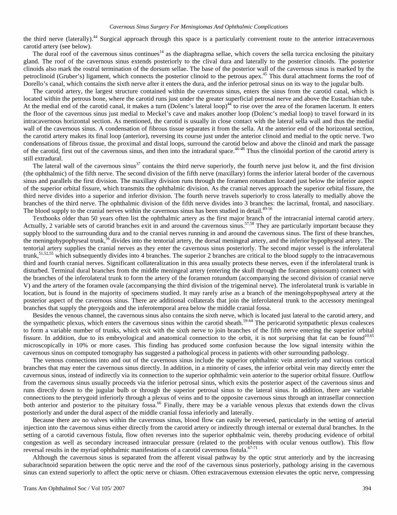
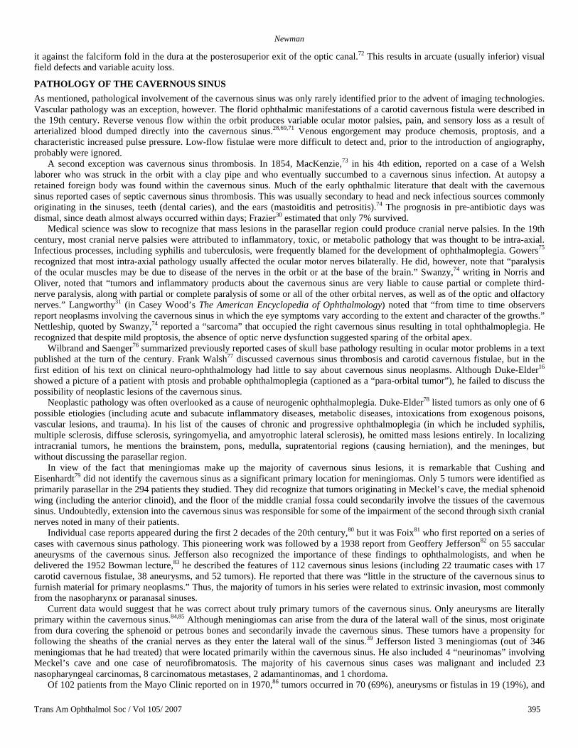
FIGURE 1
Case 2. Computed tomography (left) and magnetic resonance imaging (right) scans of a patient with a long history of a known skull base meningioma. The tumor can be seen to be encasing the carotid and middle cerebral arteries.
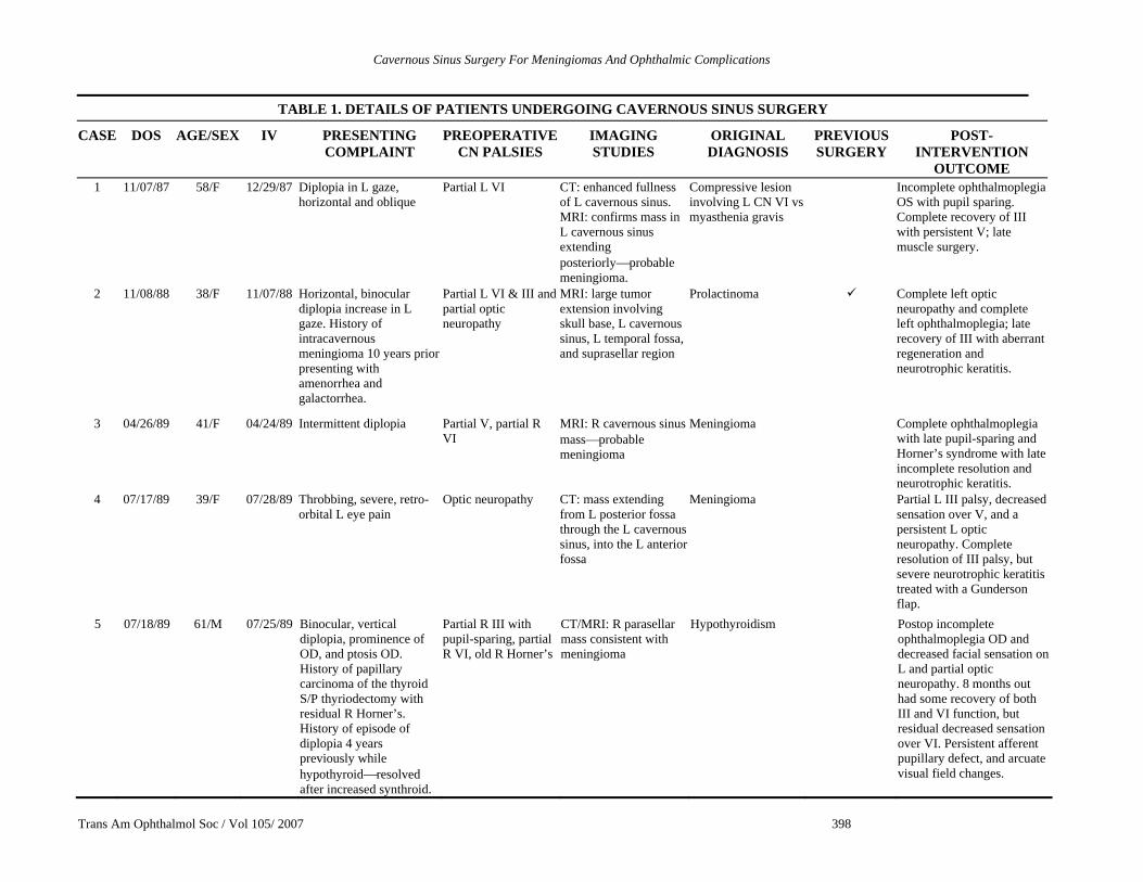
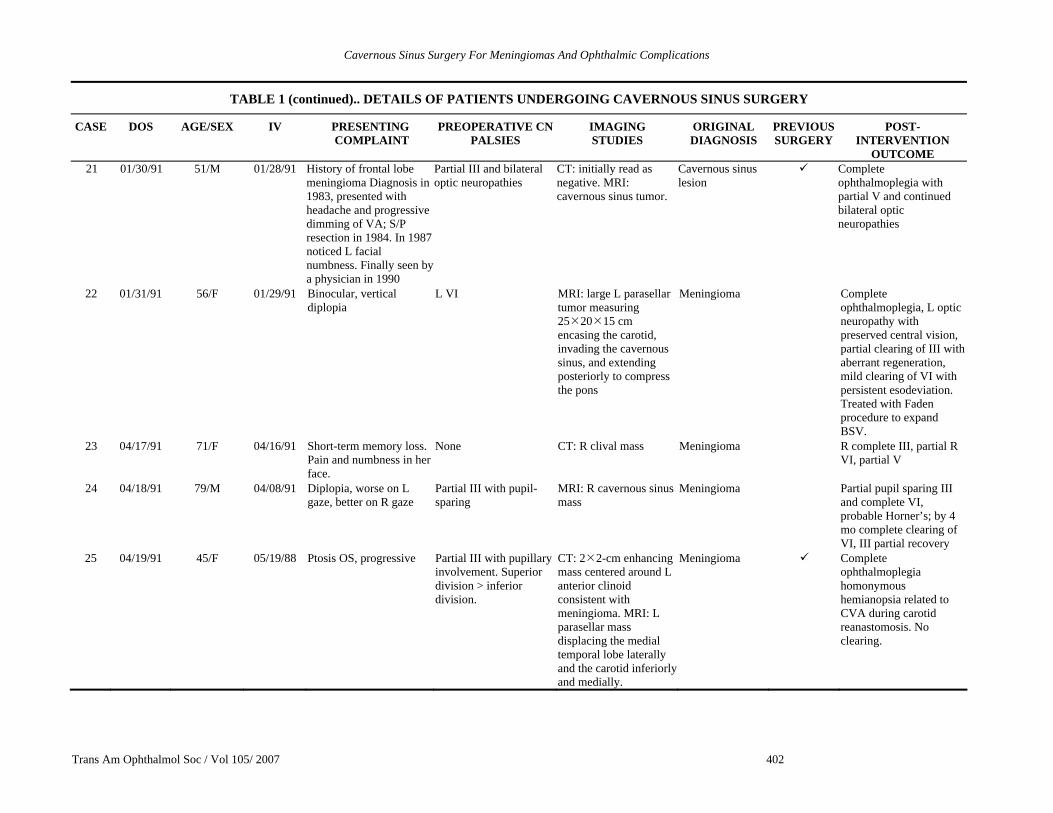
The initial series consisted of a total of 81 patients who underwent 82 cavernous sinus surgical procedures. Of these patients undergoing cavernous sinus surgery, 56 were found postoperatively to have meningiomas (Table 1). This series included 16 men and 40 women whose ages ranged from 23 to 81 years with a mean of 51 years. One man had a recurrence and underwent a second procedure to extirpate the cavernous sinus meningioma. The time from initial development of symptoms to diagnosis ranged from less than 1 week to 168 months, with a mean of 18.9 months. Diagnosis was delayed by more than 2 months in 23 of 57 cases (as much as 13 years). In one remarkable case the diagnosis was delayed for 2 years in spite of the fact that the patient had previously had a meningioma resected 10 years earlier. Diagnoses made prior to the correct identification included microvascular cranial nerve palsies in 3 patients. Sinus disease, “lazy eye,” and multiple sclerosis were each diagnosed in 2 patients. Other initial diagnoses included myasthenia, thyroid disease, labyrinthitis, trigeminal neuralgia, migraine, hypothyroidism, and “nerves.” Twenty-one patients (37%) had previous surgery.
Cavernous Sinus Surgery For Meningiomas And Ophthalmic Complications
Trans Am Ophthalmol Soc / Vol 105/ 2007 398
5 07/18/89 61/M 07/25/89 Binocular, vertical diplopia, prominence of OD, and ptosis OD. History of papillary carcinoma of the thyroid S/P thyriodectomy with residual R Horner’s. History of episode of diplopia 4 years previously while hypothyroid⎯resolved after increased synthroid.
Partial R III with pupil-sparing, partial R VI, old R Horner’s
CT/MRI: R parasellar mass consistent with meningioma
Hypothyroidism Postop incomplete ophthalmoplegia OD and decreased facial sensation on L and partial optic neuropathy. 8 months out had some recovery of both III and VI function, but residual decreased sensation over VI. Persistent afferent pupillary defect, and arcuate visual field changes.
TABLE 1. DETAILS OF PATIENTS UNDERGOING CAVERNOUS SINUS SURGERY
CASE DOS AGE/SEX IV PRESENTING COMPLAINT
PREOPERATIVE CN PALSIES
IMAGING STUDIES
ORIGINAL DIAGNOSIS
PREVIOUS SURGERY
POST-INTERVENTION
OUTCOME 1 11/07/87 58/F 12/29/87 Diplopia in L gaze,
horizontal and oblique Partial L VI CT: enhanced fullness
of L cavernous sinus. MRI: confirms mass in L cavernous sinus extending posteriorly⎯probable meningioma.
Compressive lesion involving L CN VI vs myasthenia gravis
Incomplete ophthalmoplegia OS with pupil sparing. Complete recovery of III with persistent V; late muscle surgery.
2 11/08/88 38/F 11/07/88 Horizontal, binocular diplopia increase in L gaze. History of intracavernous meningioma 10 years prior presenting with amenorrhea and galactorrhea.
Partial L VI & III and partial optic neuropathy
MRI: large tumor extension involving skull base, L cavernous sinus, L temporal fossa, and suprasellar region
Prolactinoma Complete left optic neuropathy and complete left ophthalmoplegia; late recovery of III with aberrant regeneration and neurotrophic keratitis.
3 04/26/89 41/F 04/24/89 Intermittent diplopia Partial V, partial R VI
MRI: R cavernous sinus mass⎯probable meningioma
Meningioma Complete ophthalmoplegia with late pupil-sparing and Horner’s syndrome with late incomplete resolution and neurotrophic keratitis.
4 07/17/89 39/F 07/28/89 Throbbing, severe, retro-orbital L eye pain
Optic neuropathy CT: mass extending from L posterior fossa through the L cavernous sinus, into the L anterior fossa
Meningioma Partial L III palsy, decreased sensation over V, and a persistent L optic neuropathy. Complete resolution of III palsy, but severe neurotrophic keratitis treated with a Gunderson flap.
Newman
Trans Am Ophthalmol Soc / Vol 105/ 2007 399
TABLE 1 (continued). DETAILS OF PATIENTS UNDERGOING CAVERNOUS SINUS SURGERY
CASE DOS AGE/SEX IV PRESENTING COMPLAINT
PREOPERATIVE CN PALSIES
IMAGING STUDIES
ORIGINAL DIAGNOSIS
PREVIOUS SURGERY
POST-INTERVENTION
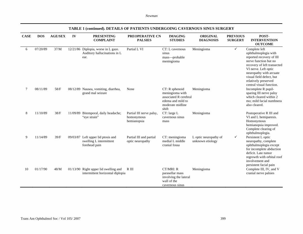
OUTCOME 6 07/20/89 37/M 12/21/86 Diplopia, worse in L gaze.
Auditory hallucinations in L ear.
Partial L VI CT: L cavernous sinus mass⎯probable meningioma
Meningioma Complete left ophthalmoplegia with reported recovery of III nerve function but no recovery of left transected VI nerve. Left optic neuropathy with arcuate visual field defect, but relatively preserved central visual function.
7 08/11/89 58/F 08/12/89 Nausea, vomiting, diarrhea, grand mal seizure
None CT: R sphenoid meningioma with associated R cerebral edema and mild to moderate midline shift
Meningioma Incomplete R pupil-sparing III nerve palsy which cleared within 2 mo; mild facial numbness also cleared.
8 11/10/89 38/F 11/09/89 Bitemporal, daily headache; “eye strain”
Partial III nerve palsy, homonymous hemianopsia
CT: large L cavernous sinus mass
Meningioma Postoperative R III and VI and L hemiparesis. Homonymous hemianopsia improved. Complete clearing of ophthalmoplegia.
9 11/14/89 39/F 09/03/87 Left upper lid ptosis and swelling L intermittent forehead pain
Partial III and partial optic neuropathy
CT: meningioma medial L middle cranial fossa
L optic neuropathy of unknown etiology
Persistent L optic neuropathy, complete ophthalmoplegia except for incomplete abduction deficit. Late tumor regrowth with orbital roof involvement and persistent facial pain
10 01/17/90 48/M 01/13/90 Right upper lid swelling and intermittent horizontal diplopia
R III CT/MRI: R parasellar mass involving the lateral wall of the cavernous sinus
Meningioma Complete III, IV, and V cranial nerve palsies
Cavernous Sinus Surgery For Meningiomas And Ophthalmic Complications
Trans Am Ophthalmol Soc / Vol 105/ 2007 400
TABLE 1 (continued). DETAILS OF PATIENTS UNDERGOING CAVERNOUS SINUS SURGERY
CASE DOS AGE/SEX IV PRESENTING COMPLAINT
PREOPERATIVE CN PALSIES
IMAGING STUDIES
ORIGINAL DIAGNOSIS
PREVIOUS SURGERY
POST- INTERVENTION
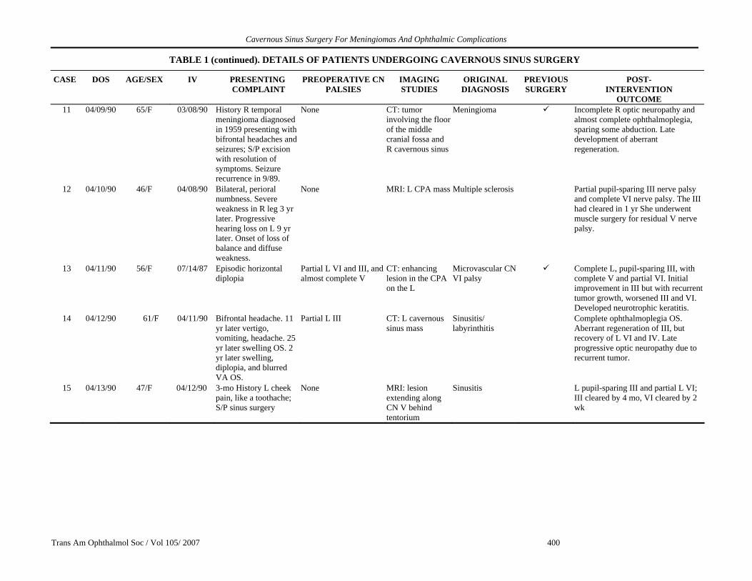
OUTCOME 11 04/09/90 65/F 03/08/90 History R temporal
meningioma diagnosed in 1959 presenting with bifrontal headaches and seizures; S/P excision with resolution of symptoms. Seizure recurrence in 9/89.
None CT: tumor involving the floor of the middle cranial fossa and R cavernous sinus
Meningioma Incomplete R optic neuropathy and almost complete ophthalmoplegia, sparing some abduction. Late development of aberrant regeneration.
12 04/10/90 46/F 04/08/90 Bilateral, perioral numbness. Severe weakness in R leg 3 yr later. Progressive hearing loss on L 9 yr later. Onset of loss of balance and diffuse weakness.
None MRI: L CPA mass Multiple sclerosis Partial pupil-sparing III nerve palsy and complete VI nerve palsy. The III had cleared in 1 yr She underwent muscle surgery for residual V nerve palsy.
13 04/11/90 56/F 07/14/87 Episodic horizontal diplopia
Partial L VI and III, and almost complete V
CT: enhancing lesion in the CPA on the L
Microvascular CN VI palsy
Complete L, pupil-sparing III, with complete V and partial VI. Initial improvement in III but with recurrent tumor growth, worsened III and VI. Developed neurotrophic keratitis.
14 04/12/90 61/F 04/11/90 Bifrontal headache. 11 yr later vertigo, vomiting, headache. 25 yr later swelling OS. 2 yr later swelling, diplopia, and blurred VA OS.
Partial L III CT: L cavernous sinus mass
Sinusitis/ labyrinthitis
Complete ophthalmoplegia OS. Aberrant regeneration of III, but recovery of L VI and IV. Late progressive optic neuropathy due to recurrent tumor.
15 04/13/90 47/F 04/12/90 3-mo History L cheek pain, like a toothache; S/P sinus surgery
None MRI: lesion extending along CN V behind tentorium
Sinusitis L pupil-sparing III and partial L VI; III cleared by 4 mo, VI cleared by 2 wk
Newman
Trans Am Ophthalmol Soc / Vol 105/ 2007 401
TABLE 1 (continued). DETAILS OF PATIENTS UNDERGOING CAVERNOUS SINUS SURGERY
CASE DOS AGE/SEX IV PRESENTING COMPLAINT
PREOPERATIVE CN PALSIES
IMAGING STUDIES
ORIGINAL DIAGNOSIS
PREVIOUS SURGERY
POST- INTERVENTION
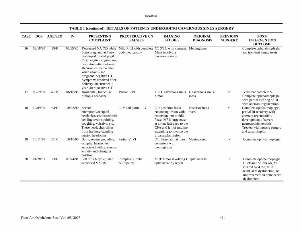
OUTCOME 16 06/18/90 29/F 06/15/90 Decreased VA OD while
5 mo pregnant; at 7 mo developed dilated pupil OD; negative angiogram; resolution after delivery. Recurrence 15 mo later when again 5 mo pregnant; negative CT. Symptoms resolved after delivery. Recurrence 1 year later; positive CT
Mild R III with complete optic neuropathy
CT 3/85: with contrast. Mass involving cavernous sinus.
Meningioma Complete ophthalmoplegia and transient hemiparesis
17 06/19/90 49/M 06/18/90 Horizontal, binocular diplopia headache
Partial L VI CT: L cavernous sinus tumor
L cavernous sinus mass
Persistent complete VI. Complete ophthalmoplegia with partial clearing of III with aberrant regeneration.
18 10/09/90 54/F 10/08/90 Severe bitemporal/occipital headaches associated with bending over, straining, coughing, valsalva, etc. These headaches differ from her long-standing tension headaches.
L IV and partial L V CT: posterior fossa enhancing lesion with extension into middle fossa. MRI: large mass at clivus just deep to the CPA and left of midline extending to involve the L parasellar region.
Posterior fossa mass
Complete ophthalmoplegia, partial III recovery with aberrant regeneration, development of severe neurotrophic keratitis. Treated with muscle surgery and tarsorrhaphy.
19 10/11/90 27/M 10/10/90 Daily, severe, pounding, occipital headaches associated with strenuous activity and changing position
Partial V, VI CT: large cranial mass consistent with meningioma
Meningioma Complete ophthalmoplegia
20 01/28/91 23/F 01/24/91 Fell off a bicycle; later decreased VA OS
Complete L optic neuropathy
MRI: tumor involving L optic nerve by report
Optic neuritis Complete ophthalmoplegia III cleared within wk, VI cleared by 4 mo, mild residual V dysfunction, no improvement in optic nerve dysfunction
Cavernous Sinus Surgery For Meningiomas And Ophthalmic Complications
Trans Am Ophthalmol Soc / Vol 105/ 2007 402
TABLE 1 (continued).. DETAILS OF PATIENTS UNDERGOING CAVERNOUS SINUS SURGERY
CASE DOS AGE/SEX IV PRESENTING COMPLAINT
PREOPERATIVE CN PALSIES
IMAGING STUDIES
ORIGINAL DIAGNOSIS
PREVIOUS SURGERY
POST-INTERVENTION
OUTCOME 21 01/30/91 51/M 01/28/91 History of frontal lobe
meningioma Diagnosis in 1983, presented with headache and progressive dimming of VA; S/P resection in 1984. In 1987 noticed L facial numbness. Finally seen by a physician in 1990
Partial III and bilateral optic neuropathies
CT: initially read as negative. MRI: cavernous sinus tumor.
Cavernous sinus lesion
Complete ophthalmoplegia with partial V and continued bilateral optic neuropathies
22 01/31/91 56/F 01/29/91 Binocular, vertical diplopia
L VI MRI: large L parasellar tumor measuring 25 20 15 cm encasing the carotid, invading the cavernous sinus, and extending posteriorly to compress the pons
Meningioma Complete ophthalmoplegia, L optic neuropathy with preserved central vision, partial clearing of III with aberrant regeneration, mild clearing of VI with persistent esodeviation. Treated with Faden procedure to expand BSV.
23 04/17/91 71/F 04/16/91 Short-term memory loss. Pain and numbness in her face.
None CT: R clival mass Meningioma R complete III, partial R VI, partial V
24 04/18/91 79/M 04/08/91 Diplopia, worse on L gaze, better on R gaze
Partial III with pupil-sparing
MRI: R cavernous sinus mass
Meningioma Partial pupil sparing III and complete VI, probable Horner’s; by 4 mo complete clearing of VI, III partial recovery
25 04/19/91 45/F 05/19/88 Ptosis OS, progressive Partial III with pupillary involvement. Superior division > inferior division.
CT: 2 2-cm enhancing mass centered around L anterior clinoid consistent with meningioma. MRI: L parasellar mass displacing the medial temporal lobe laterally and the carotid inferiorly and medially.
Meningioma Complete ophthalmoplegia homonymous hemianopsia related to CVA during carotid reanastomosis. No clearing.
Newman
Trans Am Ophthalmol Soc / Vol 105/ 2007 403
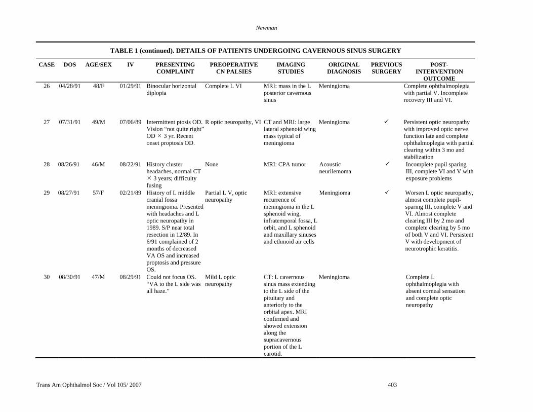
TABLE 1 (continued). DETAILS OF PATIENTS UNDERGOING CAVERNOUS SINUS SURGERY
CASE DOS AGE/SEX IV PRESENTING COMPLAINT
PREOPERATIVE CN PALSIES
IMAGING STUDIES
ORIGINAL DIAGNOSIS
PREVIOUS SURGERY
POST- INTERVENTION
OUTCOME 26 04/28/91 48/F 01/29/91 Binocular horizontal
diplopia Complete L VI MRI: mass in the L
posterior cavernous sinus
Meningioma Complete ophthalmoplegia with partial V. Incomplete recovery III and VI.
27 07/31/91 49/M 07/06/89 Intermittent ptosis OD. Vision “not quite right” OD 3 yr. Recent onset proptosis OD.
R optic neuropathy, VI CT and MRI: large lateral sphenoid wing mass typical of meningioma
Meningioma Persistent optic neuropathy with improved optic nerve function late and complete ophthalmoplegia with partial clearing within 3 mo and stabilization
28 08/26/91 46/M 08/22/91 History cluster headaches, normal CT
3 years; difficulty fusing
None MRI: CPA tumor Acoustic neurilemoma
Incomplete pupil sparing III, complete VI and V with exposure problems
29 08/27/91 57/F 02/21/89 History of L middle cranial fossa meningioma. Presented with headaches and L optic neuropathy in 1989. S/P near total resection in 12/89. In 6/91 complained of 2 months of decreased VA OS and increased proptosis and pressure OS.
Partial L V, optic neuropathy
MRI: extensive recurrence of meningioma in the L sphenoid wing, infratemporal fossa, L orbit, and L sphenoid and maxillary sinuses and ethmoid air cells
Meningioma Worsen L optic neuropathy, almost complete pupil-sparing III, complete V and VI. Almost complete clearing III by 2 mo and complete clearing by 5 mo of both V and VI. Persistent V with development of neurotrophic keratitis.
30 08/30/91 47/M 08/29/91 Could not focus OS. “VA to the L side was all haze.”
Mild L optic neuropathy
CT: L cavernous sinus mass extending to the L side of the pituitary and anteriorly to the orbital apex. MRI confirmed and showed extension along the supracavernous portion of the L carotid.
Meningioma Complete L ophthalmoplegia with absent corneal sensation and complete optic neuropathy
Cavernous Sinus Surgery For Meningiomas And Ophthalmic Complications
Trans Am Ophthalmol Soc / Vol 105/ 2007 404
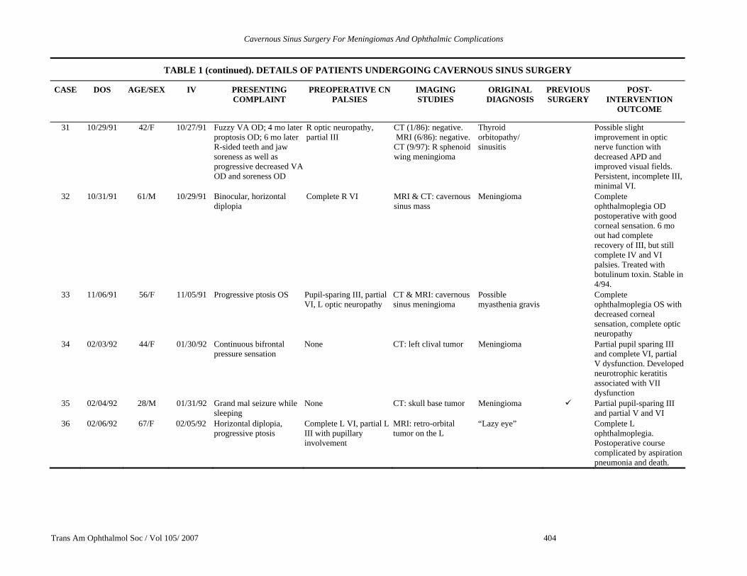
TABLE 1 (continued). DETAILS OF PATIENTS UNDERGOING CAVERNOUS SINUS SURGERY
CASE DOS AGE/SEX IV PRESENTING COMPLAINT
PREOPERATIVE CN PALSIES
IMAGING STUDIES
ORIGINAL DIAGNOSIS
PREVIOUS SURGERY
POST-INTERVENTION
OUTCOME
31 10/29/91 42/F 10/27/91 Fuzzy VA OD; 4 mo later proptosis OD; 6 mo later R-sided teeth and jaw soreness as well as progressive decreased VA OD and soreness OD
R optic neuropathy, partial III
CT (1/86): negative. MRI (6/86): negative. CT (9/97): R sphenoid wing meningioma
Thyroid orbitopathy/ sinusitis
Possible slight improvement in optic nerve function with decreased APD and improved visual fields. Persistent, incomplete III, minimal VI.
32 10/31/91 61/M 10/29/91 Binocular, horizontal diplopia
Complete R VI MRI & CT: cavernous sinus mass
Meningioma Complete ophthalmoplegia OD postoperative with good corneal sensation. 6 mo out had complete recovery of III, but still complete IV and VI palsies. Treated with botulinum toxin. Stable in 4/94.
33 11/06/91 56/F 11/05/91 Progressive ptosis OS Pupil-sparing III, partial VI, L optic neuropathy
CT & MRI: cavernous sinus meningioma
Possible myasthenia gravis
Complete ophthalmoplegia OS with decreased corneal sensation, complete optic neuropathy
34 02/03/92 44/F 01/30/92 Continuous bifrontal pressure sensation
None CT: left clival tumor Meningioma Partial pupil sparing III and complete VI, partial V dysfunction. Developed neurotrophic keratitis associated with VII dysfunction
35 02/04/92 28/M 01/31/92 Grand mal seizure while sleeping
None CT: skull base tumor Meningioma Partial pupil-sparing III and partial V and VI
36 02/06/92 67/F 02/05/92 Horizontal diplopia, progressive ptosis
Complete L VI, partial L III with pupillary involvement
MRI: retro-orbital tumor on the L
“Lazy eye” Complete L ophthalmoplegia. Postoperative course complicated by aspiration pneumonia and death.
Newman
Trans Am Ophthalmol Soc / Vol 105/ 2007 405
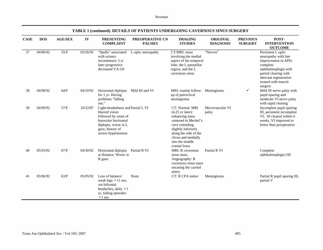
TABLE 1 (continued). DETAILS OF PATIENTS UNDERGOING CAVERNOUS SINUS SURGERY
CASE DOS AGE/SEX IV PRESENTING COMPLAINT
PREOPERATIVE CN PALSIES
IMAGING STUDIES
ORIGINAL DIAGNOSIS
PREVIOUS SURGERY
POST-INTERVENTION
OUTCOME 37 04/06/92 55/F 03/26/92 “Spells” associated
with urinary incontinence 3 yr later progressive decreased VA OS
L optic neuropathy CT/MRI: mass involving the medial aspect of the temporal lobe, the L parasellar region, and the L cavernous sinus
“Nerves” Persistent L optic neuropathy with late improvement in APD, complete ophthalmoplegia with partial clearing with aberrant regeneration treated with muscle surgery
38 04/08/92 64/F 04/10/92 Horizontal diplopia for 1 yr. Having problems “falling out.”
Mild III and VI MRI: routine follow-up of petroclival meningioma
Meningioma Mild III nerve palsy with pupil-sparing and moderate VI nerve palsy with rapid clearing
39 04/09/92 57/F 10/22/87 Light-headedness and blurred vision followed by onset of binocular horizontal diplopia, worse in L gaze, history of severe hypertension
Partial L VI CT: Normal. MRI (4.25 yr later): enhancing mass centered in Meckel’s cave extending slightly inferiorly along the side of the clivus and medially into the middle cranial fossa
Microvascular VI palsy
Incomplete pupil sparing III, persistent incomplete VI; III cleared within 6 weeks, VI improved to better than preoperative
40 05/03/92 67/F 04/30/92 Horizontal diplopia at distance. Worse in R gaze.
Partial R VI MRI: R cavernous sinus mass. Angiography: R cavernous sinus mass encasing the carotid artery
Partial R VI Complete ophthalmoplegia OD
41 05/06/92 63/F 05/05/92 Loss of balance/ weak legs 11 mo, AM bifrontal headaches, daily 1 yr, falling episodes
1 mo
None CT: R CPA tumor Meningioma Partial R pupil sparing III, partial V
Cavernous Sinus Surgery For Meningiomas And Ophthalmic Complications
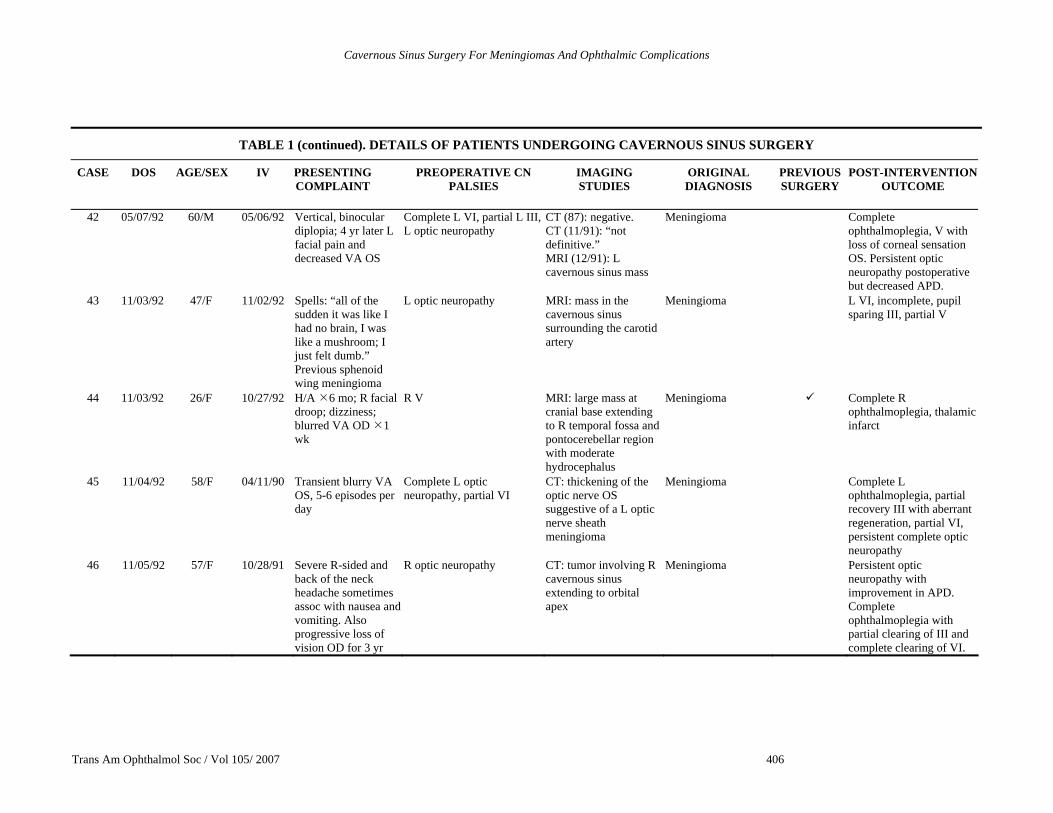
Trans Am Ophthalmol Soc / Vol 105/ 2007 406
TABLE 1 (continued). DETAILS OF PATIENTS UNDERGOING CAVERNOUS SINUS SURGERY
CASE DOS AGE/SEX IV PRESENTING COMPLAINT
PREOPERATIVE CN PALSIES
IMAGING STUDIES
ORIGINAL DIAGNOSIS
PREVIOUS SURGERY
POST-INTERVENTION OUTCOME
42 05/07/92 60/M 05/06/92 Vertical, binocular diplopia; 4 yr later L facial pain and decreased VA OS
Complete L VI, partial L III, L optic neuropathy
CT (87): negative. CT (11/91): “not definitive.” MRI (12/91): L cavernous sinus mass
Meningioma Complete ophthalmoplegia, V with loss of corneal sensation OS. Persistent optic neuropathy postoperative but decreased APD.
43 11/03/92 47/F 11/02/92 Spells: “all of the sudden it was like I had no brain, I was like a mushroom; I just felt dumb.” Previous sphenoid wing meningioma
L optic neuropathy MRI: mass in the cavernous sinus surrounding the carotid artery
Meningioma L VI, incomplete, pupil sparing III, partial V
44 11/03/92 26/F 10/27/92 H/A 6 mo; R facial droop; dizziness; blurred VA OD 1 wk
R V MRI: large mass at cranial base extending to R temporal fossa and pontocerebellar region with moderate hydrocephalus
Meningioma Complete R ophthalmoplegia, thalamic infarct
45 11/04/92 58/F 04/11/90 Transient blurry VA OS, 5-6 episodes per day
Complete L optic neuropathy, partial VI
CT: thickening of the optic nerve OS suggestive of a L optic nerve sheath meningioma
Meningioma Complete L ophthalmoplegia, partial recovery III with aberrant regeneration, partial VI, persistent complete optic neuropathy
46 11/05/92 57/F 10/28/91 Severe R-sided and back of the neck headache sometimes assoc with nausea and vomiting. Also progressive loss of vision OD for 3 yr
R optic neuropathy CT: tumor involving R cavernous sinus extending to orbital apex
Meningioma Persistent optic neuropathy with improvement in APD. Complete ophthalmoplegia with partial clearing of III and complete clearing of VI.
Newman
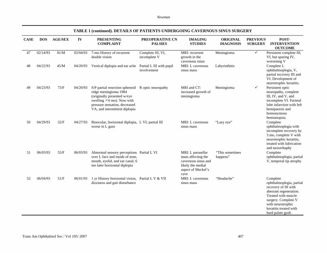
Trans Am Ophthalmol Soc / Vol 105/ 2007 407
TABLE 1 (continued). DETAILS OF PATIENTS UNDERGOING CAVERNOUS SINUS SURGERY
CASE DOS AGE/SEX IV PRESENTING COMPLAINT
PREOPERATIVE CN PALSIES
IMAGING STUDIES
ORIGINAL DIAGNOSIS
PREVIOUS SURGERY
POST-INTERVENTION
OUTCOME 47 02/14/93 81/M 02/04/93 7-mo History of recurrent
double vision Complete III, VI, incomplete V
MRI: recurrent growth in the cavernous sinus
Meningioma Persistent complete III, VI, but sparing IV, worsening V
48 04/22/93 45/M 04/20/93 Vertical diplopia and ear ache Partial L III with pupil involvement
MRI: L cavernous sinus mass
Labyrinthitis Complete L ophthalmoplegia, V, partial recovery III and VI. Development of neurotrophic keratitis.
49 04/23/93 73/F 04/20/93 S/P partial resection sphenoid ridge meningioma 1984 (originally presented w/eye swelling 6 mo). Now with pressure sensation, decreased VA, and intermittent diplopia
R optic neuropathy MRI and CT: increased growth of meningioma
Meningioma Persistent optic neuropathy, complete III, IV, and V, and incomplete VI. Parietal lobe infarction with left hemiparesis and homonymous hemianopsia.
50 04/29/93 32/F 04/27/93 Binocular, horizontal diplopia, worse in L gaze
L VI; partial III MRI: L cavernous sinus mass
“Lazy eye” Complete ophthalmoplegia with incomplete recovery by 3 mo, complete V with neurotrophic keratitis, treated with lubrication and tarsorrhaphy
51 06/03/93 55/F 06/03/93 Abnormal sensory perceptions over L face and inside of nose, mouth, eyelid, and ear canal; 6 mo later horizontal diplopia
Partial L VI MRI: L parasellar mass affecting the cavernous sinus and likely the medial aspect of Meckel’s cave
“This sometimes happens”
Complete ophthalmoplegia, partial V, temporal tip atrophy
52 06/04/93 51/F 06/01/93 1 yr History horizontal vision, dizziness and gait disturbance
Partial L V & VII MRI: L cavernous sinus mass
“Headache” Complete ophthalmoplegia, partial recovery of III with aberrant regeneration. Treated with muscle surgery. Complete V with neurotrophic keratitis treated with hard palate graft.
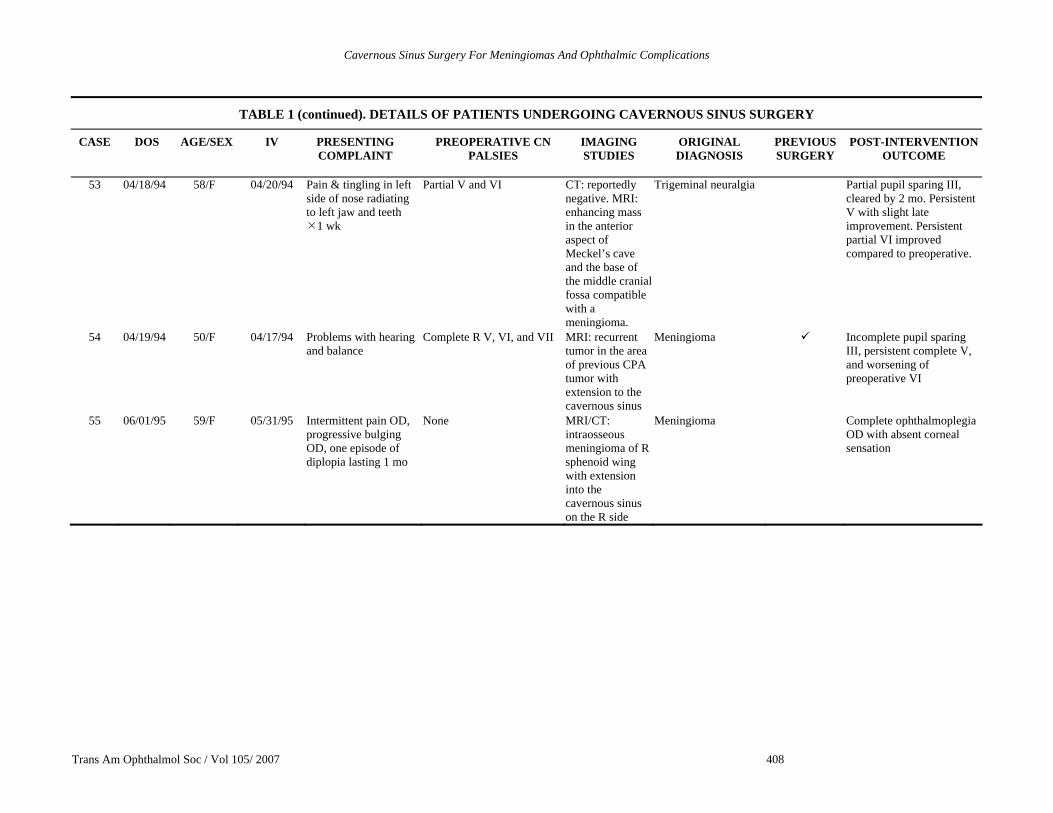
Cavernous Sinus Surgery For Meningiomas And Ophthalmic Complications
Trans Am Ophthalmol Soc / Vol 105/ 2007 408
TABLE 1 (continued). DETAILS OF PATIENTS UNDERGOING CAVERNOUS SINUS SURGERY
CASE DOS AGE/SEX IV PRESENTING COMPLAINT
PREOPERATIVE CN PALSIES
IMAGING STUDIES
ORIGINAL DIAGNOSIS
PREVIOUS SURGERY
POST-INTERVENTION OUTCOME
53 04/18/94 58/F 04/20/94 Pain & tingling in left side of nose radiating to left jaw and teeth
1 wk
Partial V and VI CT: reportedly negative. MRI: enhancing mass in the anterior aspect of Meckel’s cave and the base of the middle cranial fossa compatible with a meningioma.
Trigeminal neuralgia Partial pupil sparing III, cleared by 2 mo. Persistent V with slight late improvement. Persistent partial VI improved compared to preoperative.
54 04/19/94 50/F 04/17/94 Problems with hearing and balance
Complete R V, VI, and VII MRI: recurrent tumor in the area of previous CPA tumor with extension to the cavernous sinus
Meningioma Incomplete pupil sparing III, persistent complete V, and worsening of preoperative VI
55 06/01/95 59/F 05/31/95 Intermittent pain OD, progressive bulging OD, one episode of diplopia lasting 1 mo
None MRI/CT: intraosseous meningioma of R sphenoid wing with extension into the cavernous sinus on the R side
Meningioma Complete ophthalmoplegia OD with absent corneal sensation
Newman
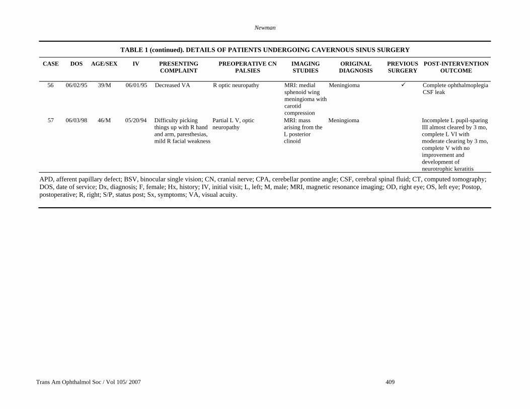
Trans Am Ophthalmol Soc / Vol 105/ 2007 409
TABLE 1 (continued). DETAILS OF PATIENTS UNDERGOING CAVERNOUS SINUS SURGERY
CASE DOS AGE/SEX IV PRESENTING COMPLAINT
PREOPERATIVE CN PALSIES
IMAGING STUDIES
ORIGINAL DIAGNOSIS
PREVIOUS SURGERY
POST-INTERVENTION OUTCOME
56 06/02/95 39/M 06/01/95 Decreased VA R optic neuropathy MRI: medial sphenoid wing meningioma with carotid compression
Meningioma Complete ophthalmoplegia CSF leak
57 06/03/98 46/M 05/20/94 Difficulty picking things up with R hand and arm, paresthesias, mild R facial weakness
Partial L V, optic neuropathy
MRI: mass arising from the L posterior clinoid
Meningioma Incomplete L pupil-sparing III almost cleared by 3 mo, complete L VI with moderate clearing by 3 mo, complete V with no improvement and development of neurotrophic keratitis
APD, afferent papillary defect; BSV, binocular single vision; CN, cranial nerve; CPA, cerebellar pontine angle; CSF, cerebral spinal fluid; CT, computed tomography; DOS, date of service; Dx, diagnosis; F, female; Hx, history; IV, initial visit; L, left; M, male; MRI, magnetic resonance imaging; OD, right eye; OS, left eye; Postop, postoperative; R, right; S/P, status post; Sx, symptoms; VA, visual acuity.
Cavernous Sinus Surgery For Meningiomas And Ophthalmic Complications
Trans Am Ophthalmol Soc / Vol 105/ 2007 410
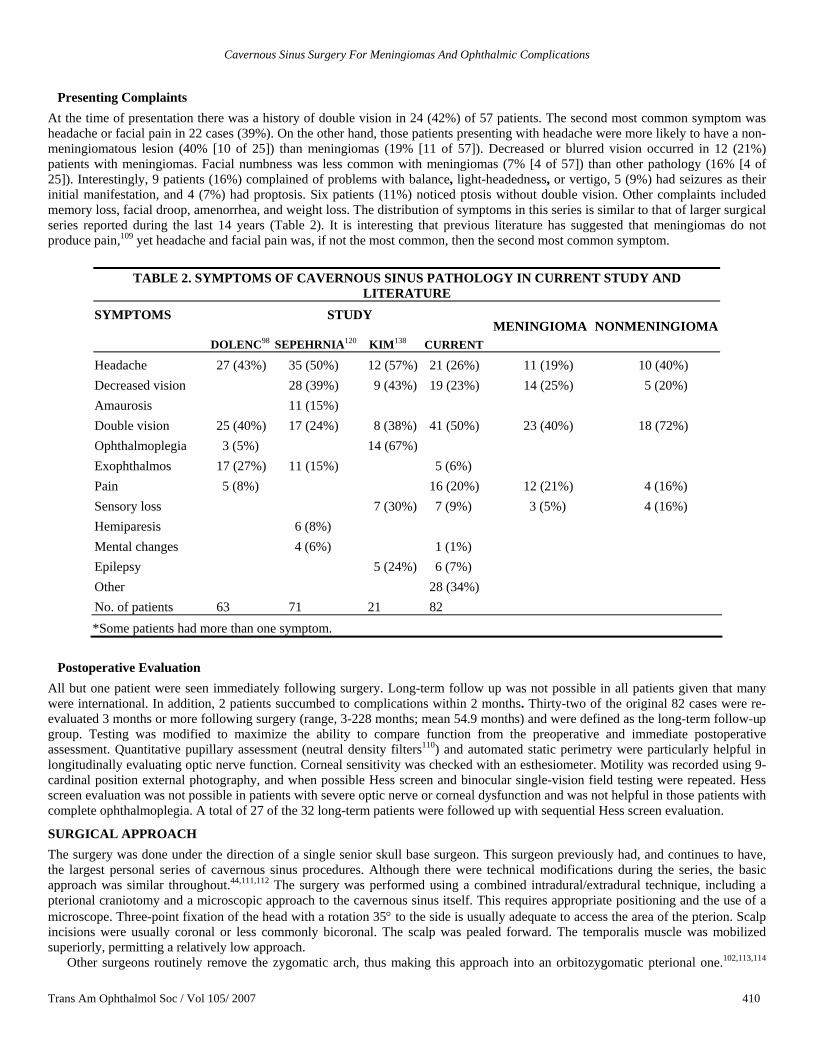
Presenting Complaints
At the time of presentation there was a history of double vision in 24 (42%) of 57 patients. The second most common symptom was headache or facial pain in 22 cases (39%). On the other hand, those patients presenting with headache were more likely to have a non-meningiomatous lesion (40% [10 of 25]) than meningiomas (19% [11 of 57]). Decreased or blurred vision occurred in 12 (21%) patients with meningiomas. Facial numbness was less common with meningiomas (7% [4 of 57]) than other pathology (16% [4 of 25]). Interestingly, 9 patients (16%) complained of problems with balance, light-headedness, or vertigo, 5 (9%) had seizures as their initial manifestation, and 4 (7%) had proptosis. Six patients (11%) noticed ptosis without double vision. Other complaints included memory loss, facial droop, amenorrhea, and weight loss. The distribution of symptoms in this series is similar to that of larger surgical series reported during the last 14 years (Table 2). It is interesting that previous literature has suggested that meningiomas do not produce pain,109 yet headache and facial pain was, if not the most common, then the second most common symptom.
TABLE 2. SYMPTOMS OF CAVERNOUS SINUS PATHOLOGY IN CURRENT STUDY AND LITERATURE
SYMPTOMS STUDY
DOLENC98 SEPEHRNIA120 KIM138 CURRENT MENINGIOMA NONMENINGIOMA
Headache 27 (43%) 35 (50%) 12 (57%) 21 (26%) 11 (19%) 10 (40%) Decreased vision 28 (39%) 9 (43%) 19 (23%) 14 (25%) 5 (20%) Amaurosis 11 (15%) Double vision 25 (40%) 17 (24%) 8 (38%) 41 (50%) 23 (40%) 18 (72%) Ophthalmoplegia 3 (5%) 14 (67%) Exophthalmos 17 (27%) 11 (15%) 5 (6%) Pain 5 (8%) 16 (20%) 12 (21%) 4 (16%) Sensory loss 7 (30%) 7 (9%) 3 (5%) 4 (16%) Hemiparesis 6 (8%) Mental changes 4 (6%) 1 (1%) Epilepsy 5 (24%) 6 (7%) Other 28 (34%) No. of patients 63 71 21 82 *Some patients had more than one symptom.
Postoperative Evaluation
All but one patient were seen immediately following surgery. Long-term follow up was not possible in all patients given that many were international. In addition, 2 patients succumbed to complications within 2 months. Thirty-two of the original 82 cases were re-evaluated 3 months or more following surgery (range, 3-228 months; mean 54.9 months) and were defined as the long-term follow-up group. Testing was modified to maximize the ability to compare function from the preoperative and immediate postoperative assessment. Quantitative pupillary assessment (neutral density filters110) and automated static perimetry were particularly helpful in longitudinally evaluating optic nerve function. Corneal sensitivity was checked with an esthesiometer. Motility was recorded using 9-cardinal position external photography, and when possible Hess screen and binocular single-vision field testing were repeated. Hess screen evaluation was not possible in patients with severe optic nerve or corneal dysfunction and was not helpful in those patients with complete ophthalmoplegia. A total of 27 of the 32 long-term patients were followed up with sequential Hess screen evaluation.
SURGICAL APPROACH The surgery was done under the direction of a single senior skull base surgeon. This surgeon previously had, and continues to have, the largest personal series of cavernous sinus procedures. Although there were technical modifications during the series, the basic approach was similar throughout.44,111,112 The surgery was performed using a combined intradural/extradural technique, including a pterional craniotomy and a microscopic approach to the cavernous sinus itself. This requires appropriate positioning and the use of a microscope. Three-point fixation of the head with a rotation 35° to the side is usually adequate to access the area of the pterion. Scalp incisions were usually coronal or less commonly bicoronal. The scalp was pealed forward. The temporalis muscle was mobilized superiorly, permitting a relatively low approach.
Other surgeons routinely remove the zygomatic arch, thus making this approach into an orbitozygomatic pterional one.102,113,114
Newman
Trans Am Ophthalmol Soc / Vol 105/ 2007 411
This was believed to be unnecessary in this series. A single burr hole was placed just behind the superior portion of the lateral orbital rim. This ideally entered the orbit as well as the anterior cranial fossa. A second burr hole was placed posteriorly in the temporal region. The dura was freed from the overlying bone, and a pterional-based bone flap was raised, which permitted immediate view of the dura of both the anterior and middle cranial fossae.
An extradural dissection was then carried down to the foramen spinosum, where the middle meningeal artery was identified and ligated. Dissection was carried forward, while identifying the foramen ovale and the foramen rotundum anteriorly, all extradurally. The course of the intrapetrous carotid artery was also identified. The carotid canal,115 within the petrous bone, was opened by dividing the greater superficial petrosal nerve (to prevent traction on the facial nerve) and by carefully removing bone back to the posterior loop of the carotid artery. Other surgeons have used this portion of the carotid artery to base a bypass graft when carotid sacrifice is planned.116-118
One of the disadvantages of this approach is the loss of the greater superficial petrosal nerve, which results in decreased reflex tearing by interrupting the parasympathetic innervation to the lacrimal gland. Wright119 has more recently advocated a longer saphenous vein bypass graft from the cervical carotid to the distal supraclinoid carotid to avoid sacrificing the greater superficial petrosal nerve. Other surgeons believe that the possible complications of carotid revascularization procedures (as part of cavernous sinus surgery) outweigh their potential usefulness in increasing resectability.120 De Monte121 has written that carotid sacrifice is rarely, if ever, indicated. In this series, although 1 carotid artery ruptured and required sacrifice and 2 carotids were oversewn, no attempts at revascularization were made.
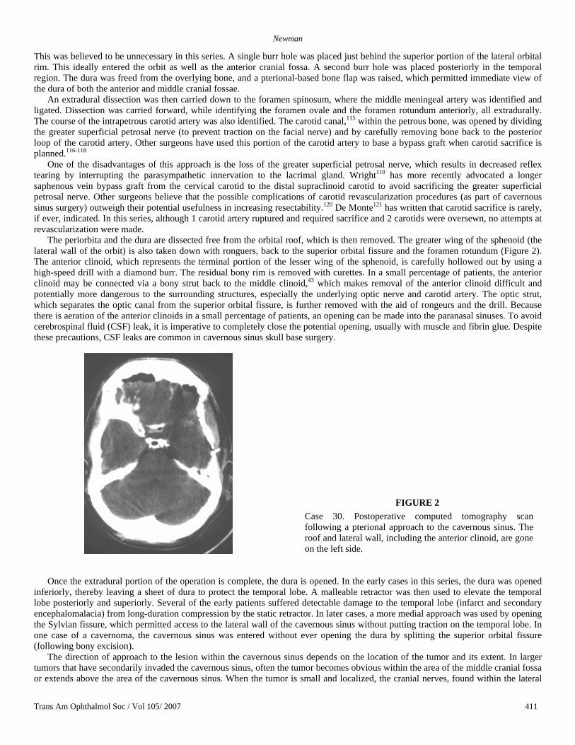
The periorbita and the dura are dissected free from the orbital roof, which is then removed. The greater wing of the sphenoid (the lateral wall of the orbit) is also taken down with ronguers, back to the superior orbital fissure and the foramen rotundum (Figure 2). The anterior clinoid, which represents the terminal portion of the lesser wing of the sphenoid, is carefully hollowed out by using a high-speed drill with a diamond burr. The residual bony rim is removed with curettes. In a small percentage of patients, the anterior clinoid may be connected via a bony strut back to the middle clinoid,43 which makes removal of the anterior clinoid difficult and potentially more dangerous to the surrounding structures, especially the underlying optic nerve and carotid artery. The optic strut, which separates the optic canal from the superior orbital fissure, is further removed with the aid of rongeurs and the drill. Because there is aeration of the anterior clinoids in a small percentage of patients, an opening can be made into the paranasal sinuses. To avoid cerebrospinal fluid (CSF) leak, it is imperative to completely close the potential opening, usually with muscle and fibrin glue. Despite these precautions, CSF leaks are common in cavernous sinus skull base surgery.
FIGURE 2 Case 30. Postoperative computed tomography scan following a pterional approach to the cavernous sinus. The roof and lateral wall, including the anterior clinoid, are gone on the left side.
Once the extradural portion of the operation is complete, the dura is opened. In the early cases in this series, the dura was opened
inferiorly, thereby leaving a sheet of dura to protect the temporal lobe. A malleable retractor was then used to elevate the temporal lobe posteriorly and superiorly. Several of the early patients suffered detectable damage to the temporal lobe (infarct and secondary encephalomalacia) from long-duration compression by the static retractor. In later cases, a more medial approach was used by opening the Sylvian fissure, which permitted access to the lateral wall of the cavernous sinus without putting traction on the temporal lobe. In one case of a cavernoma, the cavernous sinus was entered without ever opening the dura by splitting the superior orbital fissure (following bony excision).
The direction of approach to the lesion within the cavernous sinus depends on the location of the tumor and its extent. In larger tumors that have secondarily invaded the cavernous sinus, often the tumor becomes obvious within the area of the middle cranial fossa or extends above the area of the cavernous sinus. When the tumor is small and localized, the cranial nerves, found within the lateral
Cavernous Sinus Surgery For Meningiomas And Ophthalmic Complications
Trans Am Ophthalmol Soc / Vol 105/ 2007 412
wall of the cavernous sinus, must be identified to prevent direct damage. This is usually relatively easy with small lesions, but it may be more difficult when the tumors are larger and especially when they are infiltrative.
The earliest surgical opening into the cavernous sinus (in the modern era of skull base surgery) was via the potential space between the fourth cranial nerve above and the fifth cranial nerve below.1 This triangular space has become known as Parkinson’s triangle in honor of Dwight Parkinson, who initially described this surgical approach. The spaces between each of the cranial nerves also offer potential access to lesions within the cavernous sinus.44 Another common approach is through the anterior cavernous roof. By removing the anterior clinoid, the space between the optic nerve and oculomotor nerve (the anteromedial triangle) opens directly into the anterior medial cavernous sinus.
Tumors often splay the cranial nerves apart, pointing the surgeon in the direction of easiest access. The various approaches have been enumerated by Dolenc in a series of named triangles. Surgery through each may be tailored to particular lesions, depending on their location. Other less common approaches to the cavernous sinus include a direct subtemporal approach to the lateral wall,122 transmaxillary123 or transfacial124 access, and transphenoidal125 and transnasal approaches.126 Several of the latter involve endoscopy.127 Lesions that extend into the posterior cranial fossa may require more extensive surgery (beyond the scope of this review). None of our patients required these alternative procedures.
One of the factors that prevented earlier surgical approach to cavernous sinus lesions was the fear of causing uncontrollable bleeding. The earliest surgery within the cavernous sinus was performed with patients under hypothermia and hypotensive anesthesia or even in cardiac arrest. Although bleeding still may present a challenge during surgery, this issue has turned out to be less of a problem than otherwise expected, given that the lesion itself displaces the venous space. Although there may be bleeding from the tumor, cavernous sinus bleeding per se is often not problematic until the tumor boundaries are reached. Surgical packing of the residual venous spaces usually controls blood within the surgical field. Several tumors in this series had residual bleeding despite the preoperative embolization, which was routinely performed. All of these instances were controlled intraoperatively.
RESULTS
MORBIDITY AND MORTALITY Nonophthalmic complications of the cavernous sinus surgery in this series of 57 procedures included postoperative CSF leaks in 7. Most of these closed spontaneously. One patient required sacrifice of the carotid artery because of rupture during the procedure, and 5 patients had evidence of cortical infarction with resultant hemiparesis in 4 (transient in one). Additional nonophthalmic complications included one case of hydrocephalus, excessive bleeding resulting in premature termination of another case, and temporal lobe abscess in one patient. A pulmonary embolus was diagnosed in one patient while still hospitalized, and one patient had postoperative seizure activity. One patient developed aspiration pneumonia and succumbed to complications within 2 months of surgery.
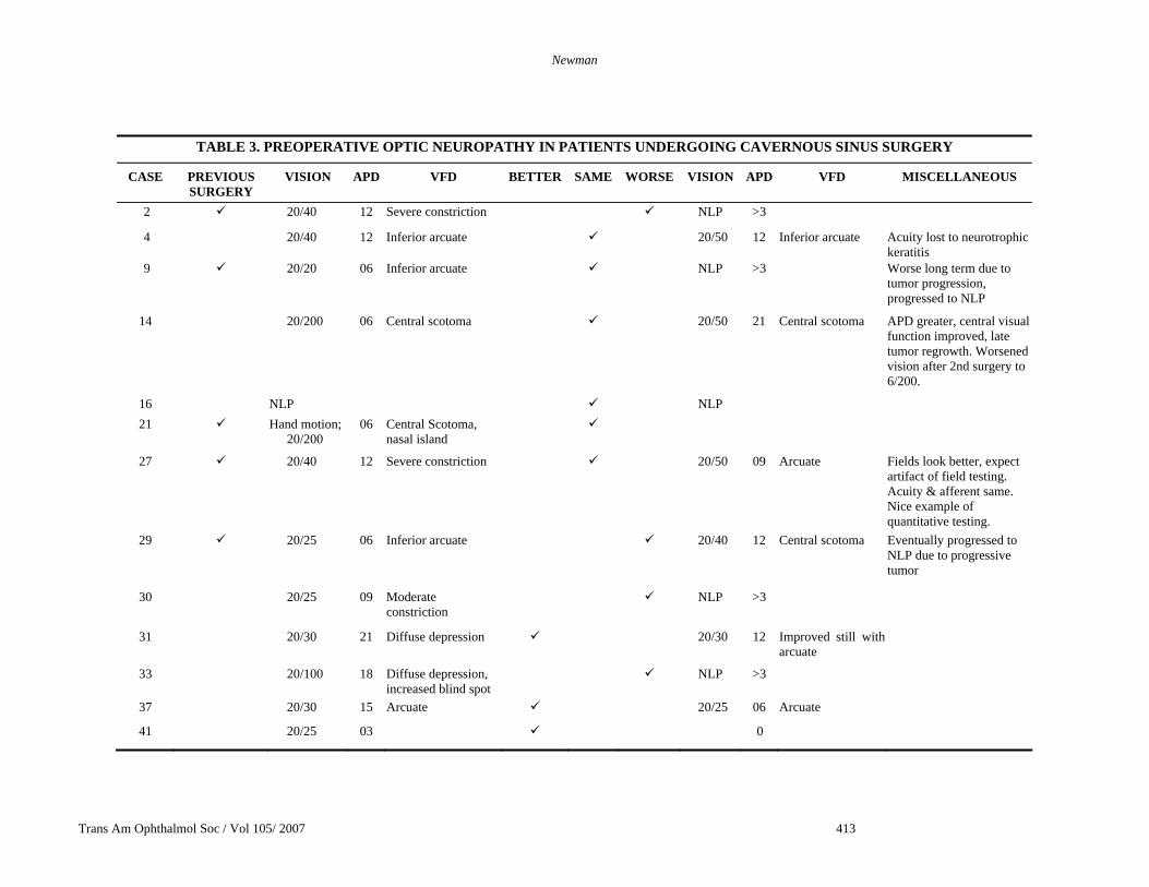
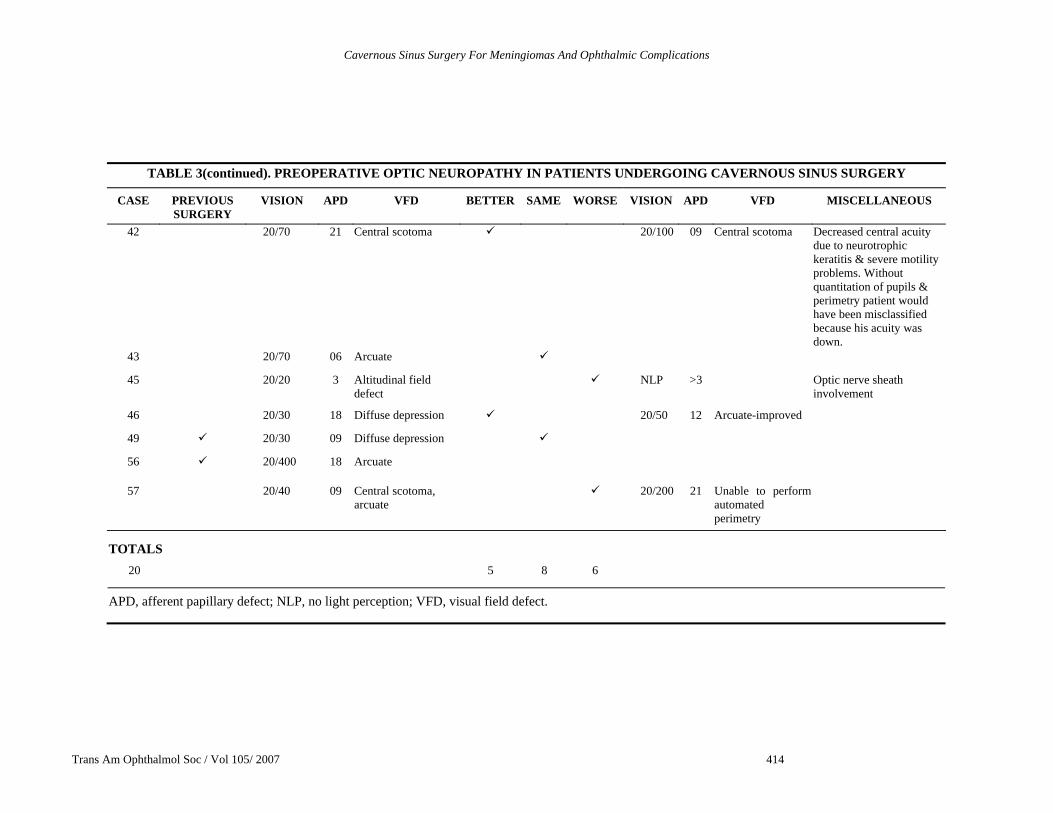
OPTIC NERVE INVOLVEMENT Twenty of the 57 patients with meningiomas had evidence of preoperative optic nerve dysfunction (Table 3). One patient had no perception of light perception (NLP), another had only hand motion vision, and one had 20/400 visual acuity. Two of these patients had previous surgery.
Five (25%) of the 20 patients showed immediate improvement, 8 (40%) experienced no change, and in 6 (30%) the dysfunction was worse postoperatively. The patient with NLP vision experienced no change. The patient with preoperative 20/400 vision could not be reassessed before leaving the hospital. Ten of the 20 patients with preoperative optic neuropathies underwent long-term follow-up (3 months or longer). Two of these (20%) had better central acuity than preoperatively, the neuropathy in 4 (40%) was the same, and 4 (40%) were worse than before surgery. Five patients (of 19 with preoperative optic neuropathies) had NLP (2 with short-term follow-up only), and the one patient preoperatively with NLP remained so. This was often anticipated preoperatively because tumor encased the optic nerve.
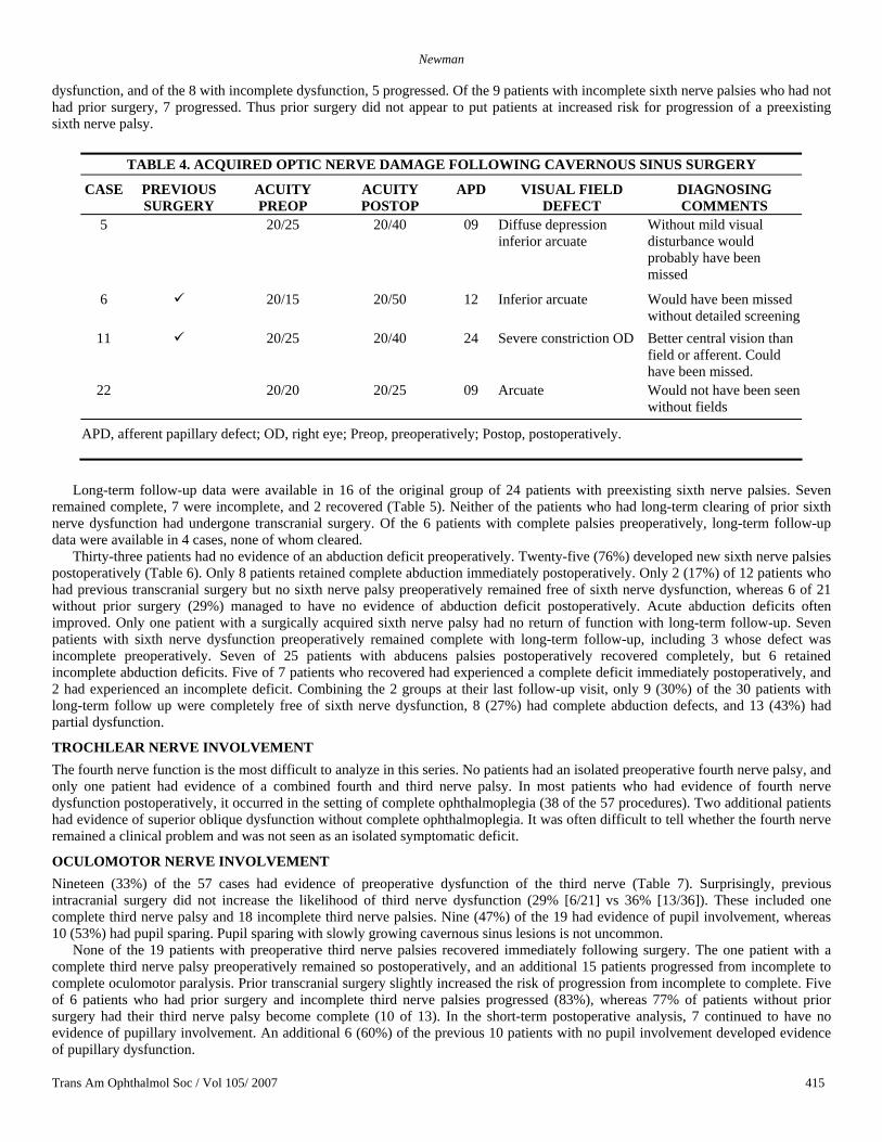
Thirty-seven patients had no evidence of preoperative optic nerve dysfunction. Thirty-three (88%) remained free of optic nerve dysfunction postoperatively, but four (11%) developed evidence of such dysfunction (Table 4). All 4 patients developed partial optic neuropathies with evidence of an afferent pupillary defect and visual field defects present on automated static perimetry (Figure 3). The field defects were characteristically arcuate in nature. There was relatively good preservation of central visual function in all 4 patients with new optic nerve dysfunction. There was marginally greater risk of a new optic neuropathy if the patient had previous surgery (2 of 14 [14%] without prior optic nerve damage) than if the patient had not had previous surgery (2 of 23 [8.7%]).
ABDUCENS NERVE INVOLVEMENT Twenty-four of the 57 patients with meningiomas who underwent cavernous sinus surgery had preoperative evidence of sixth nerve dysfunction (Table 5). Six patients (11%) had complete abduction deficits preoperatively. Prior transcranial surgery did not seem to increase the risk of sixth nerve dysfunction. There was abnormal abduction in 42% of patients with previous cranial surgery (9 of 21) and 42% of patients without prior transcranial surgery (15 of 36). No patient with a sixth nerve palsy improved immediately following surgery, but 2 did clear later. All patients whose dysfunction was complete beforehand remained the same, and 12 (67%) of the remaining 18 patients (incomplete sixth nerve palsy preoperatively) progressed to a complete sixth nerve palsy immediately following surgery. Of the 9 patients who had evidence of abducens dysfunction and prior cavernous sinus surgery, one had complete
Newman
Trans Am Ophthalmol Soc / Vol 105/ 2007 413
TABLE 3. PREOPERATIVE OPTIC NEUROPATHY IN PATIENTS UNDERGOING CAVERNOUS SINUS SURGERY
CASE PREVIOUS SURGERY
VISION APD VFD BETTER SAME WORSE VISION APD VFD MISCELLANEOUS
2 20/40 12 Severe constriction NLP >3
4 20/40 12 Inferior arcuate 20/50 12 Inferior arcuate Acuity lost to neurotrophic keratitis
9 20/20 06 Inferior arcuate NLP >3 Worse long term due to tumor progression, progressed to NLP
14 20/200 06 Central scotoma 20/50 21 Central scotoma APD greater, central visual function improved, late tumor regrowth. Worsened vision after 2nd surgery to 6/200.
16 NLP NLP 21 Hand motion;
20/200 06 Central Scotoma,
nasal island
27 20/40 12 Severe constriction 20/50 09 Arcuate Fields look better, expect artifact of field testing. Acuity & afferent same. Nice example of quantitative testing.
29 20/25 06 Inferior arcuate 20/40 12 Central scotoma Eventually progressed to NLP due to progressive tumor
30 20/25 09 Moderate constriction
NLP >3
31 20/30 21 Diffuse depression 20/30 12 Improved still with arcuate
33 20/100 18 Diffuse depression, increased blind spot
NLP >3
37 20/30 15 Arcuate 20/25 06 Arcuate
41 20/25 03 0
Cavernous Sinus Surgery For Meningiomas And Ophthalmic Complications
Trans Am Ophthalmol Soc / Vol 105/ 2007 414
TABLE 3(continued). PREOPERATIVE OPTIC NEUROPATHY IN PATIENTS UNDERGOING CAVERNOUS SINUS SURGERY
CASE PREVIOUS SURGERY
VISION APD VFD BETTER SAME WORSE VISION APD VFD MISCELLANEOUS
42 20/70 21 Central scotoma 20/100 09 Central scotoma Decreased central acuity due to neurotrophic keratitis & severe motility problems. Without quantitation of pupils & perimetry patient would have been misclassified because his acuity was down.
43 20/70 06 Arcuate
45 20/20 3 Altitudinal field defect
NLP >3 Optic nerve sheath involvement
46 20/30 18 Diffuse depression 20/50 12 Arcuate-improved
49 20/30 09 Diffuse depression
56 20/400 18 Arcuate
57 20/40 09 Central scotoma, arcuate
20/200 21 Unable to perform automated perimetry
TOTALS 20 5 8 6
APD, afferent papillary defect; NLP, no light perception; VFD, visual field defect.
Newman
Trans Am Ophthalmol Soc / Vol 105/ 2007 415
dysfunction, and of the 8 with incomplete dysfunction, 5 progressed. Of the 9 patients with incomplete sixth nerve palsies who had not had prior surgery, 7 progressed. Thus prior surgery did not appear to put patients at increased risk for progression of a preexisting sixth nerve palsy.
TABLE 4. ACQUIRED OPTIC NERVE DAMAGE FOLLOWING CAVERNOUS SINUS SURGERY
CASE PREVIOUS SURGERY
ACUITY PREOP
ACUITY POSTOP
APD VISUAL FIELD DEFECT
DIAGNOSING COMMENTS
5 20/25 20/40 09 Diffuse depression inferior arcuate
Without mild visual disturbance would probably have been missed
6 20/15 20/50 12 Inferior arcuate Would have been missed without detailed screening
11 20/25 20/40 24 Severe constriction OD Better central vision than field or afferent. Could have been missed.
22 20/20 20/25 09 Arcuate Would not have been seen without fields
APD, afferent papillary defect; OD, right eye; Preop, preoperatively; Postop, postoperatively.
Long-term follow-up data were available in 16 of the original group of 24 patients with preexisting sixth nerve palsies. Seven
remained complete, 7 were incomplete, and 2 recovered (Table 5). Neither of the patients who had long-term clearing of prior sixth nerve dysfunction had undergone transcranial surgery. Of the 6 patients with complete palsies preoperatively, long-term follow-up data were available in 4 cases, none of whom cleared.
Thirty-three patients had no evidence of an abduction deficit preoperatively. Twenty-five (76%) developed new sixth nerve palsies postoperatively (Table 6). Only 8 patients retained complete abduction immediately postoperatively. Only 2 (17%) of 12 patients who had previous transcranial surgery but no sixth nerve palsy preoperatively remained free of sixth nerve dysfunction, whereas 6 of 21 without prior surgery (29%) managed to have no evidence of abduction deficit postoperatively. Acute abduction deficits often improved. Only one patient with a surgically acquired sixth nerve palsy had no return of function with long-term follow-up. Seven patients with sixth nerve dysfunction preoperatively remained complete with long-term follow-up, including 3 whose defect was incomplete preoperatively. Seven of 25 patients with abducens palsies postoperatively recovered completely, but 6 retained incomplete abduction deficits. Five of 7 patients who recovered had experienced a complete deficit immediately postoperatively, and 2 had experienced an incomplete deficit. Combining the 2 groups at their last follow-up visit, only 9 (30%) of the 30 patients with long-term follow up were completely free of sixth nerve dysfunction, 8 (27%) had complete abduction defects, and 13 (43%) had partial dysfunction.
TROCHLEAR NERVE INVOLVEMENT The fourth nerve function is the most difficult to analyze in this series. No patients had an isolated preoperative fourth nerve palsy, and only one patient had evidence of a combined fourth and third nerve palsy. In most patients who had evidence of fourth nerve dysfunction postoperatively, it occurred in the setting of complete ophthalmoplegia (38 of the 57 procedures). Two additional patients had evidence of superior oblique dysfunction without complete ophthalmoplegia. It was often difficult to tell whether the fourth nerve remained a clinical problem and was not seen as an isolated symptomatic deficit.
OCULOMOTOR NERVE INVOLVEMENT Nineteen (33%) of the 57 cases had evidence of preoperative dysfunction of the third nerve (Table 7). Surprisingly, previous intracranial surgery did not increase the likelihood of third nerve dysfunction (29% [6/21] vs 36% [13/36]). These included one complete third nerve palsy and 18 incomplete third nerve palsies. Nine (47%) of the 19 had evidence of pupil involvement, whereas 10 (53%) had pupil sparing. Pupil sparing with slowly growing cavernous sinus lesions is not uncommon.
None of the 19 patients with preoperative third nerve palsies recovered immediately following surgery. The one patient with a complete third nerve palsy preoperatively remained so postoperatively, and an additional 15 patients progressed from incomplete to complete oculomotor paralysis. Prior transcranial surgery slightly increased the risk of progression from incomplete to complete. Five of 6 patients who had prior surgery and incomplete third nerve palsies progressed (83%), whereas 77% of patients without prior surgery had their third nerve palsy become complete (10 of 13). In the short-term postoperative analysis, 7 continued to have no evidence of pupillary involvement. An additional 6 (60%) of the previous 10 patients with no pupil involvement developed evidence of pupillary dysfunction.
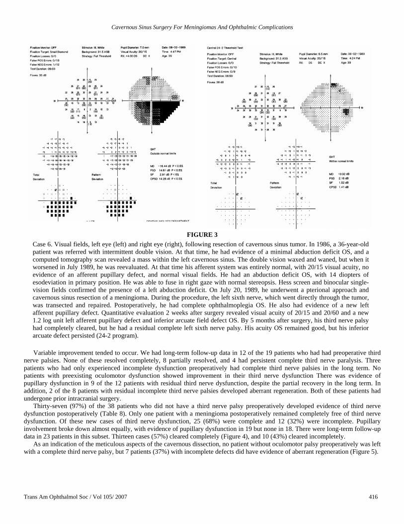
Cavernous Sinus Surgery For Meningiomas And Ophthalmic Complications
Trans Am Ophthalmol Soc / Vol 105/ 2007 416
FIGURE 3 Case 6. Visual fields, left eye (left) and right eye (right), following resection of cavernous sinus tumor. In 1986, a 36-year-old patient was referred with intermittent double vision. At that time, he had evidence of a minimal abduction deficit OS, and a computed tomography scan revealed a mass within the left cavernous sinus. The double vision waxed and waned, but when it worsened in July 1989, he was reevaluated. At that time his afferent system was entirely normal, with 20/15 visual acuity, no evidence of an afferent pupillary defect, and normal visual fields. He had an abduction deficit OS, with 14 diopters of esodeviation in primary position. He was able to fuse in right gaze with normal stereopsis. Hess screen and binocular single-vision fields confirmed the presence of a left abduction deficit. On July 20, 1989, he underwent a pterional approach and cavernous sinus resection of a meningioma. During the procedure, the left sixth nerve, which went directly through the tumor, was transected and repaired. Postoperatively, he had complete ophthalmoplegia OS. He also had evidence of a new left afferent pupillary defect. Quantitative evaluation 2 weeks after surgery revealed visual acuity of 20/15 and 20/60 and a new 1.2 log unit left afferent pupillary defect and inferior arcuate field defect OS. By 5 months after surgery, his third nerve palsy had completely cleared, but he had a residual complete left sixth nerve palsy. His acuity OS remained good, but his inferior arcuate defect persisted (24-2 program).
Variable improvement tended to occur. We had long-term follow-up data in 12 of the 19 patients who had had preoperative third
nerve palsies. None of these resolved completely, 8 partially resolved, and 4 had persistent complete third nerve paralysis. Three patients who had only experienced incomplete dysfunction preoperatively had complete third nerve palsies in the long term. No patients with preexisting oculomotor dysfunction showed improvement in their third nerve dysfunction There was evidence of pupillary dysfunction in 9 of the 12 patients with residual third nerve dysfunction, despite the partial recovery in the long term. In addition, 2 of the 8 patients with residual incomplete third nerve palsies developed aberrant regeneration. Both of these patients had undergone prior intracranial surgery.
Thirty-seven (97%) of the 38 patients who did not have a third nerve palsy preoperatively developed evidence of third nerve dysfunction postoperatively (Table 8). Only one patient with a meningioma postoperatively remained completely free of third nerve dysfunction. Of these new cases of third nerve dysfunction, 25 (68%) were complete and 12 (32%) were incomplete. Pupillary involvement broke down almost equally, with evidence of pupillary dysfunction in 19 but none in 18. There were long-term follow-up data in 23 patients in this subset. Thirteen cases (57%) cleared completely (Figure 4), and 10 (43%) cleared incompletely.
As an indication of the meticulous aspects of the cavernous dissection, no patient without oculomotor palsy preoperatively was left with a complete third nerve palsy, but 7 patients (37%) with incomplete defects did have evidence of aberrant regeneration (Figure 5).
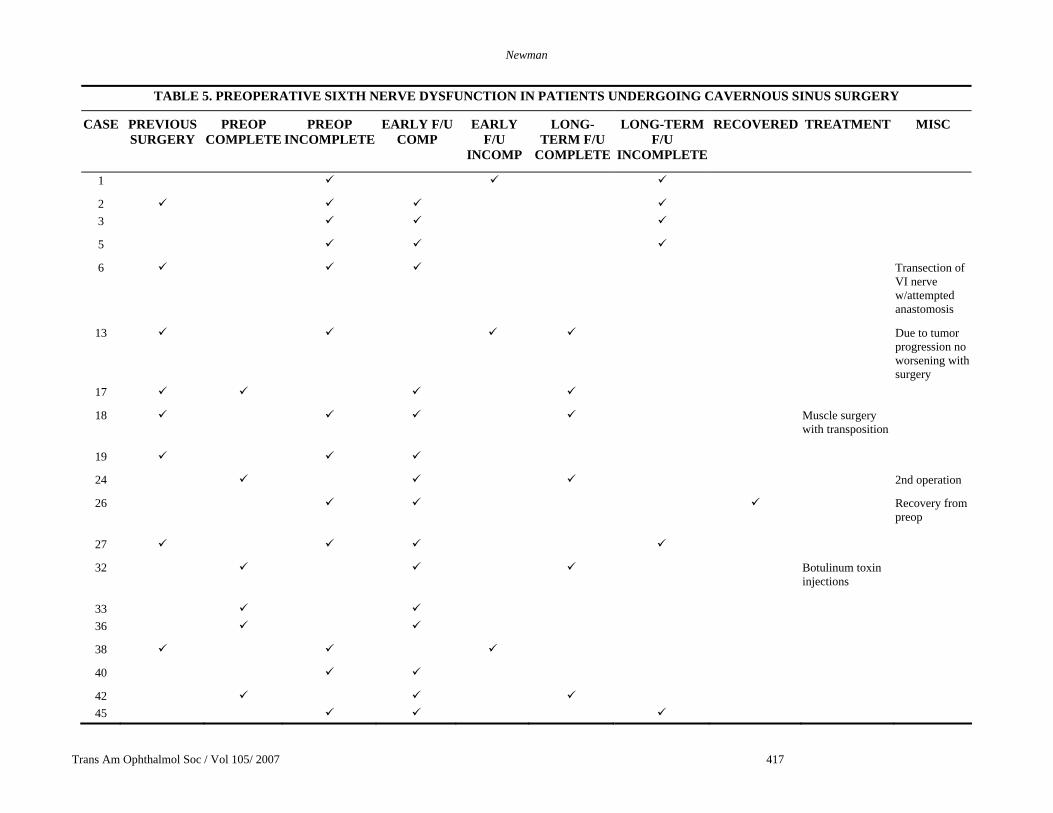
Newman
Trans Am Ophthalmol Soc / Vol 105/ 2007 417
TABLE 5. PREOPERATIVE SIXTH NERVE DYSFUNCTION IN PATIENTS UNDERGOING CAVERNOUS SINUS SURGERY
CASE PREVIOUS SURGERY
PREOP COMPLETE
PREOP INCOMPLETE
EARLY F/U COMP
EARLY F/U
INCOMP
LONG-TERM F/U
COMPLETE
LONG-TERM F/U
INCOMPLETE
RECOVERED TREATMENT MISC
1
2 3
5
6 Transection of VI nerve w/attempted anastomosis
13 Due to tumor progression no worsening with surgery
17
18 Muscle surgery with transposition
19
24 2nd operation
26 Recovery from preop
27
32 Botulinum toxin injections
33 36
38
40
42 45
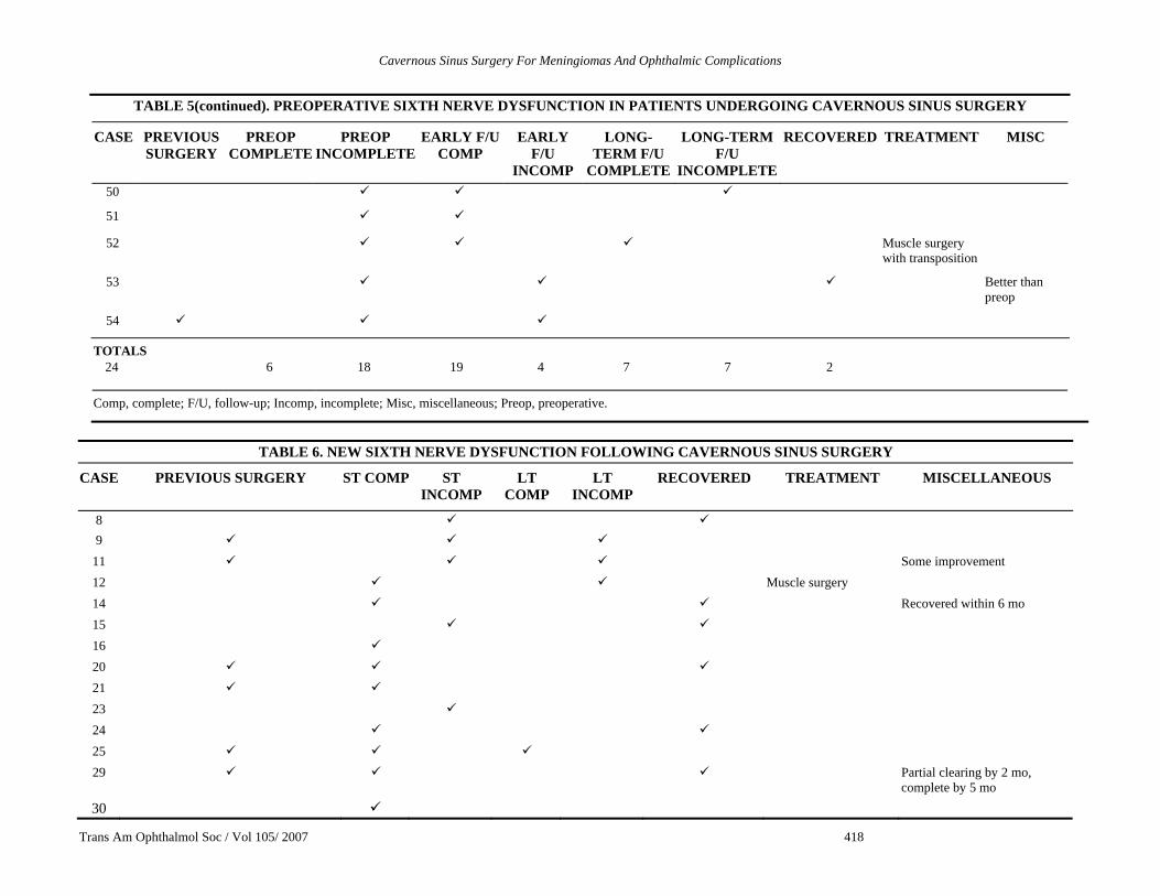
Cavernous Sinus Surgery For Meningiomas And Ophthalmic Complications
Trans Am Ophthalmol Soc / Vol 105/ 2007 418
TABLE 5(continued). PREOPERATIVE SIXTH NERVE DYSFUNCTION IN PATIENTS UNDERGOING CAVERNOUS SINUS SURGERY
CASE PREVIOUS SURGERY
PREOP COMPLETE
PREOP INCOMPLETE
EARLY F/U COMP
EARLY F/U
INCOMP
LONG-TERM F/U
COMPLETE
LONG-TERM F/U
INCOMPLETE
RECOVERED TREATMENT MISC
50
51
52 Muscle surgery with transposition
53 Better than preop
54
TOTALS 24 6 18 19 4 7 7 2
Comp, complete; F/U, follow-up; Incomp, incomplete; Misc, miscellaneous; Preop, preoperative.
TABLE 6. NEW SIXTH NERVE DYSFUNCTION FOLLOWING CAVERNOUS SINUS SURGERY
CASE PREVIOUS SURGERY ST COMP ST INCOMP
LT COMP
LT INCOMP
RECOVERED TREATMENT MISCELLANEOUS
8 9
11 Some improvement 12 Muscle surgery 14 Recovered within 6 mo 15 16 20 21 23 24 25 29 Partial clearing by 2 mo,
complete by 5 mo 30
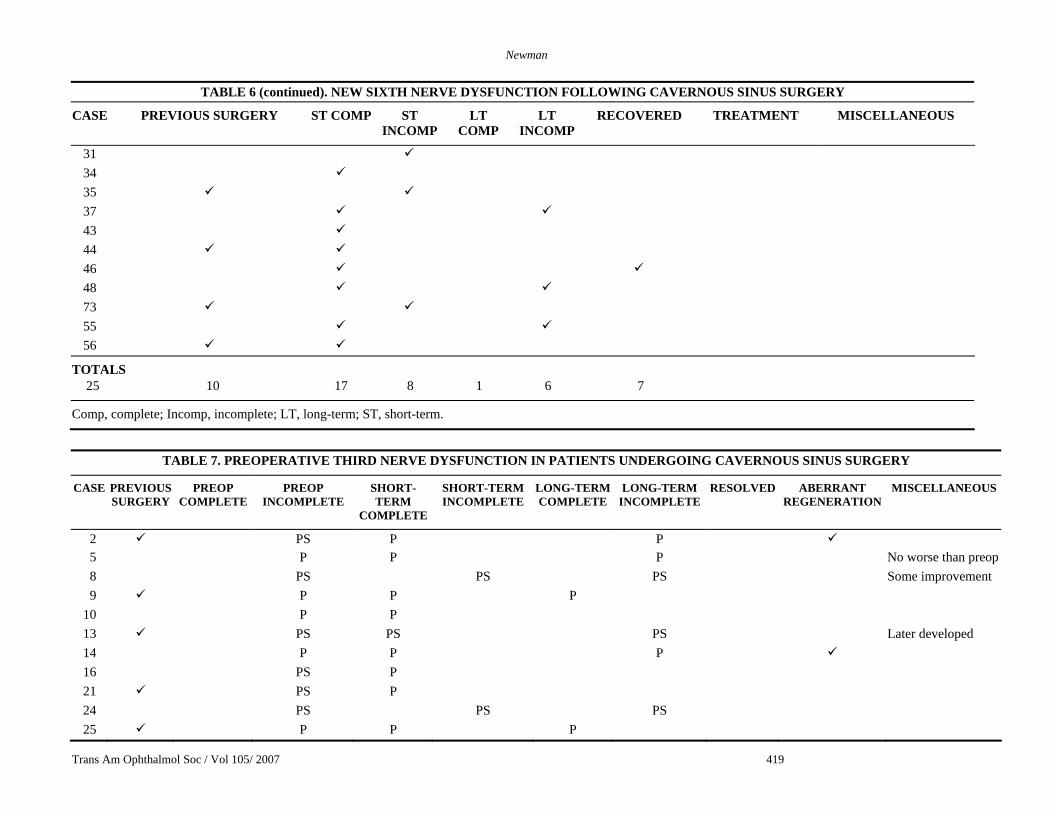
Newman
Trans Am Ophthalmol Soc / Vol 105/ 2007 419
TABLE 6 (continued). NEW SIXTH NERVE DYSFUNCTION FOLLOWING CAVERNOUS SINUS SURGERY
CASE PREVIOUS SURGERY ST COMP ST INCOMP
LT COMP
LT INCOMP
RECOVERED TREATMENT MISCELLANEOUS
31 34 35 37 43 44 46 48 73 55 56
TOTALS 25 10 17 8 1 6 7
Comp, complete; Incomp, incomplete; LT, long-term; ST, short-term.
TABLE 7. PREOPERATIVE THIRD NERVE DYSFUNCTION IN PATIENTS UNDERGOING CAVERNOUS SINUS SURGERY
CASE PREVIOUS SURGERY
PREOP COMPLETE
PREOP INCOMPLETE
SHORT-TERM
COMPLETE
SHORT-TERM INCOMPLETE
LONG-TERM COMPLETE
LONG-TERM INCOMPLETE
RESOLVED ABERRANT REGENERATION
MISCELLANEOUS
2 PS P P 5 P P P No worse than preop 8 PS PS PS Some improvement 9 P P P
10 P P 13 PS PS PS Later developed 14 P P P 16 PS P 21 PS P 24 PS PS PS 25 P P P
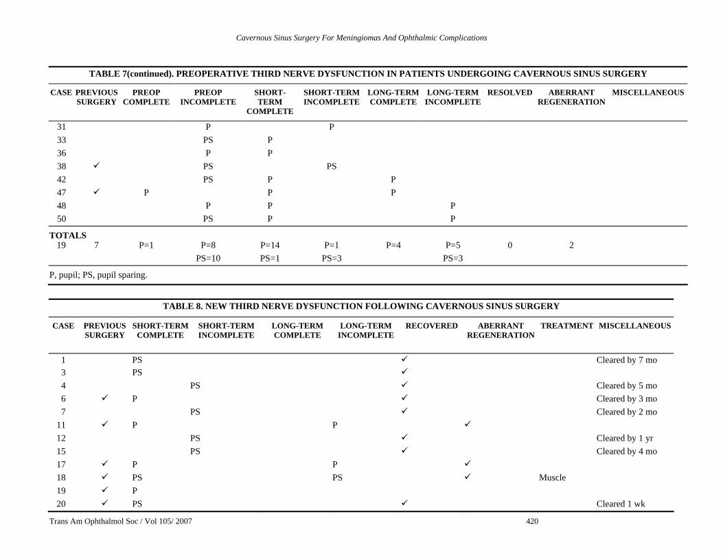
Cavernous Sinus Surgery For Meningiomas And Ophthalmic Complications
Trans Am Ophthalmol Soc / Vol 105/ 2007 420
TABLE 7(continued). PREOPERATIVE THIRD NERVE DYSFUNCTION IN PATIENTS UNDERGOING CAVERNOUS SINUS SURGERY
CASE PREVIOUS SURGERY
PREOP COMPLETE
PREOP INCOMPLETE
SHORT-TERM
COMPLETE
SHORT-TERM INCOMPLETE
LONG-TERM COMPLETE
LONG-TERM INCOMPLETE
RESOLVED ABERRANT REGENERATION
MISCELLANEOUS
31 P P 33 PS P 36 P P 38 PS PS 42 PS P P 47 P P P 48 P P P 50 PS P P
TOTALS 19 7 P=1 P=8 P=14 P=1 P=4 P=5 0 2
PS=10 PS=1 PS=3 PS=3
P, pupil; PS, pupil sparing.
TABLE 8. NEW THIRD NERVE DYSFUNCTION FOLLOWING CAVERNOUS SINUS SURGERY
CASE PREVIOUS SURGERY
SHORT-TERM COMPLETE
SHORT-TERM INCOMPLETE
LONG-TERM COMPLETE
LONG-TERM INCOMPLETE
RECOVERED ABERRANT REGENERATION
TREATMENT MISCELLANEOUS
1 PS Cleared by 7 mo 3 PS 4 PS Cleared by 5 mo 6 P Cleared by 3 mo 7 PS Cleared by 2 mo
11 P P 12 PS Cleared by 1 yr 15 PS Cleared by 4 mo 17 P P 18 PS PS Muscle 19 P 20 PS Cleared 1 wk
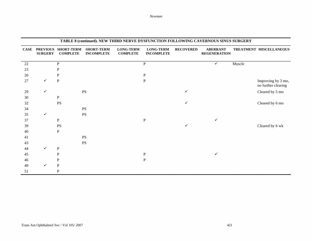
Newman
Trans Am Ophthalmol Soc / Vol 105/ 2007 421
TABLE 8 (continued). NEW THIRD NERVE DYSFUNCTION FOLLOWING CAVERNOUS SINUS SURGERY
CASE PREVIOUS SURGERY
SHORT-TERM COMPLETE
SHORT-TERM INCOMPLETE
LONG-TERM COMPLETE
LONG-TERM INCOMPLETE
RECOVERED ABERRANT REGENERATION
TREATMENT MISCELLANEOUS
22 P P Muscle 23 P 26 P P 27 P P Improving by 3 mo,
no further clearing 29 PS Cleared by 5 mo 30 P 32 PS Cleared by 6 mo 34 PS 35 PS 37 P P 39 PS Cleared by 6 wk 40 P 41 PS 43 PS 44 P 45 P P 46 P P 49 P 51 P
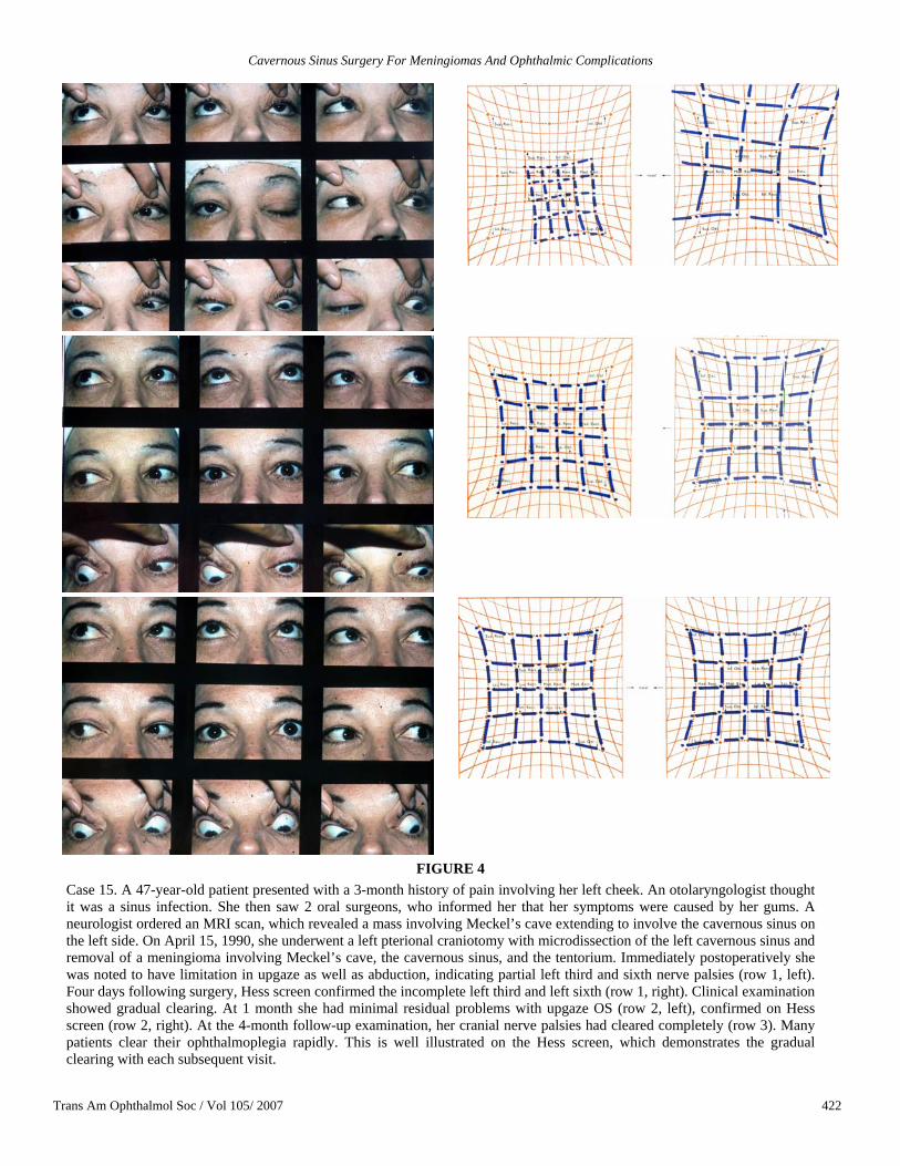
Cavernous Sinus Surgery For Meningiomas And Ophthalmic Complications
Trans Am Ophthalmol Soc / Vol 105/ 2007 422
FIGURE 4 Case 15. A 47-year-old patient presented with a 3-month history of pain involving her left cheek. An otolaryngologist thought it was a sinus infection. She then saw 2 oral surgeons, who informed her that her symptoms were caused by her gums. A neurologist ordered an MRI scan, which revealed a mass involving Meckel’s cave extending to involve the cavernous sinus on the left side. On April 15, 1990, she underwent a left pterional craniotomy with microdissection of the left cavernous sinus and removal of a meningioma involving Meckel’s cave, the cavernous sinus, and the tentorium. Immediately postoperatively she was noted to have limitation in upgaze as well as abduction, indicating partial left third and sixth nerve palsies (row 1, left). Four days following surgery, Hess screen confirmed the incomplete left third and left sixth (row 1, right). Clinical examination showed gradual clearing. At 1 month she had minimal residual problems with upgaze OS (row 2, left), confirmed on Hess screen (row 2, right). At the 4-month follow-up examination, her cranial nerve palsies had cleared completely (row 3). Many patients clear their ophthalmoplegia rapidly. This is well illustrated on the Hess screen, which demonstrates the gradual clearing with each subsequent visit.
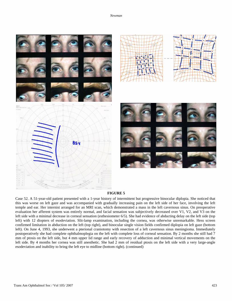
Newman
Trans Am Ophthalmol Soc / Vol 105/ 2007 423
FIGURE 5 Case 52. A 51-year-old patient presented with a 1-year history of intermittent but progressive binocular diplopia. She noticed that this was worse on left gaze and was accompanied with gradually increasing pain on the left side of her face, involving the left temple and ear. Her internist arranged for an MRI scan, which demonstrated a mass in the left cavernous sinus. On preoperative evaluation her afferent system was entirely normal, and facial sensation was subjectively decreased over V1, V2, and V3 on the left side with a minimal decrease in corneal sensation (esthesiometer 6/5). She had evidence of abducting delay on the left side (top left) with 12 diopters of esodeviation. Slit-lamp examination, including the cornea, was otherwise unremarkable. Hess screen confirmed limitation in abduction on the left (top right), and binocular single vision fields confirmed diplopia on left gaze (bottom left). On June 4, 1993, she underwent a pterional craniotomy with resection of a left cavernous sinus meningioma. Immediately postoperatively she had complete ophthalmoplegia on the left with complete loss of corneal sensation. By 2 months she still had 7 mm of ptosis on the left side, but 4 mm upper lid range and early recovery of adduction and minimal vertical movements on the left side. By 4 months her cornea was still anesthetic. She had 2 mm of residual ptosis on the left side with a very large-angle esodeviation and inability to bring the left eye to midline (bottom right). (continued)
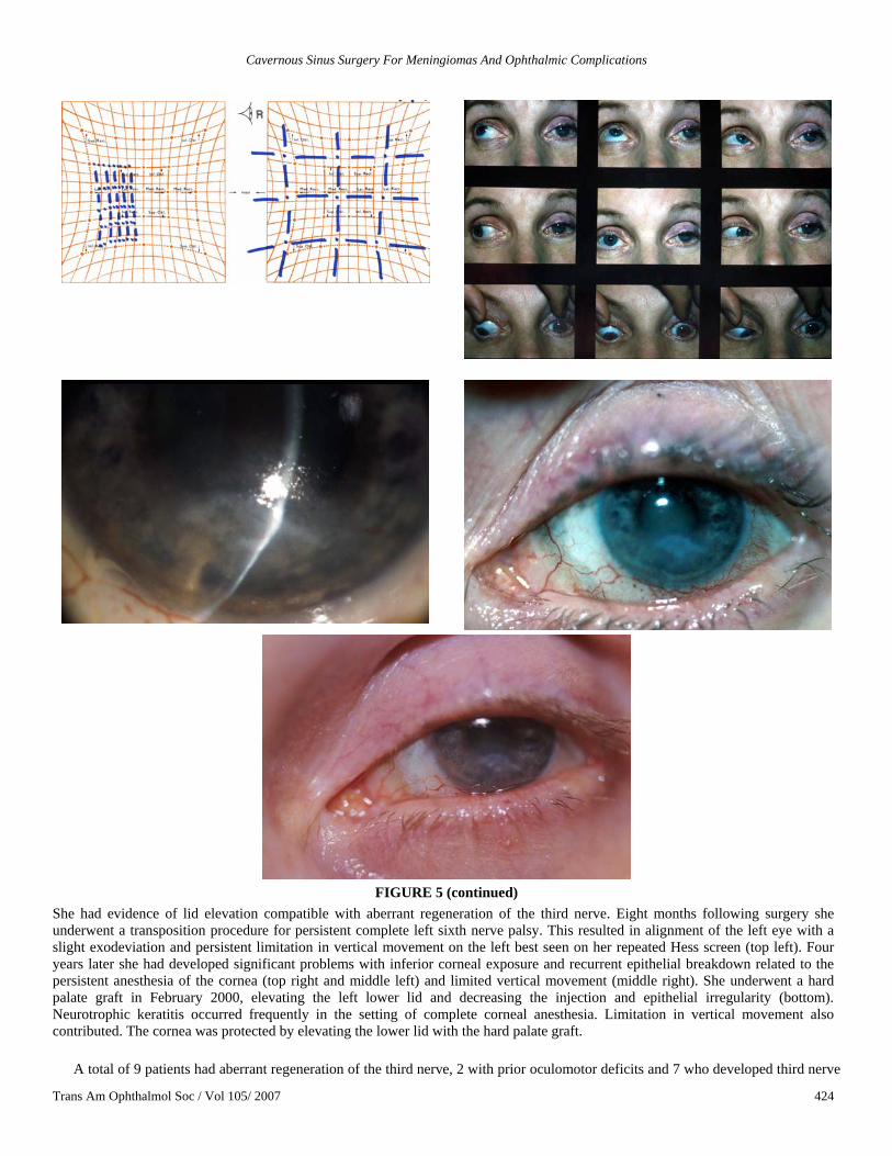
Cavernous Sinus Surgery For Meningiomas And Ophthalmic Complications
Trans Am Ophthalmol Soc / Vol 105/ 2007 424
FIGURE 5 (continued)
She had evidence of lid elevation compatible with aberrant regeneration of the third nerve. Eight months following surgery she underwent a transposition procedure for persistent complete left sixth nerve palsy. This resulted in alignment of the left eye with a slight exodeviation and persistent limitation in vertical movement on the left best seen on her repeated Hess screen (top left). Four years later she had developed significant problems with inferior corneal exposure and recurrent epithelial breakdown related to the persistent anesthesia of the cornea (top right and middle left) and limited vertical movement (middle right). She underwent a hard palate graft in February 2000, elevating the left lower lid and decreasing the injection and epithelial irregularity (bottom). Neurotrophic keratitis occurred frequently in the setting of complete corneal anesthesia. Limitation in vertical movement also contributed. The cornea was protected by elevating the lower lid with the hard palate graft.
A total of 9 patients had aberrant regeneration of the third nerve, 2 with prior oculomotor deficits and 7 who developed third nerve
Newman
Trans Am Ophthalmol Soc / Vol 105/ 2007 425
dysfunction postoperatively. Of the preexisting third nerve dysfunction, one case of aberrant regeneration developed in a patient with prior surgery, and the other had no prior history of intervention. Of the 7 cases in patients without a prior third dysfunction, 3 occurred in patients with prior surgery (3 of 13 [23%]) and 4 were in patients without surgery (4 of 24) [17%].
The chance of complete recovery was substantially better if the palsy was incomplete following surgery. Fifteen of 16 patients (94%) who had incomplete clearing immediately postoperatively and had long-term follow-up recovered. When the third nerve palsy was complete following surgery, only 6 (32%) of 19 cases recovered totally. No cases of new oculomotor dysfunction remained complete. Of the 10 cases improving but not clearing, 7 demonstrated evidence of aberrant regeneration. There did not seem to be an increased risk associated with prior surgery. Although 4 of 10 patients who did not clear completely had prior intracranial surgery, 6 did not, and 1 of the 6 patients who demonstrated complete clearing had prior surgery. Both patients who developed new complete third nerve palsies had not had previous surgery. Thus, although patients with immediate worsening of third nerve function postoperatively usually improved, this was not universal and definitely not always complete.
TRIGEMINAL NERVE INVOLVEMENT Thirteen (23%) of 57 patients had preoperative evidence of trigeminal dysfunction (Table 9). Sensory loss was more common in patients who had undergone prior surgery (29% [6 of 21] vs 19% [7 of 36]). Two patients had no corneal sensation (most commonly related to previous surgery). Immediately following surgery, 4 of 11 cases that had incomplete sensory dysfunction remained incomplete, and 6 of the other 7 lost all corneal sensation. Only 1 patient (8%) experienced improvement. None of the patients with complete fifth nerve dysfunction demonstrated immediate improvement. Long-term follow-up data were available in 9 of the 13 patients who had preoperative trigeminal dysfunction. Six remained complete, 2 partially recovered, and 1 completely recovered.
Loss of corneal sensation was of considerable clinical significance. Only 1 of 7 preoperatively anesthetic patients partially recovered with long-term follow-up (representing functional improvement). Both patients with completely anesthetic corneas preoperatively developed neurotrophic keratitis. Four additional patients with partial anesthesia preoperatively developed neurotrophic keratitis after becoming completely anesthetic postoperatively.
Thirty-four (77%) of the 44 patients who did not have evidence of preoperative fifth nerve dysfunction developed evidence of postoperative hypesthesia (Table 10). The risk of developing a new trigeminal palsy was slightly greater if the patient had undergone previous transcranial surgery (87% [13 of 15] vs 72% [21/29]). Twenty-one of these newly affected patients (62%) were completely anesthetic, and 13 (38%) had decreased sensation. Long-term follow-up data in 19 of these 34 patients revealed recovery in 2, incomplete dysfunction in 7, and complete anesthesia in 10. Five of the 13 who acquired anesthesia improved to hypesthesia with long-term follow-up. Of the 2 who completely recovered, both were partially compromised postoperatively. Six (60%) of the 10 persistently anesthetic patients developed neurotrophic keratitis.
MISCELLANEOUS OPHTHALMIC COMPLICATIONS Forty-seven additional complications occurred in 32 of the 82 procedures, including several ophthalmic complications. Horner’s syndrome developed in 3 patients and homonymous hemianopsia in 4.
SUMMARY OF RESULTS Patients undergoing cavernous sinus surgery can usually expect new ophthalmic complications together with an exacerbation of preexisting neuro-ophthalmic deficits. Although improvement in function can be achieved, it is distinctly uncommon. Worsening of ophthalmoplegia postoperatively appears to be the rule rather than the exception. Transient postoperative ocular motor palsy may improve especially if incomplete in the immediate postoperative phase. Aberrant regeneration of the third nerve is not uncommon, resulting in persistent motility problems. Worsening of optic nerve function may be underappreciated, especially if quantitative assessment is not performed. Trigeminal dysfunction may have a significant effect on visual function, especially if complete, as an anesthetic cornea is at high risk for development of neurotrophic keratitis.
DISCUSSION
OPTIC NEUROPATHY The worsening of optic nerve function in patients who were previously compromised is likely a manifestation of the extent of the tumor as well as attempts at aggressive surgical removal. In at least 3 cases, the meningioma was preoperatively noted to extend completely around the optic nerve, thus ensuring loss of optic nerve function with complete resection. In these cases, the potential for visual loss was discussed with each patient.
A more interesting question involves those preoperatively normal patients who developed optic nerve dysfunction postoperatively. The mechanism is unclear. While it is possible vasospasm may play a role, the most likely culprit is direct damage at the time of the clinoid removal. In this series the clinoid was removed extradurally, which may increase the risk over an intradural approach. This has been noted, although not emphasized, in the literature. In a series of 40 patients undergoing clinoid removal, 31 of which were done extradurally, 3 patients suffered postoperative diminution of visual acuity or visual field.128 None of the 9 patients in whom an intradural approach was used had similar complications.
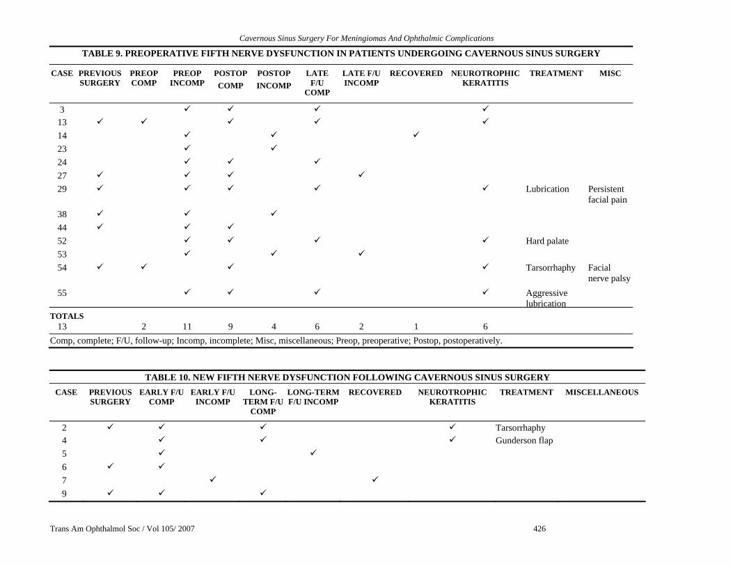
Cavernous Sinus Surgery For Meningiomas And Ophthalmic Complications
Trans Am Ophthalmol Soc / Vol 105/ 2007 426
TABLE 9. PREOPERATIVE FIFTH NERVE DYSFUNCTION IN PATIENTS UNDERGOING CAVERNOUS SINUS SURGERY
CASE PREVIOUS SURGERY
PREOP COMP
PREOP INCOMP
POSTOPCOMP
POSTOP INCOMP
LATE F/U
COMP
LATE F/U INCOMP
RECOVERED NEUROTROPHIC KERATITIS
TREATMENT MISC
3 13 14 23 24 27 29 Lubrication Persistent
facial pain 38 44 52 Hard palate 53 54 Tarsorrhaphy Facial
nerve palsy55 Aggressive
lubrication
TOTALS 13 2 11 9 4 6 2 1 6
Comp, complete; F/U, follow-up; Incomp, incomplete; Misc, miscellaneous; Preop, preoperative; Postop, postoperatively.
TABLE 10. NEW FIFTH NERVE DYSFUNCTION FOLLOWING CAVERNOUS SINUS SURGERY CASE PREVIOUS
SURGERY EARLY F/U
COMP EARLY F/U
INCOMP LONG-
TERM F/U COMP
LONG-TERM F/U INCOMP
RECOVERED NEUROTROPHIC KERATITIS
TREATMENT MISCELLANEOUS
2 Tarsorrhaphy 4 Gunderson flap 5 6 7 9
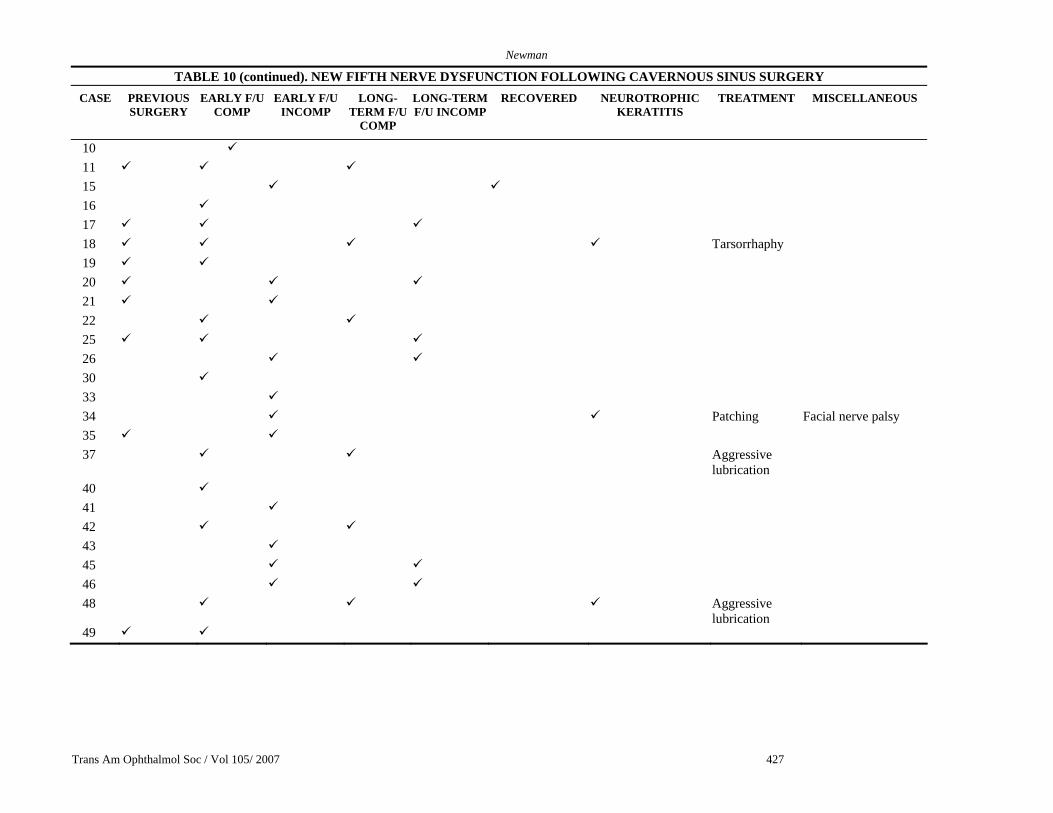
Newman
Trans Am Ophthalmol Soc / Vol 105/ 2007 427
TABLE 10 (continued). NEW FIFTH NERVE DYSFUNCTION FOLLOWING CAVERNOUS SINUS SURGERY CASE PREVIOUS
SURGERY EARLY F/U
COMP EARLY F/U
INCOMP LONG-
TERM F/U COMP
LONG-TERM F/U INCOMP
RECOVERED NEUROTROPHIC KERATITIS
TREATMENT MISCELLANEOUS
10 11 15 16 17 18 Tarsorrhaphy 19 20 21 22 25 26 30 33 34 Patching Facial nerve palsy 35 37 Aggressive
lubrication
40 41 42 43 45 46 48 Aggressive
lubrication
49
Cavernous Sinus Surgery For Meningiomas And Ophthalmic Complications
Trans Am Ophthalmol Soc / Vol 105/ 2007 428
It is presumed that surgical manipulation around the optic nerve was responsible for the development of the optic nerve
dysfunction in the 4 patients who developed partial but incomplete optic nerve damage. In particular, even the careful attempt to core the anterior clinoid with a high-speed drill puts the optic nerve at risk. There can be tremendous heat transfer with the use of a diamond burr (used to decrease bleeding and limit kickback in this tight space). Dolenc44 specifically stressed the importance of adequate irrigation during drilling to minimize heat transfer. It is possible that significant temperature elevation occurred, despite the best efforts at continuous irrigation.
The incidence of new optic nerve damage in this case (7%) is substantially higher than that in previously reported series of cavernous sinus cases undergoing surgery. The easiest explanation for this incidence (not previously reported) is that without quantitative assessment of optic nerve function, these cases would likely have been missed clinically. Although the afferent pupillary defect ranged from 0.9 to 2.4 log units, central acuity was no worse than 20/50 in the 4 patients. Without measuring the afferent pupillary defect or obtaining quantitative perimetry for evidence of arcuate visual field defects, this dysfunction would have been overlooked. Thus, more careful attention to optic nerve function postoperatively may well detect unrecognized optic neuropathy.
ABDUCENS PALSY A sixth cranial nerve palsy was the most common preoperative manifestation of cavernous sinus lesions in this series. None of the patients who had complete defects preoperatively recovered function, and although transient postoperative sixth nerve palsies did clear in a substantial portion of cases, only 9 of 30 (30%) were completely free of an abduction deficit when evaluated during a follow-up of 3 or more months postsurgery. Two patients (8%) with preoperative (all incomplete) sixth nerve palsies did recover normal function, but 13 (72%) of 18 had worse function than preoperatively, and all 6 who were complete preoperatively remained so Of the 33 patients without abducens palsies at the time of surgery, 25 (76%) developed an abduction deficit acutely. Although 50% of these cleared with long-term follow-up, 6 remained incomplete and 1 complete sixth nerve palsy failed to improve at all.
It has been suggested by neurosurgeons that an “easy muscle procedure will correct a patient with VI nerve palsy.” Although it is true that muscle surgery can be done to provide binocularity centrally and probably with at least some degree of eccentric gaze, patients with complete sixth nerve palsies are much more difficult to align, with a resultant much smaller binocular field.
TROCHLEAR PALSY In this review of prior discussions of complications in cavernous sinus surgery, the most difficult data to sort out are those involving the fourth cranial nerve. The fourth nerve should be at the same risk of damage as the third nerve and the first division of the fifth nerve because it runs within the lateral wall of the cavernous sinus. In addition, the fourth nerve is often damaged because it runs under the tentorial edge, prior to entering the cavernous sinus. Any incision in the tentorial edge may result in fourth nerve dysfunction. Gordy129 reported a transient fourth nerve palsy following the removal of a Gasserian neurilemoma. Most important, however, it is often very difficult to determine whether the fourth nerve is involved. In the setting of a complete third nerve palsy, preservation or loss of incyclotorsion with an attempted downward gaze will indicate whether the fourth nerve is functional. When the third nerve involvement is partial, however, fourth nerve function may be indeterminate. Previous reports of fourth nerve involvement must be viewed in light of this clinical difficulty.
OCULOMOTOR PALSY A pupil-sparing third nerve palsy due to an aneurysm of the posterior communicating artery is extremely rare. With the slow progression of cavernous sinus pathology, sparing of the pupil is not unusual, as demonstrated in our preoperative evaluation. Less often, more rapidly growing parasellar lesions may also produce pupil-sparing third nerve dysfunction.130 Previous compromise of the third nerve by tumor influences a patient’s prognosis. None of the 19 patients who had preoperative third nerve palsies experienced complete recovery or substantial improvement. Since one of the indications for intervention was a patient’s desire to eliminate diplopia, evidence of third nerve dysfunction was a very poor prognostic marker of complete recovery following cavernous sinus surgery.
Our data support the initial observation of Dolenc and colleagues,98 who noted that although there was a substantial incidence of third nerve palsies postoperatively, most cleared. One hundred four (90.4%) of 115 patients with intracavernous aneurysms who had undergone surgery developed third nerve palsies; all but 7 (6.1%) recovered.131 All of our patients with incomplete third nerve palsies postoperatively (and without evidence of third nerve dysfunction preoperatively) recovered completely. On the other hand, there are long-term follow-up data on 30 of the 40 patients who had complete postoperative third nerve palsies and only 8 cases (27%) cleared completely. Another 4 patients (13%) failed to recover at all (all of whom had partial involvement preoperatively) (Figure 6), and of the remaining 18 (60%) who had partial recovery, 9 (50%) developed aberrant regeneration (Figure 5).
TRIGEMINAL DYSFUNCTION Although some of the neurosurgical literature mentions “dry eye,” little emphasis has been given to developing severe corneal problems in anesthetic corneas. Recovery of impaired trigeminal function occurred less often than recovery of ocular motor function. Those patients who had a persistently anesthetic cornea were at risk for developing neurotrophic keratitis. Of the 28 patients with long-term follow-up, neurotrophic keratitis developed in 12 (43%). Additional risk factors for the development of keratitis included compromise of facial nerve function and aqueous tear deficiency. There is not enough data to analyze the effect of sectioning the
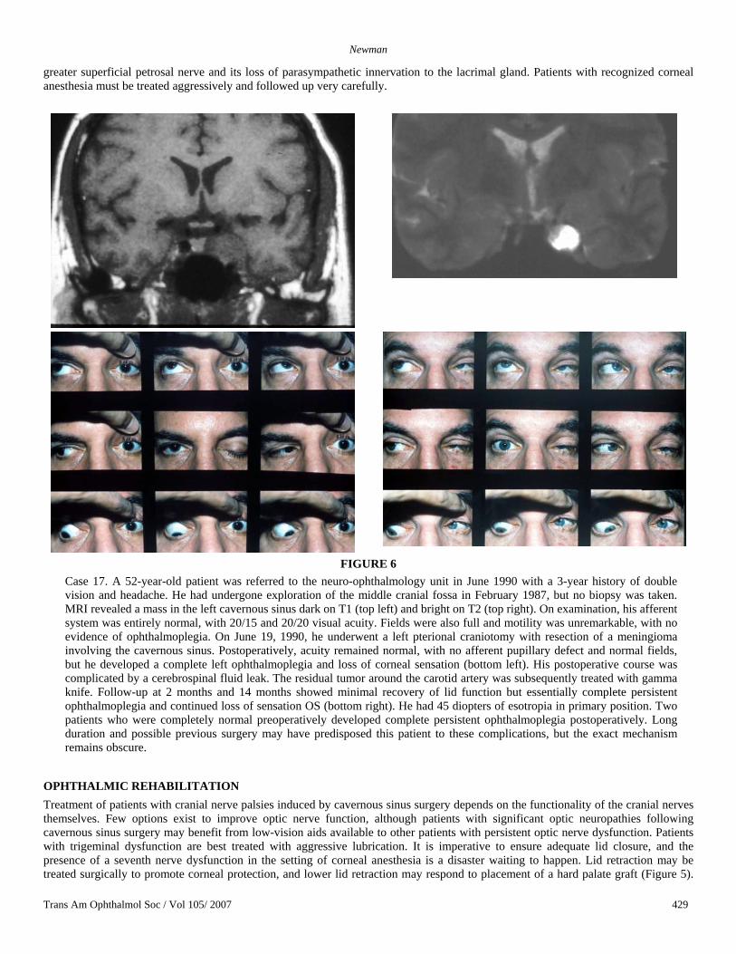
Newman
Trans Am Ophthalmol Soc / Vol 105/ 2007 429
greater superficial petrosal nerve and its loss of parasympathetic innervation to the lacrimal gland. Patients with recognized corneal anesthesia must be treated aggressively and followed up very carefully.
FIGURE 6 Case 17. A 52-year-old patient was referred to the neuro-ophthalmology unit in June 1990 with a 3-year history of double vision and headache. He had undergone exploration of the middle cranial fossa in February 1987, but no biopsy was taken. MRI revealed a mass in the left cavernous sinus dark on T1 (top left) and bright on T2 (top right). On examination, his afferent system was entirely normal, with 20/15 and 20/20 visual acuity. Fields were also full and motility was unremarkable, with no evidence of ophthalmoplegia. On June 19, 1990, he underwent a left pterional craniotomy with resection of a meningioma involving the cavernous sinus. Postoperatively, acuity remained normal, with no afferent pupillary defect and normal fields, but he developed a complete left ophthalmoplegia and loss of corneal sensation (bottom left). His postoperative course was complicated by a cerebrospinal fluid leak. The residual tumor around the carotid artery was subsequently treated with gamma knife. Follow-up at 2 months and 14 months showed minimal recovery of lid function but essentially complete persistent ophthalmoplegia and continued loss of sensation OS (bottom right). He had 45 diopters of esotropia in primary position. Two patients who were completely normal preoperatively developed complete persistent ophthalmoplegia postoperatively. Long duration and possible previous surgery may have predisposed this patient to these complications, but the exact mechanism remains obscure.
OPHTHALMIC REHABILITATION Treatment of patients with cranial nerve palsies induced by cavernous sinus surgery depends on the functionality of the cranial nerves themselves. Few options exist to improve optic nerve function, although patients with significant optic neuropathies following cavernous sinus surgery may benefit from low-vision aids available to other patients with persistent optic nerve dysfunction. Patients with trigeminal dysfunction are best treated with aggressive lubrication. It is imperative to ensure adequate lid closure, and the presence of a seventh nerve dysfunction in the setting of corneal anesthesia is a disaster waiting to happen. Lid retraction may be treated surgically to promote corneal protection, and lower lid retraction may respond to placement of a hard palate graft (Figure 5).
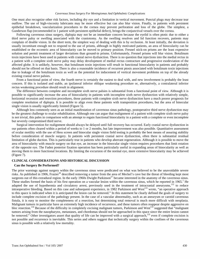
Cavernous Sinus Surgery For Meningiomas And Ophthalmic Complications
Trans Am Ophthalmol Soc / Vol 105/ 2007 430
One must also recognize other risk factors, including dry eye and a limitation in vertical movement. Punctal plugs may decrease tear outflow. The use of high-viscosity lubricants may be more effective but can also blur vision. Finally, in patients with persistent epithelial breakdown, vascularization procedures to the cornea may prevent perforation and loss of the globe. The simplest, a Gunderson flap (recommended in 1 patient with persistent epithelial defect), brings the conjunctival vessels over the cornea.
Following cavernous sinus surgery, diplopia may not be an immediate concern because the eyelid is often ptotic due to either a third nerve palsy or swelling associated with the craniotomy. As the swelling resolves and lid function recovers, patients may complain of double vision. In the short term, the simplest way of dealing with this is by occlusion. At least initially, the deviation is usually incomitant enough not to respond to the use of prisms, although in highly motivated patients, an area of binocularity can be established or the eccentric area of binocularity can be moved to primary position. Fresnel stick-on prisms are the least expensive solution and permit treatment of larger deviations than ground-in prisms. Unfortunately, Fresnel prisms will blur vision. Botulinum toxin injections have been used in patients with cranial nerve dysfunction. There is no question that injections into the medial rectus in a patient with a complete sixth nerve palsy may delay development of medial rectus contracture and progressive esodeviation of the affected globe. It is unlikely, however, that botulinum toxin injections will result in functional binocularity in patients and probably should not be offered on that basis. There is also a reasonable incidence of recurrent ptosis associated with botulinum toxin injections due to leakage of the botulinum toxin as well as the potential for inducement of vertical movement problems on top of the already existing cranial nerve palsies.
From a functional point of view, the fourth nerve is certainly the easiest to deal with, and new involvement is probably the least concern. If this is isolated and stable, an ipsilateral inferior oblique weakening procedure or, alternatively, a contralateral inferior rectus weakening procedure should result in alignment.
The difference between complete and incomplete sixth nerve palsies is substantial from a functional point of view. Although it is possible to significantly increase the area of binocularity in patients with incomplete sixth nerve dysfunction with relatively simple, horizontal recession and resection procedures, those patients with complete sixth nerve dysfunction have a much poorer prognosis for complete resolution of diplopia. It is possible to align even these patients with transposition procedures, but the area of binocular single vision is usually significantly limited (Figure 5).
Although less commonly seen as an initial manifestation of cavernous sinus pathology, postoperative third nerve dysfunction may be the greatest challenge to ocular rehabilitation. Although one can argue that realignment of a patient with complete sixth nerve palsy is not trivial, this pales in comparison with an attempt to regain functional binocularity in a patient with a complete or even incomplete but severely compromised third nerve.
Surgical intervention for realignment should always be delayed until full recovery has occurred. Early cranial nerve dysfunction in our patients often cleared within a period of weeks to 1 or 2 months, but late improvement was also possible. Quantitative assessment of ocular motility with the use of Hess screen and binocular single vision field testing is probably the best means of assuring stability before consideration of muscle surgery. In patients with persistent cranial nerve dysfunction, often there is substantial residual limitation of globe duction. This is particularly true in patients who develop aberrant regeneration. Although it is possible to move the area of binocularity with muscle surgery on that eye, an increase in the binocular single vision requires procedures that limit rotation of the opposite eye. The Faden posterior fixation operation has been particularly useful in expanding areas of binocularity as well as moving them to more functional locations. By limiting the excursion of the normal eye, more extensive binocularity may be achieved (Figure 7).
CLINICAL CONSIDERATIONS AND HISTORICAL DISCUSSION Can the Surgery Be Performed?
The prior warnings against surgery within the cavernous sinus were predicated on what was believed to be the unavoidable severe risks. As published in 1906, Frazier30 described removing a tumor from the area of Meckel’s cave but the threat of bleeding kept most surgeons out of this extradural region. In the early 1960s Dwight Parkinson57 became interested in the anatomy of the cavernous sinus. These studies formed the basis of his first operation on a vascular lesion within the cavernous sinus, which he reported in 1965.1 He adapted the use of hypothermia and circulatory arrest, previously used in the treatment of intracranial aneurysms,132 to reduce intraoperative bleeding. Based on this case and subsequent experience, in 1982 Parkinson and West133 wrote, “an operative approach to this space is indicated when it is anticipated the lesion can be removed.” In this statement he clearly defined the goals of surgery to include complete excision of the pathology present. In the case of a vascular abnormality, such as an aneurysm or carotid cavernous fistula, it is easy to monitor the completeness of a resection, but determining total removal is much more difficult with neoplasia. Malignant tumors in particular have an extremely high incidence of recurrence, and these tumors often reappear despite aggressive en bloc resection.134 Because of the difficulty in completely excising malignant tumors, Parkinson and West133 suggested that “malignant tumors arising from the nasopharynx, metastases, and chordomas should not be approached in this space since the entire lesion cannot be removed.” Other investigators assert that quality of life can be improved with a surgical approach,135 even if complete excision is not possible and recurrence is inevitable. This series and others suggest that technically surgery within the confines of the cavernous sinus is possible with a relatively low mortality.
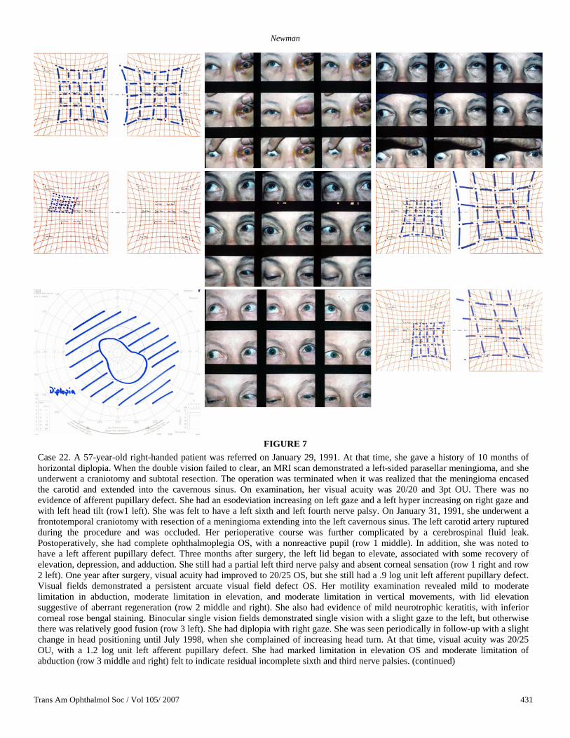
Newman
Trans Am Ophthalmol Soc / Vol 105/ 2007 431
FIGURE 7 Case 22. A 57-year-old right-handed patient was referred on January 29, 1991. At that time, she gave a history of 10 months of horizontal diplopia. When the double vision failed to clear, an MRI scan demonstrated a left-sided parasellar meningioma, and she underwent a craniotomy and subtotal resection. The operation was terminated when it was realized that the meningioma encased the carotid and extended into the cavernous sinus. On examination, her visual acuity was 20/20 and 3pt OU. There was no evidence of afferent pupillary defect. She had an esodeviation increasing on left gaze and a left hyper increasing on right gaze and with left head tilt (row1 left). She was felt to have a left sixth and left fourth nerve palsy. On January 31, 1991, she underwent a frontotemporal craniotomy with resection of a meningioma extending into the left cavernous sinus. The left carotid artery ruptured during the procedure and was occluded. Her perioperative course was further complicated by a cerebrospinal fluid leak. Postoperatively, she had complete ophthalmoplegia OS, with a nonreactive pupil (row 1 middle). In addition, she was noted to have a left afferent pupillary defect. Three months after surgery, the left lid began to elevate, associated with some recovery of elevation, depression, and adduction. She still had a partial left third nerve palsy and absent corneal sensation (row 1 right and row 2 left). One year after surgery, visual acuity had improved to 20/25 OS, but she still had a .9 log unit left afferent pupillary defect. Visual fields demonstrated a persistent arcuate visual field defect OS. Her motility examination revealed mild to moderate limitation in abduction, moderate limitation in elevation, and moderate limitation in vertical movements, with lid elevation suggestive of aberrant regeneration (row 2 middle and right). She also had evidence of mild neurotrophic keratitis, with inferior corneal rose bengal staining. Binocular single vision fields demonstrated single vision with a slight gaze to the left, but otherwise there was relatively good fusion (row 3 left). She had diplopia with right gaze. She was seen periodically in follow-up with a slight change in head positioning until July 1998, when she complained of increasing head turn. At that time, visual acuity was 20/25 OU, with a 1.2 log unit left afferent pupillary defect. She had marked limitation in elevation OS and moderate limitation of abduction (row 3 middle and right) felt to indicate residual incomplete sixth and third nerve palsies. (continued)
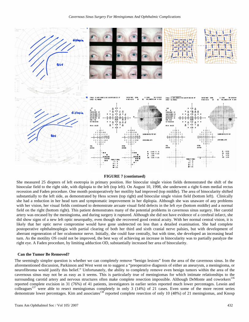
Cavernous Sinus Surgery For Meningiomas And Ophthalmic Complications
Trans Am Ophthalmol Soc / Vol 105/ 2007 432
FIGURE 7 (contintued) She measured 25 diopters of left esotropia in primary position. Her binocular single vision fields demonstrated the shift of the binocular field to the right side, with diplopia to the left (top left). On August 10, 1998, she underwent a right 6-mm medial rectus recession and Faden procedure. One month postoperatively her motility had improved (top middle). The area of binocularity shifted substantially to the left side, as demonstrated by Hess screen (top right) and binocular single vision field (bottom left). Clinically she had a reduction in her head turn and symptomatic improvement in her diplopia. Although she was unaware of any problems with her vision, her visual fields continued to demonstrate arcuate visual field defects in the left eye (bottom middle) and a normal field on the right (bottom right). This patient demonstrates many of the potential problems in cavernous sinus surgery. Her carotid artery was encased by the meningioma, and during surgery it ruptured. Although she did not have evidence of a cerebral infarct, she did show signs of a new left optic neuropathy, even though she recovered good central acuity. With her normal central vision, it is likely that her optic nerve compromise would have gone undetected on less than a detailed examination. She had complete postoperative ophthalmoplegia with partial clearing of both her third and sixth cranial nerve palsies, but with development of aberrant regeneration of her oculomotor nerve. Initially, she could fuse centrally, but with time, she developed an increasing head turn. As the motility OS could not be improved, the best way of achieving an increase in binocularity was to partially paralyze the right eye. A Faden procedure, by limiting adduction OD, substantially increased her area of binocularity.
Can the Tumor Be Removed?
The seemingly simpler question is whether we can completely remove “benign lesions” from the area of the cavernous sinus. In the aforementioned discussion, Parkinson and West went on to suggest a “preoperative diagnosis of either an aneurysm, a meningioma, or neurofibroma would justify this belief.” Unfortunately, the ability to completely remove even benign tumors within the area of the cavernous sinus may not be as easy as it seems. This is particularly true of meningiomas for which intimate relationships to the surrounding carotid artery and nervous structures often make complete resection impossible. Although DeMonte and coworkers136 reported complete excision in 31 (76%) of 41 patients, investigators in earlier series reported much lower percentages. Lesoin and colleagues137 were able to resect meningiomas completely in only 3 (14%) of 21 cases. Even some of the more recent series demonstrate lower percentages. Kim and associates138 reported complete resection of only 10 (48%) of 21 meningiomas, and Knosp
Newman
Trans Am Ophthalmol Soc / Vol 105/ 2007 433
and colleagues139 achieved only a 17% complete resection rate. In a recent series O’Sullivan, and coworkers140 reported complete resections in 26%. Involvement of the lateral aspect of the cavernous sinus may permit a higher rate of complete excision.141
Recurrence can happen even with complete excision, and a 10% to 25% incidence has been reported.136,138,140,142 The rate of recurrence seems to increase with the duration of follow-up, and previous studies of meningiomas in general suggest that the longer the follow-up, the greater the recurrence rate. Mirimanoff and colleagues143 reported recurrence rates for all meningiomas as 7%, 20%, and 32% at 5, 10, and 15 years of follow-up, respectively. Cavernous sinus numbers are likely to be higher.
The extremely slow growth and variable nature of these tumors are major factors in making it difficult to determine recurrence rates. Another cause for frequent recurrence is the invasion of the vascular wall98,144,145 and the involvement of the cranial nerves themselves.146 As seen in our cases, carotid and cranial nerve invasion occurs even with benign tumors and limits possible complete resection.
Progression rates are clearly higher if tumor is intentionally left along the carotid and around the cranial nerves. In reviewing a series of 225 subtotally resected meningiomas in all locations, Mirimanoff and colleagues143 found progression in 37% at 5 years, 55% at 10 years, and 91% at 15 years following surgery. Mathiesen and associates147 found that 42 (61%) of 69 patients (out of a series of 315 skull base meningiomas operated on between 1947 and 1982) who were treated with subtotal resection died of disease usually by 10 years. There is less data (and of shorter duration) on incompletely resected cavernous sinus meningiomas, but after 2 years of follow-up, O’Sullivan, and associates140 reported 2 (6.5%) of 31 cases with progression, and De Jesus and colleagues148 detected 7 (15%) of 46 patients who showed progression (mean follow-up, 34 months). The frequency of progression was up to 38% in those patients who had been followed for more than 5 years. Earlier studies also found that the extent of the original excision was the best prognostic factor determining recurrence.149 These data support an argument for more aggressive attempts at complete excision, but the recurrence rates clearly indicate that even with total surgical resection, meningiomas may recur. Unfortunately, more aggressive surgery probably carries a higher risk of morbidity.150
Recent advances in molecular genetics reveal chromosomal changes151-153 that put patients at high risk for recurrence and progression. There is hope that these and other molecular genetic markers may indicate the risk of recurrence in the future.
In our series the acknowledged residual tumor and the high incidence of subsequent gamma knife treatment underscore the difficulty in achieving complete resection of lesions involving the cavernous sinus. It is often difficult, if not impossible, to make definitive statements about complete excision without more detailed long-term follow-up. The slow growth of many of these lesions continues to be a challenge to surgery in the parasellar region.
Are the Results Different for Nonmeningiomatous Benign Tumors? Theoretically, patients with nonmeningiomatous benign tumors involving the cavernous sinus should have better prognoses. Day and Fukushima154 reported complete clinical resection in 29 (76%) of 38 trigeminal neurilemomas. Sekhar and colleagues155 published a series on complete clinical resection in 30 (79%) of 38 patients with benign nonmeningiomatous tumors involving the cavernous sinus. These tumors included neurilemomas, pituitary tumors, cavernomas, craniopharyngiomas, epidermoids, and juvenile angiofibromas. Eisenberg and associates156 reported similar statistics in a mixed series of nonmeningiomatous tumors affecting the cavernous sinus, with complete clinical resection in 33 (82.5%) of 40 patients.
Unfortunately, recurrences are still possible even in completely resected nonmeningiomatous tumors. This is particularly true of invasive pituitary tumors in which complete surgical excision may not be possible. Eisenberg and colleagues156 reported a 28% incidence of recurrence. Based on the results of transphenoidal surgery series for pituitary marcoadenomas, it is likely that longer-term follow-up will reveal even higher rates of recurrence.
Dolenc111 noted one of 40 trigeminal neurilemomas recurring in 1994, and Taha and associates157 found a 10% incidence of recurrence of trigeminal neurilemomas. Sekhar and coworkers158 reported 4 (13.3%) of 30 nonmeningiomatous benign lesions recurring within the area of the cavernous sinus.
All of these numbers are substantially better than the statistics from series published before the development of skull base approaches. Recurrence rates of up to 65% have been reported in patients previously treated. This likely reflects not only the improvements in surgical approaches, but also the ability to better define the anatomy with the advent of neuroimaging studies. Although the numbers are an improvement, recurrence is still possible, even with reported complete clinical excision.
Unfortunately, the effect of the nature of the pathology on the incidence of neuro-ophthalmic complications has not been previously addressed. Somewhat surprisingly in this study, when the data was subject to rigorous quantitative assessment, there was little difference between the complication rates for patients that were found to have meningiomas and those with nonmeningiomatous pathology. This was particularly true for the chance of optic nerve damage. Nonmeningiomas did have a better chance of complete recovery of pre-existing cranial nerve palsies, although the incidence was low.
Can We Improve Patient Symptoms? The goal in operating on lesions within the cavernous sinus (other than complete cure of the tumor) is to improve the patient’s symptoms. Cavernous sinus lesions may enlarge to secondarily involve the optic nerve even though it is not within the sinus proper. Improvement in compromised optic nerve function can be expected with optic nerve and canal decompression during cavernous sinus surgery. Five (15%) of 33 patients showed improvement in optic nerve function in the series reported on by DeMonte and colleagues.136 Kim and associates138 found improvement in optic nerve function in 1 of 7 patients. In the present series, improvement occurred in 5 (25%) of 20 patients.
Unfortunately, but not surprisingly, no patient with complete optic neuropathy improved following surgical decompression in the
Cavernous Sinus Surgery For Meningiomas And Ophthalmic Complications
Trans Am Ophthalmol Soc / Vol 105/ 2007 434
area of the cavernous sinus.98 This has also been demonstrated in the present study; none of the 4 patients with hand motion vision or worse, including 1 meningioma, experienced improvement. On the other hand, one could argue that decompression of the optic nerve might be accomplished without invading the cavernous sinus, inviting the attendant morbidity described.159
Given that the majority of patients with cavernous sinus pathology present with evidence of ocular motor abnormalities, one of the hoped-for results of cavernous sinus surgery is an improvement in ocular motility. Early reports were encouraging. Lesoin and associates137 reported improvement in ocular motor function in 10 (48%) of 21 patients. Sekhar and colleagues158 reported improvement in 7 of 9 patients with third nerve palsies, 2 of 6 patients with fourth nerve palsies, and 6 (50%) of 12 patients with sixth nerve palsies in a series of mixed pathology. Somewhat more ominous was the report in 1991120 that while 24 (44%) of 54 patients showed improvement in ocular motor function following cavernous sinus surgery, none of the 18 patients with meningiomas improved.
This poorer prognosis in meningiomas has been confirmed in subsequent reports. In a series restricted to meningiomas, DeMonte and associates136 found improvement in ocular motor function in only 13%, whereas patients with nonmeningiomatous tumors reportedly had a 50% improvement.156 In all patients, the third nerve was much less likely to recover than the sixth nerve.156 The ability to improve preexisting ocular palsies was very limited in the present series. In only 1 patient was there felt to be improvement in a preexistent third nerve palsy (none cleared completely), and only 2 patients with sixth nerve palsies improved (clearing in one).
How Do We Assess Cranial Nerve Function? Throughout these published series there are few data to indicate how cranial nerve function was assessed. Biglan and associates160 established a clinical grading system of binocularity in an attempt to improve clinical relevance. In this system, binocularity is graded as “excellent” if the patient has no diplopia in the primary position and downward reading gaze, and at least 20 degrees to either side. Binocularity is classified as “good” if there is no diplopia in the primary and reading gaze but double vision with any eccentric gaze. Patients were recorded as having a “fair” condition if elimination of diplopia required a head turn and a “poor” condition if there was diplopia in all fields of gaze.
This classification system was used by Cusimano and colleagues142 to follow 124 patients who underwent cavernous sinus surgery. Four (27%) of the 15 who had poor preoperative binocularity improved to “good” or “excellent” status, and 6 (20%) of 30 patients with “good” preoperative binocularity improved to “excellent” status. The series included meningiomas and nonmeningiomatous tumors. Even in this series, there was little quantitative assessment.
The potential of completely excising a lesion and improving function must be balanced against the surgical potential for creating new problems as well as the natural history of the lesions themselves. The variability in the natural history of cavernous sinus tumors is a major limitation. In particular, menigiomas involving the cavernous sinus often have an extremely indolent history, exhibiting very slow progression. One of the difficulties in analyzing previous series that focus on the natural history is the variable inclusion criteria. The natural history is far worse if tumors arising from the clivus or petrous bone and secondarily involve the cavernous sinus are included, because these tumors will often compress the brainstem. If the series are restricted to tumors involving the cavernous sinus in isolation, the natural history is far more benign.
What Is the Natural History of Lesions Affecting the Cavernous Sinus? There are few series that provide natural history data. In a series of 8 patients followed up for a mean of 5.5 years, Freidlander and colleagues161 reported progression in only 1 patient. In a series of 24 patients observed at Johns Hopkins for a mean of 76 months, there was evidence of clinical progression in only 6 (25%).162 In an updated follow-up of 29 patients with at least 10 years of study Klink and associates72 found new motility defects in only 3 (10%) but noted new optic nerve dysfunction in 7 (24%). We sometimes forget that the clinical findings are more important to the patient than are minute measurable changes on imaging studies. Patients are more concerned about worsening double vision or a decrease in acuity than radiographic changes. When dealing with the long-term natural history of a tumor, it is important to consider the clinical symptoms as paramount. This slow growth makes even most recent series of surgical results difficult to interpret as the mean follow-up is usually 5 years or less.147
What Is the Expected Morbidity of Cavernous Sinus Surgery? Can we operate safely within the cavernous sinus without inducing significant morbidity? Since the beginning of the modern era of skull base surgery, surgical mortality has been substantially reduced. Dolenc and coworkers98 reported 4 deaths (6.3%) out of 63 patients who had undergone cavernous sinus surgery. More recent series have reported the incidence of death in 2 (3.7%) of 54,120 3 (7.3%) of 41,136 and 2 (9.5%) of 21 postsurgical patients.138 In the present series, 2 patients died within the perioperative period (2.4%), only 1 with a meningioma (1.75%).
Lesser complications have been far more frequent and have included infarction with hemiparesis,136,138,142 meningitis, and hydrocephalus. A CSF leak is one of the most frequently occurring complications following cavernous sinus surgery and is probably a direct result of surgical aggressiveness. Its incidence may be as high as 28%.142
A sobering statement was made in Cusimano’s 1995 summary of the Pittsburgh142 experience: “the majority of patients in this series suffered a surgical complication.” Although he goes on to point out that most complications were minor or treatable, this potential must be weighed against the expected benefits of surgery. For psychological and other reasons, a case can still be made for aggressive surgery.163
It is not the role of the ophthalmologist to discuss nonophthalmic risks with the patient without including the referring surgeon. Unfortunately, there is often a tendency for neurosurgeons to trivialize the morbidity in any transcranial procedure. As
Newman
Trans Am Ophthalmol Soc / Vol 105/ 2007 435
ophthalmologists are often the ones to recognize cavernous sinus pathology and thus refer patients to neurosurgery, it is useful to have a feel for the interaction of the neurosurgeon with the patient so that all parties are aware of the goals as well as the risks of any type of intervention.
What Are the Problems of Nonquantitative Ophthalmic Testing? The major potential for complications in cavernous sinus surgery involves damage to those cranial nerves contained in the cavernous sinus. Given that these nerves have a profound effect on ophthalmic function, it is surprising that ophthalmologists have provided little direct input into assessing these patients both preoperatively and postoperatively.72,160,162 The lack of detailed quantitative ophthalmic testing is illustrated by the way some of the cranial nerve deficits are reported. In Hakuba and colleagues’1989 report of 10 intracavernous tumors, 26 they state that “a transient paresis of the third to sixth cranial nerves was seen in five patients.” Unfortunately, the report does not identify which nerves, the severity of the paresis, and exactly how these were measured. In addition, the enthusiasm for the results of surgery may explain why 9 of the 10 patients were said to have “excellent or good” results, despite the fact that 5 patients were noted to have permanent, complete ophthalmoplegia. Defining “good” and “excellent” is not so obvious in many of the previously reported series. Another question here concerns how well these patients were truly evaluated. In Al-Mefty and Smith’s 1988 series,164 18 patients were reported on following cavernous sinus surgery. The first 13 patients underwent surgery in Saudi Arabia, and the last 5 patients underwent surgery at the University of Mississippi. Of the 18 patients, only 4 were noted to have cranial nerve palsies, with a third nerve palsy in 3, a fourth nerve palsy in 1, and a fifth nerve palsy in 1. If the details of the series are reviewed, one finds that all 4 of the patients with cranial nerve palsies were operated on at the University of Mississippi (of 5 patients), whereas no patient (of 13 patients) who was operated on in Saudi Arabia (an earlier part of the series) was noted to have a cranial nerve palsy. One has to be suspicious that the earlier patients were not evaluated in as much detail as the subsequent patients.
As mentioned in our oculomotor results, none of the neurosurgical series discusses the concept of aberrant regeneration. The only time this is brought up is in those reports detailing intraoperative cranial nerve repair.165-167 This is somewhat surprising given that meningiomas in the parasellar area may be responsible for the development of aberrant regeneration in the absence of a history of a third nerve palsy.168 The combination of the classical findings of aberrant regeneration without a history of a third nerve palsy (primary aberrant regeneration) is almost pathognomonic of a meningioma or aneurysm in the parasellar region.
What Are the Particular Problems Associated With Ocular Motor Nerve Dysfunction? Even at the outset of the modern era in skull base surgery, concerns were expressed about the potential for damage to the third nerve. Lesoin and colleagues137 stated that it was “not possible to completely remove an intracavernous meningioma without disturbing the ocular motor nerves.” Despite these concerns, initial reports seemed to be encouraging. Only 5 (24%) of 21 cases137 demonstrated worsening of third nerve function postoperatively. Perneczky and colleagues122 reported 2 cases (5.7%) of third nerve palsy in 35 patients who had undergone surgery in which a subtemporal lateral approach was used. Of 42 original patients reported on (from Pittsburgh), 10 (23.8%) had a “temporary” palsy, but none had permanent third nerve dysfunction. By 1991 Sehkar and associates169 recognized 6 (5.9%) of 101 patients with permanent third nerve dysfunction. Al-Mefty reported transient third nerve palsies in 3 (4.1%) of 74 cases and permanent third nerve dysfunction in 3 others (4.1%) (in 7 patients [9.5%] it was “too early to tell.”).99 DeMonte and colleagues136 found 2 (14.3%) of 14 patients with preoperative third nerve palsies to have worse dysfunction and 2 (14.3%) of 14 patients without preoperative third nerve palsies to have developed new ones. Even with nonmeningiomatous tumors, third nerve palsies may develop or worsen. Eisenberg, and associates156 reported 4 (12.5%) new third nerve palsies among 32 patients who had undergone surgery and worsening palsy in 1 (12.5%) of 8.
Because of the innervation of multiple muscles, damage to the third cranial nerve often results in incomplete resolution, with evidence of variable muscle misfiring. Not until the first third of the 20th century did explanations for this misdirection syndrome appear in the literature.170 Morris Bender, a neurologist in New York, undertook experimental work with oculomotor nerve regeneration in which he sectioned the third nerve of a chimpanzee and followed its recovery.171 Walsh172 reviewed the importance of aberrant regeneration in 1947. It has been suggested that there may be alternative explanations for aberrant regeneration, including ephaptic transmission,173 but it is likely that actual reinnervation of sprouting axons terminating in the wrong extraocular muscles is responsible for the majority of the findings. The classic finding of lid elevation with attempted depression and adduction is probably less consequential than the coinnervation of the superior and inferior rectus muscles, which leads to persistent limitation in the vertical gaze. Aberrant regeneration is probably underdiagnosed and is the responsible mechanism in the majority of patients with incomplete third nerve palsies and vertical compromise.
It is unlikely that the attempts at surgical repair of the third nerve during cavernous sinus surgery will reduce the incidence of aberrant regeneration. At this time, the best treatment for aberrant regeneration is to prevent its occurrence. Aberrant regeneration does not occur with presumed microvascular third nerve palsies, and its high incidence in patients undergoing cavernous sinus surgery suggests that perhaps there is more than simple vascular damage to the third nerve. It is possible that retained tumor within the nerve or direct surgical trauma results in misdirection in patients following cavernous sinus surgery. The possible development of aberrant regeneration has been one of the strongest arguments in favor of limiting surgery.72,161
The current series has a much higher incidence of third nerve dysfunction. Only 1 (1.8%) of 57 patients showed no evidence of third nerve dysfunction immediately postoperatively. Exclusion of the 19 patients with preoperative third nerve dysfunction still results in a 97% incidence of acquired third nerve palsy immediately following cavernous sinus surgery. Although most palsies cleared rapidly, 4 (11%) of the 35 patients with long-term follow-up had complete third nerve dysfunction, and an additional 18 (51%) had an incomplete third. Nine (26%) demonstrated evidence of aberrant regeneration. Probably the closest numbers to the current
Cavernous Sinus Surgery For Meningiomas And Ophthalmic Complications
Trans Am Ophthalmol Soc / Vol 105/ 2007 436
series were reported by Cusimano and colleagues,142 who noted that 35 (60%) of 58 patients undergoing cavernous sinus surgery had binocularity that was “worse than excellent.”
Sindou and Pelissou,174 in a literature review, reported on new fourth nerve palsies in 4 (7.3%) of 55 patients undergoing surgery for trigeminal neurilemomas. In Al-Mefty and Smith’s original report of 18 patients,164 only 1 (5.6%) had a fourth nerve palsy. Nine (12.2%) of 74 patients in their expanded series99 were said to have fourth nerve dysfunction (although the situation in 7 patients was listed as “too early to tell.”). Perneczky and colleagues122 found 2 (5.7%) fourth nerve palsies among 35 cases. The Pittsburgh group originally reported 9 (20.9%) temporary and 4 (9.3%) permanent fourth nerve palsies in 43 patients.158 The incidence had changed to 7 (7.7%) of 91 cases165 by 1991, and in an article later that same year, 11 (10.9%) of 101 cases.169 In more recent series, new fourth nerve palsies occurred in only 1 (3.7%) of 27 patients undergoing cavernous sinus surgery.136 Although the fourth nerve is theoretically at greater risk during surgery for meningiomas than nonmeningiomatous tumors within the cavernous sinus, Eisenburg and associates156 reported 4 (11.1%) of 36 patients with new fourth nerve dysfunction.
In the current series, fourth nerve palsies could not be separated from other cranial nerve palsies. When the third and sixth nerve palsies were complete, the fourth nerve usually showed no function.
The sixth nerve is the most difficult to see during surgery because it is situated deep within the cavernous sinus, in relation to the carotid artery. Early cavernous sinus surgical series report worsening sixth nerve function in patients in whom it already existed. In the original series of cases from Pittsburgh, there were 12 preoperative abducens palsies and 14 temporary palsies postoperatively. In only 4 (9.5%) of the 42 cases was sixth nerve dysfunction said to be permanent.158 When the series was updated to include 101 patients,169 there were 10 permanent palsies (10%). Interestingly, a report earlier that year had revealed only 5 sixth nerve palsies (5.5%) among 91 patients undergoing surgery.166 This difference raises the question of how closely the patients were monitored for evidence of sixth nerve dysfunction. Five (9.3%) of 54 cases in the series from Hanover had permanent sixth nerve dysfunction, and 10 had temporary abduction abnormalities.120 Only one (1.4%) of 74 cases reported in 199199 was said to have a permanent sixth nerve palsy, although 7 were listed as “too early to tell.” Two (11%) of 18 patients with sixth nerve dysfunction preoperatively were reported to be worse following surgery.136 When broken down into nonmeningiomatous benign tumors, there were still a number of new sixth nerve palsies following surgery (4 [14.3%] of 28 reported by Eisenberg156).
There was no indication of the means used to assess cranial nerve function in any of these reports. None of these previously reported series include the numbers of patients with cranial nerve dysfunction seen in this current series. Only 8 patients (10%) had no evidence of sixth nerve dysfunction immediately after surgery. Although many of these dysfunctions resolved, 35 (69%) of 51 patients had some evidence of sixth nerve dysfunction at the follow-up more than 3 months after surgery, and 14 (27%) had a complete sixth nerve palsy.
Although there may be several explanations for the higher incidence of cranial nerve palsies in our postoperative patients, it is likely that quantitative detailed assessment may be largely responsible for explaining the higher numbers. Incomplete ophthalmoplegia is not incompatible with binocularity and good visual function, but may well lead to significant patient dissatisfaction. Future studies will need to include methods to evaluate functional outcome.
What Are the Implications of Trigeminal Dysfunction? The clinical importance of the fifth cranial nerve is seldom emphasized, although it is among the cranial nerves which may be injured during cavernous sinus surgery. The true implications of neurotrophic keratitis as a potential complication of cavernous sinus surgery have largely been ignored, although dry eye and its potential connection to the greater superficial petrosal nerve (responsible for reflex tearing) have been discussed. Fortunately, even with complete corneal denervation, neurotrophic changes do not always occur. Twenty (36.4%) of 55 patients who had undergone surgery for trigeminal neurilemomas174 demonstrated decreased sensation, but only 5 (9.1%) reportedly developed corneal complications. In the early series of cavernous sinus surgery, trigeminal dysfunction was mentioned in a minority of patients. In the first 7 patients reported, only the third division of the fifth nerve was noted to be affected. Six (14.3%) of 42 patients were said to have temporary, and 3 (7.1%) permanent, loss of sensation in the V1 distribution.158 Trigeminal dysfunction was recognized in only 3 (3.3%) of 91 patients following surgery,175 but in a later review that same year there were 10 (9.9%) of 101 cases. Al-Mefty and colleagues99 reported on 9 transient (12.2%) and 1 permanent (1.4%) fifth nerve dysfunction among 74 patients who had undergone surgery. The group in Hanover reported 16 (29.6%) of 54 patients with preoperative trigeminal dysfunction, 17 (31.5%) with temporary or partial dysfunction following surgery, and 10 patients (18.5%) with permanent fifth nerve dysfunction.120 In a more recent review, DeMonte136 reported 6 (19%) of 31 patients who developed new fifth nerve dysfunction following surgery. Forty-seven (76%) of 62 patients without trigeminal dysfunction preoperatively were noted to have some loss of corneal sensation in our series with more quantitative assessment. This may be more serious given that the fifth nerve seems to have a worse prognosis for recovery, and in the present series 15 patients (32% of those with trigeminal dysfunction) eventually developed neurotrophic keratitis.
The potential for developing neurotrophic keratitis following the loss of fifth nerve function can be exacerbated by several factors. These include loss of tear production, seventh nerve dysfunction with problems in eyelid closure, and problems with ocular motility resulting in the inability to move the eye out of the palpebral fissure. None of these issues has been discussed in any prior neurosurgical series, however. Aggressive lubrication is an essential treatment in patients at risk for neurotrophic keratitis. The use of gold weight implants, tarsorrhaphy, or other protective procedures is probably best completed early when the loss of corneal sensation is associated with decreased seventh nerve function. A vascularization procedure (Gunderson flap) may be required to keep the eye from perforating with recurrent epithelial defects, erosion, or infection. Nerve growth factor treatment may be an additional mode of
Newman
Trans Am Ophthalmol Soc / Vol 105/ 2007 437
therapy in patients with persistent sensory loss.176,177 Unfortunately, initial studies suggest that patients with surgically induced neurotrophic keratitis are probably the most resistant to treatment.
What Are the Alternative Treatment Options for Patients With Cavernous Sinus Pathology? In discussing surgery and its consequences, we must compare surgical results to alternative treatment modalities. Radiation therapy has been used to treat neoplasia almost since x-rays were discovered by Roentgen at the turn of the last century. Malignant lesions (especially lymphoproliferative tumors including lymphoma and plasmacytoma) may be very radiosensitive. It was thought that benign slow-growing tumors would respond poorly to radiation. Somewhat surprisingly, the growth rate of meningiomas can be altered with conventional fractionated radiation.178-180 Regression based on imaging or improvement in cranial nerve function, however, is exceptional. Radiation may play a more active role in other types of skull base tumors. This is particularly true for malignancies but also for chordomas.181
Over the last decade an increasing interest has been expressed for the use of radiosurgery. This is defined as a single-dose application, either of a gamma source or through a linear accelerator. Additional smaller studies of the use of proton beam have also been reported for meningiomas involving the cavernous sinus. The development of single-dose focal radiation therapy (LINAC182,183) and gamma knife184-194 has offered additional options. The exact role of these modalities is yet to be defined.191 Significant series reported on the use of gamma knife have included upward of 1,000 meningiomas involving the cavernous sinus treated either primarily or following subtotal resection.189,190,192-199 The emphasis has been on “control.”197 Unfortunately, although some of the case follow-ups have been 8 years or more, the majority of series have been limited to 3 to 4 years.190,197,200,201 Based on previous experience, this is much too early in order to be able to truly assess the effectiveness of gamma knife in these patients.184-188,190
While complications have been low, they do occur. Several studies have addressed the potential for radiation optic neuropathy in patients undergoing gamma knife. The optic nerve and the visual pathways are more susceptible to potential radiation damage.202,203 Some investigators have emphasized the ability to treat lesions directly applied to the optic nerve.201,204 On the other hand, it has been suggested that optic neuropathy could be as high as 77.8% when the optic nerve received a dose of 15 Gy or more.205 Therefore, lesions should be situated at least 3 mm away from the afferent visual pathways for safe treatment. The “safe dose” of radiation to the optic nerve is yet to be determined, and this fact argues for a conservative approach.206 The oculomotor and trigeminal nerves are relatively resistant to radiation therapy.196,204,207 In spite of this, trigeminal and ocular motor problems have been reported.208,209
In order to decrease the chance of damage, several investigators have suggested lowering doses.204 Others have emphasized the importance of treating the entire tumor to at least a dose of 14 Gy.210 Because of the restriction of radiosurgery to a limited size, some investigators have continued to advocate fractionated radiation therapy for those tumors that are larger.191 Because of extremely low morbidity, several investigators still advocate the use of fractionated radiation therapy for cavernous sinus meningiomas in general. 211,212 The use of gamma knife treatment with a particular interest for neuro-ophthalmologists was summarized by Carvounis and Katz in 2003.213
During the last 2 decades several medical treatments have been proposed for meningiomas. Unfortunately, data on hormonal therapy214 and hydroxyurea have been disappointing after initial optimistic reports. At this time there are no proven efficacious therapies for meningiomas beyond surgery and radiation. It is hoped that the recent mapping of chromosomal defects in meningiomas151-153 will lead to alternative treatments.
In view of the slow progression of parasellar meningiomas, a number of surgeons, including several with significant clinical experience, have advocated a limited approach to these tumors consisting of resection of only the extracavernous portion possibly to be followed with radiation therapy.72,162,215,216 This offers the advantage of decompressing the optic nerve when involved, reducing the tumor size to something that can be treated with radiosurgery, and most important, limiting the complications of aggressive intracavernous surgery.217-221 Their attitude might best be summed up by a statement made by Madjid Samii: “Why should we risk a carotid artery and eye function for a lesion that hardly ever endangers the patient’s life as long as the tumor part involving other structures such as the brain stem are resected.”
CONCLUSIONS
Pathology that affects the cavernous sinus is more common than previously thought, and recognition is largely due to the advent of modern imaging studies. Ophthalmic manifestations remain the most common indication of cavernous sinus pathology, although often patients are diagnosed when imaging studies are obtained for other reasons. The natural history of pathology affecting the cavernous sinus seems largely undetermined. Benign tumors may be asymptomatic for years, but progressive growth is common. Quantitative evaluation of the visual system is imperative to improve our understanding of the true natural history of these lesions. Failure to measure function accurately can lead to erroneous underestimation of the true incidence of dysfunction. Quantitative evaluation is important to direct therapeutic intervention. The timing of rehabilitative surgery depends on the degree of postoperative ocular motor stabilization.
Our study supports the literature that cavernous sinus surgery is technically feasible with relatively low mortality in patients with meningiomas. Nonetheless, quantitative assessment indicates that cavernous sinus surgery only infrequently benefits preexisting cranial nerve dysfunction. The one exception to this rule is extracavernous extension with compression of the optic nerve, which can be benefited by decompression in the area of the orbital apex. One caveat not previously reported in the literature is the danger to the optic nerve posed by the skull base pterional approach, which requires take-down of the anterior clinoid and portions of the optic canal. Although central acuity may remain fairly good, quantitative assessment of optic nerve function may detect a previously
Cavernous Sinus Surgery For Meningiomas And Ophthalmic Complications
Trans Am Ophthalmol Soc / Vol 105/ 2007 438
unrecognized postoperative incidence of visual field abnormalities. The mechanism of damage to the optic nerve is unknown, but may be related to the surgical removal of the anterior clinoid, during which there may be heat transfer to the optic nerve or, alternatively, hemorrhage-induced vasospasm. It is also possible that there may be direct mechanical damage to the optic nerve within the canal. Improvement in compromised optic nerve function could be obtained by simply excising the extracavernous portion of the tumor without the inherent risks of surgery within the cavernous sinus proper.
Oculomotor dysfunction following cavernous sinus surgery occurred in 97% of cases. This deficit is transient in the majority, but may also be permanent. The prognosis for complete resolution is better if the cranial nerve palsy is incomplete following surgery. Patients with complete oculomotor dysfunction postoperatively have an 84% chance of partially improved function with time, but a 32% incidence of developing aberrant regeneration, which is not necessarily incompatible with binocularity. Surgeons should discuss the potential for diplopia and the need for extraocular muscle surgery prior to proceeding with intervention.
Trigeminal dysfunction is common. The fact that neurotrophic keratitis can be expected in more than one-quarter of patients argues strongly for the importance of ophthalmic input into long-term follow-up of patients with cavernous sinus lesions. In this way, many of the consequences of neurotrophic keratitis may be prevented, as indicated in the section “Ophthalmic Rehabilitation.”
To keep this series uniform, only the results of a single surgeon were analyzed. Additional cavernous sinus surgeries performed by other neurosurgeons were excluded from this study. Although it is theoretically possible that other surgeons could have better results, this particular surgeon had more experience with cavernous sinus surgery than all others present and maintains the largest volume of cavernous sinus procedures. Given that many previously reported procedures were completed at other institutions, it is impossible quantitatively to assess ophthalmic function elsewhere. We can only conclude that more intense scrutiny from a neuro-ophthalmic point of view would reveal a higher incidence of ophthalmic pathology. Because neurosurgeons are not specifically trained to quantitatively assess ophthalmic function, it may be misleading to accept previous papers that report patient improvement based on subjective assessment. It is imperative that patients be presented with accurate data before making decisions with regard to this mode of therapy. With other treatment options potentially available, patients should be fully educated before a decision is made to perform this procedure.
This study would have benefited from a longer follow-up, but because many of the patients were coming from great distances, it was not possible to get them all back for a quantitative assessment. We chose not to accept nonquantitative data reported from other institutions because of the lack of detailed assessment, thus limiting the percentage of long-term follow-up. This is important if definitive statements are going to be made with regard to risk percentages. At this time, however, the thesis that cavernous sinus surgery has low ophthalmic morbidity is not justified. Patients undergoing cavernous sinus surgery can expect to have worsening of cranial nerve function that, even when transient, may leave them with substantial residual problems, including development of neurotrophic keratitis, and evidence of aberrant regeneration, which may produce persistent diplopia. There is a low incidence of improvement in cranial nerve function, and therefore patients should not be promised ocular motor improvement with less diplopia following surgery.
The last 2 decades of surgical refinement in dealing with skull base lesions has largely taught us what we can do. What we should do depends on a frank discussion of the options and a realistic assessment of the risks and benefits of surgery and its alternatives. It is likely that in the future, surgery will once again play a smaller, rather than a greater, role in treating cavernous sinus lesions. A combination of limited surgery and some form of radiation may reduce the morbidity associated with surgical treatment.195 This course has been suggested and probably represents the current mainstream opinion in neurosurgery. At this time the lack of many other good options will still require the consideration of surgical options, and in the right case, surgery, especially if tailored to a realistic goal, may provide significant benefit to our patients.
ACKNOWLEDGMENTS Funding/Support: None Financial Disclosures: None
REFERENCES 1. Parkinson D. A surgical approach to the cavernous portion of the carotid artery. Anatomical studies and case report. J Neurosurg
1965;23:474-483. 2. Winslow J. Exposition anatomique de la structure du corps humain. London: Prevost; 1732. 3. Donley J. John James Wepfer, a renaissance student of apoplexy. Bull Johns Hopkins Hosp 1909;20:25. 4. Vieussens R. Neurographia Universalis. Lyons; 1685. 5. Ridley H. The Anatomy of the Brain. London: Smith & Walford; 1695. 6. Weizenhoffer A. Contralateral cavernous sinus thrombosis following furuncle of the external auditory canal; recovery. N Y State
Med J 1932;32:139. 7. Bedford MA. The “cavernous” sinus. Br J Ophthalmol 1966;50:41-46. 8. Dietemann JL, Kehrli P, Maillot C, et al. Is there a dural wall between the cavernous sinus and the pituitary fossa? Anatomical
and MRI findings. Neuroradiology 1998;40:627-630. 9. Kehrli P, Maillot C, Wolff MJ. The venous system of the lateral sellar compartment (cavernous sinus): an histological and
embryological study. Neurol Res 1996;18:387-393.
Newman
Trans Am Ophthalmol Soc / Vol 105/ 2007 439
10. Kehrli P, Ali MM, Maillot C, et al. Comparative microanatomy of the lateral wall of the “cavernous sinus” in humans and the olive baboon. Neurol Res 1997;19:571-576.
11. Kehrli P, Ali M, Reis M Jr, et al. Anatomy and embryology of the lateral sellar compartment (cavernous sinus) medial wall. Neurol Res 1998;20:585-592.
12. Sadasivan B, Ma SH, Dujovny M, et al. The anterior cavernous sinus space. Acta Neurochir 1991;108:154-158. 13. San Millan Ruiz D, Gailloud P, de Miquel Miquel MA, et al. Laterocavernous sinus. Anat Rec 1999;254:7-12. 14. Umansky F, Valarezo A, Elidan J. The superior wall of the cavernous sinus: a microanatomical study. J Neurosurg 1994;81:914-
290. 15. Kawase T, Shiobara R, Ohira T, et al. Developmental patterns and characteristic symptoms of petroclival meningiomas. Neurol
Med Chir 1996;36:1-6. 16. Duke-Elder S. Text Book of Ophthalmology, ed 5. St Louis: CV Mosby; 1952:4123. 17. Taptas J. La loge du sinus caverneux. Rev Otoneuroophthalmol 1949;21:193-199. 18. Taptas JN. The so-called cavernous sinus: a review of the controversy and its implications for neurosurgeons. Neurosurgery
1982;11:712-717. 19. Taptas J. Must we still call cavernous sinus the parasellar vascular and nervous crossroads? The necessity of a definite
topographical description of the region. In: Dolenc VV, ed. The Cavernous Sinus: A Multidiciplinary Approach to Vascular and Tumorous Lesions. Wien: Springer-Verlag; 1987:30-40.
20. Bonnet P. La loge caverneuse et les syndromes de la loge caverneuse. Arch Ophtalmol 1955;15:357-372. 21. Parkinson D. Surgical anatomy of the lateral sellar compartment (cavernous sinus). Clin Neurosurg 1990;36:219-239. 22. Parkinson D. Lateral sellar compartment: history and anatomy. J Craniofac Surg 1995;6:55-68. 23. Parkinson D. Lateral sellar compartment O.T. (cavernous sinus): history, anatomy, terminology. Anat Rec 1998;251:486-490. 24. Krivosic I. Histoarchitecture of the cavernous sinus. In: Dolenc VV, ed. The Cavernous Sinus. A Multidisciplinary Approach to
Vascular and Tumorous Lesions. Wien: Springer-Verlag; 1987:117-129. 25. Rhoton AL Jr, Hardy DG, Chambers SM. Microsurgical anatomy and dissection of the sphenoid bone, cavernous sinus and sellar
region. Surg Neurol 1979;12:63-104. 26. Hakuba A, Tanaka K, Suzuki T, et al. A combined orbitozygomatic infratemporal epidural and subdural approach for lesions
involving the entire cavernous sinus. J Neurosurg 1989;71:699-704. 27. Adams J. Aneurism of internal carotid in the cavernous sinus causing paralysis of the third, fourth, fifth and sixth nerves. Lancet
1869;2:768. 28. Bartholow R. Aneurisms of the arteries at the base of the brain, their symptomatology, diagnosis, and treatment. Am J Med Sci
1872;64:373-386. 29. Krogius A. Om operative Behandlund of Tumoren i Fossa Media Cranii. Rev Chir 1896;16:434-445. 30. Frazier CH. The surgical treatment of intracranial lesions causing disturbances of vision. In: Posey WP, Spiller WG, eds. The
Eye and the Nervous System. Philadelphia: JB Lippincott; 1906:867-873. 31. Langworthy HG. Ocular relations of the cavernous sinus. In: Wood CA, ed. The American Encyclopedia of Ophthalmology. Vol
III. Chicago: Cleveland Press; 1914:1794-1809. 32. Trobe JD, Glaser JS, Post JD. Meningiomas and aneurysms of the cavernous sinus. Neuro-ophthalmologic features. Arch
Ophthalmol 1978;96:457-467. 33. Smith JL. Editorial note to: Post MJD, Glaser JS, Trobe JD: Elusive lesions of the cavernous sinus. In: Smith JL, ed. Neuro-
ophthalmology Focus 1980. New York: Masson; 1979:260. 34. Dolenc V. Direct microsurgical repair of intracavernous vascular lesions. J Neurosurg 1983;58:824-831. 35. Hakuba A, Ohata K, Soares SB Jr, et al. Embryology of the cavernous sinus. In: Eisenberg MB, Al-Mefty O, eds. The Cavernous
Sinus: A Comprehensive Text. Philadelphia: Lippincott Williams & Wilkins; 2000:5-20. 36. Weninger WJ, Muller GB. The parasellar region of human infants: cavernous sinus topography and surgical approaches. J
Neurosurg 1999;90:484-490. 37. Umansky F, Nathan H. The cavernous sinus. An anatomical study of its lateral wall. In: Dolenc VV, ed. The Cavernous Sinus. A
Multidisciplinary Approach to Vascular and Tumorous Lesions. Wien: Springer-Verlag; 1987:56-66. 38. Umansky F, Nathan H. The lateral wall of the cavernous sinus. With special reference to the nerves related to it. J Neurosurg
1982;56:228-234. 39. Kawase T, van Loveren H, Keller JT, et al. Meningeal architecture of the cavernous sinus: clinical and surgical implications.
Neurosurgury 1996;39:527-534; discussion 534-536. 40. Rhoton AL, Inoue T. Microsurgical approaches to the cavernous sinus. Clin Neurosurg 1991;37:391-439. 41. Goel A. The extradural approach to lesions involving the cavernous sinus. Br J Neurosurg 1997;11:134-138. 42. Conesa HA, Zadorecki EA, Lozano MC. Gross anatomy of the cavernous region. In: Dolenc VV, ed. The Cavernous Sinus. A
Multidisciplinary Approach to Vascular and Tumorous Lesions. Wien: Springer-Verlag; 1987:43-55. 43. Borba LAB, Al-Mefty O. Normal anatomy of the cavernous sinus. In: Eisenberg MB, Al-Mefty O, eds. The Cavernous Sinus: A
Comprehensive Text. Philadelphia: Lippincott Williams & Wilkins; 2000:21-34. 44. Dolenc VV. General approach to the cavernous sinus. In: Anatomy and Surgery of the Cavernous Sinus. Wein: Springer-Verlag;
1989:139-169.
Cavernous Sinus Surgery For Meningiomas And Ophthalmic Complications
Trans Am Ophthalmol Soc / Vol 105/ 2007 440
45. Ziyal IM, Salas E, Wright DC, et al. The petrolingual ligament: the anatomy and surgical exposure of the posterolateral landmark of the cavernous sinus. Acta Neurochir 1998;140:201-204; discussion 204-205.
46. De Jesus O. The clinoidal space: anatomical review and surgical implications. Acta Neurochir 1997;139:361-365. 47. Seoane E, Rhoton AL Jr, de Oliveira E. Microsurgical anatomy of the dural collar (carotid collar) and rings around the clinoid
segment of the internal carotid artery. Neurosurgery 1998;42:869-884; discussion 884-886. 48. Kim JM, Romano A, Sanan A, et al. Microsurgical anatomic features and nomenclature of the paraclinoid region. Neurosurgery
2000;46:670-680; discussion 680-682. 49. Knosp E, Muller G, Perneczky A. The blood supply of the cranial nerves in the lateral wall of the cavernous sinus. In: Dolenc
VV, ed. The Cavernous Sinus. A Multidisciplinary Approach to Vascular and Tumorous Lesions. Wien: Springer-Verlag; 1987:67-80.
50. Nuza AB, Taner D. Anatomical variations of the intracavernous branches of the internal carotid artery with reference to the relationship of the internal carotid artery and sixth cranial nerve. A microsurgical study. Acta Anat 1990;138:238-245.
51. Capo H, Kupersmith MJ, Berenstein A, et al. The clinical importance of the inferolateral trunk of the internal carotid artery. Neurosurgery 1991;28:733-737; discussion 737-738.
52. Jimenez-Castellanos J, Carmona A, Catalina-Herrera CJ. Anatomical study of the branches emerging along the intracavernous course of the internal carotid artery in humans. Acta Anat 1993;148:57-61.
53. Miyazaki Y, Yamamoto I, Shinozuka S, et al. Microsurgical anatomy of the cavernous sinus. Neurol Med Chir 1994;34:150-163. 54. Krisht A, Barnett DW, Barrow DL, et al. The blood supply of the intracavernous cranial nerves: an anatomic study.
Neurosurgery 1994;34:275-279; discussion 279. 55. Cahill M, Bannigan J, Eustace P. Anatomy of the extraneural blood supply to the intracranial oculomotor nerve. Br J Ophthalmol
1996;80:177-181. 56. Reisch R, Vutskits L, Patonay L, et al. The meningohypophyseal trunk and its blood supply to different intracranial structures.
An anatomical study. Minim Invasive Neurosurg 1996;39:78-81. 57. Parkinson D. Collateral circulation of cavernous carotid artery. Can J Surg 1964;7:251-268. 58. Wallace S, Goldberg HI, Leeds NE, et al. The cavernous branches of the internal carotid artery. Am J Roentgenol Radium Ther
Nucl Med 1967;101:34-46. 59. Parkinson D, Johnston J, Chaudhuri A. Sympathetic connections to the fifth and sixth cranial nerves. Anat Rec 1978;191:221-
226. 60. Parkinson D. Bernard, Mitchell, Horner syndrome and others? Surg Neurol 1979;11:221-223. 61. Abad JM, Alvarez F, Blazquez MG. An unrecognized neurological syndrome: sixth-nerve palsy and Horner’s syndrome due to
traumatic intracavernous carotid aneurysm. Surg Neurol 1981;16:140-144. 62. Mariniello G. Microsurgical anatomy of sympathetic fibres running inside the cavernous sinus. J Neurosurg Sci 1994;38:1-10. 63. Mariniello G, Annecchiarico H, Sardo L, et al. Connections of sympathetic fibres inside the cavernous sinus: a microanatomical
study. Clin Neurol Neurosurg 2000;102:1-5. 64. van Overbeeke JJ, Dujovny M, Troost D. Anatomy of the sympathetic pathways in the cavernous sinus. Neurol Res 1995;17:2-8. 65. Weninger WJ, Streicher J, Muller GB. Anatomical compartments of the parasellar region: adipose tissue bodies represent
intracranial continuations of extracranial spaces. J Anat 1997;191:269-275. 66. Kaplan HA, Browder J, Krieger AJ. Intercavernous connections of the cavernous sinuses. The superior and inferior circular
sinuses. J Neurosurg 1976;45:166-168. 67. Hamby W. Carotid-cavernous fistula. Report of 32 surgically treated cases and suggestions for definitive operation. J Neurosurg
1964;21:859-866. 68. Hamby WB, Dohn DF. Carotid-cavernous fistulas: report of thirty-six cases and discussion of their management. Clin Neurosurg
1965;11:150-170. 69. Hamby WB. Carotid-Cavernous Fistula. Springfield, IL: Charles C Thomas; 1966. 70. Parkinson D. Carotid cavernous fistula. In: Vinken P, Bruyn GW, eds. Handbook of Clinical Neurology. Vol 12. Amsterdam:
North-Holland; 1972:267-288. 71. Parkinson D. Carotid cavernous fistula. History and anatomy. In: Dolenc VV, ed. The Cavernous Sinus. A Multidisciplinary
Approach to Vascular and Tumorous Lesions. Wien: Springer-Verlag; 1987:3-29. 72. Klink DF, Sampath P, Miller NR, et al. Long-term visual outcome after nonradical microsurgery in patients with parasellar and
cavernous sinus meningiomas. Neurosurgery 2000;47:24-32. 73. MacKenzie W. Practical Treatise on the Diseases of the Eye. 4th ed. Philadelphia: Blanchard & Lea; 1855:311. 74. Swanzy HR. Eye-diseases and eye-symptoms in their relation to organic diseases of the brain and spinal cord. In: Norris WF,
Oliver CA, eds. System of Diseases of the Eye. Philadelphia: JB Lippincott; 1900:539-610. 75. Gowers WR. A Manual of Diseases of the Nervous System. 2nd ed. Phildelphia: P Blakiston Son & Co; 1893:189. 76. Wilbrand H, Saenger A. Die Pathologie der Bahen und Centren der Augenmuskeln. München: JF Bergmann; 1921:269-273. 77. Walsh F. Clinical Neuro-Ophthalmology. Baltimore: Williams & Wilkins; 1947:993-1006. 78. Duke-Elder S, Scott GI. System of Ophthalmology. Neuro-Ophthalmology. Vol XII. St Louis, MO: CV Mosby; 1971:745-747. 79. Cushing H, Eisenhardt L. Meningiomas. Springfield, IL: Charles C Thomas; 1938:298-319.
Newman
Trans Am Ophthalmol Soc / Vol 105/ 2007 441
80. Jacod M. Sur la propagation intracranienne des sarcomes de la trompe d’eustache, syndrome du carrefour petro-sphenoidal paralyde des 2e, 3e, 5e, et 6e paires craniennes. Rev Neurol 1921;37:33-38.
81. Foix C. Syndrome de la paroi extreme du sinus caverneux. Rev Neurol 1922;22:827-832. 82. Jefferson G. On the saccular aneurysms of the internal carotid artery in the cavernous sinus. Br J Surg 1938;26:267-302. 83. Jefferson G. The Bowman Lecture. Concerning injuries, aneurysms and tumours involving the cavernous sinus. Trans
Ophthalmol Soc U K 1953;73:117-152. 84. Henderson JW. Intracranial arterial aneurysms. Trans Am Ophthalmol Soc 1956;53:349-462. 85. Kupersmith MJ, Hurst R, Berenstein A, et al. The benign course of cavernous carotid artery aneurysms. J Neurosurg
1992;77:690-693. 86. Thomas JE, Yoss RE. The parasellar syndrome: problems in determining etiology. Mayo Clin Proc 1970;45:617-623. 87. Keane JR. Cavernous sinus syndrome. Analysis of 151 cases. Arch Neurol 1996;53:967-971. 88. Bolsen B. CT scanning of the brain: a revolution in only eight years. JAMA 1981;246:2667-2675. 89. Kline LB, Acker JD, Post MJD. Computed tomographic evaluation of the cavernous sinus. Ophthalmology 1982;89:374-385. 90. Kline LB, Chandra-Sekar B. Pitfalls in computed tomographic evaluation of the cavernous sinus. Surv Ophthalmol 1985;29:293-
296. 91. Sartor K, Karnaze MG, Winthrop JD, et al. MR imaging in infra-, para- and retrosellar mass lesions. Neuroradiology
1987;29:19-29. 92. Savino PJ. The present role of magnetic resonance imaging in neuro-ophthalmology. Can J Ophthalmol 1987;22:4-12. 93. Bradac GB, Riva A, Schorner W, et al. Cavernous sinus meningiomas: an MRI study. Neuroradiology1987;29:578-581. 94. Hirsch WL Jr, Hryshko FG, Sekhar LN, et al. Comparison of MR imaging, CT, and angiography in the evaluation of the
enlarged cavernous sinus. AJR Am J Roentgenol 1988;151:1015-1023. 95. Hirsch WL, Sekhar LN, Lanzino G, et al. Meningiomas involving the cavernous sinus: value of imaging for predicting surgical
complications. AJR Am J Roentgenol 1993;160:1083-1088. 96. Blake PY, Mark AS, Kattah J, et al. MR of oculomotor nerve palsy. AJNR Am J Neuroradiol 1995;16:1665-1672. 97. Post MJD, Glaser JS, Trobe JD. Elusive lesions of the cavernous sinus. In: Smith JL, ed. Neuro-ophthalmology Focus 1980. New
York: Masson; 1979:247-260. 98. Dolenc VV, Kregar T, Ferluga M, et al. Treatment of tumors invading the cavernous sinus. In: Dolenc VV, ed. The Cavernous
Sinus. A Multidisciplinary Approach to Vascular and Tumorous Lesions. Wien: Springer-Verlag; 1987:377-391. 99. Al-Mefty O, Ayoubi S, Smith RR. Direct surgery of the cavernous sinus: patient selection. Acta Neurochir (Suppl) 1991;53:117-
121. 100. Wilson CB, Mindermann T, Tyrrell JB. Extrasellar, intracavernous sinus adrenocorticotropin-releasing adenoma causing
Cushing’s disease. J Clin Endocrinol Metab 1995;80:1774-1777. 101. Sanno N, Tahara S, Yoshida Y, et al. Ectopic corticotroph adenoma in the cavernous sinus: case report. Neurosurgery
1999;45:914-917; discussion 917-918. 102. Al-Mefty O. Clinoidal meningiomas. J Neurosurg 1990;73:840-849. 103. Delfini R, Innocenzi G, Ciappetta P, et al. Meningiomas of Meckel’s cave. Neurosurgery 1992;31:1000-1006; discussion 1006-
1007. 104. Couldwell WT, Fukushima T, Giannotta SL, et al. Petroclival meningiomas: surgical experience in 109 cases. J Neurosurg
1996;84:20-28. 105. Sekhar LN, Jannetta PJ, Burkhart LE, et al. Meningiomas involving the clivus: a six-year experience with 41 patients.
Neurosurgery 1990;27:764-781; discussion 781. 106. Newman SA. The cavernous sinus. Neurosurg Clin North Am 1999;10:731-757. 107. Hess WR. Ein einfaches Messendes. Verfahren zur Motilitatsprufung der Augen. AZ Augenheilkd 1916;35:201-219. 108. Zee DS, Che FC, Optican LM, et al. Graphic analysis of paralytic strabismus with the Lancaster red-green test. Am J Ophthalmol
1984;97:587-592. 109. Harr DL, Quencer RM. Acute cavernous sinus syndrome. J Clin Neuroophthalmol 1981;1:291-294. 110. Thompson HS. Putting a number on the relative afferent pupillary defect. In: Thompson HS, Daroff R, Frisen L, et al, eds.
Topics in Neuro-Ophthalmology. Baltimore: Williams & Wilkins; 1979:157-158. 111. Dolenc V. Frontotemporal epidural approach to trigeminal neurinomas. Acta Neurochir (Suppl) 1994;130:55-65. 112. van Loveren HR, Keller JT, el-Kalliny M, et al. The Dolenc technique for cavernous sinus exploration (cadaveric prosection).
Technical note. J Neurosurg 1991;74:837-844. 113. Al-Mefty O. Supraorbital-pterional approach to skull base lesions. Neurosurgery 1987;21:474-477. 114. Lee JP, Tsai MS, Chen YR. Orbitozygomatic infratemporal approach to lateral skull base tumors. Acta Neurol Scand
1993;87:403-409. 115. Paullus WS, Pait TG, Rhoton AI Jr. Microsurgical exposure of the petrous portion of the carotid artery. J Neurosurg
1977;47:713-726. 116. Spetzler RF, Fukushima T, Martin N, et al. Petrous carotid to intradural carotid saphenous vein graft for intracavernous giant
aneurysm, tumor, and occlusive cerebrovascular disease. J Neurosurg 1990;73:496-501. 117. Sekhar LN, Sen CN, Jho HD. Saphenous vein graft bypass of the cavernous internal carotid artery. J Neurosurg 1990;72:35-41.
Cavernous Sinus Surgery For Meningiomas And Ophthalmic Complications
Trans Am Ophthalmol Soc / Vol 105/ 2007 442
118. Sen C, Sekhar LN. Direct vein graft reconstruction of the cavernous, petrous, and upper cervical internal carotid artery: lessons learned from 30 cases. Neurosurgery 1992;30:732-742; discussion 742-743.
119. Wright DC. Meningiomas. In: Eisenberg MB, Al-Mefty O, eds. The Cavernous Sinus: A Comprehensive Text. Philadelphia: Lippincott, Williams & Wilkins; 2000:263-280.
120. Sepehrnia A, Samii M, Tatagiba M. Management of intracavernous tumours: an 11-year experience. Acta Neurochir (Suppl) 1991;53:122-126.
121. DeMonte F. Malignancies invading the cavernous sinus. In: Eisenberg MB, Al-Mefty O, eds. The Cavernous Sinus: A Comprehensive Text. Philadelphia: Lippincott, Williams & Wilkins; 2000:329-338.
122. Perneczky A, Knosp E, Matula C. Cavernous sinus surgery. Approach through the lateral wall. Acta Neurochir (Suppl) 1988;92:76-82.
123. Couldwell WT, Sabit I, Weiss MH, et al. Transmaxillary approach to the anterior cavernous sinus: a microanatomic study. Neurosurgery 1997;40:1307-1311.
124. Janecka IP, Sen CN, Sekhar LN, et al. Facial translocation: a new approach to the cranial base. Otolaryngol Head Neck Surg 1990;103:413-419.
125. Hashimoto N, Kikuchi H. Transsphenoidal approach to infrasellar tumors involving the cavernous sinus. J Neurosurg 1990;73:513-517.
126. Loyo M, Sanchez-Marle JF, Del Valle R, et al. The transnasal approach to the cavernous sinus. In: Dolenc VV, ed. The Cavernous Sinus. A Multidisciplinary Approach to Vascular and Tumorous Lesions. Wien: Springer-Verlag; 1987:397-403.
127. Jho HD, Carrau RL. Endoscopic endonasal transsphenoidal surgery: experience with 50 patients. J Neurosurg 1997;87:44-51. 128. Yonekawa Y, Ogata N, Imhof H, et al. Selective extradural anterior clinoidectomy for supra- and parasellar processes. Technical
note. J Neurosurg 1997;87:636-642. 129. Gordy PD. Neurinoma of the Gasserian ganglion. Report of a case and review of the literature. J Neurosurg 1965;22:90-94. 130. Hall S, Carlin L, Roach ES, McLean WT. Pupillary sparing oculomotor palsy from metastatic carcinoma. Surg Neurol
1983;19:363-364. 131. Dolenc VV. Extradural approach to intracavernous ICA aneurysms. Acta Neurochir (Suppl) 1999;72:99-106. 132. Uihlein A, Terry HR Jr, Payne WS, et al. Operations on intracranial aneurysms with induced hypothermia below 15°C and total
circulatory arrest. J Neurosurg 1962;19:237-239. 133. Parkinson D, West M. Lesions of the cavernous plexus region. In: Youmans JR, ed. Neurological Surgery: A Comprehensive
Reference Guide to the Diagnosis and Management of Neurosurgical Problems. Vol 5. Philadelphia: WB Saunders; 1982:3004-3023.
134. Saito K, Fukuta K, Takahashi M, et al. Management of the cavernous sinus in en bloc resections of malignant skull base tumors. Head Neck 1999;21:734-742.
135. Donald PJ, Boggan J. Sphenoidal and cavernous sinus resection for tumor. J Otolaryngol 1990;19:122-129. 136. DeMonte F, Smith HK, Al-Mefty O. Outcome of aggressive removal of cavernous sinus meningiomas. J Neurosurg
1994;81:245-251. 137. Lesoin F, Pellerin P, Autricque A, et al. The direct microsurgical approach to intracavernous tumors. In: Dolenc VV, ed. The
Cavernous Sinus. A Multidisciplinary Approach to Vascular and Tumorous Lesions. Wien: Springer-Verlag; 1987:323-331. 138. Kim DK, Grieve J, Archer DJ, et al. Meningiomas in the region of the cavernous sinus: a review of 21 patients. Br J Neurosurg
1996;10:439-444. 139. Knosp E, Perneczky A, Koos WT, et al. Meningiomas of the space of the cavernous sinus. Neurosurgery 1996;38:434-442;
discussion 442-444. 140. O’Sullivan MG, van Loveren HR, Tew JM Jr. The surgical resectability of meningiomas of the cavernous sinus. Neurosurgery
1997;40:238-244; discussion 245-247. 141. Abdel-Aziz KM, Froelich SC, Dagnew E, et al. Large sphenoid wing meningiomas involving the cavernous sinus: conservative
surgical strategies for better functional outcomes. Neurosurgery 2004;54:1375-1383; discussion 1383-1384. 142. Cusimano MD, Sekhar LN, Sen CN, et al. The results of surgery for benign tumors of the cavernous sinus. Neurosurgery
1995;37:1-9; discussion 9-10. 143. Mirimanoff RO, Dosoretz DE, Linggood RM, et al. Meningioma: analysis of recurrence and progression following neurosurgical
resection. J Neurosurg 1985;62:18-24. 144. Kotapka MJ, Kalia KK, Martinez AJ, et al. Infiltration of the carotid artery by cavernous sinus meningioma. J Neurosurg
1994;81:252-255. 145. Shaffrey ME, Dolenc VV, Lanzino G, et al. Invasion of the internal carotid artery by cavernous sinus meningiomas. Surg Neurol
1999;52:167-171. 146. Larson JJ, van Loveren HR, Balko MG, et al. Evidence of meningioma infiltration into cranial nerves: clinical implications for
cavernous sinus meningiomas. J Neurosurg 1995;83:596-599. 147. Mathiesen T, Lindquist C, Kihlstrom L, et al. Recurrence of cranial base meningiomas. Neurosurgery 1996;39:2-7; discussion 8-
9. 148. De Jesus O, Sekhar LN, Parikh HK, et al. Long-term follow-up of patients with meningiomas involving the cavernous sinus:
recurrence, progression, and quality of life. Neurosurgery 1996;39:915-919; discussion 919-920.
Newman
Trans Am Ophthalmol Soc / Vol 105/ 2007 443
149. Adegbite AB, Khan MI, Paine KW, et al. The recurrence of intracranial meningiomas after surgical treatment. J Neurosurg 1983;58:51-56.
150. Landeiro JA, Ribeiro CH, Lapenta MA, et al. Meningiomas of the cavernous sinus: the surgical respectability and complications. Arquivos de Neuro-Psiquiatria 2001;59:746-753.
151. Seizinger BR, Rouleau G, Ozelius LJ, et al. Common pathogenetic mechanism for three tumor types in bilateral acoustic neurofibromatosis. Science 1987;236:317-319.
152. Trofatter JA, MacCollin MM, Rutter JL, et al. A novel moesin-, ezrin-, radixin-like gene is a candidate for the neurofibromatosis 2 tumor suppressor. Cell 1993;72:791-800.
153. Wertelecki W, Rouleau GA, Superneau DW, et al. Neurofibromatosis 2: clinical and DNA linkage studies of a large kindred. N Engl J Med 1988;319:278-283.
154. Day JD, Fukushima T. The surgical management of trigeminal neuromas. Neurosurgery 1998;42:233-240; discussion 240-241. 155. Sekhar LN, Patel S, Cusimano M, et al. Surgical treatment of meningiomas involving the cavernous sinus: evolving ideas based
on a ten year experience. Acta Neurochir (Suppl) 1996;65:58-62. 156. Eisenberg MB, Al-Mefty O, DeMonte F, et al. Benign nonmeningeal tumors of the cavernous sinus. Neurosurgery 1999;44:949-
954; discussion 954-955. 157. Taha JM, Tew JM Jr, van Loveren HR, et al. Comparison of conventional and skull base surgical approaches for the excision of
trigeminal neurinomas. J Neurosurg 1995;82:719-725. 158. Sekhar LN, Sen CN, Jho HD, et al. Surgical treatment of intracavernous neoplasms: a four year experience. Neurosurgery
1989;24:18-30. 159. Long DM. The treatment of meningiomas in the region of the cavernous sinus. Childs Nervous System 2001;17:168-172. 160. Biglan AW, Sekhar LN, Cheng KP, et al. A protocol for measuring ophthmologic morbidity and recovery after cranial base
surgery. Skull Base Surg 1994;4:26-31. 161. Friedlander RM, Ojemann RG, Thornton AF. Management of meningiomas of the cavernous sinus: conservative surgery and
adjuvant therapy. Clin Neurosurg 1999;45:279-282. 162. Golnik KC, Miller NR, Long DM. Rate of progression and severity of neuro-ophthalmologic manifestations of cavernous sinus
meningiomas. Skull Base Surg 1992;2:129-133. 163. Sekhar LN, Buchanan RI, Wright DC, Broemeling LD. The case for aggressive resection. Clin Neurosurg 1999;45:263-278. 164. Al-Mefty O, Smith RR. Surgery of tumors invading the cavernous sinus. Surg Neurol 1988;30:370-381. 165. Sekhar LN, Linskey ME, Sen CN, et al. Surgical management of lesions within the cavernous sinus. Clin Neurosurg
1991;37:440-489. 166. Sekhar LN, Lanzino G, Sen CN, et al. Reconstruction of the third through sixth cranial nerves during cavernous sinus surgery. J
Neurosurg 1992;76:935-943. 167. Harrison MJ, Levine Z, Wright D, et al. Cranial nerve repair and reconstruction in cavernous sinus surgery. In: Eisenberg MB,
Al-Mefty O, eds. The Cavernous Sinus: A Comprehensive Text. Philadelphia: Lippincott Williams & Wilkins; 2000:281-287. 168. Schatz NJ, Savino PJ, Corbett JJ. Primary aberrant oculomotor regeneration. A sign of intracavernous meningioma. Arch Neurol
1977;34:29-32. 169. Sekhar LN, Pomeranz S, Sen CN. Management of tumours involving the cavernous sinus. Acta Neurochir (Suppl) 1991;53:101-
112. 170. Ford FR, Woodhall B. Phenomena due to misdirection of regenerating fibers of cranial, spinal and autonomic nerves. Clinical
observations. Arch Surg 1938;36:480-496. 171. Bender MB, Fulton JF. Factors in functional recovery following section of the oculomotor nerve in monkeys. J Neurol Psychiatr
1939;2:285-292. 172. Walsh FB. Retraction of the upper lid and the pseudo-Graefe sign following oculomotor paralysis. Clinical Neuro-
Ophthalmology. Baltimore: Williams & Wilkins; 1947:235-237. 173. Lepore FE, Glaser JS. Misdirection revisited. A critical appraisal of acquired oculomotor nerve synkinesis. Arch Ophthalmol
1980;98:2206-2209. 174. Sindou M, Pelissou I. Trigeminal neurinomas. A special type of cavernous sinus tumors. In: Dolenc VV, ed. The Cavernous
Sinus. A Multidisciplinary Approach to Vascular and Tumorous Lesions. Wien: Springer-Verlag; 1987:355-376. 175. Sekhar LN, Sen CN, Lanzino G, et al. Carotid and cranial nerve reconstruction after removal of cavernous sinus lesions. Keio J
Med 1991;40:187-193. 176. Smith RE, Sadun AA. Clearing the cornea with nerve growth factor. N Engl J Med 1998;338:1222-1223. 177. Lambiase A, Rama P, Bonini S, et al. Topical treatment with nerve growth factor for corneal neurotrophic ulcers. N Engl J Med
1998;338:1174-1180. 178. Goldsmith BJ, Wara WM, Wilson CB, et al. Postoperative irradiation for subtotally resected meningiomas. A retrospective
analysis of 140 patients treated from 1967 to 1990. J Neurosurg 1994;80:195-201. 179. Maguire PD, Clough R, Friedman AH, et al. Fractionated external-beam radiation therapy for meningiomas of the cavernous
sinus. Int J Radiat Oncol Biol Phys 1999;44:75-79. 180. Wara WM, Sheline GE, Newman H, et al. Radiation therapy of meningiomas. Am J Roentgenol Radium Ther Nucl Med
1975;123:453-458.
Cavernous Sinus Surgery For Meningiomas And Ophthalmic Complications
Trans Am Ophthalmol Soc / Vol 105/ 2007 444
181. Suit HD, Goitein M, Munzenrider J, et al. Definitive radiation therapy for chordoma and chondrosarcoma of base of skull and cervical spine. J Neurosurg 1982;56:377-385.
182. Chang SD, Adler JR Jr, Martin DP. LINAC radiosurgery for cavernous sinus meningiomas. Stereotact Funct Neurosurg 1998;71:43-50.
183. Spiegelmann R, Nissim O, Menhel J, et al. Linear accelerator radiosurgery for meningiomas in and around the cavernous sinus. Neurosurgery 2002;51:1373-1379; discussion 1379-1380.
184. Kondziolka D, Lunsford LD, Coffey RJ, et al. Stereotactic radiosurgery of meningiomas. J Neurosurg 1991;74:552-559. 185. Valentino V, Schinaia G, Raimondi AJ. The results of radiosurgical management of 72 middle fossa meningiomas. Acta
Neurochir 1993;122:60-70. 186. Ganz JC, Backlund EO, Thorsen FA. The results of gamma knife surgery of meningiomas, related to size of tumor and dose.
Stereotact Funct Neurosurg 1993;61 Suppl 1:23-29. 187. Duma CM, Lunsford LD, Kondziolka D, et al. Stereotactic radiosurgery of cavernous sinus meningiomas as an addition or
alternative to microsurgery. Neurosurgery 1993;32:699-704; discussion 704-705. 188. Lunsford LD. Contemporary management of meningiomas: radiation therapy as an adjuvant and radiosurgery as an alternative to
surgical removal? J Neurosurg 1994;80:187-190. 189. Kida Y, Kobayashi T, Tanaka T, et al. Radiosurgery of cavernous sinus meningiomas with gamma-knife. No Shinkei Geka
1996;24:529-533. 190. Subach BR, Lunsford LD, Kondziolka D, et al. Management of petroclival meningiomas by stereotactic radiosurgery.
Neurosurgery 1998;42:437-443; discussion 443-445. 191. Sibtain A, Plowman PN. Stereotactic radiosurgery. VII. Radiosurgery versus conventionally-fractionated radiotherapy in the
treatment of cavernous sinus meningiomas. Br J Neurosurg 1999;13:158-166. 192. Liscak R, Simonova G, Vymazal J, et al. Gamma knife radiosurgery of meningiomas in the cavernous sinus region. Acta
Neurochir 1999;141:473-480. 193. Roche PH, Regis J, Dufour H, et al. Gamma knife radiosurgery in the management of cavernous sinus meningiomas. J
Neurosurg 2000;93 Suppl 3:68. 194. Kobayashi T, Kida Y, Mori Y. Long-term results of stereotactic gamma radiosurgery of meningiomas. Surg Neurol 2001;55:325-
331. 195. Pendl G, Schrottner O, Eustacchio S, et al. Cavernous sinus meningiomas—what is the strategy: upfront or adjuvant gamma
knife surgery? Stereotact Funct Neurosurg 1998;70 Suppl 1:33-40. 196. Morita A, Coffey RJ, Foote RL, et al. Risk of injury to cranial nerves after gamma knife radiosurgery for skull base
meningiomas: experience in 88 patients. J Neurosurg 1999;90:42-49. 197. Lee JY, Niranjan A, McInerney J, et al. Stereotactic radiosurgery providing long-term tumor control of cavernous sinus
meningiomas. J Neurosurg 2002;97:65-72 198. Nicolato A, Foroni R, Alessandrini F, et al. Radiosurgical treatment of cavernous sinus meningiomas: experience with 122
treated patients. Neurosurgery 2002;51:1153-1159; discussion 1159-1161. 199. Kuo JS, Chen JC, Yu C, et al. Gamma knife radiosurgery for benign cavernous sinus tumors: quantitative analysis of treatment
outcomes. Neurosurgery 2004;54:1385-1393; discussion 1393-1394. 200. Selch MT, Ahn E, Laskari A, et al Stereotactic radiotherapy for treatment of cavernous sinus meningiomas. Int J Radiat Oncol
Biol Phys 2004;59:101-111. 201. Iwai Y, Yamanaka K, Ishiguro T. Gamma knife radiosurgery for the treatment of cavernous sinus meningiomas. Neurosurgery
2003;52:517-524; discussion 523-524. 202. Leber KA, Bergloff J, Langmann G, et al. Radiation sensitivity of visual and oculomotor pathways. Stereotact Funct Neurosurg
1995;64 Suppl 1:233-238. 203. Girkin CA, Comey CH, Lunsford LD, et al. Radiation optic neuropathy after stereotactic radiosurgery. Ophthalmology
1997;104:1634-1643. 204. Iwai Y, Yamanaka K, Yasui T, et al. Gamma knife surgery for skull base meningiomas. The effectiveness of low-dose treatment.
Surg Neurol 1999;52:40-4; discussion 44-45 205. Leber KA, Bergloff J, Pendl G. Dose-response tolerance of the visual pathways and cranial nerves of the cavernous sinus to
stereotactic radiosurgery. J Neurosurg 1998;88:43-50. 206. Stafford SL, Pollock BE, Leavitt JA, et al. A study on the radiation tolerance of the optic nerves and chiasm after stereotactic
radiosurgery. Int J Radiat Oncol Biol Phys 2003;55:1177-1181. 207. Tishler RB, Loeffler JS, Lunsford LD, et al. Tolerance of cranial nerves of the cavernous sinus to radiosurgery. Int J Radiat
Oncol Biol Phys 1993;27:215-221. 208. Malik I, Rowe JG, Walton L, et al. The use of stereotactic radiosurgery in the management of meningiomas. Br J Neurosurg
2005;19:13-20. 209. Pollock BE. Stafford SL. Results of stereotactic radiosurgery for patients with imaging defined cavernous sinus meningiomas.
Int J Radiat Oncol Biol Phys 2005;62:1427-1431. 210. Shin M, Kurita H, Sasaki T, et al. Analysis of treatment outcome after stereotactic radiosurgery for cavernous sinus
meningiomas. J Neurosurg 2002;96:640-641.
Newman
Trans Am Ophthalmol Soc / Vol 105/ 2007 445
211. Metellus P, Regis J, Muracciole X, et al. Evaluation of fractionated radiotherapy and gamma knife radiosurgery in cavernous sinus meningiomas: treatment strategy. Neurosurgery 2005;57:873-886; discussion 873-886.
212. Brell M, Villa S, Teixidor P, et al. Fractionated stereotactic radiotherapy in the treatment of exclusive cavernous sinus meningioma: functional outcome, local control, and tolerance. Surg Neurol 2006;65:28-33; discussion 33-34.
213. Carvounis PE, Katz B. Gamma knife radiosurgery in neuro-ophthalmology. Curr Opin Ophthalmol 2003;14:317-324. 214. Schrell UMH, Fahlbusch R. Hormonal manipulation of cerebral meningiomas. In: Al-Mefty O, ed. Meningiomas. New York:
Raven Press; 1991:273-280. 215. Ojemann RG. Skull-base surgery: a perspective. J Neurosurg 1992;76:569-570. 216. Ojemann RG, Thornton AF, Harsh GR. Management of anterior cranial base and cavernous sinus neoplasms with conservative
surgery alone or in combination with fractionated photon or stereotactic proton radiotherapy. Clin Neurosurg 1995;42:71-98. 217. Zentner J, Meyer B, Vieweg U, et al. Petroclival meningiomas: is radical resection always the best option? J Neurol Neurosurg
Psychiatry 1997;62:341-345. 218. Kurita H, Sasaki T, Kawamoto S, et al, Role of radiosurgery in the management of cavernous sinus meningiomas. Acta Neurol
Scand 1997;96:297-304. 219. Maruyama K, Shin M, Kurita J, et al. Proposed treatment stategy for cavernous sinus meningiomas: a prospective study.
Neurosurgery 2004;55:1068-1075. 220. Pamir MN, Kilic T, Bayrakli F, Peker S. Changing treatment stategy of cavernous sinus meningiomas: experience of a single
institution. Surg Neurol 2005;64:58-66. 221. Couldwell WT, Kan P, Liu JK, Apfelbaum RI. Decompression of cavernous sinus meningioma for preservation and
improvement of cranial nerve function. Technical note. J Neurosurg 2006;105:148-152. 222. Spector RH, Fiandaca MS. The “sinister” Tolosa-Hunt syndrome. Neurology 1986;36:198-203. 223. Sananman ML, Weintraub MI. Remitting ophthalmoplegia due to rhabdomyosarcoma. Arch Ophthalmol 1971;86:459-461. 224. Phillips PH, Newman NJ. Here today ... gone tomorrow. Surv Ophthalmol 1997;41:354-356. 225. Lenzi GL, Fieschi C. Superior orbital fissure syndrome. Review of 130 cases. Eur Neurol 1977;16:23-30. 226. Rucker CW. Paralysis of the third, fourth and sixth cranial nerves. Am J Ophthalmol 1958;46:787-794. 227. Lanzino G, Hirsch WL, Pomonis S, et al. Cavernous sinus tumors: neuroradiologic and neurosurgical considerations on 150
operated cases. J Neurosurg Sci 1992;36:183-196.
APPENDIX A For the last 12 years diagnostic files have been kept in a computerized database in the Neuro-ophthalmology Unit at the University of Virginia. A search of the database for patients with cavernous sinus involvement produced 347 records, including 230 women and 117 men with an overall average age at presentation of 51 years. Patients broken down by diagnosis revealed a relatively similar age range across pathology; both vascular and neoplastic lesions presented at a mean age between the late fifth and early sixth decades. When evaluated by subgroups, the carcinomatous lesions tended to appear later with a mean age of 58, while the neurilemomas were diagnosed earliest with a mean age of 31. As opposed to most of the early series published in the pre-imaging era, inflammatory lesions made up a tiny portion of this series. One of the potential problems in the diagnosis of inflammatory lesions prior to the advent of imaging studies was the presumption of inflammation when symptoms remitted with steroid therapy. A course of steroid therapy, in fact, was suggested as a way of diagnosing Tolosa-Hunt syndrome. Spector222 pointed out the frequency of misdiagnosing inflammatory disease in the setting of neoplasia. In 1986 he reported 4 cases of neoplastic lesions originally diagnosed as having Tolosa-Hunt syndrome. A response to steroids was taken as evidence of inflammation. Unfortunately, it is quite clear that neoplastic processes, especially the lymphoproliferative disorders, but also including meningiomas and other benign tumors, may have some response to steroid therapy with resultant diminution of symptoms and signs. The distinction between inflammatory and neoplastic lesions may be quite difficult, exacerbated by possibility of variation223 or even spontaneous recovery of a neoplastic induced cranial nerve palsy.224 Even in the modern era of imaging, neoplasia may sometimes simulate an inflammatory etiology. Only 20 patients were inflammatory or infectious etiologies, including cavernous sinus thrombosis in 4 patients, mucormycosis in 2 patients, and aspergillomas in 3 patients. There were 7 patients with Tolosa-Hunt syndrome, and most of these were diagnosed earlier in the series.
Aneurysm and fistulae continued to make up a large number of the patients within this series, representing 13% and 15% of the total, respectively.
The largest group pathologically was neoplastic. This is dramatically different from the series published between 1922 and 1977.81,83,86,225 In Rucker’s 1958 series,226 of 1,000 cases of ocular motor paralysis, neoplasms made up only 17% of the cases. Of those series that had a significant number of mass lesions, malignancy was the most common, especially nasopharyngeal carcinoma with secondary involvement of the skull base. In the most recent large series of cavernous sinus lesions, published by Keane in 1996,87 tumors made up the largest single group (30%), but the majority were still malignant, including nasopharyngeal carcinoma and metastatic disease. Only 4 cases were meningiomas (9% of the 45 tumors). This was similar to 3 (6%) of 50 cases in Jefferson’s series83 and 3 (4.3%) of 70 cases in the Mayo Clinic series published by Thomas.86 As late as 1982, Parkinson133 still felt that malignancy was the most common tumor involving the skull base.
Malignant tumors still make up a significant percentage of cavernous sinus lesions in our series (48 [13.8%] of 347). Some of these were due to direct extension from the nasopharynx and the paranasal sinuses; more were metastatic or due to neurotrophic
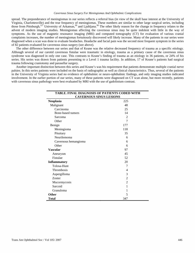
Cavernous Sinus Surgery For Meningiomas And Ophthalmic Complications
Trans Am Ophthalmol Soc / Vol 105/ 2007 446
spread. The preponderance of meningiomas in our series reflects a referral bias (in view of the skull base interest at the University of Virginia, Charlottesville) and the true frequency of meningiomas. These numbers are similar to other large surgical series, including those from Pittsburgh,227 University of Arkansas,99 and Ljubljana.98 The other likely reason for the change in frequency relates to the advent of modern imaging studies. Meningiomas affecting the cavernous sinus may be quite indolent with little in the way of symptoms. As the use of magnetic resonance imaging (MRI) and computed tomography (CT) for evaluation of various cranial complaints increases, the number of meningiomas fortuitously discovered will likely increase. Many of the patients in our series were diagnosed when a scan was done to evaluate headaches. Headache and facial pain was the second most frequent symptom in the series of 82 patients evaluated for cavernous sinus surgery (see above).
The other difference between our series and that of Keane was the relative decreased frequency of trauma as a specific etiology. Although several of our carotid cavernous fistulae were traumatic in etiology, trauma as a primary cause of the cavernous sinus syndrome was diagnosed in only one case. This contrasts to Keane’s finding of trauma as an etiology in 36 patients, or 24% of his series. His series was drawn from patients presenting to a Level 1 trauma facility. In addition, 17 of Keane’s patients had surgical trauma following craniotomy and parasellar surgery.
Another important distinction between this series and Keane’s was his requirement that patients demonstrate multiple cranial nerve palsies. In this series patients were included on the basis of radiographic as well as clinical characteristics. Thus, several of the patients in the University of Virginia series had no evidence of ophthalmic or neuro-ophthalmic findings, and only imaging studies indicated involvement. In the earlier portion of our series, many of these patients were diagnosed on CT scan alone, but more recently, patients with cavernous sinus pathology were best evaluated by MRI with the use of gadolinium contrast.
TABLE. FINAL DIAGNOSIS OF PATIENTS CODED WITH CAVERNOUS SINUS LESIONS
Neoplasia 225 Malignant 48
Carcinoma 25 Bone-based tumors 14 Sarcoma 2 Other 7
Benign 177 Meningioma 118 Pituitary 35 Neurilemoma 6 Cavernous hemangioma 6 Other 6
Vascular 97 Aneurysm 45 Fistulae 52
Inflammatory 20 Tolosa-Hunt 7 Thrombosis 4 Aspergilloma 3 Zoster 2 Mucormycosis 2 Sarcoid 1 Granuloma 1
Other 5 Total 347
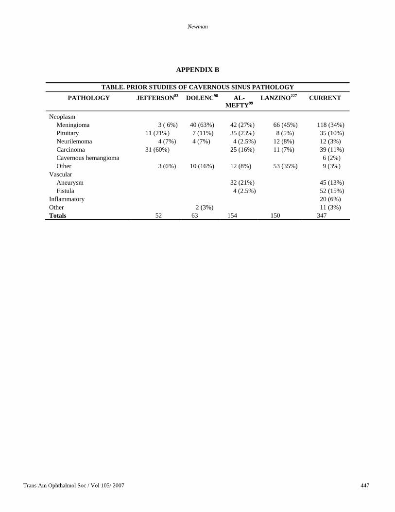
Newman
Trans Am Ophthalmol Soc / Vol 105/ 2007 447
APPENDIX B
TABLE. PRIOR STUDIES OF CAVERNOUS SINUS PATHOLOGY PATHOLOGY
JEFFERSON83 DOLENC98 AL-
MEFTY99 LANZINO227 CURRENT
Neoplasm Meningioma 3 ( 6%) 40 (63%) 42 (27%) 66 (45%) 118 (34%) Pituitary 11 (21%) 7 (11%) 35 (23%) 8 (5%) 35 (10%) Neurilemoma 4 (7%) 4 (7%) 4 (2.5%) 12 (8%) 12 (3%) Carcinoma 31 (60%) 25 (16%) 11 (7%) 39 (11%) Cavernous hemangioma 6 (2%) Other 3 (6%) 10 (16%) 12 (8%) 53 (35%) 9 (3%)
Vascular Aneurysm 32 (21%) 45 (13%) Fistula 4 (2.5%) 52 (15%)
Inflammatory 20 (6%) Other 2 (3%) 11 (3%) Totals 52 63 154 150 347