Embed Size (px)
Citation preview
Central Venous Access Device Policy Document Ref: CP82 Version 01
Page 1 of 39 Quality & Standards Team Template 2014/15
CENTRAL VENOUS ACCESS DEVICE POLICY (CVAD)
Document reference:
CP82 Version: V01
Document owner:
Stephanie Jenkins, Deputy Chief Operating Officer, Cancer Centre Services
Document author:
Steve Hill, Procedure Team Manager
Accountable committee:
Patient Safety Committee
Date approved: 29/09/2016
Ratified by: Document Ratification Committee
Date ratified: 07/11/2016
Date issued: Review date: 30/09/2019
Target audience:
The Christie clinical staff, local community nursing teams, peripheral sites of The Christie
Equality impact assessment:
03/11/2016
Key points
Types of CVADS
How to refer for a CVAD
How to manage CVAD associated complications
Central Venous Access Device Policy Document Ref: CP82 Version 01
Page 2 of 39 Quality & Standards Team Template 2014/15
CONTENTS 1. ASSOCIATED DOCUMENTS ............................................................................... 3 2. INTRODUCTION ................................................................................................... 3
2.1 Statement of intent ........................................................................................... 3 2.2 Equality Impact Analysis .................................................................................. 3 2.3 Good Corporate Citizen ................................................................................... 3 2.4 The Christie Commitment ................................................................................ 3 2.5 Purpose ........................................................................................................... 4 2.6 Scope .............................................................................................................. 4
3. DEFINITIONS ....................................................................................................... 4 4. DUTIES ................................................................................................................. 4
4.1 Patient Safety Committee ................................................................................ 4 4.2 Chief Executive ................................................................................................ 5 4.3 Procedure team ............................................................................................... 5 4.4 Clinical skills training team ............................................................................... 5 4.5 Matrons and Ward Managers ........................................................................... 5 4.6 Clinical Skills Team ......................................... 5Error! Bookmark not defined. 4.7 Chemotherapy Team ....................................................................................... 5 4.8 Lead Nurse ...................................................................................................... 5
5. CVADs .................................................................................................................. 5 5.1 Device Selection – Vessel Health Preservation ................................................ 5 5.2 CVAD insertion with ultrasound guidance ........................................................ 7 5.3 Referring for a CVAD ....................................................................................... 8 5.4 Informed consent ............................................................................................. 8 5.5 Who can insert a central venous access device? ............................................. 9 5.6 Insertion bundle ............................................................................................... 9 5.7 Who can access and provide care and maintenance for CVADs...................... 9 5.8 PICC ................................................................................................................ 9 5.9 Catheter related Thrombosis .......................................................................... 14 5.10 Non-tunnelled or Percutaneous central venous catheters ............................ 14 5.11 Haematological blood tests for CVADs (Vascular devices other than PICC) 16 5.12 Anticoagulants and CVADs (Not relevant for PICCs) ................................... 16 5.13 Direct Oral Anticoagulants ........................................................................... 16 5.14 Thrombosis Management and CVADs ......................................................... 18 5.15 Tunnelled central venous catheter ............................................................... 19 5.16 TIVAD/Implanted Ports ................................................................................ 22 5.17 CVAD Complications & Management CVAD infection ................................. 26 5.18 Anti-infective strategies for CVADs ............................................................. 28
6. CONSULTATION PROCESS .............................................................................. 30 7. DISSEMINATION, IMPLEMENTATION & TRAINING ......................................... 30
7.1 Dissemination ................................................................................................ 30 7.2 Implementation .............................................................................................. 30 7.3 Training/Awareness ....................................................................................... 30
8. PROCESS FOR MONITORING EFFECTIVE IMPLEMENTATION...................... 31 9. REFERENCES (IF APPLICABLE) ....................................................................... 31 10. VERSION CONTROL SHEET ........................................................................... 36 11. APPENDICES ................................................................................................... 37
Central Venous Access Device Policy Document Ref: CP82 Version 01
Page 3 of 39 Quality & Standards Team Template 2014/15
1. ASSOCIATED DOCUMENTS
Standard operating procedure for referral for CVC insertion (Appendix 17) PICC Occlusion Algorithm (Appendix 4) Tunnelled CVC Occlusion Algorithm (Appendix 5) CVAD Occlusion Algorithm using 3-Way tap (Appendix 9) Management of CVC related problems in the community (Appendix 13) Urokinase administration policy (Appendix 6) CVAD request form (Appendix 18) MRSA screening policy Anticoagulation policy therapy and management Policy for consent to examination and treatment Care of you central venous catheter (patient information for tunnelled CV) Care of you peripherally inserted central venous catheter (Patient information)
2. INTRODUCTION
This policy is to provide information on central venous access devices that are used at The Christie, to ensure patients receive the most appropriate device, the devices are managed correctly and any complications related to them are minimised as much as possible. A central venous access device is defined by the internal portion of the catheter being placed in the central veins, ideally the tip location should be at the lower one-third of the Superior Vena Cava (SVC) at the level of the cavoatrial junction (CAJ) (INS 2016). CVADs provide access to the venous bloodstream of the body and are used for blood sampling, administration of medications, fluids, blood products and pressure monitoring.
2.1 Statement of intent
The intention of this policy is to set out the standards expected by the Christie NHS Foundation Trust, to ensure safe, effect practice in relation to the insertion and removal of CVADs.
2.2 Equality Impact Analysis
As part of its development, this policy was analysed to consider its effect on different groups protected from discrimination by the Equality Act 2010. The requirement is to consider if there are any unintended consequences for some groups, and to consider if the policy will be fully effective for all protected groups. This analysis has been undertaken and recorded using the trust’s e-tool, and appropriate measures taken to remove barriers or advance equality in the delivery of this policy.
2.3 Good Corporate Citizen
As part of its development, this policy was reviewed in line with the Trust’s Corporate Citizen ideals. As a result, the document is designed to be used electronically in order to reduce any associated printing costs.
2.4 The Christie Commitment
We aim to reward our staff who are committed and motivated to do their best for patients every day. The trusts principles and behaviours describe what our patients and their families or carers can expect from us, and what our staff can expect from each other. The trusts behaviours are; We always give the best quality care
Central Venous Access Device Policy Document Ref: CP82 Version 01
Page 4 of 39 Quality & Standards Team Template 2014/15
We treat everybody with compassion, dignity and respect We listen to our patients and each other We work together as one Christie team We share knowledge and learning We support staff to develop to their full potential We look for new ideas and better ways of working We promote a fair culture We provide a safe, clean and tidy environment All staff are expected to behave in a way that reflects the trusts principles and behaviours.
2.5 Purpose
This policy will provide staff information and guidance around CVADs (Central venous access devices) used with the Trust.
2.6 Scope
This policy is for all staff involved in any aspect of insertion, care/maintenance and removal of CVADS at The Christie NHS Foundation Trust
Non-tunnelled (percutaneous) acute CVC
PICC - Peripherally inserted central venous catheter
Skin tunnelled central venous catheter (sometimes known as a Hickman line™)
TIVAD - Totally implantable vascular access device (sometimes known as a Portacath™).
3. DEFINITIONS
Term Meaning
CAJ Cavoatrial junction; Acceptable tip location for CVAD
CVAD Central venous access device
CVC Central venous catheter
ECG Electrocardiograph; ECG based systems are used to confirm catheter tip position during insertion
IV Intravenous
Normal working hours
Monday to Friday 9am-5pm
Procedure team Nurse lead team responsible for vascular access and other day case procedures
SVC Superior Vena Cava; Acceptable tip location for CVAD
SVCO Superior Vena Cava Obstruction; A medical condition that contraindicates placement of CVAD in the upper body, femoral approach maybe appropriate
VHP Vessel Health and Preservation; evidenced based strategy for vascular access
4. DUTIES
4.1 Patient Safety Committee
Responsible for:
Providing the Risk and Quality Governance Committee with assurance that comprehensive approach to improving patient safety is in place.
Requesting in depth investigation of any clinical concerns.
Central Venous Access Device Policy Document Ref: CP82 Version 01
Page 5 of 39 Quality & Standards Team Template 2014/15
Escalate any procedure related issues to Risk and Quality Governance Committee.
Medical Devices and Safety Committee must approve any new vascular access devices or medical devices related to vascular access introduced to the Trust.
4.2 Chief Executive
The chief executive has overall responsibility for ensuring that the organisation adheres to the standards set out in this policy. This duty may be delegated to an executive/senior manager but accountability to the Board remains with the chief executive.
4.3 Procedure team
Responsible for setting the standards in relation to best practice to CVAD insertion, removal and the provision of a clinical service.
4.4 Clinical skills training team
Responsible for the dissemination of training for the care and maintenance of CVAD’s in accordance with this policy.
4.5 Matrons and Ward Managers
Ensure that staff are adequately trained in each clinical area to provide routine maintenance, care and trouble-shooting of CVADs during normal working hours, evening and weekends where applicable.
4.7 Chemotherapy Team
To ensure all relevant clinical staff adhere to the standards set out in the central venous access device policy.
4.8 Lead Nurse
Responsible for ensuring that clinical staff in their responsible clinical areas are appropriately trained and maintain standards in accordance with this policy
5. CVADs
CVADs are inserted for a variety of reasons including:
Access for blood sampling
Administration of fluids, drugs, blood and blood products
Parenteral nutrition (PN)
Safe administration of vesicant drugs
Ambulatory chemotherapy
Monitoring of central venous pressure
Patients with poor peripheral venous access or needle phobia
Photopheresis.
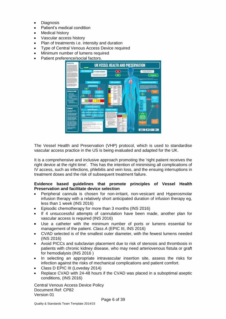
5.1 Device Selection – Vessel Health Preservation
Site selection for vascular access must include assessment of the patient’s condition, age and diagnosis, vascular condition, infusion device history, and the type and duration of the therapy as well as the potential complications associated with vascular access devices (Wise et al., 2001; Dougherty, 2006; Gabriel, 2008; Scales, 2008a). Factors in patient assessment:
Age
Central Venous Access Device Policy Document Ref: CP82 Version 01
Page 6 of 39 Quality & Standards Team Template 2014/15
Diagnosis
Patient’s medical condition
Medical history
Vascular access history
Plan of treatments i.e. intensity and duration
Type of Central Venous Access Device required
Minimum number of lumens required
Patient preference/social factors.
The Vessel Health and Preservation (VHP) protocol, which is used to standardise vascular access practice in the US is being evaluated and adapted for the UK. It is a comprehensive and inclusive approach promoting the 'right patient receives the right device at the right time'. This has the intention of minimising all complications of IV access, such as infections, phlebitis and vein loss, and the ensuing interruptions in treatment doses and the risk of subsequent treatment failure. Evidence based guidelines that promote principles of Vessel Health Preservation and facilitate device selection
Peripheral cannula is chosen for non-irritant, non-vesicant and Hyperosmolar infusion therapy with a relatively short anticipated duration of infusion therapy eg, less than 1 week (INS 2016)
Episodic chemotherapy for more than 3 months (INS 2016)
If 4 unsuccessful attempts of cannulation have been made, another plan for vascular access is required (INS 2016)
Use a catheter with the minimum number of ports or lumens essential for management of the patient. Class A (EPIC III, INS 2016)
CVAD selected is of the smallest outer diameter, with the fewest lumens needed (INS 2016)
Avoid PICCs and subclavian placement due to risk of stenosis and thrombosis in patients with chronic kidney disease, who may need arteriovenous fistula or graft for hemodialysis (INS 2016 )
In selecting an appropriate intravascular insertion site, assess the risks for infection against the risks of mechanical complications and patient comfort.
Class D EPIC III (Loveday 2014)
Replace CVAD with 24-48 hours if the CVAD was placed in a suboptimal aseptic conditions, (INS 2016)
Central Venous Access Device Policy Document Ref: CP82 Version 01
Page 7 of 39 Quality & Standards Team Template 2014/15
PICCs are associated with higher risk of thrombosis in patients who are critically ill and/or have a cancer diagnosis when compared to other CVADs (INS 2016).
Use the upper extremity for non-tunnelled catheter placement unless medically contraindicated. (EPIC III 2014)
Modified Seldinger Technique (MST) as the preferred method for PICC insertion due to advantages of decreased vein trauma, decreased insertion complications (INS 2016)
Site selection for vascular access shall include assessment of the patient’s condition; age; diagnosis; comorbidities; condition of the vasculature at the insertion site and proximal to the intended insertion site; condition of skin at insertion site
Catheter selection should be based on the prescribed therapy or treatment regimen, length of treatment, duration of dwell, vascular integrity, patient preference, and ability and resources available to care for the device. (INS 2016)
Promptly remove any intravascular catheter that is no longer essential Category 1A (CDC 2011).
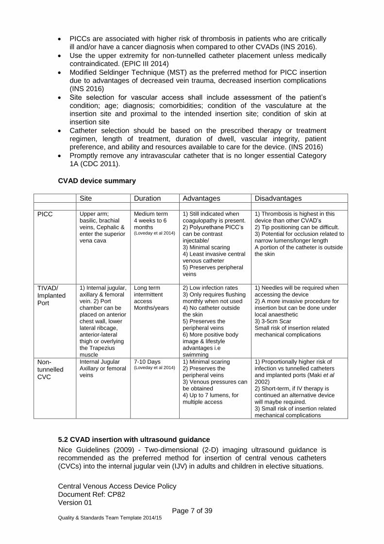
CVAD device summary
Site Duration Advantages Disadvantages
PICC Upper arm; basilic, brachial veins, Cephalic & enter the superior vena cava
Medium term 4 weeks to 6 months (Loveday et al 2014)
1) Still indicated when coagulopathy is present. 2) Polyurethane PICC’s can be contrast injectable/ 3) Minimal scaring 4) Least invasive central venous catheter 5) Preserves peripheral veins
1) Thrombosis is highest in this device than other CVAD’s 2) Tip positioning can be difficult. 3) Potential for occlusion related to narrow lumens/longer length A portion of the catheter is outside the skin
TIVAD/ Implanted Port
1) Internal jugular, axillary & femoral vein. 2) Port chamber can be placed on anterior chest wall, lower lateral ribcage, anterior-lateral thigh or overlying the Trapezius muscle
Long term intermittent access Months/years
2) Low infection rates 3) Only requires flushing monthly when not used 4) No catheter outside the skin 5) Preserves the peripheral veins 6) More positive body image & lifestyle advantages i.e swimming
1) Needles will be required when accessing the device 2) A more invasive procedure for insertion but can be done under local anaesthetic 3) 3-5cm Scar Small risk of insertion related mechanical complications
Non-tunnelled CVC
Internal Jugular Axillary or femoral veins
7-10 Days (Loveday et al 2014)
1) Minimal scaring 2) Preserves the peripheral veins 3) Venous pressures can be obtained 4) Up to 7 lumens, for multiple access
1) Proportionally higher risk of infection vs tunnelled catheters and implanted ports (Maki et al 2002) 2) Short-term, if IV therapy is continued an alternative device will maybe required. 3) Small risk of insertion related mechanical complications
5.2 CVAD insertion with ultrasound guidance
Nice Guidelines (2009) - Two-dimensional (2-D) imaging ultrasound guidance is recommended as the preferred method for insertion of central venous catheters (CVCs) into the internal jugular vein (IJV) in adults and children in elective situations.
Central Venous Access Device Policy Document Ref: CP82 Version 01
Page 8 of 39 Quality & Standards Team Template 2014/15
The use of two-dimensional (2-D) imaging ultrasound guidance should be considered in most clinical circumstances where CVC insertion is necessary either electively or in an emergency situation. It is recommended that all those involved in placing CVCs using two-dimensional (2-D) imaging ultrasound guidance should undertake appropriate training to achieve competence. Audio-guided Doppler ultrasound guidance is not recommended for CVC insertion. All patients undergoing insertion of a CVAD must have associated veins assessed using ultrasound. The assessment should look at patency, stenosis and thrombosis and general suitability of the vessel.
5.3 Referring for a CVAD
IMPORTANT - Prior to the insertion of any central venous access device it is the responsibility of the referrer to ensure patients are swabbed for MRSA (within 2 weeks of procedure) and take necessary bloods tests. Failure to prepare patient for procedure may lead to cancellation of the procedure. Process: 1) Phone 3916 to book an insertion time with the Procedure team 2) Complete referral Discover - Documents and guidelines database 3) Obtain Full blood count, coagulation screen (INR) required for Tunnelled CVC’s, Port
implantation, non-tunnelled CVC 4) Referrers please forward results to 0161-446-8265 (fax) or telephone 0161-446-3916 5) Screen all patients for MRSA (within 2 weeks of the prcedure)
5.4 Informed consent
Who can seek consent? It is important that the person who obtains the consent has sufficient knowledge about the procedure, including the potential risks and is trained in line with the Trust Consent to examination or treatment policy. For patients who are unable to consent to investigation or treatment a consent form 4 must be used. Please see consent to examination or treatment policy for further information; Patient information Patients should receive information regarding their vascular access device in advance of their procedure, though in urgent cases this might not always be possible but fully informed consent must always be obtained. A contact number will be provided should the patient have any questions in advance and allay any anxieties. Information sheets are also available electronically; PICC; http://discover/documents/upload/8/70.pdf Tunnelled CVC patient information; http://www.christie.nhs.uk/booklets/10.pdf TIVAD patient information; see Appendix 16 For medical purposes the term ‘informed consent’ means “permission granted in the knowledge of possible consequences (GMC 1998) Patients have a fundamental legal and ethical right to determine what happens to their own bodies. If the principle of consent is not respected the individual may be liable to legal action or action by a professional body (DOH 2009)
Central Venous Access Device Policy Document Ref: CP82 Version 01
Page 9 of 39 Quality & Standards Team Template 2014/15
5.5 Who can insert a central venous access device?
Registered healthcare professionals that have;
Been assessed as competent in obtaining written informed consent
Undergone specific training with a structured programme of training including training elements listed in Appendix 1.
Undertaken a minimum of 25 supervised insertions (depending upon experience, aptitude and learning efficiency) and has been assessed as competent by a Consultant Interventional Radiologist or suitably competent healthcare professional.
Inserters should be familiar with policies, guidelines and relevant documents related to vascular access procedures such as; Epic III, NICE, BCSH, Saving lives etc and relevant Trust policies.
5.6 Insertion bundle
Prior to insertion a checklist should be undertaken including (Example; Appendix 2) Use a central venous catheter Insertion bundle ensures a structured process for instituting best practice (Loveday et al 2014).
Hand hygiene
Maximal sterile barrier precautions during insertion (Hat, mask, gown, gloves, full body drapes)
Use of 2% Chlorhexidine Gluconate (CHG) in 70% isopropyl alcohol for skin antisepsis at insertion site
Avoid femoral site
Remove CVC’s as soon as no longer indicated (EPIC III - Loveday et al 2014)
5.7 Who can access and provide care and maintenance for CVADs
CVADs can only be accessed for blood sampling and flushing by staff Band 3 and above who have received appropriate Trust training. Band 5’s and above who have received appropriate training, can access them to also administer via them. For staff at The Christie initial training with the clinical skills training team or a standardised practice facilitator must occur before any supervised practice takes place. Healthcare workers must work under the supervision of a member of staff, assessed as competent to access CVADs, until they themselves have been assessed as competent to work without supervision. This assessment must be carried out by member of the clinical skills training team, a standard practice facilitator or a standardised clinical link worker. It is the responsibility of the healthcare worker to ensure this prior to working with central venous catheters. Following the initial assessment, a date will be arranged for update to be undertaken. The healthcare worker must contact the clinical skills training team or their designated link worker to arrange their scheduled update. Staff must also be reassessed following a period of prolonged absence or incident.
5.8 PICC
A PICC is a peripherally inserted central venous catheter. It is a thin flexible tube that is inserted into a vein in the upper arm. The PICC is then threaded along the vein so that the tip lies in the optimum tip position in the should be at the lower one-third of the Superior Vena Cava (SVC) at the level of the cavoatrial junction (CAJ) (INS 2016) Currently there are two types of PICC used at The Christie;
Central Venous Access Device Policy Document Ref: CP82 Version 01
Page 10 of 39 Quality & Standards Team Template 2014/15
Groshong Valved® - This PICC differs from tunnelled central venous catheters in that they have a pressure sensitive valve at the internal end which allows fluid to be injected into the catheter and blood to be withdrawn When not in use the valve remains closed, thus preventing blood from flowing back into the catheter and air entering the venous circulation, clamps or switches are not required, this catheter is made of silicone Open ended pressure injectable PICC with clamp – This PICC is open ended does not have any integral valves. This catheter is made from polyurethane and is compatible with contrast pressure injection (please see manufacture guidelines for details). PICC Indications
For the delivery of antibiotic therapy, IV fluids, TPN, Blood sampling and the administration of irritants/vesicants, chemotherapy and other anti-cancer intravenous therapies (BCSH 2006)
Episodic chemotherapy for more than 3 months (INS 2016)
Patients with limited venous access
Previous complications or failed with short PIVs
Patient requires irritating or hyperosmolar medications
A patient who has bleeding disorder/low platelet levels (<50 · 109/l) where central insertion is contraindicated (BCSH 2006, Hertzog & Waybill 2008)
Total Parenteral Nutrition (Espen 2009)
Conditions that contraindicate use of other VAD types
Need for high pressure injection of contrast media
Patients consideration
Intended dwell time – medium term 4 weeks to 6 months (Loveday et al 2014). Benefits
A PICC can be used to give chemotherapy, anti-cancer drugs, fluids, antibiotics and other drugs directly into the vein.
It can also be used for taking blood samples PICC lines are inserted when a central line is needed for short term use
PICCs are useful when peripheral veins are difficult to find or access, or for people who are very anxious about needles. A PICC is sometimes used temporarily for people who should ideally have a tunnelled central venous catheter or an implantable Port but are not able to because of their clinical condition i.e. anatomy abnormal, blood results or are unable to lie flat. In this situation a PICC means treatment can be started without delay.
Relative Contraindications
Inability to visualise, view suitable vein (Ultrasound)
Circulatory impairment, peripheral neuropathies
History of CVA (same side as limb weakness), same side as mastectomy with node dissection surgery, upper extremity fistulas, surgery or neck/head trauma
Venous thrombosis in vasculature where the PICC is proposed
Same side as; existing CVAD, cardiac pacemaker, implanted cardiovascular defibrillator, (SVC Vena Cava filter) (Hertzog & Waybill 2008)
Skin conditions, dermatitis, infection at insertion site (Hertzog & Waybill 2008)
End stage renal disease, chronic renal insufficiency, to preserve upper limb vasculature for formation of fistula (NKF-DOQI)
Allergy to material contained in the device.
Central Venous Access Device Policy Document Ref: CP82 Version 01
Page 11 of 39 Quality & Standards Team Template 2014/15
PICCs and Ultrasound pre-assessment Use Ultrasound pre-assessment to assess optimal vessel size. Ideal vessel should ideally be 3 times greater than the external diameter of the catheter. As a rough guide; 4fr vein size should be no less than 4mm, 5fr no less than 5mm (Nifong & McDevitt 2011). A higher catheter vein ratio may increase thrombosis risk and should be balanced accordingly on the clinical urgency for the PICC CVC vs catheter related and patient thrombotic risk factors. Undertaking comprehensive ultrasound assessment including upper arm veins, axillary, subclavian, internal jugular, brachio-cephalic including compression to exclude major abnormalities such as pre-existing thrombosis and select the most appropriate vein using the SIP protocol (Safe insertion of PICCs) advocated by (Pittiruiti et al 2014). Insertion; general principles. In addition to the insertion bundle advocated in EPIC guidelines (Loveday et al 2014) other considerations for PICC insertion are;
Optimal catheter site selection; Upper arm approach unless contraindicated The Basilic vein is the preferred vein (INS 2016), the Cephalic and Brachial veins may be considered but with caution as the Brachial vein is often in close proximity nerve bundles and artery. The Cephalic vein follows narrower more tortuous pathway and connects to the axillary vein at an acute angle. It is essential ultrasound assessments take place and the optimal vein is selected on an patient individual basis
PICCs may be preferred CVAD in patients with coagulopathy disorders or receiving anticoagulants as direct pressure can be applied to control bleeding and in patients with respiratory diagnoses or intracranial bleeding when Trendelenburg position is difficult or contraindicated (INS 2016)
Cannulation should be ultrasound guided (NICE 2002)
Modified Seldinger Technique, to reduce vessel trauma (INS 2016)
Secure with sutureless device (CDC 2011). Catheter tip position maybe confirmed with ECG tip confirmation system (Nice 2015) and/or fluoroscopy chest X-ray. If unable to gain satisfactory P-Wave (ECG tip confirmation system) or the patient does not have a discernible P-Wave (Atrial fibrillation or arhythmia). Tip position will be confirmed with chest x-ray and documented. Patients with arm ports or PICCs should not have additional IV devices placed on the same arm i.e. IV cannula. Complications
Misplacement of catheter tip following insertion sometimes requires adjustment, this sometimes means includes going to X-ray dept
Accidental puncture of the artery which may cause bleeding
Inability to thread catheter into vein/SVC
Inability to advance sheath/dilator assembly
Bleeding and some bruising around the insertion site, especially in people whose blood does not clot normally
Arrhythmias
Haematoma
Phlebitis
Infection, insertion site or systemic
Central Venous Access Device Policy Document Ref: CP82 Version 01
Page 12 of 39 Quality & Standards Team Template 2014/15
Nerve injury – patients may feel a shooting pain down their arm if the needle touches a nerve
Embolism, air or equipment
Cardiac tamponade, secondary to vessel wall, atrial or ventricular perforation
Occlusion
Sepsis
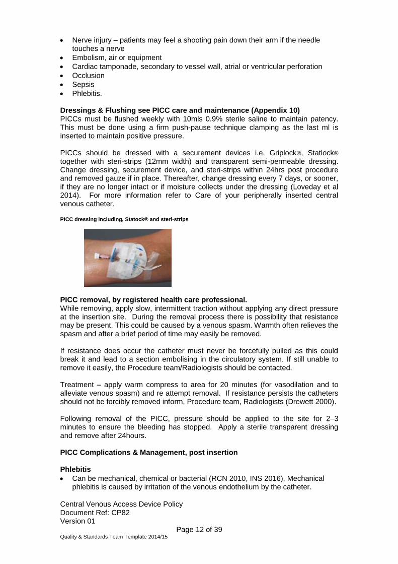
Phlebitis. Dressings & Flushing see PICC care and maintenance (Appendix 10) PICCs must be flushed weekly with 10mls 0.9% sterile saline to maintain patency. This must be done using a firm push-pause technique clamping as the last ml is inserted to maintain positive pressure. PICCs should be dressed with a securement devices i.e. Griplock®, Statlock® together with steri-strips (12mm width) and transparent semi-permeable dressing. Change dressing, securement device, and steri-strips within 24hrs post procedure and removed gauze if in place. Thereafter, change dressing every 7 days, or sooner, if they are no longer intact or if moisture collects under the dressing (Loveday et al 2014). For more information refer to Care of your peripherally inserted central venous catheter.
PICC dressing including, Statock® and steri-strips
PICC removal, by registered health care professional. While removing, apply slow, intermittent traction without applying any direct pressure at the insertion site. During the removal process there is possibility that resistance may be present. This could be caused by a venous spasm. Warmth often relieves the spasm and after a brief period of time may easily be removed. If resistance does occur the catheter must never be forcefully pulled as this could break it and lead to a section embolising in the circulatory system. If still unable to remove it easily, the Procedure team/Radiologists should be contacted. Treatment – apply warm compress to area for 20 minutes (for vasodilation and to alleviate venous spasm) and re attempt removal. If resistance persists the catheters should not be forcibly removed inform, Procedure team, Radiologists (Drewett 2000). Following removal of the PICC, pressure should be applied to the site for 2–3 minutes to ensure the bleeding has stopped. Apply a sterile transparent dressing and remove after 24hours. PICC Complications & Management, post insertion Phlebitis
Can be mechanical, chemical or bacterial (RCN 2010, INS 2016). Mechanical phlebitis is caused by irritation of the venous endothelium by the catheter.
Central Venous Access Device Policy Document Ref: CP82 Version 01
Page 13 of 39 Quality & Standards Team Template 2014/15
Symptoms – Tenderness, erythema, warmth, swelling, induration, pain (INS 2016). Treatment
Anti-inflammatory analgesics for inflammation & pain (Higginson 2011)
Culture and begin antimicrobial therapy if infection present
Site care and condition/appearance using standardised local assessment scales for phlebitis and/or infiltration/extravasation (RCN 2010)
Refer to Physician, remove if symptoms don’t improve (Simcock 2001). Prevention –
Regular monitoring of site, Aseptic technique during insertion and care/maintenance (INS 2016)
Stabilisation: Catheter securement to reduce movement (CDC 2011)
Site: Upper arm U/S rather than ante-cubital (Espen 2009)
Modified Seldinger Technique reduces trauma (INS 2016)
Prevention – Upper arm, Modified Seldinger Technique
Use the Device with ‘Smallest gauge’ (INS 2016).
‘The diameter of the catheter should be one third or less of the diameter of the vein, as checked by ultrasound’ (Espen 2009).
Catheter tip Malposition - Ideal Catheter tip position:- Lower one third of the SVC level with the cavoatrial junction (INS 2016) Symptoms
Alteration of external catheter length, inability to aspirate blood, “gurgling” sound in ear, discomfort/pain in chest, neck or back arrhythmias (INS 2016)
Treatment
If the PICC has been pulled out from its original insertion length; please follow PICC Occlusion Algorithm (Appendix 4)
A chest X-ray may be required to ascertain tip position
If too short or malpositioned refer to procedure team/Interventional Radiology for repositioning, catheter exchange/replacement
The nurse should not advance any external portion of the CVAD that has been in contact with skin into the insertion site. (INS 2016)
A CVAD with a malpositioned catheter tip location that cannot be repositioned to a central vein should be removed (INS 2016)
Catheters that are malposition have a higher risk of thrombosis if the patient is symptomatic of thrombosis an ultrasound Doppler should be undertaken to assess the associated vessels.
Prevention
Take meticulous care not to pull PICC out when changing dressings
Optimal patient positioning during insertion
Use griplock/statlock securement with Steri-strips
Thread catheter slowly, do not thread against resistance
Use of tip navigation/positioning systems/Fluoroscopy. Insertion related bleeding Symptoms
Persistent bleeding, haematoma/swelling from insertion site Treatment
Optimal pressure (RCN 2010) should be applied with a sterile dressing following the venipuncture and maintained until bleeding stops (INS 2011)
Take full blood count & full coagulation screen, is there an underlying cause.

Central Venous Access Device Policy Document Ref: CP82 Version 01
Page 14 of 39 Quality & Standards Team Template 2014/15
Use ultrasound guided & Modified Seldinger technique to minimise trauma to vein, visualise needle tip at all time during insertion
Assess patient history & current medications prior to insertion
Re; Use of blood of products; Assess need on an individual basis rather than routine correction of all minor abnormalities of platelet count and coagulation studies (BCSH 2006).
5.9 Catheter related Thrombosis
Symptoms;
Discomfort/pain/Oedema/engorged veins; arm, shoulder, neck, chest
Leakage of fluid at entry site, discoloration of extremity, unexplained fever Treatment
Refer to Respective medical team and to Radiologist to undertake ultrasound Doppler
Do not remove the catheter CVAD in the presence of a CVAD-associated vein thrombosis when the catheter is correctly positioned at the cavo-atrial junction, the catheter is functioning with a blood return, there is no evidence of any infection, and there is absence of severe deep vein thrombosis (DVT) - related symptoms causing pain Prevention (INS 2016). This is ultimately the medical team’s decision based upon, the need to keep the device in place and balanced individual hypercoagulability factors of the patient. Monitoring and anticoagulation should be followed in accordance with the Anticoagulant Therapy Management Policy.
Prevention
Smallest outer diameter (INS 2016)
‘the diameter of the catheter should be one third or less of the diameter of the vein, as checked by ultrasound’ (Espen 2009)
Assess patient risk factors i.e. malignancy, critically ill (Chopra et al Lancet 2013)
Catheter tip position (RCN 2010)
Modified Seldinger for insertion reduces trauma. Catheter Occlusion Treatment
Eliminate mechanical causes (kinking, pinching of line)
If chemical, determine which infusates caused precipitates and instill proper solution (alkaline/acidic) to clear (INS 2016)
If blood occlusion, instill Urokinase® per protocol.
Prevention
Flush before and after all infusions and blood draws with an adequate amount of solution to fully clear device
Be aware of solution incompatibilities prior to infusion
Pulsatile flushing technique.
5.10 Non-tunnelled or Percutaneous central venous catheters
Usually found in acute-care settings, critical care, theatre and high dependency units. A non-tunnelled CVC or percutaneous CVC is a centrally inserted catheter. It is a polyurethane line, which has multiple entry ports between 3 and 6 and they are known as lumens. It is threaded through a large vein and usually sutures are used to hold it in place with a transparent semi-permeable dressing.
Central Venous Access Device Policy Document Ref: CP82 Version 01
Page 15 of 39 Quality & Standards Team Template 2014/15
(Matching Michigan, 2012, EPIC III 2014). Do not routinely replace CVC access devices to prevent catheter related infection. Indications
In an acute situation, where the patient deteriorates quickly.
When the patient urgently needs IV fluids, drugs and anti-biotics
When patient’s need lots of IV medication at the same time
If the patient needs specialist monitoring, carried out in Critical Care
For intended dwell time 7-10 days. Benefits
Allows patients to receive several amounts of drugs simultaneously
Allows patients to receive drugs in an emergency
Allows patients to receive drugs that could irritate smaller vessels
Allows patients to be ‘centrally monitored’
Can be inserted quickly and removed quickly. Risks
Carries high infection risk potential
Pnuemothorax on insertion
Air emboli on insertion or removal
Patient discomfort
Patients often need nursing in specific areas (Morton, 2011, Matching Michigan 2012)
Infection: as with all surgical procedures there is a risk of infection occurring.
Accidental puncture of the artery
Embolus: a small risk of embolus (air/equipment) if the catheter or guide-wire breaks, or air embolus (bubble of air) within the blood stream.
Cardiac arrhythmia
Malpositioning.
Specific points;
Axillary vein; insertion can increase risk of pneumothorax secondary to the close proximity of the pleura to the needle insertion site. Damage to the brachial plexus is a risk of this approach (INS 2016)
Femoral vein; insertion sites present higher risk of infection and should be avoided when possible since catheter stabilistion and occlusive dressing adherence can be difficult to maintain. Femoral insertion does not require Tendelenburg position.
Subclavian vein; Ultrasound evaluation of the infraclavicular subclavian vein is difficult secondary to the overlying clavicle. Moving outward to the axillary vein make use of the ultrasound more useful. Pinch-off syndrome, where the catheter gets trapped between the first rib and clavicle, can be a risk of subclavian insertions.
Internal jugular vein insertion sites may be difficult to secure secondary to head movement, and carotid artery puncture is a risk due to its proximity to the vein
External jugular veins; are more tortuous than internal jugular veins, making catheter threading more difficult, the vessel is superficial which may create difficulty with catheter securement.
Central Venous Access Device Policy Document Ref: CP82 Version 01
Page 16 of 39 Quality & Standards Team Template 2014/15
5.11 Haematological blood tests for CVADs (Vascular devices other than PICC)
Insertion Criteria Skin tunnelled CVC’s and Implantable Ports, minimum requirements
Hb 80g/L
Platelets 50 x10^9/L
INR less than 1.5 (BCSH 2010) For further information see referral SOP (Appendix 17).
5.12 Anticoagulants and CVADs (Not relevant for PICCs)
Intravenous unfractionated (UFH) Should be stopped 6 hours before catheter insertion and restarted when haematostasis is secured (Martindale). Low molecular weight heparin. In patients receiving prophylactic subcutaneous low molecular weight heparin (LMWH) catheter insertion can be undertaken 12 hours after the last injection; for patients receiving therapeutic subcutaneous LMWH, the time to catheter insertion should be extended to 18 hours after the last injection. Heparin can be recommenced once haemostasis is secure, usually within 2 hours of catheter insertion. Substitution with intravenously infused UFH or insertion of an IVC filter should be considered if there is a very high thrombotic risk. Expert haematology advice should be sought.
LMWH may continue >4hrs post removal of the CVAD in patient without abnormal clotting or high risk of bleeding.
5.13 Direct Oral Anticoagulants
Warfarin Patients should be given advice to stop warfarin prior to admission for their vascular device insertion. Provided adequate oral intake and absorption of vitamin-K-containing foods omission of warfarin for 3 days will allow reduction from range 2.0-3.0 down to level safe (eg less than 1.5) for invasive procedures such as central access/biopsies (5 days if in range 3.0-4.0).
Dabigatran (Pradaxa) which is a direct thrombin inhibitor and the direct factor Xa inhibitors Rivaroxaban (Xarelto) and Apixaban (Elquis) and Edoxoban are new drugs which are direct oral anticoagulants and may replace warfarin and other anticoagulants. There is currently no proven method to reverse their anticoagulant effect in bleeding patients and no agreed national guidelines.
Dabigatran discontinue 2 days prior to the procedure has a half-life of
approximately 12-17 hours. Rivaroxaban discontinue 2 days prior to the procedure has a half-life of approximately 7-11 hours. The effects of these drugs extend in patients with renal impairment. (https://www.medicinescomplete.com/mc/martindale/current/ms-20786-b.htm?q=dabigatran&t=search&ss=text&p=1#_hit) Apixaban discontinue 2 days prior to the procedure
Central Venous Access Device Policy Document Ref: CP82 Version 01
Page 17 of 39 Quality & Standards Team Template 2014/15
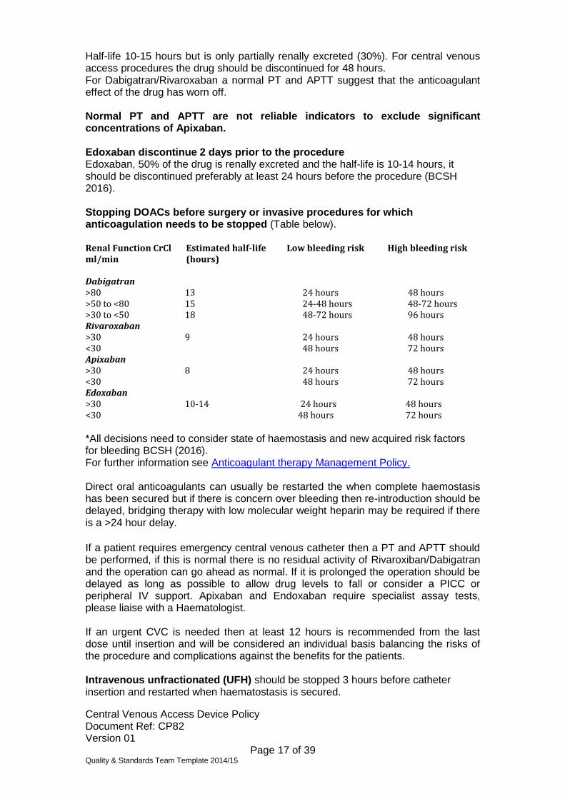
Half-life 10-15 hours but is only partially renally excreted (30%). For central venous access procedures the drug should be discontinued for 48 hours. For Dabigatran/Rivaroxaban a normal PT and APTT suggest that the anticoagulant effect of the drug has worn off. Normal PT and APTT are not reliable indicators to exclude significant concentrations of Apixaban. Edoxaban discontinue 2 days prior to the procedure Edoxaban, 50% of the drug is renally excreted and the half-life is 10-14 hours, it should be discontinued preferably at least 24 hours before the procedure (BCSH 2016). Stopping DOACs before surgery or invasive procedures for which anticoagulation needs to be stopped (Table below). Renal Function CrCl ml/min
Estimated half-life (hours)
Low bleeding risk High bleeding risk
Dabigatran >80 13 24 hours 48 hours >50 to <80 15 24-48 hours 48-72 hours >30 to <50 18 48-72 hours 96 hours Rivaroxaban >30 9 24 hours 48 hours <30 48 hours 72 hours Apixaban >30 8 24 hours 48 hours <30 48 hours 72 hours Edoxaban >30 10-14 24 hours 48 hours <30 48 hours 72 hours *All decisions need to consider state of haemostasis and new acquired risk factors for bleeding BCSH (2016). For further information see Anticoagulant therapy Management Policy. Direct oral anticoagulants can usually be restarted the when complete haemostasis has been secured but if there is concern over bleeding then re-introduction should be delayed, bridging therapy with low molecular weight heparin may be required if there is a >24 hour delay.
If a patient requires emergency central venous catheter then a PT and APTT should be performed, if this is normal there is no residual activity of Rivaroxiban/Dabigatran and the operation can go ahead as normal. If it is prolonged the operation should be delayed as long as possible to allow drug levels to fall or consider a PICC or peripheral IV support. Apixaban and Endoxaban require specialist assay tests, please liaise with a Haematologist. If an urgent CVC is needed then at least 12 hours is recommended from the last dose until insertion and will be considered an individual basis balancing the risks of the procedure and complications against the benefits for the patients. Intravenous unfractionated (UFH) should be stopped 3 hours before catheter insertion and restarted when haematostasis is secured.
Central Venous Access Device Policy Document Ref: CP82 Version 01
Page 18 of 39 Quality & Standards Team Template 2014/15
Anti-platelet therapy Patients taking anti-platelet medication (e.g. Aspirin, Dipyridamol, Clopidogrel) should discontinue 5 days prior to CVAD insertion. Emergency/unplanned patients on low dose Aspirin 75mg will be considered for central venous access REMOVAL on an individual basis balancing the risks of the procedure and complications against the benefits for the patients and urgency of device and treatment. The patient should not have problems with their blood clotting and not on concomitant anticoagulants/Antiplatelet therapy. AVASTIN (Bevacizumab) Bevacizumab interrupts angiogenesis by inhibiting vascular endothelial growth factor. Tunnelled CVC – Stop Bevacizumab 14 days prior to insertion and it should not be administered until 24hrs after insertion, providing there is no bleeding at the site. TIVAD – Stop Bevacizumab 4 weeks prior to insertion and not to be used until implantation site has healed, usually 7-10 days. Complications that occur from patients commenced on Bevacizumab within the above time frame will be the responsibility of the managing Consultant.
5.14 Thrombosis Management and CVADs
Do not remove the catheter CVAD in the presence of a CVAD-associated vein thrombosis when the catheter is correctly positioned at the cavo-atrial junction, the catheter is functioning with a blood return, there is no evidence of any infection, and there is absence of severe deep vein thrombosis (DVT) - related symptoms causing pain (INS 2016). When a CVAD related thrombus is present and the removal is indicated, anticoagulation should be undertaken prior to the procedure, for thrombosis extending to the Superior Vena Cava or Right Atrium up to 7 days anticoagulation should be considered. This is ultimately the medical team’s decision based upon, the need to keep the device in place and balanced individual hypercoagulability factors of the patient. Monitoring and anticoagulation should be followed in accordance with the Anticoagulant Therapy Management Policy. Risk factors;
History of deep vein thrombosis
Presence of chronic disease associated with a hypercoaguable state such as cancer, diabetes, irritable bowel syndrome, congenital heart disease and end stage renal failure.
Surgical and trauma patients
Critical care patients, hyperglycemia in critical care
Known presence of genetic coagulation abnormalities (eg Factor V Leiden, prothrombin mutation)
Pregnancy or the use of oral contraceptives
Fluid volume deficit
History of multiple CVAD, especially with difficult or traumatic insertion and the presence of other intravascular devices (eg, pacemakers).
Central Venous Access Device Policy Document Ref: CP82 Version 01
Page 19 of 39 Quality & Standards Team Template 2014/15
The presence of carcinoma, metastatic disease predisposes oncology patients to hypercoagulability and as a consequence device related thrombosis is high in this group of patients (Chopra et al Lancet 2013).
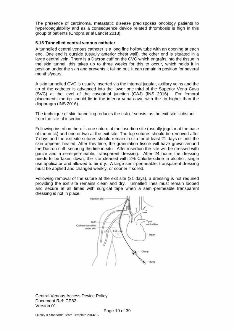
5.15 Tunnelled central venous catheter
A tunnelled central venous catheter is a long fine hollow tube with an opening at each end. One end is outside (usually anterior chest wall), the other end is situated in a large central vein. There is a Dacron cuff on the CVC which engrafts into the tissue in the skin tunnel, this takes up to three weeks for this to occur, which holds it in position under the skin and prevents it falling out. It can remain in position for several months/years. A skin tunnelled CVC is usually inserted via the internal jugular, axillary veins and the tip of the catheter is advanced into the lower one-third of the Superior Vena Cava (SVC) at the level of the cavoatrial junction (CAJ) (INS 2016). For femoral placements the tip should lie in the inferior vena cava, with the tip higher than the diaphragm (INS 2016). The technique of skin tunnelling reduces the risk of sepsis, as the exit site is distant from the site of insertion. Following insertion there is one suture at the insertion site (usually jugular at the base of the neck) and one or two at the exit site. The top sutures should be removed after 7 days and the exit site sutures should remain in situ for at least 21 days or until the skin appears healed. After this time, the granulation tissue will have grown around the Dacron cuff, securing the line in situ. After insertion the site will be dressed with gauze and a semi-permeable, transparent dressing. After 24 hours the dressing needs to be taken down, the site cleaned with 2% Chlorhexidine in alcohol, single use applicator and allowed to air dry. A large semi-permeable, transparent dressing must be applied and changed weekly, or sooner if soiled. Following removal of the suture at the exit site (21 days), a dressing is not required providing the exit site remains clean and dry. Tunnelled lines must remain looped and secure at all times with surgical tape when a semi-permeable transparent dressing is not in place.
Central Venous Access Device Policy Document Ref: CP82 Version 01
Page 20 of 39 Quality & Standards Team Template 2014/15
Tip position maybe confirmed with ECG tip confirmation system and/or fluoroscopy, chest X-ray and ultrasound maybe used to assess for and identify the “sliding lung sign” to assess for pneumothorax. If unable to gain satisfactory P-Wave (ECG tip confirmation system) or the patient does not have a discernible P-Wave (Atrial fibrillation or arrhythmia). Tip position will be confirmed with chest x-ray and documented. Indications
Long term IV therapy Months/years (EPIC III 2014)
As well as the delivery of antibiotic therapy, IV fluids, TPN, Blood sampling and the administration of irritants/vesicants, chemotherapy and other anti-cancer intravenous therapies.
Patients with limited venous access
Previous complications or failed with short PIVs
Patient requires irritating or hyperosmolar medications
Patients consideration
Plasmaphereis – In this instance the catheter must be designed and licensed for Plasmaphereis and be compatible with Plasmapharesis machines.
Conditions requiring multiple punctures, such as patient’s requiring daily blood withdrawals.
Total parenteral nutrition. Benefits
More comfortable and discreet and lower infection rates than the non – tunnelled catheters.
Can remain insitu for long periods
It allows some therapies to be given at home
It may also be used for taking blood samples which are needed regularly, avoiding the need for repeated needle stabs in your arm.
Disadvantages Requires a minimally invasive surgical procedure for placement. Potential complications on insertion Body image of external catheter Lifestyle, no swimming. Risks Insertion related complications;
Air embolus
Infection: as with all surgical procedures there is a risk of infection occurring.
Brachial plexus injury
Cardiac tamponade
Endocarditis
Pnemothorax of the lung: Rate 1 per 13,000 cases
Accidental puncture of the artery: Rate 1 per 1,500 cases
Embolus: a small risk of embolus such as air or equipment, if the catheter or guide-wire breaks, or air embolus (bubble of air) within the blood stream.
Haematoma
Scarring
Central Venous Access Device Policy Document Ref: CP82 Version 01
Page 21 of 39 Quality & Standards Team Template 2014/15
Cardiac arrhythmia
Malpositioning
Breakage between first rib/clavicle (Subclavian approach)
Hydrothorax
Laceration of the vessel
Perforation of vessel
Thoracic duct injury
Risks normally associated with local anaesthesia
Bard Hickman® instructions for use; http://www.bardpv.com/wp-content/uploads/2013/07/Hickman_TriFusion_IFU.pdf
Complications of indwelling catheter;
Blockage
Catheter/cuff erosion through skin
Exit site necrosis/scarring over the catheter site
Infection, at exit site or systemically
Fracture of catheter
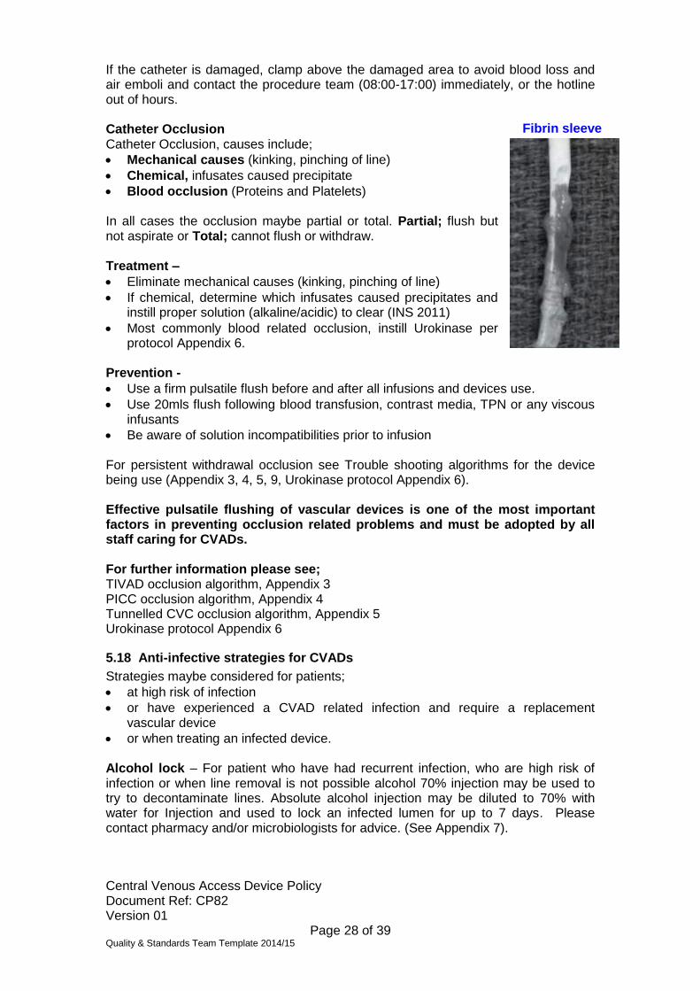
Fibrin sheath formation and persistent withdrawal occlusion (Appendix 5)
Damage, fracture, burst, trauma
Thrombosis: a blood clot may develop. This is a longer term risk associated with lines. Signs of this include pain, swelling and discomfort in the neck or arm on the side the catheter is placed.
Extravasation
Intolerance to the device material
Spontaneous catheter tip malposition
Intracardiac thrombosis.
Flushing Tunnelled CVCs must be flushed weekly with 10mls 0.9% normal sterile saline to maintain patency. This must be done using a firm push-pause technique clamping as the last ml is inserted to maintain positive pressure. See tunnelled CVC care and maintenance Appendix 9. Apheresis catheters – These types of catheters require flushing with 0.9% normal sterile saline and locking with higher does Heparin according to individual patient prescription. This is usually 5000 units Heparin per ml (or as per manufacturer instructions) the volume required is usually printed on each lumen of the catheter. Tunnelled CVC removal The device should be removed when no longer required or if there is thrombosis or infection related to the catheter or if it is fractured. The procedure may be undertaken by registered health care professionals who have been trained to undertake the procedure and has a documented assessment of competency, or under the supervision of an experienced operator. Incorrect removal may lead to catheter fracture and embolism, dislodgment of a portion of the catheter in to the bloodstream. Removal must be according to SOP for the removal of cuffed tunnelled central venous catheter.
Central Venous Access Device Policy Document Ref: CP82 Version 01
Page 22 of 39 Quality & Standards Team Template 2014/15
Criteria:
The haemoglobin and white cell count are not critical Platelets must be above 30x109/litre Formal coagulation check is not required. Patient with history of a known bleeding disorder, history of bleeding should undergo full coagulation screen and take advice from Haematologist if required. Removal and Anticoagulants For anticoagulants see Tunnelled CVCs and Anticoagulants. Aspirin and Aspirin containing products.
Emergency/unplanned patients on low dose Aspirin 75mg will be considered for central venous access REMOVAL on an individual basis balancing the risks of the procedure and complications against the benefits for the patients and urgency of device and treatment. The patient should not have problems with their blood clotting and not on concomitant anticoagulants/Antiplatelet therapy.
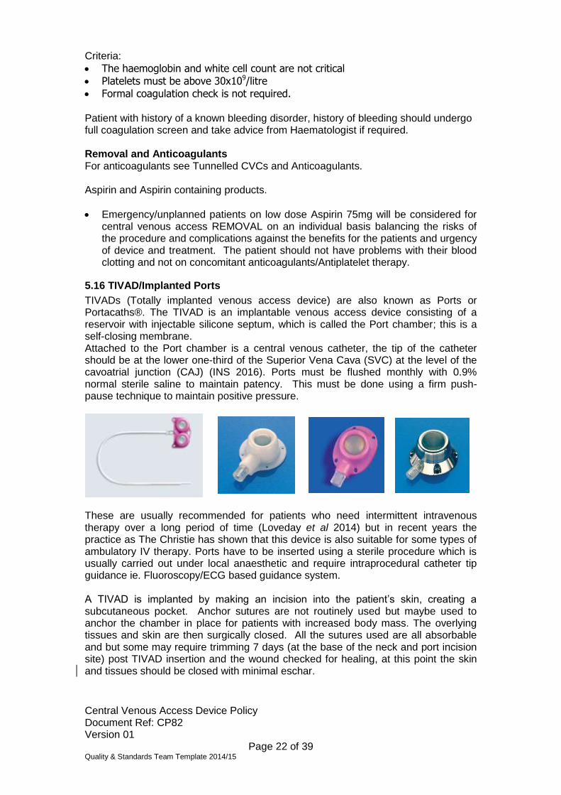
5.16 TIVAD/Implanted Ports
TIVADs (Totally implanted venous access device) are also known as Ports or Portacaths®. The TIVAD is an implantable venous access device consisting of a reservoir with injectable silicone septum, which is called the Port chamber; this is a self-closing membrane. Attached to the Port chamber is a central venous catheter, the tip of the catheter should be at the lower one-third of the Superior Vena Cava (SVC) at the level of the cavoatrial junction (CAJ) (INS 2016). Ports must be flushed monthly with 0.9% normal sterile saline to maintain patency. This must be done using a firm push-pause technique to maintain positive pressure.
These are usually recommended for patients who need intermittent intravenous therapy over a long period of time (Loveday et al 2014) but in recent years the practice as The Christie has shown that this device is also suitable for some types of ambulatory IV therapy. Ports have to be inserted using a sterile procedure which is usually carried out under local anaesthetic and require intraprocedural catheter tip guidance ie. Fluoroscopy/ECG based guidance system. A TIVAD is implanted by making an incision into the patient’s skin, creating a subcutaneous pocket. Anchor sutures are not routinely used but maybe used to anchor the chamber in place for patients with increased body mass. The overlying tissues and skin are then surgically closed. All the sutures used are all absorbable and but some may require trimming 7 days (at the base of the neck and port incision site) post TIVAD insertion and the wound checked for healing, at this point the skin and tissues should be closed with minimal eschar.
Central Venous Access Device Policy Document Ref: CP82 Version 01
Page 23 of 39 Quality & Standards Team Template 2014/15
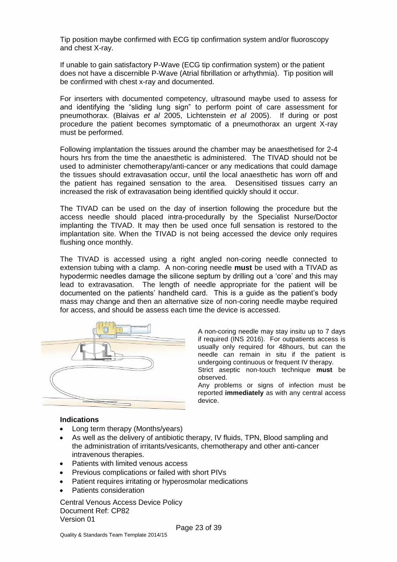
Tip position maybe confirmed with ECG tip confirmation system and/or fluoroscopy and chest X-ray. If unable to gain satisfactory P-Wave (ECG tip confirmation system) or the patient does not have a discernible P-Wave (Atrial fibrillation or arhythmia). Tip position will be confirmed with chest x-ray and documented. For inserters with documented competency, ultrasound maybe used to assess for and identifying the “sliding lung sign” to perform point of care assessment for pneumothorax. (Blaivas et al 2005, Lichtenstein et al 2005). If during or post procedure the patient becomes symptomatic of a pneumothorax an urgent X-ray must be performed. Following implantation the tissues around the chamber may be anaesthetised for 2-4 hours hrs from the time the anaesthetic is administered. The TIVAD should not be used to administer chemotherapy/anti-cancer or any medications that could damage the tissues should extravasation occur, until the local anaesthetic has worn off and the patient has regained sensation to the area. Desensitised tissues carry an increased the risk of extravasation being identified quickly should it occur. The TIVAD can be used on the day of insertion following the procedure but the access needle should placed intra-procedurally by the Specialist Nurse/Doctor implanting the TIVAD. It may then be used once full sensation is restored to the implantation site. When the TIVAD is not being accessed the device only requires flushing once monthly. The TIVAD is accessed using a right angled non-coring needle connected to extension tubing with a clamp. A non-coring needle must be used with a TIVAD as hypodermic needles damage the silicone septum by drilling out a ‘core’ and this may lead to extravasation. The length of needle appropriate for the patient will be documented on the patients’ handheld card. This is a guide as the patient’s body mass may change and then an alternative size of non-coring needle maybe required for access, and should be assess each time the device is accessed.
Indications
Long term therapy (Months/years)
As well as the delivery of antibiotic therapy, IV fluids, TPN, Blood sampling and the administration of irritants/vesicants, chemotherapy and other anti-cancer intravenous therapies.
Patients with limited venous access
Previous complications or failed with short PIVs
Patient requires irritating or hyperosmolar medications
Patients consideration
A non-coring needle may stay insitu up to 7 days if required (INS 2016). For outpatients access is usually only required for 48hours, but can the needle can remain in situ if the patient is undergoing continuous or frequent IV therapy. Strict aseptic non-touch technique must be observed. Any problems or signs of infection must be reported immediately as with any central access device.
Central Venous Access Device Policy Document Ref: CP82 Version 01
Page 24 of 39 Quality & Standards Team Template 2014/15
Prolonged intravenous therapy. Benefits (TIVAD) Less impact on patient body image No risk of pulling out the device Lifestyle advantages, swimming Lower risk of infection versus other CVADs Less community care input, devices only need flushing monthly when not in use. Disadvantages
Still requires a needle to access device
More invasive to insert than other vascular devices. Flushing TIVADs must be flushed monthly with 10mls 0.9% normal sterile saline to maintain patency. This must be done using a firm pulsating technique (See Appendix 12). Risks Bruising or pain at the site of insertion may occur following implantation this usually settles over a few days. Infection - The procedure is carried out using full sterile conditions but there are still risks of the Port getting infected – this may be local infection at the skin or a more general bloodstream infection. Port erosion - There is a small chance that the Port may wear thin the tissue above the Port or even break the skin, patients who lose significant body mass may are at increased risk of this occurring. Emboli – Air or equipment lost into the bloodstream. Thrombosis - Signs of this include pain, swelling in the neck or arm on the side the TIVAD is placed, the arm may show signs of discolouration. Arterial puncture - Puncture of the artery that may cause bleeding, approximately 1 in 1000 patients may have an arterial puncture. Lung puncture - This happens in approximately than 1 every 13,000 (The Christie data) of patients and may need further treatment to avoid breathing complications, further X-rays, a stay in hospital and possible chest drain insertion. Stenosis - The vein may become narrowed or stenosed. If this happens then we may need to put the catheter into a different vein. Extravasation - Damage caused by leakage of solution from the vein to the surrounding tissue spaces during intravenous administration. Extravasation of some Chemotherapy/anti-cancer drugs from Ports can cause tissue necrosis (tissue damage) and may need surgical removal of the Port and the affected tissue. Established fibrin sheath formation have in rare instances lead to extravasation, symptoms of this are pain, erythema, swelling at the site where the catheter enters the vein, i.e. base of neck.
Central Venous Access Device Policy Document Ref: CP82 Version 01
Page 25 of 39 Quality & Standards Team Template 2014/15
Mechanical failure. A break in the catheter or the connection that may lead to extravasation. Port rotation/movement. Refers to the Port moving out of place or turning round within the chest wall in this instance the Port may have to be repositioned or removed. Scarring. There will be a small scar at the base of the neck and a 3-5cm scar where the Port is inserted. The catheter under the skin at the base of the neck may also be visible, scarring may appear worse if the device becomes infected. Other complications that may occur include;
Haematoma formation
Brachial plexus injury
Cardiac arrhythmias
Endocarditis
Hydrothorax
Intolerance reaction to the implanted biomaterial
Inflamation, necrosis, or scarring of skin over the implanted area
Thoracic duct injury
Pericardial tamponade (thrombus formation)
Spontaneous catheter tip malposition or retraction
Vessel erosion PFM medical – Instructions for use (PFMmedical.com). Removal Patients should be prepared for the removal of the Port using the same haematological requirements for insertion. Femoral/Trapezius/tunnelled CVCs/implanted Ports On occasion situations arise where placing a central venous device in the upper body is contraindicated, SVCO (Superior vena cava obstruction) or upper body/limb veins are unable to accommodate a CVAD. Femoral - These devices should be placed under fluoroscopy guidance the tip of the catheter should dwell in the inferior vena cava above the level of the diaphragm (INS 2011). The device exit site if inserted in the femoral vein should be on the antro-lateral thigh unless contraindicated. Femoral catheters are at increased risk of thrombosis and persistent withdrawal occlusion. They should be flushed routinely as a normally placed tunnelled CVC but if catheter occlusion problems are on-going, using a lock of higher dose Heparin lock solution may be required i.e Heparin 5000 international units p/ml. Non-tunnelled CVC placed via the femoral vein, consider using biopatch®, as the site is higher risk of infection (Loveday et al 2014). Trapezius – the placement of tunnelled CVC and implanted port overlying the Trapezius muscle maybe appropriate if the usual sites are contraindicated i.e localised skin infection, previous radiotherapy where tissue is at risk of breaking down, subcutaneous disease on the anterior chest wall that may spread over the exit or implantation site. Arm TIVADs
Central Venous Access Device Policy Document Ref: CP82 Version 01
Page 26 of 39 Quality & Standards Team Template 2014/15
Arm ports maybe used as an alternative site for patients whom chest placement, and access to internal jugular and axillary veins is not possible. The ports can be placed in the forearm or upper arm (ideally). Accessing, care and maintenance of an arm TIVAD is done in the same way as a TIVAD placed on the chest wall. Patients with arm ports or PICCs should not have additional IV devices placed on the same arm i.e IV cannula.
5.17 CVAD Complications & Management CVAD infection
If a patient develops a pyrexia of unknown origin, blood cultures should be taken from each lumen of the catheter and from a peripheral vein. If attempting to salvage an infected CVAD then intravenous antibiotics should be given via the CVAD.
This should be considered if there is a subcutaneous tunnel or periport infection, septic emboli, hypotension associated with catheter use, or a non-patent catheter. Specific infections where line removal is recommended include Candida spp and other fungi,and P. aeruginosa, and S. aureus. Other bacteria that may require removal due to persistent infection/colonisation of the line include Corynebacterium jeikeium, Stenotrophomonas maltophilia, Bacillus spp and Acinetobacter spp. Single isolates require confirmation with a repeat blood culture ideally from all lumens of the line and peripherally. Persistent coagulase negative staphylococcal infections may also necessitate line removal. Where line removal is not possible alcohol 70% injection may be used to try to decontaminate lines. Absolute alcohol injection may be diluted to 70% with water for Injection and used to lock an infected lumen for up to 7 days. Please contact pharmacy and/or microbiologists for advice. Not to be used in catheters composed of polyurethane (or check manufacturer guidelines), i.e. open ended clamped PICC, PFM® implanted Ports (TIVADs).
Guidelines for the management of sepsis (including Neutropenic Sepsis)
Infection Exit site – Skin/tissue immediately around the catheter exit site has erythema, swelling or is exuding pus. Send swab for MC&S and commence appropriate antibiotics. Monitor for signs of systemic infection. Tunnel - (erythema tracking along the subcutaneous tunnel) Send swab for MC&S and commence appropriate antibiotics. Monitor for signs of systemic infection. If erythema extends to the cuff it becomes very difficult to eradicate infection and in most instances it is appropriate the device is removed. Intra-luminal/Systemic - (resulting in systemic sepsis) If a patient develops a pyrexia of unknown origin, blood cultures should be taken from each lumen of the catheter and from a peripheral vein. Intravenous antibiotics should be given via the CVAD. See Guidelines for Managing sepsis; http://discover/documents/upload/1/Guidelines%20for%20the%20Management%20of%20Sepsis%20(including%20Neutropenic%20Sepsis)v1%209.pdf Misplacement of catheter tip
Central Venous Access Device Policy Document Ref: CP82 Version 01
Page 27 of 39 Quality & Standards Team Template 2014/15
Ideal Catheter tip position:- should be at the lower one-third of the Superior Vena Cava (SVC) at the level of the cavoatrial junction (INS 2016). Symptoms
More of the catheter showing externally, inability to draw back blood, added sounds in ear, arrhythmias (INS 2011).
Treatment
Chest X-ray
Seek support from Procedure team/Radiologists
Misplaced catheters are at increased risk of thrombosis, arrange ultrasound Doppler if the patient symptomatic of thrombosis.
Prevention
Optimal patient position during placement
Tip placement lower one third of SVC/CAJ (INS 2016)
Use catheter securement device
PICC is are most prone to this -Take meticulous care not to pull PICC out when changing dressings.
Tip position Should be at the lower one-third of the Superior Vena Cava (SVC) at the level of the cavoatrial junction (CAJ) (INS 2016), optimal position reduces risk of thrombosis. Symptoms – Discomfort, pain, oedema, engorged veins, enhanced venous pattern; extremity, shoulder neck or chest (INS 2011). Leakage of fluid at the insertion site, discolouration of extremity, unexplained pyrexia. Treatment – Inform medical team/Radiologist to undertake ultrasound Doppler. Do not remove the catheter CVAD in the presence of a CVAD-associated vein thrombosis when the catheter is correctly positioned at the cavo-atrial junction, the catheter is functioning with a blood return, there is no evidence of any infection, and there is absence of severe deep vein thrombosis (DVT) - related symptoms causing pain Prevention (INS 2016) This is ultimately the medical team’s decision based upon, the need to keep the device in place and balanced individual hypercoagulability factors of the patient. Monitoring and anticoagulation should be followed in accordance with the Anticoagulant Therapy Management Policy. Use ultrasound pre-assessment to assess optimal vessel size. Ideal vessel should Systemic anticoagulation with or without CVAD removal (INS 2011). Prevention – Use the device with the smallest gauge (INS 2011) The diameter of the catheter should be one third or less of the diameter of the vein, as checked by ultrasound (Espen 2009). If the vein is smaller than ideal the risk of thrombosis should be balanced against the urgency of the CVAD. Assess patient risk factors i.e. malignancy, critically ill (Chopra et al Lancet 2013) Catheter tip position, RCN (2010). Modified Seldinger technique reduces trauma. Extravasation and CVADs See policy for Management of Extravasation. Catheter Fracture
Central Venous Access Device Policy Document Ref: CP82 Version 01
Page 28 of 39 Quality & Standards Team Template 2014/15
If the catheter is damaged, clamp above the damaged area to avoid blood loss and air emboli and contact the procedure team (08:00-17:00) immediately, or the hotline out of hours. Catheter Occlusion Catheter Occlusion, causes include;
Mechanical causes (kinking, pinching of line)
Chemical, infusates caused precipitate
Blood occlusion (Proteins and Platelets) In all cases the occlusion maybe partial or total. Partial; flush but not aspirate or Total; cannot flush or withdraw. Treatment –
Eliminate mechanical causes (kinking, pinching of line)
If chemical, determine which infusates caused precipitates and instill proper solution (alkaline/acidic) to clear (INS 2011)
Most commonly blood related occlusion, instill Urokinase per protocol Appendix 6.
Prevention -
Use a firm pulsatile flush before and after all infusions and devices use.
Use 20mls flush following blood transfusion, contrast media, TPN or any viscous infusants
Be aware of solution incompatibilities prior to infusion For persistent withdrawal occlusion see Trouble shooting algorithms for the device being use (Appendix 3, 4, 5, 9, Urokinase protocol Appendix 6). Effective pulsatile flushing of vascular devices is one of the most important factors in preventing occlusion related problems and must be adopted by all staff caring for CVADs. For further information please see; TIVAD occlusion algorithm, Appendix 3 PICC occlusion algorithm, Appendix 4 Tunnelled CVC occlusion algorithm, Appendix 5 Urokinase protocol Appendix 6
5.18 Anti-infective strategies for CVADs
Strategies maybe considered for patients;
at high risk of infection
or have experienced a CVAD related infection and require a replacement vascular device
or when treating an infected device. Alcohol lock – For patient who have had recurrent infection, who are high risk of infection or when line removal is not possible alcohol 70% injection may be used to try to decontaminate lines. Absolute alcohol injection may be diluted to 70% with water for Injection and used to lock an infected lumen for up to 7 days. Please contact pharmacy and/or microbiologists for advice. (See Appendix 7).
Fibrin sleeve
Central Venous Access Device Policy Document Ref: CP82 Version 01
Page 29 of 39 Quality & Standards Team Template 2014/15
Alcohol lock is not compatible with many polyurethane catheters and may degrade the composition catheter i.e. open ended clamped PICCs, PFM® implanted Ports (TIVADs), please check with manufacturer guidance or seek advice from the procedure team 0161-446-3916. Vygon Broviac Lifecath catheters are compatible with alcohol lock. Other anti-infective strategies currently being considered (subject to Trust & business case approvals). Taurolock® - To be administered as a lock solution, this solution is compatible with all CVADs (Subject to Safe Medicines Committee/Medical device committee approval). Coated catheters - may be considered if a patient is at higher risk of infection or who have had CVAD related infection and require a replacement vascular device. Examples of impregnated, coated catheters are those with Endexo technology® (Angiodynamics, Queensbury, N.Y, U.S) Chloragard® (Teleflex, Morrisville, NC 27560). Please note all medical device are subject to proper approvals via the medical devices committee and must follow Trust procurement process. “Use an antimicrobial-impregnated central venous access device for adult patients those central venous catheter is expected to remain in place for >5 days if catheter-related bloodstream infection rates remain above the locally agreed benchmark, despite the implementation of a comprehensive strategy to reduce catheter-related bloodstream infection”. EPIC III, Class A, Recommendation (Loveday 2014). Chlorhexidine dressing – “Consider the use of a chlorhexidine-impregnated sponge dressing in adult patients with a central venous catheter as a strategy to reduce catheter-related bloodstream infection”. Class B Revised Recommendation(Loveday 2014). Biopatch® - may be considered if a patient is at higher risk of infection or who have had CVAD related infection and require a replacement vascular device. “Consider the use of a chlorhexidine impregnated sponge dressing in adult patients with a central venous catheter as a strategy to reduce catheter related bloodstream infection”. EPIC III, Class B, Recommendation (Loveday 2014). Chlorhexidine Gluconate – Sensitivity
Chlorhexidine is a potential allergenic antiseptic that is present in many products and is widely used in health care for skin antisepsis, insertion of urinary catheters or coating CVCs.406 In susceptible individuals, initial contact will cause a minor hypersensitivity reaction that, although not severe, should not go undocumented as subsequent exposures to Chlorhexidine may lead to anaphylaxis.423,424 The Medicines and Healthcare Products Regulatory Agency has alerted all healthcare providers in the UK to the risk of Chlorhexidine allergy425 and requires them to have systems in place that ensure:
Awareness of the potential for an anaphylactic reaction to Chlorhexidine;
Known allergies are recorded in patient notes;
Labels and instructions for use are checked to establish
Central Venous Access Device Policy Document Ref: CP82 Version 01
Page 30 of 39 Quality & Standards Team Template 2014/15
if products contain chlorhexidine prior to use on patients
with a known allergy;
If a patient experiences an unexplained reaction, checks
are carried out to identify whether chlorhexidine was used
or was impregnated in a medical device that was used; and
Reporting of allergic reactions to products containing
chlorhexidine to the Medicines and Healthcare Products
Regulatory Agency. EPIC III.
Saline vs Heparin CVAD flush/locking. Central venous access devices should only be flushed routinely with 0.9% sterile saline. Heparinised saline and higher strength heparin locks maybe beneficial for devices (Appendix 15).
6. CONSULTATION PROCESS
The policy has been developed in collaboration with the Clinical Skills Team, Chemotherapy Team, Lead Nurses and clinical colleagues specialising in the vascular access. It has been updated into the standardised policy template. Overall approval of clinical content has been given by the Patient Safety Committee and ratified by the document ratification committee.
7. DISSEMINATION, IMPLEMENTATION & TRAINING
7.1 Dissemination
Once ratified the procedural document will be sent to the web team who will replace the historical version which will subsequently be archived. It will be place the document intranet database and on The Christie external website.
Dissemination of the policy will be via the intranet, nursing structure Matrons, ward managers and via relevant clinical committees with in The Christie and clinical skills team. Peripheral sites will be emailed directly, including chemotherapy teams and AONS, the policy will be forwarded to community nurse liaison to assist with informing community nursing colleagues
The governance team will be notified of the policy ratification which will enable update of the procedural document register.
Awareness will be raised clinically via an email to all clinicians, departmental managers and at on-going essential training sessions to The Christie and peripheral site.
7.2 Implementation
This policy will be effective from the date of ratification.
7.3 Training/Awareness
All permanent clinical staff training to use central venous access devices must complete the relevant workbook utilising the Clinical Skills team or Link Staff for advice. All staff must be signed off as competent. For further information refer to the Clinical skills web page.
Insertion of CVC Procedure team only, appropriately trained registered healthcare professionals
According to training schedule and assessment by competent, experienced practitioner i.e Consultant
Central Venous Access Device Policy Document Ref: CP82 Version 01
Page 31 of 39 Quality & Standards Team Template 2014/15
interventional radiologist
Access CVC Registered healthcare professionals; Radiographers, Nurse Doctors etc.
Level 3 training
8. PROCESS FOR MONITORING EFFECTIVE IMPLEMENTATION
The effectiveness of this policy will be monitored the Clinical Skills Team relating to care and maintenance through IV training and updates.
Standard to be monitored
Process for monitoring e.g. audit, ongoing evaluation etc
Frequency e.g. annually 3 yearly
Person responsible for: undertaking monitoring & developing action plans
Committee accountable for: review of results, monitoring action plan & implementation
Frequency of monitoring e.g. monthly, quarterly
Training of care and maintenance of all CVADs
On-going Yearly (CVAD skills should be assessed 3 yearly and annually for implanted ports).
Clinical skills Training Team
Patient Safety Committee
Annual
Compliance with care and maintenance standards set out in CVAD and Christie policies.
Audit Datix
Annual Procedure team Manager
Patient Safety Committee
Annual
9. REFERENCES (IF APPLICABLE)
1) Alexander et al (2010) Infusion nursing; An Evidence Based Approach.
Saunders, Elsivier, p487 2) Aseptic non-touch technique, (ANTT 2010), The Christie Hospital NHS foundation
trust. 3) Bard Limited Forest House, Tilgate, Forest Business Park, Brighton Rd,
Crawley, West Sussex, England RH11-9BP WWW.Bardaccess.com 4) BCSH Guidelines for the Insertion and Management of Central Venous Access
Devices (2006 page 8). 5) Bertoglio S, Solari N, Meszaros P et al. Efficacy of normal saline versus
heparinized saline solution for locking catheters of totally implantable long-term central vascular access devices in adult cancer patients. Cancer Nurse 2012; 35: E35–E42.
6) Blaivas M, Lyon M, Duggal (2005) A Prospective Comparison of Supine Chest Radiography and Bedside Ultrasound for the Diagnosis of Traumatic Pneumothorax. DOI: 10.1197/j.aem.2005.05.005. Vol 12, Iss 9, p 844-849
7) L. Bowers, K. G. Speroni, L. Jones, and M. Atherton (2008), “Comparison of occlusion rates by flushing solutions for peripherally inserted central catheters with positive pressure luer-activated devices,” Journal of Infusion Nursing, vol. 31, no. 1, pp. 22–27.
Central Venous Access Device Policy Document Ref: CP82 Version 01
Page 32 of 39 Quality & Standards Team Template 2014/15
8) Khanderia E, S (2014) British National Formulary, Accessed on line London: British Medical Association & The Royal Pharmaceutical Society of Great Britain
9) Crnich CY, Maki DG (2002) The promise of novel technology in the prevention of intravascular devices related blood stream infection. Pathogenesis and short term device. Clin infe dis 34 (9):1232-1242
10) Vineet Chopra, Sarah Anand, Andy Hickner, Michael Buist, Mary A M Rogers, Sanjay Saint, Scott A Flanders (2013) Risk of venous thromboembolism associated with peripherally inserted central catheters: a systematic review and meta-analysis. 382: 311-25
11) Clifton GD et al (1991) Comparison of normal saline and heparin solutions for maintenance of arterial catheter patency. Heart Lung 1991;20:115-118
12) Dawson RB (2011) PICC zone insertional method: a systematic approach to determine the ideal insertion site for PICCs in the upper arm. JAVA 16 (3) 156-165
13) Dougherty L and Lamb J (2008) Intravenous Therapy in Nursing Practice: Second edition. Oxford, Blackwell Publishing
14) Drewett (2000) Central venous catheter removal: procedures and rationale, British Journal of Nursing,VOL 9, NO 22.
15) DOH (2009) Reference Guide to consent for examination, Second Edition See; www.dh.gov.uk/consent.
16) Fuentes IPC et al (2007) Comparative study of maintenance of patency of triple lumen central venous catheter. Enferm Intensiva 2007;18:25-35
17) G. A. Goossens,M. Jérôme1, C. Janssens1, W. E. Peetermans, S. Fieuws, 18) P. Moons, J. Verschakelen, K. Peerlinck, M. Jacquemin and M. Stas (2013)
Comparing normal saline versus diluted heparin to lock non-valved totally implantable venous access devices in cancer patients: a randomised, non-inferiority, open trial. Ann Oncol doi: 10.1093/annonc/mdt114
19) Gordon CR Rojavin Y, Patel M, Zins JE, Grana G, Kann B, Simons R, Atabek U (2009) A review on bevacizumab and surgical wound healing: an important warning to all surgeons. Ann Plast Surg. 2009 Jun;62(6):707-9. doi: 10.1097/SAP.0b013e3181828141.
20) Guleri A, Kumar A, Morgan RJ, Hartley M, Roberts DH. Anaphylaxis to chlorhexidine-coated central venous catheters: a case series and review of the literature. Surg Infect 2012;13:171–174.
21) Hallam C, Weston V, Denton A, Hill S, Bodenham, Dunn H, Jackson T (2016) Development of the UK Vessel Health and Preservation (VHP) framework; a multi-organisational collaborative. Journal of infection prevention, March 7 DOI: 10.1177/1757177415624752
22) Hayden, M.K. and Goodman, M. (2005) Chemotherapy: principles of administration, in Cancer Nursing Principles and Practice, 6th edn (eds C.H. Yarbro, M. Goodman and M.H. Frogge). Jones and Bartlett, Sudbury, MA, pp.351–411.
23) Hill, S (2015) Totally implanted ports; The Trapezius approach in practice. British Journal of Nursing (suppl 19, Oncology Supplement). S22-6. Doi:10.12968/bjon.2015.24.Sup19S22.
24) Hill, S (2016) Trapezius placement of implanted port; understanding the procedure. British Journal of Nursing Vol 25, No 2. P9-15
25) Hertzog & Waybill 2008 (Journal of Infusion Nursing) Journal of Infusion Nursing May/June 2008 Volume 31 Number 3 Pages 159 - 163 http://www.nursingcenter.com/lnc/static?pageid=822659#38
26) Higginson R, Parry A (2011) Phlebitis: treatment, care and prevention.Nursing Times; 107: 36, 18-21.
Central Venous Access Device Policy Document Ref: CP82 Version 01
Page 33 of 39 Quality & Standards Team Template 2014/15
27) Infusion Nursing Society. Standards of Practice. Journal of Infusion Nursing (2006); 29(1S) Gravenstein et al (1991) In vitro evaluation of relative perforating potential of central venous catheter: comparison of matherials, selected models, number of lumens, and angle of incidence to simulated membrane - . – J. Clint Monit Jan; 7(1):1-6.
28) http://www.teleflex.com/en/usa/productAreas/vascularAccess/productGroups/arrowErgoPack/safety-information/documents/pressure-injectable-picc-ifu.pdf
29) Jonker MA (2010) et al Does low dose heparin maintain central venous access device patency? A comparison of heparin versus saline during a period of heparin shortage.
30) Kabi Pharmacia Nu Tech Inc (1983) Port-a-Cath Implantable Drug Delivery System Professional Manual.
31) Kelly, L, Buchan, E, Brown, A, Criggie, L (2011) The Greater Glasgow and Clyde NHS. Care and maintenance of central venous catheter devices.
32) Lamperti M, Bodenham A, Pittiruti M, Blaivias M, Augoustides J, Elbarbary M, Pirotte T, Karakitos D, LeDone J (2012) International evidenced recommendations on ultrasound vascular access. Intensive care medicine. 38 (7): 1105-1117
33) Lichtenstein, Daniel A. MD; Mezière, Gilbert MD; Lascols, Nathalie MD; Biderman, Philippe MD; Courret, Jean-Paul MD; Gepner, Agnès MD; Goldstein, Ivan MD; Tenoudji-Cohen, Marc MD. Ultrasound diagnosis of occult pneumothorax. Critical Care Medicine: June 2005 - Volume 33 - Issue 6 - pp 1231-1238 doi: 10.1097/01.CCM.0000164542.86954.B4
34) H.P. Loveday, J.A. Wilson, R.J. Pratt, M. Golsorkhi, A. Tingle, A. Bak, J. Browne, J. Prieto, M. (2014) Epic III, National Evidence-Based Guidelines for Preventing Healthcare-Associated Infections in NHS Hospitals in England. Wilcox Journal of Hospital Infection 86S1 (2014) S1–S70
35) Loveday HP, Wilson JA, Prieto J, Wilcox M, epic3: revised recommendation for intravenous catheter and catheter site care, Journal of Hospital Infection (2016), doi: 10.1016/j.jhin.2015.11.011.
36) F. Lovecchio (2014), “Heparin-induced thrombocytopenia,” Clinical Toxicology, vol. 52, no. 6, pp. 579–583, 2014.
37) E. Lopez-Briz, G. Ruiz, V. J. B. Cabello, S. Bort-Marti, S. R. Carbonell, and A. Burls, “Heparin versus 0.9% sodium chloride intermittent flushing for prevention of occlusion in central venous catheters in adults, ”The Cochrane Database of Systematic Reviews, vol. 10, Article ID CD008462, 2014.
38) Maki D G (1993) Complications associated with IV therapy. Paper presented at the 7th NAVAN conference, Washington.
39) Maki D G et al (1997) Prevention of central venous catheter related blood stream infection by the use of an antiseptic impregnated catheter. Annals if internal medicine. 127. 4. Pp257-266.
40) ‘Matching Michigan’, www.BMJQua Safe.com, September 2012 (last viewed 26.08.14)
41) Martindale: The Complete Drug Reference https://www.medicinescomplete.com/mc/martindale/current/ms-20786-b.htm?q=dabigatran&t=search&ss=text&p=1#_hit
42) Mayo DJ et al (1996) 43) Morton et al 2011 ‘Critical Care Nursing: a holistic approach’, , Lippincott 44) National Association of Vascular Access Networks. NAVAN Position Statement.
Journal of Vascular Access Devices 1998; 3: 8 -10 45) National Institute for Health and Care Excellence. Guidance on the use of
ultrasound locating devices for placing central venous catheters (No.49). London: National Insititute for Clinical Excellence; 2002. Available at:
Central Venous Access Device Policy Document Ref: CP82 Version 01
Page 34 of 39 Quality & Standards Team Template 2014/15
http://www.nice.org.uk/nicemedia/live/11474/32461/32461.pdf. Accessed July 25, 2013.
46) NICE MTG24 march (2015) The Sherlock 3CG Tip confirmation system for
placement of peripherally inserted central venous catheters. https://www.nice.org.uk/guidance/mtg24
47) Nifong T, McDevitt T (2011) The effect of catheter to vein ratio on blood flow rates in a simulated model of peripherally inserted central venous catheters. Jul;140(1):48-53. doi: 10.1378/chest.10-2637. Epub 2011 Feb 24.
48) Nakonechna A, Dorea P, Dixon T, et al. Immediate hypersensitivity to chlorhexidine is increasingly recognised in the United Kingdom. Allergol Immunopathol 2012; Dec19. pii: S0301-0546(12)00262-5. doi: 10.1016 /j.aller.2012. 08.001.[Epub ahead of print] 424.
49) De Neef M et al (2002) The efficacy of heparinization in prolonging patency of arterial and central venous catheters in children: A randomized double-blind trial. Pediatric Hematol Oncol 2002;19:553-560
50) Patient Identification Policy (2010), The Christie Hospital NHS foundation trust. 51) PFM medical Uk Limited, Poynton, Cheshire, England 52) Mauro Pittiruti, Alessandro Emoli, Patrizia Porta, Bruno Marche, Rosa DeAngelis,
Giancarlo Scoppettuolo (2014) A prospective, randomized comparison of three different types of valved and non-valved peripherally inserted central catheters J Vasc Access; 15(6): 519 - 523
53) Pittiruiti M, La Grecca A, Scoppettuolo G (2011) The electrocardiographic method of positioning the tip of the central venous catheters. J Vascular access. 12 (4): 280-291
54) C. Rabe, T. Gramann, X. Sons et al., “Keeping central venous lines open: a prospective comparison of heparin, vitamin C and sodium chloride sealing solutions in medical patients,” Intensive Care Medicine, vol. 28, no. 8, pp. 1172–1176, 2002.
55) RCN (2003/Nov 2005) Standards for infusion therapy 56) Richardson D, Bruso P (1993) Vascular access devices – management of
common complications. Journal of Intravenous Nursing 16 (1): pp44-49. 57) Safe use and disposal of sharps policy (2009). The Christie NHS foundation trust 58) Scales K (1999) Vascular access in the acute care setting in Dougherty L, Lamb
J (Eds) IV therapy in nursing practice. Edinburgh, Churchill Livingstone. 59) Charles A. Schiffer, Pamela B. Mangu, James C. Wade, Dawn Camp-Sorrell,
Diane G. Cope, Bassel F. El-Rayes (2013) Central Venous Catheter Care for the Patient With Cancer: American Society of Clinical Oncology Clinical Practice Guideline; J Oncol Pract doi:10.1200/JOP.2012.000780
60) Mark Gorman, Jennifer Ligibel, Paul Mansfield, and Mark Levine 61) See accompanying article in J Oncol Pract doi:10.1200/JOP.2012.000780 62) C. Sona, D. Prentice, and L. Schallom, “National survey of central venous
catheter flushing in the intensive care unit,” Critical Care Nurse, vol. 32, no. 1, pp. 12–19, 2012.
63) S. Smith, S. Dawson, R. Hennessey, and M. Andrew, “Maintenance of the patency of indwelling central venous catheters: is heparin necessary?” The American Journal of Pediatric Hematology/ Oncology, vol. 13, no. 2, pp. 141–143, 1991.
64) Stephens LC (1997) et al Normal saline versus heparin flush for maintaining central venous catheter patency during apheresis collection of peripheral blood stem cells (PBSC).
65) Whitta RKS et al (2006) Comparison of normal or heparinised saline flushing on function of arterial. Crit Care Resusc 2006;8:205-208
Central Venous Access Device Policy Document Ref: CP82 Version 01
Page 35 of 39 Quality & Standards Team Template 2014/15
66) The Royal Marsden Hospital (1993) Care of patients with central venous lines. Manual of Standards of Care. 3rd edition. 69.
67) Todd J (Feb 1998) Peripherally inserted catheters. Professional Nurse. 13 (5). 68) Weinstein TSJ et al (1997) Plumer’s principles and practice of intravenous
therapy. Sixth edition. Philadelphia PA. JB Lippincott. 69) UKCC(1992) Code of Professional Conduct for the nurse, midwife and health
visitor. UKCC. London 70) UKCC (1992) The scope of professional Practice. UKCC. London 71) Woodrow p (2002) Central Venous Catheters and central venous pressure.
Nursing Standard 2002: 16 (26) 45-51.
Central Venous Access Device Policy Document Ref: CP82 Version 01
Page 36 of 39 Quality & Standards Team Template 2014/15
10. VERSION CONTROL SHEET
Version Date Author Status Comment
V01 07/11/2016 Steve Hill Live The previous policy that pertained to central venous access devices was the; Greater Manchester and Cheshire Cancer Network Policy for the Care of long term Central Venous Catheters. The CVAD policy covers the content of the older Network policy and provides additional information relating to CVADs.
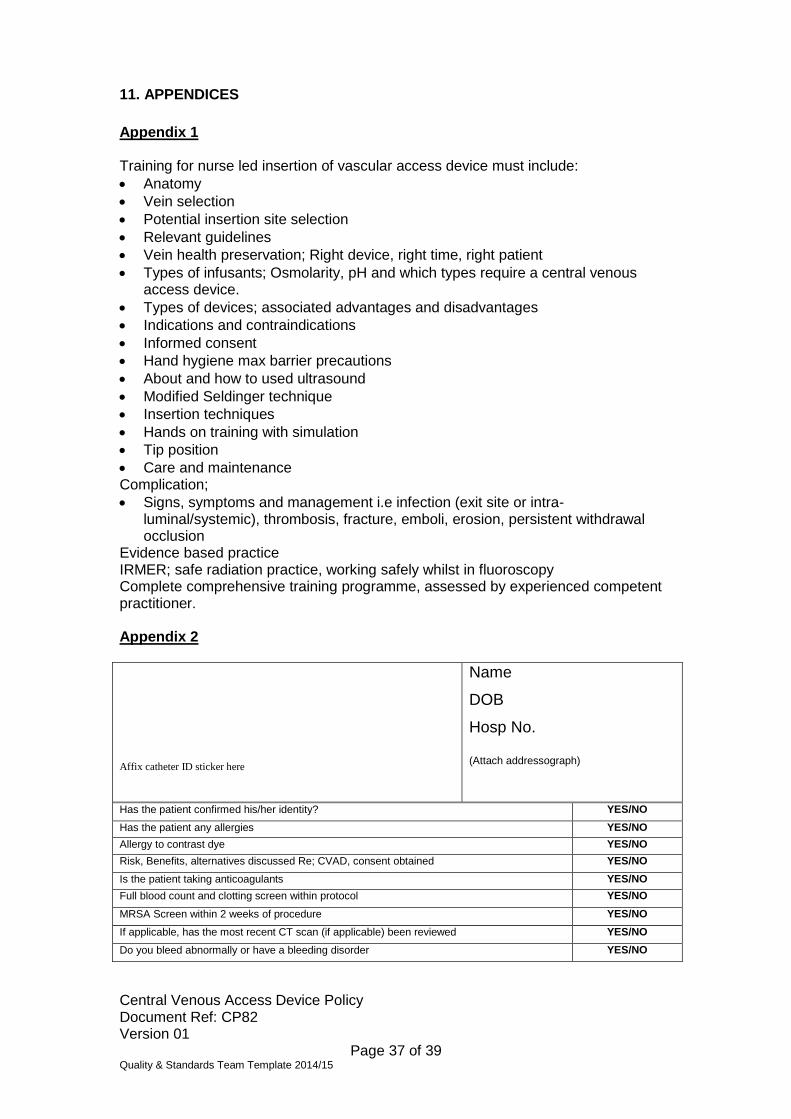
Central Venous Access Device Policy Document Ref: CP82 Version 01
Page 37 of 39 Quality & Standards Team Template 2014/15
11. APPENDICES
Appendix 1 Training for nurse led insertion of vascular access device must include:
Anatomy
Vein selection
Potential insertion site selection
Relevant guidelines
Vein health preservation; Right device, right time, right patient
Types of infusants; Osmolarity, pH and which types require a central venous access device.
Types of devices; associated advantages and disadvantages
Indications and contraindications
Informed consent
Hand hygiene max barrier precautions
About and how to used ultrasound
Modified Seldinger technique
Insertion techniques
Hands on training with simulation
Tip position
Care and maintenance Complication;
Signs, symptoms and management i.e infection (exit site or intra-luminal/systemic), thrombosis, fracture, emboli, erosion, persistent withdrawal occlusion
Evidence based practice IRMER; safe radiation practice, working safely whilst in fluoroscopy Complete comprehensive training programme, assessed by experienced competent practitioner. Appendix 2
Has the patient confirmed his/her identity? YES/NO
Has the patient any allergies YES/NO
Allergy to contrast dye YES/NO
Risk, Benefits, alternatives discussed Re; CVAD, consent obtained YES/NO
Is the patient taking anticoagulants YES/NO Full blood count and clotting screen within protocol YES/NO
MRSA Screen within 2 weeks of procedure YES/NO
If applicable, has the most recent CT scan (if applicable) been reviewed YES/NO
Do you bleed abnormally or have a bleeding disorder YES/NO
Affix catheter ID sticker here
Name
DOB
Hosp No. (Attach addressograph)
Central Venous Access Device Policy Document Ref: CP82 Version 01
Page 38 of 39 Quality & Standards Team Template 2014/15
Appendix 3
TIVAD Occlusion Algorithm 2016.doc
Appendix 4
PICC Algorithm (Hospital) 2016.doc
Appendix 5
Tunnelled CVC algorithm (Hospital) 2016.doc
Appendix 6
Urokinase Administration protocol.doc
Appendix 7
Alcohol lock 2016.doc
Appendix 8 Guidelines for the management of sepsis (including Neutropenic sepsis) Discover - Documents and guidelines database Appendix 9 Procedure for unblocking a CVAD (3-Way tap method)
Procedure for unblocking CVAD.doc
Appendix 10
Blood sampling from a picc.doc
Appendix 11
Blood sampling from a CVC.doc
Central Venous Access Device Policy Document Ref: CP82 Version 01
Page 39 of 39 Quality & Standards Team Template 2014/15
Appendix 12 TIVAD care, community guide
Community Nurse Information 2016.docx
Polyperf removal guide.pdf
Blood sampling from TIVAD.doc
Appendix 13 PICC occlusion algorithm - Community
PICC occlusion algorithm community 2016.docx
Appendix 14 Tunnelled CVC occlusion algorithm - Community
Tunnelled CVC algorithm - Community 2016.docx
Appendix 15
Heparin vs Saline Lit 2016.doc
Appendix 16
Patient information implanted port 2016.doc
Appendix 17
Referral SOP CVAD 2016.docx
Appendix 18
CVAD REQUEST 2016.doc