Embed Size (px)
Citation preview
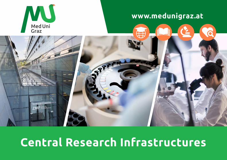
www.medunigraz.at
Central Research Infrastructures
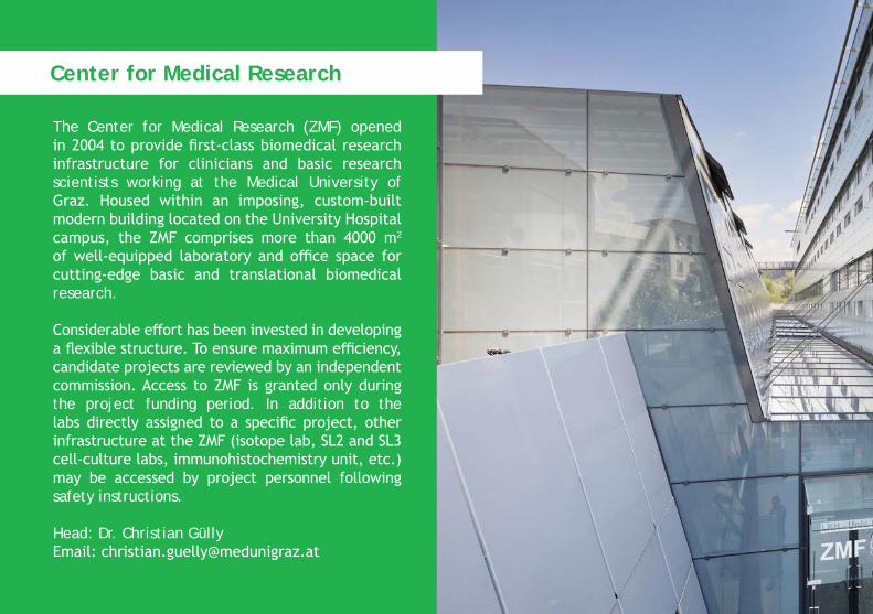
The Center for Medical Research (ZMF) opened in 2004 to provide first-class biomedical research infrastructure for clinicians and basic research scientists working at the Medical University of Graz. Housed within an imposing, custom-built modern building located on the University Hospitalcampus, the ZMF comprises more than 4000 m2
of well-equipped laboratory and office space for cutting-edge basic and translational biomedical research.
Considerable effort has been invested in developing a flexible structure. To ensure maximum efficiency, candidate projects are reviewed by an independent commission. Access to ZMF is granted only during the project funding period. In addition to the labs directly assigned to a specific project, other infrastructure at the ZMF (isotope lab, SL2 and SL3 cell-culture labs, immunohistochemistry unit, etc.) may be accessed by project personnel following safety instructions.
Head: Dr. Christian GüllyEmail: [email protected]
Center for Medical Research
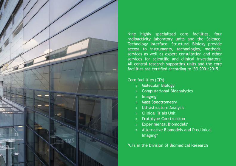
Nine highly specialized core facilities, four radioactivity laboratory units and the Science-Technology Interface: Structural Biology provide access to instruments, technologies, methods, services as well as expert consultation and other services for scientific and clinical investigators. All central research supporting units and the core facilities are certified according to ISO 9001:2015.
Core facilities (CFs): » Molecular Biology » Computational Bioanalytics » Imaging » Mass Spectrometry » Ultrastructure Analysis » Clinical Trials Unit » Prototype Construction » Experimental Biomodels* » Alternative Biomodels and Preclinical
Imaging*
*CFs in the Division of Biomedical Research
Who we are…
Our core expertise lies in the field of microbial community characterization with multi OMICS technologies, eukaryotic gene expression analysis and single cell analysis tools. CF Molecular Biology offers trendsetting technologies in a multiplicity of nucleic acid research methods.
Core instrumentation: » Illumina MiSeq » Nanostring nCounter® » 10X Genomics Chromium Single Cell
Controller » Digital PCR systems » Affymetrix microarray platform » Various qRT-PCR cyclers
Methods can be customized or newly developed on special request.
…and what we can do for you: Microbiome analyses (NGS based)
» Amplicon based determination of microbial pattern (16S, ITS, 18S) » Shot gun de-novo sequencing of small
genomes » Prokaryotic transcriptomics
Core Facility Molecular Biology
Gene expression analyses » Nanostring analyses (mRNA, PlexSet,
microRNA, miRGE Assays, 3D analyses) » Illumina based mRNA Seq, microRNA Seq,
amplicon based analyses » Droplet digital PCR for rare event detection » Affymetrix microarray solutions for gene
expression, SNP or CNV
10X Single Cell Analysis » Single Cell Gene Expression » Single Cell Immune Profiling » Single Cell ATAC
Additional NGS based methods » Customized applications upon request » Ready to run libraries for MiSeq and FASTQ
file delivery
Contact: Ingeborg Klymiuk, PhDEmail: [email protected].: +43 316 385 - 72830
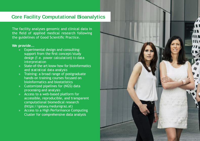
The facility analyses genomic and clinical data in the field of applied medical research following the guidelines of Good Scientific Practice.
We provide… » Experimental design and consulting:
support from the first concept/study design (f.e. power calculation) to data interpretation » State-of-the art know how for bioinformatics
and statistical data analysis » Training: a broad range of postgraduate
hands-on training courses focused on bioinformatics and biostatistics » Customized pipelines for (NGS) data
processing and analysis » Access to a web-based platform for
accessible, reproducible, and transparent computational biomedical research (https://galaxy.medunigraz.at) » Access to a High Performance Computing
Cluster for comprehensive data analysis
Core Facility Computational Bioanalytics
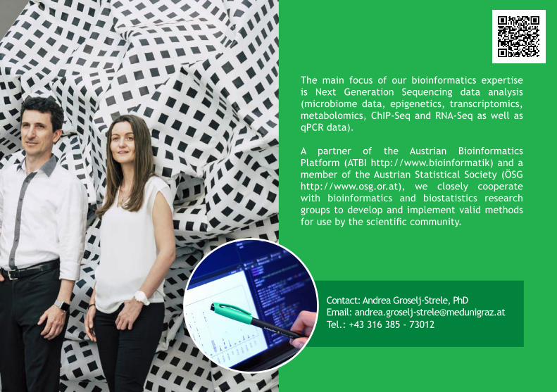
The main focus of our bioinformatics expertise is Next Generation Sequencing data analysis (microbiome data, epigenetics, transcriptomics, metabolomics, ChIP-Seq and RNA-Seq as well as qPCR data).
A partner of the Austrian Bioinformatics Platform (ATBI http://www.bioinformatik) and a member of the Austrian Statistical Society (ÖSG http://www.osg.or.at), we closely cooperate with bioinformatics and biostatistics research groups to develop and implement valid methods for use by the scientific community.
Contact: Andrea Groselj-Strele, PhDEmail: [email protected].: +43 316 385 - 73012
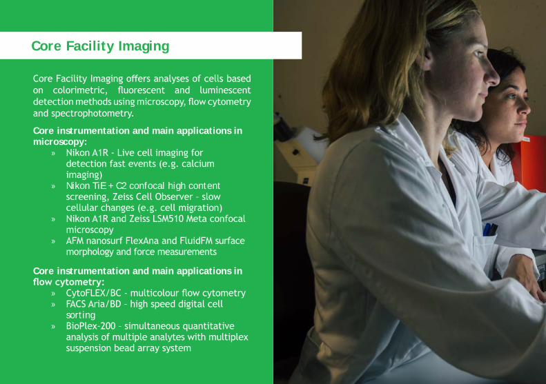
Core Facility Imaging offers analyses of cells based on colorimetric, fluorescent and luminescent detection methods using microscopy, flow cytometry and spectrophotometry.
Core instrumentation and main applications inmicroscopy:
» Nikon A1R - Live cell imaging for detection fast events (e.g. calcium imaging) » Nikon TiE + C2 confocal high content
screening, Zeiss Cell Observer – slow cellular changes (e.g. cell migration) » Nikon A1R and Zeiss LSM510 Meta confocal
microscopy » AFM nanosurf FlexAna and FluidFM surface
morphology and force measurements
Core instrumentation and main applications in flow cytometry:
» CytoFLEX/BC - multicolour flow cytometry » FACS Aria/BD – high speed digital cell
sorting » BioPlex-200 – simultaneous quantitative
analysis of multiple analytes with multiplex suspension bead array system
Core Facility Imaging
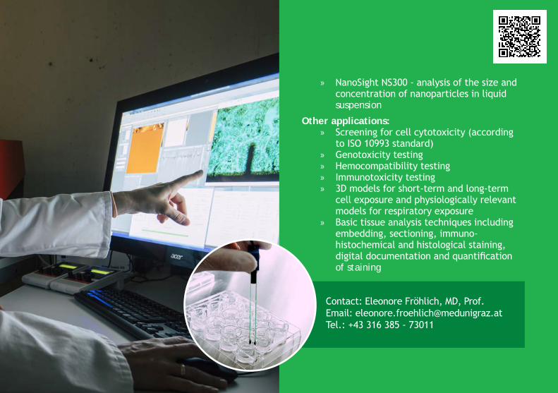
» NanoSight NS300 - analysis of the size and concentration of nanoparticles in liquid suspension
Other applications: » Screening for cell cytotoxicity (according
to ISO 10993 standard) » Genotoxicity testing » Hemocompatibility testing » Immunotoxicity testing » 3D models for short-term and long-term
cell exposure and physiologically relevant models for respiratory exposure » Basic tissue analysis techniques including
embedding, sectioning, immuno- histochemical and histological staining, digital documentation and quantification of staining
Contact: Eleonore Fröhlich, MD, Prof.Email: [email protected].: +43 316 385 - 73011
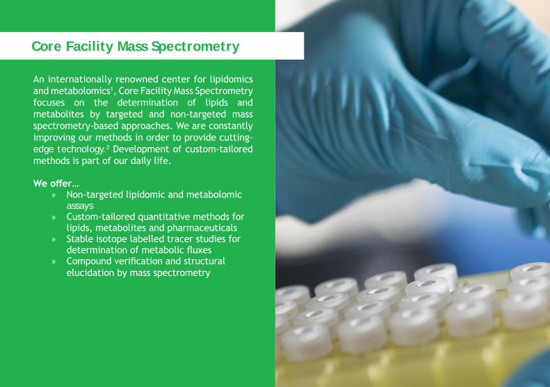
An internationally renowned center for lipidomics and metabolomics1, Core Facility Mass Spectrometry focuses on the determination of lipids and metabolites by targeted and non-targeted mass spectrometry-based approaches. We are constantly improving our methods in order to provide cutting-edge technology.2 Development of custom-tailored methods is part of our daily life.
We offer… » Non-targeted lipidomic and metabolomic
assays » Custom-tailored quantitative methods for
lipids, metabolites and pharmaceuticals » Stable isotope labelled tracer studies for
determination of metabolic fluxes » Compound verification and structural
elucidation by mass spectrometry
Core Facility Mass Spectrometry
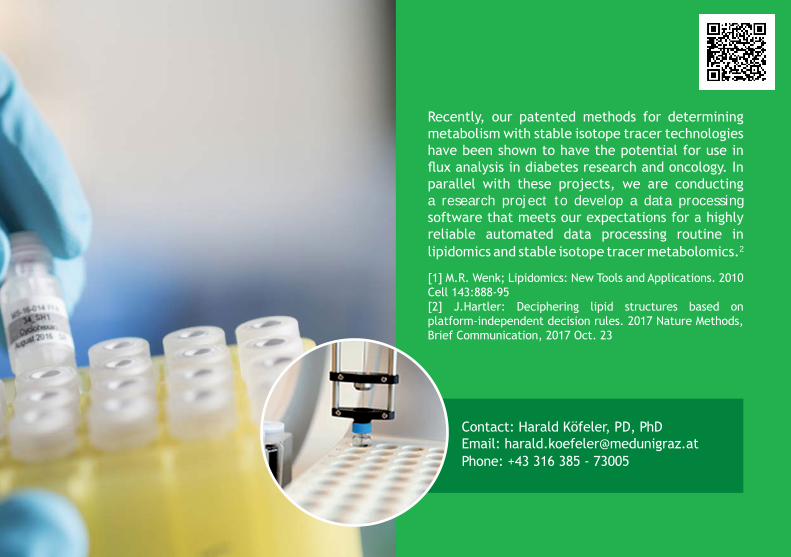
Recently, our patented methods for determining metabolism with stable isotope tracer technologies have been shown to have the potential for use in flux analysis in diabetes research and oncology. In parallel with these projects, we are conducting a research project to develop a data processing software that meets our expectations for a highly reliable automated data processing routine in lipidomics and stable isotope tracer metabolomics.2
[1] M.R. Wenk; Lipidomics: New Tools and Applications. 2010 Cell 143:888-95[2] J.Hartler: Deciphering lipid structures based on platform-independent decision rules. 2017 Nature Methods, Brief Communication, 2017 Oct. 23
Contact: Harald Köfeler, PD, PhDEmail: [email protected]: +43 316 385 - 73005
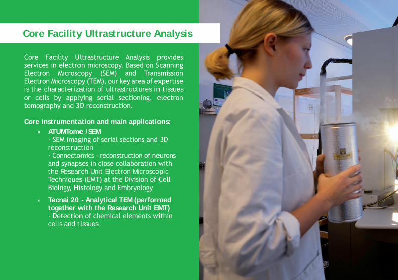
Core Facility Ultrastructure Analysis provides services in electron microscopy. Based on Scanning Electron Microscopy (SEM) and Transmission Electron Microscopy (TEM), our key area of expertise is the characterization of ultrastructures in tissues or cells by applying serial sectioning, electron tomography and 3D reconstruction.
Core instrumentation and main applications: » ATUMTome /SEM
- SEM imaging of serial sections and 3D reconstruction - Connectomics – reconstruction of neurons and synapses in close collaboration with the Research Unit Electron Microscopic Techniques (EMT) at the Division of Cell Biology, Histology and Embryology
» Tecnai 20 - Analytical TEM (performed together with the Research Unit EMT)
- Detection of chemical elements within cells and tissues
Core Facility Ultrastructure Analysis
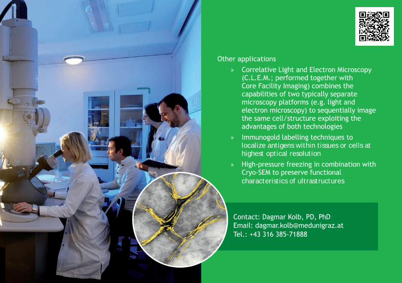
Other applications » Correlative Light and Electron Microscopy
(C.L.E.M.; performed together with Core Facility Imaging) combines the capabilities of two typically separate microscopy platforms (e.g. light and electron microscopy) to sequentially image the same cell/structure exploiting the advantages of both technologies » Immunogold labelling techniques to
localize antigens within tissues or cells at highest optical resolution » High-pressure freezing in combination with
Cryo-SEM to preserve functional characteristics of ultrastructures
Contact: Dagmar Kolb, PD, PhDEmail: [email protected].: +43 316 385-71888
Medical Research Academy Graz
Medical Research Academy Graz offers high-quality courses to post-graduates in different areas of life science technologies. In practice-oriented courses, participants gain comprehensive knowledge and profound practical experience in an environment with state-of-the-art equipment.
Biostatistics and Bioinformatics » Statistical Data Analysis with SPSS for Life Science Researchers SPSS Basics and Group Comparison » Statistical Data Analysis with SPSS for Life Science Researchers Survival Analysis and Analysing Categorical » Statistical Data Analysis with SPSS for Life Science Researchers Analysis of Variance and Regression
Analysis » Statistical Errors in Medical Research » Data Management » Introduction into R for Life Science Researchers » Advanced R for Life Sciences » Introduction to NGS Data Analysis » Short Introduction to Linux and Command Line Data Analysis » Introduction into Galaxy for Life Science Researchers » 16s rRNA Microbiome Data Analysis in Galaxy » Working with qPCR data
Galaxy » 16s rRNA Microbiome Data Analysis in Galaxy » Introduction into Galaxy for Life Science Researchers
Cell Culture » Cell Culture Basic Course » Mathematics and Troubleshooting in Cell Culture » High Content Screening for Multiparameter Phenotypic Profiling of Cells -
Staining, Image Acquisition and Statistical Analysis of Data
Animal Experimentation » Course in Basics of Laboratory Animal Science Equivalent to FELASA B Guidelines » The Pig in Biomedical Research - Training Course » National Legislation for Animal Experimentation - Guidelines for Project
Application » Mouse Course for Animal Experimentation
Lab Techniques » Real time PCR Workshop » Immunohistochemical Staining » Flow Cytometry Basic Course
For more information, please check the Medical Research Academy Graz website: https://www.medunigraz.at/medical-research-academy-graz
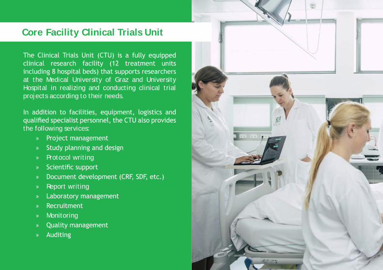
The Clinical Trials Unit (CTU) is a fully equipped clinical research facility (12 treatment units including 8 hospital beds) that supports researchers at the Medical University of Graz and University Hospital in realizing and conducting clinical trial projects according to their needs.
In addition to facilities, equipment, logistics and qualified specialist personnel, the CTU also provides the following services:
» Project management » Study planning and design » Protocol writing » Scientific support » Document development (CRF, SDF, etc.) » Report writing » Laboratory management » Recruitment » Monitoring » Quality management » Auditing
Core Facility Clinical Trials Unit
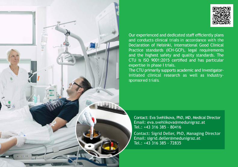
Our experienced and dedicated staff efficiently plans and conducts clinical trials in accordance with the Declaration of Helsinki, international Good Clinical Practice standards (ICH-GCP), legal requirements and the highest safety and quality standards. The CTU is ISO 9001:2015 certified and has particular expertise in phase I trials.The CTU primarily supports academic and investigator-initiated clinical research as well as industry- sponsored trials.
Contact: Eva Svehlikova, PhD, MD, Medical DirectorEmail: [email protected].: +43 316 385 - 80416Contact: Sigrid Deller, PhD, Managing DirectorEmail: [email protected].: +43 316 385 - 72835
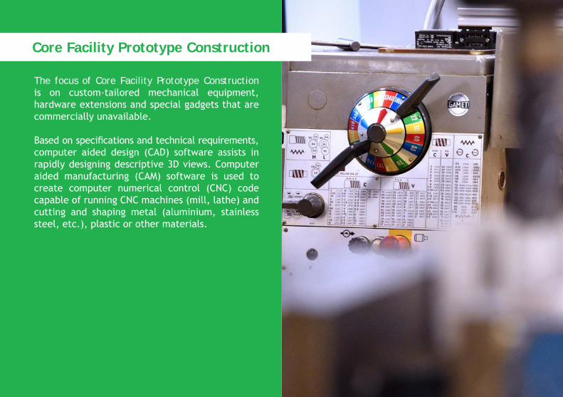
The focus of Core Facility Prototype Construction is on custom-tailored mechanical equipment, hardware extensions and special gadgets that are commercially unavailable.
Based on specifications and technical requirements,computer aided design (CAD) software assists in rapidly designing descriptive 3D views. Computer aided manufacturing (CAM) software is used to create computer numerical control (CNC) code capable of running CNC machines (mill, lathe) and cutting and shaping metal (aluminium, stainless steel, etc.), plastic or other materials.
Core Facility Prototype Construction

In-house services include: » 3D-construction (Creo 5.0) » CAM programming (Creo 5.0) » Turning » Milling » Fabrication
In cooperation with external strategic partners, various innovative technologies are available: 3D printing (metal or plastics)...and much more.
Contact: Markus PichlerEmail: [email protected].: +43 316 385 - 71509
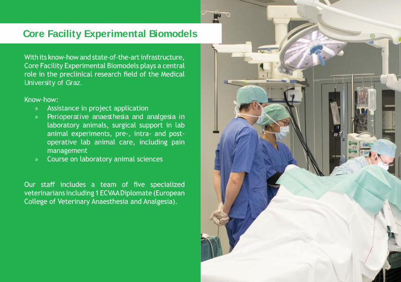
With its know-how and state-of-the-art infrastructure, Core Facility Experimental Biomodels plays a central role in the preclinical research field of the Medical University of Graz.
Know-how: » Assistance in project application » Perioperative anaesthesia and analgesia in
laboratory animals, surgical support in lab animal experiments, pre-, intra- and post-operative lab animal care, including pain management
» Course on laboratory animal sciences
Our staff includes a team of five specialized veterinarians including 1 ECVAA Diplomate (European College of Veterinary Anaesthesia and Analgesia).
Core Facility Experimental Biomodels
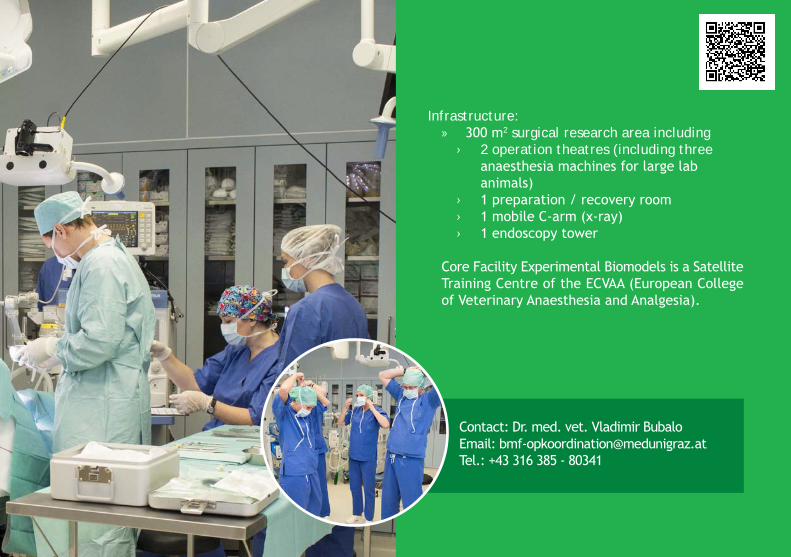
Infrastructure: » 300 m2 surgical research area including
› 2 operation theatres (including three anaesthesia machines for large lab animals)
› 1 preparation / recovery room › 1 mobile C-arm (x-ray) › 1 endoscopy tower
Core Facility Experimental Biomodels is a Satellite Training Centre of the ECVAA (European College of Veterinary Anaesthesia and Analgesia).
Contact: Dr. med. vet. Vladimir BubaloEmail: [email protected].: +43 316 385 - 80341
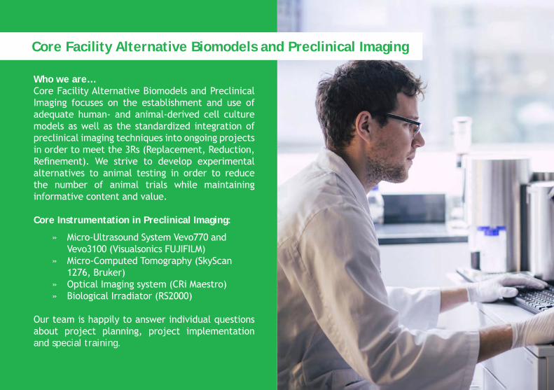
Who we are…Core Facility Alternative Biomodels and Preclinical Imaging focuses on the establishment and use of adequate human- and animal-derived cell culture models as well as the standardized integration of preclinical imaging techniques into ongoing projects in order to meet the 3Rs (Replacement, Reduction, Refinement). We strive to develop experimental alternatives to animal testing in order to reduce the number of animal trials while maintaining informative content and value.
Core Instrumentation in Preclinical Imaging:
» Micro-Ultrasound System Vevo770 and Vevo3100 (Visualsonics FUJIFILM)» Micro-Computed Tomography (SkyScan 1276, Bruker)» Optical Imaging system (CRi Maestro)» Biological Irradiator (RS2000)
Our team is happily to answer individual questions about project planning, project implementation and special training.
Core Facility Alternative Biomodels and Preclinical Imaging
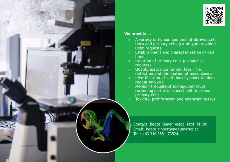
We provide … » A variety of human and animal-derived cell
lines and primary cells (catalogue provided upon request)
» Establishment and characterization of cell lines
» Isolation of primary cells (on special request)
» Quality assurance for cell labs – f.e. detection and elimination of mycoplasma
» Identification of cell lines by short tandem repeat analysis
» Medium throughput (compound/drug) screening on (rare cancer) cell lines and primary cells
» Toxicity, proliferation and migration assays
Contact: Beate Rinner, Assoc. Prof. PD Dr.Email: [email protected].: +43 316 385 - 73524
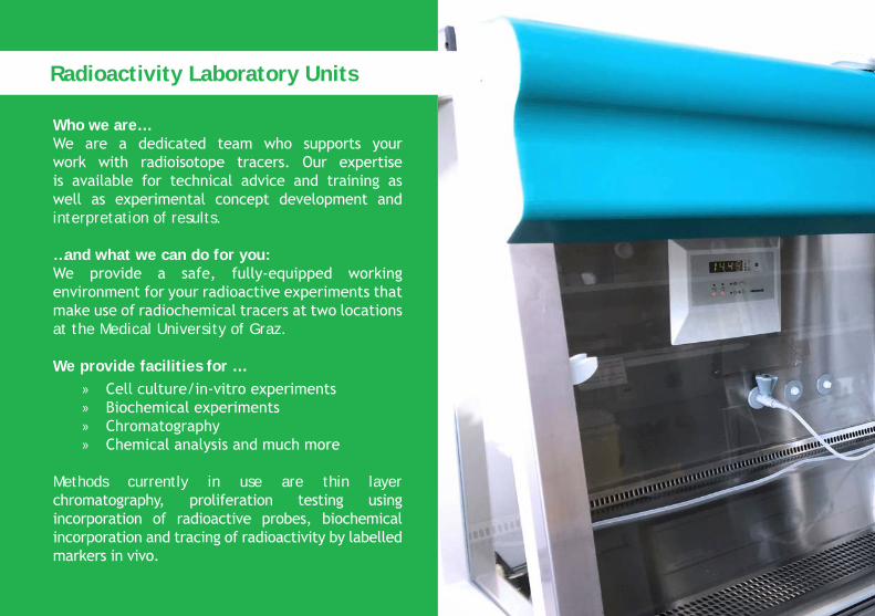
Who we are…We are a dedicated team who supports your work with radioisotope tracers. Our expertise is available for technical advice and training as well as experimental concept development and interpretation of results.
…and what we can do for you:We provide a safe, fully-equipped working environment for your radioactive experiments that make use of radiochemical tracers at two locations at the Medical University of Graz.
We provide facilities for … » Cell culture/in-vitro experiments » Biochemical experiments » Chromatography » Chemical analysis and much more
Methods currently in use are thin layer chromatography, proliferation testing using incorporation of radioactive probes, biochemical incorporation and tracing of radioactivity by labelled markers in vivo.
Radioactivity Laboratory Units
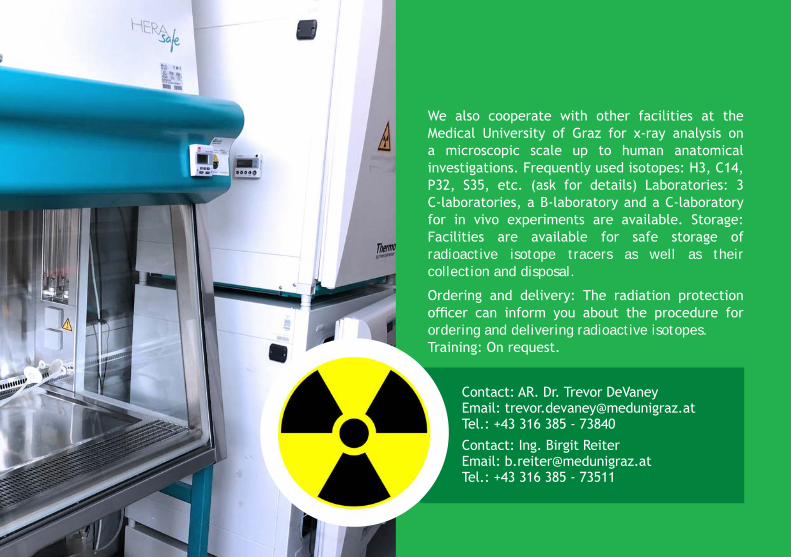
We also cooperate with other facilities at the Medical University of Graz for x-ray analysis on a microscopic scale up to human anatomical investigations. Frequently used isotopes: H3, C14, P32, S35, etc. (ask for details) Laboratories: 3 C-laboratories, a B-laboratory and a C-laboratory for in vivo experiments are available. Storage: Facilities are available for safe storage of radioactive isotope tracers as well as their collection and disposal.
Ordering and delivery: The radiation protection officer can inform you about the procedure for ordering and delivering radioactive isotopes.Training: On request.
Contact: AR. Dr. Trevor DeVaney Email: [email protected].: +43 316 385 - 73840 Contact: Ing. Birgit ReiterEmail: [email protected] Tel.: +43 316 385 - 73511
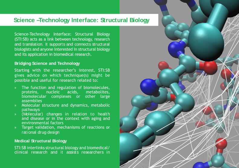
Science-Technology Interface: Structural Biology (STI:SB) acts as a link between technology, research and translation. It supports and connects structural biologists and anyone interested in structural biology and its application in biomedical research.
Bridging Science and Technology
Starting with the researcher’s interest, STI:SB gives advice on which technique(s) might be possible and useful for research related to:
» The function and regulation of biomolecules, proteins, nucleic acids, metabolites, biomolecular complexes or other large assemblies
» Molecular structure and dynamics, metabolic pathways
» (Molecular) changes in relation to health and disease or in the context with aging and environmental factors
» Target validation, mechanisms of reactions or rational drug design
Medical Structural Biology
STI:SB interlinks structural biology and biomedical/clinical research and it assists researchers in
Science –Technology Interface: Structural Biology
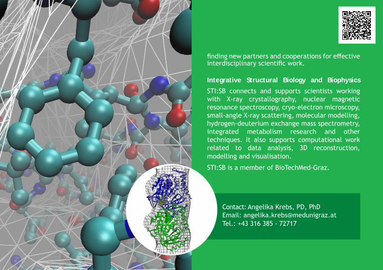
finding new partners and cooperations for effective interdisciplinary scientific work.
Integrative Structural Biology and Biophysics
STI:SB connects and supports scientists working with X-ray crystallography, nuclear magnetic resonance spectroscopy, cryo-electron microscopy, small-angle X-ray scattering, molecular modelling, hydrogen-deuterium exchange mass spectrometry, integrated metabolism research and other techniques. It also supports computational work related to data analysis, 3D reconstruction, modelling and visualisation.
STI:SB is a member of BioTechMed-Graz.
Contact: Angelika Krebs, PD, PhD Email: [email protected].: +43 316 385 - 72717
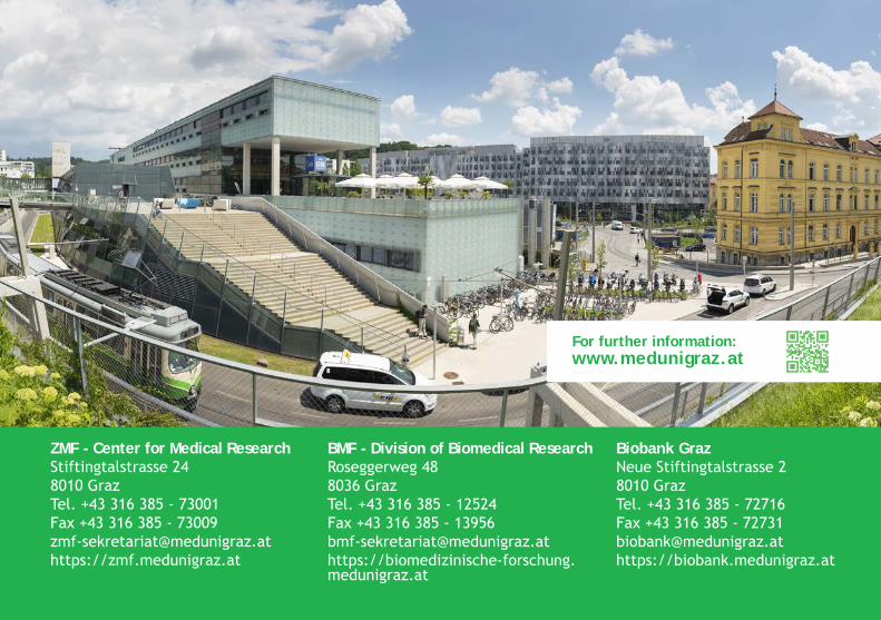
ZMF - Center for Medical ResearchStiftingtalstrasse 248010 GrazTel. +43 316 385 - 73001Fax +43 316 385 - [email protected]://zmf.medunigraz.at
BMF - Division of Biomedical ResearchRoseggerweg 488036 GrazTel. +43 316 385 - 12524Fax +43 316 385 - [email protected]://biomedizinische-forschung.medunigraz.at
Biobank GrazNeue Stiftingtalstrasse 28010 GrazTel. +43 316 385 - 72716Fax +43 316 385 - [email protected]://biobank.medunigraz.at
For further information:www.medunigraz.at