Embed Size (px)
Citation preview
18 nucleotides, respectively), these mini- filaments formed ordered crystals.
The structures reveal an ordered filament with 6.2 monomeric units per turn and a pitch of 92–95 ångstroms. The DNA is close to the filament axis, is extended relative to B-form DNA, and has global features compat-ible with the electron microscopy. The ATP is completely buried at an interface between monomers. Each RecA monomer interacts with three nucleotides of the DNA (a triplet) adjacent to itself in the structure, as well as with two more nucleotides, one from each of the preceding and following triplets. As a result, each nucleotide triplet is bound by three monomers.
Perhaps the most remarkable feature of the nucleoprotein filament is the DNA structure (Fig. 1b). The 50% extension is not manifest as an isotropic extension at the nucleotide level; instead, the DNA is seen to comprise a three-nucleotide segment with a nearly nor-mal B-form distance between bases (an axial rise of 3.5–4.2 Å for ssDNA and 3.2–3.5 Å for dsDNA), followed by a long untwisted inter-nucleotide stretch (approximately 7.1–7.8 Å in ssDNA and 8.4 Å in dsDNA) before the next three-nucleotide element, and so on. This was a surprising result, because most people assumed that the DNA within the RecA–DNA complexes was uniformly stretched to an aver-age of about 5.2 Å between bases.
The unusual repeat pattern of DNA exten-sion in the RecA nucleoprotein filament offers a structural basis for understanding the dynam-ics of filament assembly. Assembly occurs by rate-limiting initiation of polymer forma-tion (nucleation) followed by growth2. The structure shows that it would be energetically unfavourable for a single monomer to make the full repertoire of molecular contacts with DNA because of the need to both unstack the bases and extend the DNA to the next nucleotide triplet. Thus, the free energy for binding of the first monomer will be unfavourable relative to the binding of a second protein to an existing monomer, explaining the observed cooperativ-ity of RecA binding to DNA2. Binding of a third monomer provides additional net free energy, because now two of the three monomers ben-efit from the added free energy of cooperative interactions. As more monomers bind, the ener-getic cost of extending the DNA is ‘amortized’ over an increasingly greater number of RecA monomers, until the net free energy of nucleus formation is sufficiently negative to permit stable nucleation. Although more complex scenarios can be envisaged, the structures of Chen and colleagues1 now permit detailed energetic modelling of filament formation.
The results also highlight the physical mis-match between the ssDNA within the filament and the naked duplex DNA target (Fig. 1b). How does RecA align these sequences? The structures1 offer provocative insights into how the transient three-stranded intermedi-ate might look, and how the fidelity of DNA
CELL BIOLOGY
Viruses in camouflage Kirsten Sandvig and Bo van Deurs
The vaccinia virus acts like a Trojan Horse to enter its host cells: it envelops itself in the membrane of a dying cell, and is then taken up by healthy cells.
Endocytosis is the process by which cells internalize extracellular material. It is crucial to cell survival and the proper functioning of tissues, being involved in processes as diverse as growth, neural transmission and pathogen clearance. But several opportunistic molecules (bacterial and plant toxins) and pathogens (viruses and bacteria) can exploit the endocytic machinery of a host cell for their own gain1,2. Writing in Science, Mercer and Helenius3 report how vaccinia virus (Fig. 1) — a cousin of variola virus, which causes smallpox — deceives host cells into taking it up through endocytosis.
When cells are damaged or dying, for exam-ple during programmed cell death (apoptosis), they show several characteristic features. For instance, phosphatidylserine, a lipid that is
abundant in the inner (cytoplasmic) layer of the cell membrane, is redistributed to the outer layer. This phospholipid is thus available to bind to receptors on the surface of phagocytic cells that initiate the apoptotic cell’s destruc-tion and engulf it4. Another sign of apoptosis is membrane blebbing, or the formation of irregular bulges on the cell membrane. Bleb-bing also occurs during other processes, such as cell migration and division, but its function is unclear.
When enveloped viruses bud off from their host cell, they inherit a lipid coating (envelope) that has the same composition as the host cell membrane. Mercer and Helenius3 report that the outer layer of the vaccinia virus envelope contains phosphatidylserine and that this is crucial for infection. They propose that the
strand exchange might be enforced. It is easy to imagine the pairing between an ssDNA triplet within the filament and the naked dsDNA, as both have approximately B-form dimensions. However, pairing of the next three base pairs of DNA requires extension of the dsDNA to conform to the observed extension of the ssDNA in the filament. This energeti-cally unfavourable base unstacking and chain extension could be compensated both by the now lower entropic cost (because the next triplet is part of the already paired dsDNA) and by the favourable base-pairing interac-tions that would form if the next triplet were fully homologous. However, if even one of the base pairs was non-complementary, then the nascent paired molecule might not be suf-ficiently stable, and homologous pairing with a partially homologous sequence would be aborted.
Furthermore, the structures show that the strand complementary to the ssDNA in the presynaptic filament makes few contacts with the protein. Hence, it is largely stabilized by correct Watson–Crick base-pairing, thereby requiring accurate DNA pairing. Successful DNA pairing requires at least 15 base pairs of homology10, and the structures suggest how such fidelity is enforced.
Determination of the three-dimensional structure of the active state of RecA nucleo-protein filaments by Chen and colleagues1 is a watershed in recombination biochemistry and mechanics. Not only do the structures inform us about this central protein, but they also
enable the formulation of structural hypoth-eses that relate to the RecA orthologues and to interacting proteins. Although the eukaryotic and archaeal RecA homologues differ in many functional and mechanistic details, the RecA structures will provide a valuable foundation for understanding them. Also, many proteins interact with the various forms of RecA family members to regulate assembly and disassem-bly of the filament. Having structures of both the ATP– and ADP–RecA nucleoprotein fila-ments will help clarify the mechanistic basis of their biological functions. Clearly, more (DNA) partner-swapping experiments will be forthcoming. ■
Stephen C. Kowalczykowski is in the Departments of Microbiology, and of Molecular and Cellular Biology, University of California, Davis, One Shields Avenue, Davis, California 95616, USA.e-mail: [email protected]
1. Chen, Z., Yang, H. & Pavletich, N. P. Nature 453, 489–494 (2008).
2. Kowalczykowski, S. C. Annu. Rev. Biophys. Biophys. Chem. 20, 539–575 (1991).
3. Menetski, J. P., Bear, D. G. & Kowalczykowski, S. C. Proc. Natl Acad. Sci. USA 87, 21–25 (1990).
4. Mazin, A. V. & Kowalczykowski, S. C. Genes Dev. 13, 2005–2016 (1999).
5. Yu, X. et al. Curr. Protein Pept. Sci. 5, 73–79 (2004).6. Bianco, P. R., Tracy, R. B. & Kowalczykowski, S. C.
Front. Biosci. 3, d570–d603 (1998).7. Story, R. M., Weber, I. T. & Steitz, T. A. Nature 355, 318–325
(1992).8. Story, R. M. & Steitz, T. A. Nature 355, 374–376 (1992).9. McGrew, D. A. & Knight, K. L. Crit. Rev. Biochem. Mol. Biol.
38, 385–432 (2003).10. Ferrin, L. J. & Camerini-Otero, R. D. Science 254, 1494–1497
(1991).
466
NATURE|Vol 453|22 May 2008NEWS & VIEWS
underlying mechanism is as follows. When a cell becomes infected with the virus, it displays apoptotic features, including the presence of phosphatidylserine in its outer membrane layer. Thus, when the virus buds off from the cell, it inherits this as part of its envelope. Con-sequently, cells probably ‘mistake’ the unusu-ally large vaccinia virus for an apoptotic body (the debris of dying cells) and engulf it.
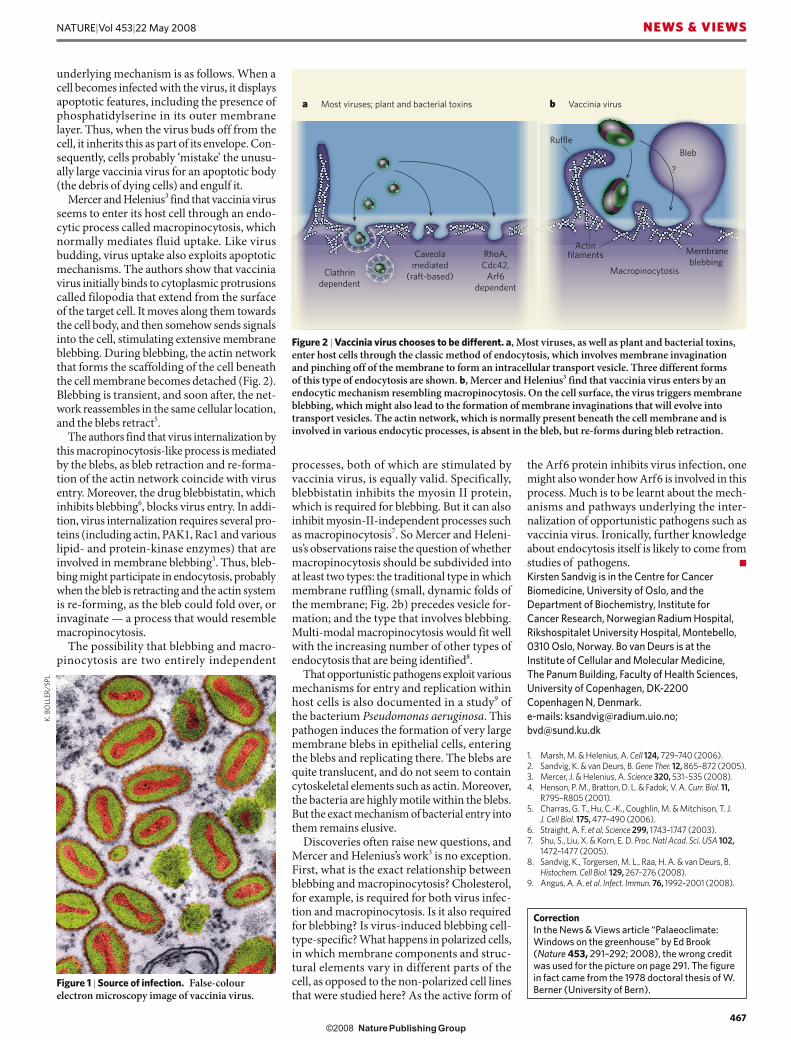
Mercer and Helenius3 find that vaccinia virus seems to enter its host cell through an endo-cytic process called macropinocytosis, which normally mediates fluid uptake. Like virus budding, virus uptake also exploits apoptotic mechanisms. The authors show that vaccinia virus initially binds to cytoplasmic protrusions called filopodia that extend from the surface of the target cell. It moves along them towards the cell body, and then somehow sends signals into the cell, stimulating extensive membrane blebbing. During blebbing, the actin network that forms the scaffolding of the cell beneath the cell membrane becomes detached (Fig. 2). Blebbing is transient, and soon after, the net-work reassembles in the same cellular location, and the blebs retract5.
The authors find that virus internalization by this macropinocytosis-like process is mediated by the blebs, as bleb retraction and re-forma-tion of the actin network coincide with virus entry. Moreover, the drug blebbistatin, which inhibits blebbing6, blocks virus entry. In addi-tion, virus internalization requires several pro-teins (including actin, PAK1, Rac1 and various lipid- and protein-kinase enzymes) that are involved in membrane blebbing3. Thus, bleb-bing might participate in endocytosis, probably when the bleb is retracting and the actin system is re-forming, as the bleb could fold over, or invaginate — a process that would resemble macropinocytosis.
The possibility that blebbing and macro-pinocytosis are two entirely independent
processes, both of which are stimulated by vaccinia virus, is equally valid. Specifically, blebbistatin inhibits the myosin II protein, which is required for blebbing. But it can also inhibit myosin-II-independent processes such as macropinocytosis7. So Mercer and Heleni-us’s observations raise the question of whether macropinocytosis should be subdivided into at least two types: the traditional type in which membrane ruffling (small, dynamic folds of the membrane; Fig. 2b) precedes vesicle for-mation; and the type that involves blebbing. Multi-modal macropinocytosis would fit well with the increasing number of other types of endocytosis that are being identified8.
That opportunistic pathogens exploit various mechanisms for entry and replication within host cells is also documented in a study9 of the bacterium Pseudomonas aeruginosa. This pathogen induces the formation of very large membrane blebs in epithelial cells, entering the blebs and replicating there. The blebs are quite translucent, and do not seem to contain cytoskeletal elements such as actin. Moreover, the bacteria are highly motile within the blebs. But the exact mechanism of bacterial entry into them remains elusive.
Discoveries often raise new questions, and Mercer and Helenius’s work3 is no exception. First, what is the exact relationship between blebbing and macropinocytosis? Cholesterol, for example, is required for both virus infec-tion and macropinocytosis. Is it also required for blebbing? Is virus-induced blebbing cell-type-specific? What happens in polarized cells, in which membrane components and struc-tural elements vary in different parts of the cell, as opposed to the non-polarized cell lines that were studied here? As the active form of
the Arf6 protein inhibits virus infection, one might also wonder how Arf6 is involved in this process. Much is to be learnt about the mech-anisms and pathways underlying the inter-nalization of opportunistic pathogens such as vaccinia virus. Ironically, further knowledge about endocytosis itself is likely to come from studies of pathogens. ■
Kirsten Sandvig is in the Centre for Cancer Biomedicine, University of Oslo, and the Department of Biochemistry, Institute for Cancer Research, Norwegian Radium Hospital, Rikshospitalet University Hospital, Montebello, 0310 Oslo, Norway. Bo van Deurs is at the Institute of Cellular and Molecular Medicine, The Panum Building, Faculty of Health Sciences, University of Copenhagen, DK-2200 Copenhagen N, Denmark.e-mails: [email protected]; [email protected]
1. Marsh, M. & Helenius, A. Cell 124, 729–740 (2006).2. Sandvig, K. & van Deurs, B. Gene Ther. 12, 865–872 (2005).3. Mercer, J. & Helenius, A. Science 320, 531–535 (2008).4. Henson, P. M., Bratton, D. L. & Fadok, V. A. Curr. Biol. 11,
R795–R805 (2001).5. Charras, G. T., Hu, C.-K., Coughlin, M. & Mitchison, T. J.
J. Cell Biol. 175, 477–490 (2006).6. Straight, A. F. et al. Science 299, 1743–1747 (2003).7. Shu, S., Liu, X. & Korn, E. D. Proc. Natl Acad. Sci. USA 102,
1472–1477 (2005).8. Sandvig, K., Torgersen, M. L., Raa, H. A. & van Deurs, B.
Histochem. Cell Biol. 129, 267–276 (2008).9. Angus, A. A. et al. Infect. Immun. 76, 1992–2001 (2008).
CorrectionIn the News & Views article “Palaeoclimate: Windows on the greenhouse” by Ed Brook (Nature 453, 291–292; 2008), the wrong credit was used for the picture on page 291. The figure in fact came from the 1978 doctoral thesis of W. Berner (University of Bern).
Figure 2 | Vaccinia virus chooses to be different. a, Most viruses, as well as plant and bacterial toxins, enter host cells through the classic method of endocytosis, which involves membrane invagination and pinching off of the membrane to form an intracellular transport vesicle. Three different forms of this type of endocytosis are shown. b, Mercer and Helenius3 find that vaccinia virus enters by an endocytic mechanism resembling macropinocytosis. On the cell surface, the virus triggers membrane blebbing, which might also lead to the formation of membrane invaginations that will evolve into transport vesicles. The actin network, which is normally present beneath the cell membrane and is involved in various endocytic processes, is absent in the bleb, but re-forms during bleb retraction.
����������������������������������������
�����������������
���������������
������������
���������������
���������
����������������
������
�����������������
����
�
��������������
� ���������������
Figure 1 | Source of infection. False-colour electron microscopy image of vaccinia virus.
K. B
OLL
ER/S
PL
467
NATURE|Vol 453|22 May 2008 NEWS & VIEWS