Embed Size (px)
Citation preview
1
DISTRIBUTION STATEMENT A. Approved for public release; distribution is unlimited.
Dynamic Camouflage in Benthic and Pelagic Cephalopods: An Interdisciplinary Approach to Crypsis Based on Color,
Reflection, and Bioluminescence
Sönke Johnsen Biology Department, Duke University
Durham, NC 27708 phone: (919) 660-7321 fax: (919) 660-7293 email: [email protected]
Alison Sweeney
Physics and Astronomy Department, U. of Pennsylvania Philadelphia, PA 19104
phone: (215) 573-7569 fax: (215) 898-2010 email: [email protected]
Daniel Morse Marine Science Institute, U. of California at Santa Barbara
Santa Barbara, CA 93106 phone: (805) 893-8982 fax: (805) 893-7998 email: [email protected]
Dariusz Stramski
Marine Physical Lab, Scripps Inst. of Oceanography, UCSD La Jolla, CA 92093
phone: (858) 534-3353 fax: (858) 534-7641 email: [email protected]
Jules Jaffe Marine Physical Lab, Scripps Inst. of Oceanography, UCSD
La Jolla, CA 92093 phone (858) 534-6101 fax: (858) 534-7641 email: [email protected]
Award Number: N00014-09-1-1053
http://www.biology.duke.edu/johnsenlab LONG-TERM GOALS
Our overall goal is to understand the perceptual and mechanistic principles that underlay camouflage framed in the context of the animals’ environment. In particular, we hope to characterize and understand the perceptual abilities of several species of benthic and pelagic cephalopods, the aspects of their optical environment that affect their camouflage behavior, the characterization of that behavior, and the molecular mechanisms inside the skin by which those responses are accomplished.
2
OBJECTIVES 1. To characterize the spatiotemporal characteristics of the near-surface and shallow benthic
underwater light field, including ultraviolet radiation and polarization. 2. To determine the visual abilities of several species of cephalopod and model both the shallow and
deep-water world from the animals’ points of view. 3. To incorporate the knowledge gained from objectives 1 and 2 in order to study the camouflage
behavior of these species under simulated ocean conditions. 4. To understand the underlying molecular and biophysical mechanisms governing changes in the
skin that produce the observed optical effects, to provide a platform for future translational efforts. APPROACH Objective 1, Light measurements: Our approach to characterize the underwater light fields includes radiometric measurements with custom-built SQUID and Porcupine instruments and commercial hyperspectral sensors from the RAMSES radiometer family (TriOS, Germany). SQUID (SeQuence of Underwater Irradiance Detectors) was developed for this project to provide a unique capability to measure both spatial and temporal statistical properties of irradiance fluctuations produced by surface-wave focusing within the surface ocean layer. SQUID consists of 25 irradiance sensors, which are positioned at different distances from one another along a 2.5 m long linear array. The SQUID sensors measure downward plane irradiance Ed at a selected light wavelength (typically 532 nm) with a high sampling rate of 1 kHz using a cosine collector of very small size (2.5 mm in diameter). The Porcupine instrument was developed for the ONR RaDyO program to measure temporal properties of high-frequency fluctuations in downwelling radiance and irradiance at several light wavelengths, and it complements SQUID measurements in this project. The RAMSES sensors provide measurements of downward and upward plane and scalar irradiances as well as upwelling radiance with an averaging time typically from about 0.002 to >1 s (depending on light intensity) and with high spectral resolution (~3 nm from 350 to 850 nm). Because the characteristics of underwater light field depend on the inherent optical properties (IOPs) of water, our approach also includes in situ IOP measurements and collection of discrete water samples for the determinations of the spectral particulate absorption coefficient as well as parameters characterizing the bulk concentration of suspended particulate matter. Objective 2, visual physiology: our approach involves using microscopy, microspectrophotometry and the optomotor response to measure six primary visual parameters of the study species: field of view, spectral sensitivity, acuity, temporal resolution, and contrast and polarization sensitivity. Field of view is determined from the placement and orientation of the eyes and the geometry of the retina and pupil. Spectral sensitivity will be investigated using microspectrophotometry (MSP), which measures the absorption spectra of individual photoreceptors. Spatial and temporal resolution will be estimated from the spacing of photoreceptors in the retina and via the optomotor response. Contrast sensitivity is estimated by determining photon catch and also via optomotor assays using stripes of decreasing contrast. Polarization sensitivity will also be assayed via retinal morphology and optomotor response. Objective 3, camouflage behavior: Camouflage behavior is studied both in situ and within various controlled environments, including a “holodeck”, a tank surrounded by monitors that project natural environments or controlled visual stimuli. The top of the tank has a plexiglass “floatee” that will make
3
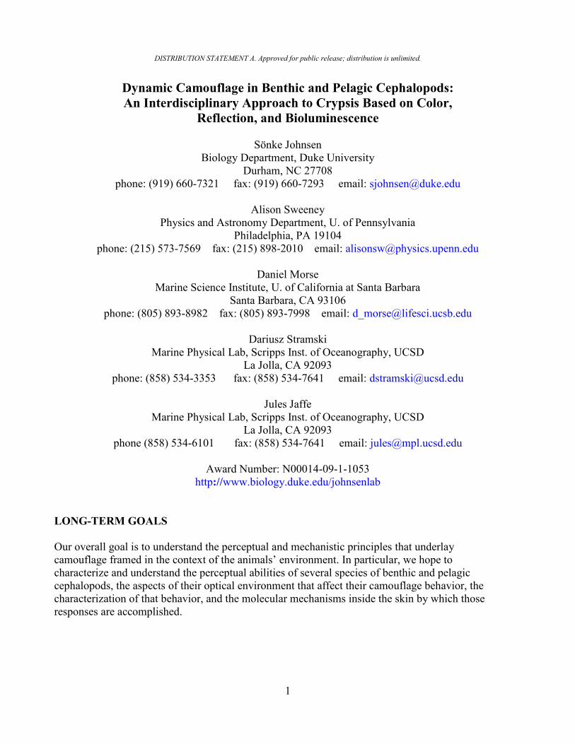
the surface optically flat and permit undistorted observation of the animals from the outside as well as permitting images to be projected into the tank by two DLP projection systems. Objective 4, biophotonics: We will characterize the optics of the skin using fiber-optic spectroscopy coupled with goniometry, measuring the polarization-specific bidirectional reflectance of the skin of the target species and correlate these with the statistical analyses of the light measurements from objective 1 to determine which aspects of this complex reflectance have specifically evolved for camouflage. We will also determine the ultrastructure of the reflectin-based structures using transmission electron microscopy and model their optical effects to determine what aspects of the biological structures are important for the observed environmental optical match. We will also investigate the biophysical mechanisms governing tunable, self-assembling reflectance. WORK COMPLETED Objective 1, Light measurements: During the reporting period, the Stramski team completed three major tasks: (i) a field experiment in Santa Catalina Island, (ii) measurements on the KM12-10 cruise in the Pacific waters off Hawaii Islands, and (iii) analysis of power spectra of wave-induced light fluctuations. Six personnel from Stramski lab participated in the Catalina experiment. During the experiment we successfully tested the newly developed SQUID instrument. We collected 140 time-series measurements, each with 10 min duration, accompanied with measurements taken with the Porcupine instrument and other optical instruments. Four personnel from Stramski lab participated in the KM cruise. A total of 10 stations were completed, located about 20-30 miles offshore on the leeward side of Lanai and the Big Island. At each station we completed the following measurements: (i) the inherent optical property (IOP) package deployment; (ii) underwater and above-water hyperspectral radiometry; (iii) CTD deployment for water sampling and measurement of beam attenuation coefficient; and (iv) particle size distribution, particulate organic carbon, suspended particulate matter, phytoplankton pigments, and particulate absorption analyses on discrete water samples. Unique radiometric measurements included vertical profiles of horizontal radiance from two opposite directions within the principal solar plane, which were taken at different solar angles. With regard to the study of power spectra of light fluctuations, we completed the analysis of our data collected with the Porcupine instrument during recent RaDyO experiments. The analysis of RaDyO dataset is important to this project because it provides a unique resource for characterizing frequency content of wave-induced light fluctuations within the near-surface ocean. Some results from this analysis were presented at the Ocean Sciences Meeting in February 2012, and more extensive analysis will be presented at the upcoming Ocean Optics XXI Conference in Glasgow in October 2012. Objectives 2 and 3, visual physiology and camouflage behavior: The Sub-sea Holodeck has been transported to and reassembled at the Duke University Cephalopod Lab to facilitate behavioral experiments with the cuttlefish Sepia officinalis and the octopus Octopus bimaculoides. We have undertaken preliminary experiments with hatching S. officinalis (figure 1). We have completed comprehensive reviews on our understanding of visual cognition in S. officinalis and in deep-sea cephalopods, which are to be published in a book on cephalopod cognition (see publications). We have completed data collection for behavioral studies investigating aspects of camouflage and are in the process of analyzing the results. We have investigated the optics of potential predators of cephalopods by measuring lens point spread function for 24 taxa during our 2012 research cruise.
4
Figure 1: Hatchling cuttlefish on a dynamic background of varying intensities and spatial
frequencies. The background changed in seconds, as did the disruptive pattern of the animal (notice the head-band and bottom lines).
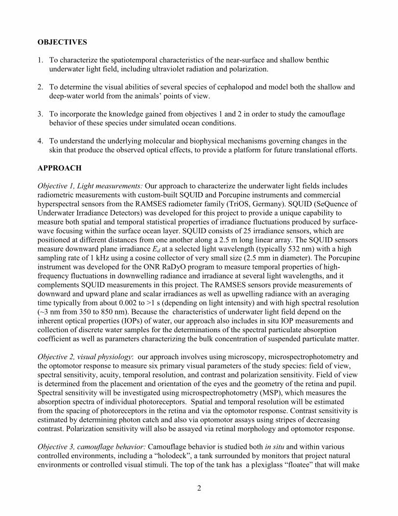
Objective 4, biophotonics and instrumentation development: The optical instrument seen in figure 2 was designed and built to measure the bidirectional reflectance distribution function (BRDF), defined as the ratio of reflected radiance to incident irradiance. A prototype version of the instrument was developed in 2011 and was revised earlier this year. Measurement of the BRDF provides a method of characterizing the complex reflected light patterns of marine animals and allows for quantitative comparison between specimens. Using a laser projector as the light source (λ = 450 nm, 532 nm, 640 nm), we direct a small incident beam onto the sample from a specified direction. Light reflected from the sample within the numerical aperture of the objective lens is relayed to the camera and is captured as a reflectance map. A pinhole aperture is used to block light from off-axis sample points and stray light. The available range for both incident and viewing angles is approximately ±45 degrees.
Figure 2: Optical schematic of improved instrument for measuring directional light scatter from
marine animal skin. Inset shows original design for the camera (imaging) lenses.
5
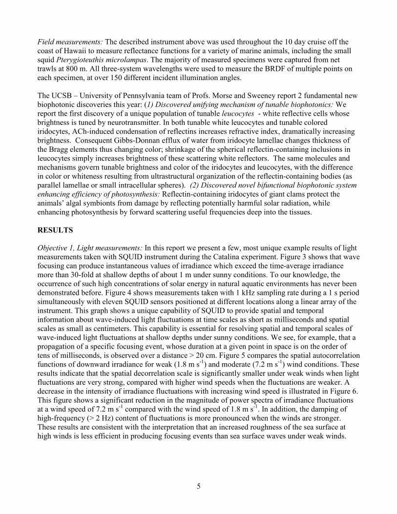
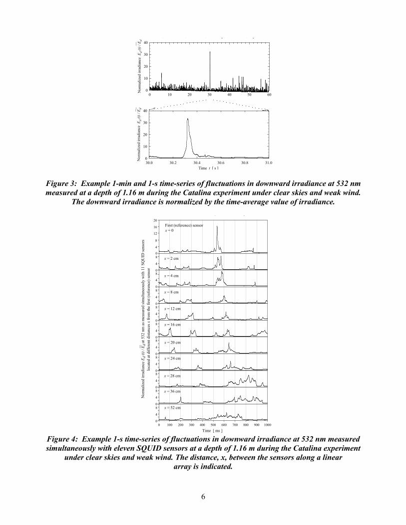
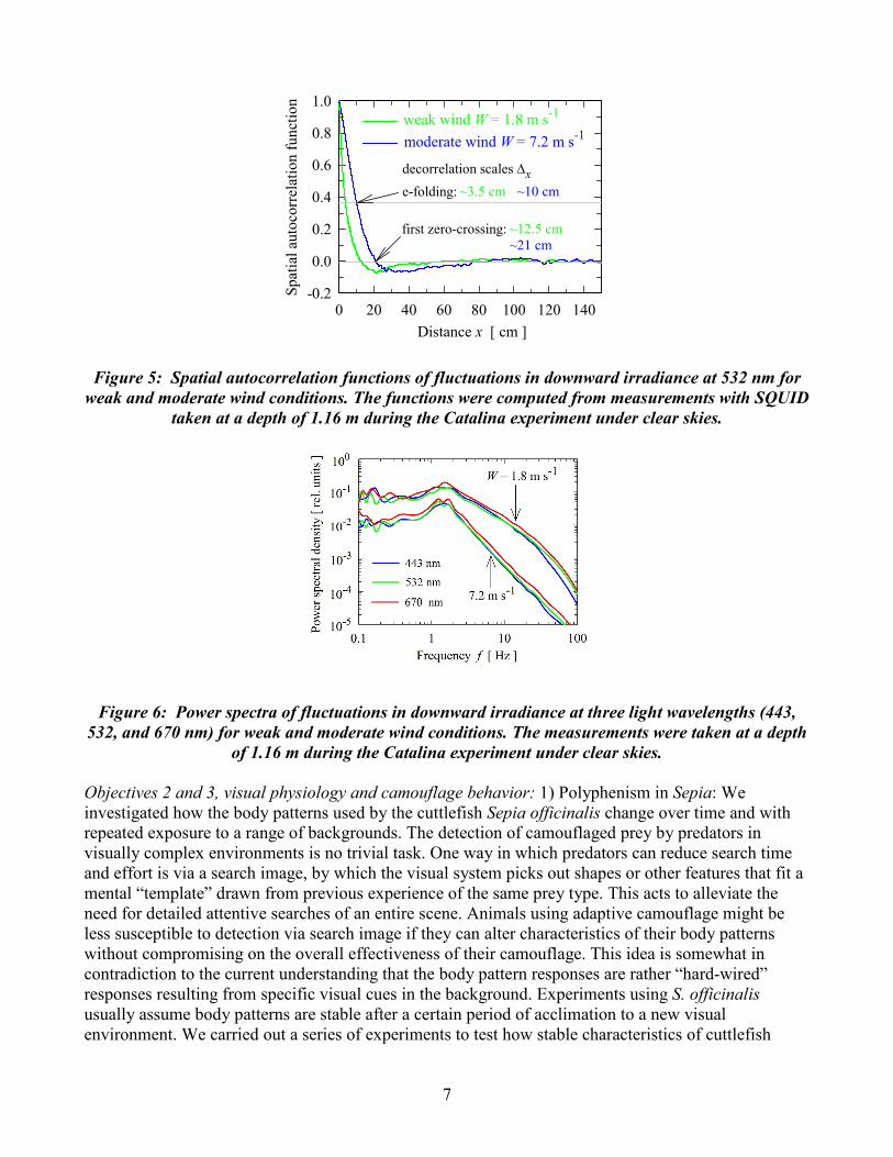
Field measurements: The described instrument above was used throughout the 10 day cruise off the coast of Hawaii to measure reflectance functions for a variety of marine animals, including the small squid Pterygioteuthis microlampas. The majority of measured specimens were captured from net trawls at 800 m. All three-system wavelengths were used to measure the BRDF of multiple points on each specimen, at over 150 different incident illumination angles. The UCSB – University of Pennsylvania team of Profs. Morse and Sweeney report 2 fundamental new biophotonic discoveries this year: (1) Discovered unifying mechanism of tunable biophotonics: We report the first discovery of a unique population of tunable leucocytes - white reflective cells whose brightness is tuned by neurotransmitter. In both tunable white leucocytes and tunable colored iridocytes, ACh-induced condensation of reflectins increases refractive index, dramatically increasing brightness. Consequent Gibbs-Donnan efflux of water from iridocyte lamellae changes thickness of the Bragg elements thus changing color; shrinkage of the spherical reflectin-containing inclusions in leucocytes simply increases brightness of these scattering white reflectors. The same molecules and mechanisms govern tunable brightness and color of the iridocytes and leucocytes, with the difference in color or whiteness resulting from ultrastructural organization of the reflectin-containing bodies (as parallel lamellae or small intracellular spheres). (2) Discovered novel bifunctional biophotonic system enhancing efficiency of photosynthesis: Reflectin-containing iridocytes of giant clams protect the animals’ algal symbionts from damage by reflecting potentially harmful solar radiation, while enhancing photosynthesis by forward scattering useful frequencies deep into the tissues. RESULTS Objective 1, Light measurements: In this report we present a few, most unique example results of light measurements taken with SQUID instrument during the Catalina experiment. Figure 3 shows that wave focusing can produce instantaneous values of irradiance which exceed the time-average irradiance more than 30-fold at shallow depths of about 1 m under sunny conditions. To our knowledge, the occurrence of such high concentrations of solar energy in natural aquatic environments has never been demonstrated before. Figure 4 shows measurements taken with 1 kHz sampling rate during a 1 s period simultaneously with eleven SQUID sensors positioned at different locations along a linear array of the instrument. This graph shows a unique capability of SQUID to provide spatial and temporal information about wave-induced light fluctuations at time scales as short as milliseconds and spatial scales as small as centimeters. This capability is essential for resolving spatial and temporal scales of wave-induced light fluctuations at shallow depths under sunny conditions. We see, for example, that a propagation of a specific focusing event, whose duration at a given point in space is on the order of tens of milliseconds, is observed over a distance > 20 cm. Figure 5 compares the spatial autocorrelation functions of downward irradiance for weak (1.8 m s-1) and moderate (7.2 m s-1) wind conditions. These results indicate that the spatial decorrelation scale is significantly smaller under weak winds when light fluctuations are very strong, compared with higher wind speeds when the fluctuations are weaker. A decrease in the intensity of irradiance fluctuations with increasing wind speed is illustrated in Figure 6. This figure shows a significant reduction in the magnitude of power spectra of irradiance fluctuations at a wind speed of 7.2 m s-1 compared with the wind speed of 1.8 m s-1. In addition, the damping of high-frequency (> 2 Hz) content of fluctuations is more pronounced when the winds are stronger. These results are consistent with the interpretation that an increased roughness of the sea surface at high winds is less efficient in producing focusing events than sea surface waves under weak winds.
6
0 10 20 30 40 50 60
Nor
mal
ized
irra
dian
ce E
d (t)
/ Ed
0
10
20
30
40
30.0 30.2 30.4 30.6 30.8 31.0
Nor
mal
ized
irra
dian
ce E
d (t)
/ Ed
0
10
20
30
40
Time t [ s ]
, , , y, , p
Figure 3: Example 1-min and 1-s time-series of fluctuations in downward irradiance at 532 nm measured at a depth of 1.16 m during the Catalina experiment under clear skies and weak wind.
The downward irradiance is normalized by the time-average value of irradiance.
_
N
orm
aliz
ed ir
radi
ance
Ed
(t) /
E d a
t 532
nm
as m
easu
red
sim
ulta
neou
sly
with
11
SQU
ID se
nsor
slo
cate
d at
diff
eren
t dis
tanc
es x
from
the
first
(ref
eren
ce) s
enso
r
0
4
8
12
16
20
0
4
8
0
4
8
0
4
8
0
4
8
0
4
8
0
4
8
0
4
8
0
4
8
0
4
8
0
4
8
, , y, , p
0 100 200 300 400 500 600 700 800 900 1000
Time [ ms ]
First (reference) sensorx = 0
x = 2 cm
x = 4 cm
x = 8 cm
x = 12 cm
x = 16 cm
x = 20 cm
x = 24 cm
x = 28 cm
x = 36 cm
x = 52 cm
Figure 4: Example 1-s time-series of fluctuations in downward irradiance at 532 nm measured simultaneously with eleven SQUID sensors at a depth of 1.16 m during the Catalina experiment
under clear skies and weak wind. The distance, x, between the sensors along a linear array is indicated.
7
0 20 40 60 80 100 120 140Sp
atia
l aut
ocor
rela
tion
func
tion
-0.2
0.0
0.2
0.4
0.6
0.8
1.0
Distance x [ cm ]
decorrelation scales ∆x
first zero-crossing: ~12.5 cm~21 cm
weak wind W = 1.8 m s-1
moderate wind W = 7.2 m s-1
~3.5 cm ~10 cme-folding:
Figure 5: Spatial autocorrelation functions of fluctuations in downward irradiance at 532 nm for weak and moderate wind conditions. The functions were computed from measurements with SQUID
taken at a depth of 1.16 m during the Catalina experiment under clear skies.
Figure 6: Power spectra of fluctuations in downward irradiance at three light wavelengths (443, 532, and 670 nm) for weak and moderate wind conditions. The measurements were taken at a depth
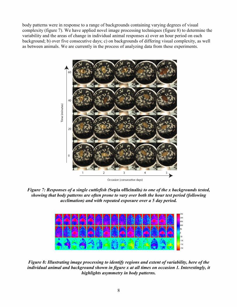
of 1.16 m during the Catalina experiment under clear skies. Objectives 2 and 3, visual physiology and camouflage behavior: 1) Polyphenism in Sepia: We investigated how the body patterns used by the cuttlefish Sepia officinalis change over time and with repeated exposure to a range of backgrounds. The detection of camouflaged prey by predators in visually complex environments is no trivial task. One way in which predators can reduce search time and effort is via a search image, by which the visual system picks out shapes or other features that fit a mental “template” drawn from previous experience of the same prey type. This acts to alleviate the need for detailed attentive searches of an entire scene. Animals using adaptive camouflage might be less susceptible to detection via search image if they can alter characteristics of their body patterns without compromising on the overall effectiveness of their camouflage. This idea is somewhat in contradiction to the current understanding that the body pattern responses are rather “hard-wired” responses resulting from specific visual cues in the background. Experiments using S. officinalis usually assume body patterns are stable after a certain period of acclimation to a new visual environment. We carried out a series of experiments to test how stable characteristics of cuttlefish
8
body patterns were in response to a range of backgrounds containing varying degrees of visual complexity (figure 7). We have applied novel image processing techniques (figure 8) to determine the variability and the areas of change in individual animal responses a) over an hour period on each background; b) over five consecutive days; c) on backgrounds of differing visual complexity, as well as between animals. We are currently in the process of analyzing data from these experiments.
Figure 7: Responses of a single cuttlefish (Sepia officinalis) to one of the x backgrounds tested, showing that body patterns are often prone to vary over both the hour test period (following
acclimation) and with repeated exposure over a 5 day period.
Figure 8: Illustrating image processing to identify regions and extent of variability, here of the individual animal and background shown in figure x at all times on occasion 1. Interestingly, it
highlights asymmetry in body patterns.
9
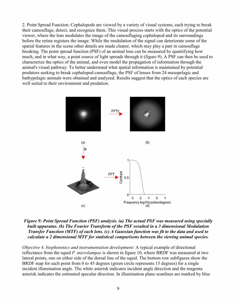
2. Point Spread Function: Cephalopods are viewed by a variety of visual systems, each trying to break their camouflage, detect, and recognize them. This visual process starts with the optics of the potential viewer, where the lens modulates the image of the camouflaging cephalopod and its surroundings before the retina registers the image. While the modulation of the signal can deteriorate some of the spatial features in the scene other details are made clearer, which may play a part in camouflage breaking. The point spread function (PSF) of an animal lens can be measured by quantifying how much, and in what way, a point source of light spreads through it (figure 9). A PSF can then be used to characterize the optics of the animal, and even model the propagation of information through the animal's visual pathway. To better understand what spatial information is maintained by potential predators seeking to break cephalopod camouflage, the PSF of lenses from 24 mesopelagic and bathypelagic animals were obtained and analyzed. Results suggest that the optics of each species are well suited to their environment and predation.
Figure 9: Point Spread Function (PSF) analysis. (a) The actual PSF was measured using specially built apparatus. (b) The Fourier Transform of the PSF resulted in a 3 dimensional Modulation Transfer Function (MTF) of each lens. (c) A Gaussian function was fit to the data and used to calculate a 2 dimensional MTF for statistical comparisons between the viewing animal species.
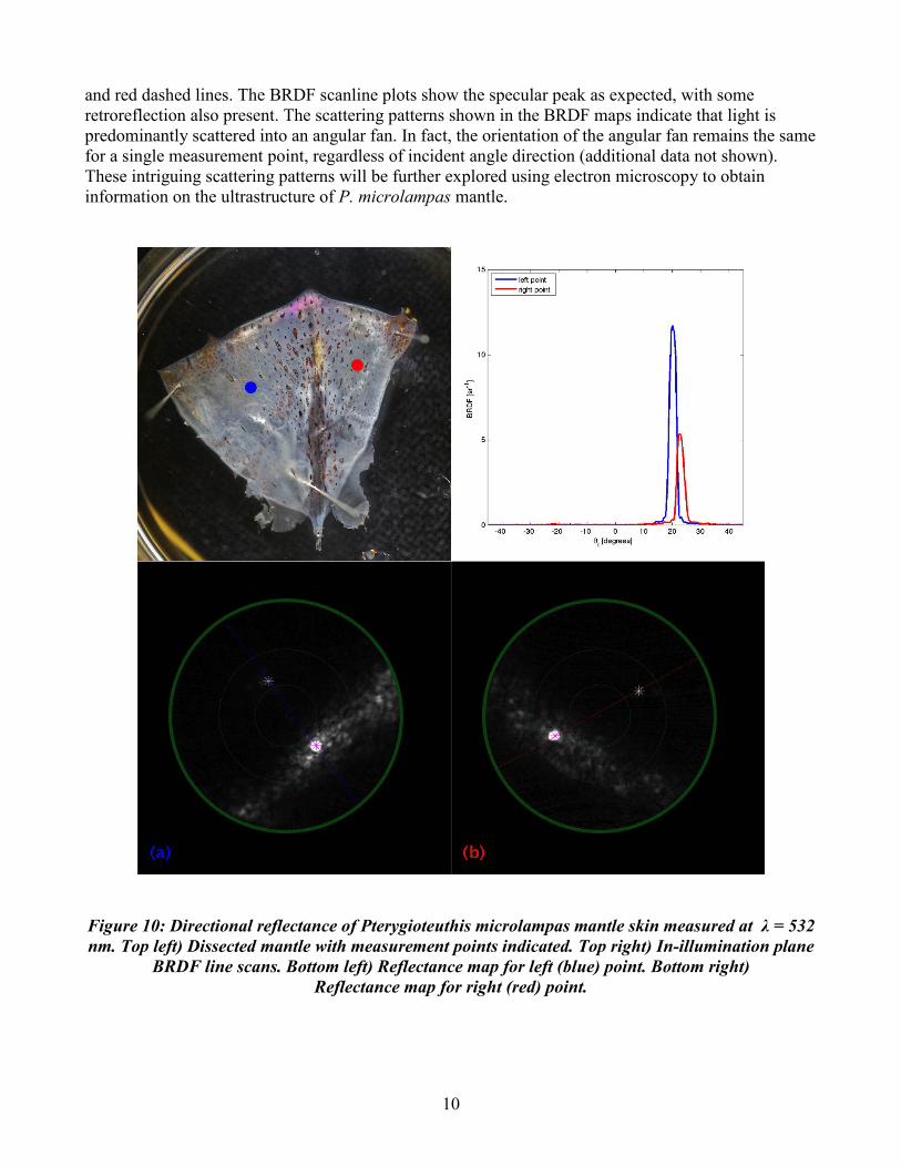
Objective 4, biophotonics and instrumentation development: A typical example of directional reflectance from the squid P. microlampas is shown in figure 10, where BRDF was measured at two lateral points, one on either side of the dorsal line of the squid. The bottom row subfigures show the BRDF map for each point from 0 to 45 degrees (green circle represents 15 degrees) for a single incident illumination angle. The white asterisk indicates incident angle direction and the magenta asterisk indicates the estimated specular direction. In illumination plane scanlines are marked by blue
10
and red dashed lines. The BRDF scanline plots show the specular peak as expected, with some retroreflection also present. The scattering patterns shown in the BRDF maps indicate that light is predominantly scattered into an angular fan. In fact, the orientation of the angular fan remains the same for a single measurement point, regardless of incident angle direction (additional data not shown). These intriguing scattering patterns will be further explored using electron microscopy to obtain information on the ultrastructure of P. microlampas mantle.
Figure 10: Directional reflectance of Pterygioteuthis microlampas mantle skin measured at λ = 532 nm. Top left) Dissected mantle with measurement points indicated. Top right) In-illumination plane
BRDF line scans. Bottom left) Reflectance map for left (blue) point. Bottom right) Reflectance map for right (red) point.
11
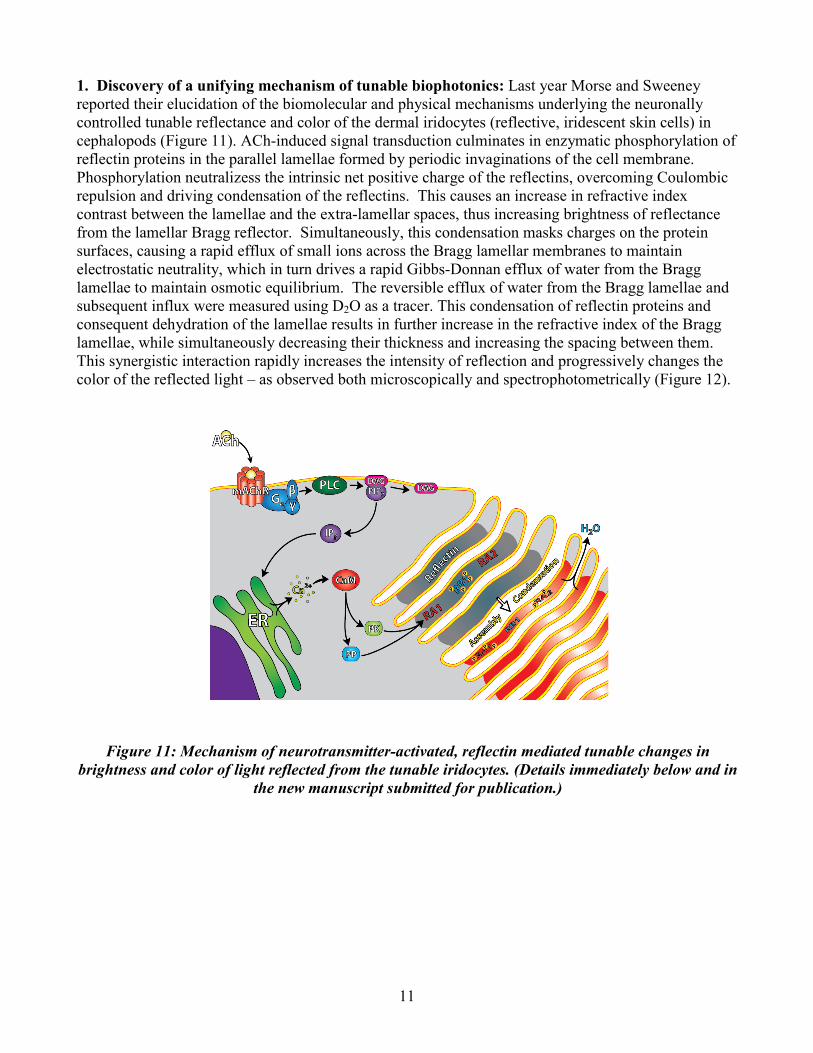
1. Discovery of a unifying mechanism of tunable biophotonics: Last year Morse and Sweeney reported their elucidation of the biomolecular and physical mechanisms underlying the neuronally controlled tunable reflectance and color of the dermal iridocytes (reflective, iridescent skin cells) in cephalopods (Figure 11). ACh-induced signal transduction culminates in enzymatic phosphorylation of reflectin proteins in the parallel lamellae formed by periodic invaginations of the cell membrane. Phosphorylation neutralizess the intrinsic net positive charge of the reflectins, overcoming Coulombic repulsion and driving condensation of the reflectins. This causes an increase in refractive index contrast between the lamellae and the extra-lamellar spaces, thus increasing brightness of reflectance from the lamellar Bragg reflector. Simultaneously, this condensation masks charges on the protein surfaces, causing a rapid efflux of small ions across the Bragg lamellar membranes to maintain electrostatic neutrality, which in turn drives a rapid Gibbs-Donnan efflux of water from the Bragg lamellae to maintain osmotic equilibrium. The reversible efflux of water from the Bragg lamellae and subsequent influx were measured using D2O as a tracer. This condensation of reflectin proteins and consequent dehydration of the lamellae results in further increase in the refractive index of the Bragg lamellae, while simultaneously decreasing their thickness and increasing the spacing between them. This synergistic interaction rapidly increases the intensity of reflection and progressively changes the color of the reflected light – as observed both microscopically and spectrophotometrically (Figure 12).
Figure 11: Mechanism of neurotransmitter-activated, reflectin mediated tunable changes in brightness and color of light reflected from the tunable iridocytes. (Details immediately below and in
the new manuscript submitted for publication.)
12
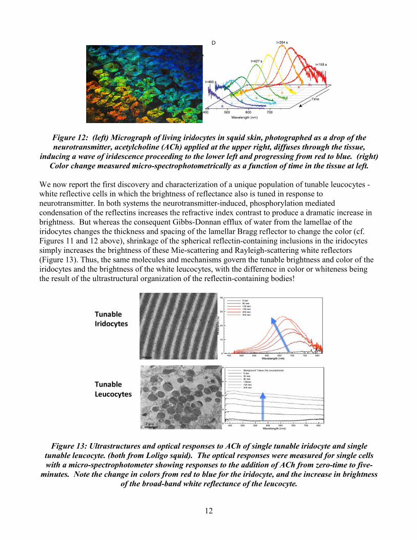
Figure 12: (left) Micrograph of living iridocytes in squid skin, photographed as a drop of the neurotransmitter, acetylcholine (ACh) applied at the upper right, diffuses through the tissue,
inducing a wave of iridescence proceeding to the lower left and progressing from red to blue. (right) Color change measured micro-spectrophotometrically as a function of time in the tissue at left.
We now report the first discovery and characterization of a unique population of tunable leucocytes - white reflective cells in which the brightness of reflectance also is tuned in response to neurotransmitter. In both systems the neurotransmitter-induced, phosphorylation mediated condensation of the reflectins increases the refractive index contrast to produce a dramatic increase in brightness. But whereas the consequent Gibbs-Donnan efflux of water from the lamellae of the iridocytes changes the thickness and spacing of the lamellar Bragg reflector to change the color (cf. Figures 11 and 12 above), shrinkage of the spherical reflectin-containing inclusions in the iridocytes simply increases the brightness of these Mie-scattering and Rayleigh-scattering white reflectors (Figure 13). Thus, the same molecules and mechanisms govern the tunable brightness and color of the iridocytes and the brightness of the white leucocytes, with the difference in color or whiteness being the result of the ultrastructural organization of the reflectin-containing bodies!
Figure 13: Ultrastructures and optical responses to ACh of single tunable iridocyte and single tunable leucocyte. (both from Loligo squid). The optical responses were measured for single cells with a micro-spectrophotometer showing responses to the addition of ACh from zero-time to five-
minutes. Note the change in colors from red to blue for the iridocyte, and the increase in brightness of the broad-band white reflectance of the leucocyte.
13
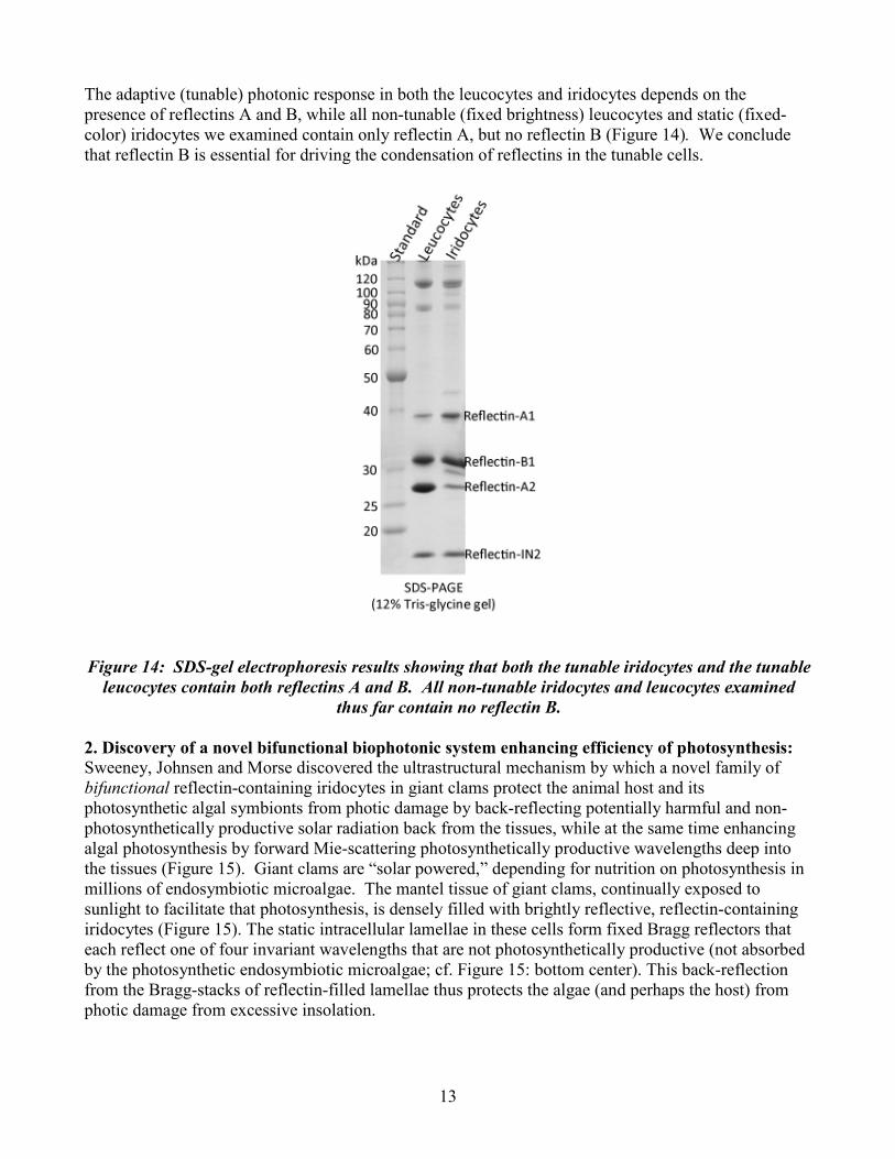
The adaptive (tunable) photonic response in both the leucocytes and iridocytes depends on the presence of reflectins A and B, while all non-tunable (fixed brightness) leucocytes and static (fixed-color) iridocytes we examined contain only reflectin A, but no reflectin B (Figure 14). We conclude that reflectin B is essential for driving the condensation of reflectins in the tunable cells.
Figure 14: SDS-gel electrophoresis results showing that both the tunable iridocytes and the tunable leucocytes contain both reflectins A and B. All non-tunable iridocytes and leucocytes examined
thus far contain no reflectin B.
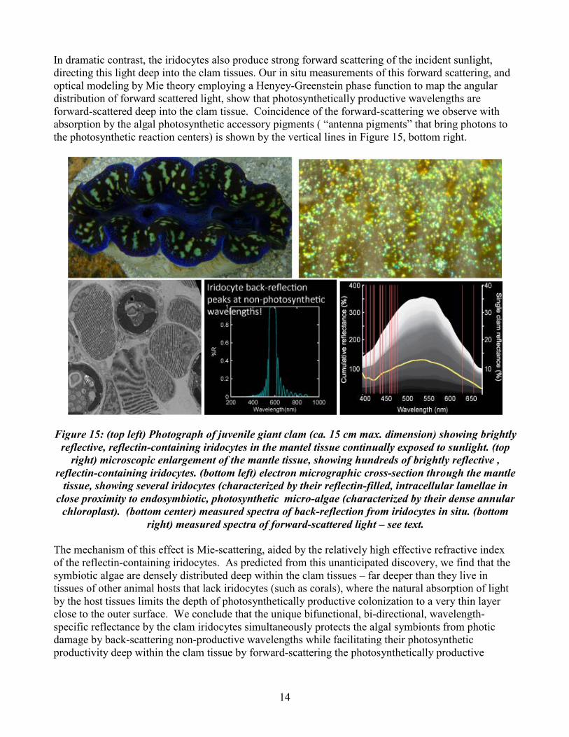
2. Discovery of a novel bifunctional biophotonic system enhancing efficiency of photosynthesis: Sweeney, Johnsen and Morse discovered the ultrastructural mechanism by which a novel family of bifunctional reflectin-containing iridocytes in giant clams protect the animal host and its photosynthetic algal symbionts from photic damage by back-reflecting potentially harmful and non-photosynthetically productive solar radiation back from the tissues, while at the same time enhancing algal photosynthesis by forward Mie-scattering photosynthetically productive wavelengths deep into the tissues (Figure 15). Giant clams are “solar powered,” depending for nutrition on photosynthesis in millions of endosymbiotic microalgae. The mantel tissue of giant clams, continually exposed to sunlight to facilitate that photosynthesis, is densely filled with brightly reflective, reflectin-containing iridocytes (Figure 15). The static intracellular lamellae in these cells form fixed Bragg reflectors that each reflect one of four invariant wavelengths that are not photosynthetically productive (not absorbed by the photosynthetic endosymbiotic microalgae; cf. Figure 15: bottom center). This back-reflection from the Bragg-stacks of reflectin-filled lamellae thus protects the algae (and perhaps the host) from photic damage from excessive insolation.
14
In dramatic contrast, the iridocytes also produce strong forward scattering of the incident sunlight, directing this light deep into the clam tissues. Our in situ measurements of this forward scattering, and optical modeling by Mie theory employing a Henyey-Greenstein phase function to map the angular distribution of forward scattered light, show that photosynthetically productive wavelengths are forward-scattered deep into the clam tissue. Coincidence of the forward-scattering we observe with absorption by the algal photosynthetic accessory pigments ( “antenna pigments” that bring photons to the photosynthetic reaction centers) is shown by the vertical lines in Figure 15, bottom right.
Figure 15: (top left) Photograph of juvenile giant clam (ca. 15 cm max. dimension) showing brightly
reflective, reflectin-containing iridocytes in the mantel tissue continually exposed to sunlight. (top right) microscopic enlargement of the mantle tissue, showing hundreds of brightly reflective ,
reflectin-containing iridocytes. (bottom left) electron micrographic cross-section through the mantle tissue, showing several iridocytes (characterized by their reflectin-filled, intracellular lamellae in
close proximity to endosymbiotic, photosynthetic micro-algae (characterized by their dense annular chloroplast). (bottom center) measured spectra of back-reflection from iridocytes in situ. (bottom
right) measured spectra of forward-scattered light – see text. The mechanism of this effect is Mie-scattering, aided by the relatively high effective refractive index of the reflectin-containing iridocytes. As predicted from this unanticipated discovery, we find that the symbiotic algae are densely distributed deep within the clam tissues – far deeper than they live in tissues of other animal hosts that lack iridocytes (such as corals), where the natural absorption of light by the host tissues limits the depth of photosynthetically productive colonization to a very thin layer close to the outer surface. We conclude that the unique bifunctional, bi-directional, wavelength-specific reflectance by the clam iridocytes simultaneously protects the algal symbionts from photic damage by back-scattering non-productive wavelengths while facilitating their photosynthetic productivity deep within the clam tissue by forward-scattering the photosynthetically productive
15
wavelengths. Application of these principles for the enhancement of photosynthesis in other systems (e.g., for production of bio-fuels) is under investigation. IMPACT/APPLICATIONS
The systems evolved by marine animals in order to hunt, hide, and mate over hundreds of million years surpass our contemporary engineering designs for underwater vehicles. Hiding and hunting are natural tasks for our military and we believe that valuable clues will be provided by the results of our studies. The impact will hopefully affect all branches of the armed forces that have aquatic missions. This includes Special Forces, mine hunting vehicles, the submarine community, and a newest generation of underwater vehicles that could all benefit from the option of “stealth”. Since visual methods play an important role in the mission profiles of all of these groups, the ability to enhance and hide from detection should be an important payoff. TRANSLATIONS (1) Electrically switchable, polymer-based shutters for IR detectors: As described in our report, we discovered the molecular structures and mechanisms responsible for the dynamically tunable reflectance in skin cells of the squid, and are now working with Raytheon Vision Systems Inc. (with support from ARL and DARPA) to translate these finding to develop a prototype electrically switchable, polymer-based shutter for infrared detectors. Using solution-processable conjugated polymers, we developed working prototypes that are activated by low voltage (2-3 V) to display significant changes in absorption and reflection in the IR. As the polymer-based materials transition from semiconducting to conducting, free carriers and conformational changes absorb and scatter broad bands of infrared radiation. The resulting change in refractive index resulting from the simultaneous production of absorbing species and their increased density closely parallels the synergistic simultaneous changes in the reflectin-based Bragg layers that provide the high gain exhibited by the biological system. Defense applications include noiseless IR shutters for forward Special Forces operations, graded neutral density and tunable hyperspectral IR filters, apertures, and lightweight coded apertures for IR image formation without a lens. (2) Broadband, omnidirectional IR reflectors: Also as described above, we recently discovered that the silver, broadband reflective tissue surrounding the eyes of the squid (providing omnidirectional camouflage of that structure) is composed of a unique array of reflectin-filled, spindle-shaped cells densely packed together to form an unusual, quasi-disordered, “distributed Bragg reflector.” The optical contrast between the high refractive index within these reflectin-packed cells and the low refractive index in the extracellular medium is responsible for the very high reflectivity of the tissue, while the infinite number of spacings between the nested, tapered cells in the quasi-disordered array is responsible for the broadband (i.e., multi-wavelength, silver) and omnidirectional reflection. Translating the underlying principles found in this biological broadband reflector (in research supported by ARO and Acumen, Inc.), we produced prototype broadband reflective coatings by evaporative self-assembly of asymmetric polymer rods to form quasi-disordered and “distributed” Bragg reflectors. The dimensions of the quasi-ordered polystyrene rods are sufficiently large to ensure that reflectance occurs in the IR, while the random quasi-disorder of the film ensures omnidirectionality of the reflectance. Silica and silicone casts of these organic films retain the optical properties in a rugged form suitable for device manufacture. Because the biologically inspired fabrication process is facile, error-tolerant and inexpensive and can be scaled to larger, flexible and curved surfaces; because silica and silicone casts preserve the desired optical features in a robust form
16
suitable for manufacture of coatings; and because the optical and IR properties of those coating can be readily tuned, they offer numerous applications of potential importance to ONR and other branches of DoD. RELATED PROJECTS
"Bioinspired Dynamically Tunable Polymer-Based Filters for Multi-Spectral Infrared Imaging"; DARPA; W911NF-08-1-0494; $150,000; 10-01/08-09/30/09. This work represents a "translation" of what we learned from the biomolecular mechanisms governing dynamically tunable reflectance in cephalopods to novel routes for synthetic optical materials. Performed in collaboration with Raytheon, Inc. This funding has ended; proposal for continuation is pending. “Bio-inspired Visual Information Processing and Dynamically Tunable Multispectral IR Detection: Learning from the Octopus.” ARL/ARO; W911NF-09-D-0001; $200,000; 1/1/09-12/31/09. To D.E. Morse and R. N. Hanlon. This work represents a "translation" of what we learned from the biomolecular mechanisms governing dynamically tunable reflectance in cephalopods to novel routes for synthetic optical materials. This funding has ended. ”Bio-Inspired Photonics: Polymer-Based, Dynamically Tunable Multi-Spectral Filters for IR Detection”; DARPA; proposal pending for continuation of effort described immediately above. This work represents a "translation" of what we learned from the biomolecular mechanisms governing dynamically tunable reflectance in cephalopods to novel routes for synthetic optical materials. PUBLICATIONS DeMartini, D. G., D. Krogstad and D. E. Morse (in prep). Dynamic biophotonics: Membrane
invaginations facilitate water fluxes driving tunable iridescence.
DeMartini, D.G., A. Ghoshal, E. Eck and D.E. Morse (in prep). Discovery of tunable leucocytes, and a unified mechanism governing tunable biophotonics in squid.
DeMartini, D. G., A.M. Sweeney, and D.E. Morse (in prep). Phosphorylation of native and recombinant squid skin reflectins in vivo and in vitro.
Gagnon, Y. and Johnsen, S. (in prep.). Visual acuity in pelagic fish and mollusks Journal of Experimental Biology.
Ghoshal, A., E. Eck, D. Martini and D.E. Morse (in prep). Calculation of biophotonic Bragg reflector characteristics from subcellular micro-spectrophotometric analyses.
Haag, J. M., Sweeney, A. M., and Jaffe, J. S. (in prep). Measurement system for marine animal reflectance functions.
Haag, J. M., Sweeney, A. M., and Jaffe, J. S. (2012). Measurement system for obtaining marine animal reflectance functions. Proceedings of Ocean Optics XXI Conference, Glasgow, Scotland, United Kingdom, October 8-12, 2012.
Holt, A.L., Wehner, J.G.A., Hampp, A., and Morse, D.E. (2010). Plastic transmissive infrared electrochromic devices. Macromolecular Chemistry and Physics 211:1701-1707.
17
Holt, A.L.., Sweeney, A.M., Johnsen, S., and Morse, D.E. (2011). A highly-distributed Bragg stack with unique geometry provides effective camouflage for Loligo squid eyes. Journal of the Royal Society: Interface 8, 1386-1399.
Izumi, M., A. M. Sweeney, D. G. DeMartini, J. C. Weaver, M. L. Powers, A.R. Tao, T. V. Silvas, R. M. Kramer, W. J. Crookes-Goodson, L. M. Mäthger, R. R. Naik, R. T. Hanlon and Morse, D. E. (2010). Changes in reflectin protein phosphorylation are associated with dynamic iridescence in squid. Journal of the Royal Society: Interface 7: 549-560.
Jaffe, J. S., Simonet, F., Laxton, B., Roberts, P. L. D., Zylinski, S., Johnsen, S., and A. Sweeney (in review). Omni-Cam and the Sub Sea Holodeck: Systems for recording in-situ radiance and simulating underwater optical environments in the lab. Mar. Tech. Soc. J.
Jaffe, J. S., Laxton, B., Zylinski, S. (2011). Omni-Cam and the Sub Sea Holodeck: Systems for recording in-situ radiance and simulating underwater optical environments in the lab. Proc. IEEE Oceans Conf., Santander, Spain June 6-9, 2011.
Johnsen, S., Marshall, N. J., and Widder, E. A. (2011). Polarization sensitivity as a contrast enhancer in pelagic predators: Lessons from in situ polarization imaging of transparent zooplankton. Philosophical Transactions of the Royal Society of London, Series B. 366: 655–670.
Li, Z., Zhang, Y., Holt, A.L., Kolasa, B.P., Wehner, J.G., Hampp, A., Bazan, B.C., Nguyen, T., and Morse, D.E. Electrochromic devices and thin film transistors from a new family of ethylenedioxythiophene based conjugated polymers. New Journal of Chemistry 35: 1327-1334.
Marshall, N. J., and Johnsen, S. (2011). Camouflage in Marine fish. In Animal Camouflage: Current issues and new perspectives. Cambridge University Press: Cambridge UK.
Nilsson, D. E., Warrant, E. J., Johnsen, S., Hanlon, R. T., and N. Shashar (2012). A unique advantage for giant eyes in giant squid. Current Biology 22, 683-688.
Sawicka, E., D. Stramski, M. Darecki, and J. Dubranna, 2012b. Power spectral analysis of wave-induced fluctuations in downward irradiance within the near-surface ocean under sunny conditions. Proceedings of Ocean Optics XXI, Glasgow.
Sweeney, A., A. Holt, S. Johnsen and D.E. Morse (in prep). Bifunctional biophotonic system supports photosynthesis in Tridacnid giant clam symbiosis.
Sweeney, A., A. Holt and D.E. Morse (in prep). Natural camouflage in the infrared? Predators and prey in the ocean’s far-red Raman glow.
Tao, A.R., D. G. DeMartini, M. Izumi, A. M. Sweeney, and Morse, D. E. (2010). The role of protein assembly in dynamically tunable bio-optical tissues. Biomaterials 31:793-801
Zylinski, S., and Osorio, D. (2011). What can camouflage tell us about non-human visual perception? A case study of the cuttlefish. In Animal Camouflage: Current issues and new perspectives. Cambridge University Press: Cambridge UK.
Zylinski, S. How, M., Osorio, D., Hanlon, R. T., and Marshall, N. J. (2011). To be seen or to hide: visual characteristics of body patterns for camouflage and communication in the Australian giant cuttlefish, Sepia apama. American Naturalist.
Zylinski, S. and S. Johnsen (2012) Camouflage without compromise: Mesopelagic cephalopods switch between transparency and pigmentation to optimize camouflage in the deep. Current Biology 21, 1937-1941.
18
Zylinski, S., A.-S. Darmaillacq, & N. Shashar (2012). Visual interpolation for contour completion by the European cuttlefish (Sepia officinalis) and its use in dynamic camouflage. Proceedings of the Royal Society B: Biological Sciences 276 (1675): 3963-3969.
Zylinski, S. and S. Johnsen (in press). Visual cognition in deep-sea cephalopods: what we don’t know and why we don’t know it. In: Cephalopod Cognition, Editors: S. Darmaillacq and J. Mather.
Zylinski, S. and D. Osorio (in press). Cuttlefish camouflage: Vision and cognition. In: Cephalopod Cognition, Editors: S. Darmaillacq and J. Mather.
Zylinski, S., Gagnon, Y., Gross, T., Wheeler, J. and Johnsen, S. (in prep.). Octopus bimaculoides substrate choice: hierarchy and interactions between visual and tactile information.


![OPTICS] Optical Camouflage - Electronics Makerelectronicsmaker.com/em/admin/pdfs/free/Optical.pdfoptical camouflage is a part of Active camouflage (or Adaptive camouflage) is a group](https://img.pdfslide.us/doc/110x75/5f01e08f7e708231d40178cf/optics-optical-camouflage-electronics-m-optical-camouflage-is-a-part-of-active.jpg)