Embed Size (px)
Citation preview
CCRN Review:
Pulmonary
Audrey Roberson, MS, RN, CPAN, CNS-BC Nurse Manager, Medical Respiratory Intensive Care Unit
Virginia Commonwealth University Health System

Please make sure all phones and pagers are switched to mute or vibrate!

Objectives
At the end of this presentation, the participants will:
Apply knowledge of pulmonary physiology and arterial blood gases to
collaboratively manage acute and chronic pulmonary disorders, with and without mechanical ventilation.
Differentiate acute hypoxic pulmonary failures (Pulmonary Embolis, ARDs, Pneumonia, Airleaks) and determine collaborative management strategies for each.
Describe collaborative interventions for managing patients with airway disorders (COPD, Asthma, Emphysema).
Relate nursing interventions for thoracic traumas/surgeries and pulmonary hypertension.
Test Plan
Acute Lung Injury ARDS
Acute Pulmonary Embolus
Acute Respiratory Failure
Acute Respiratory Infections Pneumonia
Bronchiolitis
Air-leak Syndromes
Aspiration Pneumonia
COPD, Asthma, Chronic Bronchitis, Emphysema
Pulmonary Hypertension
Status Asthmaticus
Thoracic Surgery
Thoracic Trauma Fractured Ribs
Lung Contusions
Tracheal Perforation
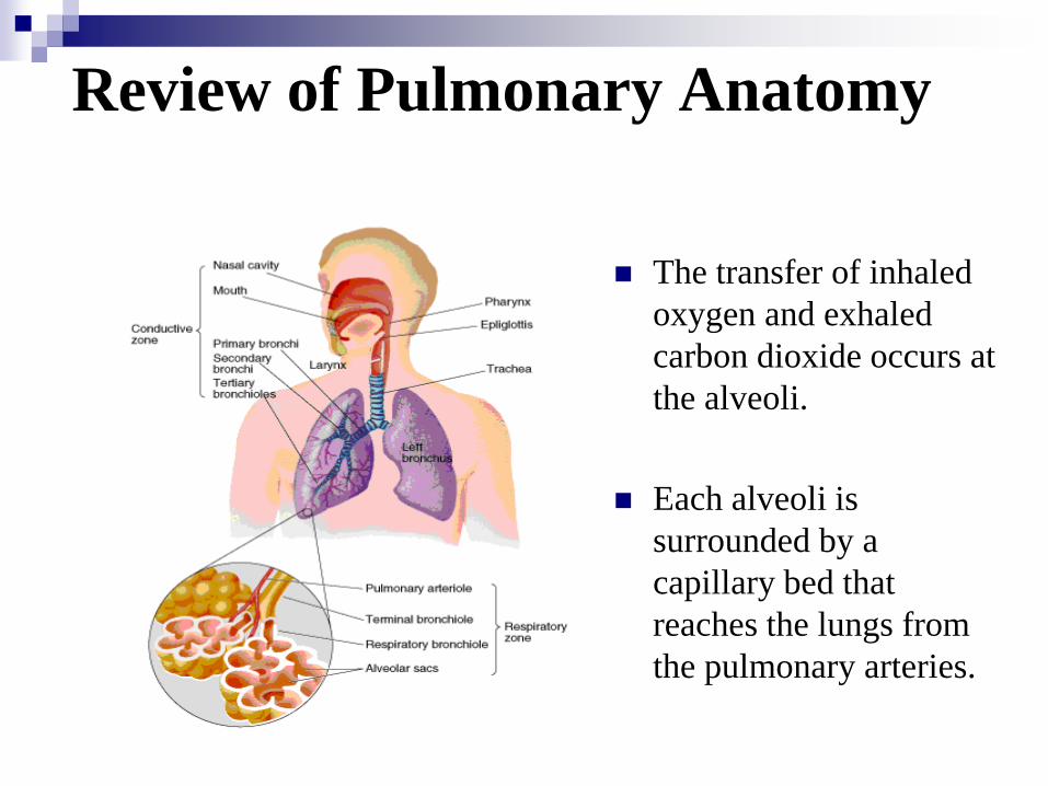
Review of Pulmonary Anatomy
The transfer of inhaled
oxygen and exhaled
carbon dioxide occurs at
the alveoli.
Each alveoli is
surrounded by a
capillary bed that
reaches the lungs from
the pulmonary arteries.
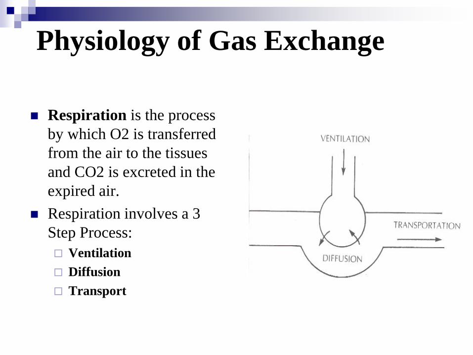
Physiology of Gas Exchange
Respiration is the process
by which O2 is transferred
from the air to the tissues
and CO2 is excreted in the
expired air.
Respiration involves a 3
Step Process:
Ventilation
Diffusion
Transport

Control of Breathing
Respiratory pacemaker is located in medulla Generates rhythmic cycle
Breathing is spontaneous, but becomes irregular if input from the pons is disrupted.
Chemoreceptors Oxygen receptors are located in carotid / aortic bodies
PaO2 must be <60 to activate
Carbon Dioxide receptors located in the medulla are the main respiratory regulators. PaCO2 > 70-80 can depress CNS
Work of Breathing
The amount of effort required to maintain a given level of ventilation.
Determined by:
Lung Compliance - Measure of elasticity of the lungs and thorax.
Airway Resistance - The opposition to gas flow in the airways. Mainly due to diameter of airways.
Small changes in diameter produce large changes in resistance.
Autonomic nervous system and inflammatory mediators affect
resistance: Parasympathetic
Sympathetic
Histamine

Oxygen Transport
Oxygen is carried in the
blood in two ways:
Bound to hemoglobin in
RBC’s (SaO2)
Dissolved in plasma
(PaO2)
Oxyhemoglobin dissociation
curve
Shows the relationship
between O2 saturation
and PaO2.
Describes the ability of
hemoglobin to bind to O2

Carbon Dioxide Transport
Carried in the blood in three ways:
Dissolved in the plasma (PaCO2)
Chemically combined with hemoglobin
As bicarbonate through a conversion reaction:
CO2 +H20 H2CO3 H + HCO3
KEY CONCEPT: The amount of CO2 in
the plasma determines the acidity of the
blood.
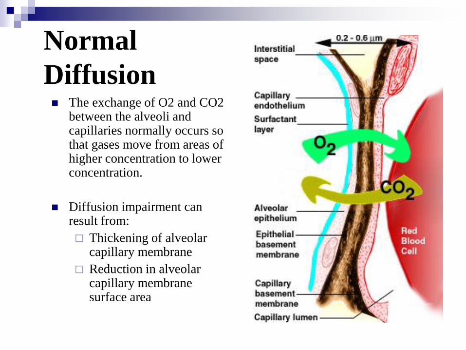
Normal
Diffusion The exchange of O2 and CO2
between the alveoli and capillaries normally occurs so that gases move from areas of higher concentration to lower concentration.
Diffusion impairment can result from:
Thickening of alveolar capillary membrane
Reduction in alveolar capillary membrane surface area
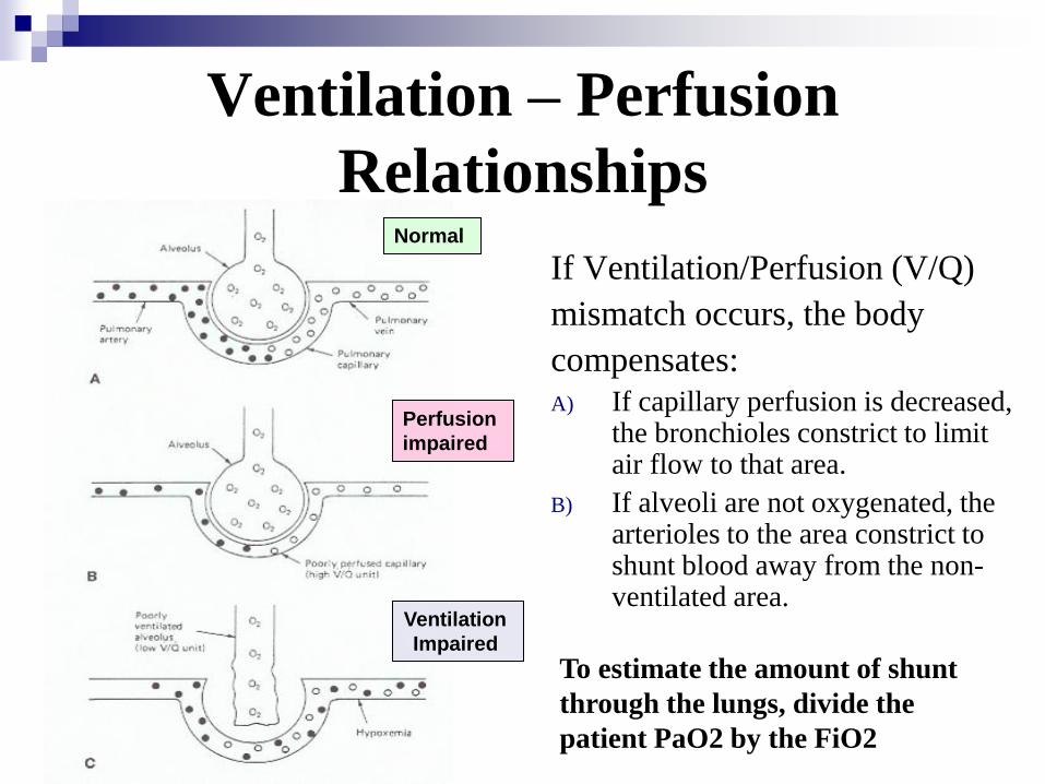
Ventilation – Perfusion
Relationships
If Ventilation/Perfusion (V/Q)
mismatch occurs, the body
compensates:
A) If capillary perfusion is decreased, the bronchioles constrict to limit air flow to that area.
B) If alveoli are not oxygenated, the arterioles to the area constrict to shunt blood away from the non-ventilated area.
Normal
Perfusion
impaired
Ventilation
Impaired
To estimate the amount of shunt
through the lungs, divide the
patient PaO2 by the FiO2
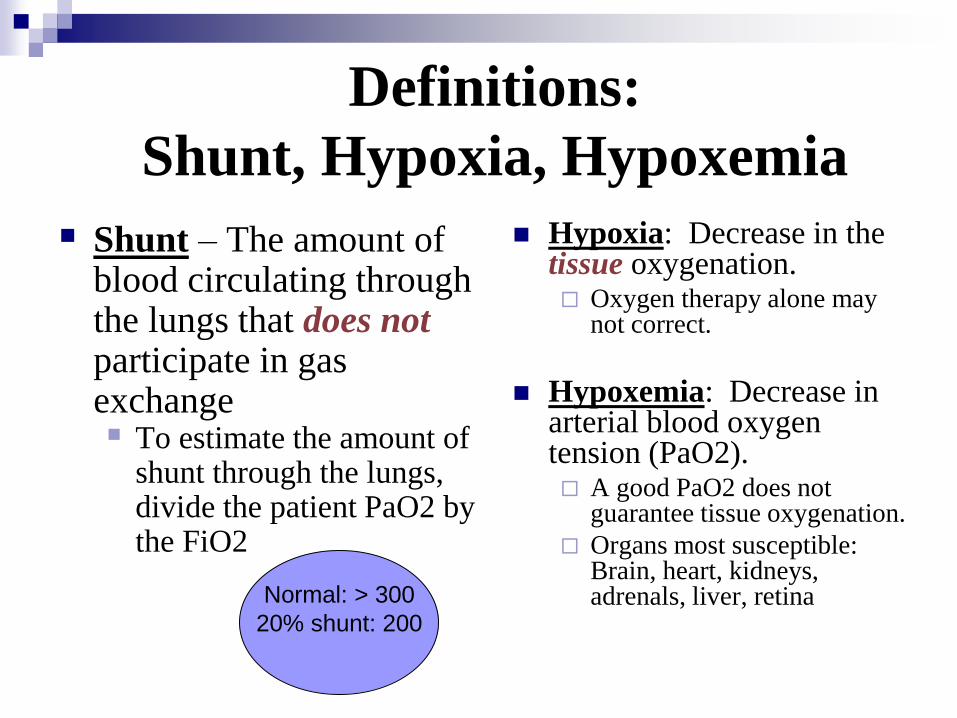
Definitions:
Shunt, Hypoxia, Hypoxemia Hypoxia: Decrease in the
tissue oxygenation. Oxygen therapy alone may
not correct.
Hypoxemia: Decrease in arterial blood oxygen tension (PaO2). A good PaO2 does not
guarantee tissue oxygenation.
Organs most susceptible: Brain, heart, kidneys, adrenals, liver, retina
Shunt – The amount of blood circulating through the lungs that does not participate in gas exchange To estimate the amount of
shunt through the lungs, divide the patient PaO2 by the FiO2
Normal: > 300
20% shunt: 200
Arterial Blood Gases
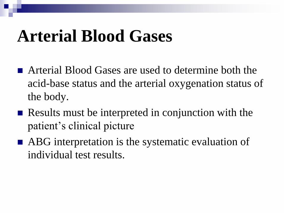
Arterial Blood Gases
Arterial Blood Gases are used to determine both the
acid-base status and the arterial oxygenation status of
the body.
Results must be interpreted in conjunction with the
patient’s clinical picture
ABG interpretation is the systematic evaluation of
individual test results.

Acid - Base Balance
The body pH must remain within normal limits or the
body will die.
The respiratory and metabolic systems work together
to maintain balance
The respiratory system begins to make adjustments
immediately when there are imbalances.
The metabolic system may take days to adjust to
imbalances.

pH
The pH of blood is a measurement of the
concentration of hydrogen ions in the plasma.
Normal range: 7.35 – 7.45 (mean 7.40)
If a patient’s pH is below 7.35, the patient is
experiencing acidosis.
If a patient’s pH is above 7.45, the patient is
experiencing alkalosis.
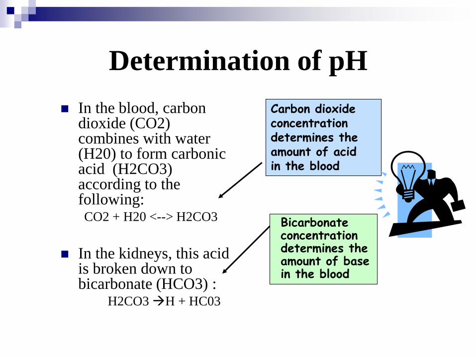
Determination of pH
In the blood, carbon dioxide (CO2) combines with water (H20) to form carbonic acid (H2CO3) according to the following: CO2 + H20 <--> H2CO3
In the kidneys, this acid is broken down to bicarbonate (HCO3) :
H2CO3 H + HC03
Carbon dioxide concentration determines the amount of acid in the blood
Bicarbonate concentration determines the amount of base in the blood

Respiratory Component:
CO2 The CO2 level of the blood is controlled by the respiratory
system.
Normal range is 35-45 mmHg When the PaCO2 is below 40, there is LESS CO2 to form
acid. This occurs when the patient hyperventilates or blows off
CO2.
The patient becomes alkalotic
When the PaCo2 is above 40, there is MORE CO2 to form acid.
This occurs when the patient is hypoventilated.
The patient becomes acidotic.

Metabolic Component:
HC03 The amount of bicarbonate ion, HCO3, is controlled by the
kidney.
Normal range is 22 –26 mEq/l
When HCO3 is above 24, there is MORE base.
This occurs when the kidneys retain more bicarbonate ion
The patient becomes alkalotic
When HCO3 is below 24, there is LESS base.
This occurs when bicarbonate ion is excreted by the kidney or lost through other sources
The patient becomes acidotic

Steps of ABG Interpretation
Step I – Determine oxygenation
PaO2 is the partial pressure of oxygen dissolved in arterial blood. It reflects only 3% of the total oxygen in the blood.
Normal level : 80 –100 mmHg.
SaO2 is the measure of oxygen bound to hemoglobin.
Normal SaO2 is 95% or greater on room air
Special Considerations:
Normal PaO2 is decreased in the elderly and neonates.
Panic PaO2 at any age: Below 40
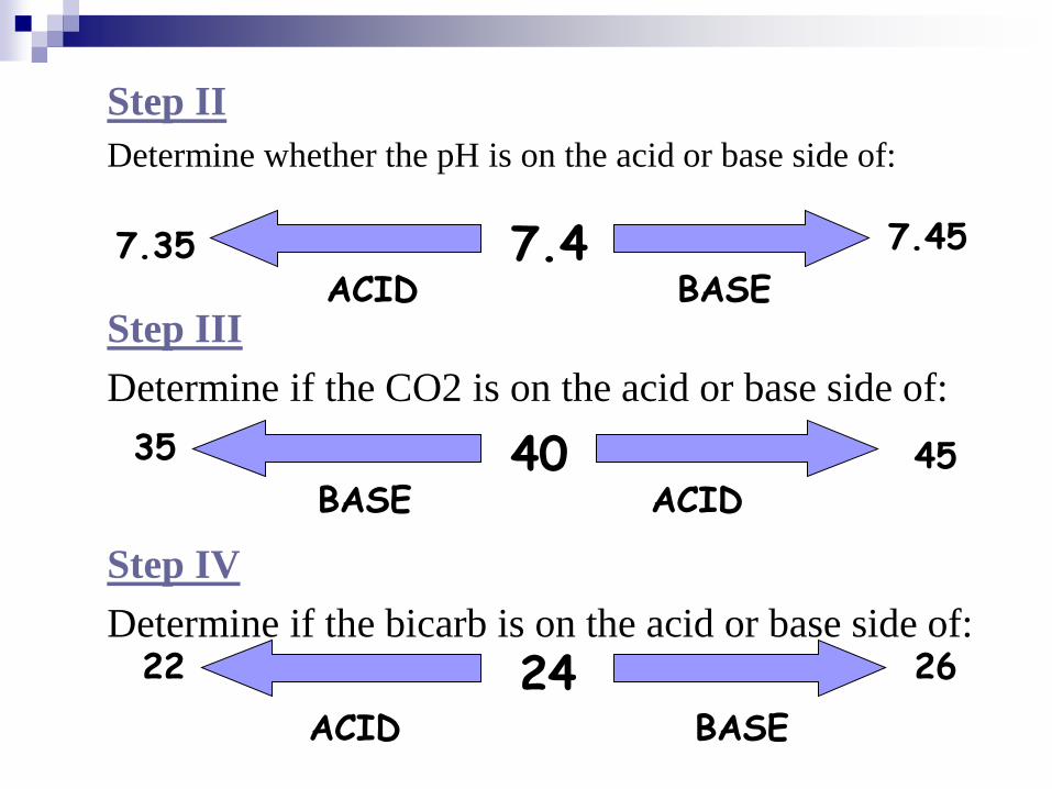
Step II
Determine whether the pH is on the acid or base side of:
Step III
Determine if the CO2 is on the acid or base side of:
Step IV
Determine if the bicarb is on the acid or base side of:
ACID BASE 7.35 7.45 7.4
40 ACID BASE
35 45
24 BASE ACID
26 22

Step V: Match it! The component that matches the
PH is the system controlling the ABG!
Acidosis:
If CO2 is elevated, the pH is under
respiratory control
If HCO3 is low, the pH is under
metabolic control
Alkalosis:
If CO2 is low, the pH is under respiratory control
If HCO3 is elevated, the pH is under metabolic control
If both systems match the pH,
the patient is having problems with both systems!
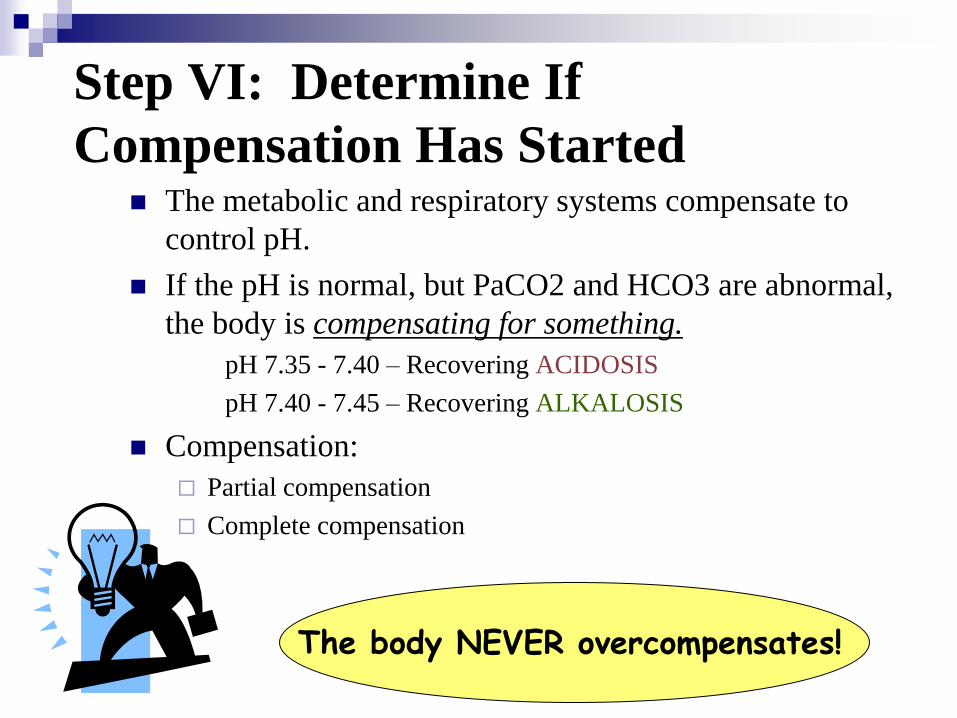
Step VI: Determine If
Compensation Has Started The metabolic and respiratory systems compensate to
control pH.
If the pH is normal, but PaCO2 and HCO3 are abnormal,
the body is compensating for something.
pH 7.35 - 7.40 – Recovering ACIDOSIS
pH 7.40 - 7.45 – Recovering ALKALOSIS
Compensation:
Partial compensation
Complete compensation
The body NEVER overcompensates!
ACID BASE 7.35 7.45 7.4
40 ACID BASE
35 45
24 BASE ACID
26 22
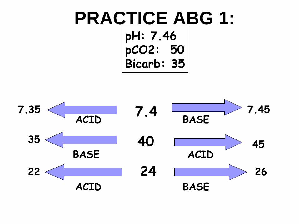
PRACTICE ABG 1:
pH: 7.46 pCO2: 50 Bicarb: 35

ACID BASE 7.35 7.45 7.4
40 ACID BASE
35 45
24 BASE ACID
26 22
PRACTICE ABG 2:
pH 7.24 PaCO2 60 HCO3 30
ACID BASE 7.35 7.45 7.4
40 ACID BASE
35 45
24 BASE ACID
26 22
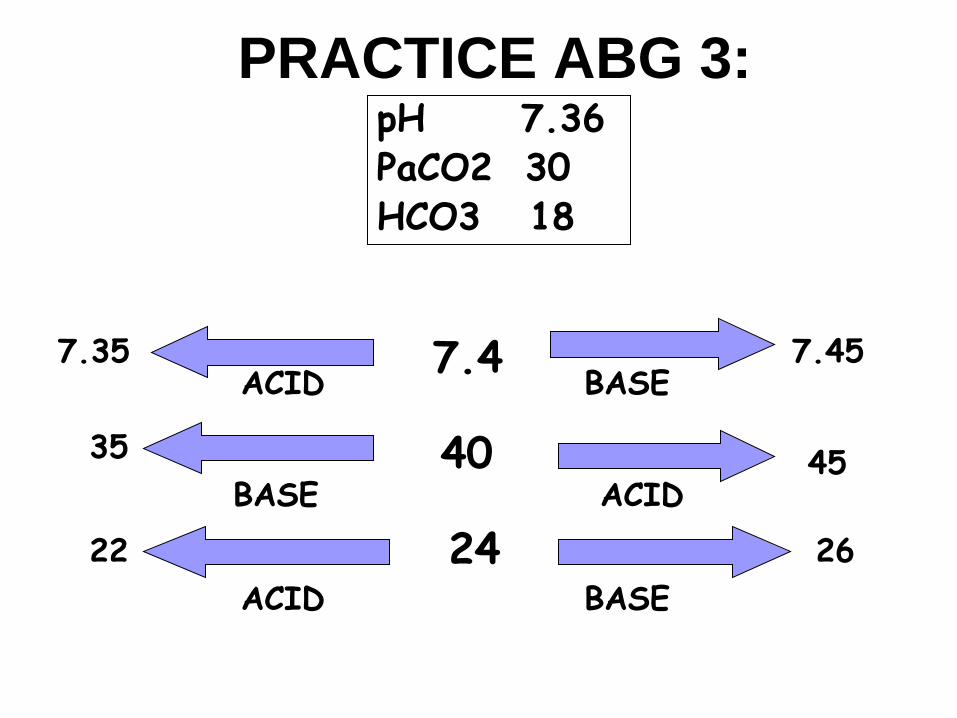
PRACTICE ABG 3:
pH 7.36 PaCO2 30 HCO3 18

ACID BASE 7.35 7.45 7.4
40 ACID BASE
35 45
24 BASE ACID
26 22
PRACTICE ABG 4:
pH: 7.44 pCO2: 29 Bicarb: 19
ACID BASE 7.35 7.45 7.4
40 ACID BASE
35 45
24 BASE ACID
26 22
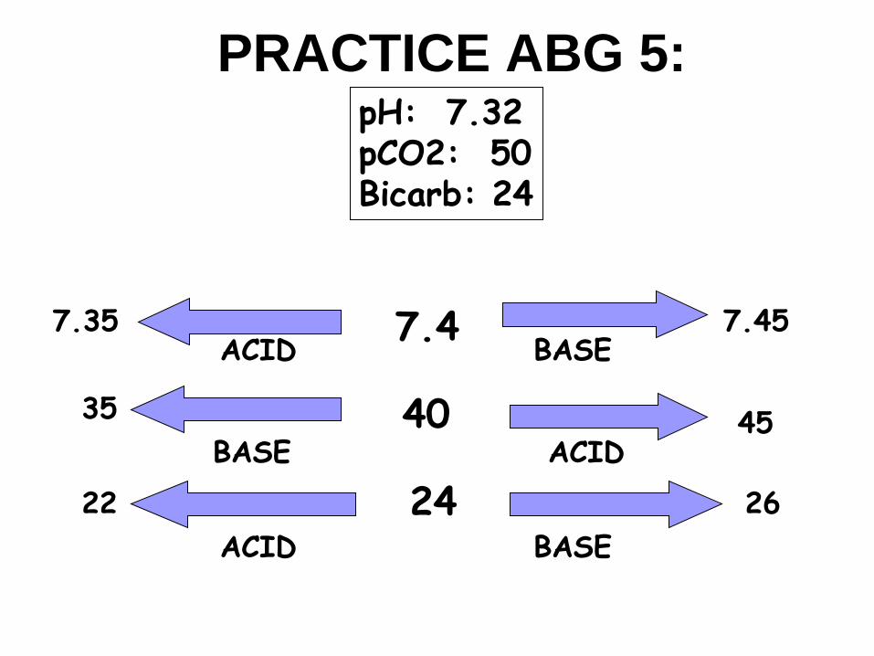
PRACTICE ABG 5:
pH: 7.32 pCO2: 50 Bicarb: 24
ACID BASE 7.35 7.45 7.4
40 ACID BASE
35 45
24 BASE ACID
26 22
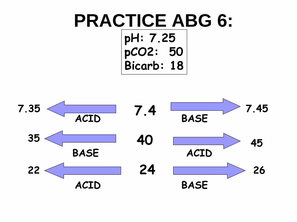
PRACTICE ABG 6:
pH: 7.25 pCO2: 50 Bicarb: 18

Managing Acute Hypoxic Pulmonary Failure
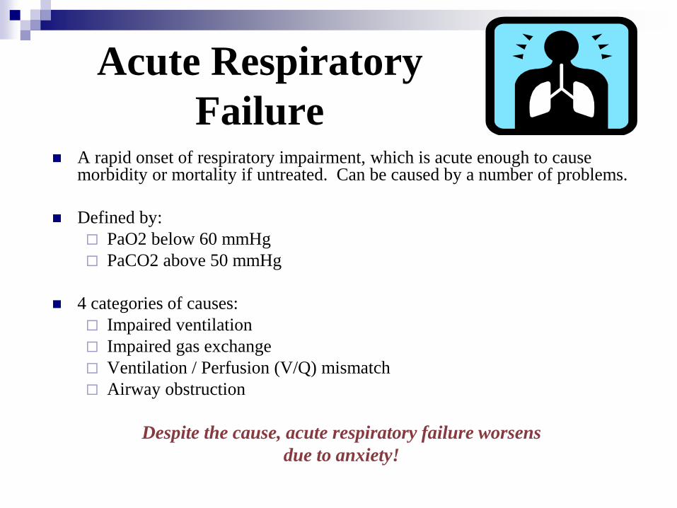
Acute Respiratory
Failure A rapid onset of respiratory impairment, which is acute enough to cause
morbidity or mortality if untreated. Can be caused by a number of problems.
Defined by:
PaO2 below 60 mmHg
PaCO2 above 50 mmHg
4 categories of causes:
Impaired ventilation
Impaired gas exchange
Ventilation / Perfusion (V/Q) mismatch
Airway obstruction
Despite the cause, acute respiratory failure worsens
due to anxiety!
General Treatment Principles
Assure airway patency
Airway adjuncts or suctioning if the patient is having difficulty managing secretions
Initiate aggressive pulmonary hygiene
Provide supplemental oxygen.
Non invasive ventilation is usually preferred if acceptable PaO2 can be achieved
Improve ventilation
May need to administer medications such as bronchodilators or mucolytics
Correct the underlying cause
Reduce anxiety
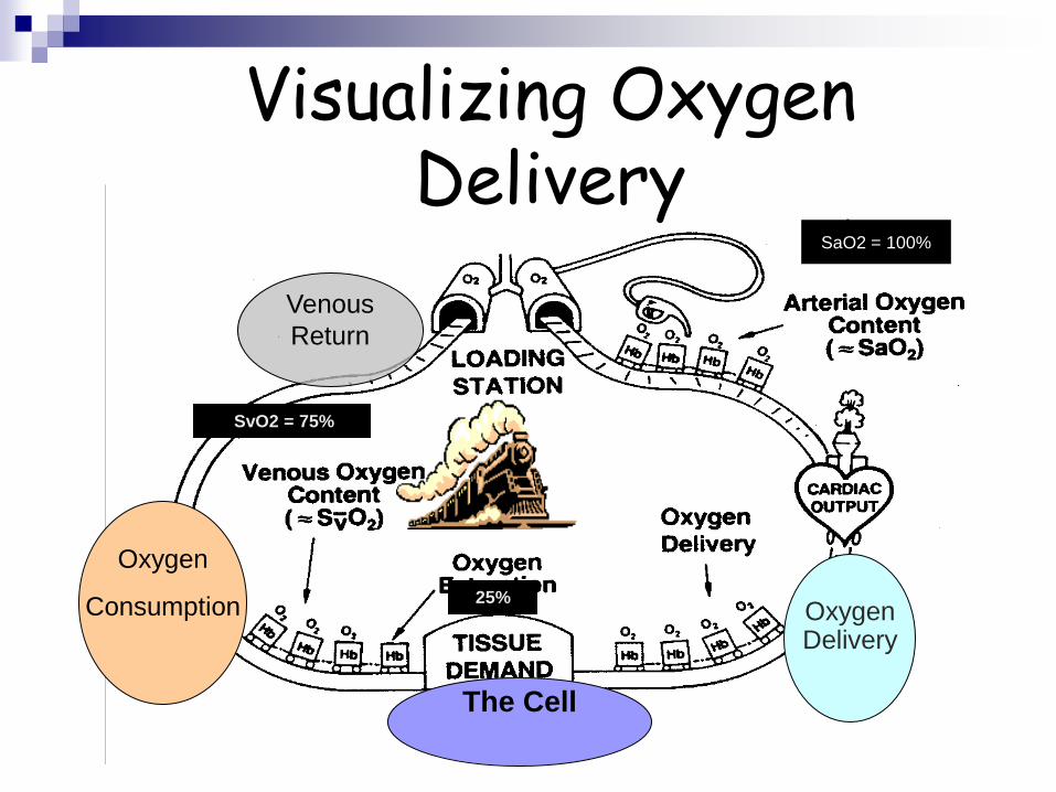
SvO2 = 75%
25%
SaO2 = 100%
Visualizing Oxygen Delivery
Oxygen Delivery
Oxygen
Consumption
The Cell
Venous
Return

Mechanical Ventilation

Lung Volumes
To decrease CO2, work here
To increase O2, work here.
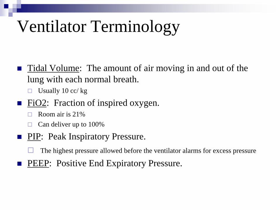
Ventilator Terminology
Tidal Volume: The amount of air moving in and out of the
lung with each normal breath.
Usually 10 cc/ kg
FiO2: Fraction of inspired oxygen.
Room air is 21%
Can deliver up to 100%
PIP: Peak Inspiratory Pressure.
The highest pressure allowed before the ventilator alarms for excess pressure
PEEP: Positive End Expiratory Pressure.
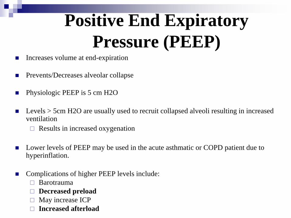
Positive End Expiratory
Pressure (PEEP) Increases volume at end-expiration
Prevents/Decreases alveolar collapse
Physiologic PEEP is 5 cm H2O
Levels > 5cm H2O are usually used to recruit collapsed alveoli resulting in increased ventilation
Results in increased oxygenation
Lower levels of PEEP may be used in the acute asthmatic or COPD patient due to hyperinflation.
Complications of higher PEEP levels include:
Barotrauma
Decreased preload
May increase ICP
Increased afterload
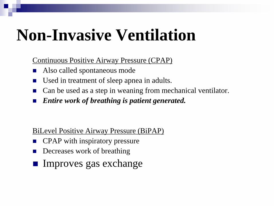
Non-Invasive Ventilation
Continuous Positive Airway Pressure (CPAP)
Also called spontaneous mode
Used in treatment of sleep apnea in adults.
Can be used as a step in weaning from mechanical ventilator.
Entire work of breathing is patient generated.
BiLevel Positive Airway Pressure (BiPAP)
CPAP with inspiratory pressure
Decreases work of breathing
Improves gas exchange

Ventilation Decision Tree (Woodruff, D., 2002)
Airway Patent?
Therapy < 48 hours? Therapy > 48 hours?
CPAP BiPAP Mechanical Ventilation
Yes No
Intubate Mask
Is WOB increased?

Modes of Mechanical Ventilation
AC: Assist Control
Every breath is supported by a
ventilator breath
Used when patient should
have no metabolic work
Post arrest
Pulmonary edema
ARDS
Anxiety
SIMV: Intermittent Mandatory
Ventilation
Patient is able to initiate breaths
between ventilator breaths
Machine breaths are
synchronized to patient pattern
Used as a weaning mode in some
patients
Minimizes barotrauma and
hemodynamic effects

Ventilator Modes (con’t)
Pressure Support (PSV)
oEach patient breath is supported by the ventilator during inhalation
oOvercomes resistance of tubing
oUsed for weaning
Volume controlled
oMachine is set to deliver a set volume
oPressures generated by each breath will vary (PIP)
oSet pressure limit where machine alarms
oMost commonly used mode in adults
Pressure Controlled
oMachine is set to deliver until certain airway pressure is reached
oVolumes of each breath will vary
oWill alarm if minimal volume is not delivered
oMost commonly used modes in pediatrics
oMay be used for patients with ARDS
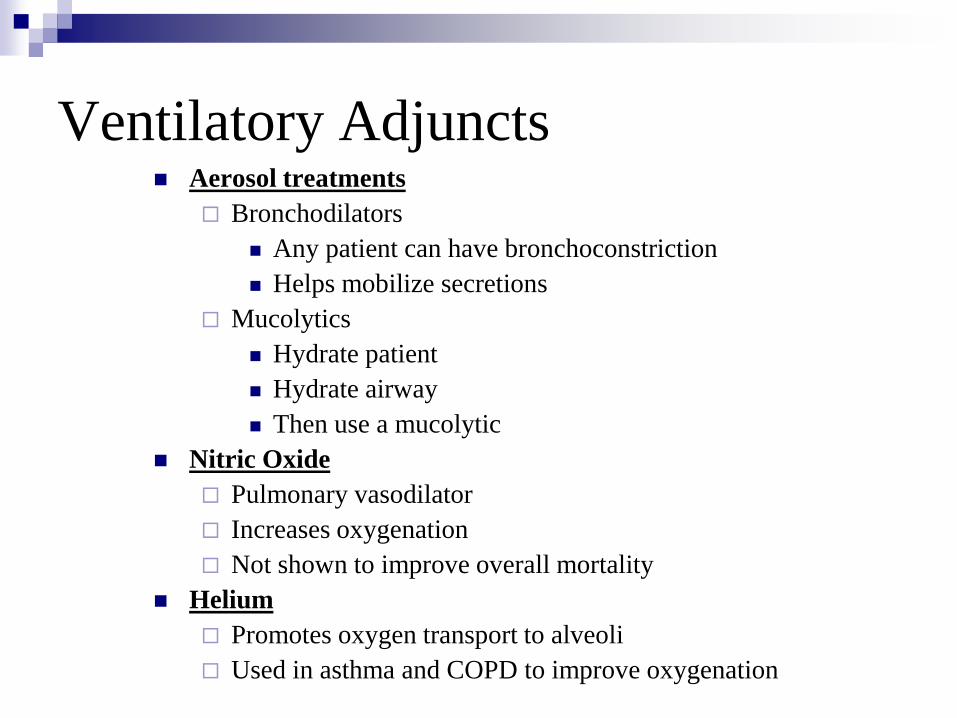
Ventilatory Adjuncts Aerosol treatments
Bronchodilators
Any patient can have bronchoconstriction
Helps mobilize secretions
Mucolytics
Hydrate patient
Hydrate airway
Then use a mucolytic
Nitric Oxide
Pulmonary vasodilator
Increases oxygenation
Not shown to improve overall mortality
Helium
Promotes oxygen transport to alveoli
Used in asthma and COPD to improve oxygenation
Ventilatory Adjuncts (con’t) Prone positioning
Redistributes lung fluid
Relieves heart weight on lower lobes
Improves oxygenation
Decreases CO2
Complications can be avoided by:
Limiting time to less than 2 hours
Adequate staff to prone
Rotational beds
If cannot move to chair, use chair position of bed
Turn and position every 2 hours
Rotational therapy for high risk patients
Vibration and percussion
Helps mobilize secretions
VEST therapy or percussion mode on bed

Pulmonary Embolism Fat Embolism

Pulmonary Embolism
An obstruction to blood flow to one or more of the arteries of the lung.
Most thrombi develop in deep veins of upper extremities (above knee)
In PE, the deep vein thrombus (DVT) has been dislodged and moved into the pulmonary vessel.
Virchow’s Triad (Risk Factors):
Hypercoagulability
Alteration to vessel wall
Venous stasis
Factors contributing to dislodgement of thrombi:
Intravascular pressure changes

Pulmonary Embolus (con’t)
Clot moves into pulmonary vessel. Ventilation
continues but perfusion is decreased
No gas exchange, so alveolar CO2 decreases
Results in bronchoconstriction to affected alveoli
Cessation of blood flow damages pneumocytes
Production of surfactant decreases
Atelectasis occurs and work of breathing increases

Presentation / Diagnostic Findings ABG: Decreased PaO2 , SaO2;
pH= elevated, then decreased
ECG: Tall peaked P waves, atrial dysrhythmias,
sinus tachycardia, S1, Q3, T3
V/Q scan / Spiral CT:
Shows perfusion defect with normal
ventilation. Similar sensitivity
and specificity.
Pulmonary Angiography:
“Gold Standard”
Labs: D-dimer
Common symptoms:
Tachypnea
Dyspnea
Chest pain
+ Homan’s sign
Restless, apprehension
If the embolus is large the presenting
symptom may be PEA!

Treatment
Prevention of DVT is the key!
Provide supplemental oxygen/circulatory/ventilatory support
Thrombolytic therapy may be used in massive PE
Heparin – Prevents further clot formation
Inferior vena cava filter – May be inserted in high risk patients to catch future clots
Pulmonary embolectomy – A very high risk interventional procedure
Pulmonary vasodilators have been used in some cases.
Fat Embolus Syndrome Patients at increased risk:
Long bone fracture
Hip replacements
Onset 24 – 48 hours after event
Present with ARDS-type syndrome:
Pulmonary edema
Hypoxia
Axillary / subconjunctival petechiae
CNS disturbances
May see: Tachycardia, fever, drop in platelets, fat globules in urine, retina, sputum
Treatment is same as treatment for PE.

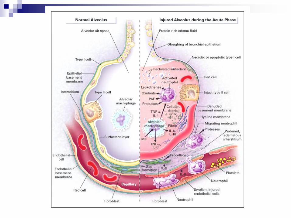
Acute Respiratory Distress Syndrome

Acute Respiratory Distress
Syndrome
Acute respiratory failure in adults characterized by pulmonary edema manifested by right to left shunting through collapsed or fluid-filled alveoli.
Specific findings: Oxygenation – PaO2 / FiO2 < 200 regardless of PEEP levels
Chest x-ray – Bilateral infiltrates seen on frontal chest x-ray
No elevated pulmonary pressures

ARDS Lungs

Predisposing Factors
Direct Pulmonary Injury due to:
Aspiration of gastric contents
Pulmonary contusion
Near drowning
Smoke inhalation
Pneumonia
Barotrauma from mechanical ventilator
Indirect injury caused by inflammatory mediator release. Mediator release may be triggered by:
Sepsis or Multiple organ dysfunction syndrome (MODS)
Shock
Pancreatitis
Trauma
DIC
Multiple transfusions
Risk of ARDS increases if patient has more than one risk factor: •One risk factor = 25% chance of ARDS •Two risk factors = 42% chance of ARDS •Three risk factors = 85% chance of ARDS
Pathophysiology of ARDS
Diffuse injury to the alveoli – capillary membrane
Increased lung permeability
Flooding of alveoli causes injury to Type II pneumocytes
Results in decreased surfactant production
Decreased surfactant causes increased alveolar surface tension
Increased alveolar surface tension causes atelectasis
Now blood begins to “shunt” through the lungs without passing by alveoli that are ventilated
Lungs become “stiff” or less compliant due to hypoxemic pulmonary vasoconstriction
Refractory hypoxemia worsens

Clinical Manifestations
Latent:
Beginning a-c membrane changes; PaO2/FiO2
Acute Interstitial:
Alveolar edema and decreased lung compliance
Dyspnea, restless on room air, anxious
Lung sounds = ___________
Oxygen saturation is decreased
Patient begins to hyperventilate
ABG will demonstrate respiratory
Chest x-ray will be unchanged at this phase
Clinical Manifestations: Acute Intra-alveolar/Chronic Phase
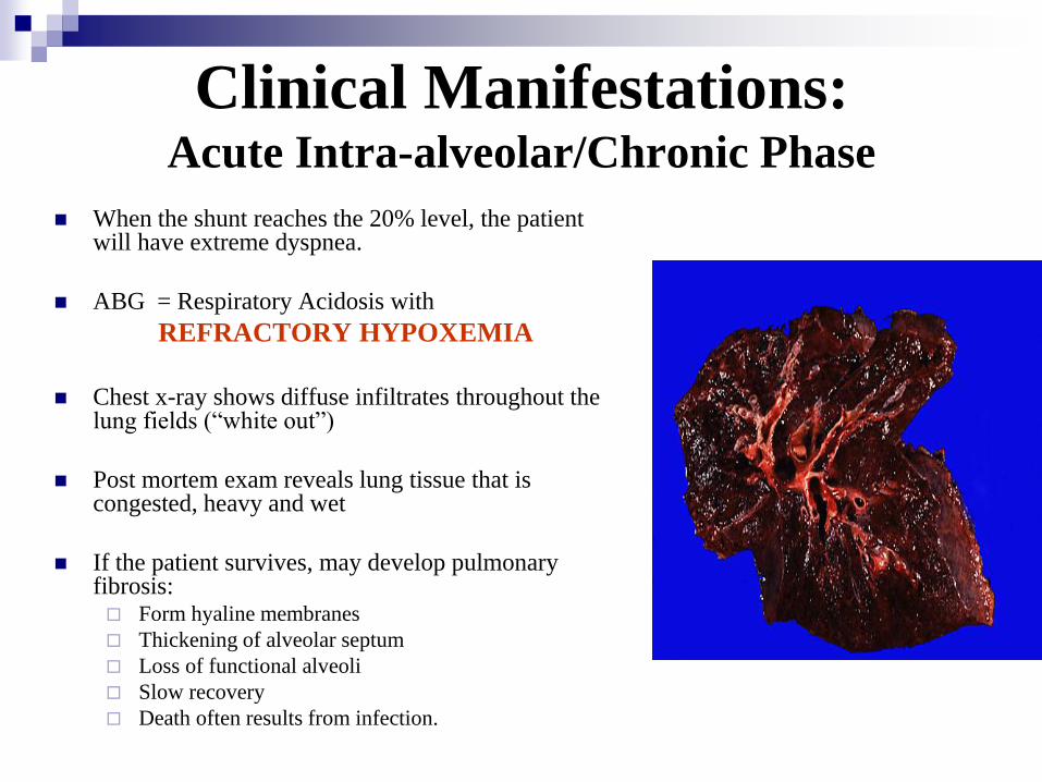
When the shunt reaches the 20% level, the patient
will have extreme dyspnea.
ABG = Respiratory Acidosis with
REFRACTORY HYPOXEMIA
Chest x-ray shows diffuse infiltrates throughout the
lung fields (“white out”)
Post mortem exam reveals lung tissue that is congested, heavy and wet
If the patient survives, may develop pulmonary fibrosis: Form hyaline membranes
Thickening of alveolar septum
Loss of functional alveoli
Slow recovery
Death often results from infection.

Evidence- Based
Multidisciplinary Plan of Care
Goals of ARDS Therapy:
Prevent further injury
Maintain adequate pulmonary oxygenation
Optimize oxygen delivery to the tissues using the six P’s

ARDS - Prevention Initiate nursing care that reduces bacterial colonization and risk of
aspiration
Handwashing
Elevate head of bed at least 30 degrees
Oral Care
Consider therapy to block injury at the alveoli- capillary interface (controversial):
Nitric oxide
Xigris
Corticosteroids
Monoclonal antibodies
Non steroidal anti-inflammatories
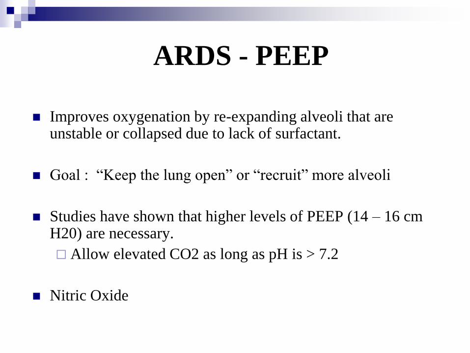
ARDS - PEEP
Improves oxygenation by re-expanding alveoli that are unstable or collapsed due to lack of surfactant.
Goal : “Keep the lung open” or “recruit” more alveoli
Studies have shown that higher levels of PEEP (14 – 16 cm H20) are necessary.
Allow elevated CO2 as long as pH is > 7.2
Nitric Oxide

ARDS - Pumps and Pipes
Adjust fluids and medications to maximize oxygen delivery to the cells
Use SVO2 to monitor cellular oxygenation
Make sure you have enough hemoglobin molecules (“trucks”) to get the oxygen to the cells. Transfuse early!
Make sure that is enough fluid in the pipes (blood vessels) to supply adequate tissue perfusion
Monitor CVP to assess volume status.
Use vasoactive medications to keep the “pipes” toned up and “pumps” squeezing the blood to the tissue.
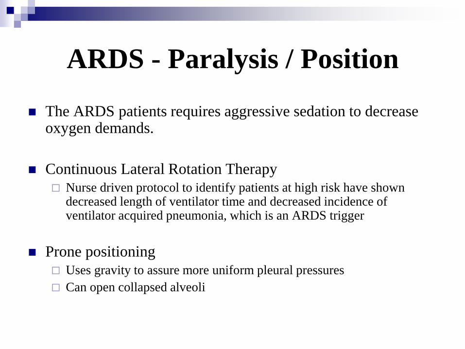
ARDS - Paralysis / Position
The ARDS patients requires aggressive sedation to decrease oxygen demands.
Continuous Lateral Rotation Therapy
Nurse driven protocol to identify patients at high risk have shown decreased length of ventilator time and decreased incidence of ventilator acquired pneumonia, which is an ARDS trigger
Prone positioning
Uses gravity to assure more uniform pleural pressures
Can open collapsed alveoli
Acute Respiratory Infections
Pneumonia An acute infection of the lung parenchyma, including
alveolar spaces and interstitial tissue. Community-/Health care associated-/Hospital acquired
Causative organisms are different.
Causative agent is inhaled / enters pharynx May be transmitted from one patient to the next
Subglottic secretions pool above ETT cuff Within 24 hours, 95% of ETT were partially covered with bacteria
Nasal Nasogastric tubes lead to colonization of nasopharynx
Factors that increase risk of colonization: Decreased salivary flow rate
Poor oral hygiene
Systemic antibiotics
No oral fluid or food

Pneumonia (con’t) Causative agent moves into lungs from pharynx:
Alveoli become inflamed and edematous.
Alveoli spaces fill with exudate and consolidate.
Patient may complain of cold or flu-like symptoms
Alveoli spaces fill with exudate and consolidate.
Diffusion of oxygen is obstructed, causing hypoxemia
WBC will be elevated with increase of immature WBC’s , if bacterial.
Pneumonia - Treatment
Prevent nosocomial pneumonia!! Keep HOB elevated
Perform frequent oral care
Strict handwashing
If suspected: Obtain culture to identify causative organism
Start antibiotic promptly
Hydrate unless contraindicated
2- 3 liters / 24 hours
Initiate enteral feeding early to improve nutrition
Air Leak Syndromes

Air-Leak Syndromes - Types
Air in the pleural space with
complete or partial collapse of the
lung. Several types:
Open pneumothorax
Closed pneumothorax
Iatrogenic pneumothorax
Spontaneous pnemothorax
Tension pneumothorax

Tension Pneumothorax
Occurs when air flows freely into the pleural space during inspiration and becomes trapped
Results in lung collapse and mediastinal shift to the opposite side
Clinical findings:
Shortness of breath, progressing to extreme dyspnea
Unilateral absence of breath sounds
Asymmetry of chest movement
May see tracheal deviation and subcutaneous emphysema
May see distended neck veins and hypotension
MAY NOT BE ABLE TO WAIT FOR CHEST X-RAY TO CONFIRM
Needle Decompression
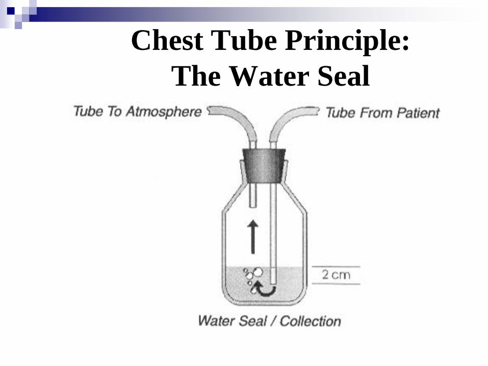
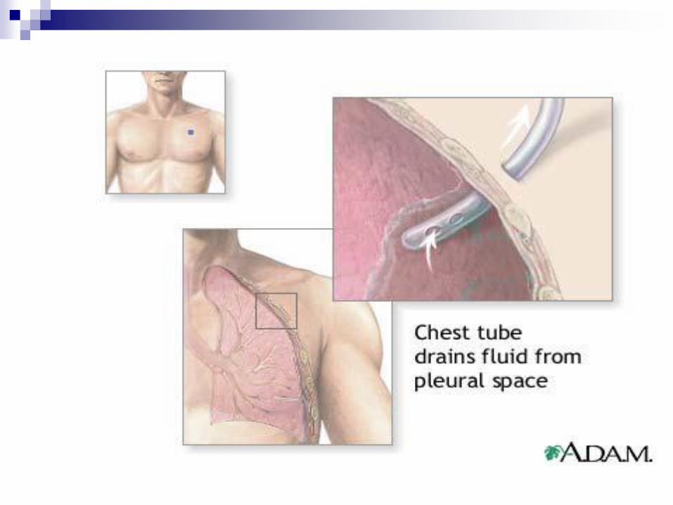
Chest Tube Principle:
The Water Seal
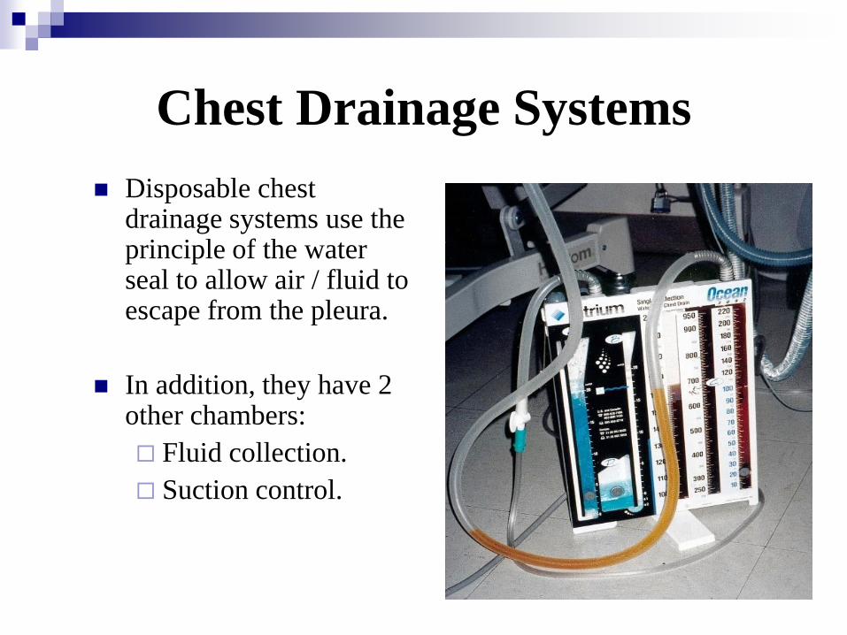
Chest Drainage Systems
Disposable chest drainage systems use the principle of the water seal to allow air / fluid to escape from the pleura.
In addition, they have 2 other chambers:
Fluid collection.
Suction control.

Chest Trauma - Hemothorax
Collection of blood in pleural space
Source:
Left hemothorax
Rib fracture 36%
Pulmonary tissue 35%
Aorta 15%
Right hemothorax
Rib fracture 51%
Pulmonary tissue 27%
Liver 10%
Manifestations:
Dyspnea, tachypnea
Cyanosis, hypoxemia
Shock
Treatment:
Chest drainage
Volume replacment
Thorocotomy More than 1500 ml blood with initial chest
tube insertion
Bleeding more than 300 /hr for 3 hours Hemodynamic instability
Tension hemothorax

Chest Tube Management
Air Leaks
Identified by bubbling in the water seal chamber.
An air leak is not uncommon immediately after tube placement.
Indicates that the lung has not fully reexpanded or that there is a leak in the system.
To prevent air leaks in the tubing or drainage system, ensure all connections are secure.
All new leaks should be investigated
Tidaling
Pressure changes that occur in the
pleural space with breathing can
be viewed as fluctuations
(tidaling) in the level of water
within the tube.
In normal spontaneous breathing,
water levels will go up with
inspiration (more negative) and
return to baseline during
exhalation

Chest Tube Management
Check collection chamber for:
Volume / rate of drainage
Appearance of drainage
“Milk” clots out gently
NO STRIPPING
Keep collection chamber below chest level
Do not clamp the chest tube
The only time a chest tube should be clamped is if the drainage unit is disrupted or is being changed.
If the chest tube is accidentally dislodged:
Apply occlusive dressing to site
Monitor patient’s respiratory status, notify physician, and obtain chest x-ray.
If the drainage system is damaged:
Immerse distal end of chest tube into a bottle of sterile water, notify physician, and attach new drainage unit per policy

Thoracic Surgery /Trauma
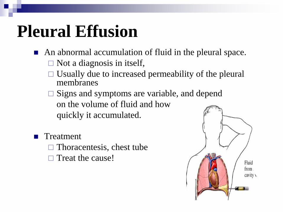
Pleural Effusion An abnormal accumulation of fluid in the pleural space.
Not a diagnosis in itself,
Usually due to increased permeability of the pleural membranes
Signs and symptoms are variable, and depend
on the volume of fluid and how
quickly it accumulated.
Treatment
Thoracentesis, chest tube
Treat the cause!
Pulmonary Resection
Type and location of surgery will dictate
the type of surgical approach used. Most common is postero-lateral thoracotomy
Care is taken to avoid drainage of blood or secretions into unaffected lung during surgery
Hemorrhage is an early, life-threatening complication that can occur after lung resection. Chest tube output more than 100 cc/hr, fresh blood, or sudden increase in
drainage signals possible hemorrhage
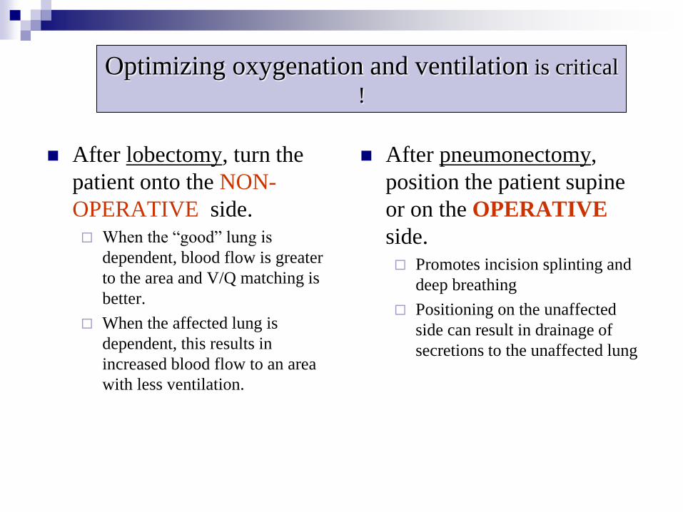
After lobectomy, turn the
patient onto the NON-
OPERATIVE side.
When the “good” lung is
dependent, blood flow is greater
to the area and V/Q matching is
better.
When the affected lung is
dependent, this results in
increased blood flow to an area
with less ventilation.
After pneumonectomy,
position the patient supine
or on the OPERATIVE
side.
Promotes incision splinting and
deep breathing
Positioning on the unaffected
side can result in drainage of
secretions to the unaffected lung
Optimizing oxygenation and ventilation is critical
!
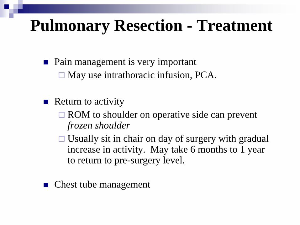
Pulmonary Resection - Treatment
Pain management is very important
May use intrathoracic infusion, PCA.
Return to activity
ROM to shoulder on operative side can prevent frozen shoulder
Usually sit in chair on day of surgery with gradual increase in activity. May take 6 months to 1 year to return to pre-surgery level.
Chest tube management
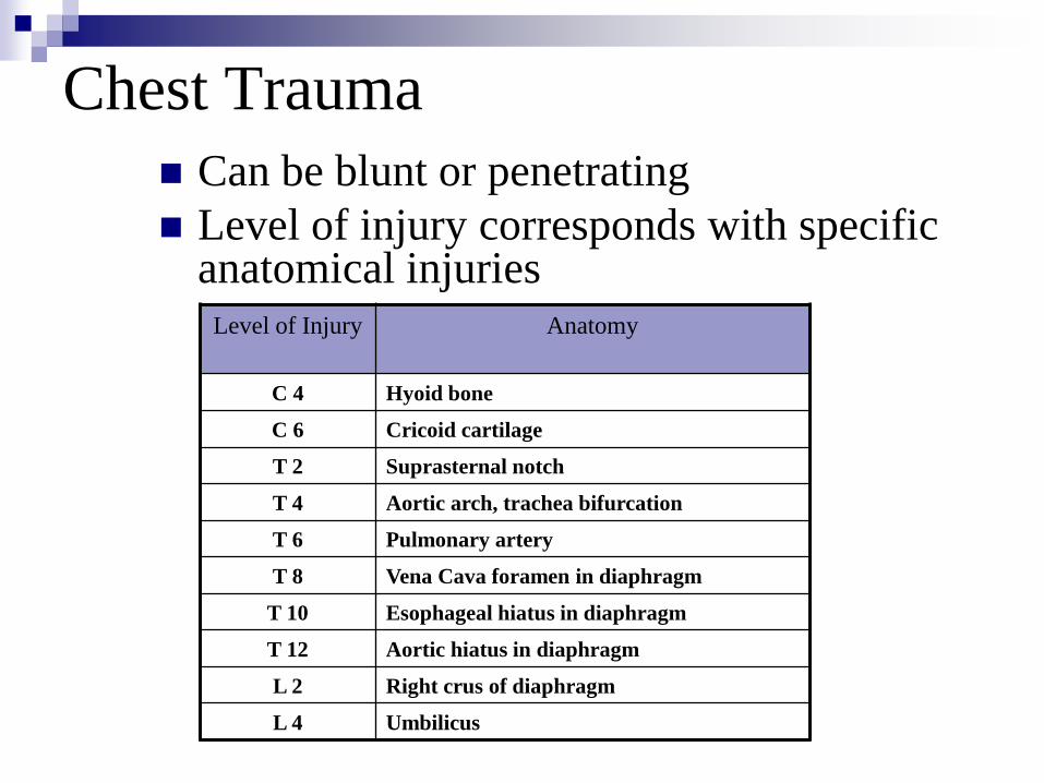
Chest Trauma Can be blunt or penetrating
Level of injury corresponds with specific anatomical injuries
Level of Injury Anatomy
C 4 Hyoid bone
C 6 Cricoid cartilage
T 2 Suprasternal notch
T 4 Aortic arch, trachea bifurcation
T 6 Pulmonary artery
T 8 Vena Cava foramen in diaphragm
T 10 Esophageal hiatus in diaphragm
T 12 Aortic hiatus in diaphragm
L 2 Right crus of diaphragm
L 4 Umbilicus
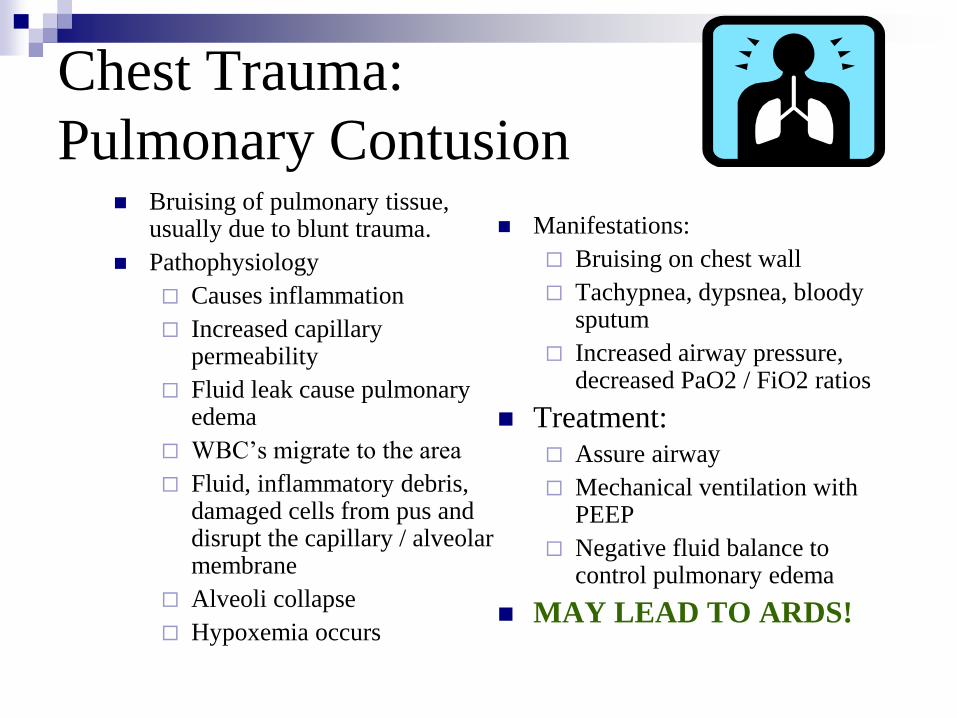
Chest Trauma:
Pulmonary Contusion Bruising of pulmonary tissue,
usually due to blunt trauma.
Pathophysiology
Causes inflammation
Increased capillary permeability
Fluid leak cause pulmonary edema
WBC’s migrate to the area
Fluid, inflammatory debris, damaged cells from pus and disrupt the capillary / alveolar membrane
Alveoli collapse
Hypoxemia occurs
Manifestations:
Bruising on chest wall
Tachypnea, dypsnea, bloody sputum
Increased airway pressure, decreased PaO2 / FiO2 ratios
Treatment: Assure airway
Mechanical ventilation with PEEP
Negative fluid balance to control pulmonary edema
MAY LEAD TO ARDS!
Chest Trauma: Rib Fractures
Simple fractures may result in decreased ventilation due to pain
1st rib fractures are associated with higher incidence of great vessel injury and cervical spine injury
Lower rib fractures are associated with abdominal injuries
Manifestations:
Pleuritic chest pain
Contusion
Decreased respiratory effort
Treatment:
Splinting
Monitor for underlying tissue damage, development of pneumothorax or hemothorax

Chest Trauma: Flail Chest
Multiple fractures may result in flail segments
Result from 2 or more segments of fractured ribs
Allows a free floating segment that moves paradoxically
Lungs do not expand as usual, resulting in hypoxemia
May damage underlying tissue
Manifestations:
Pleuritic pain
Dyspnea
Crepitus
Hypoxemia
Treatment
Oxygen, ventilation
Stabilize with tape (one side only, do not wrap chest)
ORIF
Complications
Pneumothorax
ARDS
Atelectasis

Chest Trauma: Hemothorax
Collection of blood in pleural space
Source:
Left hemothorax
Rib fracture 36%
Pulmonary tissue 35%
Aorta 15%
Right hemothorax
Rib fracture 51%
Pulmonary tissue 27%
Liver 10%
Manifestations:
Dyspnea, tachypnea
Cyanosis, hypoxemia
Shock
Treatment:
Chest drainage
Volume replacment
Thorocotomy
More than 1500 ml blood with initial chest tube insertion
Bleeding more than 300 /hr for 3 hours
Hemodynamic instability
Tension hemothorax

Airway Disorders

Chronic Obstructive
Pulmonary Disease (COPD)
Patients with COPD may have frequent exacerbations that can
cause acute respiratory failure
Asthma
Emphysema
Chronic Bronchitis
Most common precipitating events:
Airway infections
Right sided heart failure, due to high pulmonary pressures
common in COPD
Non –compliance with COPD treatment
Chronic Obstructive
Pulmonary Disease (COPD)
More than 14 million Americans affected
Cigarette smoking (85-90%, per ALA, 2011)
Occupation – coal miners, firefighters
Alpha- 1 anti-trypsin deficiency
Results in:
Emphysema –chronic inflammation
Results in air trapping in the alveoli
Chronic bronchitis – mucus production
Results in chronic, productive cough for more than 3 months in 2 consecutive years.
Symptoms:
Productive cough in AM
Resistance to airflow causes wheezing, dyspnea,
Incidence of pulmonary infections increases

COPD - Treatment Bronchodilation – Treats disease immediately
Beta 2 agonist
Anticholinergic
Steroids – Reduces airway edema, but effect will not be seen until next day.
Advair – anti-inflam/bronchodilator
Aminophylline: Smooth muscle relaxant
Oxygen – Best to use controlled delivery device.
Maintain airway patency – To mobilize thick, tenacious secretions, consider use of: Humidification
Hydration
Suctioning, percussion, vibration, postural drainage
Treat infections with appropriate antibiotics Use antipyretics to decrease any fever and O2 consumption

Assisted Ventilation (BiPAP) in COPD
Avoid mechanical ventilation as long as possible!
Criteria for ventilation:
Respiratory muscle fatigue
Refractory hypoxemia
Respiratory acidosis (pH < 7.30)
Cardiovascular instability
If pCO2 is elevated with normal pH, probably a chronic CO2 retainer
Try Non-Invasive ventilation first!
If pCO2 is elevated and pH is decreased will likely require mechanical ventilation
Remember:
For non-invasive ventilation
to work, must be alert, cooperative and
able to handle secretions
Status Asthmaticus
A recurrent, reversible airway disease characterized by increase airway responsiveness to a variety of stimuli that produce airway narrowing.
Triggers cause IgE release, which stimulates mast cells to release histamine, causing swelling and inflammation of the smooth muscles of the larger bronchi and mucous membrane swelling and excessive secretion of mucus.
Airway narrowing is greatest during expiraton. Air is trapped in alveoli, which become hyperinflated.
Excess mucus causes V/Q mismatch and shunt Has circadian influence:
Worse around 3 am.
Best around 3 pm.
Warnings of impending severe attack: Increased sleep disturbances and use of nocturnal bronchodilators
Morning chest stiffness or heaviness
Runny nose, sneezing, increase in cough

Asthma - Presentation
Tachypnea, dyspnea, wheezing due to bronchoconstriction
May have increased sputum
Absence of rhonchi and wheezing indicates absence of airflow
Not a good sign!
Anxious, diaphoresis, use of accessory muscles, tachycardia
Elevation of pCO2 is also a late sign. Usually pCO2 is
decreased / normal.

Asthma - Treatment
Bronchodilators
Beta adrenergic agonists – Alupent, Bronchosol
Anticholinergic agents – Atrovent
Steroids to decrease mucosal swelling and histamine release
IV magnesium Acts as bronchodilator, decreases inflammation
Antibiotics Strong link between sinus infections and asthma exacerbations
Hydration – More effective than expectorant Mucolytics are contraindicated because they may cause increased
bronchospasm.
If ventilation is required, avoid high PIP and PEEP Sedation with propofol may increase bronchodilation
Emphysema
Damaged air sacs in a person's lungs, causing
them to lose their elasticity.
Permanent fissures in the tissues of a person's
lungs.
Limited air supply
Chronic Bronchitis
Inflammation and swelling of the lining of the
airways, leading to narrowing and obstruction of the
airways.
Production of mucous, which can cause further
obstruction of the airways.
Increases the likelihood of bacterial lung infections.
Daily Cough
Pulmonary Hypertension
Pulmonary Hypertension

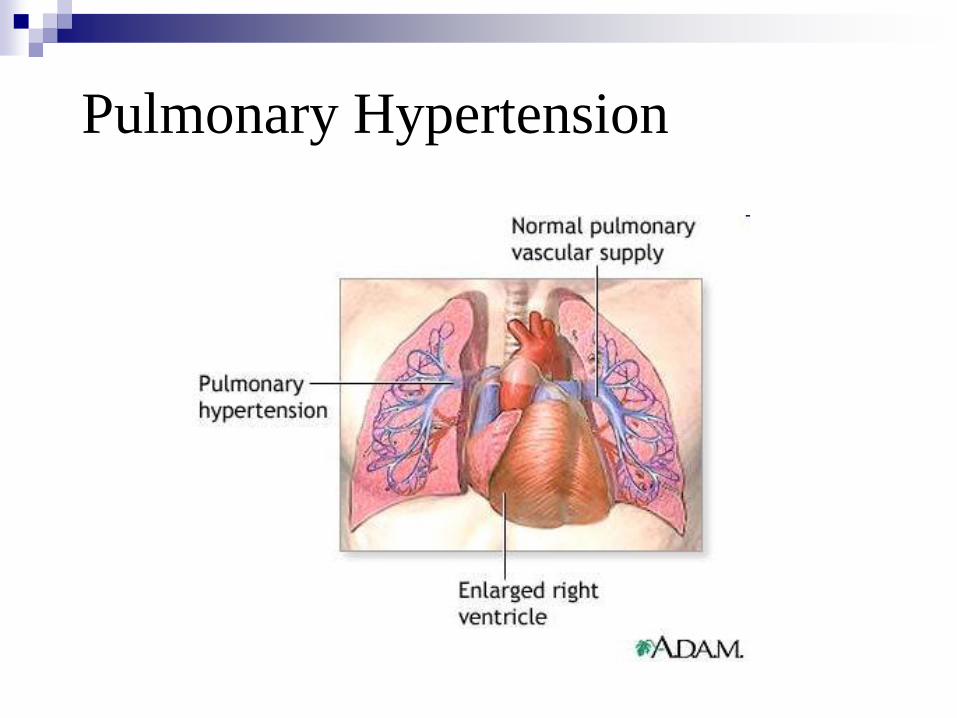
Pulmonary Hypertension
A progressive, life threatening disorder of the pulmonary circulation characterized by high pulmonary artery pressures, leading to right ventricular failure.
Primary pulmonary HTN
Associated with autoimmune diseases
Mostly effects women in childbearing years
Believed to be caused by endothelial dysfunction that leads to re-modeling of the pulmonary artery
Secondary Pulmonary HTN
is due to chronic disorders such as pulmonary fibrosis / sarcoidosis, collagen vascular disease, liver disease, portal hypertension, diet supplements, sleep apnea, HIV
Signs / symptoms
Dyspnea
Weakness / fatigue
Recurrent syncope
Signs of right heart failure
Tricuspid murmur
Jugular vein distension, pulsation
Increased pulmonary pressures
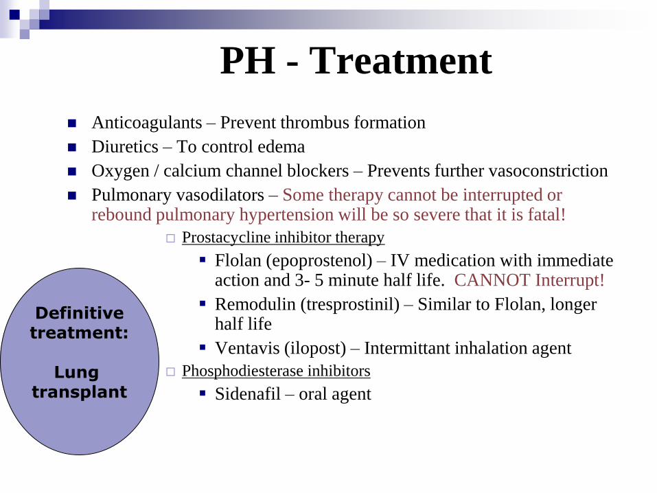
PH - Treatment
Anticoagulants – Prevent thrombus formation
Diuretics – To control edema
Oxygen / calcium channel blockers – Prevents further vasoconstriction
Pulmonary vasodilators – Some therapy cannot be interrupted or rebound pulmonary hypertension will be so severe that it is fatal!
Prostacycline inhibitor therapy
Flolan (epoprostenol) – IV medication with immediate action and 3- 5 minute half life. CANNOT Interrupt!
Remodulin (tresprostinil) – Similar to Flolan, longer half life
Ventavis (ilopost) – Intermittant inhalation agent
Phosphodiesterase inhibitors
Sidenafil – oral agent
Definitive treatment:
Lung
transplant
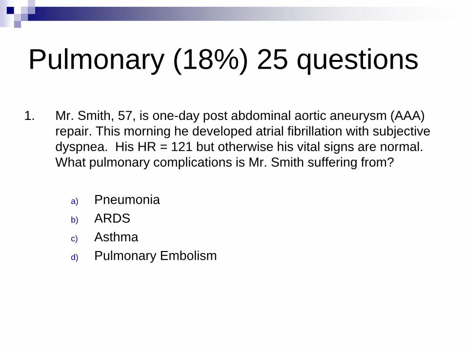
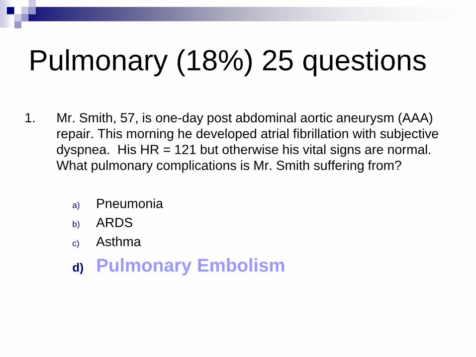
Pulmonary (18%) 25 questions
1. Mr. Smith, 57, is one-day post abdominal aortic aneurysm (AAA)
repair. This morning he developed atrial fibrillation with subjective
dyspnea. His HR = 121 but otherwise his vital signs are normal.
What pulmonary complications is Mr. Smith suffering from?
a) Pneumonia
b) ARDS
c) Asthma
d) Pulmonary Embolism
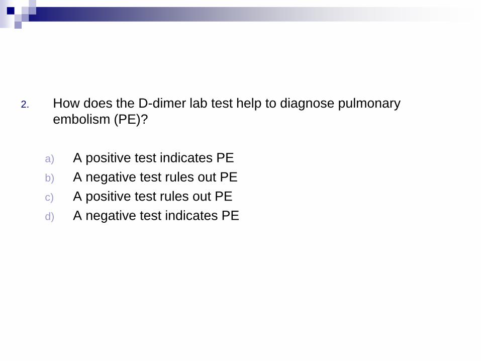
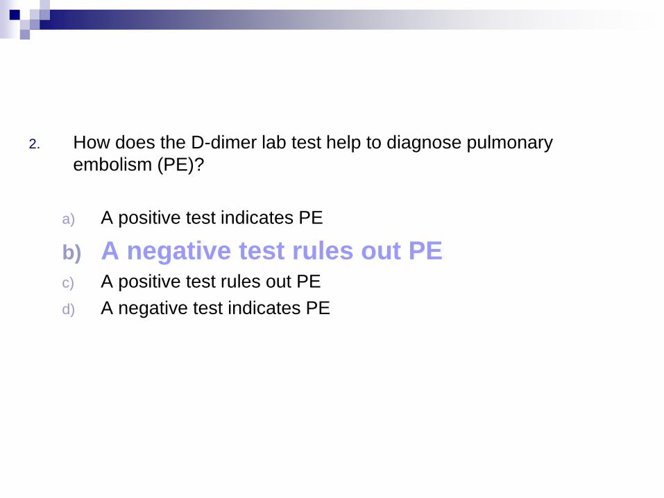
2. How does the D-dimer lab test help to diagnose pulmonary
embolism (PE)?
a) A positive test indicates PE
b) A negative test rules out PE
c) A positive test rules out PE
d) A negative test indicates PE
2. How does the D-dimer lab test help to diagnose pulmonary
embolism (PE)?
a) A positive test indicates PE
b) A negative test rules out PE c) A positive test rules out PE
d) A negative test indicates PE
3. Nursing interventions that decrease the incidence of hospital-
acquired pneumonia include:
a) Placing gastric tubes through the nose
b) Administering systemic antibiotics
c) Brushing the patient’s teeth with a toothbrush
d) Keeping the patient NPO
3. Nursing interventions that decrease the incidence of hospital-
acquired pneumonia include:
a) Placing gastric tubes through the nose
b) Administering systemic antibiotics
c) Brushing the patient’s teeth with a
toothbrush d) Keeping the patient NPO

Questions??
You Guys ROCK !!!
Review questions DAILY!
Stay Confident!