Embed Size (px)
Citation preview
Catching RIP in the act.Catching RIP in the act. Part I: A PCR assay to Part I: A PCR assay to detect DNA methylationdetect DNA methylation
Paul DoneganPaul Donegan
Freitag LabFreitag Lab
Biochemistry and Biophysics DepartmentBiochemistry and Biophysics Department
Oregon State UniversityOregon State University

Background
• MUTAGENESIS: Mutations of base pairs in genetic material– Induced by UV, X-ray, viruses, etc.– Spontaneous occurrence – triggers DNA repair
• Hypermutagenesis– Induced and controlled by cells– Not spontaneous
--AID deaminase--ApoBec (HIV)
--RIP

R I P
• RIP = Repeat Induced Point Mutation
• Genomic defense mechanism– Silences repetitive DNA (no expression)
• Targets duplicated DNA segments – linked or unlinked sequences
• Induces C to T transition mutations
RIP triggered by repeated sequence
Identical Sequences
Mutated Sequences
C to T point mutations induced by RIP
GCATATTAGTTATGTTTAGCGCATTCTAGTATATCAGTTATGTTCAGTGCACTTTA
GCATATCAGTCATGCTCAGCGCACCTAGCATATCAGTCATGCTCAGCGCACCTA

Relevance
We are interested in RIP because we want to:
– gain insights into evolutionary mechanisms that shape genomes.
– understand genome defense mechanisms and mutagenesis.
Summer Research Objective• To differentiate between two possible molecular
mechanism that can explain RIP
QuickTime™ and aTIFF (Uncompressed) decompressor
are needed to see this picture.
Neurosporacrassa
Rosette of sexual spores,nuclei labelled with GFP

Possible Mechanisms for C to T Mutations caused by RIP (1)
• Methylation by a specific cytosine DNA methyltransferase, followed by deamination
Methyl Group Donor- S-adenosylmethionine (SAM)
QuickTime™ and aTIFF (Uncompressed) decompressor
are needed to see this picture.
C CMe T
QuickTime™ and aTIFF (Uncompressed) decompressor
are needed to see this picture.QuickTime™ and a
TIFF (Uncompressed) decompressorare needed to see this picture.
H3C
METHYLATION DEAMINATION

Possible Mechanisms for C to T Mutations caused by RIP (2)
• Cytosine is never methylated but instead deaminated to uracil, which will be replaced with thymine by DNA replication or repair
z
QuickTime™ and aTIFF (Uncompressed) decompressor
are needed to see this picture.
QuickTime™ and aTIFF (Uncompressed) decompressor
are needed to see this picture.
C U
QuickTime™ and aTIFF (Uncompressed) decompressor
are needed to see this picture.
Enz
DEAMINATION
Intermediate

Image from: Shiu et al. (2001) Cell
RIP timeline
• RIP occurs during the sexual cycle
• RIP occurs after fertilization but before karyogamy.
• ~10 mitotic divisions while RIP can occur.
FERTILIZATION
KARYOGAMY RIP ZONE!

• DNA was extracted during the expected RIP timeframe
• Methylation of interest should occur between fertilization and karyogamy (nuclear fusion).
0 1 2 3 4 5 6 7
DAY
RIP ZONE (between fertilization and karyogamy)
Methylation Assay Timeline
Controls
Days of Interest

PCR after Digest
Methylation-sensitive vs. methylation-insensitive restriction enzymes:
Sau3AI tests for cytosine methylation, based on the presence or absence of bands
Methylated site
Digest
PCR
GATCme
Digest with Sau3AI
DpnII is not sensitive to cytosine methylation:
-cuts regardless-control (never
amplifies)
Unmethylated site
Bands cannot be amplified when
site is cut
GATC

RFP
• ‘tdimerRed’ has two identical segments that trigger RIP• integrated into the Neurospora genome (not in WT)• here, we look for DNA methylation induced by RIP
• EVIDENCE OF METHYLATION SUGGESTS MECHANISM 1
Mutations in the RFP region
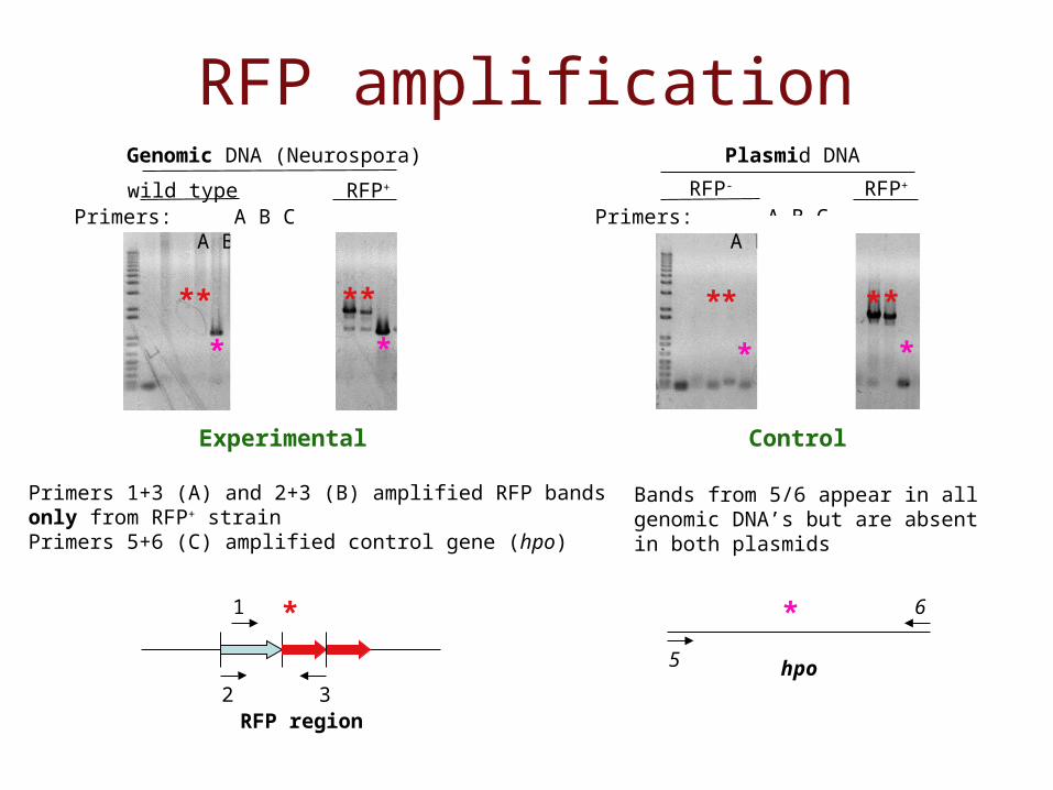
RFP amplification
Primers 1+3 (A) and 2+3 (B) amplified RFP bands only from RFP+ strainPrimers 5+6 (C) amplified control gene (hpo)
*
RFP region
1
2 3hpo5
6*
Bands from 5/6 appear in all genomic DNA’s but are absent in both plasmids
Genomic DNA (Neurospora) Plasmid DNA
wild typePrimers: A B C A B C
*
RFP- RFP+
**
* **
*
*
*
Experimental Control
RFP+
**
Primers: A B C A B C
**
*

BUT: Assay never worked with positive controls of methylated DNA
25 cycles 28 cycles 31 cycles
G S D G S D
hpo
G = genomic DNA, no digestS = Sau3AI, C-methylation sensitiveD = DpnII, C-methylation insensitive
Positive control:Methylated region
Negative control:Unmethylated region
Expected band in S lane,but no band in D lane
Expected no band in S or D lane

Catching RIP in the act.Part II: Tagging of duplicated DNA with fluorescent DNA
binding proteins

Goals• Tag DNA of Neurospora crassa with fluorescent proteins:
– to visualize pairing of duplications during RIP;
– to track chromosome territory movement (e.g., centromeres, telomeres, nucleolar DNA, specific genes)
– to track movements of DNA binding proteins from nucleus to nucleus
– to target enzymes to specific regions on chromosomes
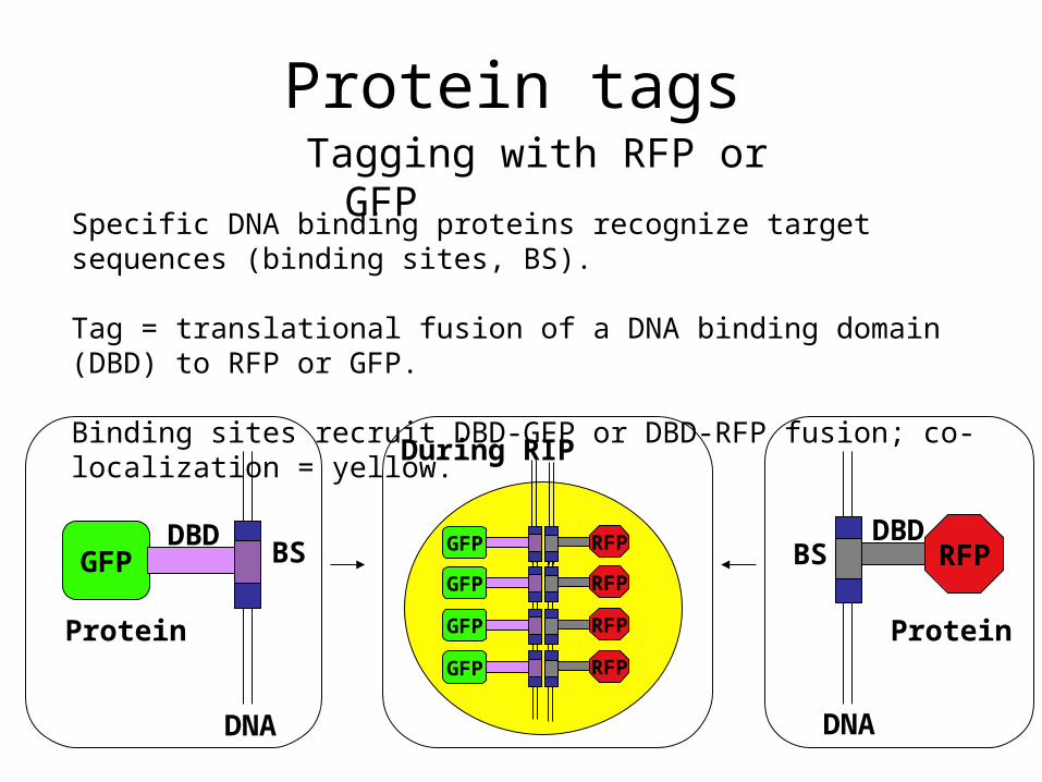
Protein tagsTagging with RFP or GFP
Specific DNA binding proteins recognize target sequences (binding sites, BS).
Tag = translational fusion of a DNA binding domain (DBD) to RFP or GFP.
Binding sites recruit DBD-GFP or DBD-RFP fusion; co-localization = yellow.
GFP RFPBSDBD
BSDBD
DNA DNA
Protein Protein
During RIP
GFP RFP
GFP RFP
GFP RFP
GFP RFP

Construction of protein tags
3 Transformed E. coli
4 Purified plamids, digested DNA and confirmed correct plasmids
5 Linearized plasmid and transformed into Neurospora his-3 mutant
1 Amplified DBD from Aspergillus AflR and AlcR by PCR
2 Generated translational fusions by cloning into gfp and rfp plasmids
6 Selected His+ Neurospora transformants that showed fluorescence
AlcR-RFP AflR-GFP
Fusion proteins localized in nuclei

Construction of DNA binding sites
2 Binding site: DNA sequences specifically recognized by AflR or AlcR
AflR:TCGNNNNNCGA AlcR: GCGGRRCCGC
Need 200+ copies of recognized sequence to bind enough fluorescent protein for visibility.

Summary
1 PCR assay: Did not work in many attempts. We need a new approach.
2 DNA tagging: The protein tags are expressed, binding sites still needed.

Acknowledgements
• HHMI (Howard Hughes Medical Institute)• URISC (Undergraduate Research,
Innovation,Scholarship & Creativity)• Kevin Ahern• Michael Freitag • Kristina Smith• Freitag Lab