Embed Size (px)
Citation preview
1
Catalytic Properties of MGAT3, a Putative Triacylgycerol Synthase
Jingsong Cao , Long Cheng2, and Yuguang Shi
Endocrine Research, Lilly Research Laboratories, Eli Lilly and Company,
Indianapolis, Indiana 46285;
Department of Cellular and Molecular Physiology, Pennsylvania State University
School of Medicine, 500 University Drive, Hershey, PA 17033
Running title: MGAT3 functions as a TAG synthase
* To whom correspondences should be addressed:
Yuguang Shi, Ph.D. Associate Professor, Department of Cellular and Molecular Physiology, Penn State University School of Medicine, Hershey, PA 17033 Tel: 717-531-0003 ex. 283789 Email: [email protected]
The abbreviations used are: MGAT, acyl-CoA:monoacylglycerol acyltransferase;
DGAT, acyl-CoA:diacylglycerol acyltransferase; ER, endoplasmic reticulum; PBS,
phosphate-buffered saline, TAG, triacylglycerol; DAG, diacylglycerol; MAG,
monoacylglycerol; G3P, glycerol-3-phosphate.
by guest, on July 30, 2018w
ww
.jlr.orgD
ownloaded from
2
ABSTRACTMGAT3 is a member of monoacylglycerol acyltransferase (MGAT) family of
enzymes that catalyze the synthesis of diacylglycerol (DAG) from monoacylglycerol
(MAG), a committed step in dietary fat absorption. Although named after initial
identification of its MGAT activity, MGAT3 shares higher sequence homology to DGAT2
than other MGAT enzymes, suggesting that MGAT3 may also possess significant
DGAT activity. The present study compared the catalytic properties of MGAT3 with
those of MGAT1 and MGAT2 enzymes using both MAG and DAG as substrates. Our
results showed that in addition to the expected MGAT activity, the recombinant MGAT3
enzyme expressed in Sf-9 insect cells displayed a strong DGAT activity relative to that
of MGAT1 and MGAT2 enzymes in the order of MGAT3 > MGAT1 >MGAT2. In
contrast, none of the three MGAT enzymes recognized biotinylated acyl-CoA or MAG
as a substrate. Although MGAT3 possesses full DGAT activity, it differs from DGAT1 in
catalytic properties and subcellular localization. The MGAT3 activity was sensitive to
inhibition by the presence of 1% CHAPS, whereas DGAT1 activity was stimulated by
the detergent. Consistent with high sequence homology to DGAT2, the MGAT3
enzyme demonstrated a similar subcellular distribution pattern to that of DGAT2, but not
DGAT1 when expressed in COS-7 cells. Our data suggest that MGAT3 functions as a
novel TAG synthase that catalyzes efficiently the two consecutive acylation steps in
TAG synthesis.
by guest, on July 30, 2018w
ww
.jlr.orgD
ownloaded from

3
INTRODUCTION
In mammals, synthesis of triacylglycerol (TAG) serves a critical function in
multiple important physiological processes including intestinal nutrition absorption,
surplus energy storage in cells, lactation, attenuation of lipotoxicity, lipid transportation,
and signal transduction (1-4). There are two main biochemical pathways for
triacylglycerol synthesis. The monoacylglycerol (MAG) pathway begins with the
acylation of MAG and fatty acyl-CoA catalyzed by acyl-CoA:MAG acyltransferase
(MGAT), producing diacylglycerol (DAG). This dominates the synthesis of TAG inside
enterocytes after a feeding because of the large amount of 2-MAG and fatty acids
released from dietary lipids (5). The MAG pathway is also active in adipose tissue that
stores excess energy in the form of TAG (6). Another is the glycerol-3-phosphate (G3P)
pathway, a de novo pathway involved in triacylglycerol synthesis in most tissues
including the small intestine. The G3P pathway begins with acylation of G3P with fatty
acyl-CoA producing lysophosphatidic acid, followed sequentially by further acylation and
dephosphorylation to yield DAG (1). The two pathways share the final step in
converting DAG to triacylglycerol that is catalyzed by acyl-CoA:DAG acyltransferase
(DGAT) (1, 7).
In the past few years, great strides have been made in the identification and
characterization of the enzymes of the two pathways as well as elucidation of underlying
mechanisms that regulate TAG synthesis, due to rapid progress in genomics,
bioinformatics, and transgenics. The utilization of bioinformatics approaches bypassed
by guest, on July 30, 2018w
ww
.jlr.orgD
ownloaded from

4
the difficulties in purification of these enzymes from primary tissues because most of the
enzymes are intrinsic membrane proteins. The identification of the first DGAT, DGAT1,
was achieved by a sequence homology search to acyl-CoA:cholesterol acyltransferase
1(ACAT1) (7). The recent purification and cloning of a DGAT2 from the oleaginous
fungus Mortierella remmaniana sparked the molecular identification of a family of
mammalian MGAT/DGAT enzymes, including DGAT2 and three isoforms of MGAT
enzymes (MGAT1, 2, and 3) (8-11). Intriguingly, DGAT2 enzyme shares very high
sequence homology with the MGAT enzymes but not DGAT1, suggesting that DGAT2
and MGAT genes likely share a common genetic origin. This is in part supported by co-
localization of DGAT2 gene with MGAT2 gene on the same region of human
chromosome 11.
Although multiple isoforms are involved in catalyzing the same step in TAG
synthesis, they may play distinct functional roles, as suggested by differential tissue
distribution and subcellular localization of the DGAT/MGAT family of enzymes. The
MGAT1 mRNAs are mainly detected from stomach, kidney, and adipose tissue,
whereas MGAT2 and MGAT3 exhibit highest expression in the small intestine (10-12).
Similarly, DGAT1 is ubiquitously expressed in many tissues with the highest level in
small intestine, while DGAT2 expression is most abundant in liver (7, 9). The functional
distinction of these isoforms can also be achieved by different subcellular localization
within the cells (13, 14). In yeast cell, there are two distinct pools of DGAT enzymes
that exhibit different subcellular localization. Although such studies have not been
carried out with mammalian DGAT enzymes, this notion is partly supported by distinct
by guest, on July 30, 2018w
ww
.jlr.orgD
ownloaded from
5
phenotypes of mice deficient in DGAT enzymes. For example, DGAT1 knockout mice
developed resistance to diet-induced obesity, whereas DGAT2-/- mice developed
lipopenia and neonatal lethality, which were not compensated by the presence of the
other isoform in the same tissue (15).
The recent cloning and characterization of the three MGAT isoforms has laid a
foundation to investigate their functional differences. Among all the three MGAT
isoforms identified thus far, MGAT3 exhibits some unique features. In comparison to
MGAT1 and MGAT2 genes, The MGAT3 gene only exists in higher mammals and
humans, but not in rodents from bioinfomatic analysis. Although the MGAT3 gene was
named according to its MGAT activity initially discovered, the MGAT3 enzyme actually
shares higher sequence homology to DGAT2 than MGAT1 and MGAT2, suggesting that
the enzyme may possess significant DGAT activity. In this report, we investigated
catalytic properties of MGAT3 and compared with those of MGAT1 and MGAT2. Our
results demonstrated that MGAT3 exhibited significantly higher DGAT activity than
MGAT1 and MGAT2 enzymes when either MAGs or DAGs were used as substrates,
suggesting that MGAT3 functions as a putative TAG synthase. Furthermore,
subcullular localization analysis indicated that MGAT3 was exclusively localized in the
endoplasmic reticulum (ER) in a pattern similar to that of DGAT2, but quite distinct from
that of DGAT1 enzyme.
by guest, on July 30, 2018w
ww
.jlr.orgD
ownloaded from

6
MATERIALS AND METHODS
Materials – rac-1-monolauroylglycerol, rac-1-monooleoylglycerol, sn-2-
monooleoylglycerol, sn-1, 2-dioleoylglycerol, rac-1, 2-dioleoylglycerol, sn-1, 3-
dioleoylglycerol, 1,2,3-trioleoylglycerol, oleic acid, lauroyl-CoA, and oleoyl-CoA were
purchased from Sigma-Aldrich (St. Louis, MO). 1-[14C]monooleoylglycerol, 2-
[14C]monooleoylglycerol, and [14C]oleoyl-CoA (50 mCi/mmol) were obtained from
American Radiolabeled Chemicals Inc. (St. Louis, MO). Biotinylated
monolauroylglycerol, dilauroylglycerol, and lauroyl-CoA were obtained from Avanti Polar
Lipids, Inc. (Alabaster, AL).
Cloning and Expression of MGATs and DGATs – The full-length coding
sequences of mouse MGAT1, human MGAT2, human MGAT3, and human DGAT1
were cloned by PCR amplification with Marathon-ready cDNA prepared from tissues as
suggested previously as templates (7, 9-11, 16). PCR amplification was performed
using Pfu DNA polymerase (Strategene, La Jolla, CA). The resulting PCR products
were cloned into the pPCR-script Amp SK(+) vector (Strategene) and verified by
sequencing analysis. The cDNA insert of each gene was then subcloned into the
pFastBac vector for expression in Spodoptera frugiperda 9 (Sf9) insect cells by using a
Bac-to-Bac Baculovirus Expression System (Invitrogen, Carlsbad, CA). The pFastBac
vector containing cDNA fragment of the relevant gene was transformed into DH10BacTM
E. Coli cells to generate a recombinant bacmid. High-titer recombinant baculovirus was
generated by transfecting the bacmid DNA into Sf-9 cells followed by several rounds of
amplification to increase viral titer. After infection with recombinant baculoviruses for an
by guest, on July 30, 2018w
ww
.jlr.orgD
ownloaded from

7
optimized time, Sf-9 cells were harvested in ice-cold phosphate-buffered saline (PBS),
pelleted by centrifugation, lysed, and assayed immediately for enzyme activity or frozen
in liquid N2 for later use. Cell pellets were lysed by homogenizing in 20 mM NaCl with
20 up-and-down strokes in a motor-driven dounce homogenizer (Heidolph, Germany)
followed by 3 passages through a 27-gaugle needle. The protein concentration in
homogenate was determined by a BCA Protein Assay Kit (Pierce Biotechnology, Inc.,
Rockford, IL) according to the manufacturer’s instructions. The protein expression
levels were checked by Western blot analysis for each enzyme, and similar amount of
proteins were used in each of the enzyme assays.
In vitro assays for MGAT and DGAT activity - MGAT and DGAT activity was
determined as previously described (9). Briefly, MGAT activity was determined by
measuring the incorporation of oleoyl moiety into dioleoylglycerol with oleoyl-CoA (acyl
donor) and sn-2-[14C]monooleoylglycerol or sn-1-[14C]monooleoylglycerol (acyl
acceptor). DGAT activity was measured by analyzing the incorporation of [14C]oleoyl
moiety into trioleoylglycerol with [14C ]oleoyl-CoA and sn-1,2-dioleoylglycerol or sn-1,3-
dioleoylglycerol. The acyl acceptors were introduced into the reaction mixture by
dissolving in ethanol (less than 1% in volume). Unless indicated otherwise, the reaction
mixture contained 100 mM Tris/HCl, pH 7.0, 5 mM MgCl2, 1 mg/ml BSA free fatty acids
(Sigma), 200 mM sucrose, 40 M of cold oleoyl-CoA or 20 M of [14C]-oleoyl-CoA, 20
M of [14C]-monooleoylglycerols (50 Ci/mmol) or 200 M of cold monoacylglycerols, and
100 g of cell homogenate protein from Sf-9 cells as a source of enzyme as indicated
above. The cell homogenate from Sf-9 cells infected with the wild type control virus was
by guest, on July 30, 2018w
ww
.jlr.orgD
ownloaded from

8
used as a negative control for the enzyme assays. The effect of detergent on enzyme
activity was determined by addition of 1% CHAPS to the enzymatic reactions. After 10
mins’ incubation at room temperature, lipids were extracted with chloroform/methanol
(2:1, v/v) and centrifuged to remove debris. The organic phase containing lipids were
dried under a speed vacuum, and separated by the Linear-K Preadsorbent TLC Plate
(Whatman Inc., Clifton, NJ) with hexane:ethyl ether:acetic acid (80:20:1, v/v/v), a
solvent that resulted in good separation of sn-1, 2 (2, 3)- from sn-1, 3-DAGs. For
resolution of biotinylated DAG and TAG, TLC plates were resolved by
toluene:chloroform:methanol (85:15:5, v/v/v). Individual lipid moieties were identified by
standards with exposure to I2 vapor. The TLC plates were exposed to a Phosphor
Screen to assess the incorporation of 14C- acyl moieties into respective lipid products.
PhosphoImaging signals were visualized and quantified by using a Storm 860
(Molecular Dynamics Inc., Sunnyvale, CA) and ImageQuant software.
Immunocytohistochemistry – To facilitate subcellular colocalization analysis of
MGAT and DGAT enzymes, mammalian expression vectors were engineered by PCR
amplification using anchored primers that carry the sequence for the FLAG
(DYKDDDDK) or myc epitope. A set of expression plasmids were engineered by this
method, which resulted in the attachment of the FLAG sequence to the C-terminus of
MGAT2 and MGAT3 or the N-terminus of DGAT1. Similarly, a myc epitope was
attached to N-terminus of DGAT2 using the method. The tagged cDNA fragments were
subcloned into the pcDNA3.1(+) vector for transient expression in COS-7 cells. For
immunohistochemical analysis, cells were grown and transfected on a cover-slip (BD
by guest, on July 30, 2018w
ww
.jlr.orgD
ownloaded from
9
Biosciences, Bedford, MA). Forty-eight hours after transfection, cells were washed two
times with PBS (2 min each), and fixed with freshly prepared 4.0% paraformaldehyde
prewarmed at 37oC. The samples were rinsed twice with PBS (5 min each) and
permeabilized with 0.2% Triton X-100 in PBS, followed by incubation for 1 h in 5%
normal donkey serum to block nonspecific binding. The samples were then incubated
for 2 h at room temperature with mouse monoclonal or rabbit polyclonal anti-FLAG
antibody (5.0 g/ml, Sigma), monoclonal anti-myc antibody (5.0 g/ml, Sigma), or rabbit
anti-calnexin amino terminal polyclonal antibody (1.0 g/ml, StressGen Biotechnologies
Corp, Victoria, Canada). After brief wash with PBS for three times, the samples were
incubated for 1 h at room temperature with Cy2-conjugated donkey anti-mouse IgG or
Cy3-conjugated donkey anti-rabbit IgG (Jackson ImmunoResearch Laboratories Inc.,
West Grove, PA). The samples were washed four times with PBS, and analyzed with a
confocal fluorescence microscope (Olympus BX61, Nashua, NH).
RESULTS
Comparative analyses of DGAT activity associated with MGAT enzymes using
MAGs as substrates– MGAT3 is a member of MGAT family of enzymes, but shares
higher sequence homology to DGAT2 than members of MGAT enzymes (data not
shown). To test the hypothesis that MGAT3 functions as TAG synthase that catalyzes
two consecutive steps in TAG synthesis, we compared both MGAT and DGAT activities
of MGAT3 with those of MGAT1 and MGAT2 using MAG as a substrate. We first
analyzed the three MGAT enzymes for their DGAT activities using 2-
[14C]monooleoylglycerol substrate and lauroyl-CoA or oleoyl-CoA as acyl donors. As
by guest, on July 30, 2018w
ww
.jlr.orgD
ownloaded from

10
shown in Fig 1, all the three MGAT enzymes demonstrated significant DGAT enzyme
activities, as evidenced by the presence of high level of radio-labeled TAG.
Additionally, all the three isoforms exhibited preference of lauroyl-CoA to oleoyl-CoA as
an acyl donor for their DGAT activities, as evidenced by relative abundance of radio-
labeled TAG over DAG. Consistent with high sequence homology to DGAT2 enzyme,
the MGAT3 enzyme demonstrated the highest DGAT activity, as shown by the near
absence of radio-labeled DAG.
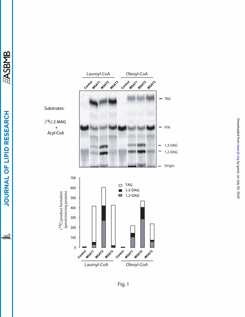
We next compared the DGAT activity of the three MGAT enzymes towards
different MAG isomers by using [14C]oleoyl-CoA as an acyl donor. As expected, the
major MGAT enzymatic products for sn-1-MAG and sn-2-MAG are 1,3-DAG and 1,2- or
2,3-DAG, respectively (Fig. 2). In contrast to MGAT1 and MGAT2, MGAT3 displayed
much stronger DGAT activity, which is supported by the high level of [14C]-TAG
produced when different sn-1-MAGs were used as substrates (Fig. 2). Again, MGAT2
displayed strong MGAT activity while its DGAT activity is relatively low.
Profiling of DGAT activity of MGAT enzymes using DAGs as substrates – We
next directly examined the DGAT activity of the three MGAT enzymes with 1,2-DAG or
1,3-DAG as acyl acceptor and [14C]oleoyl-CoA as acyl donor, using DGAT1 as a
positive control. As shown in Fig. 3, all the three MGAT enzymes possessed DGAT
activity in the order of MGAT3 > MGAT1 > MGAT2, which is consistent with the data
from the previous experiments utilizing MAG as an acyl acceptor.
by guest, on July 30, 2018w
ww
.jlr.orgD
ownloaded from

11
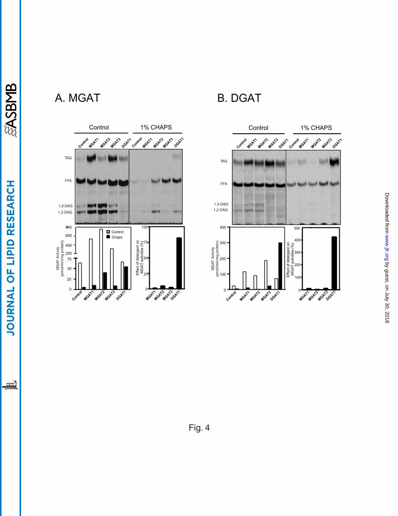
Differential effects of zwitterionic detergent CHAPS on the activity of the MGAT
and DGAT enzymes – Detergents have been widely used in soluablizing membrane-
associated proteins and protein complexes, including MGAT and DGAT from various
primary tissues. The use of detergent often led to substantial loss of enzyme activities
(17, 18). Although both MGAT3 and DGAT1 enzymes recognize DAG as substrate, the
two enzymes share no sequence homology in protein sequence. To differentiate the
two enzymes in catalytic features, we next analyzed the responses of MGAT and DGAT
enzymes to the treatment of zwitterionic detergent. The results showed that the activity
of all the three MGAT enzymes was severely inhibited by 1% CHAPS, as evidenced by
a dramatic decrease in the formation of DAG and TAG in the presence of detergent (Fig
4A). In contrast, both MGAT and DGAT activities of DGAT1 enzyme were largely
retained in the presence of 1% CHAPS (Fig. 4A). Although the DGAT activity of the
MGAT enzymes were also inhibited by the detergent, it is not clear whether this was
caused by the reduction of DAG that was used as substrate for the DGAT activity of the
MGAT enzymes. To address the issue, we next analyzed the effect of detergent on
DGAT activity of MGAT enzymes by directly using DAG as substrate. As shown in Fig.
4B, the DGAT activity of all the three MGAT enzymes was inactivated by the presence
of 1% CHAPS. In contrast to a slight reduction in DGAT activity when MAG was used
as a substrate, the DGAT1 activity was significantly stimulated by the presence of
detergent when DAG was used as a substrate (Fig 4B). The results suggest that DGAT
and MGAT activities of the MGAT enzymes are inseparable, whereas the MGAT and
DGAT activities of the DGAT1 enzyme were dispensable.
by guest, on July 30, 2018w
ww
.jlr.orgD
ownloaded from

12
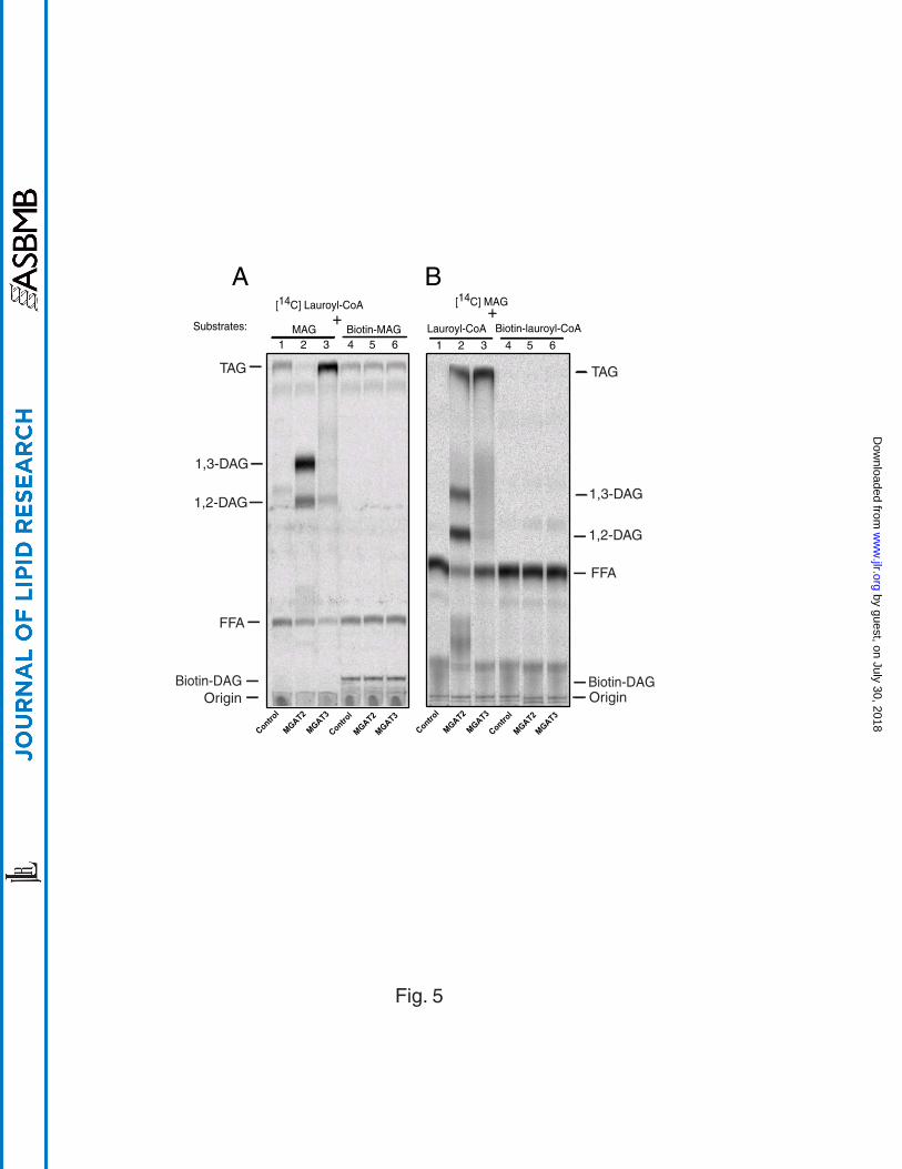
The effect of substrate biotinylation on MGAT enzyme activities– Biotin is
routinely used for assay development due to its high affinity to streptavidin. Attachment
of biotin to the acyl chain of MAG or acyl-CoA would facilitate separation of biotinylated
DAG from its substrates by using scintillation proximity assay (SPA). To evaluate the
effect of biotinylation of fatty acyl chains on MGAT activity, we analyzed MGAT activity
of MGAT2 and MGAT3 by using either biotinylated lauroyl-CoA or biotinylated
monolauroylglycerol as substrates. As shown in Fig 5, the recombinant MGAT2 and
MGAT3 showed no catalytic activity toward either biotinylated monolauroylglycerol (Fig.
5A, lanes 4 to 6) or biotinylated lauroyl-CoA (Fig. 5B, lanes 4 to 6). In contrast, both
enzymes demonstrated high acyltransferase activity towards unconjugated lauroyl-CoA
and monolauroylglycerol (Fig. 5A and 5B, lanes 1 to 3) as evidenced by the increased
production of radio-labeled 1,2-DAG by MGAT2 and TAG by MGAT3 as compared with
the wild type control. MGAT1 did not show catalytic activity towards biotinylated
substrates, either (data not shown). The results suggest that biotinylated MAG and
acyl-CoA are no longer recognized as substrates by the MGAT enzymes.
Subcellular localization of MGAT3 and DGAT enzymes in COS-7 cells – To
examine differences in the subcellular localization of MGAT3 from that of DGAT1, we
next performed immunocytohistochemical analyses of the recombinant MGAT3 and
DGAT1 that are tagged with FLAG epitope as well as myc-tagged DGAT2 in transiently
transfected COS-7 cells. Forty eight hours after transfection, cells were processed for
indirect immunofluorescence staining with antibodies specific for the FLAG epitope
(green) as well as calnexin (red), an ER-resident protein used as a positive marker.
by guest, on July 30, 2018w
ww
.jlr.orgD
ownloaded from
13
Cells were also counterstained with DAPI to visualize nuclei (blue). The FLAG-MGAT3
protein expressed in COS-7 cells displayed a perinuclear and punctated pattern (Fig. 6,
a) that co-localized with the ER marker, calnexin (Fig. 6, b), as demonstrated by the
yellow color in the merged image (Fig. 6, c). The results demonstrated conclusively that
MGAT3 was an ER-associated protein. Likewise, myc-DGAT2 also localized in the ER,
as suggested by colocalization with the ER-marker, calnexin (Fig. 6, d to f). In contrast,
the staining pattern of FLAG-DGAT1 was quite different, and is not restricted to the ER,
which is evidenced by a lack of complete overlap between FLAG-DGAT1 and calnexin
(Fig. 6, g to i). Such difference is further supported by a lack of completed co-
localization between DGAT1 and DGAT2 when co-expressed in the same cell (Fig. 6, j
to l). Furthermore, co-expression of DGAT1 and DGAT2 resulted in enlarged ER
structure (Fig. 6, j to l), which is independent from the amount of DNA used in the
transfection. As a negative control of the immunostaining process, no significant
staining was observed in mock-transfected cells stained with anti-FLAG or anti-myc
antibodies or with normal mouse IgG (data not shown). The data demonstrated that
DGAT1 and DGAT2 exhibited some differences in subcellular distribution.
DISCUSSION
In eukaryotes, TAG synthetic pathways play important roles in energy storage,
synthesis of phospholipids, lipoprotein trafficking, and detoxification of free fatty acids.
TAG also plays pivotal roles in maintenance of skin integrity as evidenced by the
phenotype of DGAT2 knockout mice (15). An efficient process of TAG synthesis and
breakdown is required to maintain energy homeostasis under different physiological
by guest, on July 30, 2018w
ww
.jlr.orgD
ownloaded from

14
conditions. Thus, excessive storage of TAG in fat tissues causes obesity, whereas
depletion of fat tissues results in lipodystrophy that is associated with metabolic
diseases such as insulin resistance and diabetes (19). Hence, modulation of TAG
synthesis by targeting appropriate enzymes involved in the process may offer treatment
options for obesity (20, 21), an ongoing epidemic of the developed nations.
The importance of TAG synthesis is also reflected by the complexity of TAG
synthesis pathways. For example, each step of the TAG synthesis pathways is
catalyzed by multiple isoforms of enzymes that differ in tissue distribution and/or
subcellular localization. Among the three MGAT isoforms identified so far, MGAT3
possesses some unique features. The MGAT3 gene is only found in higher mammals
and humans, but not in rodents. Although named after its enzyme activity, MGAT3
actually shares higher sequence homology with DGAT2 than other MGAT isoforms. In
the present report, we investigated catalytic properties and subcellular localization of
human MGAT3 and compared these features with those of MGAT isoforms and DGAT
enzymes. Our results showed that the recombinant MGAT3 enzyme demonstrated
significantly higher DGAT activity than that of MGAT1 and MGAT2 in the order of
MGAT3 > MGAT1 > MGAT2 when either MAG or DAG was used as substrate,
suggesting that MGAT3 functions as a TAG synthase. The results also support the
notion that MGAT and DGAT2 enzymes evolve from a common ancestral gene, and
may provide guidance for identification of residues responsible for the DGAT activity of
MGAT enzymes.
by guest, on July 30, 2018w
ww
.jlr.orgD
ownloaded from

15
While the MGAT3 enzyme exhibited strong DGAT activity, its catalytic properties
are quite different from those of DGAT1. MGAT3 was very sensitive to treatment with
1% CHAPS, whereas DGAT1 activity was stimulated by the treatment, indicating a
different catalytic mechanism. Interestingly, MGAT3 activities were sensitive to
detergent inactivation when either MAG or DAG was used as substrates, suggesting
that DGAT and MGAT activities of the MGAT enzymes are inseparable.
In mammals, there are two isoforms of DGAT enzymes that catalyze the final
step in TAG synthesis. The two DGAT enzymes share little sequence homology, and
exhibit different phenotypes when inactivated in mice. Mice deficient in DGAT1
enzymes were viable and resistant to diet-induced obesity, whereas DGAT2 knockout
mice developed lethal lipopenia (2, 15). The pathophysiological conditions were not
compensated by DGAT1 that is ubiquitously expressed in all the tissues, suggesting
that the two enzymes are localized in different subcellular localizations. To provide
direct evidence on how the MGAT3 enzyme differs from the DGAT enzymes in
subcellular localization, we compared subcellular distribution of MGAT3 with that of
DGAT1 and DGAT2 enzymes by immunohistochemistry. Consistent with high
sequence homology to DGAT2, both MGAT3 and DGAT2 protein exhibited typical
staining pattern of ER, as evidenced by co-localization with the ER resident protein
calnexin. Similar results are also obtained with MGAT1 and MGAT2 enzymes (data not
shown). Although DGAT1 is also localized in the ER, but the protein appears to have a
wider distribution so that its localization goes beyond the site of ER, which is supported
by a lack of complete co-localization of DGAT1 with calnexin or DGAT2 enzyme when
by guest, on July 30, 2018w
ww
.jlr.orgD
ownloaded from
16
co-expressed in the same cells. In support of this notion, overexpression of DGAT1
results in accumulation of small lipid droplets around cell periphery, whereas
overexpression of DGAT2 leads to increase in large cytosolic lipid droplets (15). Such
difference is also supported from previous reports on rat liver and yeast DGAT
enzymes. Two type of DGAT activities were detected from liver microsomes, one is
overt that regulates the cytosolic TAG pools, whereas the other is a latent that plays a
role in TAG secretion (22, 23). In yeast cells, inactivation of the yeast DGAT1 gene
resulted in reduction of DGAT activity on lipid particles, but without significant effect on
DGAT activity in the ER (24). Since the MGAT enzymes shares similar subcellular
localization with DGAT2, it can be envisaged that they may share common functional
roles, which remains to be elucidated in future studies of mice with targeted deletion of
MGAT enzymes.
by guest, on July 30, 2018w
ww
.jlr.orgD
ownloaded from
17
FIGURE LEGEND
Fig 1. Acylation activities of MGAT1, 2, & 3 towards 2-[14C]monooleoylglycerol
and two different acyl-CoAs, lauroyl-CoA or oleoyl-CoA. The acyltransferase
assays were conducted by incubating 40 M of acyl-CoA with 20 M of 2-
[14C]monooleoylglycerol in the presence of 100 g of cell lysates from Sf-9 cells infected
with either the wild-type baculovirus (control) or recombinant baculovirus expressing
MGATs, as described under “Experimental Procedures”. Representative TLC analyses
are shown in upper panel and quantitative data of specific activity are shown in the
lower bar graph. Note that the formation of radiolabeled triacylglycerol represents DGAT
activity, whereas total formation of radiolabeled diacylglycerol and triacylglycerol
represents MGAT activity because radiolabeled monooleoylglycerols were used as
substrate. MAG, monoacylglycerol; DAG, diacylglycerol; FFA, free fatty acid, TAG,
triacylglycerol.
Fig 2. Acylation activities of MGAT1, 2, & 3 towards [14C]oleoyl-CoA and different
monoacylglycerols. The experiment was conducted as exactly described in the legend
of Fig. 1 with the exception of different application of substrates. In this experiment, 20
M of [14C]oleoyl-CoA and 200 M of various MAGs were used. Representative TLC
analyses are shown in upper panel and quantitative data of specific activity are shown
in the lower bar graph.
Fig 3. Analysis of DGAT activities of MGAT1, 2, & 3 and DGAT1 towards 1, 2-
diacylglycerol (A) or 1,3-diacylglycerol (B). DGAT enzyme assays were conducted
by guest, on July 30, 2018w
ww
.jlr.orgD
ownloaded from

18
by incubating 20 M of [14C]oleoyl-CoA with 200 M of 1,2-dioleoylglycerol or 1,3-
dioleoylglycerol in the presence of 100 g of cell lysates from Sf-9 cells infected with
either the wild-type baculovirus (control) or recombinant baculovirus expressing MGATs
or DGAT1, as described under “Materials and Methods”. Representative TLC analyses
are shown in upper panel and quantitative data of specific DGAT activity are shown in
the lower bar graph.
Fig 4. Effect of CHAPS on MGAT activity (A) and DGAT activity (B) of MGAT1, 2, &
3 and DGAT1. The reaction was conducted by incubating 200 M of sn-2-
monooleoylglycerol (A) or sn-1, 2-dioleoylglycerol (B) and 20 M of [14C]Oleoyl-CoA for
10 min at room temperature with 100 g of protein in the absence or presence of 1%
CHAPS, followed by lipids extraction and TLC analysis. Specific MGAT and DGAT
activities are shown by a representative TLC analysis (upper panel) and quantified as
shown in the lower bar graph. The effect of CHAPS on the enzyme activities is also
expressed as percent of the enzyme activity obtained in the absence of the detergent.
Fig 5. Loss of recognition to biotinylated substrates by MGAT2 and MAGT3.
Reaction was conducted by incubating 100 g of cell lysates with 20 M 14C-labeled
and 40 M biotin-labeled substrates for 10 min at room temperature. While a significant
acylation activity in MGAT2- and MGAT3-containing cell lysates towards unconjugated
substrates was detected (lanes 1 to 3 from both panels), such an activity was not
observed towards biotinylated counterparts (lanes 4 to 6).
by guest, on July 30, 2018w
ww
.jlr.orgD
ownloaded from

19
Fig 6. Subcellular localization of MGAT and DGAT enzymes expressed in COS-7
cells. COS-7 cells were transiently transfected with a FLAG-tagged MGAT3, FLAG-
tagged DGAT1, and myc-tagged DGAT2, respectively, as described under
“Experimental Procedure”. Forty-eight hours after transfection, cells were processed for
indirect immunofluorescence staining with monoclonal antibodies specific for the tag
peptide or calnexin, a resident ER transmembrane protein. The merged pictures were
shown on the right panel (Merge). The arrow heads in panel i and j highlight staining
pattern of DGAT1 that did not co-localize with that of calnexin and DGAT2, respectively.
The yellow color indicates the co-localization of the two proteins. Bar, 20 m.
by guest, on July 30, 2018w
ww
.jlr.orgD
ownloaded from

20
REFERENCE
1 Coleman, R. A. and D. P. Lee. 2004. Enzymes of triacylglycerol synthesis and their
regulation. Prog Lipid Res. 43: 134-176.
2 Smith, S. J., S. Cases, D. R. Jensen, H. C. Chen, E. Sande, B. Tow, D. A. Sanan, J. Raber,
R. H. Eckel and R. V. Farese, Jr. 2000. Obesity resistance and multiple mechanisms of
triglyceride synthesis in mice lacking Dgat. Nat Genet. 25: 87-90.
3 Listenberger, L. L., X. Han, S. E. Lewis, S. Cases, R. V. Farese, Jr., D. S. Ory and J. E.
Schaffer. 2003. Triglyceride accumulation protects against fatty acid-induced
lipotoxicity. Proc Natl Acad Sci U S A. 100: 3077-3082. Epub 2003 Mar 3010.
4 Toker, A. 2005. The biology and biochemistry of diacylglycerol signalling. Meeting on
molecular advances in diacylglycerol signalling. EMBO Rep. 6: 310-314.
5 Phan, C. T. and P. Tso. 2001. Intestinal lipid absorption and transport. Front Biosci. 6:
D299-319.
6 Polheim, D., J. S. David, F. M. Schultz, M. B. Wylie and J. M. Johnston. 1973.
Regulation of triglyceride biosynthesis in adipose and intestinal tissue. J Lipid Res. 14:
415-421.
7 Cases, S., S. J. Smith, Y. W. Zheng, H. M. Myers, S. R. Lear, E. Sande, S. Novak, C.
Collins, C. B. Welch, A. J. Lusis, S. K. Erickson and R. V. Farese, Jr. 1998. Identification
of a gene encoding an acyl CoA:diacylglycerol acyltransferase, a key enzyme in
triacylglycerol synthesis. Proc Natl Acad Sci U S A. 95: 13018-13023.
8 Lardizabal, K. D., J. T. Mai, N. W. Wagner, A. Wyrick, T. Voelker and D. J. Hawkins.
2001. DGAT2 is a new diacylglycerol acyltransferase gene family: purification, cloning,
by guest, on July 30, 2018w
ww
.jlr.orgD
ownloaded from

21
and expression in insect cells of two polypeptides from Mortierella ramanniana with
diacylglycerol acyltransferase activity. J Biol Chem. 276: 38862-38869.
9 Cases, S., S. J. Stone, P. Zhou, E. Yen, B. Tow, K. D. Lardizabal, T. Voelker and R. V.
Farese, Jr. 2001. Cloning of DGAT2, a second mammalian diacylglycerol
acyltransferase, and related family members. J Biol Chem. 276: 38870-38876.
10 Cao, J., J. Lockwood, P. Burn and Y. Shi. 2003. Cloning and functional characterization
of a mouse intestinal acyl-CoA:monoacylglycerol acyltransferase, MGAT2. J Biol Chem.
278: 13860-13866.
11 Cheng, D., T. C. Nelson, J. Chen, S. G. Walker, J. Wardwell-Swanson, R. Meegalla, R.
Taub, J. T. Billheimer, M. Ramaker and J. N. Feder. 2003. Identification of acyl
coenzyme A:monoacylglycerol acyltransferase 3, an intestinal specific enzyme
implicated in dietary fat absorption. J Biol Chem. 278: 13611-13614.
12 Yen, C. L. and R. V. Farese, Jr. 2003. MGAT2, a Monoacylglycerol Acyltransferase
Expressed in the Small Intestine. J Biol Chem. 278: 18532-18537.
13 Binaglia, L., R. Roberti, A. Vecchini and G. Porcellati. 1982. Evidence for a
compartmentation of brain microsomal diacylglycerol. J Lipid Res. 23: 955-961.
14 Rustow, B. and D. Kunze. 1985. Diacylglycerol synthesized in vitro from sn-glycerol 3-
phosphate and the endogenous diacylglycerol are different substrate pools for the
biosynthesis of phosphatidylcholine in rat lung microsomes. Biochim Biophys Acta. 835:
273-278.
15 Stone, S. J., H. M. Myers, S. M. Watkins, B. E. Brown, K. R. Feingold, P. M. Elias and
R. V. Farese, Jr. 2004. Lipopenia and skin barrier abnormalities in DGAT2-deficient
mice. J Biol Chem. 279: 11767-11776. Epub 12003 Dec 11710.
by guest, on July 30, 2018w
ww
.jlr.orgD
ownloaded from
22
16 Yen, C. L., S. J. Stone, S. Cases, P. Zhou and R. V. Farese, Jr. 2002. Identification of a
gene encoding MGAT1, a monoacylglycerol acyltransferase. Proc Natl Acad Sci U S A.
99: 8512-8517.
17 Bhat, B. G., E. S. Bardes and R. A. Coleman. 1993. Solubilization and partial purification
of neonatally expressed rat hepatic microsomal monoacylglycerol acyltransferase. Arch
Biochem Biophys. 300: 663-669.
18 Lehner, R. and A. Kuksis. 1995. Triacylglycerol synthesis by purified triacylglycerol
synthetase of rat intestinal mucosa. Role of acyl-CoA acyltransferase. J Biol Chem. 270:
13630-13636.
19 Simha, V. and A. Garg. 2006. Lipodystrophy: lessons in lipid and energy metabolism.
Curr Opin Lipidol. 17: 162-169.
20 Chen, H. C. and R. V. Farese, Jr. 2000. DGAT and triglyceride synthesis: a new target for
obesity treatment? Trends Cardiovasc Med. 10: 188-192.
21 Shi, Y. and P. Burn. 2004. Lipid metabolic enzymes: emerging drug targets for the
treatment of obesity. Nat Rev Drug Discov. 3: 695-710.
22 Owen, M. R., C. C. Corstorphine and V. A. Zammit. 1997. Overt and latent activities of
diacylglycerol acytransferase in rat liver microsomes: possible roles in very-low-density
lipoprotein triacylglycerol secretion. Biochem J. 323: 17-21.
23 Waterman, I. J. and V. A. Zammit. 2002. Activities of overt and latent diacylglycerol
acyltransferases (DGATs I and II) in liver microsomes of ob/ob mice. Int J Obes Relat
Metab Disord. 26: 742-743.
by guest, on July 30, 2018w
ww
.jlr.orgD
ownloaded from

23
24 Sorger, D. and G. Daum. 2002. Synthesis of triacylglycerols by the acyl-coenzyme
A:diacyl-glycerol acyltransferase Dga1p in lipid particles of the yeast Saccharomyces
cerevisiae. J Bacteriol. 184: 519-524.
by guest, on July 30, 2018w
ww
.jlr.orgD
ownloaded from

0
50
100
150
150
200
250
0
50
100
150
200
250
1,2-Dioleoylglycerol + [14C]oleoyl-CoA 1,3-Dioleoylglycerol + [14C]oleoyl-CoA
MGAT1
MGAT2
MGAT3
DGAT1
Control
MGAT1
MGAT2
MGAT3
DGAT1
Control
A B
TAG
FFA
1,3-DAG1,2-DAG
Origin
[14 C
]TA
G fo
rmed
(pm
ol/m
in/m
g pr
otei
n)
[14 C
]TA
G fo
rmed
(pm
ol/m
in/m
g pr
otei
n)
MGAT1
MGAT2
MGAT3
DGAT1
Control
MGAT1
MGAT2
MGAT3
DGAT1
Control
Fig.3
by guest, on July 30, 2018w
ww
.jlr.orgD
ownloaded from
25
50
75
Control
Chaps
200
400
600
800
0 0
25
50
75
100
Eff
ect
of
de
terg
en
t o
n
MG
AT
activitie
s (
%)
MG
AT A
ctiv
ity(p
mol
/min
/mg
prot
ein)
TAG
FFA
1,3-DAG
1,2-DAG
0
100
200
300
400
MG
AT A
ctiv
ity(p
mol
/min
/mg
prot
ein)
TAG
FFA
1,3-DAG
1,2-DAG
0
100
200
300
400
500
Eff
ect
of
de
terg
en
t o
n
MG
AT
activitie
s (
%)
MGAT1
MGAT2
MGAT3
DGAT1
Control
MGAT1
MGAT2
MGAT3
DGAT1
Control
Control 1% CHAPS
A. MGAT
MGAT1
MGAT2
MGAT3
DGAT1
Control
MGAT1
MGAT2
MGAT3
DGAT1
Control
Control 1% CHAPS
B. DGAT
MGAT1
MGAT2
MGAT3
DGAT1
Control
MGAT1
MGAT2
MGAT3
DGAT1
MGAT1
MGAT2
MGAT3
DGAT1
Control
MGAT1
MGAT2
MGAT3
DGAT1
Fig. 4
by guest, on July 30, 2018w
ww
.jlr.orgD
ownloaded from
MGAT2
MGAT3
Control
MGAT2
MGAT3
Control
MGAT2
MGAT3
Control
MGAT2
MGAT3
Control
MAG
[14C] Lauroyl-CoA
Biotin-MAGSubstrates: +[14C] MAG
Lauroyl-CoA Biotin-lauroyl-CoA+
TAG
FFA
1,3-DAG
1,2-DAG
TAG
FFA
1,3-DAG
1,2-DAG
OriginBiotin-DAG
Origin
1 2 3 4 5 6 1 2 3 4 5 6
A B
Biotin-DAG
Fig. 5
by guest, on July 30, 2018w
ww
.jlr.orgD
ownloaded from

d e f
MGAT3 Calnexin
Fig. 6
a b c
DGAT1 Calnexin
g h i
DGAT2 Calnexin
j k l
DGAT1 DGAT2
by guest, on July 30, 2018w
ww
.jlr.orgD
ownloaded from