Embed Size (px)
Citation preview


HumanGene Therapy
A Guide to
7406 tp.indd 1 5/4/10 10:36:23 AM

May 4, 2010 11:0 SPI-B903 9in x 6in b903-fm
This page intentionally left blankThis page intentionally left blank

N E W J E R S E Y • L O N D O N • S I N G A P O R E • B E I J I N G • S H A N G H A I • H O N G K O N G • TA I P E I • C H E N N A I
World Scientific
HumanGene
TherapyEditors
Roland W HerzogSergei ZolotukhinUniversity of Florida, USA
A Guide to
7406 tp.indd 2 5/4/10 10:36:25 AM

British Library Cataloguing-in-Publication DataA catalogue record for this book is available from the British Library.
For photocopying of material in this volume, please pay a copying fee through the CopyrightClearance Center, Inc., 222 Rosewood Drive, Danvers, MA 01923, USA. In this case permission tophotocopy is not required from the publisher.
ISBN-13 978-981-4280-90-7ISBN-10 981-4280-90-9
Typeset by Stallion PressEmail: [email protected]
All rights reserved. This book, or parts thereof, may not be reproduced in any form or by any means,electronic or mechanical, including photocopying, recording or any information storage and retrievalsystem now known or to be invented, without written permission from the Publisher.
Copyright © 2010 by World Scientific Publishing Co. Pte. Ltd.
Published by
World Scientific Publishing Co. Pte. Ltd.
5 Toh Tuck Link, Singapore 596224
USA office: 27 Warren Street, Suite 401-402, Hackensack, NJ 07601
UK office: 57 Shelton Street, Covent Garden, London WC2H 9HE
Printed in Singapore.
A GUIDE TO HUMAN GENE THERAPY
Sanjeed - A Guide to Human Gene Therapy.pmd 8/2/2010, 6:19 PM1

May 4, 2010 11:0 SPI-B903 9in x 6in b903-fm
Preface
Kenneth I. Berns∗
Ever since the discovery of the DNA double helix by Watson and Cricktremendous advances in our knowledge of molecular genetics and cell biol-ogy have made it one of the most exciting areas of science for more than 50years. We have now progressed beyond the realm of basic research in genet-ics to the development of biotechnology, which has allowed us to producehuman proteins in bacteria and to use these, as in the case of insulin, fortherapy of human disease. In many cases the therapy derived from biotech-nology is designed as replacement therapy to make up for defects at the DNAlevel, which result in either the wrong protein being made or no protein atall. A more sophisticated approach than replacement therapy would be tocorrect a deficit at the level of the gene by either regulating gene expressionor replacing or substituting for a defective gene. Once the discovery andcharacterization of restriction enzymes made it possible to identify, isolate,and clone specific genes the way was open to begin to think about genetherapy. This book describes the state of the art of gene therapy, which isdesigned to either directly introduce a good copy of a defective gene or tohave the cell express a new protein or regulatory RNA which will block thedeleterious effects of a defective gene.
The requirements for executing gene therapy successfully are relativelyfew. First the molecular nature of the defect to be corrected needs to beunderstood. This has been achieved for a large number of diseases, althoughmultifactorial diseases still pose a challenge. Second, the corrective DNAsequence (i.e., the gene) needs to be determined and sufficient amounts for
∗Correspondence: Director, University of Florida Genetics Institute, Gainesville, FL 32610.E-mail: [email protected]
v
May 4, 2010 11:0 SPI-B903 9in x 6in b903-fm
A Guide to Human Gene Therapy
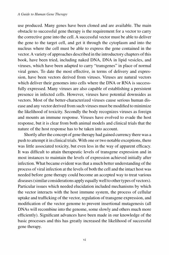
use produced. Many genes have been cloned and are available. The mainobstacle to successful gene therapy is the requirement for a vector to carrythe corrective gene into the cell. A successful vector must be able to deliverthe gene to the target cell, and get it through the cytoplasm and into thenucleus where the cell must be able to express the gene contained in thevector.A variety of approaches described in the introductory chapters of thisbook, have been tried, including naked DNA, DNA in lipid vesicles, andviruses, which have been adapted to carry “transgenes” in place of normalviral genes. To date the most effective, in terms of delivery and expres-sion, have been vectors derived from viruses. Viruses are natural vectorswhich deliver their genomes into cells where the DNA or RNA is success-fully expressed. Many viruses are also capable of establishing a persistentpresence in infected cells. However, viruses have potential downsides asvectors. Most of the better-characterized viruses cause serious human dis-ease and any vector derived from such viruses must be modified to minimizethe likelihood of toxicity. Secondly the body recognizes viruses as foreignand mounts an immune response. Viruses have evolved to evade the hostresponse, but it is clear from both animal models and clinical trials that thenature of the host response has to be taken into account.
Shortly after the concept of gene therapy had gained currency there was apush to attempt it in clinical trials. With one or two notable exceptions, therewas little associated toxicity, but even less in the way of apparent efficacy.It was difficult to attain therapeutic levels of transgene expression and inmost instances to maintain the levels of expression achieved initially afterinfection. What became evident was that a much better understanding of theprocess of viral infection at the levels of both the cell and the intact host wasneeded before gene therapy could become an accepted way to treat variousdiseases (similar considerations apply equally well to other types of vectors).Particular issues which needed elucidation included mechanisms by whichthe vector interacts with the host immune system, the process of cellularuptake and trafficking of the vector, regulation of transgene expression, andmodification of the vector genome to prevent insertional mutagenesis (allDNAs will recombine into the genome, some slowly and others much moreefficiently). Significant advances have been made in our knowledge of thebasic processes and this has greatly increased the likelihood of successfulgene therapy.
vi
May 4, 2010 11:0 SPI-B903 9in x 6in b903-fm
Preface
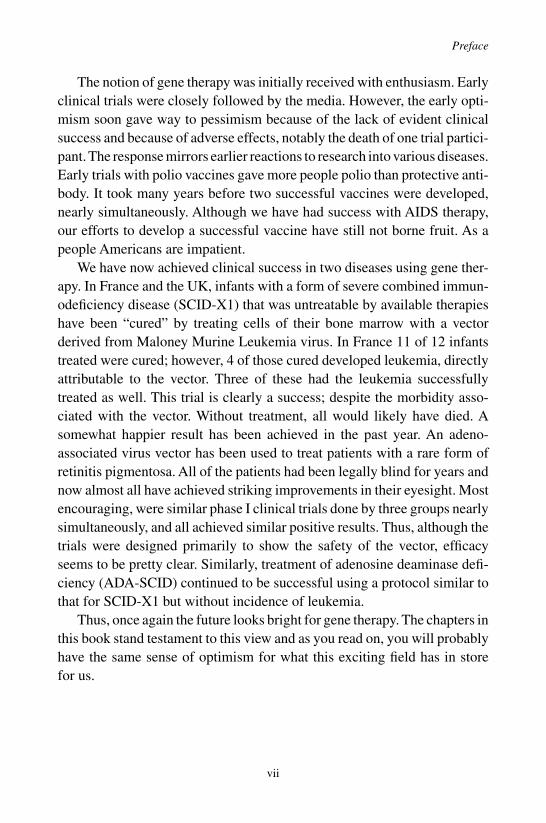
The notion of gene therapy was initially received with enthusiasm. Earlyclinical trials were closely followed by the media. However, the early opti-mism soon gave way to pessimism because of the lack of evident clinicalsuccess and because of adverse effects, notably the death of one trial partici-pant. The response mirrors earlier reactions to research into various diseases.Early trials with polio vaccines gave more people polio than protective anti-body. It took many years before two successful vaccines were developed,nearly simultaneously. Although we have had success with AIDS therapy,our efforts to develop a successful vaccine have still not borne fruit. As apeople Americans are impatient.
We have now achieved clinical success in two diseases using gene ther-apy. In France and the UK, infants with a form of severe combined immun-odeficiency disease (SCID-X1) that was untreatable by available therapieshave been “cured” by treating cells of their bone marrow with a vectorderived from Maloney Murine Leukemia virus. In France 11 of 12 infantstreated were cured; however, 4 of those cured developed leukemia, directlyattributable to the vector. Three of these had the leukemia successfullytreated as well. This trial is clearly a success; despite the morbidity asso-ciated with the vector. Without treatment, all would likely have died. Asomewhat happier result has been achieved in the past year. An adeno-associated virus vector has been used to treat patients with a rare form ofretinitis pigmentosa. All of the patients had been legally blind for years andnow almost all have achieved striking improvements in their eyesight. Mostencouraging, were similar phase I clinical trials done by three groups nearlysimultaneously, and all achieved similar positive results. Thus, although thetrials were designed primarily to show the safety of the vector, efficacyseems to be pretty clear. Similarly, treatment of adenosine deaminase defi-ciency (ADA-SCID) continued to be successful using a protocol similar tothat for SCID-X1 but without incidence of leukemia.
Thus, once again the future looks bright for gene therapy. The chapters inthis book stand testament to this view and as you read on, you will probablyhave the same sense of optimism for what this exciting field has in storefor us.
vii

May 4, 2010 11:0 SPI-B903 9in x 6in b903-fm
This page intentionally left blankThis page intentionally left blank
May 4, 2010 11:0 SPI-B903 9in x 6in b903-fm
Contents
Preface v
Contributors xxiii
1. Non-Viral Gene Therapy 1
Sean M. Sullivan
1. Introduction . . . . . . . . . . . . . . . . . . . . . . . . 12. Plasmid DNA . . . . . . . . . . . . . . . . . . . . . . . 3
2.1 Plasmid DNA Manufacture . . . . . . . . . . . . . 53. Plasmid DNA Gene Transfer Methods . . . . . . . . . . 6
3.1 Plasmid DNA or “Naked DNA” as a Gene DeliverySystem . . . . . . . . . . . . . . . . . . . . . . . 63.1.1 Electroporation of Naked DNA . . . . . . . 83.1.2 Sonoporation of Naked DNA . . . . . . . . 9
3.2 Plasmid DNA Formulations . . . . . . . . . . . . 93.2.1 Cationic Lipids . . . . . . . . . . . . . . . 9
3.2.1.1 In vitro transfection . . . . . . . . 103.2.1.2 Systemic in vivo gene transfer . . . 113.2.1.3 Local administration of cationic
lipid/pDNA transfectioncomplexes . . . . . . . . . . . . . 12
3.3 Polymer . . . . . . . . . . . . . . . . . . . . . . . 143.3.1 Cationic Polymers . . . . . . . . . . . . . . 143.3.2 Neutral Polymer . . . . . . . . . . . . . . . 15
Conclusions . . . . . . . . . . . . . . . . . . . . . . . . 17References . . . . . . . . . . . . . . . . . . . . . . . . . . . 17
ix
May 4, 2010 11:0 SPI-B903 9in x 6in b903-fm
A Guide to Human Gene Therapy
2. Adenoviral Vectors 21
Stuart A. Nicklin and Andrew H. Baker
1. Introduction . . . . . . . . . . . . . . . . . . . . . . . . 212. Adenoviral Capsid Structure . . . . . . . . . . . . . . . 223. Adenoviral Cell Entry . . . . . . . . . . . . . . . . . . . 234. Production of Adenoviral Vectors . . . . . . . . . . . . . 245. Production of Targeted Adenoviral Vectors . . . . . . . . 266. Gene Therapy Applications . . . . . . . . . . . . . . . . 287. Immune Responses to Ad Vectors . . . . . . . . . . . . 308. Safety and Regulatory Issues . . . . . . . . . . . . . . . 329. Conclusions . . . . . . . . . . . . . . . . . . . . . . . . 33References . . . . . . . . . . . . . . . . . . . . . . . . . . . 33
3. Retroviral Vectors and Integration Analysis 37
Cynthia C. Bartholomae, Romy Kirsten, Hanno Glimm,Manfred Schmidt and Christof von Kalle
1. Introduction . . . . . . . . . . . . . . . . . . . . . . . . 372. Design, Production and Mechanism of Transduction . . . 383. In vivo Application . . . . . . . . . . . . . . . . . . . . 414. Side Effects in Retroviral Gene Therapy . . . . . . . . . 42
4.1 Distribution of Retroviral Integration Sitesin the Cellular Genome . . . . . . . . . . . . . . . 42
4.2 Side Effects in Clinical and Preclinical GeneTherapy Studies . . . . . . . . . . . . . . . . . . . 45
5. New Strategies for Vector Biosafety in Gene Therapy . . 47References . . . . . . . . . . . . . . . . . . . . . . . . . . . 49
4. Lentiviral Vectors 53
Janka Mátrai, Marinee K. L. Chuahand Thierry VandenDriessche
1. Basic Viral Biology . . . . . . . . . . . . . . . . . . . . 532. Vector Design and Production . . . . . . . . . . . . . . 56
2.1 Vector Development . . . . . . . . . . . . . . . . 562.2 Vector Production . . . . . . . . . . . . . . . . . . 58
x
May 4, 2010 11:0 SPI-B903 9in x 6in b903-fm
Contents
3. Gene Transfer Concepts and Potential Applications . . . 593.1 Target Cells and Diseases . . . . . . . . . . . . . . 593.2 Pseudotyping . . . . . . . . . . . . . . . . . . . . 593.3 Cell Type Specific Targeting . . . . . . . . . . . . 603.4 Integration-Defective Lentiviral Vectors . . . . . . 60
4. Immune Consequences . . . . . . . . . . . . . . . . . . 625. Safety Issues . . . . . . . . . . . . . . . . . . . . . . . 636. Conclusions and Perspectives . . . . . . . . . . . . . . . 64References . . . . . . . . . . . . . . . . . . . . . . . . . . . 64
5. Herpes Simplex Virus Vectors 69
William F. Goins, David M. Krisky, James B. Wechuck,Darren Wolfe, Justus B. Cohen and Joseph C. Glorioso
1. Introduction . . . . . . . . . . . . . . . . . . . . . . . . 692. HSV Biology in the Design of Replication DefectiveVectors 743. HSV Vector Design Technology . . . . . . . . . . . . . 774. Gene Transfer/Therapy Applications . . . . . . . . . . . 795. Immunology . . . . . . . . . . . . . . . . . . . . . . . 806. Safety and Regulatory Issues . . . . . . . . . . . . . . . 817. Summary . . . . . . . . . . . . . . . . . . . . . . . . . 81References . . . . . . . . . . . . . . . . . . . . . . . . . . . 82
6. Adeno-Associated Viral (AAV) Vectors 87
Nicholas Muzyczka
1. Introduction . . . . . . . . . . . . . . . . . . . . . . . . 872. Biology of AAV . . . . . . . . . . . . . . . . . . . . . . 883. Vector Technology . . . . . . . . . . . . . . . . . . . . 934. Vector Characteristics In Vivo . . . . . . . . . . . . . . . 965. Next Generation Vectors . . . . . . . . . . . . . . . . . 986. Conclusions and Outlook . . . . . . . . . . . . . . . . . 99References . . . . . . . . . . . . . . . . . . . . . . . . . . . 99
xi
May 4, 2010 11:0 SPI-B903 9in x 6in b903-fm
A Guide to Human Gene Therapy
7. Regulatory RNA in Gene Therapy 103
Alfred. S. Lewin
1. Introduction . . . . . . . . . . . . . . . . . . . . . . . . 1032. Delivery of Therapeutic RNAs . . . . . . . . . . . . . . 1063. Ribozymes . . . . . . . . . . . . . . . . . . . . . . . . 1094. RNAi for Gene Therapy . . . . . . . . . . . . . . . . . 1115. Gene Therapy Using miRNA . . . . . . . . . . . . . . . 1146. Aptamers, Decoys and Bi-Functional RNAs . . . . . . . 1157. Modification of Cis-Acting Regulatory
RNA Sequences . . . . . . . . . . . . . . . . . . . . . . 1168. Conclusions . . . . . . . . . . . . . . . . . . . . . . . . 119References . . . . . . . . . . . . . . . . . . . . . . . . . . . 120
8. DNA Integrating Vectors (Transposon, Integrase) 123
Lauren E. Woodard and Michele P. Calos
1. Basic Vector Biology . . . . . . . . . . . . . . . . . . . 1231.1 Transposon Systems . . . . . . . . . . . . . . . . 1241.2 Integrase Systems . . . . . . . . . . . . . . . . . . 126
2. Vector Design and Production . . . . . . . . . . . . . . 1282.1 Design of Transposon Systems . . . . . . . . . . . 1282.2 Design of Integrase Systems . . . . . . . . . . . . 1282.3 Production of Plasmid DNA . . . . . . . . . . . . 129
3. Gene Transfer Protocols and Potential Applications . . . 1303.1 Hepatocyte Transfection via Hydrodynamic
Injection . . . . . . . . . . . . . . . . . . . . . . . 1303.2 Lipophilic Complexes to Transfect Endothelial
Cells and Glioblastoma . . . . . . . . . . . . . . . 1313.3 Direct DNA Injection and Electroporation
to Target Muscle, Retina, and Joints . . . . . . . . 1313.4 Integration into Cultured Cells for
Ex vivo Gene Therapy . . . . . . . . . . . . . . . 1314. Immunology . . . . . . . . . . . . . . . . . . . . . . . 132
xii
May 4, 2010 11:0 SPI-B903 9in x 6in b903-fm
Contents
5. Safety and Regulatory Issues . . . . . . . . . . . . . . . 1335.1 Integration Profiles and Associated Hazards . . . . 1335.2 Efforts to Enhance Integration Specificity . . . . . 1335.3 Effects on Tumor Latency in Mouse Models
of Cancer . . . . . . . . . . . . . . . . . . . . . . 134References . . . . . . . . . . . . . . . . . . . . . . . . . . . 135
9. Homologous Recombination and Targeted GeneModification for Gene Therapy 139
Matthew Porteus
1. Introduction . . . . . . . . . . . . . . . . . . . . . . . . 1392. Problems with Using Gene Targeting by Homologous
Recombination . . . . . . . . . . . . . . . . . . . . . . 1403. Homologous Recombination in Embryonic
Stem Cells . . . . . . . . . . . . . . . . . . . . . . . . . 1414. Homologous Recombination using
Adeno-Associated Virus . . . . . . . . . . . . . . . . . 1445. Site-Specific Modification of the Genome using
Double-Strand Breaks . . . . . . . . . . . . . . . . . . 1446. Double-Strand Break Repair . . . . . . . . . . . . . . . 1447. Double-Strand Break Induced Homologous
Recombination . . . . . . . . . . . . . . . . . . . . . . 1468. Re-design of Homing Endonucleases to Recognize
New Target Sites . . . . . . . . . . . . . . . . . . . . . 1469. Development of Zinc Finger Nucleases . . . . . . . . . 14710. Using Zinc Finger Nucleases to Stimulate
Gene Targeting . . . . . . . . . . . . . . . . . . . . . . 14711. Using Zinc Finger Nucleases to Site-Specifically Modify
Genes by Mutagenic Non-HomologousEnd-Joining . . . . . . . . . . . . . . . . . . . . . . . . 149
12. Strategies of Zinc Finger Nuclease Design . . . . . . . . 15113. Aspects of Zinc Finger Binding Sites and Structure
of Zinc Finger Nucleases . . . . . . . . . . . . . . . . . 15314. Zinc Finger Nuclease Toxicity: Measuring
and Minimizing . . . . . . . . . . . . . . . . . . . . . . 154
xiii

May 4, 2010 11:0 SPI-B903 9in x 6in b903-fm
A Guide to Human Gene Therapy
15. The Challenge of Delivery . . . . . . . . . . . . . . . . 15616. Future Directions and Promise of Homologous
Recombination as a Gene Correction Approachto Gene Therapy . . . . . . . . . . . . . . . . . . . . . 157
References . . . . . . . . . . . . . . . . . . . . . . . . . . . 157
10. Gene Switches for Pre-Clinical Studies in Gene Therapy 163
Caroline Le Guiner, Knut Stieger, Alice Toromanoff,Fabienne Rolling, Philippe Moullier and Oumeya Adjali
1. Introduction . . . . . . . . . . . . . . . . . . . . . . . . 1632. Rapamycin-Dependent Regulatable System . . . . . . . 165
2.1 Molecular Mechanisms Involved in TransgeneRegulation . . . . . . . . . . . . . . . . . . . . . 165
2.2 Pharmacology of Rapamycin . . . . . . . . . . . . 1662.3 Translation Development of the Rapamycin
Dependent Regulation System . . . . . . . . . . . 1663. Tetracycline-Dependent Regulatable Systems . . . . . . 168
3.1 Molecular Mechanisms Involved in TransgeneRegulation . . . . . . . . . . . . . . . . . . . . . 168
3.2 Pharmacology of Doxycycline (Dox) . . . . . . . . 1713.3 Translational Development of Tet-dependant
Regulation Systems . . . . . . . . . . . . . . . . . 1714. Other Regulatable Systems . . . . . . . . . . . . . . . . 1755. General Conclusions . . . . . . . . . . . . . . . . . . . 177References . . . . . . . . . . . . . . . . . . . . . . . . . . . 177
11. Gene Therapy for Central Nervous System Disorders 181
Deborah Young and Patricia A. Lawlor
1. Introduction . . . . . . . . . . . . . . . . . . . . . . . . 1812. Gene Therapy for Parkinson’s Disease . . . . . . . . . . 1823. Gene Therapy for Temporal Lobe Epilepsy . . . . . . . 1864. Huntington’s Disease Gene Therapy . . . . . . . . . . . 1875. Amyotrophic Lateral Sclerosis (ALS) . . . . . . . . . . 1896. Gene Therapy for Canavan Disease . . . . . . . . . . . . 190
xiv

May 4, 2010 11:0 SPI-B903 9in x 6in b903-fm
Contents
7. Gene Therapy for Alzheimer’s Disease . . . . . . . . . . 1918. Conclusions and Outlook . . . . . . . . . . . . . . . . . 193References . . . . . . . . . . . . . . . . . . . . . . . . . . . 194
12. Gene Therapy of Hemoglobinopathies 197
Angela E. Rivers and Arun Srivastava
1. Introduction . . . . . . . . . . . . . . . . . . . . . . . . 1982. β-Thalassemia . . . . . . . . . . . . . . . . . . . . . . 1983. Sickle Cell Disease . . . . . . . . . . . . . . . . . . . . 1994. Gene Therapy . . . . . . . . . . . . . . . . . . . . . . . 200
4.1 Oncoretroviral Vector-Mediated GlobinGene Transfer . . . . . . . . . . . . . . . . . . . . 202
4.2 Lentiviral Vector-Mediated GlobinGene Transfer . . . . . . . . . . . . . . . . . . . . 203
4.3 Adeno-Associated Viral Vector-Mediated GlobinGene Transfer . . . . . . . . . . . . . . . . . . . . 204
References . . . . . . . . . . . . . . . . . . . . . . . . . . . 208
13. Gene Therapy for Primary Immunodeficiencies 213
Aisha Sauer, Barbara Cassani and Alessandro Aiuti
1. Introduction . . . . . . . . . . . . . . . . . . . . . . . . 2142. Adenosine Deaminase (ADA)-deficient SCID . . . . . . 2153. X-linked Severe Combined Immunodeficiency
(SCID-X1) . . . . . . . . . . . . . . . . . . . . . . . . 2184. Gene Therapy for Other SCIDs . . . . . . . . . . . . . . 220
4.1 V(D)J Recombination Defects . . . . . . . . . . . 2204.2 Purine Nucleoside Phosphorylase (PNP)
Deficiency . . . . . . . . . . . . . . . . . . . . . . 2224.3 Janus Kinase 3 (Jak3) Deficiency . . . . . . . . . . 2224.4 IL-7R Deficiency . . . . . . . . . . . . . . . . . . 2234.5 Zeta Associated 70 kDa Phosphoprotein
(ZAP-70) Deficiency . . . . . . . . . . . . . . . . 2235. Wiskott-Aldrich-Syndrome (WAS) . . . . . . . . . . . . 2246. Chronic Granulomatous Disease . . . . . . . . . . . . . 2257. Conclusions and Outlook . . . . . . . . . . . . . . . . . 227References . . . . . . . . . . . . . . . . . . . . . . . . . . . 228
xv

May 4, 2010 11:0 SPI-B903 9in x 6in b903-fm
A Guide to Human Gene Therapy
14. Gene Therapy for Hemophilia 233
David Markusic, Babak Moghimi and Roland Herzog
1. Introduction . . . . . . . . . . . . . . . . . . . . . . . . 2332. Limitations of Hemophilia Treatment With
Coagulation Factor Concentrates or RecombinantCoagulation Factors . . . . . . . . . . . . . . . . . . . . 235
3. Gene Transfer for Correction of Hemophilia . . . . . . . 2363.1 Ex Vivo Gene Transfer of F.VIII and F.IX . . . . . 2363.2 In Vivo Gene Transfer of F.VIII and F.IX . . . . . . 237
4. AAV is a Preferred Gene Therapy Vector for In VivoGene Transfer to Correct of Hemophilia . . . . . . . . . 238
5. Immunological Considerations for Efficient F.IXGene Transfer . . . . . . . . . . . . . . . . . . . . . . . 239
6. Advancements from Small and Large Animal Modelsof Hemophilia . . . . . . . . . . . . . . . . . . . . . . . 2426.1 Murine Hemophilia Models . . . . . . . . . . . . 2426.2 Canine Hemophilia Models . . . . . . . . . . . . . 242
7. Gene Therapy Trials for Hemophilia Past, Present, andFuture . . . . . . . . . . . . . . . . . . . . . . . . . . . 244
8. Conclusions . . . . . . . . . . . . . . . . . . . . . . . . 245References . . . . . . . . . . . . . . . . . . . . . . . . . . . 246
15. Gene Therapy for Obesity and Diabetes 251
Sergei Zolotukhin and Clive H. Wasserfall
1. Introduction . . . . . . . . . . . . . . . . . . . . . . . . 2512. Understanding Obesity: Why We Get Fat . . . . . . . . 252
2.1 Genetic Factors: Human Obesity Gene Map . . . . 2522.2 Environmental Factors: The Big Two and Other
Causal Contributors . . . . . . . . . . . . . . . . . 2533. General Strategies in Gene Therapy for Obesity . . . . . 2534. Gene Delivery Vehicles . . . . . . . . . . . . . . . . . . 2555. Gene Targets for Obesity . . . . . . . . . . . . . . . . . 255
5.1 Leptin . . . . . . . . . . . . . . . . . . . . . . . . 2555.2 Neurocytokines . . . . . . . . . . . . . . . . . . . 256
xvi
May 4, 2010 11:0 SPI-B903 9in x 6in b903-fm
Contents
5.3 AMP-Activated Protein Kinase (AMPK) . . . . . . 2565.4 Adiponectin . . . . . . . . . . . . . . . . . . . . . 2575.5 Wnt-10b . . . . . . . . . . . . . . . . . . . . . . . 2575.6 Obesity Gene Menu à la Carte . . . . . . . . . . . 2585.7 Obesity and Diabetes . . . . . . . . . . . . . . . . 259
References . . . . . . . . . . . . . . . . . . . . . . . . . . . 260
16. Gene Therapy for Duchenne MuscularDystrophy 261
Takashi Okada and Shin’ichi Takeda
1. Introduction . . . . . . . . . . . . . . . . . . . . . . . . 2611.1 Background of Duchenne Muscular Dystrophy . . 261
2. Gene-replacement Strategies using Virus Vectors . . . . 2622.1 Choice of Vector . . . . . . . . . . . . . . . . . . 2622.2 Modification of the Dystrophin Gene
and Promoter . . . . . . . . . . . . . . . . . . . . 2642.3 Use of Surrogate Genes . . . . . . . . . . . . . . . 266
3. AAV-Mediated Transduction of Animal Models . . . . . 2663.1 Vector Production . . . . . . . . . . . . . . . . . . 2663.2 Animal Models for the Gene
Transduction Study . . . . . . . . . . . . . . . . . 2673.3 Immunological Issues of rAAV . . . . . . . . . . . 2683.4 Intravascular Vector Administration
by Limb Perfusion . . . . . . . . . . . . . . . . . 2693.5 Global Muscle Therapies . . . . . . . . . . . . . . 269
4. Safety and Potential Impact of Clinical Trials . . . . . . 2705. Development of Alternative Strategies . . . . . . . . . . 271
5.1 Design of Read-through Drugs . . . . . . . . . . . 2715.2 Modification of mRNA Splicing . . . . . . . . . . 2725.3 Ex Vivo Gene Therapy . . . . . . . . . . . . . . . 272
6. Future Perspectives . . . . . . . . . . . . . . . . . . . . 2736.1 Pharmacological Intervention . . . . . . . . . . . . 2736.2 Capsid Modification . . . . . . . . . . . . . . . . 273
7. Conclusions and Outlook . . . . . . . . . . . . . . . . . 273References . . . . . . . . . . . . . . . . . . . . . . . . . . . 274
xvii
May 4, 2010 11:0 SPI-B903 9in x 6in b903-fm
A Guide to Human Gene Therapy
17. Cancer Gene Therapy 279
Kirsten A.K. Weigel-Van Aken
1. Introduction . . . . . . . . . . . . . . . . . . . . . . . . 2802. Targeting the Tumor Cell . . . . . . . . . . . . . . . . . 280
2.1 DNA Electroporation . . . . . . . . . . . . . . . . 2802.2 Non-Oncolytic Viral Vectors . . . . . . . . . . . . 281
2.2.1 Retrovirus . . . . . . . . . . . . . . . . . . 2812.2.2 Lentivirus . . . . . . . . . . . . . . . . . . 282
2.3 Oncolytic Viruses . . . . . . . . . . . . . . . . . . 2822.3.1 Herpesvirus . . . . . . . . . . . . . . . . . 2832.3.2 Adenovirus . . . . . . . . . . . . . . . . . . 2832.3.3 Poxvirus . . . . . . . . . . . . . . . . . . . 2842.3.4 Measles virus . . . . . . . . . . . . . . . . 2852.3.5 Vesicular stomatitis virus . . . . . . . . . . 285
3. Targeting the Immune System . . . . . . . . . . . . . . 2863.1 Cancer Vaccines . . . . . . . . . . . . . . . . . . . 287
3.1.1 Vaccinia virus . . . . . . . . . . . . . . . . 2873.1.2 Lentivirus . . . . . . . . . . . . . . . . . . 2873.1.3 Adenovirus . . . . . . . . . . . . . . . . . . 2883.1.4 Parvoviruses . . . . . . . . . . . . . . . . . 288
3.2 Mesenchymal Stem Cells (MSC)as Delivery Vehicles . . . . . . . . . . . . . . . . 288
3.3 Adoptive T Cell Transfer . . . . . . . . . . . . . . 2884. Targeting the Tumor Microenvironment . . . . . . . . . 2895. Challenges and Risks of Cancer Gene Therapy . . . . . 2896. Novel Strategies . . . . . . . . . . . . . . . . . . . . . . 290
6.1 Prime/Boost Regimens . . . . . . . . . . . . . . . 2906.2 Immune Cells as Carriers for Viruses . . . . . . . . 290
7. Conclusions . . . . . . . . . . . . . . . . . . . . . . . . 291References . . . . . . . . . . . . . . . . . . . . . . . . . . . 291
18. Gene Therapy for Autoimmune Disorders 295
Daniel F. Gaddy, Melanie A. Ruffner and Paul D. Robbins
1. Introduction . . . . . . . . . . . . . . . . . . . . . . . . 2952. Rheumatoid Arthritis . . . . . . . . . . . . . . . . . . . 296
xviii

May 4, 2010 11:0 SPI-B903 9in x 6in b903-fm
Contents
2.1 Background . . . . . . . . . . . . . . . . . . . . . 2962.2 Existing Therapies . . . . . . . . . . . . . . . . . 2962.3 Target Tissues and Routes of Delivery . . . . . . . 297
2.3.1 Local RA Gene Therapy . . . . . . . . . . . 2972.3.2 Systemic RA Gene Therapy . . . . . . . . . 297
2.4 Immunomodulation . . . . . . . . . . . . . . . . . 2982.5 Overview of Preclinical Gene Therapy Studies . . 2992.6 Overview of Clinical Gene Therapy Studies . . . . 301
3. Type I Diabetes Mellitus . . . . . . . . . . . . . . . . . 3013.1 Background . . . . . . . . . . . . . . . . . . . . . 3013.2 Existing Therapies . . . . . . . . . . . . . . . . . 3023.3 Target Tissues and Routes of Delivery . . . . . . . 3033.4 Immunomodulation . . . . . . . . . . . . . . . . . 3033.5 Overview of Preclinical Gene Therapy Studies . . 3053.6 Overview of Clinical Gene Therapy Studies . . . . 306
4. Conclusions and Outlook . . . . . . . . . . . . . . . . . 307References . . . . . . . . . . . . . . . . . . . . . . . . . . . 308
19. Gene Therapy for Inherited Metabolic Storage Diseases 311
Cathryn Mah
1. Introduction . . . . . . . . . . . . . . . . . . . . . . . . 3112. Lysosomal Storage Diseases . . . . . . . . . . . . . . . 3123. Glycogen Storage Diseases . . . . . . . . . . . . . . . . 3144. Animal Models . . . . . . . . . . . . . . . . . . . . . . 3155. Cross-Correction Strategies . . . . . . . . . . . . . . . . 3196. Direct Correction of Target Tissues . . . . . . . . . . . . 3217. Conclusions and Outlook . . . . . . . . . . . . . . . . . 324References . . . . . . . . . . . . . . . . . . . . . . . . . . . 324
20. Retinal Diseases 327
Shannon E. Boye, Sanford L. Boye and William W. Hauswirth
1. Introduction . . . . . . . . . . . . . . . . . . . . . . . . 3272. Rod and Cone Photoreceptors . . . . . . . . . . . . . . 3303. Cone Photoreceptors . . . . . . . . . . . . . . . . . . . 3334. Retinal Ganglion Cells . . . . . . . . . . . . . . . . . . 335
xix

May 4, 2010 11:0 SPI-B903 9in x 6in b903-fm
A Guide to Human Gene Therapy
5. Retinal Pigment Epithelium . . . . . . . . . . . . . . . 3376. LCA2 Gene Therapy, a Perspective on Translational
Research . . . . . . . . . . . . . . . . . . . . . . . . . 339References . . . . . . . . . . . . . . . . . . . . . . . . . . . 342
21. A Brief Guide to Gene Therapy Treatmentsfor Pulmonary Diseases 345
Ashley T. Martino, Christian Muellerand Terence R. Flotte
1. Introduction . . . . . . . . . . . . . . . . . . . . . . . . 3452. Common Disorders . . . . . . . . . . . . . . . . . . . . 346
2.1 Cystic Fibrosis . . . . . . . . . . . . . . . . . . . 3462.2 Alpha-1 Antitrypsin (A1AT) . . . . . . . . . . . . 348
3. Development of Viral Vectors for Lung Disease . . . . . 3483.1 Adenoviral Vectors . . . . . . . . . . . . . . . . . 3493.2 Adeno-Associated Viral Vectors . . . . . . . . . . 3493.3 Early Conclusions . . . . . . . . . . . . . . . . . 349
4. Enhancing Efficiency . . . . . . . . . . . . . . . . . . . 3504.1 Alternative AAV Viral Vector Serotypes . . . . . . 350
4.1.1 Addition of Expression EnhancingElements . . . . . . . . . . . . . . . . . . . 351
4.2 Adenoviral Vectors . . . . . . . . . . . . . . . . . 3514.3 Physiological Hurdles in the Lung
Environment . . . . . . . . . . . . . . . . . . . . 3525. Non-Viral Vectors . . . . . . . . . . . . . . . . . . . . . 352
5.1 Cationic Liposomes . . . . . . . . . . . . . . . . . 3525.2 Compacted DNA Nanoparticles . . . . . . . . . . 353
6. Gene Therapy Development for Alpha-1Anti-trypsin . . . . . . . . . . . . . . . . . . . . . . . . 353
7. Lung Cancer Gene Therapy Development . . . . . . . . 3548. Cystic Fibrosis Animal Models . . . . . . . . . . . . . . 3559. Cell-Based Therapy for Cystic Fibrosis . . . . . . . . . 35610. Conclusion and Outlooks . . . . . . . . . . . . . . . . . 357References . . . . . . . . . . . . . . . . . . . . . . . . . . . 358
xx

May 4, 2010 11:0 SPI-B903 9in x 6in b903-fm
Contents
22. Cardiovascular Disease 361
Darin J. Falk, Cathryn S. Mah and Barry J. Byrne
1. Introduction . . . . . . . . . . . . . . . . . . . . . . . . 3612. Therapeutic Targets . . . . . . . . . . . . . . . . . . . . 362
2.1 Congenital Heart Disease . . . . . . . . . . . . . . 3622.2 Coronary Artery Disease and Ischemia/
Reperfusion Injury . . . . . . . . . . . . . . . . . 3652.3 Oxidative Stress . . . . . . . . . . . . . . . . . . . 3652.4 Antioxidants . . . . . . . . . . . . . . . . . . . . 3662.5 Cardiac Contractility . . . . . . . . . . . . . . . . 367
3. Animal Models . . . . . . . . . . . . . . . . . . . . . . 3674. Vector Delivery . . . . . . . . . . . . . . . . . . . . . . 3685. Conclusions and Outlook . . . . . . . . . . . . . . . . . 374Acknowledgments . . . . . . . . . . . . . . . . . . . . . . . 374References . . . . . . . . . . . . . . . . . . . . . . . . . . . 374
Index 379
xxi

May 4, 2010 11:0 SPI-B903 9in x 6in b903-fm
This page intentionally left blankThis page intentionally left blank
May 4, 2010 11:0 SPI-B903 9in x 6in b903-fm
Contributors
Oumeya AdjaliINSERM UMR 649CHU Hôtel-Dieu30 Boulevard Jean Monnet,44035, Nantes, France02 28 08 04 15/1702 28 08 04 [email protected]
Alessandro AiutiHS Raffaele Institute/HSR-TIGET,San Raffaele Telethon Institute
for Gene TherapyDIBIT, HSR-TIGET,Via Olgettina, 58,20132 Milano (MI), Italy+39 (0)2 2643 4671+39 (0)2 2643 [email protected]
Kenneth I. BernsUniversity of Florida-Director,Genetics InstituteDepartment of Molecular
Genetics and MicrobiologyCancer/Genetics Research Complex1376 Mowry Road, Room CG-110,Gainesville FL 32610-3610352-273-8100 (Tel)352-273-8284 (Fax)[email protected]
Shannon BoyeUniversity of FloridaDepartment of Ophthalmic
Molecular GeneticsAcademic Research Building1600 SW Archer Road,Room R1-236, Gainesville
Barry ByrneUniversity of FloridaDepartment of Pediatrics
CardiologyPowell Gene Therapy CenterAcademic Research Building1600 SW Archer Road,Room RG-165, Gainesville
Michele CalosStanford UniversityDepartment of GeneticsStanford University School
of MedicineAlway Building, Room M334
xxiii
May 4, 2010 11:0 SPI-B903 9in x 6in b903-fm
A Guide to Human Gene Therapy
300 Pasteur Drive, Stanford,CA 94305-5120
Dr. William F. GoinsUniversity of Pittsburgh School
of MedicineDepartment of Microbiology &
Molecular GeneticsW1157 BSTWR,200 Lothrop StreetPittsburgh, PA 15261412-383-9751 (Tel)412-383-9760 (Fax)[email protected]
Roland HerzogUniversity of FloridaDivision of Cellular
and Molecular TherapyCancer/Genetics Research Complex1376 Mowry Road, Room CG-203,Gainesville FL [email protected]
Christof Von KalleNational Center for Tumor
Diseases, DirectorDepartment of Translation
OncologyGerman Cancer Research
Center (DKFZ)D-69120 Im Neuenheimer Feld 350TPA 69120 Heidelberg,Germany49-6221-56-6990 ext 6991 (Tel)49-6211-56-6967 (Fax)
Alfred S. LewinUniversity of FloridaDepartment of Molecular Genetics
and MicrobiologyAcademic Research Building1600 SW Archer Road,Room R1-295, GainesvilleFL [email protected]
Cathryn S. MahUniversity of FloridaCancer/Genetics Research ComplexDivision of Cellular and
Molecular Therapy1376 Mowry Road, room 493,Gainesville FL [email protected]
Ashley T. MartinoUniversity of FloridaCancer/Genetics Research ComplexDivision of Cellular and
Molecular Therapy1376 Mowry Road, room 225,Gainesville FL [email protected]
Nicholas MuzyczkaUniversity of FloridaDepartment of Molecular Genetics
and Microbiology, PowellGene Therapy
Center Cancer/Genetics ResearchComplex
xxiv
May 4, 2010 11:0 SPI-B903 9in x 6in b903-fm
Contributors
1376 Mowry Road, Room CG-208,Gainesville FL 32610-3610352-273-8114352 [email protected]
Stuart NicklinUniversity of GlasgowBHF GCRC,University of Glasgow,126 University Place,Glasgow G12 8TA, UK+44-(0)141-3302521 (Tel)+44-(0)141-3306997 (Fax)[email protected]
Takashi OkadaNational Institute of NeuroscienceDepartment of Molecular TherapyNational Center of Neurology
and Psychiatry4-1-1 Ogawa-Higashi, Kodaira,Tokyo 187-8502, Japan+81-42-341-2712 [email protected]
Matthew PorteusUT Southwestern Medical CenterDepartment of Pediatrics MC 90635323 Harry Hines BoulevardDallas, Texas 75390214-648-7222 (Tel)214-648-3122 (Fax)[email protected]
Paul D. RobbinsUniversity of PittsburghUniversity of Pittsburgh School
of MedicineDepartment of Microbiology and
Molecular Genetics
W1246 BST, 200 Lothrop Street,Pittsburgh, PA [email protected]
Arun SrivastavaUniversity of FloridaCancer/Genetics Research ComplexDivision of Cellular and
Molecular Therapy1376 Mowry Road, Room 492,Gainesville FL [email protected]
Sean M. SullivanVical, Inc.Pharmaceutical Sciences10390 Pacific Center Court,San Diego, CA 9212858-646-1108 (Tel)858-646-1150 (Fax)[email protected]
Thierry VandenDriesscheUniversity of Leuven and Flanders
Institute of Biotechnology (VIB)Vesalius Research CenterUniversity Hospital Campus
Gasthuisberg, Herestraat 493000 Leuven, Belgium+32-16-330558 (Tel)+32-16-3345990 (Fax)thierry.vandendriessche@med.
kuleuven.be
Kirsten A.K. Weigel-Van AkenUniversity of FloridaDivision of Cellular and
Molecular Therapy
xxv

May 4, 2010 11:0 SPI-B903 9in x 6in b903-fm
A Guide to Human Gene Therapy
Cancer/Genetics Research Complex1376 Mowry Road, Room CG-204,Gainesville FL [email protected]
Deborah YoungUniversity of AucklandDepartment of Pharmacology &
Clinical PharmacologyFaculty of Medical and
Health Sciences85 Park Road, Grafton, Auckland,New Zealand
+649 373 7599 extension 84491+ 649 373 [email protected]
Sergei ZolotukhinUniversity of FloridaDivision of Cellular and
Molecular TherapyCancer/Genetics Research Complex1376 Mowry Road, Room CG-202,Gainesville FL [email protected]
xxvi

May 4, 2010 11:0 SPI-B903 9in x 6in b903-ch01
Chapter 1
Non-Viral Gene Therapy
Sean M. Sullivan∗
This chapter is meant to serve as an introduction to non-viral gene trans-fer by highlighting therapeutic applications that have transitioned frompreclinical research into the clinic. Non-viral gene therapy is the adminis-tration of plasmid DNA encoding a transgene gene locally or systemicallyyielding expression of a therapeutic protein, thereby correcting a diseasestate. Local administration of plasmid DNA results in gene transfer tocells at the site of injection. Gene transfer efficiency can be increasedby applying electric current (electroporation) or sound waves (sonopo-ration). Alternatively, the plasmid DNA can be formulated with cationiclipids or polymers to increase gene transfer. All of these methods result inincreased uptake by cells and therefore in increased gene expression. Clin-ical applications of this technology include: treatment of peripheral vascu-lar disease following local administration at the sites of muscle ischemia;development of genetic vaccines resulting in immune activation againstthe specific expressed antigen; development of therapeutic cancer vac-cines that induce surveillance and killing of tumor cells by the immunesystem; correction of genetic disease by expressing a functional wild typeprotein in cells that lack a functional protein.
1. Introduction
It is difficult to pinpoint a specific discovery that initiated the field of plasmidDNA based gene therapy. There have been several milestones that led toits development. Table 1 lists a series of events that have impacted thedevelopment of the field.
∗Correspondence: Vical, Inc. 10390 Pacific Center Court, San Diego CA 92130.E-mail: [email protected]
1

May 4, 2010 11:0 SPI-B903 9in x 6in b903-ch01
A Guide to Human Gene Therapy
Table 1.1. Scientific milestones that impacted the field of non-viral gene therapy.
Scientific Milestone Year Refs.
First Liposome Based DNA Delivery Patent filed 1983 1First publications describing the use of cationic lipids to
transfect cells1987–89 2–4
Demonstration that “Naked DNA” can Transfect musclecells in vivo
1990 5
First human clinical trial conducted for development ofmelanoma cancer vaccine using cationic lipid formulatedplasmid DNA
1996 6, 7
First indications of clinical efficacy demonstrated fortreatment of Chronic Limb Ischemia following IMadministration of VEGF Naked pDNA
1996 8, 9
Electroporation yields order of magnitude increase in geneexpression following local administration
1998 10
Aqua Health (Novartis) anti-viral vaccine for salmonreceives approval in Canada.
2005
Successful demonstration of efficacy for treatment ofchronic limb ischemia following IM administration ofpDNA expressing hepatocyte growth factor.
2007
Merial receives conditional USDA approval of caninemelanoma therapeutic genetic vaccine.
2008
The discovery of cationic lipids was the segue into therapeutic applica-tions of plasmid based gene delivery. It provided a methodology for gettingDNA into cells resulting in expression. This was not a new gene transferconcept in that calcium phosphate and poly cationic polypeptides, such aspoly-lysine and poly-L-ornithine had been used to introduce plasmid DNAand RNA into cells.11 However, this methodology had many uncontrolledvariables resulting in a high degree of variability and not being applicablefor clinical development.
The transition of cationic liposomes from an in vitro transfection reagentto a clinical application was first realized with the testing of the first cancervaccine where a non-self major histocompatibility antigen, HLA-B7 wasencoded along with β-2 microglobulin in an expression plasmid, complexed
2
May 4, 2010 11:0 SPI-B903 9in x 6in b903-ch01
Non-Viral Gene Therapy
with cationic liposomes composed of DC-Chol/DOPE and injected intotumors. Gene transfer of the foreign major histocompatibility antigen com-plex triggers a T cell mediated immune response that not only results inthe killing of antigen expressing tumor cells but also results in the killingof non-antigen expressing tumor cells. This local priming of the immunesystem against tumor cells activates immune surveillance of the body toseek and destroy neoplasms distil to the initial tumor immunization site.
As in any new therapy, the initial clinical trials provided lessons thatwould impact the design of future clinical trials and focus improvements inthe technology that increased performance and safety. Two major technol-ogy improvement categories included increased expression of the therapeu-tic protein and increased duration of expression. The subsequent sectionswill describe the basic features of the plasmid DNA, the formulations andthe gene transfer techniques that have been employed to overcome tech-nology deficiencies. Though these deficiencies have not completely beenovercome, the lessons learned have been applied to yield commercializationof animal health products and produce successful late stage human clinicaltrials.
2. Plasmid DNA
Plasmid DNA is a closed circular double stranded helix DNA molecule.When isolated from bacteria, pDNA is in a supercoiled, dimer or con-catamer form. The isolation conditions can cause single strand or doublestrand nicks producing relaxed or linear forms. Isolation conditions areoptimized to yield the highest percentage of supercoiled pDNA and mini-mize the production of the other forms because there are studies that showincreased supercoil content yields higher levels of transgene expression.12
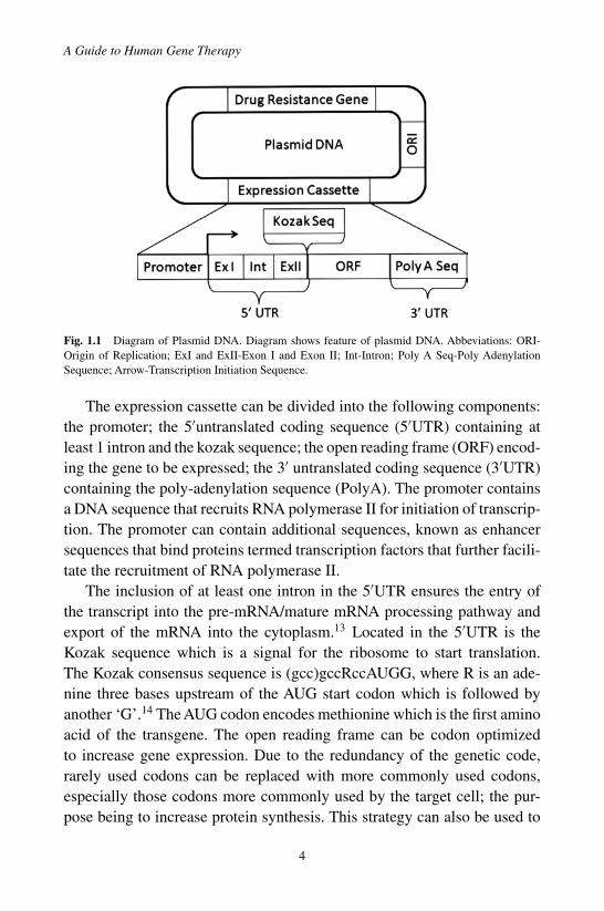
Furthermore, the FDA has deemed the percent supercoil content a prod-uct shelf life determinant. The fundamental features of pDNA are shownin Fig. 1.1. These are: the expression cassette, the origin of replicationand the drug resistance gene. The origin of replication (ORI) is a DNAsequence of 13 mer and 9 mer repeats that initiates plasmid DNA replica-tion in bacteria. The drug resistant gene, Kanamycin resistance gene orAmpicillin resistance gene, allows for the selection of plasmid transformedbacteria.
3
May 4, 2010 11:0 SPI-B903 9in x 6in b903-ch01
A Guide to Human Gene Therapy
Fig. 1.1 Diagram of Plasmid DNA. Diagram shows feature of plasmid DNA. Abbeviations: ORI-Origin of Replication; ExI and ExII-Exon I and Exon II; Int-Intron; Poly A Seq-Poly AdenylationSequence; Arrow-Transcription Initiation Sequence.
The expression cassette can be divided into the following components:the promoter; the 5′untranslated coding sequence (5′UTR) containing atleast 1 intron and the kozak sequence; the open reading frame (ORF) encod-ing the gene to be expressed; the 3′ untranslated coding sequence (3′UTR)containing the poly-adenylation sequence (PolyA). The promoter containsa DNA sequence that recruits RNA polymerase II for initiation of transcrip-tion. The promoter can contain additional sequences, known as enhancersequences that bind proteins termed transcription factors that further facili-tate the recruitment of RNA polymerase II.
The inclusion of at least one intron in the 5′UTR ensures the entry ofthe transcript into the pre-mRNA/mature mRNA processing pathway andexport of the mRNA into the cytoplasm.13 Located in the 5′UTR is theKozak sequence which is a signal for the ribosome to start translation.The Kozak consensus sequence is (gcc)gccRccAUGG, where R is an ade-nine three bases upstream of the AUG start codon which is followed byanother ‘G’.14 The AUG codon encodes methionine which is the first aminoacid of the transgene. The open reading frame can be codon optimizedto increase gene expression. Due to the redundancy of the genetic code,rarely used codons can be replaced with more commonly used codons,especially those codons more commonly used by the target cell; the pur-pose being to increase protein synthesis. This strategy can also be used to
4

May 4, 2010 11:0 SPI-B903 9in x 6in b903-ch01
Non-Viral Gene Therapy
reduce the amount of CpG sequences that can activate the immune system.Activation of the immune system has been shown to reduce the amount andduration of gene expression.15 However, in developing genetic vaccines,codon optimization can be used to increase CpG content thereby increasingimmune activation.16 The 3′UTR contains the polyadenylation sequencethat is a binding site for a multi-protein complex that cleaves the end of themRNA transcript and polyadenylate polymerase adds approximately 250adenine nucleotide monophosphates. The poly adenylation takes place inthe nucleus and promotes nuclear export of the mRNA and translation, andinhibits degradation.
Endogenous microRNA cleavage sequences approximately 20 base-pairs in length are located in the 3′UTR of endogenous mRNAs. Trans-fection of cells with endogenous microRNA activity against a latenttarget in the 3′UTR of a therapeutic gene could inhibit protein synthe-sis by removing the poly A tail resulting in immediate degradation of themRNA. Databases, such as the Wellcome Trust Sanger Institute siRNAdatabase (http://microrna.sanger.ac.uk/), are continually being updated formicroRNA target sequences as they are identified. Scanning the sequenceof the 3′UTR using these microRNA target sequence databases will avoidinactivation of the transcript by endogenously expressed microRNAs.
2.1 Plasmid DNA Manufacture
The therapeutic gene is ligated into the plasmid backbone and standardmicrobiological protocols are used to identify a bacterial clone that con-tains the plasmid DNA. The bacterial clones are also selected for the high-est specific activity with regard to plasmid DNA/bacterium. Master cellbanks are created using this clone. The bacteria are fermented at lab scalein a shaker flask or can be fermented using a fermenter. The bacteria arepelleted by centrifugation and resuspended in resuspension buffer. The bac-teria are lysed opened by alkaline lysis, neutralized and then centrifuged.The supernatant is extracted with phenol/chloroform followed by ethanolprecipitation of the pDNA. The precipitant is resuspended in buffer anddouble banded using CsCl equilibrium centrifugation with a vertical rotor.The ethidium bromide is extracted with buffer saturated butanol followedby dialysis of the DNA against buffer. There are commercially available kits
5

May 4, 2010 11:0 SPI-B903 9in x 6in b903-ch01
A Guide to Human Gene Therapy
to purify pDNA from bacteria. However, the quality of the pDNA can vary.The method outlined above yields highly purified plasmid DNA with regardto elimination of bacterial protein, RNA and endotoxin. This is especiallyimportant when formulating the pDNA with polymers and cationic lipids.Low-speed and high-speed centrifugation are not suitable for gram scalepharmaceutical manufacture. Substitution of filters for low speed centrifu-gation, and replacement of high speed CsCl density gradient centrifugationwith anion exchange chromatography combined with hydrophobic interac-tion chromatography (HIC) makes this process amenable to pharmaceuticalscale pDNA manufacture.17,18 Depending upon the bacterial strain and theplasmid DNA backbone, a single 500 L fermentation run can yield 10 to20 grams of plasmid DNA.
3. Plasmid DNA Gene Transfer Methods
3.1 Plasmid DNA or “Naked DNA” as a Gene DeliverySystem
As stated in the introduction, plasmid DNA can be injected by itself andyield gene expression. This was first discovered by intramuscular injection5
expressing a reporter gene resulting in the marking of muscle cells at thesite of injection. The expression levels are low compared to other formsof non-viral gene therapy. However, the DNA is not toxic and low levelsof expression can be compensated for by increasing the dose and dosingfrequency.
Two late stage clinical applications take advantage of this form ofplasmid based gene delivery. Both treat peripheral vascular disease byexpressing a therapeutic gene encoding for angiogenic growth factors, basicfibroblast growth factor (FGF-1)19 or hepatocyte growth factor (HGF),20,21
to induce new blood vessel growth in ischemic limbs. Both non-viral genetherapies have similar pDNA doses but different dosing schedules and dif-ferent endpoints. For the FGF Phase 2 clinical trial, 125 patients, whererevascularization surgery was not an option and had non-healing ulcers,were randomized and double blind placebo controlled for 2.5 ml injectionof FGF-1 pDNA, [pDNA] = 0.2 mg/ml, on days 1, 15, 30 and 45. Theprimary end point was healing of at least one ulcer and secondary end
6

May 4, 2010 11:0 SPI-B903 9in x 6in b903-ch01
Non-Viral Gene Therapy
points were ankle brachial index, amputation and death. The gene therapywas well tolerated. There was no significant difference in ulcer healingbetween treatment and placebo group. However, there was a significantreduction in risk of amputations and there was a trend in the reduced riskof death.
For expression of HGF, multiple clinical trials were conducted showingthat the therapy was well tolerated and no severe complications or adverseevents were observed for any of the patients.20,21 A multicenter Phase 3 trialwas conducted in Japan comprised of patients with arteriosclerosis oblit-erans with critical limb ischemia that could not undergo revascularizationand did not respond to conventional drug therapies. Patients were random-ized 2:1 therapy (55 patients) to placebo (26 patients) groups and receivedtwo intramuscular injections at the site of ischemia at 4 week intervals.Patients were followed for 8 weeks after the last administration. The pri-mary endpoints were decreased rest pain or improvement in ischemic ulcer.The treatment group showed a 70% response in rest pain reduction and ulcerimprovement whereas the placebo group showed a 30% response rate. Atthis time, these two therapeutic applications are in the latest stages of clinicaldevelopment for plasmid DNA delivery.
Another active area of plasmid DNA therapy is the development ofgenetic vaccines. Purification of protein antigen can be problematic withregard to the yield and purity, often giving rise to antibody responses to theimpurity rather than the intended protein antigen. This can be especiallychallenging for water insoluble proteins, such as integral membrane pro-teins.Also, combinations of different protein antigens can be prohibitive dueto formulation incompatibility. Expression of the protein antigen followingintramuscular, intradermal or subcutaneous administration of pDNA dra-matically simplifies the immunization process and insures immune responseto the native protein antigen. Genetic vaccines using pDNA alone are beingdeveloped for pandemic flu, HIV and Hepatitis C. In some cases, more thanone gene is being expressed, such as HIV where six to 7 different genes arebeing expressed at the same time.22–24
One technique that has been used as a research tool is to systemicallyadminister plasmid DNA in a large volume of vehicle, termed “hydrody-namic” gene delivery. This was initially discovered using a mouse animalmodel in which the pDNA is administered via the tail vein in a 1 ml injection
7
May 4, 2010 11:0 SPI-B903 9in x 6in b903-ch01
A Guide to Human Gene Therapy
volume rapidly (10 seconds). The result is a high gene transfer efficiencyto the liver. Hydrodynamic gene delivery while not practical for clinicaldevelopment of systemic non-viral gene therapies has been used to screenfor biological activity of secreted proteins into the blood, such as blood clot-ting factors. Therapeutic applications for Naked DNA are limited by the lowlevel of gene expression. The level of gene expression can be improved byincreasing the amount of DNA that gets into the cells, enters the nucleusand is expressed. The following section will describe technologies designedto increase gene transfer efficiencies.
3.1.1 Electroporation of Naked DNA
Applying energy following local administration of pDNA results in largeincreases in gene expression. The forms of energy can be electrical — usedin “electroporation” or ultrasound — used in “sonoporation”. Electropora-tion consists of applying voltage to the site of administration in a series ofelectrical pulses lasting microseconds for each pulse. The hypothesis is thatthe electrical pulses induce a transient depolarization of the smooth mus-cle plasma membrane allowing the pDNA to enter the cell. The number ofpulses, duration of pulses and the electrical strength of the pulse are of a par-ticular magnitude to minimize permanent damage to the cell membrane andmaximize gene transfer. Voltage was initially applied through calipers andthe muscle was sandwiched between the plates. However the administrationtechnology has been modified to use needles arranged in a hexagonal arraywith the electrical field alternating between opposing needles creating anelectrical field around the injection site. Electroporation can increase geneexpression of pDNA from one to several orders of magnitude comparedto pDNA alone.25 The fold increase is dependent on the transgene to beexpressed, the administration route and the optimization of the electricalfield pulse. A phase 1 clinical trial has been conducted involving the elec-troporation of an IL-12 expression plasmid into surface accessible tumorsof melanoma patients.26 There were 24 patients in the study. This was adose ranging study with the most serious adverse event being pain at thesite of injection. IL-12 and interferon gamma were observed at the tumorinjection site. Also induction of infiltrating lymphocytes into the tumor wasobserved.
8

May 4, 2010 11:0 SPI-B903 9in x 6in b903-ch01
Non-Viral Gene Therapy
3.1.2 Sonoporation of Naked DNA
Substitution of high frequency sound waves in sonoporation, for electric-ity can achieve similar effects as electroporation. The hypothetical mecha-nism for facilitating gene transfer is similar to electroporation, in that thesound waves produce a very short lived transient breach in the integrityof the plasma membrane facilitating entry of pDNA into the cell. The keycomponents are development of a probe that can effectively deliver thesound waves to the site of administration and application of pDNA effec-tively to maximize transfection efficiency.27–29 Gene transfer efficiencycan be increased by applying ultrasound contrast reagent along with theultrasound.30,31
3.2 Plasmid DNA Formulations
3.2.1 Cationic Lipids
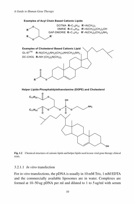
Cationic lipids are synthetic amphiphiles comprised of a hydrophobicdomain (R), a linker and a hydrophilic domain (R′). For the cholesterolbased cationic lipids, the hydrophobic domain is cholesterol and the cationichead group is denoted by R. Chemical structures of cationic lipids used innon-viral gene therapy clinical trials used are shown in Fig. 1.2. The lipidanchor is linked to the head group by either an ether linkage or a carbamatelinkage, as shown for DC-CHOL and GL-67.
Cationic lipids bind to pDNA by electrostatic interactions between thecationic lipid moiety and the phosphate pDNA backbone or through hydro-gen bonding between the amines and hydroxyl groups of the cationic lipidsand the pDNA. The cationic lipids shown in Fig. 1.2 require a helper lipid,DOPE or cholesterol for the diacyl cationic lipids, and DOPE for the choles-terol based cationic lipids, to transfect cells. Cationic liposomes are preparedby first mixing the lipids in an organic solvent; the solvent is removed byevaporation producing a lipid film and the lipid film is hydrated with abuffer to form liposomes. The liposome size can be reduced by extrusionthrough a filter with fixed pore size, or energy can be applied to the sus-pension by strong bath or probe sonication to reduce the diameter of theliposomes.
9
May 4, 2010 11:0 SPI-B903 9in x 6in b903-ch01
A Guide to Human Gene Therapy
O
O
R′
R
R
O
OO
P
C17H33
C17H33
O
O
O
O
OH
NH2
OH
Examples of Acyl Chain Based Cationic Lipids
DOTMA R=C18H35 R′=N(CH3)3
DMRIE R=C14H29 R′=N(CH3)2(CH2)2OHGAP-DMORIE R=C14H27 R′=N(CH3)2(CH2)3NH2
OR
O
Examples of Cholesterol Based Cationic Lipid
GL-67TM R=N((CH2)2NH2)(CH2)4NH(CH2)3NH2
DC-CHOL R=NH (CH2)2N(CH3)2
Helper Lipids-Phosphatidylethanolamine (DOPE) and Cholesterol
Fig. 1.2 Chemical structures of cationic lipids and helper lipids used in non-viral gene therapy clinicaltrials.
3.2.1.1 In vitro transfection
For in vitro transfections, the pDNA is usually in 10 mM Tris, 1 mM EDTAand the commercially available liposomes are in water. Complexes areformed at 10–50 ug pDNA per ml and diluted to 1 to 5 ug/ml with serum
10
May 4, 2010 11:0 SPI-B903 9in x 6in b903-ch01
Non-Viral Gene Therapy
free media and added to cells also in serum free media. After 4 hrs, thetransfection complexes are aspirated off and replaced with serum contain-ing media. The amount of cationic lipid to pDNA phosphate is titrated overa range of 2/1 to 8/1. The cationic lipids can be toxic to certain cells so therea balanced optimal ratio is required to achieve maximal gene expressionwith minimal toxicity. The biological activity of the transfection complexeshas a short half life (hours) and maximal gene expression is achieved within0.5 hr to 1 hr after formation.
3.2.1.2 Systemic in vivo gene transfer
Formulation of cationic lipids with pDNA for in vivo gene transfer is morecomplicated. First, there is very little correlation between optimal parame-ters, such as cationic lipid to helper lipid, ratio of cationic lipid to pDNA,liposome size or vehicle composition, developed for in vitro gene expressionand in vivo gene expression. Secondly, the resulting transfection complexesare dependent on the type of cationic lipid used and how the complexes areformed. Binding of endogenous polyamines, such as spermine and spermi-dine, to plasmid DNA transforms supercoiled pDNA from a random coilinto a toroid. Several approaches have been taken in developing cationiclipids for systemic gene transfer.
The first is to use cationic moieties known to bind to pDNA as thecationic head group. The resulting lipids are formulated with a helper lipid,such as DOPE or cholesterol, to form liposomes. The mole ratio of cationiclipid to helper lipid can vary from 100/0 to 10/90; most commonly it is setat 50:50. The mole ratio of cationic lipid to DNA phosphate can vary from2/1 to 6/1 depending on the affinity of the cationic lipid in the context ofa liposome or micelle to pDNA yielding a positively charged transfectioncomplex. Non-reducing carbohydrates, such as sucrose, can be substitutedfor salt, such as NaCl, to maintain isogenicity.
An alternative approach is the synthesis of cationic lipid libraries, com-plex the cationic amphiphiles to the plasmid DNA and then test for geneexpression.32 Assays can use expression of reporter genes such as greenfluorescent protein (GFP) or β-galactosidase (Lac-Z), or a secreted pro-tein, such as human placental secreted alkaline phosphatase. It is imprac-tical to screen the library other than by in vitro tissue culture due tothe number of animals needed and the sample processing. Formulation
11

May 4, 2010 11:0 SPI-B903 9in x 6in b903-ch01
A Guide to Human Gene Therapy
of active cationic lipid candidates can then be optimized for in vivogene transfer using the same reporter genes or a more popular in vivoscreen is to detect expression by expression of a bioluminescent activereporter gene, such as firefly luciferase. This technique is excellent forliver, lung, spleen and heart imaging but poor for brain and muscle dueto the limited biodistribution of the luciferase substrate following systemicadministration.
The ideal particle size should range between 50 nm to 200 nm. Particleswith diameters greater than 200 nm result in the reduction of the endocy-totic index, except for monocyte derived cells such as macrophage, Kupffercells or dendritic cells, which are specialized phagocytic cells. Once insidethe endocytotic vesicle, the transfection complexes are disassembled andthe pDNA must escape the internal vesicle and migrate to the nucleus fortranscription of the transgene.
Modifications to the lipid composition have been made to increase sys-temic gene transfer efficiency. The circulation half life of the transfectioncomplexes can be increased by covering the surface of the transfectioncomplex with high molecular weight polyethylene glycol (2kdalt–10kdalt)inhibiting opsonization of the transfection complexes, thereby reducingnon-specific uptake by the reticuloendothelium system (RES). Internaliza-tion of the transfection complex can be restricted to a specific cell typeby derivatizing ligands to the surface of the transfection complex that bindto a receptor expressed on a specific cell type.33–35 Intracellular releaseof the pDNA from either the endosome or lysosome can be achieved bychanging the cationic lipid composition to one that is susceptible to fusionat acidic pH.36 Lastly, nuclear uptake of the pDNA can be increased byadding nuclear localization sequences in the form of DNA sequences thatbind proteins manufactured in the cytoplasm that contain nuclear localiza-tion sequences allowing the pDNA to “hitch a ride” into the nucleus.37 Thereare currently no clinical trials using systemically administered cationiclipid/plasmid DNA transfection complexes.
3.2.1.3 Local administration of cationic lipid/pDNAtransfection complexes
Local administration of cationic lipid/pDNA transfection complexes refersto intratumoral administration and intramuscular administration. From a
12

May 4, 2010 11:0 SPI-B903 9in x 6in b903-ch01
Non-Viral Gene Therapy
safety perspective, the bulk of gene delivery and gene expression is lim-ited to the site of administration. Therapeutic applications of intratumoraladministration have focused on expression of molecules, such as cytokines38
and self antigens39 for the purpose of priming the immune system to killnot only the transfected cells but in so doing, prime the immune system tokill non-transfected tumors, thus creating a systemic therapy from a localadministration.
With regard to the expression of self antigens, the first non-viral clinicaltrial was conducted by Dr. G. Nabel in collaboration with Dr. L. Huang.Intratumoral administration of cationic lipid formulated HLA B-7 pDNAwas shown to be safe and tumor regression was observed. Subsequentclinical trials modified the cationic lipid formulation from the use of DC-Chol/DOPE to DMRIE/DOPE, increased the pDNA dose and tested dif-ferent dosing schedules. A phase 2 dose ranging study identified a 2 mgper intratumoral injection as an effective antitumor dose and administra-tion once a week for 6 weeks in a single tumor to be the dosing cycle.The clinical results from the phase 2 dose escalation study were used todefine the patient entry criteria for a Phase 3 clinical study. The Phase 3clinical study compares the cationic lipid/pDNA non-viral gene therapy(Allovectin-7) to Dacarbazine or Temozolomide. This therapy is currentlyin Phase 3 clinical trial.40 Results from this trial should be available in2012.
Expression of foreign antigens by administration of pDNA results in animmune response to the expressed protein creating the potential for develop-ment of genetic vaccines. However, mg quantities of pDNA injected multi-ple times have been required to sustain an immune response. Certain cationiclipids when formulated with pDNA not only increased transfection effi-ciency but were also immunostimulatory. A cationic liposome formulationcomposed of GAP-DMROIE and diphytanolylphosphatidylethanolamine(DPyPE) was shown to be immunostimulatory for several expressed anti-gens in rodent and non-human primates.41,42 Multiple demonstrations ofproof of concept advanced this formulation into a phase 1 human clinicaltrial for a pandemic influenza vaccine. The phase 1 trial evaluated tolera-bility and immunogenicity. These parameters were tested for this clinicaltrial included a comparison of a needle free device, Biojector 2000, vs.needle; a single plasmid expressing the H5 hemmaglutinin viral envelope
13

May 4, 2010 11:0 SPI-B903 9in x 6in b903-ch01
A Guide to Human Gene Therapy
protein vs. the H5 plasmid plus two additional plasmids expressing viralproteins whose amino acid sequences are highly conserved amongst multi-ple pandemic influenza virus clades. One hundred and three patients wereenrolled and 86 were evaluable for immunogenicity. A 65% response ratewas observed for neutralizing antibody equivalent to or exceeding protect-ing titers of 1/40. This was a durable response being observed out to 182 days(unpublished results).
3.3 Polymer
Polymer based plasmid DNA gene therapy can be divided into two cate-gories: cationic polymer and neutral polymers. Examples of cationic poly-mers are polylysine, polyethleneimine, or panamdendrimers. Examples ofneutral polymers are Polylactide glycolic anhydride (PLGA), poloxamerand polyvinylpyrolidone (PVP).
3.3.1 Cationic Polymers
Cationic polymers behave similarly to cationic lipids in that they containa cationic moiety which either electrostatically bonds with the phosphatebackbone of the pDNA or hydrogen bonds to the pDNA. The first cationicpolymers were naturally derived from poly amino acids such as poly-L-ornithine and poly-L-lysine. Utility was derived from the virology field thatfirst used poly-L-ornithine to facilitate viral infection of cells.11 It was laterdiscovered that transfection of cells with viral RNA complexed to poly-L-ornithine produced infectious virus. Lessons learned from the virology fieldwere applied to pDNA delivery with the substitution of poly-L-ornithinefor poly-L-lysine. The polymer provided a versatile backbone for chemicalmodification of ligands to target the complexes to cells by additional peptidesequences or chemical modification of small molecular weight ligands, suchas folate or carbohydrates.43 One application that has evolved from thebench to the clinic is the use of poly-L-lysine/pDNA complexes for thetreatment of cystic fibrosis.44 Several key developments in transitioningfrom the research lab to the clinic were the development of an aerosolizedtransfection complex, reduction in the CpG content of the plasmid to avoidsecondary cytokine activation,15 use of a polyethylene glycol-substituted30-mer lysine peptides.45
14

May 4, 2010 11:0 SPI-B903 9in x 6in b903-ch01
Non-Viral Gene Therapy
Improvements in polycationic amino acids polymers have included otherpositively charged amino acids such as arginines and histidines.46 The latternot only serves as an alternate DNA binding moiety but also has a pK thatcan impact the transfection complex when the pH environment acidifies,as is found in the lysosome and endosome, facilitating decomplexation andpotentially lysing open the endocytic vacuole.47
Polyethyleneimine (PEI) is a completely synthetic polymer that hassimilar intracellular release properties as the poly-L-histidine, first intro-duced into the field of nonviral gene therapy by JP Behr.48 The imine moi-ety of the polymer provides electrostatic bonding and hydrogen bondingto pDNA and also serves as a protonateable buffer that prevents acidifi-cation of the endosome/lysosome resulting in intracellular plasmid DNArelease. This polymer has been modified with polyethylene glycol andtargeting ligands. The polymer/pDNA complex is currently in preclinicaldevelopment.
3.3.2 Neutral Polymer
Neutral polymers are defined as polymers carrying no net charge. Examplesare poloxamers, polyvinylpyrolidone (PVP), polyanhydride (PLGA) poly-mers. The first two form simple mixtures of polymer and pDNA whereasthe PLGA polymer forms nanoparticles in which the pDNA is packaged.Both PVP49,50 and poloxamer have been used in clinical trials for localadministration of pDNA. The applications have been in cancer, cardio-vascular disease and vaccine development against infectious disease. Forcancer applications, plasmid DNA encoding cytokines, such as IL-2, IL-12,or interferon-α in combination with IL-12,49,50,51 is formulated with PVPand injected intratumorally resulting in expression and secretion of thecytokines from the transfected cells. The chemoattracting cytokine gra-dient causes immune cells to migrate to the tumor and kill the tumor cells.The therapeutic hypothesis is similar to that of the cationic lipid basedimmunotherapy, in that T cell mediates tumor cell killing programs andactivates tumor surveillance of the immune system, thus preventing newtumor formation.
Poloxamers are block copolymers of polyethylene oxide (a) andpolypropylene oxide (b) that are linked by ether linkages shown in Fig. 1.3with molecular weights ranging from 1,000 daltons to 12,000 daltons. In
15
May 4, 2010 11:0 SPI-B903 9in x 6in b903-ch01
A Guide to Human Gene Therapy
HO O O O OH
CH3
aba
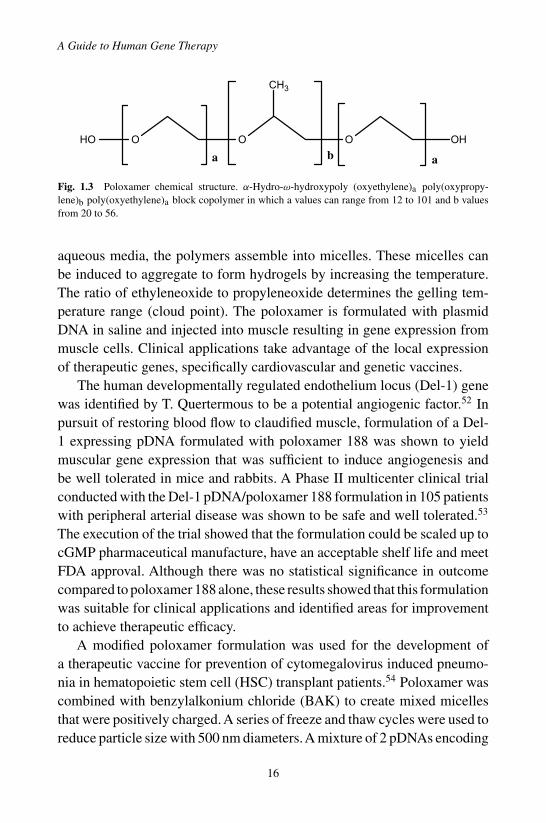
Fig. 1.3 Poloxamer chemical structure. α-Hydro-ω-hydroxypoly (oxyethylene)a poly(oxypropy-lene)b poly(oxyethylene)a block copolymer in which a values can range from 12 to 101 and b valuesfrom 20 to 56.
aqueous media, the polymers assemble into micelles. These micelles canbe induced to aggregate to form hydrogels by increasing the temperature.The ratio of ethyleneoxide to propyleneoxide determines the gelling tem-perature range (cloud point). The poloxamer is formulated with plasmidDNA in saline and injected into muscle resulting in gene expression frommuscle cells. Clinical applications take advantage of the local expressionof therapeutic genes, specifically cardiovascular and genetic vaccines.
The human developmentally regulated endothelium locus (Del-1) genewas identified by T. Quertermous to be a potential angiogenic factor.52 Inpursuit of restoring blood flow to claudified muscle, formulation of a Del-1 expressing pDNA formulated with poloxamer 188 was shown to yieldmuscular gene expression that was sufficient to induce angiogenesis andbe well tolerated in mice and rabbits. A Phase II multicenter clinical trialconducted with the Del-1 pDNA/poloxamer 188 formulation in 105 patientswith peripheral arterial disease was shown to be safe and well tolerated.53
The execution of the trial showed that the formulation could be scaled up tocGMP pharmaceutical manufacture, have an acceptable shelf life and meetFDA approval. Although there was no statistical significance in outcomecompared to poloxamer 188 alone, these results showed that this formulationwas suitable for clinical applications and identified areas for improvementto achieve therapeutic efficacy.
A modified poloxamer formulation was used for the development ofa therapeutic vaccine for prevention of cytomegalovirus induced pneumo-nia in hematopoietic stem cell (HSC) transplant patients.54 Poloxamer wascombined with benzylalkonium chloride (BAK) to create mixed micellesthat were positively charged.A series of freeze and thaw cycles were used toreduce particle size with 500 nm diameters.A mixture of 2 pDNAs encoding
16
May 4, 2010 11:0 SPI-B903 9in x 6in b903-ch01
Non-Viral Gene Therapy
the surface glycoprotein B (gB) and the internal matrix protein (pp65) wasadded to the poloxamer/BAK and stored frozen.55,56 A phase I clinical trialwas conducted involving 22 CMV seronegative and 22 seropositive healthysubjects. The vaccine was well tolerated with no serious adverse events.Immunogenicity determined by ex vivo interferon (IFN)-gamma enzyme-linked immunospot assay yielded 45.5% of CMV seronegative subjects and25% of CMV seropositive subjects in subjects receiving the full vaccineseries. The safety and immunogenicity results supported further evaluationin a phase 2 clinical trial.
Conclusions
Local administration of plasmid DNA based gene therapies seem to be themost promising and have the least potential for any side effects. pDNAalone has advanced into phase 3 clinical trials for cardiovascular applica-tions. Success of this clinical application is based upon the potent angiogenicstimulation produced by a very low level of expressed protein that remainslocalized at the site of administration. Improvements in gene transfer effi-ciency from the result of pDNA formulation with cationic liposomes andpolymers are also combined with selection of expressed genes that producea potent secondary effect, such as the case of immunostimulation againsttumor antigens and pathogenic antigens. Research focused on increasingthe transfection activity of plasmid DNA based gene delivery systems isvery active. The hope is to combine the development of cell specific tar-geted gene delivery along with tissue specific promoters that further restrictgene expression to a specific cell type with the application for systemicadministration.
References
1. Szoka FC, Papahadjopoulos, DP (1983). Method of inserting DNA into living cells. InUnited States Patent Office.
2. Behr JP, Demeneix B, Loeffler JP, Perez-Mutul J (1989). Efficient gene transfer intomammalian primary endocrine cells with lipopolyamine-coated DNA. Proc Natl AcadSci USA 86: 6982–6986.
3. Felgner PL, et al. (1987). Lipofection: a highly efficient, lipid-mediated DNA-transfection procedure. Proc Natl Acad Sci USA 84: 7413–7417.
17
May 4, 2010 11:0 SPI-B903 9in x 6in b903-ch01
A Guide to Human Gene Therapy
4. Felgner PL, Ringold GM (1989). Cationic liposome-mediated transfection. Nature 337:387–388.
5. Wolff JA, et al. (1990). Direct gene transfer into mouse muscle in vivo. Science 247:1465–1468.
6. Nabel GJ, et al. (1996). Immune response in human melanoma after transfer of an allo-geneic class I major histocompatibility complex gene with DNA-liposome complexes.Proc Natl Acad Sci USA 93: 15388–15393.
7. Nabel GJ, et al. (1993). Direct gene transfer with DNA-liposome complexes inmelanoma: expression, biologic activity, and lack of toxicity in humans. Proc NatlAcad Sci USA 90: 11307–11311.
8. Isner JM, et al. (1996). Arterial gene transfer for therapeutic angiogenesis in patientswith peripheral artery disease. Hum Gene Ther 7: 959–988.
9. Isner JM, et al. (1996). Clinical evidence of angiogenesis after arterial gene transfer ofphVEGF165 in patient with ischaemic limb. Lancet 348: 370–374.
10. Mir LM, Bureau MF, Rangara R, Schwartz B, Scherman D (1998). Long-term, highlevel in vivo gene expression after electric pulse-mediated gene transfer into skeletalmuscle. C R Acad Sci III 321: 893–899.
11. Motoyoshi F, Bancroft JB, Watts JW, Burgess J (1973). The infection of tobaccoprotoplasts with cowpea chlorotic mottle virus and its RNA. J Gen Virol 20:177–193.
12. Pillai VB, Hellerstein M, Yu T, Amara RR, Robinson HL (2008). Comparative studieson in vitro expression and in vivo immunogenicity of supercoiled and open circularforms of plasmid DNA vaccines. Vaccine 26: 1136–1141.
13. Valencia P, DiasAP, Reed R (2008). Splicing promotes rapid and efficient mRNA exportin mammalian cells. Proc Natl Acad Sci USA 105: 3386–3391.
14. Lida Y, Kanagu D (2000). Quantification analysis of translation initiation signal invertebrate mRNAs: Effect of nucleotides at positions +4 upon efficiency of translationinitiation. Nucleic Acids Symposium 44: 77–78.
15. Yew NS, et al. (2002). CpG-depleted plasmid DNA vectors with enhanced safety andlong-term gene expression in vivo. Mol Ther 5: 731–738.
16. Abdulhaqq SA, Weiner DB (2008). DNA vaccines: developing new strategies toenhance immune responses. Immunol Res 42: 219–232.
17. Ferreira GNM, Monteiro GA, Prazeres DMF, Cabral JMS (2000). Downstream pro-cessing of plasmid DNA for gene therapy and DNA vaccine applications. TIBTech 18:380–388.
18. Stadler S, Lemmens R, Nyhammar Y (2004). Plasmid DNA Purification. J Gene Med6: S54–S66.
19. Nikol S, et al. (2008). Therapeutic angiogenesis with intramuscular NV1FGF improvesamputation-free survival in patients with critical limb ischemia. Mol Ther 16:972–978.
20. Powell RJ, et al. (2008). Results of a double-blind, placebo-controlled study to assessthe safety of intramuscular injection of hepatocyte growth factor plasmid to improvelimb perfusion in patients with critical limb ischemia. Circulation 118: 58–65.
21. Nakagami H, Kaneda Y, Ogihara T, Morishita R (2005). Hepatocyte growthfactor as potential cardiovascular therapy. Expert Rev Cardiovasc Ther 3:513–519.
18
May 4, 2010 11:0 SPI-B903 9in x 6in b903-ch01
Non-Viral Gene Therapy
22. Seaman MS, et al. (2005). Multiclade human immunodeficiency virus type 1 envelopeimmunogens elicit broad cellular and humoral immunity in rhesus monkeys. J Virol 79:2956–2963.
23. Gudmundsdotter L, et al. (2006). Therapeutic immunization for HIV. Springer SeminImmunopathol 28: 221–230.
24. Sandstrom E, et al. (2008). Broad immunogenicity of a multigene, multiclade HIV-1DNA vaccine boosted with heterologous HIV-1 recombinant modified vaccinia virusAnkara. J Infect Dis 198: 1482–1490.
25. Rizzuto G, et al. (1999). Efficient and regulated erythropoietin production by nakedDNA injection and muscle electroporation. Proc Natl Acad Sci USA 96: 6417–6422.
26. Daud AI, et al. (2008). Phase I Trial of Interleukin-12 Plasmid Electroporation inPatients With Metastatic Melanoma. J Clin Oncol.
27. Manome Y, Nakamura M, Ohno T, Furuhata H (2000). Ultrasound facilitates transduc-tion of naked plasmid DNA into colon carcinoma cells in vitro and in vivo. Hum GeneTher 11: 1521–1528.
28. Huber PE, Pfisterer P (2000). In vitro and in vivo transfection of plasmid DNA in theDunning prostate tumor R3327-AT1 is enhanced by focused ultrasound. Gene Ther 7:1516–1525.
29. Greenleaf WJ, Bolander ME, Sarkar G, Goldring MB, Greenleaf JF (1998). Artificialcavitation nuclei significantly enhance acoustically induced cell transfection. Ultra-sound Med Biol 24: 587–595.
30. Takeuchi D, et al. (2008). Alleviation of Abeta-induced cognitive impairment byultrasound-mediated gene transfer of HGF in a mouse model. Gene Ther 15:561–571.
31. TaniyamaY, et al. (2002). Development of safe and efficient novel nonviral gene transferusing ultrasound: enhancement of transfection efficiency of naked plasmid DNA inskeletal muscle. Gene Ther 9: 372–380.
32. Yingyongnarongkul BE, Howarth M, Elliott T, Bradley M (2004). Solid-phase synthesisof 89 polyamine-based cationic lipids for DNA delivery to mammalian cells. Chemistry10: 463–473.
33. Anwer K, Bailey A, Sullivan SM (2000). Targeted gene delivery: a two-prongedapproach. Crit Rev Ther Drug Carrier Syst 17: 377–424.
34. Driessen WH, Fujii N, Tamamura H, Sullivan SM (2008). Development of peptide-targeted lipoplexes to CXCR4-expressing rat glioma cells and rat proliferating endothe-lial cells. Mol Ther 16: 516–524.
35. Hood JD, et al. (2002). Tumor regression by targeted gene delivery to the neovascula-ture. Science 296: 2404–2407.
36. Li W, Huang Z, MacKay JA, Grube S, Szoka FC, Jr. (2005). Low-pH-sensitivepoly(ethylene glycol) (PEG)-stabilized plasmid nanolipoparticles: effects of PEG chainlength, lipid composition and assembly conditions on gene delivery. J Gene Med 7:67–79.
37. Cartier R, Reszka R (2002). Utilization of synthetic peptides containing nuclear local-ization signals for nonviral gene transfer systems. Gene Ther 9: 157–167.
38. Li D, Shugert E, Guo M, Bishop JS, O’Malley BW, Jr. (2001). Combination nonvi-ral interleukin 2 and interleukin 12 gene therapy for head and neck squamous cellcarcinoma. Arch Otolaryngol Head Neck Surg 127: 1319–1324.
19

May 4, 2010 11:0 SPI-B903 9in x 6in b903-ch01
A Guide to Human Gene Therapy
39. Plautz GE, Yang ZY, Wu BY, Gao X, Huang L, Nabel GJ (1993). Immunotherapy ofmalignancy by in vivo gene transfer into tumors. Proc Natl Acad Sci USA 90: 4645–4649.
40. Bedikian AY, Del Vecchio M (2008). Allovectin-7 therapy in metastatic melanoma.Expert Opin Biol Ther 8: 839–844.
41. Reyes L, et al. (2001).Vaxfectin enhances antigen specific antibody titers and maintainsTh1 type immune responses to plasmid DNA immunization. Vaccine 19: 3778–3786.
42. Hartikka J, et al. (2001). Vaxfectin enhances the humoral immune response to plasmidDNA-encoded antigens. Vaccine 19: 1911–1923.
43. Wu CH, Wilson JM, Wu GY (1989). Targeting genes: delivery and persistent expressionof a foreign gene driven by mammalian regulatory elements in vivo. J Biol Chem 264:16985–16987.
44. Davis PB, Cooper MJ (2007). Vectors for airway gene delivery. Aaps J 9: E11–E17.45. Konstan MW, et al. (2004). Compacted DNA nanoparticles administered to the nasal
mucosa of cystic fibrosis subjects are safe and demonstrate partial to complete cysticfibrosis transmembrane regulator reconstitution. Hum Gene Ther 15: 1255–1269.
46. Putnam D, Zelikin AN, Izumrudov VA, Langer R (2003). Polyhistidine-PEG:DNAnanocomposites for gene delivery. Biomaterials 24: 4425–4433.
47. Pack DW, Putnam D, Langer R (2000). Design of imidazole-containing endosomolyticbiopolymers for gene delivery. Biotechnol Bioeng 67: 217–223.
48. Zou SM, Erbacher P, Remy JS, Behr JP (2000). Systemic linear polyethylenimine(L-PEI)-mediated gene delivery in the mouse. J Gene Med 2: 128–134.
49. Mendiratta SK, et al. (1999). Intratumoral delivery of IL-12 gene by polyvinyl poly-meric vector system to murine renal and colon carcinoma results in potent antitumorimmunity. Gene Ther 6: 833–839.
50. Mendiratta SK, et al. (2000). Combination of interleukin 12 and interferon alpha genetherapy induces a synergistic antitumor response against colon and renal cell carcinoma.Hum Gene Ther 11: 1851–1862.
51. Kaushik A (2001). Leuvectin Vical Inc. Curr Opin Investig Drugs 2: 976–981.52. Ho HK, et al. (2004). Developmental endothelial locus-1 (Del-1), a novel angiogenic
protein: its role in ischemia. Circulation 109: 1314–1319.53. Grossman PM, et al. (2007). Results from a phase II multicenter, double-blind placebo-
controlled study of Del-1 (VLTS-589) for intermittent claudication in subjects withperipheral arterial disease. Am Heart J 153: 874–880.
54. Selinsky C, et al. (2005). A DNA-based vaccine for the prevention of humancytomegalovirus-associated diseases. Hum Vaccin 1: 16–23.
55. Vilalta A, et al. (2005). I. Poloxamer-formulated plasmid DNA-based humancytomegalovirus vaccine: evaluation of plasmid DNA biodistribution/persistence andintegration. Hum Gene Ther 16: 1143–1150.
56. Hartikka J, et al. (2008). Physical characterization and in vivo evaluation of poloxamer-based DNA vaccine formulations. J Gene Med 10: 770–782.
20
May 4, 2010 11:0 SPI-B903 9in x 6in b903-ch02
Chapter 2
Adenoviral Vectors
Stuart A. Nicklin∗ and Andrew H. Baker
Adenoviruses are one of the most widely investigated vectors for genetherapy. Their attributes include ease of genetic manipulation to producereplication-deficient vectors, ability to readily generate high titer stocks,efficiency of gene delivery into many cell types and ability to encodelarge genetic inserts. Adenoviruses have been utilized for a variety oftherapeutic applications particularly for high level transient overexpres-sion in cancer gene therapy, vaccine delivery and certain cardiovasculardiseases. With the first licensing of clinical adenoviral gene therapy forcancer treatment the use of adenoviruses is likely to expand in the future.This chapter focuses on the history of adenovirus development, interac-tions with the host both for gene delivery and immunogenicity as well aspotential therapeutic applications.
1. Introduction
The human adenovirus (Ad) family is divided into 6 sub-species (A-F).There are a total of 51 different serotypes identified to date, classified viadifferential abilities to hemagglutinate erythrocytes and characterized bydifferences in capsid structure and receptor use. Ads from species C, partic-ularly serotype 5 (Ad5) are the most widely investigated viral gene transfervectors and consequently, one of the most commonly utilized viral vectorsin clinical trials. However, it is only in recent years that important findingsregarding receptor usage and interactions with the host in vivo, particularlyfollowing intravenous (i.v.) administration, have been deciphered. There are
∗Correspondence: BHF GCRC, Faculty of Medicine, University of Glasgow, 126 University Place,Glasgow UK.E-mail: [email protected]
21
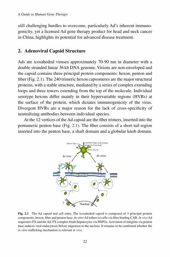
May 4, 2010 11:0 SPI-B903 9in x 6in b903-ch02
A Guide to Human Gene Therapy
still challenging hurdles to overcome, particularly Ad’s inherent immuno-genicity, yet a licensed Ad gene therapy product for head and neck cancerin China, highlights its potential for advanced disease treatment.
2. Adenoviral Capsid Structure
Ads are icosahedral viruses approximately 70-90 nm in diameter with adouble stranded linear 36 kb DNA genome. Virions are non-enveloped andthe capsid contains three principal protein components: hexon, penton andfiber (Fig. 2.1). The 240 trimeric hexon capsomeres are the major structuralproteins, with a stable structure, mediated by a series of complex extendingloops and three towers extending from the top of the molecule. Individualserotype hexons differ mainly in their hypervariable regions (HVRs) atthe surface of the protein, which dictates immunogenicity of the virus.Divergent HVRs are a major reason for the lack of cross-specificity ofneutralizing antibodies between individual species.
At the 12 vertices of the Ad capsid are the fiber trimers, inserted into thepentameric penton base (Fig. 2.1). The fiber consists of a short tail regioninserted into the penton base, a shaft domain and a globular knob domain.
Fig. 2.1 The Ad capsid and cell entry. The icosahedral capsid is composed of 3 principal proteincomponents, hexon, fiber and penton base. In vitro Ad tethers to cells via fiber binding CAR. In vivo Adsequesters FX and the Ad: FX complex binds hepatocytes via HSPGs. Activation of integrins via pentonbase induces viral endocytosis before migration to the nucleus. It remains to be confirmed whether thein vitro trafficking mechanism is relevant in vivo.
22

May 4, 2010 11:0 SPI-B903 9in x 6in b903-ch02
Adenoviral Vectors
The length of the fiber shaft is a main determinant of variation betweenindividual serotypes, as the shortest fibers belong to the B species Ad35,while fibers from species C Ads such as Ad5 are among the longest. Thereare also a number of minor capsid proteins, which mainly function as “glue”for the capsid.
3. Adenoviral Cell Entry
The method for Ad infection (at least in vitro) is well defined, particularlyfor Ad5 (species C). Ads infect host cells through receptor-mediated endo-cytosis mediated by fiber and penton base (Fig. 2.1). Initial interaction forspecies C Ads is between the fiber knob domain and the Coxsackie/Adreceptor (CAR).1,2 All Ad species, except B can interact with CAR viathe fiber knob domain, although some species D Ads also use sialic acid.3
Species B Ads utilize CD46.4 Virus internalization is through activationof αvβ3 and αvβ5 integrins by the RGD motif in the penton base at theN-terminus of the fiber protein.5 Once internalized to the cytoplasm viaclathrin-coated endosomes the penton base mediates capsid release fromthe endosome via acidification and the virus migrates to the nucleus.6
Following i.v.Ad administration transduction involves other factors. Themajor site of Ad transduction following i.v. delivery is liver hepatocytes butmutation of all Ad capsid receptor binding epitopes has limited effects onblocking liver uptake.7 This is because upon contact with blood Ad5 inter-acts with host proteins including coagulation factor IX (FIX), FX and com-plement binding protein (C4BP) to deliver virus to hepatocytes via heparansulphate proteoglycans (HSPGs) or low density lipoprotein receptor-relatedprotein (LRP), respectively (Figs. 2.1 and 2.3).8,9 However, sequestration ofFX and delivery of the virus: FX complex to liver via HSPGs is the primaryroute for i.v. delivered Ad liver transduction.10,11 This finding defines aninfectivity function for hexon, since direct interaction between hexon andFX is observed.10,11 Additionally, FX interacts with the Ad hexon via theHVRs since exchange of the Ad5 HVRs for those of a non-FX binding Ad issufficient to block liver gene transfer.10 The fact that such diverse cell entrypathways are used by the virus highlights the complexity of Ad interactionwith the host, providing further challenges for the development of efficientgene therapies.
23
May 4, 2010 11:0 SPI-B903 9in x 6in b903-ch02
A Guide to Human Gene Therapy
4. Production of Adenoviral Vectors
The fact that Ads are one of the most widely investigated gene transfervectors is in no small part due to the development of the human cell line,293, transformed with the entire left hand end of the Ad5 genome.12 Thisallowed the construction and study of recombinantAds in a helper virus-freeenvironment.
The gene products are organized into early (E1-E4) and late (L1-L5)regions, based on expression before or after initiation of DNA replication.The E1 region is the essential initiating factor for viral DNA replicationas it encodes E1a, which induces expression of other early genes. E1 alsoensures the cell enters S phase to enable viral replication by inhibiting keycell cycle components and blocking p53-mediated apoptosis. The E2 regionencodes products required for DNA replication such as DNA polymerase,single stranded DNA binding proteins and pre-terminal protein (which areessential for viral DNA synthesis, elongation and genome packaging intothe maturing viral particle). The E3 region encodes products which inhibitthe cell’s immune responses via blocking major histocompatibility complex(MHC) transport. E4 encodes products required for trafficking viral mRNAto the Golgi complex and for inhibiting transport of host cell mRNAs toenhance viral production. The late region transcripts encode structural cap-sid proteins.
First generation, replication-deficientAd vectors contain deletions in E1,enabling insertion of expression cassettes of up to approximately 5 kb. Thelimit in DNA packaging is dictated by the physical size of the virion whichis only able to package up to 105% (approximately 38 kb) of the native virusgenome size. Generation of these vectors has been historically carried outvia two plasmid transfection; the first plasmid contains sequences for the E1region in between which is inserted the expression cassette, the second plas-mid encodes the Ad genome with a large insertion in the E1 region whichblocks self packaging of virus through exceeding the maximal 38 kb pack-aging capacity. Homologous recombination between two plasmids, usingthe E1 sequence acting in trans from 293 cells excises E1 and inserts theexpression cassette, producing replication deficient Ad vectors which canbe propagated in 293 cells. This can result in generation of replication com-petent Ad (RCA) due to recombination between the Ad genome vector and
24

May 4, 2010 11:0 SPI-B903 9in x 6in b903-ch02
Adenoviral Vectors
the integrated homologous E1 region in 293 cells, however modified E1-expressing cell lines andAd generating plasmids with low homology to eachother has significantly reduced RCA. Additionally, other first generation Adproduction systems utilize cloning and construction of full length recombi-nant Ad genomes in bacteria prior to single plasmid transfection into 293cells to package virus. To increase the size of expression cassette that firstgeneration vectors can encode to 8 kb, E3 can also be deleted. The mainlimitation of first generation Ads was the discovery of leaky expression ofviral genes even in the absence of E1 due to transactivation of viral genesfrom host transcription factors, leading to transient transgene expression.Transduced cells release cytokines and are recognized and destroyed bycytotoxic CD8+ and memory CD4+ T lymphocytes (CTL) via recognitionof viral structural proteins presented on the cell surface by MHC class I(Fig. 2.3). Second generation vectors were developed via deletion of the E2and/ or E4 regions potentially enabling cloning of 14 kb of foreign DNAinto the vector. The initial study mutated E2a leading to increased trans-gene expression and decreased inflammatory responses in mouse liver andlung.13 E4 has also been deleted to provide similar advantages,14 however,such vectors were rapidly superseded by third generation Ads.
Third generation [also called gutless or helper dependent Ad vectors(hdAd)] are those devoid of all viral open reading frames, creating vectorscontaining only essential cis elements (inverted terminal repeats and con-tiguous packaging sequences). Deletion of all viral genes increases insertcapacity to 36–38 kb and limits host immune responses by ablating leakyproduction of Ad genes, leading to persistent transgene expression. Earlyproduction of helper-dependent Ad vectors was hampered by the require-ment for co-infection of cell lines with helperAd expressing all the necessaryAd genes in trans, leading to contamination of hdAd stocks with wild typehelper virus. New systems use recombinases to remove packaging signalsfrom helper virus to reduce contamination.15,16 Further refinements suchas reverse insertion of the packaging signal in the helper virus to preventrecombination between helper and vector have almost eliminated helpercontamination.17 The inability to produce stable cell lines expressing all theviral helper functions, due to toxicity of viral proteins on host cells meansthat scale up and clinical grade production of hdAds is still a limiting factor.Nonetheless, hdAds have enormous potential for gene therapy, they show
25

May 4, 2010 11:0 SPI-B903 9in x 6in b903-ch02
A Guide to Human Gene Therapy
greatly reduced pro-inflammatory responses and have been shown to drivelong term transgene expression in many different animal models, in certaininstances for the lifetime of the animal.
5. Production of Targeted Adenoviral Vectors
Modification of the tropism of the virus would be beneficial to enable i.v.delivery to be targeted selectively to diseased tissues and away from theliver to improve safety and efficiency. To date targeting has almost exclu-sively focused on modifying fiber, however with the discovery that hexon isresponsible for Ad5’s liver tropism future Ad5 targeting research is likely tofocus in this new direction. The first targeting approaches used antibodiesto neutralise the knob protein (Fig. 2.2) and retargeted gene delivery vialigands such as growth factors,18 or peptides.19 However, successful in vivo
Fig. 2.2 Ad targeting. (A) Bispecific antibodies neutralize the Ad knob domain and retarget tonovel/candidate receptors. (B) Serotype switching by exchanging either the Ad5 fiber knob or entirefiber for an alternative serotype fiber for new tropism. (C) Coating the Ad capsid with inert polymerscovalently linked to targeting ligands. (D) Insertion of peptides into the surface exposed HI loop on theAd fiber knob.
26
May 4, 2010 11:0 SPI-B903 9in x 6in b903-ch02
Adenoviral Vectors
Ad5 retargeting using antibodies has only been reported once and addi-tionally required a cell-specific promoter to silence hepatocyte expression,highlighting the difficulty of detargeting Ad5 from liver.20 Alternatives toknob-neutralising antibodies include coating the Ad capsid with inert poly-mers, e.g. polyethylene glycol (Fig. 2.2) which coats the entire Ad, mask-ing the immunogenic capsid enabling retargeting via, for example, growthfactors.21
Genetic modification of the Ad fiber and/or penton base protein werethe next logical steps in Ad targeting, in order to avoid potential scale upproblems and additional regulatory hurdles. Although many different meth-ods have been explored, utilization of the fiber HI loop is now used almostubiquitously (Fig. 2.2). The HI loop is exposed outside the knob enablingincorporation of additional protein sequence without affecting fiber trimer-ization or knob domain folding and inserting the RGD peptide into the HIloop produces Ads that transduce cells via a CAR-independent route.22 Theability to target Ad via ligands in the HI loop was improved considerablyby identification of the amino acid residues in the fiber knob that mediatedinteraction with CAR, such as S408E or G, P409A, K417G or L, K420A,Y477A or T andY491A.23 Combining these mutations with insertion of tar-geting ligands into the HI loop can provide efficient, selective gene deliveryin vitro for example.24,25 However, translating efficient and selective in vitrotargeting into efficient in vivo targeting that avoids liver transduction hasbeen less straightforward.
Many studies have pursued detargeting of Ad5 from the liver via eithersingle or multiple mutations in the knob domain, or by combining muta-tions in fiber knob, shaft and penton base with largely negative effects.26
Clearly, blocking interaction of hexon with FX will be key to i.v. targetedAd transduction.
The alternative to genetic modification of the Ad5 fiber to remove nativereceptor binding and replace with new receptor binding moieties has beenthe identification of novelAd serotypes with alternative tropism toAd5. Thishas enabled the use of other Ad serotype fibers via pseudotyping in whichthe Ad5 fiber is exchanged for another serotype fiber to provide Ad5-basedvectors with alternative tropism (Fig. 2.2). The main focus of alternativeserotypes has been those of species B, particular Ad35. Shayakhmetov et al.pseudotyped Ad5 with the Ad35 fiber to efficiently transduce hematopoietic
27
May 4, 2010 11:0 SPI-B903 9in x 6in b903-ch02
A Guide to Human Gene Therapy
cells refractory toAd5 highlighting the potential of this approach.27 Further-more, pseudotyping Ad5 with the rare species D fiber 19p modified withrenal targeting peptides in the HI loop produced significantly decreasedliver transduction and retargeting to the rat kidney.28 The prevalence ofneutralizing immunity to Ad5 in patients has also driven the developmentof vectors based on entire alternative serotypes such as Ad35.29 Ad35 is arelatively uncommon human Ad serotype and anti-Ad5 immunity does notcross-react withAd35. TheAd35 receptor CD46 is a ubiquitous complementmembrane protein, expressed in almost all human cells and up-regulated incertain cancers, but expressed only in the testes of rodents.4 It can thereforebe utilized for gene delivery to targets such as tumors that may be refractoryto transduction with Ad5.
6. Gene Therapy Applications
As one of the commonest gene transfer vectors under investigation forhuman gene therapy it has been widely investigated in many disease areas.The application of gene therapy for specific diseases is covered in greatdepth elsewhere in this book and this section is designed to merely give anoverview of Ad’s potential applications.
Cancer gene therapy aims to kill tumor cells via gene delivery, e.g. byoverexpressing p53 (which is absent in many tumors) to prevent tumor cellgrowth. In China a first generation Ad expressing p53 (Gendicine) is thefirst licensed gene therapy product.30 Alternative Ad5-based cancer genetherapy include the use of suicide gene transfer in which overexpression ofan enzyme converts a toxic pro-drug into an active anti-cancer agent, e.g.the herpes simplex virus thymidine kinase gene and ganciclovir. Other cyto-toxic cancer gene therapy strategies have taken advantage of Ad5’s modeof replication and the knowledge that many cancers have mutations in p53or retinoblastoma pathways. Modified Ads termed conditionally replica-tive Ads (CRAds) or oncolytic Ads have mutations or deletions in E1. E1-deleted/ mutant Ads are unable to replicate in cells in which normal p53function is present (as E1b 55k protein is required to inactivate it and enablecells to enter S phase) but replication takes place in tumor cells with absentp53 expression. The first example of this was the CRAd ONYX-015 with2 mutations in the E1b gene enabling it to replicate in tumor cells.31 The
28

May 4, 2010 11:0 SPI-B903 9in x 6in b903-ch02
Adenoviral Vectors
other most well investigated CRAd is Ad5-�24 which has a region of E1adeleted that normally mediates binding to the Rb protein to enable cells topass the G1-S check point, thus limiting the virus to replication in tumorcells with a mutated Rb pathway.32
Since Ad efficiently transduces cells of the cardiovascular system, theirutility for cardiovascular disease gene therapy has also been investigated.First generationAds are disadvantaged due to transient transgene expressionwhen, for many cardiovascular applications long term transgene expres-sion would be beneficial. Nonetheless, first generation Ads have a niche inthe development of gene therapy for cardiovascular disease where acuteremodeling of blood vessels occurs via migration and proliferation ofsmooth muscle cells (SMC), such as in vein graft failure following coro-nary bypass surgery. Acute inhibition of SMC migration and proliferationin vein graft failure via Ad-mediated delivery of cell cycle inhibitors suchas p53,33 as well as inhibitors of extracellular matrix remodeling (essen-tial for SMC migration) such as tissue inhibitors of metalloproteinasesare promising approaches.34 Additionally, Ad5 has been utilized in clinicaltrials for local delivery to the myocardium to overexpress pro-angiogenicfactors such as fibroblast growth factor (FGF) or vascular endothelialgrowth factor (VEGF) to relieve symptoms of angina with some positiveoutcomes.35,36
The high immunogenicity of first generationAd5 vectors highlights theirpotential for vaccination strategies. Ad5 vectors overexpressing antigensfrom different pathogens have been investigated in experimental models,but it is in the development of a vaccine for human immunodeficiency virus(HIV) that Ad strategies have advanced to a clinical trial.37 Ad deliveryof the HIV antigens gag, pol and nef in the phase 2 STEP trial assessedefficacy in lowering either HIV-1 infection rates or plasma viremia.37
Disappointingly the trial was halted prematurely due to an unforeseensafety issue (see later). NonethelessAd-mediated vaccination is a promisingapproach for immunotherapy for a number of serious infectious diseases andcancer.
There has been major research into development of Ad as a gene therapyvector for the treatment of inherited disorders. Despite the obvious limita-tions of transient transgene expression from first generation Ad vectorswhich would limit curative treatment of an inherited disorder, the use of
29

May 4, 2010 11:0 SPI-B903 9in x 6in b903-ch02
A Guide to Human Gene Therapy
hdAd has provided new impetus for research in this area. As the liver isthe primary site of Ad gene delivery it has been widely investigated forthe treatment of liver-specific disorders, and in the case of secreted solu-ble gene products, as a factory for releasing therapeutic proteins to eithertreat distal sites, or act systemically. For example, a single i.v. deliveryof hdAd expressing coagulation factor IX provided sustained improve-ment in coagulation time in a canine model of hemophilia B for over ayear,38 while a single hdAd delivery of low density lipoprotein receptor intoApoE knockout mice protected the mice from atherosclerosis for their entirelifetime.39
7. Immune Responses to Ad Vectors
The immune response to Ads is a complex process, activated rapidly uponviral administration and occuring via the innate and adaptive immune sys-tem. The Ad capsid activates the innate system independently of any viralgene expression and involves activation of mitogen activated protein kinase(MAPK), Jun N-terminal kinase (JNK) and extracellular regulated kinase(ERK) pathways, resulting in nuclear translocation of nuclear factor kappaB (NFκB) and transcription of its target genes, including a number ofcytokines.40,41 Following i.v. Ad delivery, there is rapid thrombocytopenia,cytokine production, inflammation and a rise in liver enzymes. Althoughtransduced hepatocytes contribute, Kupffer cells, resident macrophages inthe liver sinusoids, mediate the major initial host response to Ad (Fig. 2.3).Uptake of 90% ofAd by Kupffer cells takes place within minutes of deliveryto eliminate it from the circulation, resulting in rapid induction of cytokines,including interleukin (IL)-1, macrophage inflammatory proteins, IL-6 andtumor necrosis factor alpha.41 The complement system also opsonizes Adto prime it for Kupffer cell uptake and in C3- knockout mice (a key com-plement protein) reduced levels of IL-6, granulocyte macrophage colonystimulating factor and IL-5 are observed after Ad delivery.42 Ad bindingto erythrocytes has been well known for many years. Classification of Adserotypes is based on their differential agglutination of red blood cells fromdifferent species. For example, species CAds agglutinate human and rat ery-throcytes, but not those from mice, whereas species D Ads segregate into3 subgroups; DI agglutinate rat and human erythrocytes, DII agglutinate
30

May 4, 2010 11:0 SPI-B903 9in x 6in b903-ch02
Adenoviral Vectors
Fig. 2.3 Interactions of Ad with the host in vivo. Once in the bloodstream Ad5 is bound and/orphagocytosed by Kupffer cells, red blood cells (RBC), Platelets and neutrophils. Kupffer cell uptakeis enhanced by antibody opsonization or complement proteins and leads to cytokine production. Adcomplexes with FX or C4BP to transduce hepatocytes via HSPGs or LRP, respectively. Transducedhepatocytes present viral proteins via MHC and further release of cytokines leads to T cellmediated lysis.
rat erythrocytes only and DIII fail to agglutinate rat erythrocytes, or do butweakly. Ad interaction with erythrocytes also eliminates Ad from the blood-stream (Fig. 2.3).43 In addition, Ad is bound by neutrophils and platelets.Together these findings suggest that host species have evolved sophisti-cated and orchestrated methods to overcome the invading Ad. The adaptiveresponse toAd takes place onceAd delivery and uptake in cells has occurredand there is a strong humoral response, which inhibits subsequent deliveriesof Ad vectors of the same serotype.
Perhaps not unexpectedly as the most abundant capsid protein, the hexonis the major antigenic site of the virus,44 and the HVRs function as serotype-specific neutralizing antigens.45 The penton base and fiber also contributeto the generation of efficient viral neutralization as anti-fiber antibodies canmediate Ad aggregation,46 and sera from Ad5-seropositive patients blocksFX-mediated transduction of Ad5, but not Ad5 pseudotyped with the unre-lated serotype Ad45 fiber.47
31
May 4, 2010 11:0 SPI-B903 9in x 6in b903-ch02
A Guide to Human Gene Therapy
8. Safety and Regulatory Issues
One of the primary pieces of evidence that it is safe to administer Ad tohumans is the use of live, attenuated Ad as a vaccine in the Americanmilitary without any reported side effects. Wild-type Ads primarily infectthe upper respiratory tract, gastro-intestinal, or ocular epithelium. In healthypeople they cause acute, self limiting infections, although they are a signifi-cant threat to morbidity and mortality in immunocompromised individuals.Neutralizing antibodies to types 1, 2 and 5 occur in 40–60% of individ-uals by the age of 15 years and by the age of 30 this rises to 70–85%.Although naturally occurring immunity to rarer serotypes is lower, there isa robust humoral response to individual serotypes. Ads are non-oncogenicin humans, although they can induce oncogenesis in newborn rodents. Firstgeneration Ads have been injected into thousands of patients to date and onthe whole they are well tolerated and safe, with the most common findingsbeing transiently elevated liver transaminases and mild fevers.
By far the most serious safety issue for Ad is that of Jesse Gelsinger,an 18 year old participant in a phase I trial for ornithine transcarbamy-lase deficiency (a X-linked deficiency in urea synthesis) who was admin-istered 3.8x10¹³ viral particles into the hepatic artery and died followingsystemic inflammatory syndrome, disseminated intravascular coagulationand multiple organ failure.48 The study highlighted several issues withregard to recruitment selection for clinical trials and Ad safety, triggeringre-evaluation of safety and dosing regimes. Importantly, none of the pre-clinical data in rodent models or small and large non-human primate studiesrevealed similar side effects, highlighting the limitations of extrapolatingpre-clinical data into clinical trials. This has been further emphasized in theSTEP trial for Ad-mediated HIV vaccination as in individuals seropositivefor Ad5 prior to vaccine administration a two-fold rise in HIV infection wasobserved.37
There are additional regulatory issues with the use of Ad gene therapyprotocols, although many of these relate generally to the use of biologics.Clinical grade vector production facilities are becoming better establishedthroughout many countries, however there are still issues related to standard-ization of production and titration procedures to ensure equivalent dosingand quality control criteria.
32
May 4, 2010 11:0 SPI-B903 9in x 6in b903-ch02
Adenoviral Vectors
9. Conclusions
A vast amount of research over the past 50 years has lead to a detailed under-standing of the biology of Ad vectors and their use as gene therapy vectors.However, it is only in recent years that important information regarding theimmunological responses to Ad, both innate and adaptive, have begun tobe unraveled, as well as the factors which dictate tissue uptake and trans-duction in vivo. There are still important questions to be addressed in orderto be able to effectively target Ad to tissues other than the liver via the i.v.route. Combining this knowledge with avoidance of Ad interactions withthe immune system, will be key to the next generation of Ad gene therapies.It is clear that Ad-mediated gene therapy has vast potential for the futuretreatment of human diseases.
References
1. Bergelson JM, Cunningham JA, Droguett G, Kurt-Jones EA, Krithivas A, Hong JS,et al. (1997). Isolation of a common receptor for coxsackie B viruses and adenoviruses2 and 5. Science 275: 1320–1323.
2. Tomko RP, Xu R, Philipson L. (1997). HCAR and MCAR: The human and mousecellular receptors for subgroup C adenoviruses and group B coxsackieviruses. ProcNatl Acad Sci, USA 94: 3352–3356.
3. Arnberg N, Edlund K, Kidd AH, Wadell G. (2000). Adenovirus type 37 uses sialic acidas a cellular receptor. J Virol 74: 42–48.
4. Gaggar A, Shayakhmetov DM, Lieber A. (2003). CD46 is a cellular receptor for groupB adenoviruses. Nat Med 9: 1408–1412.
5. Wickham TJ, Mathias P, Cheresh DA, Nemerow GR. (1993). Integrins α5β3 and α5β5promote adenovirus internalization but not virus attachment. Cell 73: 309–319.
6. Seth, P. (1994). Mechanism of adenovirus-mediated endosome lysis: role of the intactadenovirus capsid structure. Biochem Biophys Res Comm 205: 1318–1324.
7. Huard J, Lochmuller H, Acsadi G, Jani A, Massie B, Karpati G. (1995). The route ofadministration is a major determinant of the transduction efficiency of rat tissues byadenoviral recombinants. Gene Ther 2: 107–115.
8. Shayakhmetov DM, Gaggar A, Ni S, Li ZY, Lieber A. (2005). Adenovirus binding toblood factors results in liver cell infection and hepatotoxicity. J Virol 79: 7478–7491.
9. ParkerAL,Waddington SN, Nicol CG, Shayakhmetov DM, Buckley SM, Denby L, et al.(2006). Multiple vitamin K-dependent coagulation zymogens promote adenovirus-mediated gene delivery to hepatocytes. Blood 108: 2554–2561.
10. Waddington SN, McVey JH, Bhella D, Parker AL, Barker K, Atoda H, et al. (2008).Adenovirus serotype 5 hexon mediates liver gene transfer. Cell 132: 397–409.
33

May 4, 2010 11:0 SPI-B903 9in x 6in b903-ch02
A Guide to Human Gene Therapy
11. Kalyuzhniy O, Di Paolo NC, Silvestry M, Hofherr SE, Barry MA, Stewart PL, et al.(2008).Adenovirus serotype 5 hexon is critical for virus infection of hepatocytes in vivo.Proc Natl Acad Sci USA 105: 5483–5488.
12. Graham FL, Smiley J, Russel WC, Nairu R. (1977). Characteristics of a human cellline transformed by DNA from human adenovirus type 5. J Gen Virol 36: 59–72.
13. Engelhardt JF, Ye X, Doranz B, Wilson JM. (1994). Ablation of E2a in recombinantadenoviruses improves transgene persistence and decreases inflammatory response inmouse liver. Proc Natl Acad Sci USA 91: 6196–6200.
14. Dedieu JF, Vigne E, Torrent C, Jullien C, Mahfouz I, Caillaud JM, et al. (1997). Long-term gene delivery into the livers of immunocompetent mice with E1/E4-defectiveadenoviruses. J Virol 71: 4626–4637.
15. Parks Robin J, Chen L, Anton M, Sankar U, Rudnicki Michael A, Graham Frank L.(1996). A helper-dependent adenovirus vector system: Removal of helper virus byCre-mediated excision of the viral packaging signal. Proc Natl Acad Sci USA 93:13565–13570.
16. Ng P, Beauchamp C, Evelegh C, Parks R, Graham FL. (2001). Development of a FLP/frtsystem for generating helper-dependent adenoviral vectors. Mol Ther 3: 809–815.
17. Palmer D, Ng P (2003). Improved system for helper-dependent adenoviral vector pro-duction. Mol Ther 8: 846–852.
18. Watkins SJ, Mesyanzhinov V, Kurochkina LP, Hawkins RE. (1997). The adenobodyapproach to viral targeting: specific and enhanced adenoviral delivery. Gene Ther 4:1004–1012.
19. Nicklin SA, White SJ, Watkins SJ, Hawkins RE, and Baker AH. (2000). Selectivetargeting of gene transfer to vascular endothelial cells by use of peptides isolated byphage display. Circulation 102: 231–237.
20. Reynolds PN, Nicklin SA, Kaliberova L, Boatman BG, Grizzle WE, Balyasnikova I,et al. (2001). Combined transductional and transcriptional targeting improves the speci-ficity of transgene expression in vivo. Nat Biotechnol 19: 838–842.
21. Fisher KD, Stallwood Y, Green NK, Ulbrich K, Mautner V, Seymour LW. (2001).Polymer-coated adenovirus permits efficient retargeting and evades neutralising anti-bodies. Gene Ther 8: 341–348.
22. Dmitriev I, Krasnykh V, Miller CR, Wang M, Kashentseva E, Mikheeva G, et al. (1998).An adenovirus vector with genetically modified fibers demonstrates expanded tropismvia utilization of a coxsackievirus and adenovirus receptor-independent cell entry mech-anism. J Virol 72: 9706–9713.
23. Roelvink PW, Lee GM, Einfeld DA, Kovesid I, Wickham TJ. (1999). Identification of aconserved receptor-binding site on the fiber proteins of CAR-recognizing adenoviridaeScience 286: 1568–1571.
24. Nicklin SA, Von Seggern DJ, Work LM, Pek DCK, Dominiczak AF, Nemerow GR,et al. (2001). Ablating adenovirus type 5 fiber-CAR binding and HI loop insertion ofthe SIGYPLP peptide generate an endothelial cell-selective adenovirus. Mol Ther 4:534–542.
25. Seki T, Dmitriev I, Suzuki K, Kashentseva E, Takayama K, Rots M, et al. (2002).Fiber shaft extension in combination with HI loop ligands augments infectivity forCAR-negative tumor targets but does not enhance hepatotropism in vivo. Gene Ther 9:1101–1108.
34

May 4, 2010 11:0 SPI-B903 9in x 6in b903-ch02
Adenoviral Vectors
26. Nicklin SA, Wu E, Nemerow GR, Baker AH. (2005). The influence of adenovirus fiberstructure and function on vector development for gene therapy. Mol Ther 12: 384–393.
27. Shayakhmetov DM, Papayannopoulou T, Stamatoyannopoulos G, Lieber A. (2000).Efficient gene transfer into human CD34+ cells by a retargeted adenovirus vector.J Virol 74: 2567–2583.
28. Denby L, Work LM, Seggern DJ, Wu E, McVey JH, Nicklin SA, et al. (2007). Devel-opment of renal-targeted vectors through combined in vivo phage display and capsidengineering of adenoviral fibers from serotype 19p. Mol Ther 15: 1647–1654.
29. Sakurai F, Kawabata K, Koizumi N, Inoue N, Okabe M, Yamaguchi T, et al. (2006).Adenovirus serotype 35 vector-mediated transduction into human CD46-transgenicmice. Gene Ther 13: 1118–1126.
30. Peng Z. (2005). Current status of gendicine in China: recombinant human Ad-p53 fortreatment of cancers. Hum Gen Ther 16: 1016–1027.
31. Bischoff JR, Kirn DH, Williams A, Heise C, Horn S, Muna M, et al. (1996). An ade-novirus mutant that replicates selectively in p53-deficient human tumor cells. Science274: 373–376.
32. Fueyo J, Gomez-Manzano C, Alemany R, Lee PS, McDonnell TJ, Mitlianga P, et al.(2000). A mutant oncolytic adenovirus targeting the Rb pathway produces anti-gliomaeffect in vivo. Oncogene 19: 2–12.
33. Wan S, George SJ, Nicklin SA, Yim AP, Baker AH. (2004). Overexpression of p53increases lumen size and blocks neointima formation in porcine interposition veingrafts. Mol Ther 9: 689–698.
34. George SJ, Lloyd CT, Angelini GD, Newby AC, Baker AH. (1999). Inhibition of latevein graft neointima formation in human and porcine models by adenoviral-mediatedoverexpression of tissue inhibitor of metalloproteinase-3. Circulation 101: 296–304.
35. Grines CL, Watkins MW, Helmer G, Penny W, Brinker J, Marmur JD, et al. (2002).Angiogenic gene therapy (AGENT) trial in patients with stable angina pectoris. Circu-lation 105: 1291–1297.
36. Hedman M, Hartikainen J, Syvanne M, Stjernvall J, Hedman A, Kivela A, et al. (2003).Safety and feasibility of catheter-based local intracoronary vascular endothelial growthfactor gene transfer in the prevention of postangioplasty and in-stent restenosis and inthe treatment of chronic myocardial ischemia: phase II results of the Kuopio Angio-genesis Trial (KAT). Circulation 107: 2677–2683.
37. Buchbinder SP, Mehrotra DV, Duerr A, Fitzgerald DW, Mogg R, Li D, et al. (2008).Efficacy assessment of a cell-mediated immunity HIV-1 vaccine (the Step Study): adouble-blind, randomized, placebo-controlled, test-of-concept trial. The Lancet 372:1881–1893.
38. Brunetti-Pierri N, Nichols TC, McCorquodale S, Merricks E, Palmer DJ, Beaudet AL,et al. (2005). Sustained phenotypic correction of canine hemophilia B after systemicadministration of helper-dependent adenoviral vector. Hum Gene Ther 16: 811–820.
39. Oka K, Pastore L, Kim IH, Merched A, Nomura S, Lee HJ, et al. (2001). Long-termstable correction of low-density lipoprotein receptor-deficient mice with a helper-dependent adenoviral vector expressing the very low-density lipoprotein receptor.Circulation 103: 1274–1281.
40. Muruve DA. (2004). The innate immune response to adenovirus vectors. Hum GeneTher 15: 1157–1166.
35
May 4, 2010 11:0 SPI-B903 9in x 6in b903-ch02
A Guide to Human Gene Therapy
41. Shayakhmetov DM, Li ZY, Ni S, Lieber A. (2005). Interference with the IL-1-signalingpathway improves the toxicity profile of systemically applied adenovirus vectors.J Immunol 174: 7310–7319.
42. Jiang H, Wang Z, Serra D, Frank MM, Amalfitano A. (2004). Recombinant adenovirusvectors activate the alternative complement pathway, leading to the binding of humancomplement protein C3 independent of anti-Ad antibodies. Mol Ther 10: 1140–1142.
43. Lyons M, Onion D, Green NK, Aslan K, Rajaratnam R, Bazan-Peregrino M, et al.(2006). Adenovirus type 5 interactions with human blood cells may compromise sys-temic delivery. Mol Ther 14: 118–128.
44. Sumida SM, Truitt DM, Lemckert AA, Vogels R, Custers JH, Addo MM, et al. (2005).Neutralizing antibodies to adenovirus serotype 5 vaccine vectors are directed primarilyagainst the adenovirus hexon protein. J Immunol 174: 7179–7185.
45. Gall JGD, Crystal RG, Falck-Pedersen E. (1998). Construction and characterization ofhexon-chimeric adenoviruses: specification of adenovirus serotype. J Virol 72: 10260–10264.
46. Wohlhart C. (1998). Neutralization of adenoviruses: kinetics, stoichiometry and mech-anisms. J Virol 72: 6875–6879.
47. Parker AL, Waddington SN, Buckley SM, Custers J, Havenga MJ, van Rooijen N, et al.(2009). Effect of neutralizing sera on factor X-mediated adenovirus serotype 5 genetransfer. J Virol 83: 479–483.
48. Raper SE, Chirmule N, Lee FS, Wivel NA, Bagg A, Gao GP, et al. (2003). Fatalsystemic inflammatory response syndrome in an ornithine transcarbamylase deficientpatient following adenoviral gene transfer. Mol Genet Metab 80: 148–158.
36

May 4, 2010 11:0 SPI-B903 9in x 6in b903-ch03
Chapter 3
Retroviral Vectors and IntegrationAnalysis
Cynthia C. Bartholomae, Romy Kirsten, Hanno Glimm,Manfred Schmidt and Christof von Kalle∗
Retroviral vectors are predestined for use in gene therapy because they arenot pathogenic for humans and allow efficient long-term expression of thetherapeutic gene. Intensive research has been done on murine and humanretroviruses and derived vectors during the last few decades. In recentyears the curative potential of retroviral gene transfer into hematopoieticstem cells has been demonstrated impressively in a number of clinicalstudies. At the same time it became evident that as the therapeutic effi-cacy of retroviral gene transfers increased, so did the biologically rele-vant vector-induced side effects, ranging from in vitro immortalization toclonal dominance and oncogenesis in vivo. Detection and monitoring ofthese side effects by a genome wide inspection of the integration sites hasproven to be an effective tool to elucidate a vector’s oncogenic potential.Current research efforts aim to develop safety vectors with a reduced ten-dency of unwanted activation of cellular genes that ideally integrate in amore targeted manner into the host genome. At the same time continuousmonitoring of transduced cells has to become an integral part of everytherapy using retroviral vectors.
1. Introduction
Retroviral vectors are attractive and promising gene delivery vehicles inclinical gene therapy. They stably integrate into the host genome and allow
∗Correspondence: National Center for Tumor Diseases and German Cancer Research Center (DKFZ)Heidelberg, Otto-Meyerhof-Zentrum, Im Neuenheimer Feld 350, 69120 Heidelberg Germany.Email: [email protected]
37

May 4, 2010 11:0 SPI-B903 9in x 6in b903-ch03
A Guide to Human Gene Therapy
the long-term expression of therapeutic transgenes. As of December 2009,1579 clinical gene therapy studies, most exclusively clinical phase I/II, havebeen conducted world-wide. Retroviral vectors were used in 357 trials,representing the second most commonly used gene delivery vehicles afteradenoviral vectors. (http://www.wiley.co.uk/genmed/clinical/).
For a long time it was assumed that the integration of retroviral vectorsinto the host genome is close to random and that, consequently, the chancesof integration in a critical or dangerous site are minimal. However, in recentyears evidence has been accumulating which contradicts this assumption,thus making it necessary to reevaluate the risk of insertional mutagenesiswhen using retroviral vectors for gene delivery.Accordingly, research effortsinvestigating the insertion patterns of retroviral vectors were intensified sothat the relationship between the benefits and risks of treating patients withthis method could be more accurately evaluated.
In the first part of this chapter an overview of the structure and majorcharacteristics of presently used retroviral gene therapy vectors is given. Inthe second part the most recent research on insertion mutagenesis is pre-sented and discussed in the light of the demands for safety in gene therapy.
2. Design, Production and Mechanism of Transduction
The family of retroviridae consists of seven species, the simple α-, β-,γ-, and ε-retroviruses, and the complex δ-retro-, lenti- and spumaviruses.The viral genome is formed by two positive strand-oriented single-strandRNAs (except spumaviruses) and exhibits a 5′-cap structure and a 3′-polyadenylation. After reverse transcription of the viral RNA genome intodouble stranded cDNA in the cytoplasm of the host cell, the viral cDNAexhibits the same sequence at both ends, the long terminal repeat (LTR).The LTRs are essential for viral integration and expression of viral genes.The replication cycle of the retroviridae is shown in Fig. 3.1.
The LTRs consist of three structural units: U3 (U, “unique”), R(R, “redundant”) and U5. The U3 region contains the promoter and enhancerelements (Fig. 3.2). Three coding regions are common to all infectious retro-viruses: gag, pol and env. The gag gene (group-specific antigenes) codesfor the structural proteins, the pol gene (enzymatic activities) codes forthe enzymes reverse transcriptase, integrase and protease and the env gene
38

May 4, 2010 11:0 SPI-B903 9in x 6in b903-ch03
Retroviral Vectors and Integration Analysis
Fig. 3.1 Retroviral life cycle. The retroviral life cycle starts with adsorption on the cell membraneand the penetration of the nucleocapsid into the cell. After reverse transcription in the cytoplasm,the proviral DNA integrates into the host genome, followed by DNA transcription and translation ofthe virus proteins, packaging of the RNA genome in the developing particle and budding. (Source:www.clontech.com)
(glycoproteins) for the viral envelope proteins (Figs. 3.2a and 3.2b).1 Thesimple retroviruses such as the murine leukemia virus (MLV) and the spleenfocus forming virus (SFFV) only carry information for these domains. Thecomplex lentiviruses (e.g. the human immunodeficiency virus, HIV) and thespumaviruses (e.g. the prototypical foamy virus) possess additional geneswhich code for regulatory and accessory proteins.
In clinical gene therapy, only replication deficient vectors are used asgene transfer vehicles. These vectors are produced by removing almost allof the viral genomes, replacing it with a therapeutic gene or a marker geneand regulatory elements (Fig. 3.2c). Thus, after infection of the target cellsthe possibility of further replication and spreading of the viral vectors canbe eliminated. The transfering vector codes for the transgene, the packagingsignal ψ, the primer binding site, and the retroviral 5′- and 3′-LTRs. To avoidunwanted recombination, the viral genes gag, pol and env necessary for theself-assembly of the vector are provided separately, either by using a stablepackaging cell line or by transient transfection of expression plasmids.
39
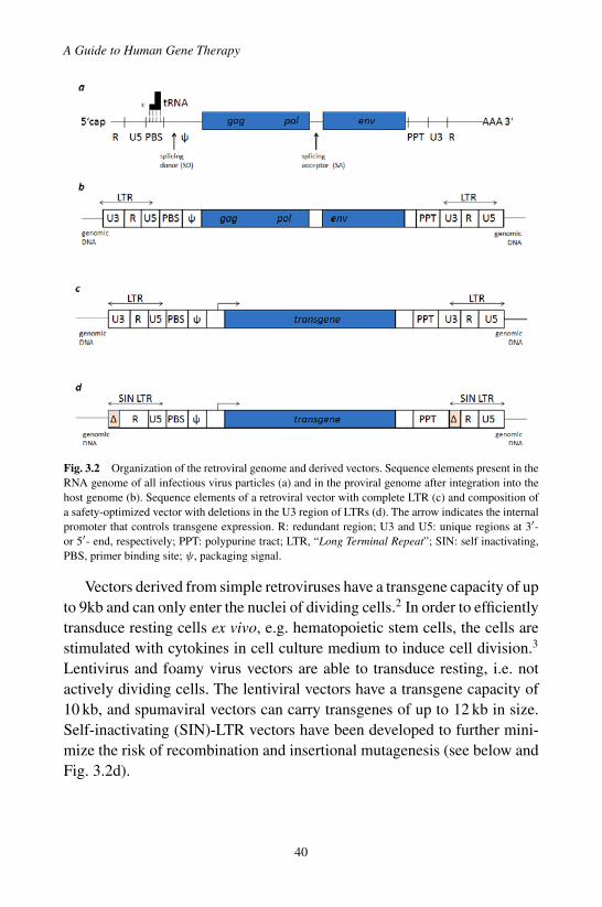
May 4, 2010 11:0 SPI-B903 9in x 6in b903-ch03
A Guide to Human Gene Therapy
Fig. 3.2 Organization of the retroviral genome and derived vectors. Sequence elements present in theRNA genome of all infectious virus particles (a) and in the proviral genome after integration into thehost genome (b). Sequence elements of a retroviral vector with complete LTR (c) and composition ofa safety-optimized vector with deletions in the U3 region of LTRs (d). The arrow indicates the internalpromoter that controls transgene expression. R: redundant region; U3 and U5: unique regions at 3′-or 5′- end, respectively; PPT: polypurine tract; LTR, “Long Terminal Repeat”; SIN: self inactivating,PBS, primer binding site; ψ, packaging signal.
Vectors derived from simple retroviruses have a transgene capacity of upto 9kb and can only enter the nuclei of dividing cells.2 In order to efficientlytransduce resting cells ex vivo, e.g. hematopoietic stem cells, the cells arestimulated with cytokines in cell culture medium to induce cell division.3
Lentivirus and foamy virus vectors are able to transduce resting, i.e. notactively dividing cells. The lentiviral vectors have a transgene capacity of10 kb, and spumaviral vectors can carry transgenes of up to 12 kb in size.Self-inactivating (SIN)-LTR vectors have been developed to further mini-mize the risk of recombination and insertional mutagenesis (see below andFig. 3.2d).
40

May 4, 2010 11:0 SPI-B903 9in x 6in b903-ch03
Retroviral Vectors and Integration Analysis
3. In vivo Application
In recent years the curative potential of retroviral gene transfer intohematopoietic stem cells was demonstrated impressively in a number ofclinical studies.4–8 In addition to obtain a therapeutic benefit, each trans-duced cell and its clonal progeny is uniquely labeled by the vector inte-gration locus. Identification of such vector integration sites not only allowsresearchers to investigate the origin of a tumor recurrence and the biology ofhuman hematopoietic regeneration, but also to follow the fate of transducedcells and to monitor their clonal behaviour over time.6,7,9–11
More than 15 years passed between the first description of a retroviralgene transfer in murine hematopoietic stem cells and the first successes in theclinical therapy of patients with X-chromosomal inherited severe combinedimmunodeficiency disorder (X-SCID).5,6,12 The limiting factor, in previousunsuccessful clinical trials, was the low efficiency of gene transfer. This hur-dle was effectively circumvented by improved transduction protocols andvectors.13,14 Further successes followed in the gene therapeutic treatmentof adenosine deaminase (ADA) SCID and chronic granulomatous disease(CGD).4,7
Since lentiviral vectors are capable of transducing proliferating as wellas non-proliferating cells, great hopes are being set for their use in clinicalapplications. Initial successes have already been reported for the applica-tion of lentiviral vectors for treating neurodegenerative diseases in animalmodels, including the treatment of Parkinson’s disease in monkeys,14 andof metachromatic leukodystrophy, a storage disorder of the central nervoussystem, in a mouse model.15
The first clinical lentiviral gene therapy trial was initiated in 2006 for thetreatment of the cerebral form of X-chromosomal linked adrenoleukodys-trophy (X-ALD), a demyelinating disease of the central nervous system.8
Mobilized hematopoietic precursor cells were harvested from two seven-year-old patients, transduced ex vivo with lentiviral SIN-vectors encodingthe ABCD1 transgene and reinfused into the patients. It was possible toarrest the progression of the disease in both patients through the constitu-tive expression of the functional ABCD1 therapeutic transgene.
41

May 4, 2010 11:0 SPI-B903 9in x 6in b903-ch03
A Guide to Human Gene Therapy
4. Side Effects in Retroviral Gene Therapy
Gene therapy using retroviral vectors is a specialized form of therapy thatis mainly applied to patients for whom no therapeutic alternatives are avail-able. Therefore, the benefit of a gene therapy always has to be opposed tothe potential risks case by case.
The integration of retroviruses in the cellular genome is per se a muta-genic event, which can lead to disruption of the gene coding regions and/orto interference with regulatory elements. However, in contrast to the use ofwild-type retroviruses and replication-competent retroviral vectors, the riskof insertional mutagenesis leading to cell transformation for a long time wasassessed to be rather low when using replication-deficient oncoviral vectorsthat were specifically developed for clinical purposes.17 The increased effi-ciency in performing ex vivo transduction of human CD34+ cells and, as aconsequence, the greater number (up to 108 − 109) of gene-corrected cellswhich can be re-infused are the factors responsible for the successful clini-cal treatment achieved with retroviral vectors since the beginning of 2000.At the same time it became evident that the risk of biologically relevantside effects could also increase along with the improved effectiveness ofthis type of treatment. Insertion-mediated mutagenesis can lead to a widespectrum of phenomena, ranging from in vitro immortalization to clonaldominance and oncogenesis in vivo.18–23
Figure 3.3 illustrates one possible mechanism of how integration ofMLV- and HIV-1-derived retroviral vectors (which integrate preferentiallyin close proximity to transcriptional start sites or gene coding regions respec-tively) may lead to a dysregulation of cellular genes.
4.1 Distribution of Retroviral Integration Sitesin the Cellular Genome
The identification and sequencing of viral integration sites involves the pre-sentation of a DNA sequence composed of the known proviral sequence andof the unknown neighboring genomic sequence. In order to characterize theunknown flanking DNA, two fundamental PCR methods were developed,inverse PCR and ligation-mediated PCR.24,25 Although further developmentin these methodologies led to improvements in sensitivity, the invention of
42

May 4, 2010 11:0 SPI-B903 9in x 6in b903-ch03
Retroviral Vectors and Integration Analysis
Fig. 3.3 Mechanism of proto-oncogene activation by insertional mutagenis. (A) Integration of viralgenomic DNA into somatic cellular DNA in the proximity of a nontranscribed oncogene. (B) Integratedproviral DNA induces the transcription of the oncogene. RV, retroviral vector. LTR, “Long TerminalRepeat”.
the linear amplification-mediated PCR (LAM-PCR) allowed for the firsttime the characterization of integration sites at the single-cell level andto monitor the polyclonal gene-modified hematopoiesis directly in limitedamounts of peripheral blood leukocytes and bone marrow cells.26,27 LAM-PCR has been shown to be the superior method to determine the clonalityof the hematopoietic repopulation after transplantation in humans.
The principle of LAM-PCR is illustrated in Fig. 3.4. The first step (a)is the preamplification of the vector–genome junctions through a linearPCR with biotinylated primers hybridizing at one end of the integratedvector. The following steps (b-f) are carried out on a semisolid strepta-vidin phase in order to capture DNA strands with an incorporated biotiny-lated vector primer. After double strand synthesis (c), a restriction digest(d), and the ligation of a linker cassette (e) on the genomic end of thefragment, two exponential PCRs with nested arranged vector- and linkercassette primers are carried out in order to amplify the fragments consist-ing of linker cassette-, genomic-, and vector-sequence (g). The generatedfragments are sequenced and the integration loci are determined. A further
43

May 4, 2010 11:0 SPI-B903 9in x 6in b903-ch03
A Guide to Human Gene Therapy
Fig. 3.4 Schematic outline of LAM-PCR to amplify 5-LTR retroviral vector-genomic fusionsequences. Steps (a-g) see text. B, biotinylated primer; LTR, long terminal repeat; LTR I/II: LTR primerfor the first/second exponential PCR; LCI/II, linker cassette primer for the first /second exponentialPCR; Schmidt et al. 2007)
development of the LAM-PCR is the non-restrictive LAM-PCR (nrLAM-PCR), which allows the genome-wide identification of retroviral integrationsites in a single reaction, circumventing the detection bias caused by the useof restriction enzymes.28 The knowledge of the genomic flanking sequencederived from a clone of interest allows the creation of primers specific foran individual integration clone. Thus, PCR analysis using these primers incombination with vector-specific primers makes it possible to monitor onesingle specific clone in vivo in different samples.27
Due to the availability of the complete human and murine genomesequence data the precise localization of viral integration sites in the hostgenome now became possible, and by the improvement of new sequencingtechnologies also feasible in a high-throughput manner.29
Severe side effects emerged in 2003, when two of the patients enrolled inthe first successful gene therapy trial for the treatment of X-SCID developeda T cell acute lymphatic leukemia between 30 and 34 months after treat-ment. As a consequence, comprehensive investigations were conducted to
44
May 4, 2010 11:0 SPI-B903 9in x 6in b903-ch03
Retroviral Vectors and Integration Analysis
observe the integration behavior of different vectors and wild-type virusesin vitro and in vivo.30–32 These analyses provided the first concrete evi-dence that retroviral integration into the host genome is much less randomthan previously assumed, and that different vector types consistently tendto integrate into specific genomic regions.
The fact that tumorigenic chromosomal aberrations (i.e. deletions,translocations and amplifications) as well as the integration of onco-genic viruses (for instance human papilloma virus 16 (HPV16), HPV18,Hepatitis-B-Virus and Epstein-Barr-Virus) often occur in so-called“common fragile sites” (CFSs) has already been known since 2003.33 In2006, Bester et al. were able to show a significant higher integration fre-quency of MLV in such CFSs.34 The integration of MLV-derived vectorspreferentially occurs close to transcription start sites and in proximity toCpG islands.32 In contrast, HIV-1-derived vectors in two thirds of casesintegrate into gene coding regions over the complete length of the gene,with a preference for active genes.30,31 Moreover, for both MLV- and HIV-derived vectors a cell type specific integration pattern was observed, due todifferences in their gene expression status.7,10,35–37 More recent investiga-tions on the integration pattern of HIV-1 revealed a relationship between theexpression of the transcription coactivator lens epithelium-derived growthfactor (LEDGF) and the integration of the vector in transcribed regionsin Jurkat cells.38,39 Lewinski et al. showed that viral components such asintegrase and group-specific antigenes influence the integration behavior ofHIV, MLV and their chimeras.40 Another group of retroviral vectors derivedfrom Avian Sarcoma Leukosis Virus (ASLV) show neither a preference fortranscription start sites nor for active genes.30 These results make it clearthat the selection of the gene transfer vector with respect to the target cellor target tissue can have a decisive influence on the clinical outcome of agene therapy trial.
4.2 Side Effects in Clinical and Preclinical GeneTherapy Studies
In 2002, the first malignant transformation that developed as a result of genetransfer using replication-deficient retroviral vectors was observed.21 Serialtransplantations of gammaretroviral gene-marked CD34+ cells derived
45
May 4, 2010 11:0 SPI-B903 9in x 6in b903-ch03
A Guide to Human Gene Therapy
from murine bone marrow led to tumor development in secondary and ter-tiary recipients. All secondary transplanted animals developed alterationsof their hematopoiesis after 22 weeks, and 6 out of ten animals developedacute myeloid leukemia. It was possible to show that all of these animals hadthe same malignant clone with a vector integrant in the Evi1 gene locus. Thefunctional promoter and enhancer elements in both proviral LTRs caused anoverexpression of Evi1. The MLV vector used here encoded the transgenethat had been used for clinical applications, a low-affinity nerve growthfactor receptor (LNGFR). It could not be proven that the growth-mediatingtransgene LNGFR had a synergistic effect, but neither could such an effectbe ruled out.
Gene marking studies in a non-human primate model revealed indica-tions of a possible clonal dominance in the retrovirally transduced cells.18
Calmels and his colleagues were able to show that clones with distinctvector integration sites (Mds1/Evi1, Prdm16) had a growth advantage com-pared to other cell clones. However, no abnormal proliferation of affectedcell clones was observed, despite numerous retroviral integrations withinthese proto-oncogenes. Since the major part of these cell clones contributesto long-term hematopoiesis, a higher engraftment or survival probabilityas a result of insertional mutagenesis was discussed. Analysis of anothermurine gene marking study proved that proviral integration within or nearbydistinct cellular genes promotes the growth of single transduced cells andcontributes to their clonal expansion in vivo.20
In the context of a murine study on the retroviral expression of an ABC-transporter (MDR1), it was observed that the vector dose has an impact onleukemogenesis. Leukemia not only occurred due to the growth advantageof single clones but also correlated with high vector doses.41 In 2006, the firstacute myeloid leukemia in a non-human primate model was described afterretroviral gene transfer in hematopoietic precursor cells. The treated animaldied five years after gene therapy due to a myeloid sarcoma.22 Two vectorintegrants (Bcl2-A1, Cdw91) were detected in the blood as well as in thetumor tissue of the animal, both of which had already achieved dominanceone year after transplantation.
The first published side effect after gene transfer using a lentiviral vec-tor was a T-cell lymphoma that occurred in the context of the constitutiveexpression of the therapeutic gene Il2RG in a murine model. The influence
46

May 4, 2010 11:0 SPI-B903 9in x 6in b903-ch03
Retroviral Vectors and Integration Analysis
of the growth-related transgene on the tumor development in this study wasa matter of much controversial debate.23,42 A gene transfer-induced tumori-genesis was also observed with an adeno-associated virus (AAV) derivedvector in a murine model.43
In three clinical gene therapy studies, integration site analyses in patienthematopoietic cells showed that the retroviral vector could affect the growthand differentiation of distinct transduced clones. Two to six years after suc-cessful gene therapy for X-SCID, 5 of 19 patients developed T-cell leukemia.In 4 of these patients, vector integrations in the proto-oncogene LMO2occurred causing its overexpression and expansion of these clones.7,36,37,44,45
Also, a combinatorial effect of insertional mutagenesis and constitutiveexpression of the transgene IL2RG as a cause of lymphoproliferative dis-ease was discussed.23 Gene therapy for correction of X-CGD in two adultpatients resulted in the restoration of oxidative antimicrobial activity inphagocytes after gene transfer. Substantial gene transfer in neutrophil cellshad produced a high number of functional phagocytes. Integration anal-yses revealed a clonal dominance of insertion sites in the genes MDS1-EVI1, PRDM16 or SETBP1, resulting in an expansion of gene-correctedmyelopoesis.7 A myelodysplastic syndrome developed in both patients inthe course of time.46
5. New Strategies for Vector Biosafety in Gene Therapy
The treatment of monogenic hereditary diseases with gammaretroviral vec-tors led to severe side effects only in certain diseases (X-SCID, X-CGD)and only in few patients.44–46 Therefore, the applied vector itself cannotaccount solely for the incidence of these side effects; in fact a number offactors seem to interact. For instance no side effects have been observed sofar in the gene therapy of ADA-SCID using gammaretroviral vectors.47 Inorder to minimize the risk of side effects in future gene therapy trials it willbe important to choose the most suitable gene transfer vectors dependenton the disease individually and to test the usage of tissue-specific promotersor non-integrating vector systems if appropriate.
During the last years several in vitro and in vivo systems were devel-oped, aiming to assess the genotoxic potential of different vectors. Alreadyin 2003 it was reported that replication-competent gammaretroviruses are
47
May 4, 2010 11:0 SPI-B903 9in x 6in b903-ch03
A Guide to Human Gene Therapy
suitable for specific detection of cancer genes. Transplantation of transducedhematopoietic cells in mice with knocked-out Cdkn2a tumor suppressorgene leads to accelerated tumor growth when a cellular proto-oncogeneis virally activated.48 Thus, the most extensive database of murine geneswith oncogenic potential, the mouse “Retrovirally Tagged Cancer GeneDatabase” (RTCGD) was developed.49
In other work, Du and colleagues showed that identification of proto-oncogenes is feasible by retroviral gene transfer in cell culture.26 Trans-duction of murine bone marrow cells with replication deficient retrovirusesexpressing marker genes resulted in immortalized cell lines, many of whichcontained integrations in the Mds1/Evi1 and Prdm16 genes. Vector integra-tion into the Evi1 locus and resulting overexpression of Evi1 appeared to besufficient for a cell to be immortalized. Modlich and colleagues adapted andimproved this method so that the results could be analyzed in a quantitativemanner.47
In order to assess the biological safety of vectors used for the treatmentof X-SCID a tumor prone mouse model was developed in which the tumorsuppression gene Arf as well as the Il2rg gene had been knocked-out.51
Since it was proven that the activity of promoter/enhancer elements inthe LTR of conventional retroviral vectors may cause insertional activationof cellular protooncogenes, the first attempts to reduce the risk of insertionalmutagenesis involved the development of SIN vectors. In these vectors, theenhancer and promoter elements in the U3-region of the LTR are deletedin order to eradicate their activity after integration.52 Expression of thetransgene is driven by an internal promoter, which, in addition, can be tissueor cell-type specific for further improvement of safety (see Fig. 3.2d).
The mouse model originally developed by Lund et al. was suitable to per-form comparative tests on the genotoxic potential of different gene transfervectors. Mice that had received hematopoietic precursor cell transplantstransduced with gammaretroviral vectors developed tumors much morerapidly than mice that had received cells harbouring lentiviral SIN-vectorswith an internal human phosphoglycerate kinase (hPGK) promoter.53 Lateron it was demonstrated that retroviral vectors with SIN-LTRs show a signif-icant reduction in genotoxicity, but when combined with strong (viral) inter-nal promoters still have the ability to induce leukemia in a mouse model.51
48

May 4, 2010 11:0 SPI-B903 9in x 6in b903-ch03
Retroviral Vectors and Integration Analysis
However, the usage of physiological (cellular) promoters further reducedthe risk of insertional mutagenesis.55
Other approaches for safety optimization include the modification ofenvelope proteins of vector particles in order to constrain their transductionto the target cell population.56 Insertion of insulator elements in the LTRsaim to reduce both the influence of genomic environment on the integratedvector as well as the influence of the integrated vector on the surroundinghost genome.57
In postmitotic tissues, no dilution of unintegrated episomal vector formsoccurs, thus allowing the usage of non-integrating vector systems. Muta-tions in the core domain of the viral integrase prevent integration, therebyreducing the risk of insertional mutagenesis intensively. Recently, the long-term functional correction of retinal degeneration in a well-establishedrodent model for ocular gene therapy was reported. This was accom-plished without apparent side effects using an integrase-deficient HIV-1vector.58
The targeted insertion of non-integrating vectors into defined, puta-tive safe gene loci via homologous recombination by the use of doublestrand break inducing enzymes (i.e. zinc finger nucleases, meganucleases)represent another promising approach for safety optimization. The decisivefactor for the successful application of this method is to avoid the recogni-tion of wrong target sequences by zinc finger nucleases that would triggeroff-site double strand breaks.59,60 Another alternative approach for targetedintegration is the usage of transposons as gene ferries.57
References
1. Coffin JM, Hughes SH, Varmus HE (1997). Cold Spring Harbour, USA: Cold SpringHarbor Laboratory Press.
2. Roe T, et al. (1993). Integration of murine leukemia virus DNA depends on mitosis.EMBO Retroviruses J 12(5): 2099–108.
3. Nolta JA, Smogorzewska EM, Kohn DB (1995). Analysis of optimal conditions forretroviral-mediated transduction of primitive human hematopoietic cells. Blood 86(1):101–110.
4. Aiuti A, et al. (2002). Correction of ADA-SCID by stem cell gene therapy combinedwith nonmyeloablative conditioning. Science 296(5577): 2410–2413.
5. Gaspar HB, et al. (2004). Gene therapy of X-linked severe combined immunodeficiencyby use of a pseudotyped gammaretroviral vector. Lancet 364(9452): 2181–2187.
49

May 4, 2010 11:0 SPI-B903 9in x 6in b903-ch03
A Guide to Human Gene Therapy
6. Hacein-Bey-Abina S, et al. (2002). Sustained correction of X-linked severe combinedimmunodeficiency by ex vivo gene therapy. N Engl J Med 346(16): 1185–1193.
7. Ott MG, et al. (2006). Correction of X-linked chronic granulomatous disease by genetherapy, augmented by insertional activation of MDS1-EVI1, PRDM16 or SETBP1.Nat Med 12(4): 401–409.
8. Cartier N, et al. (2009). Hematopoietic stem cell gene therapy with a lentiviral vectorin X-linked adrenoleukodystrophy. Science 326(5954): 818–823.
9. Brenner MK, et al. (1993). Gene-marking to trace origin of relapse after autologousbone-marrow transplantation. Lancet 341(8837): 85–86.
10. Glimm H, et al. (2005). Efficient marking of human cells with rapid but transientrepopulating activity in autografted recipients. Blood 106(3): 893–898.
11. Guenechea G., et al. (2001). Distinct classes of human stem cells that differ in prolif-erative and self-renewal potential. Nat Immunol 2(1): 75–82.
12. Cavazzana-Calvo M, et al. (2000). Gene therapy of human severe combined immun-odeficiency (SCID)-X1 disease. Science 288(5466): 669–672.
13. Kohn DB, et al. (1998). T lymphocytes with a normal ADA gene accumulate aftertransplantation of transduced autologous umbilical cord blood CD34+ cells in ADA-deficient SCID neonates. Nat Med 4(7): 775–780.
14. Stead RB, et al. (1988). Canine model for gene therapy: inefficient gene expressionin dogs reconstituted with autologous marrow infected with retroviral vectors. Blood71(3): 742–747.
15. Kordower JH, et al. (2000). Neurodegeneration prevented by lentiviral vector deliveryof GDNF in primate models of Parkinson’s disease. Science 290(5492): 767–773.
16. Consiglio A, et al. (2001). In vivo gene therapy of metachromatic leukodystrophy bylentiviral vectors: correction of neuropathology and protection against learning impair-ments in affected mice. Nat Med 7(3): 310–316.
17. Moolten FL, Cupples LA (1992). A model for predicting the risk of cancer consequentto retroviral gene therapy. Hum Gene Ther 3(5): 479–486.
18. Calmels B, et al. (2005). Recurrent retroviral vector integration at the Mds1/Evi1 locusin nonhuman primate hematopoietic cells. Blood 106(7): 2530–2533.
19. Du Y, Jenkins NA, Copeland NG (2005). Insertional mutagenesis identifies genes thatpromote the immortalization of primary bone marrow progenitor cells. Blood 106(12):3932–3939.
20. Kustikova O, et al. (2005). Clonal dominance of hematopoietic stem cells triggered byretroviral gene marking. Science 308(5725): 1171–1174.
21. Li Z, et al. (2002). Murine leukemia induced by retroviral gene marking. Science296(5567): 497.
22. Seggewiss R, et al. (2006). Acute myeloid leukemia is associated with retroviral genetransfer to hematopoietic progenitor cells in a rhesus macaque. Blood 107(10): 3865–3867.
23. Woods NB, et al. (2006). Gene therapy: therapeutic gene causing lymphoma. Nature440(7088): 1123.
24. Mueller PR, Wold B (1989). In vivo footprinting of a muscle specific enhancer byligation mediated PCR. Science 246(4931): 780–786.
25. Silver J, KeerikatteV (1989). Novel use of polymerase chain reaction to amplify cellularDNA adjacent to an integrated provirus. J Virol 63(5): 1924–1928.
50
May 4, 2010 11:0 SPI-B903 9in x 6in b903-ch03
Retroviral Vectors and Integration Analysis
26. Schmidt M, et al. (2001). Detection and direct genomic sequencing of multiple rareunknown flanking DNA in highly complex samples. Hum Gene Ther 12(7): 743–749.
27. Schmidt M, et al. (2007). High-resolution insertion-site analysis by linear amplification-mediated PCR (LAM-PCR). Nat Methods 4(12): 1051–1057.
28. Gabriel R, et al. (2009). Comprehensive genomic access to vector integration in clinicalgene therapy. Nat Med 15(12): 1431–1436.
29. Wold B, Myers RM (2008). Sequence census methods for functional genomics. NatMethods 5(1): 19–21.
30. Mitchell RS, et al. (2004). Retroviral DNA integration: ASLV, HIV, and MLV showdistinct target site preferences. PLoS Biol 2(8): E234.
31. Schroder AR, et al. (2002). HIV-1 integration in the human genome favors active genesand local hotspots. Cell 110(4): 521–529.
32. Wu X, et al. (2003). Transcription start regions in the human genome are favored targetsfor MLV integration. Science 300(5626): 1749–1751.
33. Popescu NC (2003). Genetic alterations in cancer as a result of breakage at fragile sites.Cancer Lett 192(1): 1–17.
34. Bester AC, et al. (2006). Fragile sites are preferential targets for integrations of MLVvectors in gene therapy. Gene Ther 13(13): 1057–1059.
35. Cattoglio C, et al. (2007). Hot spots of retroviral integration in human CD34+hematopoietic cells. Blood 110(6): 1770–1778.
36. Deichmann A, et al. (2007). Vector integration is nonrandom and clustered and influ-ences the fate of lymphopoiesis in SCID-X1 gene therapy. J Clin Invest 117(8): 2225–2232.
37. Schwarzwaelder K, et al. (2007). Gammaretrovirus-mediated correction of SCID-X1 isassociated with skewed vector integration site distribution in vivo. J Clin Invest 117(8):2241–2249.
38. Ciuffi A, et al. (2005). A role for LEDGF/p75 in targeting HIV DNA integration. NatMed 11(12): 1287–1289.
39. Marshall HM, et al. (2007). Role of PSIP1/LEDGF/p75 in lentiviral infectivity andintegration targeting. PLoS ONE 2(12): e1340.
40. Lewinski MK, et al. (2006). Retroviral DNA integration: viral and cellular determinantsof target-site selection. PLoS Pathog 2(6): e60.
41. Modlich U, et al. (2005). Leukemias following retroviral transfer of multidrug resis-tance 1 (MDR1) are driven by combinatorial insertional mutagenesis. Blood 105(11):4235–4246.
42. Pike-Overzet K, et al. (2006). Gene therapy: is IL2RG oncogenic in T-cell development?Nature 443(7109): E5; discussion E6–7.
43. Kay MA (2007). AAV vectors and tumorigenicity. Nat Biotechnol 25(10): 1111–1113.44. Hacein-Bey-Abina S, et al. (2008). Insertional oncogenesis in 4 patients after retrovirus-
mediated gene therapy of SCID-X1. J Clin Invest.45. Howe SJ, et al. (2008). Insertional mutagenesis combined with acquired somatic muta-
tions causes leukemogenesis following gene therapy of SCID-X1 patients. J Clin Invest.46. Grez M, et al. (2007). Update on gene therapy for chronic granulomatous disease. Hum
Gene Ther. European Society of Gene and Cell Therapy: Rotterdam. p. 959.47. Aiuti A, et al. (2009). Gene therapy for immunodeficiency due to adenosine deaminase
deficiency. N Engl J Med 360(5): 447–458.
51

May 4, 2010 11:0 SPI-B903 9in x 6in b903-ch03
A Guide to Human Gene Therapy
48. Lund AH, et al. (2002). Genome-wide retroviral insertional tagging of genes involvedin cancer in Cdkn2a-deficient mice. Nat Genet 32(1): 160–165.
49. Akagi K, et al. (2004). RTCGD: retroviral tagged cancer gene database. Nucleic AcidsRes 32(Database issue): D523–527.
50. Modlich U, et al. (2006). Cell-culture assays reveal the importance of retroviral vectordesign for insertional genotoxicity. Blood 108(8): 2545–2553.
51. ShouY, et al. (2006). Unique risk factors for insertional mutagenesis in a mouse modelof XSCID gene therapy. Proc Natl Acad Sci USA 103(31): 11730–11735.
52. Zufferey R, et al. (1998). Self-inactivating lentivirus vector for safe and efficient in vivogene delivery. J Virol 72(12): 9873–9880.
53. Montini E, et al. (2006). Hematopoietic stem cell gene transfer in a tumor-prone mousemodel uncovers low genotoxicity of lentiviral vector integration. Nat Biotechnol 24(6):687–696.
54. Montini E, et al. (2009). The genotoxic potential of retroviral vectors is strongly mod-ulated by vector design and integration site selection in a mouse model of HSC genetherapy. J Clin Invest 119(4): 964–975.
55. Zychlinski D, et al. (2008). Physiological promoters reduce the genotoxic risk of inte-grating gene vectors. Mol Ther 16(4): 718–725.
56. Waehler R, SJ Russell, DT Curiel (2007). Engineering targeted viral vectors for genetherapy. Nat Rev Genet 8(8): 573–587.
57. West AG, Gaszner M, Felsenfeld G (2002). Insulators: many functions, many mecha-nisms. Genes Dev 16(3): 271–288.
58. Yanez-Munoz RJ, et al. (2006). Effective gene therapy with nonintegrating lentiviralvectors. Nat Med 12(3): 348–353.
59. Miller JC, et al. (2007). An improved zinc-finger nuclease architecture for highly spe-cific genome editing. Nat Biotechnol 25(7): 778–785.
60. Szczepek M, et al. (2007). Structure-based redesign of the dimerization interfacereduces the toxicity of zinc-finger nucleases. Nat Biotechnol 25(7): 786–793.
52
May 4, 2010 11:0 SPI-B903 9in x 6in b903-ch04
Chapter 4
Lentiviral Vectors
Janka Mátrai, Marinee K. L. Chuah and ThierryVandenDriessche∗
Lentiviral vectors represent some of the most promising vectors for genetherapy. They have emerged as potent and versatile tools for ex vivo orin vivo gene transfer into dividing and non-dividing cells. Lentiviral vec-tors can be pseudotyped with distinct viral envelopes that influence vectortropism and transduction efficiency. In addition, it is possible to redi-rect vector transduction and generate cell-type specific targetable vectorsby incorporating cell-type specific ligands or antibodies into the vectorenvelope. Lentiviral vectors have been used to deliver genes into varioustissues, including brain, retina and liver, by direct in vivo gene deliv-ery. Since they integrate stably into the target cell genome, they are ide-ally suited to deliver genes ex vivo into bona fide stem cells, particularlyhematopoietic stem cells (HSCs), allowing for stable transgene expressionupon hematopoietic reconstitution.Though there are some safety concernsregarding the risk of insertional mutagenesis, it is possible to minimize thisrisk by modifying the vector design or by employing integration-deficientlentiviral vectors which, in conjunction with zinc-finger nuclease tech-nology, allow for site-specific gene correction or addition in pre-definedchromosomal loci. Lentiviral gene transfer has provided efficient phe-notypic correction of diseases in mouse models paving the way towardsclinical applications.
1. Basic Viral Biology
Lentiviruses are enveloped RNA viruses that are surrounded by a lipidbilayer in which the envelope proteins are embedded. The main components
∗Correspondence: Flanders Institute for Biotechnology,Vesalius Research Center, University of Leuven,Leuven, Belgium.E-mail: [email protected]
53

May 4, 2010 11:0 SPI-B903 9in x 6in b903-ch04
A Guide to Human Gene Therapy
Fig. 4.1 A. Schematic representation of the HIV-1 virus. The figure shows the single stranded diploidRNA genome of HIV-1 embedded into the nucleocapsid encoded by the gag gene. The integrase (INT)and reverse transcriptase (RT) enzymes encoded by the pol gene are also indicated. The nucleocapsidis surrounded by the capsid and the lipid bilayer membrane where the glycoproteins 41 and 120 arelocated; B. Viral life cycle. The HIV-1 virus enters the cell via the cell surface receptors. The viralgenome is endocytosed and reverse transcribed in the cytosol. The viral DNA subsequently crosses thenuclear membrane and finally integrates into the host genome. Using the transcription machinery ofthe host cell viral RNA is transcribed and transported into the cytoplasm. Here, translation and virusassembly take place and the de novo synthesized virus leaves the cell to initiate a new productiveinfection.
54

May 4, 2010 11:0 SPI-B903 9in x 6in b903-ch04
Lentiviral Vectors
of the wild-type HIV-1 virus and its life cycle are shown in Figs. 4.1A and4.1B. The envelope surrounds the viral capsid that contains the structuralproteins (matrix), viral enzymes and 2 copies of a single stranded RNAgenome, which codes for gag, pol and env genes that are common to allretroviruses (Fig. 4.1A). The gag gene encodes the structural matrix pro-teins, capsid and nucleocapsid, pol encodes the reverse transcriptase (RT),integrase (INT) and protease and env encodes the envelope glycoproteins(gp120 and gp41). Additionally, the lentiviral genome contains accessorygenes encoding the regulatory proteins Tat, Rev, Vpr, Vpu Vif and Nef.Tat and Rev are essential for viral replication, whereas the other accessorygenes contribute to viral pathogenicity. Tat is a potent trans-activator ofHIV-1 gene expression and “jump-starts” the HIV replication cycle. Rev isa post-transcriptional trans-activator that accelerates mRNA transport fromthe nucleus to the cytoplasm via binding to the Rev-responsive element(RRE). Rev is essential for the expression of the gag, pol and env genes andfor the transport of the full length RNA genome.
During infection, the Env proteins interact with their cognate cellularreceptors (i.e. CD4 and the co-receptors CCR5 or CXCR4). After receptor-mediated viral entry into the cell by membrane fusion, the RNA genomeis reverse transcribed in the cytoplasm by the reverse transcriptase into adouble stranded proviral DNA (Fig. 4.1B). This proviral DNA is associatedwith different viral proteins (nucleocapsid, RT, integrase) in an intermediateprotein-DNA complex designated as the pre-integration complex (PIC).The PIC also interacts with cellular proteins that facilitate the transportacross the nuclear membrane into the nucleus. The intrinsic properties ofthe lentiviral PIC are different from that of the γ-retroviruses and endowthe virus with the ability to enter the nucleus of non-dividing cells. In thenucleus, the integrase catalyzes the integration into the target cell genomevia interaction with the viral long terminal repeats (LTR), particularly theatt (attachment) sequence.
Cellular factors such as LEDGF/p75 play a key role in controlling lentivi-ral integration by acting as a tethering factor. Host cell factors initiate tran-scription from the integrated proviral LTRs resulting in production of newviral genomic RNA that encodes the different HIV-1 structural and regu-latory proteins. The genomic RNA and the newly produced viral proteinsare subsequently assembled as a viral particle during the budding process(Fig. 4.1B).
55
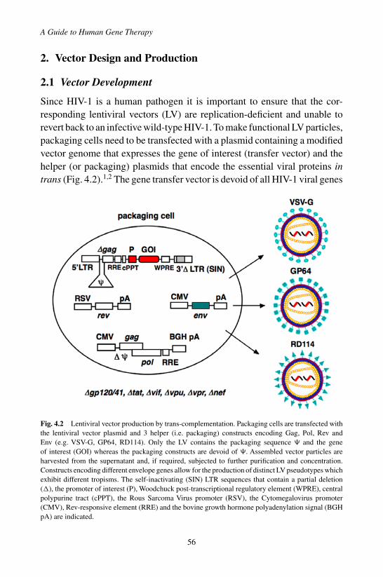
May 4, 2010 11:0 SPI-B903 9in x 6in b903-ch04
A Guide to Human Gene Therapy
2. Vector Design and Production
2.1 Vector Development
Since HIV-1 is a human pathogen it is important to ensure that the cor-responding lentiviral vectors (LV) are replication-deficient and unable torevert back to an infective wild-type HIV-1. To make functional LV particles,packaging cells need to be transfected with a plasmid containing a modifiedvector genome that expresses the gene of interest (transfer vector) and thehelper (or packaging) plasmids that encode the essential viral proteins intrans (Fig. 4.2).1,2 The gene transfer vector is devoid of all HIV-1 viral genes
Fig. 4.2 Lentiviral vector production by trans-complementation. Packaging cells are transfected withthe lentiviral vector plasmid and 3 helper (i.e. packaging) constructs encoding Gag, Pol, Rev andEnv (e.g. VSV-G, GP64, RD114). Only the LV contains the packaging sequence � and the geneof interest (GOI) whereas the packaging constructs are devoid of �. Assembled vector particles areharvested from the supernatant and, if required, subjected to further purification and concentration.Constructs encoding different envelope genes allow for the production of distinct LV pseudotypes whichexhibit different tropisms. The self-inactivating (SIN) LTR sequences that contain a partial deletion(�), the promoter of interest (P), Woodchuck post-transcriptional regulatory element (WPRE), centralpolypurine tract (cPPT), the Rous Sarcoma Virus promoter (RSV), the Cytomegalovirus promoter(CMV), Rev-responsive element (RRE) and the bovine growth hormone polyadenylation signal (BGHpA) are indicated.
56

May 4, 2010 11:0 SPI-B903 9in x 6in b903-ch04
Lentiviral Vectors
encoding structural or accessory proteins. In contrast to γ-retroviral vectors,LV can typically accommodate larger transgene cassettes (up to ∼10 kb) thatare expressed using an internal promoter. The � sequence is essential forpackaging and ensures appropriate encapsidation of the full-length vectorgenome into the viral vector particles (Fig. 4.2). The LV is flanked by the 5′
and 3′ LTR sequences that have promoter/enhancer activity, which is essen-tial for the correct expression of the full-length vector transcript, and play animportant role in reverse transcription and integration of the vector into thetarget cell genome. To prevent mobilization (the packaging of the integratedLV into HIV particles) by infection with wild-type HIV a self-inactivating(SIN) vector configuration with a 400 bp deletion in the 3′ LTR U3 region,including the TATA box was introduced.3 This deletion is subsequentlycopied onto the 5′ LTR of the proviral DNA during reverse transcription,abolishing the transcriptional activity of the LTR. Furthermore, the SINconfiguration reduces the likelihood that cellular coding sequences locatedadjacent to the vector integration site will be aberrantly expressed, either dueto the promoter activity of the 3′ LTR or through an enhancer effect. Finally,the SIN design prevents potential transcriptional interference between theLTR and the internal promoter driving the transgene.3 The introduction ofchimeric CMV/HIV-5′ LTR or RSV/HIV-5′ LTR promoters to drive theexpression of the full-length vector transcript allowed for the eliminationof the HIV-1 Tat protein during vector production.
Several small genetic sequences have been introduced into the vectorto enhance gene transfer efficiency. The woodchuck hepatitis virus post-transcriptional regulatory element (WPRE)4 when placed 3′ of the gene ofinterest leads to increased transgene expression. The RRE including a smallsegment of the gag gene was re-introduced into the transfer vector resultingin improved viral titers. Finally, it has been shown that the central polypurinetract (cPPT) facilitates nuclear translocation of the pre-integration complexand consequently increases lentiviral transduction.5,6
Of the 9 HIV-1 viral genes, only 3 are essential for the production ofHIV-1 based LV namely gag, pol, and rev. The function of these genes inthe LV is identical to that in the cognate wild-type HIV-1 virus. The gagand pol genes are expressed from the same helper (i.e. packaging) plasmid.Since expression of gag and pol is strictly dependent upon the Rev-RREinteraction, a separate construct encoding Rev needs to be cotransfected
57
May 4, 2010 11:0 SPI-B903 9in x 6in b903-ch04
A Guide to Human Gene Therapy
into the packaging cells. The generation of wild-type HIV-1 can de factobe excluded since none of these genes are present in the packaging cells.Additionally, LV particles require a viral envelope protein, which is essentialfor vector entry into the target cells. The envelope of the LV is typicallyencoded by a separate packaging construct that contains a heterologous envgene from another enveloped virus that replaces the native HIV-1 env gene(i.e. pseudotyping; see below).
2.2 Vector Production
The most common procedure to generate LV is to co-transfect 293T (humanembryonic kidney 293) cells with the vector encoding plasmid, the gag-pol,rev and env packaging constructs (Fig. 4.2). The typical titer of the non-concentrated vector batches is about 107 transducing units/ml (TU/ml).Vector titers can further be increased by means of centrifugal filter con-centration or ultracentrifugation. Concentrated vector titers typically fall inthe range of 109 − 1010 TU/ml. Although transient transfection of 293 Tcells can produce high titer LV, this method is cumbersome and diffi-cult to scale up which poses significant manufacturing hurdles. To over-come these limitations, stable packaging cell lines were developed thatconditionally express the cytotoxic Gag-Pol and VSV-G Env using aninducible promoter. This packaging cell line allows for the productionof high-titer LV (>107 TU/ml) paving the way towards industrial large-scale vector production in bio-reactors for clinical trials and therapeuticapplications.7
Typically LV titers are reported as TU/ml, which is determined with areporter or selectable marker gene by quantifying the number of transducedcells following gene transfer. Alternatively, vector titer can be determinedby assessing the number of actual vector copies in the transduced cell pop-ulation by (real-time) quantitative PCR.8 Since both of these methods fordetermining viral titers are based on a chosen cell line, additional meth-ods have been developed to allow for comparison of different pseudotypedLV titers. LV particle titers can be determined by measuring the amountof the HIV-1 capsid p24 protein by enzyme-linked immunosorbent assay(ELISA), or by estimating the RNA content of the vector particles by RNAdot-blot analysis or reverse transcriptase (RT)-qPCR.
58

May 4, 2010 11:0 SPI-B903 9in x 6in b903-ch04
Lentiviral Vectors
3. Gene Transfer Concepts and Potential Applications
3.1 Target Cells and Diseases
In contrast to γ-retroviral vectors, LV can also transduce non-dividing cells,which opened up new therapeutic avenues and applications. In particular,local administration of LV into the brain resulted in stable transduction ofterminally differentiated neurons.1,9 Stable transduction was also reportedin other tissues, including skeletal muscle. Systemic administration typi-cally resulted in relatively efficient and widespread transduction of liverand spleen. Proof of concept has been established in pre-clinical animalmodels that LV can result in effective gene therapy for hereditary, acquiredor complex disorders that affect these various organs (e.g. neurodegenera-tive disease, congenital blindness, liver disease, hemophilia, metabolic dis-eases, etc.). LV are also ideally suited to transduce various stem/progenitorcells, particularly HSCs.10,11 HSCs are attractive targets for gene therapybecause of their capacity of self-renewal and potential life-long ability toreplenish mature blood cell populations. Sustained expression of therapeu-tic transgenes at clinically relevant levels has been demonstrated follow-ing LV transduction of HSCs for a variety of hematologic and metabolicdiseases (e.g. hemoglobinopathies, severe combined immune deficiencies,hemophilia, etc.).12,13 By restricting expression of the therapeutic gene ina cell lineage-specific fashion using lineage-specific promoters, it has beenpossible to generate LV that upon transduction of HSC are only expressedin the desired lineage upon hematopoietic reconstitution.14
3.2 Pseudotyping
The most commonly used viral envelope protein used for pseudotyping LVis the vesicular stomatitis virus glycoprotein (VSV-G), but other envelopesincluding rabies, MLV-amphotropic, Ebola, baculovirus and measles virusenvelopes can be used too (Fig. 4.2).15 Pseudotyping HIV-1 vectors obvi-ates safety concerns associated with the use of HIV-1 gp120, which hasknown pathogenic consequences. Moreover, pseudotyping has a dramaticimpact on the biodistribution, vector tropism, and viral particle stability. Forinstance, filovirus envelope-pseudotyped LV enhance transduction of air-way epithelia or endothelial cells16 whereas baculovirus GP64 and hepatitis
59

May 4, 2010 11:0 SPI-B903 9in x 6in b903-ch04
A Guide to Human Gene Therapy
C E1 and E2 pseudotyping enhances hepatic transduction.17,18 RD114 pseu-dotyping favors transduction in lymphohematopoietic cells.19 LV pseudo-typed with the Edmonston measles virus (MV) glycoproteins H and Fallowed efficient transduction through the MV receptors, SLAM and CD46,both present on blood T cells.20
3.3 Cell Type Specific Targeting
Pseudotyping also provides a means to generate cell-type specific LV. Thiscan be accomplished by incorporating cell-type specific ligands or antibod-ies into the viral envelope (Fig. 4.3). Modification of the envelope withthese cell type specific retargeting moieties can redirect the binding of theLV particles to the corresponding cellular receptor. However, envelope engi-neering can potentially adversely alter the fusion domain of Env resultingin low vector titers. It is therefore important to ensure that redirecting vec-tor tropism does not compromise the post-binding step in vector entry. Toaddress this concern, fusogenic envelope proteins are incorporated alongside the retargeted envelope proteins into the LV particles. Typically, thefusogen is constructed by modifying viral envelope proteins, so that theylack the ability to bind to their cognate receptor but still retain the abil-ity to trigger membrane fusion. Thus, the specificity of such a LV is thensolely determined by the retargeting moiety (Fig. 4.3). A slightly differentparadigm was explored to achieve more efficient lentiviral transduction intoT cells and HSC. By engineering LV particles displaying T-cell activatingsingle chain antibody peptides or IL-7, resting T cells could be efficientlytransduced.21,22 Similarly, to obtain more selective and efficient gene trans-fer into HSCs recombinant membrane proteins were engineered and wereincorporated into LV particles to display “early acting” cytokines on theirsurface.
3.4 Integration-Defective Lentiviral Vectors
Since a functional integrase is required to achieve stable genomic lentiviralintegration, it is possible to reduce this integration by mutational inactiva-tion of the integrase protein. The use of integration-defective LV (IDLV)could potentially minimize concerns associated with random integration and
60

May 4, 2010 11:0 SPI-B903 9in x 6in b903-ch04
Lentiviral Vectors
Fig. 4.3 LV targeting into specific cell types. The LV Env protein is amenable to engineering allowingthe display of cell type specific ligands or single chain antibody fragment (scFv). These ligands or scFvcan then bind onto cell surface receptors that are specifically expressed on the desired target cells. Someof the ligand-receptor interaction may activate the target cells and consequently enhance transduction.Some Env proteins like the amphotropic MLV Env can be engineered to display the stem cell factor(SCF) as ligand for the SCF cellular receptor (i.e. c-kit) allowing enhanced transduction of CD34+HSC. A mutated version of the RD114 envelope is used as fusogen. An alternative retargeting paradigmis based on the display of scFv on the measles hemagluttinin envelope H protein while the native tropismof this measles Env was ablated. The measles envelope F protein acts as fusogen.
insertional oncogenesis.23 Consequently these IDLV have a defect in inte-gration characterized by accumulation of double stranded episomal DNAcircles in the host cell nucleus. IDLV are ideally suited to achieve short-termexpression of a gene of interest in dividing cells since expression declinesas the cells progress through subsequent cell cycles due to the loss of non-integrated episomes. For instance, IDLV may be used for the delivery ofzinc finger nucleases (ZNF) and a corrective or additive genetic sequence forsite-specific integration.24 However, in non-dividing target cells, in partic-ular retina, brain or skeletal muscle cells, transgene expression is typicallysustained from IDLV.23,25,26 Robust functional rescue of congenital blind-ness could be achieved with IDLV in a rodent model of autosomal recessiveretinitis pigmentosa, a severe form of Leber congenital amaurosis, resultingfrom a mutation in rpe65.23 Though genomic integration of IDLV is clearlyimpaired, low frequency residual integrations cannot be excluded.
61
May 4, 2010 11:0 SPI-B903 9in x 6in b903-ch04
A Guide to Human Gene Therapy
4. Immune Consequences
LV can trigger innate and adaptive immune reactions directed againstthe vector particles, the transduced cells and/or the transgene productencoded by the vector. Indeed, immunological responses to LV gene transfermake these vectors effective for vaccination.27 However, untoward immuneresponses may curtail expression of the gene of interest, which would beundesirable when treating hereditary diseases that warrant stable trans-gene expression. The vector particles themselves can trigger a rapid butself-limiting pro-inflammatory response leading to a transient cytokinesurge (e.g. interleukin-6)28 and IFNαβ response.29 This pro-inflammatoryimmune response can likely be ascribed to efficient interaction of the LVparticles with antigen-presenting cells (APCs)6 and it has been proposedthat this may involve engagement of toll-like receptor 7 (TLR7), a pattern-recognition receptor (PRR) for single-stranded RNA (ssRNA) and/or TLR9which recognizes unmethylated CpG. Remarkably, when LV were admin-istered to animals that lack the capacity to respond to IFNß, there was adramatic increase in hepatocyte transduction, and stable transgene expres-sion was achieved. Exposure to LV will likely induce vector-specific anti-bodies that will neutralize the vector particles and consequently preventgene transfer by subsequent vector readministration. It is also possible, inprinciple, that pre-existing antibodies to the heterologous Env protein usedto pseudotype the LV may interfere with viral transduction.
Since the immune system of many patients suffering from hereditarydiseases has not been tolerized to the functional transgene product that theyare missing, it is possible that LV transduction may evoke antigen-specificimmune reactions that could result in the elimination and/or neutraliza-tion of the transgene product by antibodies and/or the clearance of thetransduced cells by cytotoxic T cells (CTLs) in an MHC class I-restrictedfashion.30,31,32 The strength of these antigen-specific humoral and cellu-lar adaptive immune reactions following LV administration, depends onseveral parameters, including the transgene product, vector design, vectordose, route of vector administration, target cell type and genotype of therecipient animal or patient. In particular, LV transduction of APCs mayresult in ectopic expression of the transgene product. This may increasethe risk of inducing humoral and/or cellular immune response that curtail
62

May 4, 2010 11:0 SPI-B903 9in x 6in b903-ch04
Lentiviral Vectors
long-term gene expression in particularly if LV transduction enhances thematuration of APCs. To minimize this risk it is warranted to use cell type-specific promoter/enhancers to restrict transgene expression to the targettissue while preventing inadvertent ectopic transgene expression in APCs.31
Nevertheless, even with highly tissue-specific promoters, immune responseagainst the transgene product could not be prevented due to “leaky” trans-gene expression in APCs. To ensure robust tissue-specific expression ofthe transgene product and prevent, ectopic, “leaky” expression in APCs, anadditional layer of regulation was built into the LV by incorporating a tar-get sequence for the hematopoietic-specific microRNA, miR-142-3p. Thiseliminated off-target expression in hematopoietic cells, particularly APCs,allowing for possible induction of immune tolerance and resulting in sus-tained expression of the gene of interest, in casu FIX in a hemophilia Bmouse model.32,33
5. Safety Issues
The use of SIN vector configuration, heterologous envelopes and Tat-independent vector production has significantly improved the overall vectorsafety. Moreover, potential homologous overlap between vector and pack-aging constructs is minimized to reduce the risk of generating replication-competent lentiviruses (RCL) by homologous recombination. Integrationof a γ-retroviral vector encoding the IL2Rγc gene in proximity of the lmo2proto-oncogene contributed to the deregulated LMO2 expression that leadto the leukemiogenesis observed in several subjects enrolled in a clinicaltrial for SCID-X1.34 Consequently, one of the main potential safety concernsrelated to the use of LV in clinical applications is the risk of insertional onco-genesis. Both γ-retroviral and LV show an integration bias into transcrip-tional units indicating that integration is not strictly random.35,36 However,γ-retroviral vectors have a predilection towards integrating in the immedi-ate proximity of transcription start sites (TSS) and a small window aroundDNAse I hypersensitive sites, whereas LV are more likely to integrate furtheraway from the TSS.37,38 To assess the relative oncogenicity/genotoxicity ofLV, hematopoietic stem cell gene transfer studies are being conducted in atumor-prone mouse model.39 These studies uncovered low genotoxicity ofLV integration compared to when γ-retroviral vectors were used. Retroviral
63

May 4, 2010 11:0 SPI-B903 9in x 6in b903-ch04
A Guide to Human Gene Therapy
vectors triggered dose-dependent acceleration of tumor onset contingent onLTR activity. Insertions at oncogenes and cell-cycle genes were enrichedin early-onset tumors, indicating cooperation in tumorigenesis. In contrast,tumorigenesis was unaffected by the SIN LV and did not enrich for specificintegrants, despite the higher integration load and robust expression of LVin all hematopoietic lineages. Hence, the prototypical LV appeared to havelow oncogenic potential.40 Lentiviral integration profiles and/or oncogenicrisks may depend on several confounding variables including the vectorcopy number, the target cell type, the proliferation and/or activation statusof the target cells, the nature of the transgene itself, the vector design (SIN vs.non-SIN, choice of promoter/enhancers), underlying disease and possibleselective advantage of rapidly growing cells, protocol-specific cofactors andfinally the intrinsic genotypic variation of the model animals and the treatedpatients. This implies that under “permissive” conditions it may be possibleto uncover insertional oncogenic events that can be ascribed to the LV.
6. Conclusions and Perspectives
The conversion of the highly pathogenic HIV-1 into an efficient and rel-atively safe gene delivery vector serves as testimony to the worthiness ofthe impressive journey that has been undertaken. LV have now becomecommonplace in experimental research. LV gene transfer can treat or curedisease in preclinical animal models and has now moved into the clini-cal arena with multiple gene therapy trials ongoing or approved. The earlyresults from these clinical trials are encouraging and underscore the safetyand potential efficacy of using LV.41,42
References
1. Naldini L, et al. (1996). In vivo gene delivery and stable transduction of nondividingcells by a lentiviral vector. Science 272: 263–267.
2. Vigna E, Naldini L (2000). Lentiviral vectors: excellent tools for experimental genetransfer and promising candidates for gene therapy. The Journal of Gene Medicine 2:308–316.
3. Zufferey R, et al. (1998). Self-inactivating lentivirus vector for safe and efficient in vivogene delivery. Journal of Virology 72: 9873–9880.
64
May 4, 2010 11:0 SPI-B903 9in x 6in b903-ch04
Lentiviral Vectors
4. Zufferey R, Donello JE, Trono D, Hope TJ (1999). Woodchuck hepatitis virus posttran-scriptional regulatory element enhances expression of transgenes delivered by retroviralvectors. Journal of Virology 73: 2886–2892.
5. FollenziA,Ailles LE, Bakovic S, Geuna M, Naldini L (2000). Gene transfer by lentiviralvectors is limited by nuclear translocation and rescued by HIV-1 pol sequences. NatureGenetics 25: 217–222.
6. VandenDriessche T, et al. (2002). Lentiviral vectors containing the human immuno-deficiency virus type-1 central polypurine tract can efficiently transduce nondividinghepatocytes and antigen-presenting cells in vivo. Blood 100: 813–822.
7. Broussau S, et al. (2008). Inducible packaging cells for large-scale production of lentivi-ral vectors in serum-free suspension culture. Mol Ther 16: 500–507.
8. Geraerts M, Willems S, Baekelandt V, Debyser Z, Gijsbers R (2006). Comparison oflentiviral vector titration methods. BMC Biotechnol 6: 34.
9. Naldini L, Blomer U, Gage FH, Trono D, Verma IM (1996). Efficient transfer, integra-tion, and sustained long-term expression of the transgene in adult rat brains injectedwith a lentiviral vector. Proceedings of the National Academy of Sciences of the UnitedStates of America 93: 11382–11388.
10. Miyoshi H, Smith KA, Mosier DE, Verma IM, Torbett, BE (1999). Transduction ofhuman CD34+ cells that mediate long-term engraftment of NOD/SCID mice by HIVvectors. Science 283: 682–686.
11. Case SS, et al. (1999). Stable transduction of quiescent CD34(+)CD38(-) humanhematopoietic cells by HIV-1-based lentiviral vectors. Proceedings of the NationalAcademy of Sciences of the United States of America 96: 2988–2993.
12. May C, et al. (2000). Therapeutic haemoglobin synthesis in beta-thalassaemic miceexpressing lentivirus-encoded human beta-globin. Nature 406: 82–86.
13. Biffi A, et al. (2004). Correction of metachromatic leukodystrophy in the mouse modelby transplantation of genetically modified hematopoietic stem cells. The Journal ofClinical Investigation 113: 1118–1129.
14. Shi Q, et al. (2007). Lentivirus-mediated platelet-derived factor VIII gene therapy inmurine haemophilia A. J Thromb Haemost 5: 352–361.
15. Watson DJ, Kobinger GP, Passini MA, Wilson JM, Wolfe JH (2002). Targeted trans-duction patterns in the mouse brain by lentivirus vectors pseudotyped with VSV, Ebola,Mokola, LCMV, or MuLV envelope proteins. Mol Ther 5: 528–537.
16. Kobinger GP, Weiner DJ, Yu QC, Wilson JM (2001). Filovirus-pseudotyped lentiviralvector can efficiently and stably transduce airway epithelia in vivo. Nature Biotechnol-ogy 19: 225–230.
17. Bartosch B, Dubuisson J, Cosset FL (2003). Infectious hepatitis C virus pseudo-particles containing functional E1-E2 envelope protein complexes. The Journal ofExperimental Medicine 197: 633–642.
18. Kang Y, et al. (2005). Persistent expression of factor VIII in vivo following nonprimatelentiviral gene transfer. Blood 106: 1552–1558.
19. Hanawa H, et al. (2002). Comparison of various envelope proteins for their ability topseudotype lentiviral vectors and transduce primitive hematopoietic cells from humanblood. Mol Ther 5: 242–251.
65
May 4, 2010 11:0 SPI-B903 9in x 6in b903-ch04
A Guide to Human Gene Therapy
20. Frecha C, et al. (2008). Stable transduction of quiescent T cells without induction ofcycle progression by a novel lentiviral vector pseudotyped with measles virus glyco-proteins. Blood 112: 4843–4852.
21. Maurice M, Verhoeyen E, Salmon P, Trono D, Russell SJ, Cosset FL (2002). Efficientgene transfer into human primary blood lymphocytes by surface-engineered lentiviralvectors that display a T cell-activating polypeptide. Blood 99: 2342–2350.
22. Verhoeyen E, Dardalhon V, Ducrey-Rundquist O, Trono D, Taylor N, Cosset FL (2003).IL-7 surface-engineered lentiviral vectors promote survival and efficient gene transferin resting primary T lymphocytes. Blood 101: 2167–2174.
23. Yanez-Munoz RJ, et al. (2006). Effective gene therapy with nonintegrating lentiviralvectors. Nature Medicine 12: 348–353.
24. LombardoA, et al. (2007). Gene editing in human stem cells using zinc finger nucleasesand integrase-defective lentiviral vector delivery. Nature Biotechnology 25: 1298–1306.
25. Apolonia L, et al. (2007). Stable gene transfer to muscle using non-integrating lentiviralvectors. Mol Ther 15: 1947–1954.
26. Philippe S, et al. (2006). Lentiviral vectors with a defective integrase allow efficientand sustained transgene expression in vitro and in vivo. Proceedings of the NationalAcademy of Sciences of the United States of America 103: 17684–17689.
27. He Y, Zhang J, Mi Z, Robbins P, Falo LD, Jr. (2005). Immunization with lentiviralvector-transduced dendritic cells induces strong and long-lasting T cell responses andtherapeutic immunity. J Immunol 174: 3808–3817.
28. Vandendriessche T, et al. (2007). Efficacy and safety of adeno-associated viral vectorsbased on serotype 8 and 9 vs. lentiviral vectors for hemophilia B gene therapy. J ThrombHaemost 5: 16–24.
29. Brown BD, et al. (2007). In vivo administration of lentiviral vectors triggers a type Iinterferon response that restricts hepatocyte gene transfer and promotes vector clear-ance. Blood 109: 2797–2805.
30. Annoni A, et al. (2007). The immune response to lentiviral-delivered transgene is mod-ulated in vivo by transgene-expressing antigen-presenting cells but not by CD4+CD25+regulatory T cells. Blood 110: 1788–1796.
31. Follenzi A, Battaglia M, Lombardo A, Annoni A, Roncarolo MG, Naldini L (2004).Targeting lentiviral vector expression to hepatocytes limits transgene-specific immuneresponse and establishes long-term expression of human antihemophilic factor IX inmice. Blood 103: 3700–3709.
32. Brown BD, et al. (2007). A microRNA-regulated lentiviral vector mediates stable cor-rection of hemophilia B mice. Blood 110: 4144–4152.
33. Brown BD, Venneri MA, Zingale A, Sergi Sergi L, Naldini L (2006). EndogenousmicroRNA regulation suppresses transgene expression in hematopoietic lineages andenables stable gene transfer. Nature Medicine 12: 585–591.
34. Hacein-Bey-Abina S, et al. (2008). Insertional oncogenesis in 4 patients after retrovirus-mediated gene therapy of SCID-X1. The Journal of Clinical Investigation 118: 3132–3142.
35. SchroderAR, Shinn P, Chen H, Berry C, Ecker JR, Bushman F (2002). HIV-1 integrationin the human genome favors active genes and local hotspots. Cell 110: 521–529.
36. Trono D (2003). Virology. Picking the right spot. Science 300: 1670–1671.
66

May 4, 2010 11:0 SPI-B903 9in x 6in b903-ch04
Lentiviral Vectors
37. Sinn PL, Sauter SL, McCray PB, Jr. (2005). Gene therapy progress and prospects: devel-opment of improved lentiviral and retroviral vectors–design, biosafety, and production.Gene Ther 12: 1089–1098.
38. Baum C (2007). Insertional mutagenesis in gene therapy and stem cell biology. CurrOpin Hematol 14: 337–342.
39. Montini E, et al. (2006). Hematopoietic stem cell gene transfer in a tumor-prone mousemodel uncovers low genotoxicity of lentiviral vector integration. Nature Biotechnology24: 687–696.
40. Montini, et al. (2009). The genotoxic potential of retroviral vectors is strongly mod-ulated by vector design and integration site selection in a mouse model of HSC genetherapy. J Clin Invest 119: 755–758.
41. Cartier N, et al. (2009). Hematopoietic stem cell gene therapy with a lentiviral vectorin X-linked adrenoleukodystrophy. Science 326: 818–823.
42. Matrai J, Chuah MKL, VandenDriessche, T (2010). Recent advances in lentiviral vectordevelopment and applications, Mol Ther 18: 477–490.
67

May 4, 2010 11:0 SPI-B903 9in x 6in b903-ch04
This page intentionally left blankThis page intentionally left blank

May 4, 2010 11:0 SPI-B903 9in x 6in b903-ch05
Chapter 5
Herpes Simplex Virus Vectors
William F. Goins∗, David M. Krisky†,James B. Wechuck†, Darren Wolfe†, Justus B. Cohenand Joseph C. Glorioso‡
Herpes simplex virus (HSV), a member of the human herpesviruses thatinfects humans, has been developed as a gene delivery vehicle for treatingperipheral neuropathies, chronic pain and brain tumors including glioblas-toma multiforme (GBM) because of the ability of the virus to readily trans-duce cells of the peripheral (PNS) and central nervous systems (CNS) aspart of the natural biology of virus infection. In fact, HSV vectors are cur-rently employed in human clinical trials for treating numerous forms ofcancer and recently cancer-related pain. Considerable work in the designof HSV vectors has involved the engineering of vectors with reducedcytotoxicity yet are still capable of expressing sufficient amounts of thedesired therapeutic gene product. In addition, methods to purify thesevectors for use in pre-clinical animal studies have been developed.
1. Introduction
Herpes simplex virus type 1 (HSV-1) is a member of the herpesvirus familyfor which there are 8 members of the family which infect humans; HSV-1,HSV-2, varicella zoster virus (VZV), human cytomegalovirus (HCMV),Epstein-Barr virus (EBV), human herpesvirus-6 (HHV-6), human herpesvirus-7 (HHV-7) and human herpes virus-8 (HHV-8) also know as Kaposisarcoma herpes virus (KSHV). HSV-1 is responsible for 60% of the oro-facial lesion and 40% of the genital lesions.1 Although these lesions can
Correspondence: Department of Microbiology & Molecular Genetics, University of Pittsburgh Schoolof Medicine, Pittsburgh, PA 15261, †Current Address: Diamyd Inc., Pittsburgh PA 15219.Email: ∗[email protected]; ‡[email protected]
69
May 4, 2010 11:0 SPI-B903 9in x 6in b903-ch05
A Guide to Human Gene Therapy
be painful, they are self-limiting in the immune competent host. The onlyinstances where severe clinical HSV manifestation becomes a concernis when the virus enters the CNS ultimately leading to encephalitis, orwhen the virus encounters immuno-compromised patients such as transplantpatients on immunosuppressive drug therapy to prevent rejection, or inAIDSpatients.
A common feature of all the members of the herpesvirus family is theability of these viruses to persist long-term in the host, even in the presenceof high levels of neutralizing antibodies.1 During initial HSV infection,replicating virus released by infected epithelial cells may come into contactwith neuronal cell termini that innervate the site of the primary infection,resulting in the infection of these neuronal cells where it does not proceedthrough the normal lytic virus life cycle that results in the production ofprogeny virions and death of that cell, but instead the virus is capable ofentering a latent or “quiescent” state where all the HSV lytic cycle genesare shut off and no HSV proteins are made so that the host is incapableof recognizing these neurons and clearing these infected cells.2 The hostcan keep the virus in check within the neurons of the PNS where it liesdormant or latent, sometimes for the lifetime of the host. Certain stimulisuch as surgery, UV light, local trauma, fatigue, immune suppression, andfever lead to the virus exiting the latent or “quiescent” state and the re-establishment of the lytic life cycle in the PNS nerve cells, also known as“reactivation”.
HSV is an enveloped, double-stranded DNA-containing virus that pos-sesses natural neurotropism as part of its life cycle within the human host.The virus particle (Fig. 5.1A) is composed of an icosahedral-shaped nucle-ocapsid containing the 152 kb linear dsDNA molecule encased in a lipidbilayer envelope that it acquires from the host cell when it buds from thecell as part of the egress process. The viral genome is composed of twosegments, the unique long (UL) and short (US) regions each of which areflanked by inverted repeats (Fig. 5.1A). Over 85 viral genes are presentprimarily within the unique long segment with several important regula-tory genes present in the repeat sequence, and thus are present within twocopies in each viral genome. The viral genes can be categorized into twogroups; those that are essential for virus replication in standard cells usedto grow the virus in cell culture and those that are not essential to virus
70
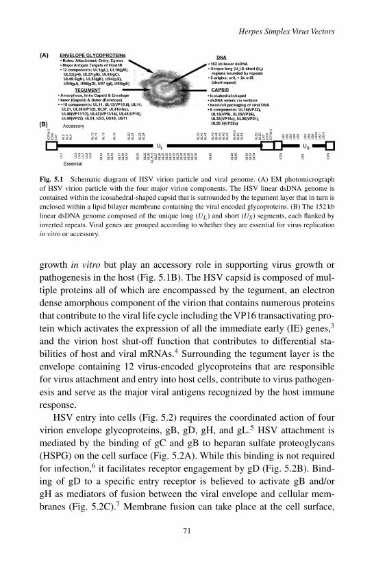
May 4, 2010 11:0 SPI-B903 9in x 6in b903-ch05
Herpes Simplex Virus Vectors
Fig. 5.1 Schematic diagram of HSV virion particle and viral genome. (A) EM photomicrographof HSV virion particle with the four major virion components. The HSV linear dsDNA genome iscontained within the icosahedral-shaped capsid that is surrounded by the tegument layer that in turn isenclosed within a lipid bilayer membrane containing the viral encoded glycoproteins. (B) The 152 kblinear dsDNA genome composed of the unique long (UL) and short (US) segments, each flanked byinverted repeats. Viral genes are grouped according to whether they are essential for virus replicationin vitro or accessory.
growth in vitro but play an accessory role in supporting virus growth orpathogenesis in the host (Fig. 5.1B). The HSV capsid is composed of mul-tiple proteins all of which are encompassed by the tegument, an electrondense amorphous component of the virion that contains numerous proteinsthat contribute to the viral life cycle including the VP16 transactivating pro-tein which activates the expression of all the immediate early (IE) genes,3
and the virion host shut-off function that contributes to differential sta-bilities of host and viral mRNAs.4 Surrounding the tegument layer is theenvelope containing 12 virus-encoded glycoproteins that are responsiblefor virus attachment and entry into host cells, contribute to virus pathogen-esis and serve as the major viral antigens recognized by the host immuneresponse.
HSV entry into cells (Fig. 5.2) requires the coordinated action of fourvirion envelope glycoproteins, gB, gD, gH, and gL.5 HSV attachment ismediated by the binding of gC and gB to heparan sulfate proteoglycans(HSPG) on the cell surface (Fig. 5.2A). While this binding is not requiredfor infection,6 it facilitates receptor engagement by gD (Fig. 5.2B). Bind-ing of gD to a specific entry receptor is believed to activate gB and/orgH as mediators of fusion between the viral envelope and cellular mem-branes (Fig. 5.2C).7 Membrane fusion can take place at the cell surface,
71
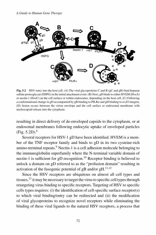
May 4, 2010 11:0 SPI-B903 9in x 6in b903-ch05
A Guide to Human Gene Therapy
Fig. 5.2 HSV entry into the host cell. (A) The viral glycoproteins C and B (gC and gB) bind heparansulfate proteoglycan (HSPG) in the initial attachment event. (B) Next, gD binds to either HVEM (HveA)or nectin-1 (HveC) on the cell surface or within endosomes, depending on the host cell. (C) Followinga conformational change in gD accompanied by gB binding to PILRα and gH binding to αvβ3 integrin,(D) fusion occurs between the virion envelope and the cell surface or endosomal membrane withnucleocapsid release into the cytoplasm.
resulting in direct delivery of de-enveloped capsids to the cytoplasm, or atendosomal membranes following endocytic uptake of enveloped particles(Fig. 5.2D).8
Several receptors for HSV-1 gD have been identified. HVEM is a mem-ber of the TNF receptor family and binds to gD in its two cysteine-richamino-terminal repeats.9 Nectin-1 is a cell adhesion molecule belonging tothe immunoglobulin superfamily where the N-terminal variable domain ofnectin-1 is sufficient for gD recognition.10 Receptor binding is believed tounlock a domain on gD referred to as the “profusion domain” resulting inactivation of the fusogenic potential of gB and/or gH.11,12
Since the HSV receptors are ubiquitous on almost all cell types andtissues,13 it may be necessary to target the virus to specific cell types throughretargeting virus binding to specific receptors. Targeting of HSV to specificcells types requires: (i) the identification of cell-specific surface receptor(s)to which viral binding/entry can be redirected and (ii) the modificationof viral glycoproteins to recognize novel receptors while eliminating thebinding of these viral ligands to the natural HSV receptors, a process that
72

May 4, 2010 11:0 SPI-B903 9in x 6in b903-ch05
Herpes Simplex Virus Vectors
ideally should be accomplished without compromising infectivity. Noveltargeting ligands have been inserted into the amino terminus of gC and/orgD to achieve targeting of HSV to cell-specific receptors.14,15 Additionally,soluble adapter molecules have been employed to target EGF receptor-bearing cells.16
Engineered HSV vector types can be broadly defined as (i) replication-competent vectors that are deleted for certain nonessential functions in orderto achieve attenuation of their pathogenic phenotype,17 but preserving theirability to carry out lytic infection of many dividing cell types, (ii) replicationdefective vectors deleted for at least one essential function,18 limiting theirability to replicate in complementing cells engineered to provide the essen-tial function(s) in trans, (iii) plasmid vectors referred to as amplicons,19
which contain HSV packaging signals and an HSV origin of replication, cre-ating a vector potentially devoid of all viral genes but requiring HSV helperfunctions supplied by helper virus or plasmids for their production, and (iv)bacterial artificial chromosome (BAC) vectors that have a bacterial originof replication and a drug resistance gene to enable efficient genome manip-ulation and propagation in bacteria.20 The replication competent vectorshave been developed for the treatment of cancer relying on restricted virusreplication within tumor cells. These vectors have the potential advantage ofintra-tumoral virus spread and tumor cell lysis and thus have been referredto as oncolytic vectors. Thus far oncolytic HSV vectors have been used inclinical trials in patients with a variety of tumors, but have failed to showadequate efficacy due to limited replication and spread within the humantumor mass.21 Replication defective and amplicon vectors were developedfor localized applications suitable for a variety of diseases and have thepotential advantages of low toxicity and large transgene payload. Recently,a replication-defective vector has been employed in a phase-I clinical trialto treat chronic pain associated with bone metastases.22 The manufacture ofthe amplicon vector type has been unusually difficult with infectious titersproduced in the order of 106–108 plaque-forming units (PFU) per mL com-pared to titers reaching 1012/mL for the replication defective and competentvectors, making their use in human trials problematic. The remainder of thechapter will concentrate on the design and use of the replication defectiveHSV vector backbone.18
73

May 4, 2010 11:0 SPI-B903 9in x 6in b903-ch05
A Guide to Human Gene Therapy
2. HSV Biology in the Design of ReplicationDefective Vectors
Although HSV has a complex genome, the genes are expressed in a well-ordered cascade in which the immediate early (IE) regulatory genes arerequired for activation of the early (E) and late (L) gene viral functions(Fig. 5.3), the latter requiring in addition viral DNA synthesis to becomeactivated. Early viral genes are mostly involved in viral DNA replicationwhile late genes largely encode viral structural proteins, the virus capsid,
Fig. 5.3 HSV lytic and latent life cycles. Lytic HSV infection results in the onset of the three-phasecascade of HSV gene expression. The five immediate early (IE) genes are the first genes transcribed,and regulate the expression of the HSV early class genes involved in viral DNA synthesis. FollowingDNA replication, these IE gene products activate late gene class expression, which enables virionformation. Virion particles released from these cells encounter nerve terminals that innervate the site ofprimary infection. The particle travels by retrograde axonal transport to the nerve cell nucleus. Insteadof entering the lytic phase, the viral genome circularizes, and all viral genes are silenced except for thelatency-associated transcripts (LATs), the hallmark of HSV latency.
74

May 4, 2010 11:0 SPI-B903 9in x 6in b903-ch05
Herpes Simplex Virus Vectors
tegument and the envelope glycoproteins (Fig. 5.3). The interdependencyof viral gene expression can be demonstrated by the removal of just oneessential IE gene in order to render the virus defective for both early andlate viral gene expression.
There are five IE genes in HSV-1 (Fig. 5.3), only two of which are essen-tial for virus replication. Infected cell protein 4 (ICP4) is an essential IE genethat transactivates expression of early and late genes by directly recruitingtranscription factors to these viral promoters.23 ICP27 is an essential IEgene whose function involves post-transcriptional activation of early andlate genes.24 ICP0, a non-essential gene, mediates the disaggregation ofnuclear structures termed PML bodies by degrading certain member com-ponents, and arrests the cell cycle without the induction of apoptosis.25
Thus ICP0 plays an important role in establishing an intracellular milieufavorable for viral replication and consequently is essential for virus reac-tivation from latency. ICP22 is involved in redirecting RNAP II to sitesof viral transcription allowing the virus to expropriate the cellular tran-scription machinery for viral transcription.26 ICP47 inhibits the loading ofimmunogenic peptides onto MHC class I molecules, thereby transientlymitigating immune recognition of infected cells.27 With the exception ofICP47, the individual IE genes are highly cytotoxic to most cell types andthus these genes must be removed in order to produce safe non-toxic vectors.Cells of the nervous system are a notable exception since ICP0 is rapidlydegraded in neurons and there is reduced expression of cytotoxic viralfunctions.28
Wild–type HSV is capable of establishing a latent infection in sensoryneurons of the TG or DRG depending on the site of initial virus interac-tion with the host (Fig. 5.3). During latency viral lytic gene expression issilenced and only a single region of the genome remains transcriptionallyactive (Fig. 5.4A) producing a family of non-coding latency-associated tran-scripts (LATs).29 The major 2.0 Kb LAT is neither capped nor polyadeny-lated and represents stable introns that result from splicing of a large 8.3 Kbunstable primary transcript. Latent viral genomes are partially methylatedand sequestered into an episomal minichromosome-like structure boundby nucleosomes in basically inactive heterochromatin.30 Moreover, theselatent viral genomes have no discernible effect on function of the host cell.Thus, HSV is particularly well suited for the delivery of genes to sensory
75

May 4, 2010 11:0 SPI-B903 9in x 6in b903-ch05
A Guide to Human Gene Therapy
Fig. 5.4 HSV latent gene expression. (A) The location of the LAT RNAs within the HSV genome isdepicted. The large 8.3 Kb LAT primary transcript has the 2.0 Kb LAT intron removed by splicing. Thelocation of the two latency active promoters, LAP1 and LAP2, are depicted in relation to the LATs.(B) Replication defective recombinant vectors deleted for ICP4 and ICP27 [D] containing either LAP1[L1], LAP2 [L2], LAP1+2 [L12], HCMV [H] or chimeric combinations driving expression of thelacZ reporter gene [Z] were used to infect the trigeminal ganglia of mice (N = 10) following cornealscarification and gene expression assayed using a kit is shown as relative light units (RLU) per mg oftotal protein.
neurons and provides an excellent vehicle to target ectoptic expression oftransgenes to specific neuronal populations.
The LAT promoter/regulatory region has been shown to be composed oftwo latency-active promoters, LAP1,31 a TATA box promoter with numer-ous cis-regulatory sites, and LAP2,32 a TATA-less promoter that possessesCT-rich repeats that contribute to the ability of this promoter region toremain active when the genome is silenced.32 LAP2 is moveable and active
76
May 4, 2010 11:0 SPI-B903 9in x 6in b903-ch05
Herpes Simplex Virus Vectors
in expressing transgenes for prolonged periods from both replication com-petent and defective vectors;32 LAP1 does not possess this capability inthe absence of LAP2 (Fig. 5.4B).33 Moreover, LAP2 has the ability to keepnormally transiently active promoters (HCMV) active in the context of repli-cation defective vector backbones (Fig. 5.4B), suggesting that LAP2 couldbe employed to drive long-term transgene or therapeutic gene expressionwhen other viral or cellular promoters are non-functional.
3. HSV Vector Design Technology
The remainder of the chapter will focus on the creation of replication defec-tive HSV-1-based vectors. The goal for the replication defective vectorswas to identify genomic configurations that provide vigorous transgeneexpression while eliminating attendant cytotoxicity. Pertinent parametersof vector characterization include target cell transduction efficiency, rela-tive abundance and longevity of transgene expression, and cell viability.The initial first generation replication defective HSV vectors evolved fromsingle gene deletions, such as ICP4 (SOZ.4 in Fig. 5.5). Since some of thesevectors were missing a gene product that is essential to the lytic life cycle,the only way to propagate these vectors was by using complementing celllines which express the deleted gene product in trans.23 Although thesevectors have been effective for gene delivery to PNS neurons, they haveshown high levels of toxicity in most non-neuronal cell types. Therefore,second generation replication defective vectors were generated to furtherdecrease the inherent toxicity to these cell types. Deletion of two IE regu-latory genes such as ICP4 and ICP27 (DOZ.1 in Fig. 5.5) lead to vectorswhich displayed increased levels of transgene expression with little to noreduction in the cytotoxicity profile depending on the IE gene combinationdeleted, again suggesting that the deletion of additional IE genes wouldbe required. The third generation vectors deleted the three IE genes ICP4,ICP27 and ICP22 (TOZ.2 in Fig. 5.5). These vectors displayed only 50%toxicity, a definite improvement over the second-generation counterparts,and there was an increase in reporter gene expression. However, since thesevectors still retained significant cytotoxicity in non-neuronal cell lines, thenext goal was to eliminate the expression of ICP0 in this background, as itappeared to display the greatest toxicity. Therefore, the fourth generation
77

May 4, 2010 11:0 SPI-B903 9in x 6in b903-ch05
A Guide to Human Gene Therapy
Fig. 5.5 Design of HSV replication defective vectors. Five subsequent generations of replicationdefective HSV vectors deleted for single or multiple IE genes are depicted; grey boxes representdeleted gene sequences. All of the vectors possess the same expression cassette composed of the ICP0promoter driving expression of the lacZ reporter gene in the UL41 locus so that the levels of transgeneexpression can be directly compared in the various mutant vectors. Toxicity of the vectors (Clear Bars)and transgene expression (Black Bars) plotted as relative light units (RLU) in Vero cells at 2 days.
vectors deleted just the single copy of ICP0 present within the internal jointsequences, leaving an intact copy of ICP0 within the terminal repeat atthe far left-hand end of the viral genome (JDTOZ in Fig. 5.5). This vectorshowed further reduced toxicity (∼79% of control) while providing robust
78
May 4, 2010 11:0 SPI-B903 9in x 6in b903-ch05
Herpes Simplex Virus Vectors
transgene expression. In an effort to further reduce vector-associated toxi-city, the final copy of ICP0 was deleted from the fourth generation vectorto provide a replication defective vector devoid of all the four IE regulatorygene products. This vector (JDQ0Z in Fig. 5.5) displayed no measurabletoxicity differences from uninfected cells, yet the loss of the second copy ofthe ICP0 gene product resulted in a tremendous loss in transgene expression.This result was similar to what has been reported in other studies where allthe IE regulatory genes were deleted.34 Some ICP0 expression is required toenable transgene expression, however, ICP0 has some inherent cytotoxicitythat may be tolerated depending on the target cell type. Overall, these exper-iments underscore the role of ICP0 in maintaining a transcriptionally activegenome and suggest that insight into the mechanism of virus genome silenc-ing will be essential to providing a vector capable of transgene expressionin the total absence of vector toxicity.
4. Gene Transfer/Therapy Applications
Due to the ability of HSV to readily transduce neurons of the PNS as partof its natural life cycle, the virus becomes the intuitive choice for treat-ing diseases of the PNS. Replication defective vectors deleted for single ormultiple IE genes have been used in animal models of various (i) peripheralneuropathies caused by diabetes35 and chemotherapeutics36 via delivery ofneurotrophic factors, (ii) pain37 with the delivery of enkephalin or GAD,and (iii) also the delivery of neurotrophins for erectile dysfunction.38 Vec-tor mediated therapeutic gene expression either prolonged neuronal sur-vival measured by neuroanatomical methods or altered the behavior of theanimals. Transgene expression could be targeted to specific neuronal pop-ulations by simply selecting the site of vector inoculation.
HSV has been employed as a vector to treat cancer, primarily thosecancers of the CNS such as GBM in which both replication-defective vectorsexpressing HSV-TK and other anti-tumor agents,39 and the conditionallyreplicating oncolytic HSV vectors have shown promise21 In fact, numerousHSV oncolytic vectors have been used in phase-I human clinical trials andhave displayed excellent safety profiles and have in some patients shownefficacy.40,41 In addition to the above-mentioned PNS and CNS disorders,
79

May 4, 2010 11:0 SPI-B903 9in x 6in b903-ch05
A Guide to Human Gene Therapy
HSV replication defective vectors have been employed in approaches totreat muscle disease, arthritis, and have also been used as vaccine vectors.
5. Immunology
A major issue in any gene transfer approach concerns the immune responseto the virus and/or transgene. One needs to determine the effects of thehost innate and adaptive immune response to the virus vector as 50–80%of the human population is seropositive for HSV. The response by NKcells, macrophages, CD8 and CD4 cells, as well as the presence of virusneutralizing antibodies determines the extent of initial virus infection andspread as well as the ability of the host immune system to keep the virus ina latent state. This plays an important role in cases where the approach mayrequire multiple or repetitive dosing to achieve a therapeutic response.
The host immune response to replication-defective genomic andoncolytic HSV vectors has been studied in great detail. Different replicationcompetent oncolytic vectors have been examined.42,43 In all instances exceptone,43 there was no observable difference in transduction, vector-mediatedgene expression or when measured therapeutic efficacy in pre-immune ani-mals versus naïve animals following administration of one of the aboveoncolytic HSV vectors. Moreover, while increased levels of neutralizingAbs, NK cells, macrophages and T-cells in the sero-positive animals didnot result in reduced transduction at the site of vector injection, virus dis-semination to off-target sites was limited by this host response.42 Similarresults were seen using a replication-defective HSV vector,44 where virustransduction was limited to the site of injection.
One indication that HSV vectors may be able to be re-dosed is that mul-tiple injections of an oncolytic vector performed over a period of days toweeks did not lead to reduced transduction of animals, but rather resulted inincreased efficacy in pre-immune mice.42 Similar results have also been seenin clinical trials40 where multiple injections of vector into HSV sero-positivepatients did not show increased toxicity but seemed to yield improved tumortransduction and efficacy. Finally, re-dosing has also been achieved usingreplication-defective HSV vectors expressing different and diverse thera-peutic transgenes.37
80
May 4, 2010 11:0 SPI-B903 9in x 6in b903-ch05
Herpes Simplex Virus Vectors
6. Safety and Regulatory Issues
Three main issues exist for the use of HSV vectors in human clinical trials.Firstly, will virus introduced into specific tissues or organs spread withinthat tissue, or spread to other tissues within the body? Second, is the issueof the immune status of a patient in relation to the potential inflammatoryresponse to the vector or vector-mediated expression of the transgene. Third,a major concern exists that the introduction of the HSV vector will causereactivation of a resident latent virus in sero-positive patients that could leadto recombination event between the vector genome and the genome of thereactivated wild-type virus.
To address the first issue, toxicology and bio-distribution studies havebeen performed using conditionally replicating oncolytic and replicationdefective HSV vectors in both rodent and non-human primate animalmodels.44,46 No vector-mediated toxicity was observed and there was noevidence of vector spread past the injection site either using qPCR for viralgenomes or HSV-specific IHC staining. As final support for the safety ofHSV vectors, multiple phase-I and I/II studies using oncolytic HSV vectorsdemonstrated no evidence of viral encephalitis or reactivation of wild-typevirus.40,41 Furthermore, no adverse events could be unequivocally attributedto the vectors. Finally the ability to reactivate latent endogenous virus wasnot observed in phase-I human clinical trials with replication competentoncolytic viruses.41 Moreover, pre-clinical studies in rodents and primatespre-infected with wild-type HSV to establish latent infections did not detectreactivation by super-infection with the oncolytic vectors.45
7. Summary
Considerable progress has been made in developing safe, non-toxic HSVvectors to express therapeutic gene products that can be tailored to a varietyof applications, primarily to those affecting the nervous system such asneuropathy, pain and cancer. In addition, methodologies to produce andpurify these vectors have enabled their production to high titers under cGMPconditions as required by the FDA. Initial phase-I trials using these vectorshave shown an excellent safety profile, yet overall efficacy may require
81
May 4, 2010 11:0 SPI-B903 9in x 6in b903-ch05
A Guide to Human Gene Therapy
other improvements to the vector delivery system as a whole to improve theclinical outcome.
References
1. Fatahzadeh M, Schwartz RA (2007). Human herpes simplex virus infections: epi-demiology, pathogenesis, symptomatology, diagnosis, and management. J Am AcadDermatol. 57: 737–763.
2. Stevens JG (1989). Human herpesviruses: a consideration of the latent state. Microbiol.Rev. 53: 318–332.
3. Herrera FJ, Triezenberg SJ (2004). VP16-dependent association of chromatin-modifying coactivators and underrepresentation of histones at immediate-early genepromoters during herpes simplex virus infection. J Virol. 78: 9689–9696.
4. Esclatine A, Taddeo B, Evans L, Roizman B (2004). The herpes simplex virus 1 UL41gene-dependent destabilization of cellular RNAs is selective and may be sequence-specific. Proc Natl Acad Sci USA. 101: 3603–3608.
5. Campadelli-Fiume G, Amasio M, Avitabile E, Cerretani A, Forghieri C, Gianni T, et al.(2007). The multipartite system that mediates entry of herpes simplex virus into thecell. Rev Med Virol. 17: 313–326.
6. Laquerre S, Argnani R, Anderson DB, Zucchini S, Manservigi R, Glorioso JC (1998).Heparan sulfate proteoglycan binding by herpes simplex virus type 1 glycoproteins Band C, which differ in their contributions to virus attachment, penetration, and cell-to-cell spread. J Virol. 72: 6119–6130.
7. Lazear E, Carfi A, Whitbeck JC, Cairns TM, Krummenacher C, Cohen GH, et al.(2008). Engineered disulfide bonds in herpes simplex virus type 1 gD separate receptorbinding from fusion initiation and viral entry. J Virol. 82: 700–709.
8. Nicola AV, Straus SE (2004). Cellular and viral requirements for rapid endocytic entryof herpes simplex virus. J Virol. 78: 7508–7517.
9. Montgomery RI, Warner MS, Lum BJ, Spear PG (1996). Herpes simplex virus 1 entryinto cells mediated by a novel member of the TNF/NGF receptor family. Cell. 87:427–436.
10. Geraghty RJ, Krummenacher C, Cohen GH, Eisenberg RJ, Spear PG (1998). Entryof alphaherpesviruses mediated by poliovirus receptor-related protein 1 and poliovirusreceptor. Science. 280: 1618–1620.
11. Bender FC, Samanta M, Heldwein EE, de Leon MP, Bilman E, Lou H, et al. (2007).Antigenic and mutational analyses of herpes simplex virus glycoprotein B reveal fourfunctional regions. J Virol. 81: 3827–3841.
12. Galdiero S, Falanga A, Vitiello M, Browne H, Pedone C, Galdiero M (2005). Fusogenicdomains in herpes simplex virus type 1 glycoprotein H. J Biol Chem. 280: 28632–28643.
13. Simpson SA, Manchak MD, Hager EJ, Krummenacher C, Whitbeck JC, Levin MJ,et al. (2005). Nectin-1/HveC Mediates herpes simplex virus type 1 entry into primaryhuman sensory neurons and fibroblasts. J Neurovirol. 11: 208–218.
14. Menotti L, Cerretani A, Hengel H, Campadelli-Fiume G (2008). Construction of afully retargeted herpes simplex virus 1 recombinant capable of entering cells solely viahuman epidermal growth factor receptor 2. J Virol. 82: 10153–10161.
82
May 4, 2010 11:0 SPI-B903 9in x 6in b903-ch05
Herpes Simplex Virus Vectors
15. Zhou G, Roizman B (2006). Construction and properties of a herpes simplex virus 1designed to enter cells solely via the IL-13alpha2 receptor. Proc Natl Acad Sci U S A.103: 5508–5513.
16. Nakano K, Asano R, Tsumoto K, Kwon H, Goins WF, Kumagai I, et al. (2005). Herpessimplex virus targeting to the EGF receptor by a gD-specific soluble bridging molecule.Mol Ther. 11: 617–626.
17. Markert JM, Parker JN, Buchsbaum DJ, Grizzle WE, Gillespie GY, Whitley RJ (2006).Oncolytic HSV-1 for the treatment of brain tumours. Herpes. 13: 66–71.
18. Krisky DM, Wolfe D, Goins WF, Marconi PC, Ramakrishnan R, Mata M, et al.(1998). Deletion of multiple immediate-early genes from herpes simplex virusreduces cytotoxicity and permits long-term gene expression in neurons. Gene Ther. 5:1593–1603.
19. Oehmig A, Fraefel C, Breakefield XO, Ackermann M (2004). Herpes simplex virustype 1 amplicons and their hybrid virus partners, EBV, AAV, and retrovirus. Curr GeneTher. 4: 385–408.
20. Sciortino MT, Taddeo B, Giuffre-Cuculletto M, Medici MA, Mastino A, RoizmanB (2007). Replication-competent herpes simplex virus 1 isolates selected from cellstransfected with a bacterial artificial chromosome DNA lacking only the UL49 genevary with respect to the defect in the UL41 gene encoding host shutoff RNase. J Virol.81: 10924–10932.
21. Currier MA, Gillespie RA, Sawtell NM, Mahller YY, Stroup G, Collins MH, et al.(2008). Efficacy and safety of the oncolytic herpes simplex virus rRp450 alone andcombined with cyclophosphamide. Mol Ther. 16: 879–885.
22. Wolfe D, Mata M, Fink DJ (2009). A human trial of HSV-mediated gene transfer forthe treatment of chronic pain. Gene Ther. 16: 455–460.
23. DeLuca NA, McCarthy AM, Schaffer PA (1985a). Isolation and characterization ofdeletion mutants of herpes simplex virus type 1 in the gene encoding immediate-earlyregulatory protein ICP4. J. Virol. 56: 558–570.
24. Sandri-Goldin RM (2008). The many roles of the regulatory protein ICP27 duringherpes simplex virus infection. Front Biosci. 13: 5241–5256.
25. Everett RD, Parsy ML, Orr A (2009). Analysis of the functions of herpes simplex virustype 1 regulatory protein ICP0 that are critical for lytic infection and de-repression ofquiescent genomes. J Virol. 83: 4963–4977.
26. Bastian TW, Rice SA (2009). Identification of sequences in herpes simplex virus type1 ICP22 that influence RNA polymerase II modification and viral late gene expression.J Virol. 83: 128–139.
27. Jugovic P, Hill AM, Tomazin R, Ploegh H, Johnson DC (1998). Inhibition of majorhistocompatibility complex class I antigen presentation in pig and primate cells byherpes simplex virus type 1 and 2 ICP47. J Virol. 72: 5076–5084.
28. Chen X, Li J, Mata M, Goss J, Wolfe D, Glorioso JC, et al. (2000). Herpes simplexvirus type 1 ICP0 protein does not accumulate in the nucleus of primary neurons inculture. J Virol. 74: 10132–10141.
29. Spivack J, Fraser N (1988). Expression of herpes simplex virus type 1 latency-associatedtranscripts in trigeminal ganglia of mice during acture infection and reactivation of latentinfection. J. Virol. 62: 1479–1485.
83
May 4, 2010 11:0 SPI-B903 9in x 6in b903-ch05
A Guide to Human Gene Therapy
30. Deshmane SL, Fraser NW (1989). During latency, herpes simplex virus type 1 DNA isassociated with nucleosomes in a chromatin structure. J. Virol. 63: 943–947.
31. DobsonAT, Sederati F, Devi-Rao G, FlanaganWM, Farrell MJ, Stevens JG, et al. (1989).Identification of the latency-associated transcript promoter by expression of rabbit β-globin mRNA in mouse sensory nerve ganglia latently infected with a recombinantherpes simplex virus. J. Virol. 63: 3844–3851.
32. Goins WF, Sternberg LR, Croen KD, Krause PR, Hendricks RL, Fink DJ, et al. (1994).A novel latency-active promoter is contained within the herpes simplex virus type 1UL flanking repeats. J. Virol. 68: 2239–2252.
33. Perez MC, Hunt SP, Coffin RS, Palmer JA (2004). Comparative analysis of genomicHSV vectors for gene delivery to motor neurons following peripheral inoculationin vivo. Gene Ther. 11: 1023–1032.
34. Samaniego LA, Neiderhiser L, DeLuca NA (1998). Persistence and expression of theherpes simplex virus genome in the absence of immediate-early proteins. J Virol. 72:3307–3320.
35. Goss JR, Goins WF, Lacomis D, Mata M, Glorioso JC, Fink DJ (2002). Herpes simplex-mediated gene transfer of nerve growth factor protects against peripheral neuropathyin streptozotocin-induced diabetes in the mouse. Diabetes. 51: 2227–2232.
36. Chattopadhyay M, Goss J, Wolfe D, Goins WC, Huang S, Glorioso JC, et al. (2004).Protective effect of herpes simplex virus-mediated neurotrophin gene transfer in cis-platin neuropathy. Brain. 127: 929–939.
37. Goss JR, Mata M, Goins WF, Wu HH, Glorioso JC, Fink DJ (2001). Antinociceptiveeffect of a genomic herpes simplex virus-based vector expressing human proenkephalinin rat dorsal root ganglion. Gene Ther. 8: 551–556.
38. Kato R, Wolfe D, Coyle CH, Huang S, Wechuck JB, Goins WF, et al. (2007). Herpessimplex virus vector-mediated delivery of glial cell line-derived neurotrophic factorrescues erectile dysfunction following cavernous nerve injury. Gene Ther. 14: 1344–1352.
39. Niranjan A, Wolfe D, Tamura M, Soares MK, Krisky DM, Lunsford LD, et al. (2003).Treatment of rat gliosarcoma brain tumors by HSV-based multigene therapy combinedwith radiosurgery. Mol Ther. 8: 530–542.
40. Hu JC, Coffin RS, Davis CJ, Graham NJ, Groves N, Guest PJ, et al. (2006). A phaseI study of OncoVEXGM-CSF, a second-generation oncolytic herpes simplex virusexpressing granulocyte macrophage colony-stimulating factor. Clin Cancer Res. 12:6737–6747.
41. Markert JM, Liechty PG, Wang W, Gaston S, Braz E, Karrasch M, et al. (2009). PhaseIb trial of mutant herpes simplex virus G207 inoculated pre-and post-tumor resectionfor recurrent GBM. Mol Ther. 17: 199–207.
42. Lambright ES, Kang EH, Force S, Lanuti M, Caparrelli D, Kaiser LR, et al. (2000).Effect of preexisting anti-herpes immunity on the efficacy of herpes simplex viraltherapy in a murine intraperitoneal tumor model. Mol Ther. 2: 387–393.
43. Herrlinger U, Kramm CM, Aboody-Guterman KS, Silver JS, Ikeda K, Johnston KM,et al. (1998). Pre-existing herpes simplex virus 1 (HSV-1) immunity decreases, butdoes not abolish, gene transfer to experimental brain tumors by a HSV-1 vector. GeneTher. 5: 809–819.
84

May 4, 2010 11:0 SPI-B903 9in x 6in b903-ch05
Herpes Simplex Virus Vectors
44. Wolfe D, Niranjan A, Trichel A, Wiley C, Ozuer A, Kanal E, et al. (2004). Safety andbiodistribution studies of an HSV multigene vector following intracranial delivery tonon-human primates. Gene Ther. 11: 1675–1684.
45. Sundaresan P, Hunter WD, Martuza RL, Rabkin SD (2000). Attenuated, replication-competent herpes simplex virus type 1 mutant G207: safety evaluation in mice. J Virol.74: 3832-3841.
85

May 4, 2010 11:0 SPI-B903 9in x 6in b903-ch05
This page intentionally left blankThis page intentionally left blank
May 4, 2010 11:0 SPI-B903 9in x 6in b903-ch06
Chapter 6
Adeno-Associated Viral (AAV)Vectors
Nicholas Muzyczka∗
Adeno-associated Virus (AAV) is a non-pathogenic virus with wide tissueand species tropism. AAV vectors have proven to be safe and efficient forgene transfer to non-dividing cells, can generate long term gene expres-sion in a variety of animal models and can accommodate a variety oftissue specific and inducible promoter elements. Progress in understand-ing the biology of the virus has helped in the development of efficient andscalable production methods, and the simplicity of the viral structure sug-gests that it might be possible to design targeted vectors. Recent clinicaltrials also suggest that AAV will be useful in treating human disease.
1. Introduction
AAV is a parvovirus, which are small, non-enveloped viruses that containa linear, single-stranded DNA (ssDNA) that is about 5 kb.1 AAVs havebeen isolated from a wide variety of vertebrate species, including humans,monkeys, cows, horses, birds, and sheep. Over a hundred variants havebeen isolated from primate and human tissues and a number of these havebeen characterized for gene therapy. Most humans (∼80%) are positivefor AAV antibodies, but no human disease has been associated with AAVinfection. The lack of disease association is one of the major safety featuresof AAV vectors. Over the last 10 years recombinant AAV (rAAV) vectorshave become widely used as a delivery vehicle for modeling the treatmentof various human diseases. More recently, rAAV has shown partial success
∗Correspondence: Powell Gene Therapy Center, University of Florida College of Medicine.E-mail: [email protected]
87

May 4, 2010 11:0 SPI-B903 9in x 6in b903-ch06
A Guide to Human Gene Therapy
in clinical trials2,3 and promises to be an important therapeutic tool. Thischapter will briefly summarize the biology of AAV, the vector technologiesused to produce and purify rAAV, the characteristics of the vector systemand recent attempts to develop the next generation of rAAV vectors thatincorporate tissue targeting.
2. Biology of AAV
AAV requires a helper virus for productive infection.1 Because AAV isoften found as a contaminant in human adenovirus isolates, it is believedthat human adenovirus (Ad) is the natural AAV helper. However, herpessimplex virus (HSV) can also provide complete helper function. In theabsence of a helper virus, it is believed that AAV can produce a persistentinfection, which can be rescued when cells are infected with a helper virus. Incell culture, AAV has been shown to integrate preferentially into a specifichuman chromosomal location, 19q2, and it has been suggested that thismay be a mechanism for AAV persistence. More recent evidence, however,suggests that wild type AAV persists in humans as an episome.4
The linearAAV genome (Fig. 6.1) contains two inverted terminal repeats(ITRs) that flank two open reading frames, rep and cap1 Three promoters,p5, p19, and p40 initiate spliced and unspliced mRNAs that code for twolarge Rep proteins, Rep78 and 68, two short Reps, Rep52 and 40, and three
Fig. 6.1 AAV genetic map. Only the most abundant mRNA species are shown.
88

May 4, 2010 11:0 SPI-B903 9in x 6in b903-ch06
Adeno-Associated Viral (AAV) Vectors
capsid proteins, VP1, 2, and 3. The two shorter capsid proteins, VP2 andVP3 are encoded by the same mRNA. VP2 (gray + yellow) is initiatedfrom an upstream ACG codon, while VP3 is initiated from the first ATGcodon (*). VP1 is coded by an alternatively spliced mRNA that uses analternative upstream splice acceptor site. The relative efficiencies of initiat-ing translation from the two initiation codons in the VP2/3 mRNA and therelative efficiency of the splice acceptor site usage accounts for the fact thatthe VP3:VP2:VP1 ratio is 10:1:1. The Rep proteins are synthesized fromtwo promoters, p5 and p19 and their mRNAs use the same splice donor andacceptor sites as the capsid mRNA, but are spliced less often than the cap-sid mRNA. This accounts for the relative ratio of unspliced to spliced Repproteins (about 2:1). The p19 promoter is approximately 5 times strongerthan the p5 promoter and this accounts for the relative amounts of Rep52/40to Rep78/68 (5:1). All of the Rep and capsid proteins contain overlappingamino acid sequences in common from their respective open reading frames.The ITRs are 145 bases long. They contain two short palindromes flankedby a long palindrome, and an additional non-palindromic sequence that isrepeated at both ends. The ITRs are the only AAV sequences required in cisfor the production of rAAV vectors; they are the origins for DNA replica-tion, packaging signals, and chromosome maintenance signals.5 The ITRsare also Rep dependent enhancers for AAV transcription from all three pro-moters, and in some tissues have been shown to have weak promoter activityby themselves.
The helper functions provided by Ad have been studied extensively;they are the E1a, E1b 55K and 19K proteins, E4 orf 6, E2a DNA bindingprotein (DBP), and VA RNA genes.1 Most of these genes by themselves canprovide partial helper function that produces semipermissive conditionsfor AAV replication, but completely permissive conditions require all ofthese genes. The effect of these genes is to induce transcription from theAAV p5 promoter (E1a and E2a), induce cellular S phase (E1a), inhibitapoptosis (E1b, E4), inhibit mitosis, promote viral mRNA transport to thecytoplasm (E1b, E4), inhibit the PKR antiviral response (VA), and inhibitnon-homologous end joining of AAV DNA (E1b, E4).6 The initial effect ofthe Ad helper functions appears to be to push a quiescent cell into S phase,freeze it in S phase by preventing mitosis, and prevent apoptosis, therebymaking cellular replication enzymes available for AAV DNA synthesis.
89
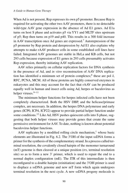
May 4, 2010 11:0 SPI-B903 9in x 6in b903-ch06
A Guide to Human Gene Therapy
When Ad is not present, Rep represses its own p5 promoter. Because Rep isrequired for activating the other two AAV promoters, there is no detectablewild-type AAV gene expression in the absence of Ad E1 genes. Ad E1aturns on host S phase and activates p5 via YY1 and MLTF sites upstreamof p5; Rep then turns on p19 and p40. This results in a 300 fold increasein AAV transcription once Ad genes are expressed.7 Autorepression of thep5 promoter by Rep protein and derepression by Ad E1 also explains whyattempts to make rAAV producer cells in some established cell lines havefailed. Integrated AAV genomes are stable in HeLa and KB cells, but not293 cells because expression of E1 genes in 293 cells presumably activatesRep expression, thereby initiating AAV replication.
AAV relies primarily on cellular replication factors for DNA synthesisin the presence of Ad, and in vitro reconstruction of AAV DNA replica-tion has identified a minimum set of protein complexes;8 these are pol δ,RFC, PCNA, MCM. All of these proteins are highly conserved enzymes ineukaryotes and this may account for the fact that rAAV can be producedequally well in human and insect cells using Ad, herpes or baculovirus ashelper viruses.9–11
The minimum helper functions for herpes infected cells have not beencompletely characterized. Both the HSV DBP, and the helicase/primasecomplex, are necessary. In addition, the herpes DNA polymerase and earlygenes (ICP0, ICP4, ICP22) appear to provide partial helper function undersome conditions.12 Like Ad, HSV pushes quiescent cells into S phase, sug-gesting that both helper viruses may provide genes that create the samepermissive environment for AAV. To date, nothing is known about potentialbaculovirus helper functions.
AAV replicates by a modified rolling circle mechanism,1 whose basicelements are illustrated in Fig. 6.2. The 3′OH of the input ssDNA forms aprimer for the synthesis of the complementary strand. In a process called ter-minal resolution, the covalently closed hairpin of the monomer turnaround(mT) genome is then cleaved at a unique position (trs, terminal resolutionsite) so as to form a new 3′ primer, which is used to repair the end to anormal duplex configuration (mE). The ITR of this intermediate is thenreconfigured to a double hairpin (reinitiation) and the 3′OH primer is usedto displace a ssDNA genome and new mT form which again undergoesterminal resolution in the next cycle. A new ssDNA progeny molecule is
90
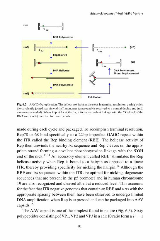
May 4, 2010 11:0 SPI-B903 9in x 6in b903-ch06
Adeno-Associated Viral (AAV) Vectors
Fig. 6.2 AAV DNA replication. The yellow box isolates the steps in terminal resolution, during whichthe covalently joined hairpin end (mT, monomer turnaround) is resolved to a normal duplex end (mE,monomer extended). When Rep nicks at the trs, it forms a covalent linkage with the 5′OH end of theDNA (red circle). See text for more details.
made during each cycle and packaged. To accomplish terminal resolution,Rep78 or 68 bind specifically to a 22 bp imperfect GAGC repeat withinthe ITR called the Rep binding element (RBE). The helicase activity ofRep then unwinds the nearby trs sequence and Rep cleaves on the appro-priate strand forming a covalent phosphotyrosine linkage with the 5′OHend of the nick.13,14 An accessory element called RBE’ stimulates the Rephelicase activity when Rep is bound to a hairpin as opposed to a linearITR, thereby providing specificity for nicking the hairpin.14 Although theRBE and trs sequences within the ITR are optimal for nicking, degeneratesequences that are present in the p5 promoter and in human chromosome19 are also recognized and cleaved albeit at a reduced level. This accountsfor the fact that ITR negative genomes that contain an RBE and a trs with theappropriate spacing between them have been observed to undergo limitedDNA amplification when Rep is expressed and can be packaged into AAVcapsids.15
The AAV capsid is one of the simplest found in nature (Fig. 6.3). Sixtypolypeptides consisting of VP1, VP2 and VP3 in a 1:1:10 ratio form a T = 1
91
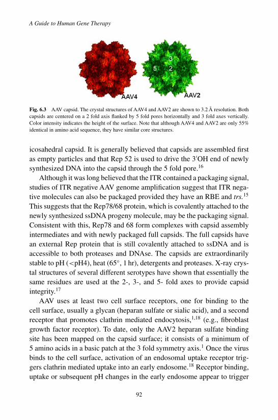
May 4, 2010 11:0 SPI-B903 9in x 6in b903-ch06
A Guide to Human Gene Therapy
Fig. 6.3 AAV capsid. The crystal structures of AAV4 and AAV2 are shown to 3.2 Å resolution. Bothcapsids are centered on a 2 fold axis flanked by 5 fold pores horizontally and 3 fold axes vertically.Color intensity indicates the height of the surface. Note that although AAV4 and AAV2 are only 55%identical in amino acid sequence, they have similar core structures.
icosahedral capsid. It is generally believed that capsids are assembled firstas empty particles and that Rep 52 is used to drive the 3′OH end of newlysynthesized DNA into the capsid through the 5 fold pore.16
Although it was long believed that the ITR contained a packaging signal,studies of ITR negative AAV genome amplification suggest that ITR nega-tive molecules can also be packaged provided they have an RBE and trs.15
This suggests that the Rep78/68 protein, which is covalently attached to thenewly synthesized ssDNA progeny molecule, may be the packaging signal.Consistent with this, Rep78 and 68 form complexes with capsid assemblyintermediates and with newly packaged full capsids. The full capsids havean external Rep protein that is still covalently attached to ssDNA and isaccessible to both proteases and DNAse. The capsids are extraordinarilystable to pH (<pH4), heat (65◦, 1 hr), detergents and proteases. X-ray crys-tal structures of several different serotypes have shown that essentially thesame residues are used at the 2-, 3-, and 5- fold axes to provide capsidintegrity.17
AAV uses at least two cell surface receptors, one for binding to thecell surface, usually a glycan (heparan sulfate or sialic acid), and a secondreceptor that promotes clathrin mediated endocytosis,1,18 (e.g., fibroblastgrowth factor receptor). To date, only the AAV2 heparan sulfate bindingsite has been mapped on the capsid surface; it consists of a minimum of5 amino acids in a basic patch at the 3 fold symmetry axis.1 Once the virusbinds to the cell surface, activation of an endosomal uptake receptor trig-gers clathrin mediated uptake into an early endosome.18 Receptor binding,uptake or subsequent pH changes in the early endosome appear to trigger
92
May 4, 2010 11:0 SPI-B903 9in x 6in b903-ch06
Adeno-Associated Viral (AAV) Vectors
conformational changes in the capsid surface that lead to the extrusion ofthe N terminal regions of VP1 and VP2.19 These contain nuclear localiza-tion signals (NLS) and the phospholipase A2 activity (PLA) that is uniqueto VP1. Genetic studies have shown that both the PLA and NLS are nec-essary for efficient infection.19,20 The role of the phospholipase domain isnot clear; it may be involved in release of capsids from endosomes or thesubsequent entry of capsids into the nucleus. Evidence exists for immediaterelease of virus from the early endosome, as well as trafficking to recyclingendosomes, lysosomes and Golgi.18
Proteosome inhibitors generally increase transduction, although to dif-ferent levels in different cell types, suggesting that virus released intocytoplasm may be functional for transduction.18 This is consistent withexperiments in which injection of neutralizing antibodies into cells inhibitstransduction.19 Recently, phosphorylation of tyrosines on the capsid surfacehas been implicated in AAV trafficking and transduction.21 Finally, thereis also no definitive agreement on whether intact virus particles enter thenucleus prior to uncoating of the nucleic acid.
3. Vector Technology
Long term, persistent expression from an AAV vector was first achieved intissue culture cells by substituting a neomycin cassette driven by a heterol-ogous SV40 promoter in place of the AAV capsid gene.10 The recombinantwas packaged by a plasmid cotransfection system in which the missing cap-sid gene was supplied in trans. Subsequently, it was shown that all of theAAV coding regions could be removed and supplied in trans, only the 145 bpterminal repeat sequences were essential in cis for vector production.5 Lit-tle attention was paid to AAV vectors until two groups demonstrated thatefficient transduction of mouse muscle tissue produced robust expressionof the transgene that persisted for up to a year with no apparent decrease inexpression.22,23 These early reports were followed by demonstrations thatrAAV could be used to efficiently transduce the eye, brain, and liver.24 AAVtransduction was unique among viral vectors in that transduction was effi-cient, and appeared to persist unabated for the lifetime of the animal. Thelongest uninterrupted expression engineered with AAV vectors thus far hasbeen in the eye, where expression of the rpe65 gene has been demonstrated
93

May 4, 2010 11:0 SPI-B903 9in x 6in b903-ch06
A Guide to Human Gene Therapy
in the deficient dog model for greater than 8 years with no loss in the levelof expression. This has made AAV the vector of choice for achieving longterm, persistent expression in animal models.
Interest in AAV vectors continued to lag until several groups solvedthe problem of making helper free rAAV.25,26 Small amounts of researchgrade vectors are typically made by cotransfecting two or three plasmidsinto human tissue culture cells (Fig. 6.4). The process takes advantage ofthe discovery that AAV and rAAV plasmids are infectious.27 Followingtransfection into cells, the AAV ITR-containing genome is rescued fromthe plasmid and undergoes standard AAV DNA replication. The rep andcap gene products as well as the Ad helper function genes are suppliedon one or two additional plasmids (Fig. 6.3) that are co-transfected.25,26
This method generally produces approximately 103 mature viral particlesper cell, much lower than the yield from a wild type AAV infection, 105
particles per cell. However, it requires the construction of only one rAAVplasmid containing the transgene and is easily achieved in most laboratories.The method allows the rapid screening of transgene mutants as well as tissuespecific promoters that might be appropriate.
For clinical and commercial applications, several groups have developedscalable methods for large scale virus production. These methods generatemuch better yields of virus (>105 vector genomes per cell). They also relyon virus infection rather than DNA transfection, and therefore, can be scaledto large fermentor or bioreactor preparations using defined media. A varietyof methods have been described but the two that currently appear to be themost robust use baculovirus or HSV as the helper viruses.11,28–30 When twoHSVs, one expressing rep and cap, and the other carrying the rAAV, are coin-fected in human cells, the yield of rAAV is similar to that seen in wild-type
Fig. 6.4 rAAV production. The most commonly used method for producing rAAV vectors that arefree of helper virus is to transfect three plasmids that contain (1) the transgene cassette flanked by theAAV ITRs, (2) a plasmid that expresses the Ad helper genes, and (3) a plasmid that expresses the AAVrep and cap genes but is lacking the ITRs. Often the second and third plasmids are combined into one.
94

May 4, 2010 11:0 SPI-B903 9in x 6in b903-ch06
Adeno-Associated Viral (AAV) Vectors
AAV infections. To minimize the production of contaminating HSV virus, areplication defective HSV is used. The use of baculovirus vectors in insectcells is equally efficient for rAAV production. The development of thesescalable production methods should make clinical therapeutic applicationspossible in the future.
Several methods have been used for rAAV purification, including CsCland iodixanol density gradient purification, ion exchange chromatogra-phy, and affinity chromatography.31,32 In addition, large scale productiontechniques have been developed that use only column chromatography forvirus purification, thereby eliminating inefficient centrifugation methods.33
Finally, the development of accurate and reproducible methods for deter-mining the titer of full and empty particles and the relative infectivity ofvector preparations has been an equally important aspect of advances inAAVvector technology, and this is discussed extensively in a recent review.34
Until recently only a limited number of AAV serotypes had been iden-tified. However, Gao et al. and others have now isolated over a hundreddifferent serotypes from human, primate and other mammalian species.35
AAV serotypes do not appear to be species specific, so all of these serotypesappear to be capable of transducing rodents, primates and other mammalianspecies. Currently, serotypes 1-11 are routinely available and experimentsfrom many laboratories have demonstrated that each serotype has a uniquetissue tropism when injected into animals. The different serotype tropismsare presumably due in part to the fact that each serotype capsid uses a dif-ferent combination of cell surface receptors. However, it is also clear thedifferent serotypes may use different trafficking strategies or have differentrates of uncoating.18,36 Fortunately, it is easy to screen multiple serotypesusing the same transduction cassette. Typically, a vector construct con-taining AAV2 ITRs can be packaged into capsids from serotypes 1-10 bysubstituting the appropriate capsid in trans.32
The most severe limitation on AAV vectors is the packaging size. Typ-ically, only 5 kilobases can be packaged into a rAAV capsid. Two strate-gies have been proposed for increasing the packaging limit. The first takesadvantage of the splicing machinery and the fact that AAV DNA typicallyforms head to tail concatemers during transduction.37 Thus, a gene can beinterrupted in an intron and the two halves (one carrying a splice donorand a portion of the intron and the other carrying a splice acceptor, and the
95

May 4, 2010 11:0 SPI-B903 9in x 6in b903-ch06
A Guide to Human Gene Therapy
remainder of the intron can be packaged in separate vectors. When the twovectors are simultaneously used to transduce cells, the resulting concatemerscan synthesize an intact mRNA that contains an AAV ITR junction withinthe intron. Splicing of the intron produces a viable mRNA and removes theITR sequence. The second strategy is similar but incorporates an intein, aprotein sequence that self splices or ligates two polypeptides after they havebeen synthesized.38 It is not yet clear whether either of these split gene–twovector approaches will provide a viable solution for delivery of genes thatare too large to package into standard AAV capsids.
4. Vector Characteristics In Vivo
When first developed it was expected that AAV vector would integrate intohost chromosomes and that this was the mechanism for achieving persistentexpression. In early cell culture experiments, rAAV was found to be inte-grated randomly throughout host chromosomes.5,39 Flotte et al. suggestedthat this might not be the case in vivo by showing that rAAV carrying the cys-tic fibrosis gene persisted in rabbit airway epithelial cells as a monomer lin-ear episome.40 Subsequently, several groups have demonstrated that rAAVpersists as an extrachromosomal piece of DNA.4,41,42 This in part explainswhy the long term transduction frequency in rapidly dividing hematopoi-etic cells tends to be low, in contrast to what is seen in organs containingnon-dividing cells, such as brain, eye, liver and muscle. Studies in mousemuscle could find no evidence of integration at a sensitivity that wouldhave detected 1 integrated vector in 200 that persisted in cells.42 Similarstudies in mouse liver also revealed that most of the rAAV genomes wereextrachromosomal with only a low level of integration (<10%).
Although it is now reasonably well established that rAAV genomespersist as episomes, the reason for the persistent expression of AAV vec-tors, compared to non-integrating Ad vectors, herpes vectors, plasmid DNAtransduction vectors, and integrating retroviral vectors is not clear. The factthat rAAV does not integrate may spare it from a variety of host epigeneticmechanisms, such as DNA methylation and histone acetylation, that havebeen implicated in shutoff of other viral vectors. An alternative possibilityis that the AAV ITR may have signals that specifically prevent eliminationor silencing of the AAV genome.
96

May 4, 2010 11:0 SPI-B903 9in x 6in b903-ch06
Adeno-Associated Viral (AAV) Vectors
AAV vectors display low immunogenicity.43 AAV vectors do not activatekey components of innate immunity, including toll-like receptors, type 1IFN responses or other cytokines. AAV capsids also blunt activation ofthe complement cascade. In addition, because AAV vectors are completelydeleted for viral genes, the frequency of generating a cytotoxicT lymphocyte(CTL) response to rAAV-expressed transgenes is reduced. This is in partdue to the fact that antigen presenting cells are less prone to be infectedby some AAV serotypes. There is also evidence that rAAV targeted to theliver can induce tolerance to transgenes, which is apparently mediated byregulatory T cells. As expected, infection with rAAV produces circulatingneutralizing antibodies to the AAV capsid, which prevent gene expressionwhen the same serotype is re-administered. Even low neutralizing titers canprevent transduction. In contrast, some serotypes appear to be capable ofre-administration in partially immuno-protected organs, such as the eye andbrain with no evidence of inflammatory response or loss of expression onsecond administration.
Because most animal models have not been exposed to human and pri-mate AAV serotypes, there are neither neutralizing antibodies nor CTLs toaffect the initial application of vector. Most humans, however, have experi-enced a prior exposure to AAV, most likely along with an Ad infection, lead-ing to the development of both circulating antibodies and a CTL responseto AAV capsid. This is believed to have led to the CTL induced loss ofgene expression in a clinical trial to restore expression of Factor IX.43 Incontrast, a recent clinical trial for Alpha-1-antitrypsin produced long termexpression of the transgene inspite of inducing both circulating antibodiesand a CTL response to capsid protein.3 It is therefore, still not clear whetherthe previous exposure that most individuals have had to AAV will make itdifficult to use rAAV for human gene therapy.
Most AAV vectors display a slow onset of gene expression in vivothat takes 1-3 months to reach the maximum level, after which expressionplateaus and remains constant. Two explanations have been proposed forthis behavior. rAAV vectors can persist as intact particles for extended peri-ods of time in infected cells,36 implying that uncoating is inherently slow.In addition, once the nucleic acid material is released, the single strandedDNA must synthesize the complementary strand before gene expression canoccur. This most likely occurs by de novo synthesis of the second strand.
97

May 4, 2010 11:0 SPI-B903 9in x 6in b903-ch06
A Guide to Human Gene Therapy
Srivastava and colleagues demonstrated that a phosphorylated cellular pro-tein, FKBP52, was an inhibitor of second strand synthesis.44 This proteinbound to the newly exposed ssDNA just downstream of the trs.
To avoid the problem of second strand synthesis McCarty et al. havedeveloped a method for packaging an rAAV genome that consists of afull length duplex DNA in which one end is covalently joined, i.e., a fulllength inverted repeat.45 The disadvantage of this method is that half of thepackaging capacity of the rAAV capsid is lost; only cassettes that are 50%of AAV genome size can be packaged. However, once the vector genome isuncoated within the cell, it immediately “snaps back” on itself to produce aduplex template for transcription. This produces dramatic increases in theefficiency of transduction (up to 100 fold) and eliminates the delay in onsetof gene expression.
TheAAV ITR does not have a typical TATA like promoter element and inmost applications no expression is seen unless a promoter is included in thetransgene cassette.A wide variety of promoter and enhancer sequences havebeen used successfully in rAAV. In general, tissue specific promoters havebehaved as expected and provided the same tropism that is seen in transgenicmouse experiments. Although the packaging capacity of AAV puts a seriousconstraint on the size of the promoter that can be used, some small tissuespecific promoters have been developed that provide cell type specificity.The CMV enhancer/promoter and a CMV-chicken β actin (CBA) hybridpromoter have been the most widely used constitutive promoters. Of thesethe CBA promoter is the strongest promoter reported and has not been shownto shut off in any tissue to date. Several groups have also demonstrated thein vivo use of inducible promoters in an rAAV background.
5. Next Generation Vectors
All rAAV serotypes are promiscuous; they infect multiple organs and tissueswhen vector is delivered systemically. Because the virus has a simple capsidstructure, a number of groups have explored the possibility of targetingAAVto specific tissues. Two general strategies have been followed. The first uses adirected approach in which novel cell surface ligands are genetically insertedinto the capsid sequence. Girod et al.46 were the first of several groups toshow that a short peptide could be inserted at aa 587 without affecting capsid
98

May 4, 2010 11:0 SPI-B903 9in x 6in b903-ch06
Adeno-Associated Viral (AAV) Vectors
viability. This approach has successfully increased the amount of rAAV thatwas taken up by one or another cell type following systemic administration.Peptides inserted at this position have the additional property that they splitthe R585,588 heparan sulfate binding site, thereby reducing AAV2 bindingto its primary receptor.
An approach designed to accommodate large ligands has been suggestedby Warrington et al.47 This group has shown that GFP can be fused to theN terminus of the minor capsid protein VP2 with no significant affect ontiter or viral stability. In this approach a ligand up to 35 kDa can be used todecorate the outside of the capsid at 3-6 copies.
Although binding of the capsid to the surface of the correct cell is impor-tant, other aspects of viral entry also presumably affect the efficiency oftransduction, including receptor endocytosis, trafficking and uncoating. Toidentify viruses that are optimized for all steps in the viral entry process,several groups have used a random library approach to identifying usefulcapsid variants.48 These groups have screened capsid libraries that havebeen shuffled from multiple existing serotypes and further mutagenized byusing error prone PCR. By selecting libraries with pooled human serum,it is also possible to enrich for new capsid variants that do not react withneutralizing antibody.
6. Conclusions and Outlook
rAAV has been a useful tool for engineering long term expression in animals.Its safety profile has been excellent and the recent partial success in twoclinical trials suggests that it may be useful for curing a variety of humandiseases.
References
1. Berns KI, Parrish CR (2007). Parvoviridae. In Fields Virology (Knipe DM and Howley,PM Eds.), pp. 2437–2477. Lippincott Williams and Wilkins, New York.
2. Cideciyan AV, Aleman TS, Boye SL, Schwartz SB, Kaushal S, Roman AJ, et al. (2008).Human gene therapy for RPE65 isomerase deficiency activates the retinoid cycle ofvision but with slow rod kinetics. Proc Nat Acad Sci USA 105: 15112–15117.
3. Brantly ML, Chulay J, Wang L, Mueller C, Humphries M, Spencer LT, et al.(2009). Sustained Expression of Alpha-1 Antitrypsin Despite Effector T Lymphocyte
99

May 4, 2010 11:0 SPI-B903 9in x 6in b903-ch06
A Guide to Human Gene Therapy
Responses in a Phase 1 Clinical Trial of Intramuscular rAAV1-AAT. J Clin Invest[submitted].
4. Schnepp BC, Jensen RL, Clark KR, Johnson PR (2009). Infectious molecular clonesof adeno-associated virus isolated directly from human tissues. J Virol 83: 1456–1464.
5. McLaughlin SK, Collis P, Hermonat PL, Muzyczka N (1988). Adeno-associated virusgeneral transduction vectors: analysis of proviral structures. J Virol 62: 1963–1973.
6. Berk AJ (2007). Adenoviridae: The viruses and their replication. In Fields Virology(Knipe DM and Howley PM Eds.), pp. 2355–2394. Lippincott Williams and Wilkins,New York.
7. Pereira DJ, McCarty DM, Muzyczka N (1997). The adeno-associated virus (AAV) Repprotein acts as both a repressor and an activator to regulate AAV transcription during aproductive infection. J Virol 71: 1079–1088.
8. Nash K, Chen W, Muzyczka N (2008). Complete in vitro reconstitution of adeno-associated virus DNA replication requires the minichromosome maintenance complexproteins. J Virol 82: 1458–1464.
9. Conway JE, Zolotukhin S, Muzyczka N, Hayward GS, Byrne BJ (1997). Recombinantadeno-associated virus type 2 replication and packaging is entirely supported by aherpes simplex virus type 1 amplicon expressing Rep and Cap. J Virol 71: 8780–8789.
10. Hermonat PL, Muzyczka N (1984). Use of adeno-associated virus as a mammalianDNA cloning vector: transduction of neomycin resistance into mammalian tissue cul-ture cells. Proc Nat Acad Sci USA 81: 6466–6470.
11. Urabe M, Ding C, Kotin RM (2002). Insect cells as a factory to produce adeno-associated virus type 2 vectors. Hum Gene Ther 13: 1935–1943.
12. Alazard-Dany N, Nicolas A, Ploquin A, Strasser R, Greco A, Epstein AL, et al. (2009).Definition of herpes simplex virus type 1 helper activities for adeno-associated virusearly replication events. PLoS pathogens 5: e1000340.
13. Im DS, Muzyczka N (1990). The AAV origin binding protein Rep68 is an ATP-dependent site-specific endonuclease with DNA helicase activity. Cell 61: 447–457.
14. Brister JR, Muzyczka N (2000). Mechanism of Rep-mediated adeno-associated virusorigin nicking. J Virol 74: 7762–7771.
15. Nony P, Chadeuf G, Tessier J, Moullier P, Salvetti A (2003). Evidence for packaging ofrep-cap sequences into adeno-associated virus (AAV) type 2 capsids in the absence ofinverted terminal repeats: a model for generation of rep-positive AAV particles. J Virol77: 776–781.
16. Bleker S, Pawlita M, Kleinschmidt JA (2006). Impact of capsid conformation and Rep-capsid interactions on adeno-associated virus type 2 genome packaging. J Virol 80:810–820.
17. Nam HJ, Lane MD, Padron E, Gurda B, McKenna R, Kohlbrenner E, et al. (2007).Structure of adeno-associated virus serotype 8, a gene therapy vector. J Virol 81: 12260–12271.
18. Ding W, Zhang L, Yan Z, Engelhardt JF (2005). Intracellular trafficking of adeno-associated viral vectors. Gene Ther 12: 873–880.
19. Sonntag F, Bleker S, Leuchs B, Fischer R, Kleinschmidt JA (2006). Adeno-associatedvirus type 2 capsids with externalized VP1/VP2 trafficking domains are generatedprior to passage through the cytoplasm and are maintained until uncoating occurs inthe nucleus. J Virol 80: 11040–11054.
100

May 4, 2010 11:0 SPI-B903 9in x 6in b903-ch06
Adeno-Associated Viral (AAV) Vectors
20. Grieger JC, Snowdy S, Samulski RJ (2006). Separate basic region motifs within theadeno-associated virus capsid proteins are essential for infectivity and assembly. J Virol80: 5199–5210.
21. Zhong L, Li B, Mah CS, Govindasamy L, Agbandje-McKenna M, Cooper M, et al.(2008). Next generation of adeno-associated virus 2 vectors: point mutations intyrosines lead to high-efficiency transduction at lower doses. Proc Nat Acad Sci USA105: 7827–7832.
22. Kessler PD, Podsakoff GM, Chen X, McQuiston SA, Colosi PC, Matelis LA, et al.(1996). Gene delivery to skeletal muscle results in sustained expression and systemicdelivery of a therapeutic protein. Proc Nat Acad Sci USA 93: 14082–14087.
23. Xiao X, Li J, Samulski RJ (1996). Efficient long-term gene transfer into muscle tissueof immunocompetent mice by adeno-associated virus vector. J Virol 70: 8098–8108.
24. Buning H, Perabo L, Coutelle O, Quadt-Humme S, Hallek M (2008). Recent develop-ments in adeno-associated virus vector technology. J Gene Med 10: 717–733.
25. Grimm D, Kern A, Rittner K, Kleinschmidt JA (1998). Novel tools for production andpurification of recombinant adenoassociated virus vectors. Hum Gene Ther 9: 2745–2760.
26. Xiao X, Li J, Samulski RJ (1998). Production of high-titer recombinant adeno-associated virus vectors in the absence of helper adenovirus. J Virol 72: 2224–2232.
27. Samulski RJ, Berns KI, Tan M, Muzyczka N (1982). Cloning of adeno-associated virusinto pBR322: rescue of intact virus from the recombinant plasmid in human cells. ProcNat Acad Sci USA 79: 2077–2081.
28. Conway JE, Rhys CM, Zolotukhin I, Zolotukhin S, Muzyczka N, Hayward GS, et al.(1999). High-titer recombinant adeno-associated virus production utilizing a recombi-nant herpes simplex virus type I vector expressing AAV-2 Rep and Cap. Gene Ther 6:986–993.
29. Kang W, Wang L, Harrell H, Liu J, Thomas DL, Mayfield TL, et al. (2008). An effi-cient rHSV-based complementation system for the production of multiple rAAV vectorserotypes. Gene Ther in press.
30. Aslanidi G, Lamb K, Zolotukhin S (2009). An inducible system for highly efficientproduction of recombinant adeno-associated virus (rAAV) vectors in insect Sf9 cells.Proc Nat Acad Sci USA in press.
31. Zolotukhin S, Byrne BJ, Mason E, Zolotukhin I, Potter M, Chesnut K, et al. (1999).Recombinant adeno-associated virus purification using novel methods improves infec-tious titer and yield. Gene Ther 6: 973–985.
32. Zolotukhin S, Potter M, Zolotukhin I, Sakai Y, Loiler S, Fraites TJ Jr., et al. (2002).Production and purification of serotype 1, 2, and 5 recombinant adeno-associated viralvectors. Methods (San Diego, Calif) 28: 158–167.
33. Kaludov N, Handelman B, Chiorini JA (2002). Scalable purification of adeno-associated virus type 2, 4, or 5 using ion-exchange chromatography. Hum Gene Ther13: 1235–1243.
34. Aucoin MG, Perrier M, Kamen AA (2008). Critical assessment of current adeno-associated viral vector production and quantification methods. Biotechnol Adv 26:73–88.
35. Gao G, Vandenberghe LH, Wilson JM (2005). New recombinant serotypes of AAVvectors. Curr Gene Ther 5: 285–297.
101
May 4, 2010 11:0 SPI-B903 9in x 6in b903-ch06
A Guide to Human Gene Therapy
36. Thomas CE, StormTA, Huang Z, Kay MA (2004). Rapid uncoating of vector genomes isthe key to efficient liver transduction with pseudotyped adeno-associated virus vectors.J Virol 78: 3110–3122.
37. Yan Z, Lei-Butters DC, ZhangY, Zak R, Engelhardt JF (2007). Hybrid adeno-associatedvirus bearing nonhomologous inverted terminal repeats enhances dual-vector recon-struction of minigenes in vivo. Hum Gene Ther 18: 81–87.
38. Li J, Sun W, Wang B, Xiao X, Liu XQ (2008). Protein trans-splicing as a means forviral vector-mediated in vivo gene therapy. Hum Gene Ther 19: 958–964.
39. Nakai H, Montini E, Fuess S, Storm TA, Grompe M, Kay MA (2003). AAV serotype2 vectors preferentially integrate into active genes in mice. Nat Genet 34: 297–302.
40. Afione SA, Conrad CK, Kearns WG, Chunduru S, Adams R, Reynolds TC, et al.(1996). In vivo model of adeno-associated virus vector persistence and rescue. J Virol70: 3235–3241.
41. Nakai H, Yant SR, Storm TA, Fuess S, Meuse L, Kay MA (2001). Extrachromosomalrecombinant adeno-associated virus vector genomes are primarily responsible for stableliver transduction in vivo. J Virol 75: 6969–6976.
42. Schnepp BC, Clark KR, Klemanski DL, Pacak CA, Johnson, PR (2003). Genetic fate ofrecombinant adeno-associated virus vector genomes in muscle. J Virol 77: 3495–3504.
43. Zaiss AK, Muruve DA (2008). Immunity to adeno-associated virus vectors in animalsand humans: a continued challenge. Gene Ther 15: 808–816.
44. Qing K, Hansen J, Weigel-Kelley KA, Tan M, Zhou S, Srivastava A (2001). Adeno-associated virus type 2-mediated gene transfer: role of cellular fkbp52 protein in trans-gene expression. J Virol 75: 8968–8976.
45. McCarty DM (2008). Self-complementary AAV vectors; advances and applications.Mol Ther 16: 1648–1656.
46. Girod A, Ried M, Wobus C, Lahm H, Leike K, Kleinschmidt JA, et al. (1999). Geneticcapsid modifications allow efficient re-targeting of adeno-associated virus type 2. NatMed 5: 1438.
47. Warrington KH Jr., Gorbatyuk OS, Harrison JK, Opie SR, Zolotukhin S, Muzyczka N(2004). Adeno-associated virus type 2 VP2 capsid protein is nonessential and cantolerate large peptide insertions at its N terminus. J Virol 78: 6595–6609.
48. Kwon I, Schaffer DV (2008). Designer gene delivery vectors: molecular engineeringand evolution of adeno-associated viral vectors for enhanced gene transfer. Pharm Res25: 489–499.
102

May 4, 2010 11:0 SPI-B903 9in x 6in b903-ch07
Chapter 7
Regulatory RNA in Gene Therapy
Alfred. S. Lewin∗
Regulatory RNAs include trans-acting molecules such as miRNAs,siRNAs, ribozymes and cis-acting elements within mRNA molecules.Trans-acting RNAs may be delivered either as modified oligonucleotidesor via gene therapy vectors, such as plasmids or viruses. Oligonucleotidetherapy requires re-administration of the therapeutic RNA, while vectormediated delivery may provide long-term expression of the siRNA orribozymes. Even though ribozymes have been studied for 25 years, RNAinterference using siRNAs has become the major tool for suppressing geneexpression for therapy. Non-specific effects of RNA mediated regulationdemands careful control experiments and testing of multiple therapeu-tic molecules for each target gene. Adding or modifying sequences in the3′ untranslated regions of mRNAs provides a powerful tool for controllingmRNA stability, localization and translation.
1. Introduction
Regulatory RNAs may be broadly defined as non-protein coding RNAsequences that in some way control the expression of genes. RNA moleculessuch as Xist or naturally occurring siRNAs may lead to stabilization of het-erochromatin and shut off entire chromosomes or chromosome regions.1
For gene therapy, in contrast, one wishes to control the expression of asingle gene and for this purpose specificity is required. For gene silencing,gene therapists have therefore turned to antisense RNA, RNA decoys or
∗Correspondence: Department of Molecular Genetics and Microbiology, University of Florida,Gainesville, FL 32610.E-mail: [email protected]
103
May 4, 2010 11:0 SPI-B903 9in x 6in b903-ch07
A Guide to Human Gene Therapy
aptamers, ribozymes, artificial siRNAs or siRNAs disguised as miRNAs.For gene regulation, cis-acting RNA elements may be added or modified.
Antisense RNA molecules are RNA molecules that are complementaryto the coding sequence of a messenger RNA. They may block gene expres-sion by interfering with translation. Antisense molecules are most effec-tive, however, when delivered as DNA oligonucleotides that can stimulateRNAse H-mediated decay of mRNAs to which they bind. Alternatively,morpholino based antisense molecules and PNAs (peptide nucleic acids)can form stable duplexes with target mRNAs and prevent their translationinto protein.2 Although these are powerful experimental and therapeuticapproaches, they do not fall under the heading of regulatory RNA, and willnot be discussed in this chapter.
RNA decoys are simply segments of RNA that contain naturally-occurring binding sites for viral or cellular proteins. If delivered andexpressed at a high level, they may sequester a protein essential for thepathogenic process. An example is an HIV TAR decoy, which has beenembedded in a small nucleolar RNA and used to sequester the Tat proteinfrom the HIV promoter.3 RNA aptamers are products of reiterative RNAselection methods designed to produce high-affinity binding sequences.Aptamers usually bind proteins via the tertiary structure of the small RNArather than the primary sequence, and affinities can be quite high, compara-ble to antigen antibody interactions. An aptamer to VEGF is commerciallyavailable (as Macugen™) for the treatment of choroidal neovascularization,a complication of age related macular degeneration.4
Ribozymes are catalytic RNA molecules. RNA catalysis was discov-ered in the context of RNAse P and self-splicing introns and almost simul-taneously was found to be a feature of self-processing satellite RNAs ofplant pathogens.5 Other RNA enzymes have been discovered in a varietyof contexts, and novel catalysts consisting of RNA or DNA have been arti-ficially selected in the test tube. Pre-clinical gene therapy has made themost use of two short ribozymes derived from tobacco ringspot virus satel-lite RNA, the hammerhead and the hairpin ribozyme.6 The RNA subunitof RNAse P can cleave targeted mRNAs, using a guide RNA to directthe ribozyme to a particular mRNA. Once ribozyme mediated digestionseparates the 5′ cap structure from the polyA tail, mRNAs are rapidlydegraded by cellular exonucleases. Like protein enzymes, ribozymes exhibit
104

May 4, 2010 11:0 SPI-B903 9in x 6in b903-ch07
Regulatory RNA in Gene Therapy
catalytic turnover, and each ribozyme may cleave many potential targetRNAs. While their turnover is not dramatic relative to protein catalysts,on the order of 10 per minute, most mRNAs are not abundant in thesteady state. Therefore, ribozymes can be useful tools for elimination ofspecific RNAs.
RNA interference (RNAi) is mediated by small interfering RNAs(siRNAs), which are 19-25 nucleotide RNA duplexes with sequence homol-ogy to cellular mRNAs.7,8 One strand of an siRNA is exactly complemen-tary to a target mRNA and is termed the guide strand, while the otherstrand has the same sense as the mRNA and is called the passenger strand.Naturally occurring siRNAs are processed from larger double strandedRNAs by an endonuclease called Dicer. Like other nucleases of its class(ribonuclease III), Dicer leaves 5′ phosphate groups and 3′ hydroxyls, with2 nucleotide overhangs at the 3′ ends. Since mammalian cells are intoler-ant of long double stranded RNA molecules (see below), for gene therapy,siRNAs are either delivered as short dsRNA molecules with 3′ overhangs,or they are expressed from DNA vectors as small hairpin RNAs that areprocessed by Dicer into siRNAs. Once siRNAs are processed and deliveredto the cytoplasm, they associate with the RNA Induced Silencing Complex(RISC), where they unwind and one RNA strand becomes incorporated intothe complex, which then directs cleavage of the complementary mRNA.
An alternative approach for delivery of siRNAs is to express them asmiRNAs.9 Like siRNAs, miRNAs are short RNA molecules that bind tomRNAs in the context of RISC. However, miRNAs are not fully comple-mentary to their target RNAs and, in general, bind to the mRNAs in the 3′
untranslated region (3′UTR). MicroRNAs block gene expression at the levelof protein synthesis and do not lead to immediate degradation of mRNA,though RNA half-life may decrease. There are hundreds of natural miRNAsin mammalian cells and each may regulate the translation of many mRNAs.Because of this complexity, miRNAs themselves have not been advanced astools for gene therapy, except in the case of certain tumors in which miRNAgenes are defective. However, an RNA with exact complementarity to thecoding region of a mRNA can be placed within the context of a miRNAprecursor. This precursor will be processed in the nucleus and exported bythe same mechanism as native miRNAs, providing a mechanism to expressan siRNA using RNA polymerase II.
105

May 4, 2010 11:0 SPI-B903 9in x 6in b903-ch07
A Guide to Human Gene Therapy
Manipulation of cis-acting regulatory RNA sequences has not developedas rapidly as the use of siRNAs for regulation of gene expression. Never-theless, this approach is more subtle and discriminating than simple RNAknockdown. Sequences in the 3′ UTR of genes delivered for therapy havebeen modified to limit their expression in unintended tissues,10 and to directmRNAs to a particular location in the target cell.11 In addition, small RNAscan be delivered in trans to affect the activity of cis-acting sequences thatgovern alternative splicing.
2. Delivery of Therapeutic RNAs
Delivery is a major concern for those trying to use small RNA molecules astools for gene therapy. There are two basic approaches for delivering ther-apeutic RNA to animals and humans (Fig. 7.1). The first is to deliver RNAmolecules directly and the second is to deliver DNA vectors that drive thetranscription of the therapeutic RNA. The development of oligonucleotidesas drugs has received support from the pharmaceutical industry. The efficacyof siRNAs as inhibitors allows access to many cellular targets that were untilrecently thought to be “undruggable”. Annotated genome sequences facil-itate design of siRNAs and identification of potential unintended mRNAtargets. There are two key advantages to delivering RNA as a gene-directedtherapy, rather than as a gene therapy. The first is that RNA drugs can be“dosed to effect” for a specific condition and a specific patient. The sec-ond is that therapy can be halted upon adverse reaction, and the inherentinstability of RNA will lead to rapid clearance from the body. DeliveringRNA has several drawbacks, however. For one, RNA drugs are not orallyavailable, so that delivery may require infusion or injection. Intravenouslyinjected RNA molecules tend to be filtered by the kidney, liver, spleen andlungs. If these are not target organs, then bioavailability may be limited.Finally, because of the susceptibility of RNA to digestion by cellular andextracellular enzymes and the clearance of nucleic acids from the circula-tion, RNA drugs must be continuously reapplied in order to treat chronicmedical conditions. This may be seen as a disadvantage to a patient, thoughnot necessarily to an investor.
Because RNA is unstable under physiologic conditions, RNA thera-pists rely on a series of modifications to the ribose-phosphate backbone to
106
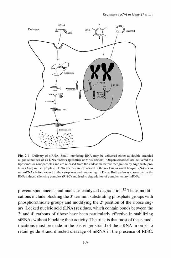
May 4, 2010 11:0 SPI-B903 9in x 6in b903-ch07
Regulatory RNA in Gene Therapy
Fig. 7.1 Delivery of siRNA. Small interfering RNA may be delivered either as double strandedoligonucleotides or as DNA vectors (plasmids or virus vectors). Oligonucleotides are delivered vialiposomes or nanoparticles and are released from the endosome before recognition by Argonaute pro-teins (Ago) in the cytoplasm. DNA vectors are expressed in the nucleus as small hairpin RNAs or asmicroRNAs before export to the cytoplasm and processing by Dicer. Both pathways converge on theRNA induced silencing complex (RISC) and lead to degradation of complementary mRNA.
prevent spontaneous and nuclease catalyzed degradation.12 These modifi-cations include blocking the 3′ termini, substituting phosphate groups withphosphorothioate groups and modifying the 2′ position of the ribose sug-ars. Locked nucleic acid (LNA) residues, which contain bonds between the2′ and 4′ carbons of ribose have been particularly effective in stabilizingsiRNAs without blocking their activity. The trick is that most of these mod-ifications must be made in the passenger strand of the siRNA in order toretain guide strand directed cleavage of mRNA in the presence of RISC.
107
May 4, 2010 11:0 SPI-B903 9in x 6in b903-ch07
A Guide to Human Gene Therapy
In addition, a compromise must be made between stability and activity interms of the number of modifications that can be made. Typically, however,the half-life of siRNA in serum can increase from seconds to hours usingmodifications that do not reduce inhibitory activity substantially. Ribose andphosphorothioate modifications are also compatible with ribozyme activity.Again, a balance must be drawn between maximizing stability and retain-ing catalytic activity: modifying purine groups in the catalytic core of thehammerhead, for example, eliminates ribozyme function.
Both liposomes and synthetic nanoparticles have been employed todeliver regulatory RNAs in vivo.13 Such particles can be directed to specificcell types by including carbohydrate or protein ligands within the particle.These approaches to nucleic acid administration are discussed elsewherein this book. A major advance in siRNA delivery was made by Soutscheket al., who covalently modified the 5′ ends of an siRNAi directed against themRNA for apolipoprotein B mRNA.14 This modification led to silencingof ApoB mRNA in the liver and jejunum, and opened the door for directtargeting of siRNAs to tissues of interest following intravenous administra-tion by adding cell penetrating peptides or single chain antibodies directlyto the RNA.15
Delivering therapeutic RNAs as DNA permits long-term expression ofthe RNA molecules and may allow more precise delivery to a particularorgan or cell type. As for other therapeutic genes, control of RNA expres-sion is determined by the specific virus and virus serotype employed, by thesite of injection and by the promoter sequence used. If longevity of expres-sion is an advantage, it may also be detrimental, if treatment leads to harmfulside effects. As for other gene therapy applications, thorough pre-clinicalsafety testing is required before human testing begins. For sequence specificinhibitors such as siRNAs and ribozymes, however, animal testing may notreveal unintended targets whose knockdown can cause human complica-tions.
RNA delivery has employed both viral vectors and liposome formulatedplasmids. These delivery systems are described in other chapters in thisvolume. Synthesis of ribozymes or siRNAs can be directed by either RNApolymerase III promoters or RNA polymerase II promoters. RNA pol III nor-mally produces small nuclear RNAs, transfer RNAs and 5S ribosomal RNA.While pol III promoters are powerful, much of the small hairpin transcripts
108
May 4, 2010 11:0 SPI-B903 9in x 6in b903-ch07
Regulatory RNA in Gene Therapy
produced by pol III, remains in the nucleus, separated from mRNA targets.In addition, pol III promoters are generally unregulated and expressed in allcell types. The need for nuclear export signal and regulated promoter sys-tems has persuaded many investigators to embed siRNA sequences withinmiRNAs. MicroRNAs are typically produced by RNA pol II in the con-text of larger precursors containing signals for processing by the nuclearenzyme Drosha and transport to the cytoplasm by Exportin 5.16 They areoften made as part of long transcripts that may contain multiple miRNAsor they may be encoded in the introns of protein coding genes. There-fore, it is possible to insert a miRNA disguised siRNA in the intron or theuntranslated region of another gene. Details of such constructs are discussedbelow.
3. Ribozymes
Ribozymes have been used primarily to suppress the expression of diseaserelated genes.6 As noted above, the hammerhead and the hairpin ribozymesare the easiest to design and have target sequences (NUX for hammerheadsand BNGUC for hairpins, where N is any nucleotide, B is any nucleotidebut adenosine and X is any nucleotide but guanosine) that can be found inany mRNA.5 To design a hammerhead ribozyme, for example, one looksfor NUX triplets (typically GUC, CUC or UUC) in the target mRNA anddesigns an antisense RNA of 12 or 13 nucleotides surrounding the cytosine,which does not form a conventional base pair with the ribozyme. The cat-alytic core of the ribozyme is then inserted within the antisense sequence,dividing it into two hybridizing arms which form mini-helices with thetarget mRNA. Since product release may be rate limiting for ribozymeturnover, these helices are kept short, typically 5–7 base pairs. Design ofthe hairpin is somewhat more complex, with an 8 nucleotide loop insertedbetween helices formed between ribozyme and target. To use RNase P forRNA knockdown, one uses the endogenous enzyme and designs a guideRNA resembling the acceptor stem of a transfer RNA when it is paired withthe mRNA to be digested.17 For all ribozymes, it is necessary to identifyaccessible target sequences: cleavage sites cannot be buried in stable inter-nal helices within the mRNA. Computer algorithms may help identify suchstems if used judiciously to analyze short stretches of sequence.
109

May 4, 2010 11:0 SPI-B903 9in x 6in b903-ch07
A Guide to Human Gene Therapy
An advantage of ribozymes relative to small interfering RNAs is thatribozymes can be tested in cell-free reactions in order to identify whichare most catalytically active. Such testing should be performed at relativelylow magnesium concentration (10 mM) in order to eliminate ribozymesthat will not function efficiently in cells. The most active ribozymes canthen be tested in transfected cells to verify that they can cleave full lengthmRNA within the cellular environment. Expressing ribozymes in humancells also provides at least a crude assessment of “off-target” effects medi-ated by ribozymes: if a ribozyme unintentionally retards the cell cycle orleads to apoptosis, then it is time to choose a new target sequence. Finally,those ribozymes that pass the in vitro tests can be cloned in a vector andtested in animal models of human disease. Ribozymes have been expressedfrom viral vectors using either pol II promoters or pol III promoters, inwhich case the ribozymes may be expressed in tandem with tRNAs. Alter-natively, RNA modifications can be introduced, including phosphorothioatenucleotides at the 5′ end of the ribozyme, an inverted abasic nucleotide atthe 3′ end and 2′ amino and 2′O-methyl substitutions at certain residues inthe core of the ribozyme.18 Such modifications will lower cleavage rate invitro by 5–10 fold, but may increase stability in cells by a 1000 fold. Mod-ified ribozymes can be delivered directly to the circulation or to affectedtissues.
Therapeutic targets for ribozymes have included dominant oncogenes,RNA viruses and retroviruses and autosomal dominant disease genes. Whileribozymes have been successful in reducing tumor growth in animal models,there have been few clinical trials of ribozyme gene therapy. Hammerheadribozymes directed against HIV-1 targets have been tested in lymphocytesor progenitor cells infused into AIDS patients.19,20,21 A ribozyme directedat proliferating cell nuclear antigen mRNA was tested, unsuccessfully, aspreventative of proliferative vitreoretinopathy.22 Despite over 20 years ofresearch, no therapeutic ribozyme has been brought to market as a dis-ease therapy. Problems with ribozymes include their relatively low affinityfor target RNAs and the lack of specific targeting mechanisms for targetbinding: ribozymes can be embedded in pol II transcripts for export fromthe nucleus, but unlike siRNAs they do not associate with a protein com-plex that promotes mRNA recognition. In addition, the active conformationof cis-acting hammerhead ribozymes is stabilized by tertiary loop-bulge
110

May 4, 2010 11:0 SPI-B903 9in x 6in b903-ch07
Regulatory RNA in Gene Therapy
interactions that are hard to recapitulate in trans.23 Nevertheless, highlyactive ribozymes can reduce mRNA levels as effectively as siRNAs, andribozymes can be partnered with small hairpin RNAs, as in the HIV exam-ple, without competing for the same nuclear export system. Relative to theRNAi user community, however, the ribozyme user community is small, andcommercial support for ribozyme technology is non-existent. For this rea-son alone, the search for effective RNA-based inhibitors of gene expressionshould begin with siRNA.
4. RNAi for Gene Therapy
As with ribozymes, siRNAs have been employed primarily to suppressgene expression by inducing mRNA degradation. Therapeutic targets haveincluded dominant oncogenes, viruses, growth factors and almost any genethat is induced or mis-regulated in disease.24 For example therapeutic siR-NAs are being clinically tested for treatment of respiratory syncytial virususing an inhaled formulation of a modified siRNA, for the down regula-tion of VEGF in the eye, and for pachyonchia congenita, an inherited skindisease.25 While it is possible to produce allele-specific siRNAs for thetreatment of dominantly inherited disease, single base changes may not besufficient to provide discrimination between the mutant and the wild-typemRNA. One method to overcome this problem is to deliver an siRNA thattargets both mutant and wild-type mRNA. This siRNA may be delivered inconjunction with an mRNA containing silent nucleotide changes renderingit resistant to attack by the siRNA and RISC.26 Such an approach is alsofeasible with ribozymes.
Effective siRNAs generally conform to certain design principles: siRNAsequences should be short (19–21 bp); they should be of moderate G+Ccontent (30–60%); they should avoid runs of 4 adenosine or 4 guanosineresidues; they should contain an A-U pair at position 10 relative to 5′ endof the antisense strand, and if possible, they should have a lower ther-mal stability at the 5′ end of the antisense strand than at the 3′ end.27
The last requirement reflects the asymmetric loading of RNA strands ontoRISC. While either strand of an siRNA can be associated with the com-plex, RNA is loaded in the 5′ to 3′ direction. Therefore, if the 5′ end of
111

May 4, 2010 11:0 SPI-B903 9in x 6in b903-ch07
A Guide to Human Gene Therapy
the guide (anti-sense) strand unwinds first, it will preferentially load intoRISC. Keeping the siRNA short is important. While longer siRNAs (25 nt)are more effective in directing cleavage of mRNA, they are more likely tostimulate pathways of innate immunity and to block the export of endoge-nous miRNAs.28 As with ribozymes, it is important to avoid siRNA targetsites in regions of stable secondary structure. Fortunately, one does not haveto remember all of the design principles in selecting siRNA target sites,as several good design algorithms are available on line.27 Some of thesesites are sponsored by biotechnology companies that sell dsRNA or vec-tors expressing short hairpin RNA. In addition, several companies sell pre-validated siRNAs or shRNA plasmids for human, mouse and rat mRNAs.These commercial sources are the best choice for initiating an RNAi relatedproject.
It is important to identify siRNAs of the greatest potency, so that the levelof siRNA delivered can be minimized. Small interfering RNAs are assayedin tissue culture before testing in animal models for disease. This testing maybe done by measuring the expression level of an endogenous gene using RT-PCR, immunoblots or ELISA. Alternatively, siRNAs may be co-transfectedas RNA or as plasmids expressing small hairpin RNAs (shRNAs) togetherwith a plasmid expressing the target mRNA. It is convenient to insert asegment of the mRNA containing the target sequence within the transcriptof an easily measured marker gene such as luciferase, GFP or secretedalkaline phosphatase. This permits rapid screening of the inhibitory activityof the siRNAs. It is important to recognize that for therapeutic applications,one or two siRNAs per target are not enough. Non-specific effects of siRNAsand pharmacodynamic properties of small RNAs are frequently sequencespecific. An siRNA that is potent in HeLa cells may be useless in vivo.
For gene therapy, siRNAs are usually delivered as small hairpin RNAs.29
Their expression is driven from DNA vectors using pol III promoters suchas U6 and H1. Alternatively, modified pol II promoters may be employed.To limit overhanging sequences, these have been engineered to provide thestart site for RNA synthesis just before the shRNA coding sequence anda polyadenylation site just afterwards. As mentioned above, an alternativeapproach employs pre-miRNA sequences in which an siRNA is embedded,replacing the sequence containing the mature miRNA. These precursorscontain processing sites for Drosha and Dicer, so that the siRNA can be
112
May 4, 2010 11:0 SPI-B903 9in x 6in b903-ch07
Regulatory RNA in Gene Therapy
expressed and exported from the nucleus by the normal miRNA pathway.Small interfering RNAs disguised as pre-miRNAs may be expressed fromeither pol II or pol III promoters. As noted, pol II promoters are more easilyregulated and may be cell type specific. On the other hand, pol III promotersare more robust: they should yield high level expression of the miRNA orshRNA in any cell type.
Off target effects are an important consideration in employing RNAifor therapy.30 Off target effects lead to non-specific suppression of geneexpression and are caused in several ways: (1) An siRNA (or ribozyme forthat matter) can bind to and stimulate the degradation of an unintendedtarget RNA. (2) An siRNA can pair imperfectly with the 3′ UTR of anmRNA and suppress its translation into protein. (3) A small hairpin RNAcan interfere with the trafficking of endogenous miRNA, and (4) siRNA canstimulate the dsRNA response. The dsRNA response protects animal cellsfrom invading viruses, retrotransposons and the transcription of repetitivesequences by activating PKR (dsRNA-dependent protein kinase) and 2′,5′-oligoadenylate synthetase. In response to dsRNA, PKR autophosphory-lates and then phosphorylates its substrates, including eukaryotic initiationfactor 2, leading to arrest of protein synthesis. PKR also initiates a signaltransduction pathway that leads to induction of interferons. Interferon pro-duction results in the up-regulation of antiviral and antiproliferative genesand mediators of apoptosis. The activation of 2′, 5′-oligoadenylate syn-thetase results in the formation of oligoadenylates that bind to and activateRNaseL, which cleaves RNA non-specifically, resulting in inhibition oftranslation. Stimulation of the interferon response by siRNAs appears to bemediated by Toll-like receptor (TLR) 7/8 or by TLR3.31,32 Incorporation of2′-O-methyl nucleotides into the siRNA suppresses the TLR7/8 pathway butmay not inhibit activation of the TLR3 pathway. In one study of VEGF siR-NAs used to inhibit laser-induced choroidal neovascularization in a mousemodel of age related macular degeneration, it was found that inhibition ofneovascularization could be attributed solely to TLR3-mediated inductionof interferon (Fig. 7.2).31
Off target effects can be minimized by several means. Sequence search-ing programs may help to identify unintended targets with annotatedmRNAs. It is best to avoid using siRNAs that can form 15 or more consecu-tive base pairs with another RNA that is expressed in the organ(s) of interest.
113

May 4, 2010 11:0 SPI-B903 9in x 6in b903-ch07
A Guide to Human Gene Therapy
Fig. 7.2 Gfp siRNA or Luc siRNA led to a modest suppression of choroidal neovascularizartion(CNV) in Tlr3+/+ but not Tlr3−/− mice. P < 0.05 compared to vehicle (buffer). From Kleinman etal. (Ref 31).
It is somewhat harder to exclude miRNA-like activity of siRNAs, becausebase-pairing with the 7 base seed region (residues 2–8) of the siRNA maybe sufficient to inhibit translation of an unintended mRNA.33 Inhibition ofmiRNA transport and stimulation of the interferon and 2′, 5′ oligoadeny-late pathways can be avoided by keeping the siRNA short. Inhibition ofmiRNA transport requires over-expression of shRNAs of 25 bp or more.28
While siRNAs of 19bp do not typically stimulate the dsRNA response, thisresponse is sequence specific, and some short interfering RNAs may turnon the interferon pathway. Indeed, it has recently been suggested that manyof the early reports of antiviral activity of siRNAs are attributable to thestimulation of interferon rather than the digestion of viral RNA by RISC.32
The best way to control for non-specific effects of siRNA is to have sev-eral independent siRNAs for the target mRNA and to demonstrate that eachhas the same physiologic impact. These positive controls should be com-bined with several negative control siRNAs that together demonstrate thatdigestion of the intended mRNA leads to a particular physiologic response.
5. Gene Therapy Using miRNA
Individual miRNAs regulate variety of genes; some modulate the translationof hundreds of mRNAs.34 We know little about the regulation of miRNAexpression in human tissues. Therefore, it seems unlikely that delivery of
114

May 4, 2010 11:0 SPI-B903 9in x 6in b903-ch07
Regulatory RNA in Gene Therapy
native pri-miRNAs or pre-miRNAs using gene therapy vectors will be usedin the near future, since they are likely to lead to unforeseen consequences.For now, pre-miRNAs are being used as vehicles for siRNAs (see above).Nevertheless, coding sequences for some miRNAs are missing or defectivein specific tumors, so that it is conceivable to use gene therapy vectors toreplace these missing genes.35 Similarly, it is possible to inhibit the activityof specific miRNAs using antisense sequences or surrogate target RNAs.These can be used to decrease the impact of over-expressed miRNAs thatmay lead to disease. Careful assessment of non-specific effects must bemade in developing these therapies.
6. Aptamers, Decoys and Bi-Functional RNAs
RNA aptamers are short oligonucleotides (usually less than 40 nt) with astable tertiary structure that permits high-affinity binding (Kd in the rangeof 10−10 to 10−12) to specific proteins or other target molecules. Conse-quently, aptamers have been developed as inhibitors of pathologic processes.Aptamers are generated by a sequential selection method called SELEX(systematic evolution of ligands by exponential enrichment).36 In this pro-cess RNA libraries containing randomized regions are incubated with theligand of interest and RNA molecules that bind the target compound areseparated from those that do not bind. Binders are amplified by reversetranscription PCR and transcribed into RNA for another round of selection(binding).After multiple rounds of selection, high affinity aptamers are ana-lyzed and counter selection with structural analogs of the target ligand maybe used to produce aptamers of greater selectivity.
For therapy, aptamers are delivered as oligonucleotides.As with siRNAsand ribozymes they may be protected using ribose modifications, but thesemodifications may affect the affinity of the aptamer, since binding involvesthe RNA (or DNA) backbone as well as the nucleotide bases. As noted,one aptamer has already found clinical application for treatment of maculardegeneration and it is delivered by direct and repeated ocular injections. Itis conceivable that aptamers could be delivered using DNA vectors for sus-tained expression, but the folding of RNA sequences during transcriptionmay not recapitulate the folding of aptamer sequences during the selection
115

May 4, 2010 11:0 SPI-B903 9in x 6in b903-ch07
A Guide to Human Gene Therapy
process in vitro, and flanking RNA sequences may affect the tertiary struc-ture as well.
Decoy oligonucleotides contain the recognition sites for nucleic acid-binding proteins or RNAs. DNA decoys have been used to interfere withthe binding of transcription factors to promoter sequences. Rossi and col-leagues have used an RNA decoy delivered by a retrovirus vector, to blockthe interaction of TAR with the HIV-1 promoter.3 Decoy RNAs have alsobeen employed to block the activity of specific miRNAs, by providing mul-tiple copies of a fully-complementary binding site. One type of miRNAinhibitor has been called “antagomir”.37 These are synthetic and chemicallystabilized (LNA or 2′-O-methyl) oligonucleotides that inhibit the functionof endogenous microRNAs by complementary base pairing. Antigomirsmay also have modifications, such as cell penetrating peptides or choles-terol, which promote their uptake by cells. They have been shown to inhibitmiRNA in vivo following intravenous injection. An alternative approachto miRNA inhibition is to express repeated miRNA binding sites from astrong pol II promoter. Such constructs are termed “microRNA sponges”and they appear to block miRNA activity without lowering the level of targetmiRNAs.38
Bi-functional RNAs represent a more complex approach to using recog-nitions sites for RNA binding proteins. In bi-function molecules, the bindingsites for regulatory proteins are linked to antisense RNA in order to directthe regulatory protein to a particular region of the target mRNA. Such bi-functional oligonucleotides have been used to regulate alternative splicingof pre-mRNA by recruiting SR proteins or hnRNP proteins to alternativeexons in order to either increase or decrease the utilization of an adjacentsplice junction. For example, Baughan et al. used AAV to deliver a chimericRNA to attract SR proteins (SF2/ASF, SC35, and hTra2h1) to the 5′ terminusof exon 7 of the SMN2 gene.39 Infection of fibroblasts from patients withspinal muscular atrophy led to an increase in exon inclusion and thereforea significant increase in SMN2 protein.
7. Modification of Cis-Acting Regulatory RNA Sequences
Mature mRNAs are bound by proteins from their time of synthesis, throughnuclear transport and their translation into protein. Sequences within the
116

May 4, 2010 11:0 SPI-B903 9in x 6in b903-ch07
Regulatory RNA in Gene Therapy
mRNA govern which proteins bind and consequently determine mRNAstability and transport. In a few instances, cis-acting sequences have beenused to control gene expression for gene therapy. However, in most cases,gene therapists are constrained by the limitation of viral packaging limits oravailable restriction sites, and they clip off important regulatory elementsin the 5′ and 3′ UTRs.
The stability of a particular mRNA is often determined by sequenceswithin the 3′ UTR of that mRNA, though RNA destabilizing sequences havebeen identified in other locations.40 The best known stability element in the3′ UTR is designated the ARE or AU rich element. Cellular proteins thatbind the ARE may either stabilize that mRNA (e.g. HuR) or destabilize themRNA (AUF1). Some of these trans-acting proteins are cell type specific,while others are ubiquitous. The localization of mRNA is one mechanism toachieve protein sorting in asymmetric eukaryotic cells. While this processhas been best described in yeast and in Drosophila oocytes, mRNA sortingis extremely important in the nervous system, where local protein synthesisin neural dendrites may explain how patterns of neural activity lead tochanges in synaptic connectivity in development and in adaptive responsesin the brain.41 In oligodendrocytes, mRNAs encoding proteins required formyelin formation are localized. Disruption of mRNA sorting and localizedmRNA protein synthesis is a contributing factor of diseases such as fragileX mental retardation and spinal muscular atrophy.
To date gene therapists have made little use of either stability ele-ments or localization elements to obtain more precise control of geneexpression. There have been exceptions, however. In order to attain tumorselective gene expression for virotherapy using adenovirus, Säkioja et al.included an ARE element in the 3′ UTR of a passenger gene (luciferase)and thereby suppressed expression in non-cancer cells but not in malig-nant cells.42 Corral-Debrinski et al. have made an interesting use ofmRNA targeting to optimize allotropic gene expression of human mito-chondrial genes. In yeast, they discovered that mRNAs translated onmitochondrial-bound polyribosomes contain common elements in their3′UTRs. They included one of these sequence elements in the 3′ UTRof a transgene encoding the mitochondrial ND1 and ND4 genes ren-dered in the nuclear genetic code and fitted with a mitochondrial proteinimport sequence.43 Including the mRNA localization element increased the
117
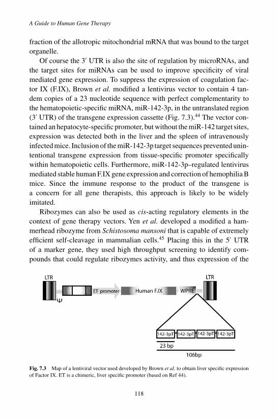
May 4, 2010 11:0 SPI-B903 9in x 6in b903-ch07
A Guide to Human Gene Therapy
fraction of the allotropic mitochondrial mRNA that was bound to the targetorganelle.
Of course the 3′ UTR is also the site of regulation by microRNAs, andthe target sites for miRNAs can be used to improve specificity of viralmediated gene expression. To suppress the expression of coagulation fac-tor IX (F.IX), Brown et al. modified a lentivirus vector to contain 4 tan-dem copies of a 23 nucleotide sequence with perfect complementarity tothe hematopoietic-specific miRNA, miR-142-3p, in the untranslated region(3′ UTR) of the transgene expression cassette (Fig. 7.3).44 The vector con-tained an hepatocyte-specific promoter, but without the miR-142 target sites,expression was detected both in the liver and the spleen of intravenouslyinfected mice. Inclusion of the miR-142-3p target sequences prevented unin-tentional transgene expression from tissue-specific promoter specificallywithin hematopoietic cells. Furthermore, miR-142-3p–regulated lentivirusmediated stable human F.IX gene expression and correction of hemophilia Bmice. Since the immune response to the product of the transgene isa concern for all gene therapists, this approach is likely to be widelyimitated.
Ribozymes can also be used as cis-acting regulatory elements in thecontext of gene therapy vectors. Yen et al. developed a modified a ham-merhead ribozyme from Schistosoma mansoni that is capable of extremelyefficient self-cleavage in mammalian cells.45 Placing this in the 5′ UTRof a marker gene, they used high throughput screening to identify com-pounds that could regulate ribozymes activity, and thus expression of the
Fig. 7.3 Map of a lentiviral vector used developed by Brown et al. to obtain liver specific expressionof Factor IX. ET is a chimeric, liver specific promoter (based on Ref 44).
118
May 4, 2010 11:0 SPI-B903 9in x 6in b903-ch07
Regulatory RNA in Gene Therapy
gene. Unfortunately, the most efficient regulators were potentially toxicnucleoside analogues, toyocamycin and 5-fluorouridine. However, Win andSmolke have developed cis-acting hammerhead ribozymes that can either beactivated or repressed based on an appended theophylline binding aptamer.46
This approach has the potential to produce regulatory riboswitches that maybe controlled by easily tolerated and approved drugs.
8. Conclusions
While gene therapy is usually designed to deliver genes for proteins, deliveryof regulatory RNAs has the capacity to suppress endogenous gene expres-sion in order to attenuate disease. Protein-directed therapy is well suited forrecessive inherited disease, but gene silencing by siRNAs or ribozymes canbe used to treat a variety of acquired, dominantly inherited and infectiousdiseases. In addition, modification of cis-acting RNA elements can make theexpression of protein genes more precise and, therefore, more effective. Themajor dichotomy in the field concerns delivery: whether regulatory RNAsare delivered as stabilized oligoribonucleotides or whether they are admin-istered via gene therapy vectors. Obviously, the application will determinethe most suitable approach. For short-term applications, such as suppressionof scarring or treating acute infections, direct RNA delivery should be better.For chronic diseases, however, gene therapy will ultimately replace the useof oligonucleotides. The major cautionary note in the field of RNA-basedgene therapy is the potential for off-target effects, and the major method toovercome this obstacle is to design and test many potential RNA-inhibitorsin order to develop a therapeutic RNA that has high activity for the intendedmRNA and a low profile of undesirable side effects.
It is now appreciated that almost the entire genome is transcribed, butthe role of the low-level transcripts produced from most of the genome isundefined. There are certainly more regulatory RNAs to discover. Recently,for example, short RNAs homologous to promoter regions were found bothto inhibit and to activate gene expression at the level of transcription.47
These promoter-directed short RNAs require Argonaute proteins and maytarget promoter-specific antisense transcripts. While these so-called anti-gene RNAs have not been adapted for gene therapy, they are likely tobecome useful tools for allele independent suppression of mutant genes.
119

May 4, 2010 11:0 SPI-B903 9in x 6in b903-ch07
A Guide to Human Gene Therapy
As the functions of other minor transcripts are discovered, gene therapistsmust stay aware of developments in the study of regulatory RNA.
References
1. Grewal SI, Elgin SC (2007). Transcription and RNA interference in the formation ofheterochromatin. Nature 447: 399–406.
2. Juliano RL, Dixit VR, Kang H, Kim TY, MiyamotoY, Xu D (2005). Epigenetic manip-ulation of gene expression: a toolkit for cell biologists. J Cell Biol 169: 847–857.
3. Li MJ, Bauer G, MichienziA,Yee JK, Lee NS, Kim J, Li S, Castanotto D, Zaia J, Rossi JJ(2003). Inhibition of HIV-1 infection by lentiviral vectors expressing Pol III-promotedanti-HIV RNAs. Mol Ther 8: 196–206.
4. Ng EW, Shima DT, Calias P, Cunningham ET Jr., Guyer DR, Adamis AP (2006).Pegaptanib, a targeted anti-VEGF aptamer for ocular vascular disease. Nat Rev DrugDiscov 5: 123–132.
5. Doudna JA, Cech TR (2002). The chemical repertoire of natural ribozymes. Nature418: 222–228.
6. Lewin AS, Hauswirth WW (2001). Ribozyme gene therapy: applications for molecularmedicine. Trends Mol Med 7: 221–228.
7. McManus MT, Sharp PA (2002). Gene silencing in mammals by small interferingRNAs. Nat Rev Genet 3: 737–747.
8. Meister G, Tuschl T (2004). Mechanisms of gene silencing by double-stranded RNA.Nature 431: 343–349.
9. Chang K, Elledge SJ, Hannon GJ (2006). Lessons from Nature: microRNA-basedshRNA libraries. Nat Methods 3: 707–714.
10. Brown BD, Gentner B, Cantore A, Colleoni S, Amendola M, Zingale A, Baccarini A,Lazzari G, Galli C, Naldini L (2007). Endogenous microRNA can be broadly exploitedto regulate transgene expression according to tissue, lineage and differentiation state.Nat Biotech 25: 1457–1467.
11. Corral-Debrinski M (2007). mRNA specific subcellular localization represents a crucialstep for fine-tuning of gene expression in mammalian cells. Biochim Biophys Acta 1773:473–475.
12. Behlke MA (2008). Chemical modification of siRNAs for in vivo use. Oligonucleotides18: 305–319.
13. Juliano R, Alam MR, Dixit V, Kang H (2008). Mechanisms and strategies for effectivedelivery of antisense and siRNA oligonucleotides. Nucleic Acids Res 36: 4158–4171.
14. Soutschek J, Akinc A, Bramlage B, Charisse K, Constien R, Donoghue M, Elbashir S,Geick A, Hadwiger P, Harborth J, John M, Kesavan V, Lavine G, Pandey RK, RacieT, Rajeev KG, Rohl I, Toudjarska I, Wang G, Wuschko S, Bumcrot D, Kotelian-sky V, Limmer S, Manoharan M, Vornlocher HP (2004). Therapeutic silencing ofan endogenous gene by systemic administration of modified siRNAs. Nature 432:173–178.
15. Kumar P, Ban HS, Kim SS, Wu H, Pearson T, Greiner DL, Laouar A, Yao J, HaridasV, Habiro K, Yang YG, Jeong JH, Lee KY, Kim YH, Kim SW, Peipp M, Fey GH,
120

May 4, 2010 11:0 SPI-B903 9in x 6in b903-ch07
Regulatory RNA in Gene Therapy
Manjunath N, Shultz LD, Lee SK, Shankar P (2008). T cell-specific siRNA deliverysuppresses HIV-1 infection in humanized mice. Cell 134: 577–586.
16. FilipowiczW, Jaskiewicz L, Kolb FA, Pillai RS (2005). Post-transcriptional gene silenc-ing by siRNAs and miRNAs. Current Opinion in Structural Biology 15: 331–341.
17. Werner M, Rosa E, Nordstrom JL, Goldberg AR, George ST (1998). Short oligonu-cleotides as external guide sequences for site- specific cleavage of RNA molecules withhuman RNase P. RNA 4: 847–855.
18. Beigelman L, McSwiggen JA, Draper KG, Gonzalez C, Jensen K, Karpeisky AM,ModakAS, Matulic-Adamic J, DiRenzoAB, Haeberli P (1995). Chemical modificationof hammerhead ribozymes. Catalytic activity and nuclease resistance. J Biol Chem 270:25702–25708.
19. Amado RG, Mitsuyasu RT, Symonds G, Rosenblatt JD, Zack J, Sun LQ, Miller M, ElyJ, Gerlach W (1999). A phase I trial of autologous CD34+ hematopoietic progenitorcells transduced with an anti-HIV ribozyme. Hum Gene Ther 10: 2255–2270.
20. Macpherson JL, Boyd MP, Arndt AJ, Todd AV, Fanning GC, Ely JA, Elliott F, Knop A,Raponi M, Murray J, Gerlach W, Sun LQ, Penny R, Symonds GP, Carr A, Cooper DA(2005). Long-term survival and concomitant gene expression of ribozyme-transducedCD4+ T-lymphocytes in HIV-infected patients. J Gene Med 7: 552–564.
21. Wong-Staal F, Poeschla EM, Looney DJ (1998). A controlled, Phase 1 clinical trial toevaluate the safety and effects in HIV-1 infected humans of autologous lymphocytestransduced with a ribozyme that cleaves HIV-1 RNA. Hum Gene Ther 9: 2407–2425.
22. Schiff WM, Hwang JC, Ober MD, Olson JL, Dhrami-Gavazi E, Barile GR, Chang S,Mandava N (2007). Safety and efficacy assessment of chimeric ribozyme to prolifer-ating cell nuclear antigen to prevent recurrence of proliferative vitreoretinopathy. ArchOphthalmol 125: 1161–1167.
23. Khvorova A, Lescoute A, Westhof E, Jayasena SD (2003). Sequence elements outsidethe hammerhead ribozyme catalytic core enable intracellular activity. Nat Struct Biol10: 708–712.
24. Lieberman J, Song E, Lee SK, Shankar P (2003). Interfering with disease: opportunitiesand roadblocks to harnessing RNA interference. Trends Mol Med 9: 397–403.
25. Haussecker D (2008). The business of RNAi therapeutics. Hum Gene Ther 19: 451–462.26. O’Reilly M, Millington-Ward S, Palfi A, Chadderton N, Cronin T, McNally N,
Humphries MM, Humphries P, Kenna PF, Farrar GJ (2008). A transgenic mouse modelfor gene therapy of rhodopsin-linked Retinitis Pigmentosa. Vision Res 48: 386–391.
27. Pei Y, Tuschl T (2006). On the art of identifying effective and specific siRNAs. NatMethods 3: 670–676.
28. Grimm D, Streetz KL, Jopling CL, Storm TA, Pandey K, Davis CR, Marion P, SalazarF, Kay MA (2006). Fatality in mice due to oversaturation of cellular microRNA/shorthairpin RNA pathways. Nature 441: 537–541.
29. Cullen BR (2006). Induction of stable RNA interference in mammalian cells. GeneTher 13: 503–508.
30. Cullen BR (2006). Enhancing and confirming the specificity of RNAi experiments. NatMethods 3: 677–681.
31. Kleinman ME, Yamada K, Takeda A, Chandrasekaran V, Nozaki M, Baffi JZ,Albuquerque, RJC, Yamasaki S, Itaya M, Pan Y, Appukuttan B, Gibbs D, Yang Z,Kariko K, Ambati BK, Wilgus, TA, DiPietro LA, Sakurai E, Zhang K, Smith JR,
121
May 4, 2010 11:0 SPI-B903 9in x 6in b903-ch07
A Guide to Human Gene Therapy
Taylor EW, Ambati J (2008). Sequence- and target-independent angiogenesis suppres-sion by siRNA via TLR3. Nature 452: 591–597.
32. Robbins M, Judge A, Ambegia E, Choi C, Yaworski E, Palmer L, McClintock K,Maclachlan I (2008). Misinterpreting the therapeutic effects of siRNA caused byimmune stimulation. Hum Gene Ther 19(10): 991–9
33. Jackson AL, Burchard J, Schelter J, Chau BN, Cleary M, Lim L, Linsley PS (2006).Widespread siRNA “‘off-target” transcript silencing mediated by seed region sequencecomplementarity. RNA 12: 1179–1187.
34. Lim LP, Lau NC, Garrett-Engele P, Grimson A, Schelter JM, Castle J, Bartel DP,Linsley PS, Johnson JM (2005). Microarray analysis shows that some microRNAsdownregulate large numbers of target mRNAs. Nature 433: 769–773.
35. Marquez RT, McCaffrey AP (2008). Advances in MicroRNAs: Implications for GeneTherapists. Hum Gene Ther 19: 27–38.
36. Marshall KA, Ellington AD (2000). In vitro selection of RNA aptamers [In ProcessCitation]. Methods Enzymol 318: 193–214.
37. Krutzfeldt J, Rajewsky N, Braich R, Rajeev KG, Tuschl T, Manoharan M, Stoffel M(2005). Silencing of microRNAs in vivo with antagomirs. Nature 438: 685–689.
38. Ebert MS, Neilson JR, Sharp PA (2007). MicroRNA sponges: competitive inhibitorsof small RNAs in mammalian cells. Nat Methods 4: 721–726.
39. Baughan T, Shababi M, Coady TH, Dickson AM, Tullis GE, Lorson CL (2006). Stim-ulating Full-Length SMN2 Expression by Delivering Bifunctional RNAs via a ViralVector. Mol Ther 14: 54–62.
40. Bolognani F, Perrone-Bizzozero NI (2008). RNA-protein interactions and control ofmRNA stability in neurons. J Neurosci Res 86: 481–489.
41. Bramham CR, Wells DG (2007). Dendritic mRNA: transport, translation and function.Nat Rev Neurosci 8: 776–789.
42. Sarkioja M, Hakkarainen T, Eriksson M, Ristimaki A, Desmond RA, Kanerva A, Hem-minkiA (2008). The cyclo-oxygenase 2 promoter is induced in nontarget cells followingadenovirus infection, but an AU-rich 3′-untranslated region destabilization element canincrease specificity. J Gene Med 10: 744–753.
43. Bonnet C, Augustin S, Ellouze S, Benit P, Bouaita A, Rustin P, Sahel JA, Corral-Debrinski M (2008). The optimized allotopic expression of ND1 or ND4 genes restoresrespiratory chain complex I activity in fibroblasts harboring mutations in these genes.Biochim Biophys Acta 1783: 1707–1717.
44. Brown BD, Cantore A, Annoni A, Sergi LS, Lombardo A, la V, Patrizia, D’A, Armando,Naldini L, (2007). A microRNA-regulated lentiviral vector mediates stable correctionof hemophilia B mice. Blood 110: 4144–4152.
45. Yen L, Magnier M, Weissleder R, Stockwell BR, Mulligan RC (2006). Identification ofinhibitors of ribozyme self-cleavage in mammalian cells via high-throughput screeningof chemical libraries. RNA 12: 797–806.
46. Win MN, Smolke CD (2007). A modular and extensible RNA-based gene-regulatoryplatform for engineering cellular function. Proc Natl Acad Sci 0703961104-.
47. Janowski BA, Hu J, Corey DR (2006). Silencing gene expression by targeting chro-mosomal DNA with antigene peptide nucleic acids and duplex RNAs. Nat Protoc 1:436–443.
122
May 4, 2010 11:0 SPI-B903 9in x 6in b903-ch08
Chapter 8
DNA Integrating Vectors(Transposon, Integrase)
Lauren E. Woodard and Michele P. Calos∗
Plasmid DNA vectors provide a way to sidestep many of the limitationsof viral vectors, such as capsid immunogenicity. However, gene expres-sion from unintegrated plasmid DNA is typically transient. DNA vectorsthat integrate into the chromosomes via either an integrase or transposaseprovide permanent gene addition. Two such systems are under intensivedevelopment. The Sleeping Beauty transposase integrates into TA din-ucleotides in an essentially random fashion, while φC31 integrase is asequence-specific recombinase that integrates at a more limited numberof endogenous sites in mammalian genomes. Both systems have medi-ated successful gene therapy in numerous animal models. The features ofthese DNA integrating systems are described, including safety issues andefforts to increase target site specificity.
1. Basic Vector Biology
While viral vectors were the first to be developed for clinical use in genetherapy, they have often proven to be less successful than originally antici-pated. Viral vectors are effective at overcoming cellular barriers such as theplasma membrane. However, immune defenses against viral vectors mayresult in an ineffective therapy or even death. Other major concerns includeoncogene activation and accidental production of a self-replicating vector.For these reasons, plasmid DNA may provide a simpler and safer alternative
∗Correspondence: Department of Genetics, Stanford University School of Medicine, 300 Pasteur Dr.,Stanford, CA 94305.E-mail: [email protected]
123

May 4, 2010 11:0 SPI-B903 9in x 6in b903-ch08
A Guide to Human Gene Therapy
that is also easier to engineer and produce. In particular, integrating DNAvectors can permanently place the therapeutic gene into the genome, pre-venting short-term gene expression due to plasmid loss. Integration has beenachieved by using transposon and integrase systems.
1.1 Transposon Systems
Mobile DNA elements, or transposons, were first discovered because theyexcise, replicate, and/or insert their DNA back into the genome, caus-ing mutations. Retrotransposons and DNA transposons are the two majorclasses of mobile DNA elements. The transposons that have been appliedto gene therapy applications thus far have been DNA transposons. Trans-posons have been grouped into families according to their similarities. Mosttransposons used for gene therapy, including Sleeping Beauty (SB), are fromthe largest such family, the Tc1/mariner family.
Because SB is by far the most developed of all of the transposons thathave been tested for gene therapy, we will focus on the mechanism ofthis transposon. By analyzing inactive transposon sequences found in afish genome, a consensus sequence was discovered that, when synthesized,was found to be an active transposon.1 Because it was “awakened” fromthe genome, it was named Sleeping Beauty. The system consists of boththe DNA sequence to be inserted (transposon) and the enzyme that accom-plishes integration into the genome (transposase). Natural transposons codefor the transposase and contain the sequences required for its insertion,called terminal inverted repeat (IR)/direct repeat (DR) elements. Only whatis between these elements will be permanently placed in the genome. Inengineered systems, the transgene cassette is placed between the two IR/DRelements so that it will be inserted (Fig. 8.1). The transposase gene mayeither be placed on the outside of the IR/DR elements so that it will be leftbehind after insertion, or it may be placed on a separate plasmid. Once thecell makes the transposase, it can insert the transposon into the genome.Two molecules of SB transposase bind to each of the two IR/DR elementsthat are located on the transposon DNA molecule. Then, the transposasecuts the transposon DNA at the IR/DR elements in a staggered manner,creating GTC overhangs on the transposon. It also cuts the host genomicDNA in a staggered manner at a TA dinucleotide, creating TA overhangs. Inthe final step, these overhangs are repaired by host cofactors. This process
124

May 4, 2010 11:0 SPI-B903 9in x 6in b903-ch08
DNA Integrating Vectors (Transposon, Integrase)
Fig. 8.1 Transposition using Sleeping Beauty for gene therapy. The transposase binds IR/DR elementsin the transposon and mediates recombination at TA dinucleotides in the genome.
125

May 4, 2010 11:0 SPI-B903 9in x 6in b903-ch08
A Guide to Human Gene Therapy
is evidenced by the duplication of the sequence GTCTA after transposoninsertion.
1.2 Integrase Systems
An integrase is a recombinase enzyme that is capable of integrating DNA,usually that of a virus, into another piece of DNA, usually the host chromo-some. Bacteriophages are omnipresent viruses that infect bacteria. In somecases, bacteriophage DNA can be integrated into the host chromosome by anintegrase. If no cofactors are required, these integrases can also be utilizedin foreign organisms to incorporate genes into the genomic DNA. Micro-bial recombinases can be broadly grouped into two categories dependent onthe amino acid responsible for cleaving the DNA, either a tyrosine or serineresidue. The serine integrases have been found to be the most useful for inte-gration into the genomes of foreign organisms. Integrases recombine twodistinct sequences to create hybrid sites that can no longer be recognizedand recombined by the integrase, making integration irreversible. Of theintegrases studied thus far φC31 integrase has been found to have the mostfavorable properties for gene therapy.
φC31 integrase attaches to specific sequences in both the phage andbacterial genomes, termed attP and attB, respectively. The sites are some-what palindromic, meaning that one half of the site is partially similar to theother half. The minimal site length is 34 bp for attB and 39 bp for attP.2 Itis believed that one molecule of φC31 integrase binds to each half of the attsite and that the two sites come together through formation of a tetramericcomplex (Fig. 8.2). The DNA is cleaved at a core sequence in the att sites,and the DNA-integrase complex rotates to form the recombination prod-ucts. The resulting hybrid att sites are termed attL and attR, because theyare found to the left (5′) and right (3′) of the phage genome. Wild-type φC31integrase cannot perform the excision reaction between attL and attR, sointegration is irreversible.3
φC31 integrase is relatively efficient at recombining at pseudo attP siteswhen given an attB-containing plasmid, but poor at recombining at pseudoattB sites.4 When used in mammalian cells for gene addition, the φC31integrase system is comprised of two plasmids. One plasmid expresses theactive integrase enzyme. Another plasmid contains the attB sequence toallow for sequence-specific integration, the transgene, and often a selectable
126
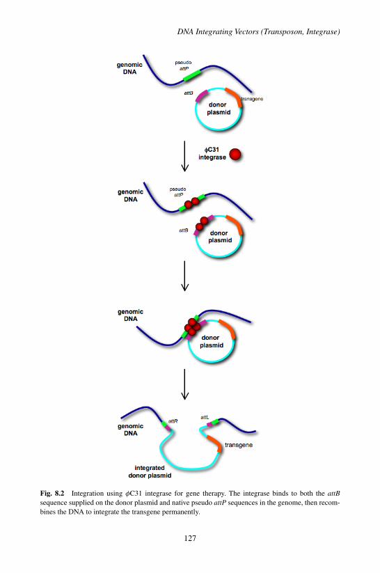
May 4, 2010 11:0 SPI-B903 9in x 6in b903-ch08
DNA Integrating Vectors (Transposon, Integrase)
Fig. 8.2 Integration using φC31 integrase for gene therapy. The integrase binds to both the attBsequence supplied on the donor plasmid and native pseudo attP sequences in the genome, then recom-bines the DNA to integrate the transgene permanently.
127

May 4, 2010 11:0 SPI-B903 9in x 6in b903-ch08
A Guide to Human Gene Therapy
marker that can be used to eliminate cells that have not received integrationevents.
2. Vector Design and Production
2.1 Design of Transposon Systems
There are many factors that can affect transposition. A phenomenon calledoverexpression inhibition has been observed with SB, which means thattoo much of the transposase actually inhibits the transposition reaction.5
This complication necessitates the optimization of transposon to transposaseDNA ratios for every new application or vector. Both the size of the trans-gene and the transposon-containing plasmid are inversely correlated withtransposition efficiency in the SB system.6 This correlation is even morepronounced if DNA of prokaryotic, rather than eukaryotic, origin is used asthe transgene. If the distance between transposon ends is decreased, trans-position increases.7 Another way to decrease the physical distance betweenthe transposon ends is to methylate the DNA, which causes DNA conden-sation. Methylation of the transposon gave an 11-fold increase in overalltransposition rates.8
The first SB transposase was SB10, and the original transposon wascalled pT. This system mediated transposition in only 0.3% of culturedhuman cells, which was an increase of 20 to 40-fold over the backgroundrate of random integration. Improvements in the IR/DR sequences createdthe pT2, pT3, and pMSZ transposons, which are 2-4 times more effectivethan pT when combined with SB10.9−12 The enzyme SB10 has also beenimproved many times. A recent improvement, HSB17 transposase, togetherwith the pMSZ transposon gave a 30-fold improvement in cell culture overthe original SB10/pT system.13 However, in mice the most effective enzymewas the HSB2 enzyme, which was 7-fold improved over SB10 when usedwith pT or pT3 transposons.9
2.2 Design of Integrase Systems
Some attractive features of the integrase system include that the donorplasmid has no size limit, there is no known overexpression inhibition
128

May 4, 2010 11:0 SPI-B903 9in x 6in b903-ch08
DNA Integrating Vectors (Transposon, Integrase)
phenomenon, and the integrase does not require cell division, at least inthe liver where long-term integration events were found in cells that did notundergo replication.14 Mutants of φC31 integrase have been identified thathave increased activity. One such mutant, P3, appeared to have increasedspecificity for the wild-type attP site, making it useful for applications inwhich a wild-type attP is present, such as plasmid excision or integrationinto an engineered cell line.15 For gene therapy, a more useful mutant is P2,which demonstrated two-fold increased integration efficiency at pseudo attPsites in cell culture and mouse liver. There was no obvious change in speci-ficity between P2 and wild-type integrase, but more extensive sequencingof pseudo attP sites is necessary.15
For gene therapy, φC31 integrase has generally been expressed undercontrol of the strong constitutive cytomegalovirus (CMV) promoter.Although adding a C-terminal nuclear localization signal was found to aid ina chromosomal excision reaction,16 it hampered integration into pseudo attPsites in vitro and did not aid integration in mouse liver.14 Also, codon opti-mization was found to be helpful for an excision reaction in the early mouseembryo.17 For increased control over enzyme expression, an inducible inte-grase has been constructed.18
Donor plasmid construction is fairly simple. Generally the full-length∼300 bp attB site is placed on the plasmid between coding regions for anantibiotic resistance gene and the therapeutic gene. In tissue culture, the34 bp site was found to be functional but somewhat inferior, with about75% activity of the full-length attB site at integration into pseudo attPsites.19 Depending on the application, the investigator may wish to usetissue-specific promoters or insulator elements to aid expression. Whilethere is no size limit for integration, it is easier to construct and produceDNA plasmids that are of a moderate size. Bacterial artificial chromosomesmay be used if the therapeutic gene is very large.20
2.3 Production of Plasmid DNA
Plasmids must have an origin of replication for E. coli and a selectablemarker to ensure that only E. coli carrying plasmid comprise the culture.The United States FDA currently prefers plasmids that will be used asmolecular medicines to avoid using ampicillin resistance, since ampicillin
129

May 4, 2010 11:0 SPI-B903 9in x 6in b903-ch08
A Guide to Human Gene Therapy
is prescribed to patients for use as an antibiotic. Another important featureis copy number, which is the number of plasmids per E. coli cell. High copynumber plasmids are preferable since they provide a better yield of plasmidDNA from the same culture volume.
Plasmids are transformed into an optimal strain of E. coli. A culture isgrown from a single colony, which may be cell banked. E. coli are obtainedfrom liquid culture by centrifugation and then lysed and neutralized, result-ing in the precipitation of protein and genomic DNA. The plasmid DNA ispurified from the supernatant by chromatography and isopropanol precipi-tation. If the DNA will be used in vivo, endotoxins must also be removedto avoid serious side effects such as fever and shock. For use in the clinic,plasmids must be validated and then carefully monitored for purity andintegrity during storage.21
3. Gene Transfer Protocols and Potential Applications
Mammalian cells do not normally take up plasmid DNA efficiently. There-fore, DNA integrating systems are limited by the availability of transfectiontechniques. We will provide a brief overview of several transfection methodsand their gene therapy applications with integrating vectors.
3.1 Hepatocyte Transfection via Hydrodynamic Injection
Hydrodynamic injection is a fast, straightforward way to introduce DNAinto the liver cells of mice.22,23 This method uses a large volume of diluteDNA solution, injected quickly through the tail vein, to introduce the DNAinto the hepatocytes. The human alpha-1 antitrypsin (hAAT) and humanFactor IX (hFIX) transgenes have been delivered to normal mice using bothSB and φC31 integrase, resulting in higher long-term transgene expressionwhen recombinase was present.24,25 SB and φC31 integrase have been com-pared directly for hFIX expression. Long-term transgene levels when wild-type integrase was used were about three-fold higher than those obtainedusing an optimized SB system.26 Both systems have been shown to cor-rect several mouse disease models using hydrodynamic injection and maybe tested in the clinic by using balloon catheters to achieve hydrodynamicdelivery.
130
May 4, 2010 11:0 SPI-B903 9in x 6in b903-ch08
DNA Integrating Vectors (Transposon, Integrase)
3.2 Lipophilic Complexes to Transfect Endothelial Cellsand Glioblastoma
Because DNA is negatively charged and must traverse the hydrophobic cellmembrane, DNA may be complexed in solution with lipophilic reagentsthat have a positively charged head and a hydrocarbon tail. These reagentsthen encapsulate the DNA in a fatty ball, allowing it to be endocytosed andthus enter the cell. This strategy is extremely effective for cell culture andis also a viable in vivo transfection method. Complexes have been used todeliver the SB system to type II pneumocyte cells in the lung and to specif-ically express in endothelial cells by way of a tissue-specific promoter.12
Lipophilic complexes were also used to target glioblastoma brain cancercells with success, shrinking the tumors and increasing survival times ofthe mice when the SB system integrated cancer-killing genes.27
3.3 Direct DNA Injection and Electroporation to TargetMuscle, Retina, and Joints
An effective way to transfect muscle fibers is to use electroporation. Themuscle fiber is injected with plasmid DNA, then electrodes are placed oneither side of the injection site and a current is passed through the tissue todisrupt the plasma membrane. This method was used to treat disease-modelmice with a dystrophin gene using φC31 integrase.28 φC31 integrase wasalso used in conjunction with electroporation to deliver the VEGF gene forhindlimb ischemia.29 Both of these strategies were carried out in juvenile(12 day old) mice.
Electroporation has also been used to deliver DNA to the rat retina.This transfection method was combined with φC31 integrase to achievelong-term marker gene expression.30 Additionally, in a rabbit model ofarthritis, joints were injected intra-articularly with naked DNA, includ-ing φC31 integrase.31 Transgene levels were higher when the integrasewas present, but overall transgene levels were still relatively low, reflectinginefficient delivery.
3.4 Integration into Cultured Cells for ex vivo Gene Therapy
Depending on the ability of progenitor cells to be grown under tissueculture conditions and transplanted, ex vivo approaches may be suitable
131
May 4, 2010 11:0 SPI-B903 9in x 6in b903-ch08
A Guide to Human Gene Therapy
for treatment of diseases using a combined gene and cell therapy. Thetherapeutic cells are propagated, transfected with the integrating vectorsystem, and used to treat the disease model, where the stem cells maycontribute to the target tissue. Another use of this strategy is to improvecells so that they are better at attacking a tumor. SB has been used to modifyperipheral blood leukocytes to stably express a suicide gene for cancer genetherapy.32
Both SB and φC31 integrase have been used effectively to inte-grate therapeutic genes into primary human keratinocytes isolated frompatients with epidermolysis bullosa.33,34 When these keratinocytes weregrafted onto immune-deficient mice, healthy skin grew, indicating a viablegene therapy strategy. Muscle precursor cells have also been subjectedto gene addition with φC31 integrase, in another example of a possibleφC31 integrase-mediated treatment for dystrophin deficiency.35 Addition-ally, a human T-cell line derived from a patient with X-linked severecombined immune deficiency was successfully corrected using φC31integrase.36 Other hematopoietic cells such as immortalized K-562 andCD34+ hematopoietic stem cells have been given SB carrying the reportergene GFP.37 While transposition was successful, engraftment was poor.However, blood outgrowth endothelial cells have been engineered with SBto produce hAAT, hFIX and hFVIII.38 Embryonic stem cells and neuralprogenitor cells have been modified with φC31 integrase and were differ-entiated after modification into several lineages.39,40
4. Immunology
One of the major challenges facing gene therapy is the immune responseof patients to the vectors used to deliver helpful genes. Systems utilizingplasmid DNA should be less immunogenic than viral vectors. No immuneresponse has been observed in response to either SB or φC31 integrase inanimals, and there is no reason to suspect that humans would be different inthis respect. The duration of SB and φC31 integrase protein expression afterhydrodynamic injection have been investigated. Both systems exhibit a shortexpression time of the recombinase, on the order of a couple of days.41,42
This brief expression time may be due to the short period of transcription
132

May 4, 2010 11:0 SPI-B903 9in x 6in b903-ch08
DNA Integrating Vectors (Transposon, Integrase)
from the viral promoter (CMV) used to express the recombinase and isfavorable to limit adverse effects.
5. Safety and Regulatory Issues
There is a risk associated with genomic modification because insertionevents could cause oncogene activation or tumor suppressor loss, therebyincreasing the likelihood of cancer.
5.1 Integration Profiles and Associated Hazards
SB transposase requires a TA dinucleotide to integrate and otherwise has fewstatistically significant sequence preferences. It prefers highly deformablesequences, probably because the DNA must be bent in order for SB torecombine it.43 SB may be less harmful than retroviruses, because SB inte-gration is essentially random, rather than favoring transcriptionally activeregions. However, a randomly integrating system will still integrate in areasthat are problematic if enough cells are subjected to transposition.
φC31 integrase, on the other hand, is naturally sequence-specific. Byevaluating integration sites rescued from human cell lines a 28bp consen-sus pseudo attP sequence was identified.44 While 60% of the 123 rescuedintegration events occurred at only 19 sites, many sites were rescued onlyonce. Using a statistical method, it was estimated that approximately 370potential integration sites may exist in the human genome. Of the rescuedsites, none were found in regions of the genome that could be expectedto contribute to malignant transformation of a cell, such as near an onco-gene or disrupting a tumor suppressor. However, it was discovered thatabout 10% of integrations occurred across chromosomes, meaning thatthe plasmid integrated between pseudo attP sites located on two differentchromosomes.44
5.2 Efforts to Enhance Integration Specificity
Integrating gene therapies may be improved by placing the therapeutic geneat a known, safe site in every cell. Strides have been made toward thisgoal. SB has been targeted to a particular genomic location in cultured
133

May 4, 2010 11:0 SPI-B903 9in x 6in b903-ch08
A Guide to Human Gene Therapy
human cells 10% of the time, which is a 107-fold enrichment over randomintegration.45 This approach used expression of another protein that boundto both the transposase and the desired genomic sequence, thus bringing thetransposase closer to the desired location. If this system could be furtherrefined to eliminate random integration and were demonstrated in vivo, itwould be a major advance for the SB system.
φC31 integrase, while many orders of magnitude more specific than SB,may also be modified to make integration more specific. Using a bacterialscreen for directed evolution of the integrase gene, φC31 was mutated tointegrate preferentially at a human pseudo attP site called psA. The bestmutant was ∼6-fold improved over the wild-type enzyme at integration intothe genomic location psA.46 φC31 integrase has also been mutagenized andscreened for increased recombination at the native attP site. Mutants with∼8-fold higher specificity for attP were found.46 This higher specificity actsas a proof of principle that the enzyme can be changed to recognize specificsequences better.
5.3 Effects on Tumor Latency in Mouse Models of Cancer
SB is so effective at random integration that it has been optimized andused for gene discovery to find cancer-causing genes in mouse and rat.Transgenic mice expressing the transposase under tissue-specific promotershave proven to be an advance over the traditional method of cancer genediscovery using retroviruses.47 Although in these instances SB has beenoptimized to find the cancer genes, this application highlights the risk thateven the more benign version of SB used in gene therapy could also causeproblems by integrating near cancer genes.
φC31 integrase is currently being tested in a transgenic mouse modelof liver cancer using hydrodynamic injection to deliver a donor plasmid.Preliminary evidence suggests that the presence of φC31 integrase does notcause acceleration of tumorigenesis in the liver, reducing concerns aboutthe carcinogenic potential of φC31 integrase.19 Upon completion, this studywill provide rigorous data to augment prior observations that cancers havenot been seen in treated mice.
In the future, integrating systems are expected to play an important rolein the formulation of gene therapies for the lifelong treatment of genetic
134
May 4, 2010 11:0 SPI-B903 9in x 6in b903-ch08
DNA Integrating Vectors (Transposon, Integrase)
disease. Further improvements in these systems, as well as upcoming clin-ical trials using them for novel gene therapies, are eagerly awaited.
References
1. Ivics Z, Hackett PB, Plasterk RH, Izsvak Z (1997). Molecular reconstruction of SleepingBeauty, a Tc1-like transposon from fish, and its transposition in human cells. Cell 91:501–510.
2. Groth AC, Olivares EC, Thyagarajan B, Calos MP (2000). A phage integrase directsefficient site-specific integration in human cells. Proc Natl Acad Sci U S A 97: 5995–6000.
3. Thorpe HM, Wilson SE, Smith MC (2000). Control of directionality in the site-specificrecombination system of the Streptomyces phage phiC31. Mol Microbiol 38: 232–241.
4. Thyagarajan B, Olivares EC, Hollis RP, Ginsburg DS, Calos MP (2001). Site-specificgenomic integration in mammalian cells mediated by phage phiC31 integrase. Mol CellBiol 21: 3926–3934.
5. Hackett PB, Ekker SC, Largaespada DA, McIvor RS (2005). Sleeping beautytransposon-mediated gene therapy for prolonged expression. Adv Genet 54: 189–232.
6. Karsi A, Moav B, Hackett P, Liu Z (2001). Effects of insert size on transpositionefficiency of the sleeping beauty transposon in mouse cells. Mar Biotechnol (NY) 3:241–245.
7. Izsvak Z, Ivics Z, Plasterk RH (2000). Sleeping Beauty, a wide host-range transposonvector for genetic transformation in vertebrates. J Mol Biol 302: 93–102.
8. Yusa K, Takeda J, Horie K (2004). Enhancement of Sleeping Beauty transpositionby CpG methylation: possible role of heterochromatin formation. Mol Cell Biol 24:4004–4018.
9. Yant SR, Park J, Huang Y, Mikkelsen JG, Kay MA (2004). Mutational analysis of theN-terminal DNA-binding domain of sleeping beauty transposase: critical residues forDNA binding and hyperactivity in mammalian cells. Mol Cell Biol 24: 9239–9247.
10. Geurts AM, et al. (2003). Gene transfer into genomes of human cells by the sleepingbeauty transposon system. Mol Ther 8: 108–117.
11. Cui Z, GeurtsAM, Liu G, Kaufman CD, Hackett PB (2002). Structure-function analysisof the inverted terminal repeats of the sleeping beauty transposon. J Mol Biol 318:1221–1235.
12. Liu L, Sanz S, Heggestad AD, Antharam V, Notterpek L, Fletcher BS (2004). Endothe-lial targeting of the Sleeping Beauty transposon within lung. Mol Ther 10: 97–105.
13. Baus J, Liu L, Heggestad AD, Sanz S, Fletcher, BS (2005). Hyperactive transposasemutants of the Sleeping Beauty transposon. Mol Ther 12: 1148–1156.
14. Woodard LE, Hillman RT, Keravala A, Lee S, Calos MP (2010). Effect of nuclear local-ization and hydrodynamic delivery-induced cell division on phiC31 integrase activity.Gene Ther 17: 217–226.
15. Keravala A, et al. (2009). Mutational Derivatives of PhiC31 Integrase With IncreasedEfficiency and Specificity. Mol Ther 17: 112–120.
135

May 4, 2010 11:0 SPI-B903 9in x 6in b903-ch08
A Guide to Human Gene Therapy
16. Andreas S, Schwenk F, Kuter-Luks B, Faust N, Kuhn R (2002). Enhanced efficiencythrough nuclear localization signal fusion on phage PhiC31-integrase: activity com-parison with Cre and FLPe recombinase in mammalian cells. Nucleic Acids Res 30:2299–2306.
17. Raymond CS, Soriano P (2007). High-efficiency FLP and PhiC31 site-specific recom-bination in mammalian cells. PLoS ONE 2: e162.
18. Sharma N, Moldt B, Dalsgaard T, Jensen TG, Mikkelsen JG (2008). Regulated geneinsertion by steroid-induced PhiC31 integrase. Nucleic Acids Res 36: e67.
19. Woodard LE, Calos MP (Unpublished results).20. Venken KJ, HeY, Hoskins RA, Bellen HJ (2006). P[acman]: a BAC transgenic platform
for targeted insertion of large DNA fragments in D. melanogaster. Science 314: 1747–1751.
21. Voss C (2007). Production of plasmid DNA for pharmaceutical use. Biotechnol AnnuRev 13: 201–222.
22. Liu F, SongY, Liu D (1999). Hydrodynamics-based transfection in animals by systemicadministration of plasmid DNA. Gene Ther 6: 1258–1266.
23. Zhang G, Budker V, Wolff JA (1999). High levels of foreign gene expression in hepato-cytes after tail vein injections of naked plasmid DNA. Hum Gene Ther 10: 1735–1737.
24. Yant SR, Meuse L, Chiu W, Ivics Z, Izsvak Z, Kay MA (2000). Somatic integrationand long-term transgene expression in normal and haemophilic mice using a DNAtransposon system. Nat Genet 25: 35–41.
25. Olivares EC, Hollis RP, Chalberg TW, Meuse L, Kay MA, Calos MP (2002). Site-specific genomic integration produces therapeutic Factor IX levels in mice. Nat Biotech-nol 20: 1124–1128.
26. Ehrhardt A, Xu H, Huang Z, Engler JA, Kay MA (2005). A direct comparison oftwo nonviral gene therapy vectors for somatic integration: in vivo evaluation of thebacteriophage integrase phiC31 and the Sleeping Beauty transposase. Mol Ther 11:695–706.
27. Ohlfest JR, et al. (2005). Combinatorial antiangiogenic gene therapy by nonviralgene transfer using the sleeping beauty transposon causes tumor regression andimproves survival in mice bearing intracranial human glioblastoma. Mol Ther 12:778–788.
28. Bertoni, C, et al. (2006). Enhancement of plasmid-mediated gene therapy for mus-cular dystrophy by directed plasmid integration. Proc Natl Acad Sci U S A 103:419–424.
29. Portlock JL, Keravala A, Bertoni C, Lee S, Rando TA, Calos MP (2006). Long-termincrease in mVEGF164 in mouse hindlimb muscle mediated by phage phiC31 integraseafter nonviral DNA delivery. Hum Gene Ther 17: 871–876.
30. Chalberg TW, Genise HL, Vollrath D, Calos MP (2005). PhiC31 integrase confersgenomic integration and long-term transgene expression in rat retina. Invest OphthalmolVis Sci 46: 2140–2146.
31. Keravala A, Portlock JL, Nash JA, Vitrant DG, Robbins PD, Calos MP (2006). PhiC31integrase mediates integration in cultured synovial cells and enhances gene expressionin rabbit joints. J Gene Med 8: 1008–1017.
32. Huang X, et al. (2006). Stable gene transfer and expression in human primary T cellsby the Sleeping Beauty transposon system. Blood 107: 483–491.
136

May 4, 2010 11:0 SPI-B903 9in x 6in b903-ch08
DNA Integrating Vectors (Transposon, Integrase)
33. Ortiz-Urda S, Lin Q, Yant SR, Keene D, Kay MA, Khavari PA (2003). Sustainablecorrection of junctional epidermolysis bullosa via transposon-mediated nonviral genetransfer. Gene Ther 10: 1099–1104.
34. Ortiz-Urda S, et al. (2002). Stable nonviral genetic correction of inherited human skindisease. Nat Med 8: 1166–1170.
35. Quenneville SP, Chapdelaine P, Rousseau J, Tremblay JP (2007). Dystrophin expressionin host muscle following transplantation of muscle precursor cells modified with thephiC31 integrase. Gene Ther 14: 514–522.
36. Ishikawa Y, et al. (2006). Phage phiC31 integrase-mediated genomic integration of thecommon cytokine receptor gamma chain in human T-cell lines. J Gene Med 8: 646–653.
37. Hollis RP, et al. (2006). Stable gene transfer to human CD34(+) hematopoietic cellsusing the Sleeping Beauty transposon. Exp Hematol 34: 1333–1343.
38. Kren BT, Yin W, Key NS, Hebbel RP, Steer, CJ (2007). Blood outgrowth endothelialcells as a vehicle for transgene expression of hepatocyte-secreted proteins via SleepingBeauty. Endothelium 14: 97–104.
39. Thyagarajan B, et al. (2008). Creation of engineered human embryonic stem cell linesusing phiC31 integrase. Stem Cells 26: 119–126.
40. Keravala A, Ormerod BK, Palmer TD, Calos MP (2008). Long-term transgene expres-sion in mouse neural progenitor cells modified with phiC31 integrase. J Neurosci Meth-ods 173: 299–305.
41. Chavez CL, Keravala A, Woodard LE, Calos MP (2009). Unpublished data.42. Bell JB, et al. (2004). Duration of Expression of Sleeping Beauty Transposase by
Hydrodynamic Injection of C57/BL6 Mice. Molecular Therapy 9: S312.43. Yant SR, Wu X, Huang Y, Garrison B, Burgess SM, Kay MA (2005). High-resolution
genome-wide mapping of transposon integration in mammals. Mol Cell Biol 25: 2085–2094.
44. Chalberg TW, et al. (2006). Integration specificity of phage phiC31 integrase in thehuman genome. J Mol Biol 357: 28–48.
45. Ivics Z, Katzer A, Stuwe EE, Fiedler D, Knespel S, Izsvak Z (2007). Targeted sleepingbeauty transposition in human cells. Mol Ther 15: 1137–1144.
46. Sclimenti CR, Thyagarajan B, Calos MP (2001). Directed evolution of a recombinasefor improved genomic integration at a native human sequence. Nucleic Acids Res 29:5044–5051.
47. Dupuy AJ, Jenkins NA, Copeland NG (2006). Sleeping beauty: a novel cancer genediscovery tool. Hum Mol Genet 15 Spec No 1: R75–79.
137

May 4, 2010 11:0 SPI-B903 9in x 6in b903-ch08
This page intentionally left blankThis page intentionally left blank

May 4, 2010 11:0 SPI-B903 9in x 6in b903-ch09
Chapter 9
Homologous Recombinationand Targeted Gene Modificationfor Gene Therapy
Matthew Porteus∗
The ability to manipulate the genome in a site-specific manner wouldincrease the specificity and perhaps the efficacy of gene therapy. Gene tar-geting by homologous recombination, whereby an endogenous segmentof the genome is replaced by an introduced segment of DNA, can be usedto both correct disease-causing mutations (gene correction) and to targetthe insertion site of transgenes (targeted transgene insertion). Gene tar-geting can be mediated by adeno-associated virus and stimulated by theinduction of gene-specific double-strand breaks. Gene specific double-strand breaks can be made by either designed zinc finger nucleases ormodified homing endonucleases. In addition, zinc finger nucleases cancreate gene specific mutations when the cell repairs an induced double-strand break in a mutagenic fashion. In this way, permanent gene-specificmutations in human somatic cells can be created with therapeutic intent.
1. Introduction
Gene therapy is based on the principle that we can manipulate the nucleicacid content of cells, usually the genome directly, for therapeutic purposes.The field was born from the recognition of the genetic origin of thousandsof diseases and the discovery of the tools of molecular biology. The abil-ity to create new DNA molecules and introduce DNA molecules into cellsseemed like a direct way to cure or ameliorate genetic diseases. In the
∗Correspondence: Department of Pediatrics and Biochemistry, UT Southwestern Medical Center.E-mail: [email protected]
139

May 4, 2010 11:0 SPI-B903 9in x 6in b903-ch09
A Guide to Human Gene Therapy
laboratory, the modification of cellular phenotype by molecular biologicaltechniques forms the backbone of contemporary biomedical research. Thetranslation of these routine techniques from the laboratory into improve-ments in care for patients, however, has been more difficult. One of themost efficient ways to alter the genome of cells is to use vectors, suchas retroviruses, lentiviruses, and transposons, that integrate new DNA intothe genome. A current characteristic of these vectors is that they integratein an uncontrolled fashion. These uncontrolled integrations have two fun-damental problems. The first is that the integrations can occur in regionsof the genome that cause the gene of interest to be expressed inappropri-ately secondary to “position-effects.” Position effect variegation can causeboth problems in safety (the gene of interest is expressed at a dangeroustime, place or amount) or efficacy (the gene of interest is not expressed atthe correct time, place or amount). The second problem is that uncontrolledintegrations cause uncontrolled mutations. That is, each integration creates anew mutation. These mutations can result in the activation of a nearby onco-gene, thus causing cancer in a relatively short-time frame (several years).They can also result in the inactivation of potentially important genes thatcan cause the cell to behave inappropriately. If a tumor suppressor was inac-tivated by an insertion, for example, one might contribute to the oncogenicprocess, the result of which might not become apparent for decades. A solu-tion to the problem of uncontrolled integrations is to use controlled genomemodification (Fig. 9.1).
2. Problems with Using Gene Targeting by HomologousRecombination
In gene targeting by homologous recombination (hereafter referred to as“gene targeting” or “homologous recombination”), a fragment of DNA isintroduced into a cell and that fragment replaces a similar but not identicalregion of the endogenous genome (Fig. 9.1). Through homologous recom-bination both large and small sequence changes can be introduced into thegenome in a site-specific fashion.
The use of homologous recombination to modify genomes has beenwidely used in many experimental systems including bacteria, yeast, murineembryonic stem cells, and a few vertebrate cell lines. The major limitation
140

May 4, 2010 11:0 SPI-B903 9in x 6in b903-ch09
Homologous Recombination and Targeted Gene Modification for Gene Therapy
Fig. 9.1 Schematic Representation of Strategies to Site-Specifically Modify a Genomic Target byEither Homologous Recombination or Mutagenic Non-Homologous End-Joining (NHEJ). The “∗”represents a correction of a small mutation, the “box” represents the targeted insertion of a transgeneincluding new regulatory elements, and “Ins/Del” represents the insertion or deletion of a small numberof nucleotides at the site of the ZFN induced double-strand break.
to using homologous recombination in mammalian cells has been two-fold.First, the spontaneous rate of gene targeting (10−6) is too low to be clinicallyuseful.1,2 Second the relative rate of gene targeting, the ratio of targeted tonon-targeted integrations, is also too low to be usable. In vertebrate cell typesin which homologous recombination has been used experimentally, the rel-ative rate of targeted vs. non-targeted integrations is higher than usual andthe targeted events can be identified using simple screening strategies.3 Inthis chapter, three approaches to using homologous recombination for genetherapy are discussed: 1) Using homologous recombination in embryonicstem cells and then differentiating embryonic stem cells into tissue specificstem cells for infusion back into a patient; 2) the use of adeno-associatedvirus; 3) the use of nucleases to create gene-specific DSBs to stimulate therate of gene targeting.
3. Homologous Recombination in Embryonic Stem Cells
A standard paradigm for gene therapy for monogenic diseases is to removetissue specific stem cells from a patient, use gene transfer technologies to
141

May 4, 2010 11:0 SPI-B903 9in x 6in b903-ch09
A Guide to Human Gene Therapy
Fig. 9.2 Using Homologous recombination in ES/iPS cells for gene therapy.
manipulate the genome of those cells, and then re-infuse those cells backinto the patient. The clinical trials for the immunodeficiencies have followedthis paradigm.4,5 An alternative paradigm is based on using embryonic stem(ES) cells (Fig. 9.2).
ES cells are immortal cells derived from the early blastocyst embryo andare capable of giving rise to all three germ layers (ectoderm, mesoderm,and endoderm). When implanted back into the early blastocyst, embryonicstem cells can give rise to every cell in the body. In addition, ES cells can beinduced to form multiple different somatic cell types, including tissue spe-cific stem cells, in vitro. In a gene therapy paradigm using ES cells, patientspecific ES cells would be generated. Patient specific ES cells can currentlybe generated two possible ways. First, by nuclear transfer in which thenucleus of a patient cell is used to replace the nucleus of an activated oocytethus creating a cell that is genetically identical to the patient.6 This modifiedoocyte can then be converted in vitro into an ES cell. Second, patient specificpluripotent cells can be created by the use of re-programming by transcrip-tion factors. In this strategy defined transcription factors (such as Oct4,Nanog, Klf4, Sox2, and c-Myc) are introduced into patient somatic cells.7
These transcription factors are able to reprogram a small fraction of the dif-ferentiated somatic cells, by a mechanism that is currently not understood,into cells with ES cell like properties (called “induced pluripotent stem
142

May 4, 2010 11:0 SPI-B903 9in x 6in b903-ch09
Homologous Recombination and Targeted Gene Modification for Gene Therapy
cells” or “iPS” cells). Patient specific ES cells would be modified in vitroby homologous recombination. The manipulation of murine ES cells byhomologous recombination has become a backbone of biomedical research,the importance of which was recognized by the awarding of the Nobel Prizein Medicine in 2007 to Capecchi, Smithies, and Evans for their developmentof the technology. The ES cells having undergone homologous recombina-tion are identified and expanded. Finally, the modified ES cells can then beinduced into tissue specific stem cells in vitro and re-infused to correct a dis-ease. In the last decade, proof-of-principle experiments have been publishedin mice demonstrating the feasibility of this approach in correcting mousemodels of immunodeficiencies and sickle cell anemia.8,9 These mouse pre-clinical experiments highlight the promise of this strategy. There remainseveral important obstacles, however, before this strategy can be adopted inhumans. First, a technique for the creation of patient specific iPS cells mustbe established such that the iPS cells have no increased propensity for can-cer and are fully capable of being converted into tissue specific stem cells.Recent studies suggest that this problem may soon be solved for humancells using non-integrating gene transfer techniques or other strategies.10
Second, while iPS cells seem to behave like true ES cells, there remains nodefinitive way to establish this for human cells. The ability to form teratomasin mice and differentiate into various tissue types in vitro are surrogates thatmay or may not accurately reflect the true potentiality of any given iPS line.Finally, reliable and non-oncogenic methods of converting ES cells intotransplantable tissue specific stem cells need to be established. The simpleinjection of ES cells without such differentiation results in teratomas. Thedifferentiation of ES cells into transplantable hematopoietic stem cells, forexample, while possible in the murine model using proto-oncogenes such asCdx2 and HoxB4, still remains relatively inefficient and requires the use ofoncogenes.11
Although critical problems remain, overall, the paradigm of creatingpatient specific stem cells using the iPS technology, correcting those cellsusing homologous recombination or site-specific modification techniques,expansion of those modified cells and then transplanting tissue specific stemcells derived from the modified ES cells remains of high interest and greatpotential.
143

May 4, 2010 11:0 SPI-B903 9in x 6in b903-ch09
A Guide to Human Gene Therapy
4. Homologous Recombination usingAdeno-Associated Virus
The major limitation to using homologous recombination for gene therapy isthe low spontaneous rate in human somatic cells. When naked DNA is intro-duced into a human somatic cell, the rate of gene targeting is on the order of10−6.1,2 An intriguing property of adeno-associated virus (AAV), is that itstimulates gene targeting in a wide variety of human cell types.12,13 Usinghigh multiplicity of infections, targeting rates of greater than 1% have beenobtained.14 Using the AAV system, the entire range of genome modifica-tions (correction of point mutations, the insertion of small or large segmentsof DNA, the creation of small deletions) have been created by homologousrecombination. The use of AAV to create human somatic cell lines withgenomes modified by homologous recombination has been adopted by anumber of laboratories. The potential of using AAV-mediated homologousrecombination for therapeutic purposes was highlighted by the work ofChamberlain, Russell and their colleagues.15 They demonstrated that theycould use AAV mediated gene targeting to selectively knockout the domi-nant negative allele in mesenchymal stem cells from patients with osteoge-nesis imperfecta.
5. Site-Specific Modification of the Genome usingDouble-Strand Breaks
A third method to site-specifically modify the genome is to use enzymes thatcreate site-specific DNA double-strand breaks (DSBs). The way the DSBis repaired determines the type of genomic modification that occurs. Genetargeting occurs when the DSB is repaired using homologous recombinationwhile insertions/deletions can occur at the site if the DSB is repaired by non-homologous end-joining (Fig. 9.1).
6. Double-Strand Break Repair
Double-strand breaks (DSBs) are among the most dangerous types of DNAdamage because they can lead to chromosomal translocations, chromosomalloss and aneuploidy. The creation of a DSB activates a complex cell
144
May 4, 2010 11:0 SPI-B903 9in x 6in b903-ch09
Homologous Recombination and Targeted Gene Modification for Gene Therapy
signaling pathway resulting in changes in chromatin structure, cell cyclearrest, the repair of the DSB or ultimately apoptosis if the DSB is notrepaired in a timely fashion. Cells have redundant mechanisms to repairDSBs; the most prominent of which are homologous recombination andnon-homologous end-joining (NHEJ).16
Conceptually the repair of a DSB by homologous recombination occursby a “copy and paste” mechanism.17 In homologous recombination, theDSB is processed to form long single-stranded 3′ tails. These tails serve asa major signal to the cell that damage has occurred and initiate the cellularresponse to a DSB. In the repair of a DSB by homologous recombina-tion, the repair machinery uses the 3′ tails to initiate strand invasion intoan undamaged, homologous repair template. The machinery then uses theundamaged template to synthesize new DNA that spans the site of the DSB.The amount of new DNA can vary from tens of bases (short-tract repair)to thousands of bases (long-tract repair).18 The usual repair template is thesister chromatid (an identical copy to the damaged DNA). The repair of aDSB by homologous recombination using the sister chromatid will result ina perfect restoration of the damaged DNA and is considered a high-fidelityform of DSB repair. If the cell chooses a repair template that is homolo-gous, but not identical, to the damaged DNA, then the cell will incorporatethe sequence differences from the template into the damaged site, therebycreating a “gene conversion” event. Gene targeting occurs when the celluses an experimentally introduced homologous, but not identical, DNAfragment as the template for repair. Thus, the stimulation of gene targetingby the induction of a site-specific DNA DSB is the result of harnessingthe cell’s own DNA repair machinery to repair the DSB by homologousrecombination.
In contrast, the repair of a DSB by NHEJ is conceptually like gluingthe two ends together.19 In NHEJ, the ends are recognized, cell signalingis activated, the ends are processed and ultimately ligated together. If theends of the break are “ragged,” that is, not easily ligated back together,the repair by NHEJ will result in small insertions and deletions at the siteof the break. Even if the ends are “clean,” where the ends can be easilyjoined, the repair of a DSB by NHEJ can sometimes lead to small insertionsand deletions at the site of the break. Thus, repeated cutting of a singlesite by an endonuclease can lead to mutations at the site of the break. This
145

May 4, 2010 11:0 SPI-B903 9in x 6in b903-ch09
A Guide to Human Gene Therapy
mutagenic effect of NHEJ is utilized to create site-specific mutations usingZFNs (discussed below and Fig. 9.1).
7. Double-Strand Break Induced HomologousRecombination
Double-strand breaks are powerful inducers of homologous recombina-tion. There are multiple examples in nature where a nuclease induced DSBis made to stimulate genome rearrangement by homologous recombina-tion including meiotic recombination, yeast mating type switching, and thegenetic transfer of homing endonucleases. In addition, homologous recom-bination is a major mechanism by which cells repair spontaneous DSBs.20
In the 1990’s several labs determined that DSBs could stimulate gene tar-geting in mammalian cells.2,21−26 In these studies, a recognition site for theI-SceI homing endonuclease (an 18 basepair sequence) was introduced intoa reporter gene thereby inactivating the reporter gene. The reporter gene wasthen chromosomally integrated as a single copy into the genome to make areporter cell line. The reporter cell line was co-transfected with an expres-sion plasmid for I-SceI and a fragment of DNA (called the repair/donor)to fix the mutation in the reporter gene. I-SceI would create a DSB inthe reporter gene, this DSB would be repaired by the cell by homologousrecombination using the repair/donor DNA fragment and 1000-50,000 foldstimulation in gene targeting was achieved when compared to when the DSBwas not induced. Thus, a DSB stimulated the process of gene targeting by3-5 orders of magnitude in human somatic cells.2 In principle, therefore, apath to using homologous recombination for gene therapy was establishedif a mechanism to create gene specific DSBs could be found.
8. Re-design of Homing Endonucleases to RecognizeNew Target Sites
Homing endonucleases, of which I-SceI is a member, are genetic par-asites that catalyze their duplication by DSB mediated homologousrecombination.27 There are now hundreds of such proteins and the impor-tant characteristic of this family is that the endonucleases all have longrecognition sites giving them excellent specificity. The widespread use of
146

May 4, 2010 11:0 SPI-B903 9in x 6in b903-ch09
Homologous Recombination and Targeted Gene Modification for Gene Therapy
I-SceI as a tool to explore DSB repair with no reports of off-target geno-toxicity, suggest that homing endonucleases could be powerful tools instimulating homologous recombination for gene therapy. The major obsta-cle, however, is that the homing endonucleases have to be re-engineered torecognize endogenous gene target sites. In the last several years, progressusing computational and selection approaches have been used to modifyhoming endonucleases to recognize novel target sites.28−32 These homingendonucleases have been shown to stimulate gene targeting using reporterassays but have not yet been shown to be able to modify an endoge-nous gene target. It is likely that with improvements in the re-design ofhoming endonucleases to new target sites, however, that milestone will bereached.
9. Development of Zinc Finger Nucleases
An alternative approach to modifying homing endonucleases to recognizenew target sites is the use of zinc finger nucleases (ZFNs) to generatesequence-specific genomic DSBs. ZFNs are artificial proteins that fuse azinc finger DNA binding domain to the nuclease domain from FokI, a typeIIS restriction endonuclease (Fig. 9.3). To create a DSB, two ZFNs needto bind their cognate binding sites in the proper orientation. In the properorientation, the nuclease domains dimerize and generate a DSB between thetwo binding sites. ZFNs were originally developed by Chandrasegaran andhis colleagues and further developed in collaboration with Carroll and hiscolleagues.33−36 Those two groups first showed that purified ZFNs couldcreate a DSB in a DNA fragment in a living cell and prompt the cell to repairthe DSB in a recombinogenic fashion to create a new DNA molecule.37
These results set the stage for using ZFNs to modify genomic DNA.
10. Using Zinc Finger Nucleases to StimulateGene Targeting
The stimulation of gene targeting by I-SceI induced DSBs in reporter assaysin mammalian cells, suggested that any method that creates a gene-specificDSB could stimulate gene targeting by several orders of magnitude. Thishypothesis was confirmed using a chromosomally integrated GFP based
147
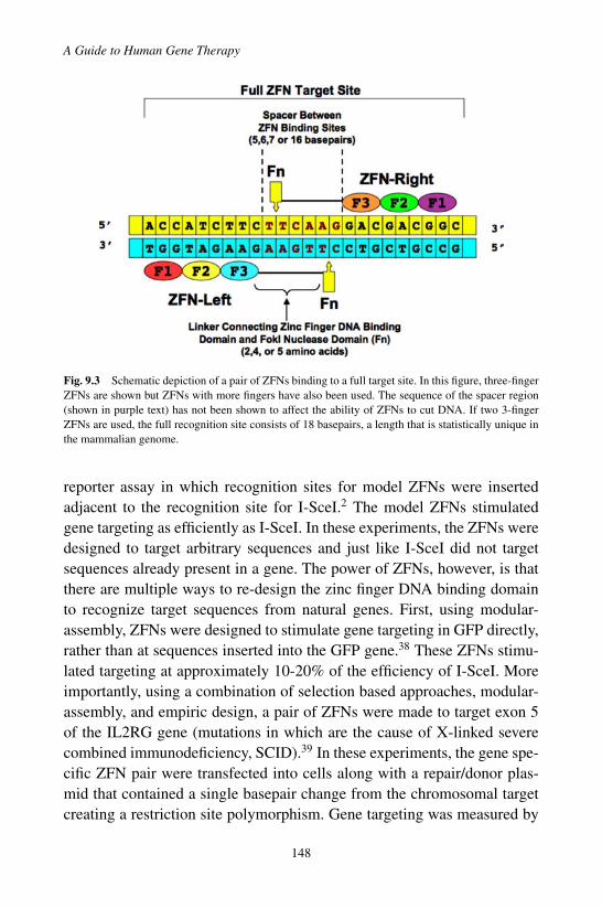
May 4, 2010 11:0 SPI-B903 9in x 6in b903-ch09
A Guide to Human Gene Therapy
Fig. 9.3 Schematic depiction of a pair of ZFNs binding to a full target site. In this figure, three-fingerZFNs are shown but ZFNs with more fingers have also been used. The sequence of the spacer region(shown in purple text) has not been shown to affect the ability of ZFNs to cut DNA. If two 3-fingerZFNs are used, the full recognition site consists of 18 basepairs, a length that is statistically unique inthe mammalian genome.
reporter assay in which recognition sites for model ZFNs were insertedadjacent to the recognition site for I-SceI.2 The model ZFNs stimulatedgene targeting as efficiently as I-SceI. In these experiments, the ZFNs weredesigned to target arbitrary sequences and just like I-SceI did not targetsequences already present in a gene. The power of ZFNs, however, is thatthere are multiple ways to re-design the zinc finger DNA binding domainto recognize target sequences from natural genes. First, using modular-assembly, ZFNs were designed to stimulate gene targeting in GFP directly,rather than at sequences inserted into the GFP gene.38 These ZFNs stimu-lated targeting at approximately 10-20% of the efficiency of I-SceI. Moreimportantly, using a combination of selection based approaches, modular-assembly, and empiric design, a pair of ZFNs were made to target exon 5of the IL2RG gene (mutations in which are the cause of X-linked severecombined immunodeficiency, SCID).39 In these experiments, the gene spe-cific ZFN pair were transfected into cells along with a repair/donor plas-mid that contained a single basepair change from the chromosomal targetcreating a restriction site polymorphism. Gene targeting was measured by
148

May 4, 2010 11:0 SPI-B903 9in x 6in b903-ch09
Homologous Recombination and Targeted Gene Modification for Gene Therapy
determining the presence or absence of this polymorphism in the gene.Using the IL2RG-ZFNs in K562 cells, a human erythroleukemia cell line,11% of cells underwent targeting at a single allele and 6% of cells underwenttargeting at both alleles. In primary T-cells, 4% of the IL2RG alleles couldbe targeted. Moreover, using these ZFNs one could create bi-allelic modifi-cation of the IL2RG gene and then subsequently re-modify both alleles. Thisestablished a proof-of-principle on the potential use of ZFNs to stimulategene targeting by homologous recombination to correct small mutations indisease-associated genes.
The repertoire of genomic changes that can be created by ZFN-mediatedhomologous recombination, however, is not limited to small point muta-tions. In addition, ZFNs can induce both small and large insertions into atarget locus by gene targeting.38,40,41 In this way, full genes including regula-tory elements such as promoters and enhancers, can be specifically targetedto integrate into specific genomic locations by homologous recombination.By targeting transgene insertion to a site that has no oncogenic potentialand is not subject to chromatin silencing, the use of ZFN-mediated genetargeting could increase both the safety and efficacy of gene therapy basedon the integration of transgenes.
In summary, the use of ZFN mediated gene targeting by homologousrecombination has been shown in pre-clinical models to be a method that candirectly correct disease causing mutations and a way of targeting transgeneinsertion in situations where direct correction of a disease causing mutationis neither desirable nor feasible.
11. Using Zinc Finger Nucleases to Site-SpecificallyModify Genes by Mutagenic Non-HomologousEnd-Joining
Carroll and his colleagues established in flies an alternative use of ZFNs toperform site-specific modifications of the genome. In addition to showingthat ZFNs could stimulate gene targeting by homologous recombination,they demonstrated that gene specific mutations could be created by themutagenic repair of a ZFN induced DSB by NHEJ.42 In their seminal work,they used ZFNs to recognize the yellow locus, a gene that controls eye color.They demonstrated that their ZFNs could induce a high rate of germline
149
May 4, 2010 11:0 SPI-B903 9in x 6in b903-ch09
A Guide to Human Gene Therapy
mutations in the yellow gene. When they sequenced the gene, they foundsmall insertions and deletions that were created by the mutagenic repairof a DSB by NHEJ. This strategy has been applied to create gene specificmutations in zebrafish and has also been applied to mammalian cells whereZFNs have been used to create gene-specific inactivating mutations in theDHFR gene, the CCR5 gene, and the glucocorticoid receptor gene.43−46 Thisstrategy of using ZFNs to create gene specific mutations by the mutagenicrepair of a DSB is one that has great potential in creating experimental celllines, particularly human lines, that could not be easily generated in otherways. The ability to create such specific cell lines should be a powerful toolin biomedical research. In addition, the possibility of using this strategyto knock-out dominant disease causing alleles is a potential use in genetherapy.
The inactivation of the CCR5 gene by CCR5 specific ZFNs deservesspecial attention because it is the first use of ZFNs in a clinical trial.46 InHIV infections, patients who have a bi-allelic deletion in the CCR5 geneare resistant to HIV infection and AIDS.47 While these patients do seemto be more susceptible to other infections, such as Listeria and West NileVirus, their immune systems are otherwise functional. The strategy is thatby harvesting T cells from HIV infected patients, treating them with CCR5specific ZFNs to inactivate both alleles of the CCR5 gene by mutagenicNHEJ, a pool of patient specific T cells would be created that would berelatively resistant to HIV infection and thus might protect the patient fromdeveloping AIDS or even help limit the proliferation of HIV itself. In con-trast to RNAi or ribozyme based strategies to target CCR5, the ZFN strategyhas the advantage of permanently modifying the CCR gene itself and thus isnot dependent on the continued trans-repression of the gene. In pre-clinicalmodels, CCR5-ZFNs have been designed that after delivery into primary Tcells by adenovirus can bi-allelically inactivate the CCR5 gene in approx-imately 1-3% of cells. When the ZFN modified cells were injected into animmunodeficient mouse (NOG mice) the modified T cells had a selectiveadvantage compared to un-modified cells when the animals are inoculatedwith HIV. Based on these results, a clinical trial to test the safety of thisapproach in humans has opened. One caveat is that the in the mouse pre-clinical studies, the ZFN modified T cells consisted of 10% of the T cellsin the animal prior to HIV challenge. In contrast, the ZFN modified T cells
150
May 4, 2010 11:0 SPI-B903 9in x 6in b903-ch09
Homologous Recombination and Targeted Gene Modification for Gene Therapy
will likely represent less than 0.1% of the total number of T cells in thebody in patients. Furthermore, in the mouse studies a single CCR-tropicvirus was used to challenge the animal. In contrast, human HIV infectedpatients harbor a mixture of both CCR5 and CXCR4 tropic viruses. Theknockout of the CCR5 gene would not be expected to have any effect onthe kinetics of the CXCR4 tropic viruses. These quantitative and qualitativefactors may be of such magnitude to prevent any clinical efficacy from beingdemonstrated. Nonetheless, this application is a logical first use of ZFNs ina human clinical trial.
12. Strategies of Zinc Finger Nuclease Design
ZFNs have been used to stimulate homologous recombination and to creategene specific mutations in mammalian cells; uses that have direct applicationto gene therapy. The key factor to ZFNs is to design the zinc finger DNAbinding domain such that it has high affinity and high specificity for itstarget binding site, as that will determine the quality of the ZFN. A zincfinger DNA binding domain consists of an array of individual zinc fingerunits. Each unit binds to a 3-4 basepair sequence of DNA. To create a zincfinger DNA binding domain with sufficient affinity for a specific sequence,at least three individual fingers must be assembled in an array and this3-finger domain recognizes a 9-10 basepair site. Proteins with more fingers,including 4, 5, or 6 fingers, which will have longer target sites, can be made.Proteins with more fingers can have increased affinity for their target sitebut may not necessarily have greater specificity. There are three generalmethods to designing the zinc finger DNA binding domain: 1) Modular-Assembly; 2) The Sangamo Biosciences Platform; 3) Selection.
In modular-assembly, new zinc finger proteins are assembled from pre-viously identified individual zinc finger units to create a new zinc fingerprotein.48 These individual units have been identified for most of the64 possible 3-basepair binding sites.49−52 These units have been identifiedusing a variety of methods, including from naturally occurring zinc fingerproteins, phage display and empiric design. Using standard molecular biol-ogy techniques individual units can be assembled into a multi-finger proteinto target a specific DNA sequence. This strategy has been used with suc-cess in creating artificial zinc finger transcription factors and several ZFNs
151

May 4, 2010 11:0 SPI-B903 9in x 6in b903-ch09
A Guide to Human Gene Therapy
(including those that target the GFP gene and the fly yellow genes).38,42,48 Abroad study of three-finger proteins assembled by modular-assembly, high-lighted the limitations of this approach.53 Modular-assembly has a relativelylow probability of being a successful design strategy for a large proportion ofpotential target sequences.54 In addition, in comparison with ZFNs designedby the approaches described below, ZFNs designed by modular-assemblyhave less specificity and more toxicity in cells.55 Thus, the ease of creatinga ZFN by modular-assembly must be balanced against the possibility thatthe ZFN will not have sufficient affinity to be active and may be more toxicthan desired.
A second approach to ZFN design is a proprietary strategy from SangamoBiosciences and now licensed for research use to Sigma-Aldrich. In thisstrategy, two-finger cassettes are selected by phage display against a widerange of 6 basepair binding sites.56 To create a ZFN, a target sequence iscompared against a private database of the two finger cassettes to identifypotential ZFN target sites. In this strategy, two two-finger cassettes areassembled to create a four-finger protein to recognize a specific twelve-basepair target site. A series of four-finger proteins are created and theseare screened using both in vitro binding assays and cell-based nucleaseassays to identify a pair of ZFNs that have the best activity at a targetsite within a given locus.44 This strategy was used to design the IL2RGand CCR5 ZFNs described above.39,46 The major limitation of this ZFNdesign strategy is that it depends on a proprietary algorithm and databasethat is not available to academic researchers. For investigators interested inusing ZFNs for research purposes, they can buy ZFNs made by this strategyfrom Sigma-Aldrich. Investigators interested in using ZFNs made with thisplatform for therapeutic use, must establish a collaborative agreement withSangamo Biosciences.
A third approach to designing ZFNs is to use a selection-basedstrategy.55,57−59 In this approach, a large library of three-finger proteinsis created and then using either iterative phage display or serial bacterialbased selections, proteins with high affinity for the desired target site areidentified. While phage display was the first type of selection strategy tobe used, this selection strategy has been replaced by the simpler, but stillrelatively complex, bacterial based systems.43,55,58,60 Using the bacterialbased systems, active ZFNs have been made to stimulate gene targeting
152
May 4, 2010 11:0 SPI-B903 9in x 6in b903-ch09
Homologous Recombination and Targeted Gene Modification for Gene Therapy
by homologous recombination in human cells, to create site-specific mod-ifications in endogenous genes in human cells and, to create heritablesite-specific gene mutations in zebrafish.43,55,58 ZFNs made by bacterialselection strategies have been shown to have greater specificity and con-sequently less cellular toxicity than ZFNs made by modular-assembly andto have similar, if not better, activity and toxicity characteristics made bythe proprietary Sangamo Biosciences approach.55,58 While this approach isavailable to all researchers, the major limitation to this strategy is that itremains relatively complex, currently requires substantial molecular biol-ogy expertise to perform and still can take weeks to months to generate thedesired proteins.
There are advantages and disadvantages to each of the ZFN design tech-niques. With the development of ZFNs, there has been a renewed interest indeveloping bioinformatic and computational design approaches to makingzinc finger proteins. It is likely that as the database of ZFNs increases, theseapproaches will become increasingly useful. Even if they do not absolutelydetermine the best ZFN in terms of activity and toxicity, if computationalapproaches can provide guidance to selecting a relatively small number ofpotential ZFNs to screen, they will be a significant advance in the field anddramatically shorten the time from experimental use to concrete results.
13. Aspects of Zinc Finger Binding Sites and Structureof Zinc Finger Nucleases
Figure 9.3 shows the general structure of a pair of ZFNs binding to theircognate binding sites. In this figure, the separation of the ZFN bindingsites (the “spacer”) is 6 basepairs. In practice, the ZFN binding sites can beseparated by 5, 6, 7, or 16 basepairs.61 Spacer distances of 3 and 4 basepairshave been shown not to work and a spacer length of 8 basepairs has beenshown to be sup-optimal in comparison to a 6 basepair spacer.2
Figure 9.3 also shows a pair of three finger ZFNs binding to their9-basepair cognate binding sites. In a 3-finger zinc finger protein, eachfinger is linked to its neighbor by a 5 amino acid linker. In 4-finger ZFNsthere is an extra serine in the linker region between fingers 2 and 3. Thisextra amino acid allows the zinc finger DNA binding domain and the majorgroove of DNA in which the zinc finger binds to remain in register. Finally,
153
May 4, 2010 11:0 SPI-B903 9in x 6in b903-ch09
A Guide to Human Gene Therapy
Fig. 9.3 depicts an amino acid linker that connects the zinc finger DNA bind-ing domain to the nuclease domain. This linker can be 2, 4, or 5 amino acidslong without affecting the activity of the ZFN. The linker length between thezinc finger DNA binding domain and the nuclease domain does affect thestructure of the ZFN target site. For example, if the linker length betweenthe two domains is 5 amino acids, the ZFNs effectively cut target siteswith a 6 or 7 basepair spacer but do not efficiently cut sites separated by5 basepairs. In contrast, if the linker length is 2 or 4 amino acids, the ZFNseffectively cut sites separated by 5 or 6 basepairs, but do not seem to cutsites separated by 7 basepairs. Thus, the target site guides what linker needsto be used to connect the zinc finger DNA binding domain and the nucleasedomain.
14. Zinc Finger Nuclease Toxicity: Measuringand Minimizing
In addition to the challenges in designing active ZFNs, the other majorproblem in the use of ZFNs is limiting their cellular toxicity. When ZFNswere compared to I-SceI in gene targeting in mammalian cells, they werefound to have significantly more cellular toxicity.2 The toxicity of ZFNs wasalso observed in flies.62 The toxicity in flies was abrogated when the nucleaseactivity of the ZFNs was mutated suggesting that the toxicity of ZFNs wasdue in major part from off-target DSBs.62 The translation of ZFN technologyto use in humans for gene therapy requires that these off-target effects bequantitatively determined, better understood, and minimized to a substantialdegree because the creation of off-target DSBs would be predicted to leadto oncogenic mutations.
ZFN toxicity has been measured in a number of different ways includ-ing the use of assays that measure cell survival after exposure to ZFNs, thefrequency with which ZFNs create extra DSBs, and by deep sequencingof potential off-target break sites.46,55 Using these assays, several differ-ent approaches have been used to minimize the off-target effects. The firstapproach was to use more sophisticated design methods for the zinc fingerDNA binding domain.39,55,58 These more sophisticated design methods haveled to improvements in ZFN cellular toxicity but as more sensitive measuresof cellular toxicity have been developed, they show that improved zinc finger
154
May 4, 2010 11:0 SPI-B903 9in x 6in b903-ch09
Homologous Recombination and Targeted Gene Modification for Gene Therapy
design does not solve the problem entirely.63 Further decreases in toxicitywere achieved by modifying the nuclease domain.55,64,65 As Fig. 9.3 depicts,cutting of the target site requires dimerization of the nuclease domains fromtwo different ZFNs. It is possible, however, that off-target cutting can arisefrom the homodimerization of a single ZFN. To prevent such homodimercutting at off-target sites, the nuclease domain has been re-engineered usinga combination of structure based approaches and empirical design to cre-ate domains that are only permissive for heterodimerization. That is, thenuclease domain from ZFN1 can only dimerize with the nuclease domainof ZFN2, not with itself, thus allowing cutting at the target site but reducingcutting at off-target sites. This modification should theoretically result ina 50% reduction of off-target cutting, but empirically it is observed thata much greater reduction in toxicity is observed with these “obligate het-erodimer” ZFNs. Better understanding of the mechanism by which thesehave reduced cellular toxicity and reduced creation of extra DSBs shouldhelp unravel the different ways that ZFNs can create off-target damage.Finally, a third approach to minimizing cellular toxicity is to use smallmolecule regulation of ZFN expression level.63 ZFN based genome modi-fication is based on a “hit and run” strategy and continued expression of theZFN is neither needed nor desired. By attaching a domain to the N-terminusof the ZFN that destabilizes the ZFN except in the presence of a smallmolecule, the expression level of the ZFN can be tightly regulated so it isonly expressed for a short window of time. This tight regulation of ZFNexpression results in reduced cellular toxicity without compromising ZFNefficacy.63
In summary, there are now multiple strategies that can reduce the off-target effects of ZFNs and increasingly sensitive assays to measure sucheffects. One of the major lessons from the development of retroviral vectorsfor gene therapy for the immunodeficiencies, however, is to push the enve-lope on developing pre-clinical models to quantitatively assess the onco-genic potential of any gene therapy approach. This lesson applies to theuse of ZFNs — the potential safety of using site-specific genome modifica-tion needs to be measured against the potential oncogenic risk of off-targetDSBs. Until better pre-clinical assays are developed, it remains unclearwhether the oncogenic risk of ZFNs falls in the tolerable range, such as thatcreated by radiation therapy for life threatening malignancies, or falls in a
155

May 4, 2010 11:0 SPI-B903 9in x 6in b903-ch09
A Guide to Human Gene Therapy
range that would be considered too high for the disease processes the ZFNsare designed to treat.
15. The Challenge of Delivery
In addition to the challenge of designing highly active and specific ZFNsand minimizing the off-target toxicity of ZFNs, the challenge of how tobest deliver the ZFN based components to therapeutically important celltypes, such as tissue specific stem cells, needs to be determined. While ZFNmanipulation of the genome can be classified as a “non-viral” strategy, theproblem of how to deliver the components for ZFN manipulations into thecell still remains. The upcoming clinical trial with the CCR5 directed ZFNsfor treatment of HIV uses ex vivo adenoviral delivery of ZFN expressionconstructs followed by expansion of transduced cells.46 The expansion ofT cells after manipulation should reduce the probability of a host immuneresponse to either the viral vector or the foreign ZFN. But this must be care-fully monitored given the experience with the in vivo use of adenovirus forthe treatment of OTC deficiency.66 The use of integration deficient lentivirushas also shown promise in pre-clinical studies, but the low rate of genetargeting in human CD34+ using this delivery method demonstrates thatfurther improvements are needed.41 Since ZFN based genome manipula-tion is a hit and run strategy, delivery methods that introduce either pro-tein or mRNA for the ZFNs rather than DNA ZFN expression constructsmay provide an added degree of safety. In fact, the delivery of mRNAto zebrafish fertilized eggs was an effective method in that organism,43,44
but scaling that process up for human clinical use, where millions if nothundreds of millions of cells might need to be manipulated remains a tech-nological hurdle. Another possible delivery method is the use of AAV. Asdiscussed above, AAV, through mechanisms not entirely clear, can stim-ulate targeting on its own. But AAV can be combined with the creationof a gene-specific DSB to stimulate gene targeting.67,68 While the packag-ing size of AAV can limit certain gene therapy applications, it should notpose as great a limitation in a gene targeting approach. Finding the optimalserotype to transduce human embryonic or tissue specific stem cells remainsan important problem for either the ex vivo or in vivo modification of cells for
156

May 4, 2010 11:0 SPI-B903 9in x 6in b903-ch09
Homologous Recombination and Targeted Gene Modification for Gene Therapy
therapeutic use. In summary, once again, gene transfer remains an importantproblem in translating an exciting laboratory based approach into humantherapy.
16. Future Directions and Promise of HomologousRecombination as a Gene Correction Approachto Gene Therapy
While the gene therapy field has worked hard to translate laboratory basedtechniques into therapy for humans and the progress has been slower thaneither expected or desired, the promise of being able to cure genetic diseaseswith genetic techniques remains great. Moreover, while short and interme-diate term successes are likely to continue using vectors that integrate in anuncontrolled fashion, the ultimate long-term broad success of gene therapyis likely to depend on finding ways to either directly correct disease causingmutations or to control the target site of transgene integration. The use ofhomologous recombination by AAV, the potential of embryonic stem celltherapy, and the progress with gene specific modifications using modifiedhoming endonucleases and zinc finger nucleases all suggest that the specifictargeting of the mammalian genome for therapeutic purposes may one daybe successful.
References
1. Sedivy JM, Dutriaux A (1999). Gene targeting and somatic cell genetics–a rebirth or acoming of age? Trends Genet 15: 88–90.
2. Porteus MH, Baltimore D (2003). Chimeric nucleases stimulate gene targeting in humancells. Science 300: 763.
3. Capecchi M (1989). Altering the genome by homologous recombination. Science 244:1288-1292.
4. Aiuti A, et al. (2009). Gene therapy for immunodeficiency due to adenosine deaminasedeficiency. N Engl J Med 360: 447–458.
5. Cavazzana-Calvo M, Lagresle C, Hacein-Bey-Abina S, FischerA (2005). Gene therapyfor severe combined immunodeficiency. Annu Rev Med 56: 585–602.
6. Hochedlinger K, Jaenisch R (2006). Nuclear reprogramming and pluripotency. Nature441: 1061–1067.
7. Yamanaka S (2007). Strategies and new developments in the generation of patient-specific pluripotent stem cells. Cell Stem Cell 1: 39–49.
157

May 4, 2010 11:0 SPI-B903 9in x 6in b903-ch09
A Guide to Human Gene Therapy
8. Rideout WM, 3rd, Hochedlinger K, Kyba M, Daley GQ, Jaenisch R (2002). Correctionof a genetic defect by nuclear transplantation and combined cell and gene therapy. Cell109: 17–27.
9. Hanna J, et al. (2007). Treatment of sickle cell anemia mouse model with iPS cellsgenerated from autologous skin. Science 318: 1920–1923.
10. Maherali N, Hochedlinger K (2008). Guidelines and techniques for the generation ofinduced pluripotent stem cells. Cell Stem Cell 3: 595–605.
11. Lengerke C, et al. (2007). The cdx-hox pathway in hematopoietic stem cell formationfrom embryonic stem cells. Ann N Y Acad Sci 1106: 197–208.
12. Russell DW, Hirata RK, Inoue N (2002). Validation of AAV-mediated gene targeting.Nat Biotechnol 20: 658.
13. Hirata R, Chamberlain J, Dong R, Russell DW (2002). Targeted transgene insertion intohuman chromosomes by adeno-associated virus vectors. Nat Biotechnol 20: 735–738.
14. Russell DW, Hirata RK (1998). Human gene targeting by viral vectors. Nat Genet 18:325–330.
15. Chamberlain JR, et al. (2004). Gene targeting in stem cells from individuals withosteogenesis imperfecta. Science 303: 1198–1201.
16. Wyman C, Kanaar R (2006). DNA double-strand break repair: all’s well that ends well.Annu Rev Genet 40: 363–383.
17. West SC, Chappell C, Hanakahi LA, Masson JY, McIlwraith MJ, Van Dyck E (2000).Double-strand break repair in human cells. Cold Spring Harb Symp Quant Biol 65:315–321.
18. Elliott B, Richardson C, Winderbaum J, Nickoloff JA, Jasin M (1998). Gene conversiontracts from double-strand break repair in mammalian cells. Mol Cell Biol 18: 93–101.
19. Burma S, Chen BP, Chen DJ (2006). Role of non-homologous end joining (NHEJ) inmaintaining genomic integrity. DNA Repair (Amst) 5: 1042–1048.
20. Wyman C, Ristic D, Kanaar R (2004). Homologous recombination-mediated double-strand break repair. DNA Repair (Amst) 3: 827–833.
21. Smih F, Rouet P, Romanienko PJ, Jasin M (1995). Double-strand breaks at the targetlocus stimulate gene targeting in embryonic stem cells. Nucleic Acids Res 23: 5012–5019.
22. Brenneman M, Gimble FS, Wilson JH (1996). Stimulation of intrachromosomal homol-ogous recombination in human cells by electroporation with site-specific endonucle-ases. Proc Natl Acad Sci USA 93: 3608–3612.
23. Sargent RG, Brenneman MA, Wilson JH (1997). Repair of site-specific double-strandbreaks in a mammalian chromosome by homologous and illegitimate recombination.Mol Cell Biol 17: 267–277.
24. Choulika A, Perrin A, Dujon B, Nicolas J-F (1995). Induction of homologous recom-bination in mammalian chromosomes by using the I-SceI system of Saccaromycescerevisiae. Molecular and Cellular Biology 15: 1968–1973.
25. Taghian DG, Nickoloff JA (1997). Chromosomal double-strand breaks inducegene conversion at high frequency in mammalian cells. Mol Cell Biol 17: 6386–6393.
26. Donoho G, Jasin M, Berg P (1998). Analysis of gene targeting and intrachromoso-mal homologous recombination stimulated by genomic double-strand breaks in mouseembryonic stem cells. Mol Cell Biol 18: 4070–4078.
158

May 4, 2010 11:0 SPI-B903 9in x 6in b903-ch09
Homologous Recombination and Targeted Gene Modification for Gene Therapy
27. Chevalier BS, Stoddard BL (2001). Homing endonucleases: structural and functionalinsight into the catalysts of intron/intein mobility. Nucleic Acids Res 29: 3757–3774.
28. Ashworth J, et al. (2006). Computational redesign of endonuclease DNA binding andcleavage specificity. Nature 441: 656–659.
29. Sussman D, et al. (2004). Isolation and characterization of new homing endonucleasespecificities at individual target site positions. J Mol Biol 342: 31–41.
30. Arnould S, et al. (2007). Engineered I-CreI derivatives cleaving sequences from thehuman XPC gene can induce highly efficient gene correction in mammalian cells. JMol Biol 371: 49–65.
31. Redondo P, et al. (2008). Molecular basis of xeroderma pigmentosum group C DNArecognition by engineered meganucleases. Nature 456: 107–111.
32. Paques F, Duchateau P (2007). Meganucleases and DNA double-strand break-inducedrecombination: perspectives for gene therapy. Curr Gene Ther 7: 49–66.
33. Kim YG, Cha J, Chandrasegaran S (1996). Hybrid restriction enzymes: zinc fingerfusions to Fok I cleavage domain. Proc Natl Acad Sci USA 93: 1156–1160.
34. Smith J, Berg JM, Chandrasegaran S (1999).A detailed study of the substrate specificityof a chimeric restriction enzyme. Nucleic Acids Res 27: 674–681.
35. Smith J, Bibikova M, Whitby FG, Reddy AR, Chandrasegaran S, Carroll D (2000).Requirements for double-strand cleavage by chimeric restriction enzymes with zincfinger DNA-recognition domains. Nucleic Acids Res 28: 3361–3369.
36. Durai S, Mani M, Kandavelou K, Wu J, Porteus MH, Chandrasegaran S (2005). Zincfinger nucleases: custom-designed molecular scissors for genome engineering of plantand mammalian cells. Nucleic Acids Res 33: 5978–5990.
37. Bibikova M, et al. (2001). Stimulation of homologous recombination through targetedcleavage by chimeric nucleases. Mol Cell Biol 21: 289–297.
38. Porteus MH (2006). Mammalian gene targeting with designed zinc finger nucleases.Mol Ther 13: 438–446.
39. Urnov FD, et al. (2005). Highly efficient endogenous human gene correction usingdesigned zinc-finger nucleases. Nature 435: 646–651.
40. Moehle EA, et al. (2007). Targeted gene addition into a specified location in the humangenome using designed zinc finger nucleases. Proc Natl Acad Sci USA 104: 3055–3060.
41. LombardoA, et al. (2007). Gene editing in human stem cells using zinc finger nucleasesand integrase-defective lentiviral vector delivery. Nat Biotechnol 25: 1298–1306.
42. Bibikova M, Beumer K, Trautman JK, Carroll D (2003). Enhancing gene targeting withdesigned zinc finger nucleases. Science 300: 764.
43. Meng X, Noyes MB, Zhu LJ, Lawson ND, Wolfe SA (2008). Targeted gene inactivationin zebrafish using engineered zinc-finger nucleases. Nat Biotechnol 26: 695–701.
44. Doyon Y, et al. (2008). Heritable targeted gene disruption in zebrafish using designedzinc-finger nucleases. Nat Biotechnol 26: 702–708.
45. Santiago Y, et al. (2008). Targeted gene knockout in mammalian cells by using engi-neered zinc-finger nucleases. Proc Natl Acad Sci USA 105: 5809–5814.
46. Perez EE, et al. (2008). Establishment of HIV-1 resistance in CD4+ T cells by genomeediting using zinc-finger nucleases. Nat Biotechnol 26: 808–816.
47. Stewart GJ, et al. (1997). Increased frequency of CCR-5 delta 32 heterozygotes amonglong-term non-progressors with HIV-1 infection. The Australian Long-Term Non-Progressor Study Group. AIDS 11: 1833–1838.
159
May 4, 2010 11:0 SPI-B903 9in x 6in b903-ch09
A Guide to Human Gene Therapy
48. Beerli RR, Barbas CF, 3rd (2002). Engineering polydactyl zinc-finger transcriptionfactors. Nat Biotechnol 20: 135–141.
49. Segal DJ, Dreier B, Beerli RR, Barbas CF, 3rd (1999). Toward controlling gene expres-sion at will: selection and design of zinc finger domains recognizing each of the 5′-GNN-3′ DNA target sequences. Proc Natl Acad Sci USA 96: 2758–2763.
50. Dreier B, Beerli RR, Segal DJ, Flippin JD, Barbas CF, 3rd (2001). Development of zincfinger domains for recognition of the 5′-ANN-3′ family of DNA sequences and their usein the construction of artificial transcription factors. J Biol Chem 276: 29466–29478.
51. Dreier B, et al. (2005). Development of zinc finger domains for recognition of the5′-CNN-3′ family DNA sequences and their use in the construction of artificial tran-scription factors. J Biol Chem 280: 35588–35597.
52. Liu Q, Xia Z, Zhong X, Case CC (2002). Validated zinc finger protein designs for all16 GNN DNA triplet targets. J Biol Chem 277: 3850–3856.
53. Ramirez CL, et al. (2008). Unexpected failure rates for modular assembly of engineeredzinc fingers. Nat Methods 5: 374–375.
54. Sander JD, Zaback P, Joung JK, Voytas DF, Dobbs D (2009). An affinity-based scor-ing scheme for predicting DNA-binding activities of modularly assembled zinc-fingerproteins. Nucleic Acids Res 37: 506–515.
55. Pruett-Miller SM, Connelly JP, Maeder ML, Joung JK, Porteus MH (2008). Comparisonof zinc finger nucleases for use in gene targeting in mammalian cells. Mol Ther 16:707–717.
56. Isalan M, Klug A, Choo Y (2001). A rapid, generally applicable method to engi-neer zinc fingers illustrated by targeting the HIV-1 promoter. Nat Biotechnol 19:656–660.
57. Hurt JA, Thibodeau SA, Hirsh AS, Pabo CO, Joung JK (2003). Highly specific zincfinger proteins obtained by directed domain shuffling and cell-based selection. ProcNatl Acad Sci USA 100: 12271–12276.
58. Maeder ML, et al. (2008). Rapid “open-source” engineering of customized zinc-fingernucleases for highly efficient gene modification. Mol Cell 31: 294–301.
59. Greisman HA, Pabo CO (1997).A general strategy for selecting high-affinity zinc fingerproteins for diverse DNA target sites. Science 275: 657–661.
60. Durai S, Bosley A, Abulencia AB, Chandrasegaran S, Ostermeier M (2006). A bacte-rial one-hybrid selection system for interrogating zinc finger-DNA interactions. CombChem High Throughput Screen 9: 301–311.
61. Handel EM, Alwin S, Cathomen T (2009). Expanding or restricting the target siterepertoire of zinc-finger nucleases: the inter-domain linker as a major determinant oftarget site selectivity. Mol Ther 17: 104–111.
62. Beumer K, Bhattacharyya G, Bibikova M, Trautman JK, Carroll D (2006). Efficientgene targeting in Drosophila with zinc-finger nucleases. Genetics 172: 2391–2403.
63. Pruett-Miller SM, Reading DW, Porter SN, Porteus MH (2009).Attenuation of Zinc Fin-ger Nuclease Toxicity by Small Molecule Regulation of Protein Levels. PLOS Geneticsin press.
64. Szczepek M, BrondaniV, Buchel J, Serrano L, Segal DJ, Cathomen T (2007). Structure-based redesign of the dimerization interface reduces the toxicity of zinc-finger nucle-ases. Nat Biotechnol 25: 786–793.
160

May 4, 2010 11:0 SPI-B903 9in x 6in b903-ch09
Homologous Recombination and Targeted Gene Modification for Gene Therapy
65. Miller JC, et al. (2007). An improved zinc-finger nuclease architecture for highly spe-cific genome editing. Nat Biotechnol 25: 778–785.
66. Raper SE, et al. (2003). Fatal systemic inflammatory response syndrome in a ornithinetranscarbamylase deficient patient following adenoviral gene transfer. Mol Genet Metab80: 148–158.
67. Porteus MH, Cathomen T, Weitzman MD, Baltimore D (2003). Efficient gene targetingmediated by adeno-associated virus and DNA double-strand breaks. Mol Cell Biol 23:3558–3565.
68. Miller DG, Petek LM, Russell DW (2003). Human gene targeting by adeno-associatedvirus vectors is enhanced by DNA double-strand breaks. Mol Cell Biol 23: 3550–3557.
161

May 4, 2010 11:0 SPI-B903 9in x 6in b903-ch09
This page intentionally left blankThis page intentionally left blank

May 4, 2010 11:0 SPI-B903 9in x 6in b903-ch10
Chapter 10
Gene Switches for Pre-ClinicalStudies in Gene Therapy
Caroline Le Guiner∗, Knut Stieger, Alice Toromanoff∗,Fabienne Rolling∗, Philippe Moullier∗and Oumeya Adjali∗,†
Numerous preclinical studies have demonstrated the efficacy of viral genedelivery vectors. However, the tight control of transgene expression islikely to be required for therapeutic applications and/or safety reasons. Forthis purpose, several ligand-dependent transcription regulatory systemshave been developed. Among these, the tetracycline and the rapamycindependent systems have been largely used. However, if long-term regu-lation of the transgene has been obtained in small animal models usingthese inducible systems, when translational studies were initiated in largeranimals, especially in nonhuman primates (NHP) the development of animmune response against proteins involved in transgene regulation wereoften observed. Such immune response was especially documented whenusing the TetOn tetracycline (Tet) regulatable system in NHP especiallyafter intramuscular injection of the vector encoding for the Tet-regulatedtransgene. This chapter will emphasize on the tetracycline and rapamycingene switch systems, which we believe are the more relevant in a preclin-ical setting and potentially in future clinical trials.
1. Introduction
The clinical efficacy and safety as well as the application range of genetherapy will be broadened by developing systems capable of fine-tuningthe expression of therapeutic genes. A regulatable system that can turn
∗Correspondence: INSERM UMR 649, Institut de Recherche Therapeutique IRT UN, Nantes, Fance.E-mail: †[email protected]
163
May 4, 2010 11:0 SPI-B903 9in x 6in b903-ch10
A Guide to Human Gene Therapy
“on” and “off” therapeutic gene expression will not only be crucial formaintaining appropriate levels of a gene product within the therapeuticrange, thus preventing toxicity, but also allow the turning “off” of therapeuticgene expression to avoid harmful side effects. The development of ligand-dependent transcription regulatory systems is thus of great importance.
In human gene therapy, a pharmacologically regulated system shouldfulfill several criteria: (1) the ligand should activate (On-switch) rather thansilence transcription (Off-switch). Indeed, Off-systems suffer from 2 draw-backs: a prolonged exposure to the drug is required to silence the systemand the induction kinetic is mainly determined by the rate of drug clear-ance; (2) the inducer drug should be orally bioavailable and penetrate thetarget tissue; (3) the system should not interfere with endogenous metabolicpathways and the inducer compound should have a drug metabolism pro-file compatible with prolonged therapeutic uses; (4) transcription shouldbe fully and rapidly reversible to enable prompt changing of the dosingregimen; (5) the system should be inactive in the absence of the inducerdrug but strongly stimulated by the drug administration; (6) a precise cor-relation must exist between drug dosage and target gene expression level;(7) the transcriptional activator should not elicit an immune responsein human.
Four drug-dependent regulatable systems were developed for the in vivocontrol of transgene expression: the tetracycline (Tet) and the rapamycinregulatable systems, the mammalian steroid receptor (mifepristone andtamoxifen) and the insect steroid receptor (ecdysteroid)-based systems. Themolecular mechanisms underlying the regulatory effect and the pharmaco-logic characteristics of the inducer drug are distinct in these different regu-latory systems, but transgene regulation has been documented in vitro andin rodent models with all of them. Only the Tet and the rapamycin regulat-able systems were tested in large animal models.1−7 However, when thesetranslational studies were initiated in larger animals, the development of animmune response against the proteins involved in transgene regulation wasobserved,1−3,6 whereas in the murine model both were able to permanentlysustain transgene regulation.8,9
Because the Tet and the rapamycin-based systems are the most potentand clinically relevant for future trials, this chapter will focus on the molec-ular mechanisms involved in transgene regulation, the pharmacology of the
164

May 4, 2010 11:0 SPI-B903 9in x 6in b903-ch10
Gene Switches for Pre-Clinical Studies in Gene Therapy
inducer drug and the immunotoxicity observed (or not) for each of the twosystems.
2. Rapamycin-Dependent Regulatable System
2.1 Molecular Mechanisms Involved in Transgene Regulation
The basic concept of the rapamycin regulatory system is to express thetwo critical domains of a transcription factor as separate polypeptides thatcan be reversibly crosslinked only in the presence of the inducer drug,the bivalent “dimerizer” drug (Fig. 10.1). In cells, the natural productrapamycin or its analogs, binds to two peptides, FKBP12 (FK506 bindingprotein of 12kDa) and FRAP (FKBP rapamycin associated protein), bringsthem into close proximity, thus, activating their transcriptional specificityas inhibitor of T-cell proliferation and immunosuppression. The fusion ofthe FKBP12 domain to a DNA binding domain, called ZFHD1 (zinc fin-ger homeo domain 1) and of one portion of FRAP, called FRB (FKBPrapamycin binding), to a transcriptional activation domain from the p65subunit of the transcription factor NF-kB, results, when dimerization ofFKBP12 and FRB occurs, in the formation of a functional transactivator.In the presence of rapamycin, the complex FKBP12-ZFHD1/FRB-p65 isable to associate to the ZFHD1 corresponding DNA sequence upstream ofa minimal Interleukin-2 promoter (pIL2min), and the transactivator allows
Fig. 10.1 Schematic representation of the rapamycin dependent regulation system. This system iscomposed of two chimeric proteins, FRB-p65 and FKBP12-ZFHD1. The FKBP12-ZFHD1 proteinis able to interact with the corresponding DNA sequence (ZFHD1), which is fused to a minimal IL2promoter. The second protein is soluble. Rapamycin allows dimerization of FKBP12 and FRB, resultingin the formation of a functional transactivator, which allows transgene expression. In the absence ofrapamycin, the FRB and FKBP12 domains cannot interact, and no transgene expression is observed.
165
May 4, 2010 11:0 SPI-B903 9in x 6in b903-ch10
A Guide to Human Gene Therapy
the expression of the transgene. In the absence of rapamycin, the two pep-tides FKBP12-ZFHD1 and FRB-p65 are unable to interact, and are thentranscriptionally inert (Fig. 10.1).9
2.2 Pharmacology of Rapamycin
The pharmacologic profile of rapamycin is well known, but one majorobstacle for its application in gene therapy clinical trial is that rapamycinis a powerful immunosuppressive agent used in humans for many years.Since a long-term treatment with rapamycin could have significant deleteri-ous effects, efforts were made to develop non-immunosuppressive analogs.Analysis of the rapamycin-FRB complex led to the development of theso called “rapalogs” carrying punctual substitutions.10,11 Several of theserapalogs are considerably less immunosuppressive and, yet, were shownto efficiently induce transgene expression in nonhuman primate (NHP)muscle.6 However, one analog was tested in the retina and failed to beas efficient as the original drug.5
2.3 Translation Development of the Rapamycin DependentRegulation System
This system employs exclusively human peptides, thus rendering the pos-sibility for immune reactions against the chimeric transactivator mini-mal when tested in other species. Since its first description in 1996, therapamycin regulatable system was used in vivo in rodents, using differentviral vector platforms.1,12−14 The long-term regulation of the erythropoietin(Epo) transgene was also obtained in NHP eye and muscle, after subreti-nal, intravitreal and intramuscular (IM) injection of recombinant Adeno-Associated Virus (rAAV) vectors.5,6 The NHP that received rapamycin-regulated expression cassettes are summarized in Table 10.1.
In 1999, Ye et al. injected a first group of NHP, with two rAAV-2vectors (in grey in Table 10.1). If the first two administrations of rapamycinresulted in the rapid induction of Epo, its expression was decreased 20 foldat the third induction, and became indistinguishable from endogenous Epoafter the fourth induction.1,6 Antibodies directed against the influenza virushemagglutinin tag contained in the transcription factor were detected inserum of at least one animal after two rounds of rapamycin induction.
166
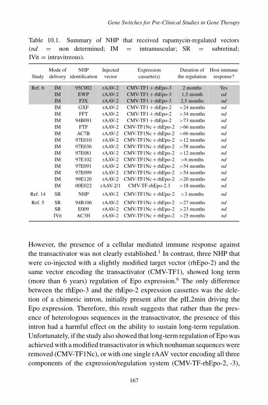
May 4, 2010 11:0 SPI-B903 9in x 6in b903-ch10
Gene Switches for Pre-Clinical Studies in Gene Therapy
Table 10.1. Summary of NHP that received rapamycin-regulated vectors(nd = non determined; IM = intramuscular; SR = subretinal;IVit = intravitreous).
Mode of NHP Injected Expression Duration of Host immuneStudy delivery identification vector cassette(s) the regulation response?
Ref. 6 IM 95C002 rAAV-2 CMV-TF1 + rhEpo-3 2 months YesIM EWP rAAV-2 CMV-TF1 + rhEpo-3 1,5 month ndIM FJX rAAV-2 CMV-TF1 + rhEpo-3 2,5 months ndIM GXF rAAV-2 CMV-TF1 + rhEpo-2 >24 months ndIM FFT rAAV-2 CMV-TF1 + rhEpo-2 >34 months ndIM 94B091 rAAV-2 CMV-TF1 + rhEpo-2 >73 months ndIM FTF rAAV-2 CMV-TF1Nc + rhEpo-2 >66 months ndIM AC7B rAAV-2 CMV-TF1Nc + rhEpo-2 >66 months ndIM 97E010 rAAV-2 CMV-TF1Nc + rhEpo-2 >12 months ndIM 97E036 rAAV-2 CMV-TF1Nc + rhEpo-2 >58 months ndIM 97E081 rAAV-2 CMV-TF1Nc + rhEpo-2 >12 months ndIM 97E102 rAAV-2 CMV-TF1Nc + rhEpo-2 >6 months ndIM 97E091 rAAV-2 CMV-TF1Nc + rhEpo-2 >54 months ndIM 97E099 rAAV-2 CMV-TF1Nc + rhEpo-2 >54 months ndIM 99E120 rAAV-2 CMV-TF1Nc + rhEpo-2 >20 months ndIM 00E022 rAAV-2/1 CMV-TF-rhEpo-2.3 >18 months nd
Ref. 14 SR NHP rAAV-2 CMV-TF1Nc + rhEpo-2 >3 months nd
Ref. 5 SR 94B106 rAAV-2 CMV-TF1Nc + rhEpo-2 >27 months ndSR E009 rAAV-2 CMV-TF1Nc + rhEpo-2 >23 months ndIVit AC3H rAAV-2 CMV-TF1Nc + rhEpo-2 >25 months nd
However, the presence of a cellular mediated immune response againstthe transactivator was not clearly established.1 In contrast, three NHP thatwere co-injected with a slightly modified target vector (rhEpo-2) and thesame vector encoding the transactivator (CMV-TF1), showed long term(more than 6 years) regulation of Epo expression.6 The only differencebetween the rhEpo-3 and the rhEpo-2 expression cassettes was the dele-tion of a chimeric intron, initially present after the pIL2min driving theEpo expression. Therefore, this result suggests that rather than the pres-ence of heterologous sequences in the transactivator, the presence of thisintron had a harmful effect on the ability to sustain long-term regulation.Unfortunately, if the study also showed that long-term regulation of Epo wasachieved with a modified transactivator in which nonhuman sequences wereremoved (CMV-TF1Nc), or with one single rAAV vector encoding all threecomponents of the expression/regulation system (CMV-TF-rhEpo-2, -3),
167

May 4, 2010 11:0 SPI-B903 9in x 6in b903-ch10
A Guide to Human Gene Therapy
the immune status against the transactivator in this NHP cohort was notreported (Table 10.1).
Finally, the retina of other NHP was injected with the same rAAV vectorsencoding for the Epo under the control of the rapamycin-regulated transac-tivator. In these animals, tight and long-term regulation of Epo expressionwas observed for more than two years.5,14 Again, if this result suggests theabsence of an immune response against the transcription factor, there wasno indication on the absence/presence of circulating antibodies against thetransactivator.
In summary, it is now clearly demonstrated that an efficient rapamycin-regulation of transgene expression can be obtained after a single IM orintraocular administration of a rAAV vector in NHP. One major advantage ofthis regulation system would be that it employs human peptides exclusively,thus minimizing the possibility for an immune reaction against the transacti-vator. However, the presence of a humoral and/or a cellular immune responseagainst this transcription factor has not yet been fully characterized.
3. Tetracycline-Dependent Regulatable Systems
3.1 Molecular Mechanisms Involved in Transgene Regulation
Tet-responsive systems are widely used because they are able to tightlycontrol transgene expression in a wide range of cultured cells as well asplants, yeast, insects and mammals in vivo.2,4,7,8,15−18 They are based onthe Escherichia coli Tn10 Tet resistance operator, which consists of theTet repressor protein (TetR) and the Tet operator DNA sequence (TetO).In the absence of Tet or its derivate doxycycline (Dox), the TetR proteininteracts with the TetO DNA sequence, while in the presence of this drug,TetR changes its conformation, thus detaching from the DNA. Based on thisbacterial operator system, three different Tet-responsive regulatory systemshave been developed (Fig. 10.2).
The first two systems contain the TetR protein fused to the transactivatorVP16 (viral protein 16) of the Herpes simplex virus (HSV).19,20 If trans-gene expression is allowed only in the absence of Dox, the system is calledTetOff. If transgene expression is allowed in the presence of Dox, the sys-tem is called TetOn. The third system contains the TetR protein fused to ahuman inhibitor protein: the krüppel associated box (KRAB) of zinc-finger
168
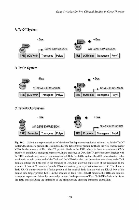
May 4, 2010 11:0 SPI-B903 9in x 6in b903-ch10
Gene Switches for Pre-Clinical Studies in Gene Therapy
Fig. 10.2 Schematic representations of the three Tet dependent regulation systems. A. In the TetOffsystem, the chimeric protein tTa is composed of the Tet repressor protein TetR and the viral transactivatorVP16. In the absence of Dox, the tTA protein binds to the TRE, which is fused to a minimal CMVpromoter, and allows transgene expression. In the presence of Dox, the tTA protein cannot interact withthe TRE, and no transgene expression is observed. B. In the TetOn system, the rtTA transactivator is alsoa chimeric protein composed of the TetR and the VP16 domains, but due to four mutations in the TetRdomain, it fixes the TRE only in the presence of Dox, thus allowing expression of the transgene. In theabsence of Dox, rtTA detaches from the DNA and no transgene expression is observed. C. The chimericTetR-KRAB transactivator is a fusion protein of the original TetR domain with the KRAB box of thehuman zinc finger protein Kox1. In the absence of Dox, TetR-KRAB binds to the TRE and inhibitstransgene expression driven by a normal promoter. In the presence of Dox, TetR-KRAB detaches fromthe TRE, thus disabling the inhibition of the promoter and allowing transgene expression.
169
May 4, 2010 11:0 SPI-B903 9in x 6in b903-ch10
A Guide to Human Gene Therapy
proteins.21 This system is designed to allow transgene expression only inthe presence of Dox.
In the TetOff system, the transactivator protein, called tTA (for Tet-controlled transcriptional activator) is a chimera made by the fusion of theTetR protein and the transactivator VP16. In the absence of Dox, tTA isable to bind to seven repeats of the TetO sequence, also referred to as TRE,for Tet responsive element. Binding of tTA to the TRE activates the TREassociated promoter (usually a minimal CMV promoter), resulting in theexpression of the adjacent gene. If Dox is present, tTA detaches from theTRE and gene expression is silenced (Fig. 10.2A).19
Four mutations in the TetR part of the transactivator tTA were shown toreverse its interaction with the TRE in the presence or absence of Dox20:rather than attaching to the TRE in the absence of Dox, this mutant,called rtTA (for reverse tTA), stays soluble. In the presence of Dox, theconformation of the rtTA changes, which in turn allows the transactiva-tor to interact with TRE. Therefore, the system changes to a TetOn sys-tem, i.e. only the presence of Dox allows expression of the transgene(Fig. 10.2B). Because the original rtTA version was prone to high back-ground leakage in the “Off” stage and poor inducibility in some instances,novel rtTA variants were generated after random and directed mutagen-esis as well as codon optimization. These variants, called rtTA-S2 andrtTA-M2, contain mutations in the DNA binding domain that connectsthe DNA with the core of the protein. They showed an improved sensi-tivity to the inducer drug and a reduced background activity in the absenceof Dox.22
In 1995, Deuschle et al. developed a system, in which the original TetRprotein was fused to the repressing KRAB domain of the human zinc-fingerprotein Kox1, generating a transrepressor called TetR-KRAB.21 Zinc-fingerproteins are mammalian regulatory proteins and their KRAB box is knownto inhibit all DNA Polymerases I, II or III within a range of 3 kilobasesof its DNA attachment site.23 In the absence of Dox, TetR-KRAB bindsspecifically to the TRE and suppresses the activity of the nearby pro-moter(s), while in the presence of Dox, TetR-KRAB detaches from theTRE, thus allowing transgene expression (Fig. 10.2C). The mechanism bywhich KRAB inhibits expression of genes is not entirely known. Threepathways seem to be involved: (i) local change in chromatin structure;(ii) local histone deacetylation, and; (iii) the indirect influence of the
170

May 4, 2010 11:0 SPI-B903 9in x 6in b903-ch10
Gene Switches for Pre-Clinical Studies in Gene Therapy
arrangement of the basal transcription machinery. Therefore, until recentlythe hypothesis was that only integrated promoters could be inhibited bythe TetR-KRAB.23,24 But, it was recently demonstrated that the TetR-KRAB system could also be functional in vitro and in vivo in the mousemodel in a non-integrative context, using non-integrative lentiviral andAAVvectors.25
This KRAB-mediated inhibition system was also incorporated in theTetOn system, in order to minimize the background expression observed inthe latter regulation system.26 For this purpose, the TetR-KRAB gene, alsocalled tTS (for Tet controlled transcriptional silencer), was cloned into abicistronic expression cassette downstream of the transactivator separatedby an IRES (internal ribosomal entry site). In the absence of Dox, the rtTAtransactivator stays detached from the TRE while the tTS transrepressorbinds and inhibit background expression of the transgene. In the presenceof Dox, the two chimeric proteins change their conformation and reversetheir respective activity, allowing transgene expression.
3.2 Pharmacology of Doxycycline (Dox)
The antibiotic Dox has been extensively documented over the past 40 years.It has a half-life of 14–22 hours after absorption and an excellent tissuepenetration, including the brain. The bioavailability of Dox after oraladministration is 100% and it is rapidly metabolized and eliminated bythe gastrointestinal tract. Dox has been extensively used in patients andis well tolerated, at least for medium term (months) applications. Sideeffects during prolonged administration of Tet include infrequent rise inblood-urea nitrogen, which is apparently dose-related as well as a dose-dependant photosensitivity. A concern when using an antibiotic-driven reg-ulatory switch remains the possibility of raising resistance to the antibioticitself. The generation of a nonantibiotic Dox analog-system would improvebiosafety in that respect.
3.3 Translational Development of Tet-dependantRegulation Systems
The different Tet-derived regulation systems were used to study trans-gene expression in several rodent models using recombinant retrovirus,
171

May 4, 2010 11:0 SPI-B903 9in x 6in b903-ch10
A Guide to Human Gene Therapy
AAV, adenovirus or naked DNA as vehicles.8,18,24,25,27−30 All these studiesdemonstrated the ability for the Tet-derived systems to sustain permanenttransgene regulation with no adverse effect. Of note, however, and evenwith more advanced versions of the chimeric transactivators, backgroundactivity in the absence of the inducer remains an issue, especially when highcopy numbers of the vector per cell are achieved.
To date, among the Tet-derived systems used to regulate gene transcrip-tion, the TetOn system is the most widely developed. Importantly, it wastested in large animal models particularly in NHP, in the liver,31 in theskeletal muscle2,4 and in the retina.7,32
The NHP that received Tet-regulated vectors are summarized inTable 10.2. Unlike in rodent models, translation of the Tet-based systemsinto NHP resulted in some studies in the rapid loss of transgene regulationwith an immune response towards the chimeric transactivators (in grey inTable 10.2). It appears that the targeted organ, as well as the route of admin-istration of the vector, are likely to impact the development (or not) of thisimmune response.
When rtTA and TetR-KRAB were used to regulate the expression ofeither macaque Epo or the secreted alkaline phosphatase (SEAP) from skele-tal muscle, after rAAV, plasmid or recombinant adenovirus-mediated genetransfer, no immune response was observed against the transgene protein,but a large majority of individuals developed an immune response againstthe rtTA or the TetR-KRAB proteins. The mechanism involved includedboth humoral and cellular responses, and resulted in the destruction of thegenetically modified cells.2,3
Such immune response even occurred when using the so-called muscle-specific desmin promoter instead of an ubiquitous promoter to drive theexpression of the transactivator rtTA [Moullier et al., unpublished data].However, one should emphasize that when the NHP immune system toler-ates the chimeric transactivator rtTA, transgene regulation is achieved forseveral years (more than 6 years) [Moullier et al., unpublished data].
Importantly, Table 10.2 indicates that skeletal muscle transduction afterRegional Intravenous delivery (RI) using an rAAV vector, leads to perma-nent TetOn-mediated transgene regulation (more than 7 Dox-inductions). Inthe fully immunocompetent injected animals, neither anti-rtTA nor lympho-cyte infiltration were detected in the muscle [Toromanoff et al., submitted].
172

May 4, 2010 11:0 SPI-B903 9in x 6in b903-ch10
Gene Switches for Pre-Clinical Studies in Gene Therapy
Table 10.2. Summary of NHP that received tetracycline-regulated vectors(nd = non determined; IM = intramuscular; IV = intravenous; RI = RegionalIntravenous; SR = subretinal).
Mode of NHP Injected Expression Duration of Host immuneStudy delivery identification vector cassette(s) the regulation response?
Ref. 3 IM M113 Adenovirus TK.TetOn.SEAP 0.5 month YesIM M114 Adenovirus TK.TetOn.SEAP 0.5 month YesIM F128 Adenovirus TK.TetOn.SEAP 0.5 month YesIM F129 Adenovirus TK.TetOn.SEAP 0.5 month YesIM F132 Adenovirus TK.TetOn.SEAP 0.5 month YesIM M102 Plasmid TK.TetOn.SEAP 0.7 month YesIM M107 Plasmid TK.TetOn.SEAP 0.7 month YesIM M109 Plasmid TK.TetOn.SEAP 0.7 month YesIM F121 Plasmid TK.TetOn.SEAP 0.7 month YesIM F124 Plasmid TK.TetOn.SEAP 0.7 month Yes
(Refs. 2, 4,Moullier et al.,unpublisheddata andToromanoffet al.,submitted (orthe referencenumber ifupdated in thelist))
IM Mac 4 rAAV-2 LTR.TetOn.Epo 5 months Yes
IM Mac 5 rAAV-2 CAG.TetOn.Epo 3 months YesIM Mac 6 rAAV-2 CAG.TetOn.Epo 3 months YesIM Mac 7 rAAV-2 CAG.TetOn.Epo 6 months YesIM Mac 8 rAAV-2 CAG.TetOn.Epo >13 months NoIM Mac 9 rAAV-2 Desmine.TetOn.Epo 4 months YesIM Mac 10 rAAV-2 Desmine.TetOn.Epo 3 months YesIM Mac 11 rAAV-2 Desmine.TetOn.Epo 2 months YesIM Mac 12 rAAV-2 Desmine.TetOn.Epo 2 months YesIM Mac 13 rAAV-2/1 Desmine.TetOn.Epo >72 months YesIM Mac 14 rAAV-2/1 Desmine.TetOn.Epo 2 months YesIM Mac 15 rAAV-2/1 Desmine.TetOn.Epo 4 months YesIM Mac 16 rAAV-2/1 Desmine.TetOn.Epo 3 months YesIM Mac 17 rAAV-2/5 Desmine.TetOn.Epo 5 months Yes
Ref. 31 IV 95-14 Adenovirus TTR-TetOn-tINFα <3 months ndIV 96-15 Adenovirus TTR-TetOn-tINFα <3 months ndIV 96-20 Adenovirus TTR-TetOn-tINFα <3 months ndIV 97-01 Adenovirus TTR-TetOn-tINFα 0 month nd
[Toromanoff etal., submitted]
RI Mac 19 rAAV-2/1 Desmine.TetOn.Epo >22 months No
RI Mac 20 rAAV-2/1 Desmine.TetOn.Epo >22 months No
(Continued)
173

May 4, 2010 11:0 SPI-B903 9in x 6in b903-ch10
A Guide to Human Gene Therapy
Table 10.2. (Continued)
Mode of NHP Injected Expression Duration of Host immuneStudy delivery identification vector cassette(s) the regulation response?
RI Mac 21 rAAV-2/1 Desmine.TetOn.Epo >22 months No
(adjaliet al., inprepa-ration)
IM Mac 22 rAAV-2/1 CAG.TetR-KRAB.cmEpo 0 month Yes
IM Mac 23 rAAV-2/1 CAG.TetR-KRAB.cmEpo 2 months YesIM Mac 24 rAAV-2/1 CAG.TetR-KRAB.cmEpo 2 months Yes
Refs. 7, 32 SR P5.1 rAAV-2/5 CAG.TetOn.Epo >48 months NoSR P5.2 rAAV-2/5 CAG.TetOn.Epo >32 months NoSR P5.3 rAAV-2/5 CAG.TetOn.Epo >32 months NoSR P4.1 rAAV-2/4 CAG.TetOn.Epo >31 months NoSR P4.2 rAAV-2/4 CAG.TetOn.Epo >31 months NoSR P4.3 rAAV-2/4 hRPE65.TetOn.Epo >12, 5 months NoSR P4.4 rAAV-2/4 hRPE65.TetOn.Epo >12, 5 months No
What makes the classical IM route of delivery rather efficient to trig-ger the host immune system against the transgene is believed to be, atleast in part, local factors including high vector concentration at the site ofinjection, poor diffusion in the muscle, and high transgene expression infocal areas of the tissue.33 In contrast, the RI route results in homogeneousand uniform transgene distribution within the injected limb with no trans-gene overexpression in muscular focal areas.33 While immune tolerance(i.e. Treg induction) or ignorance with respect to the rtTA transactivator,in this context still needs to be analyzed, this study suggested that the RIroute of administration could be an attractive procedure to achieve safeand non-immunogenic regulated gene transfer to the skeletal muscle usingTet-regulatable systems.
Also, and likewise the rapamycin regulatable system, ability for theTetOn system to establish long-term transgene regulation was systemati-cally demonstrated in the retina of NHP following subretinal injection ofrAAV vectors (Table 10.2).
The fact that no antibody directed against the rtTA could be detectedin the eye or in the serum of these NHP, and that all the animals displayedpersistent regulation of Epo secretion (more than 4 year study period), isconsistent with the hypothesis that, unlike skeletal muscle, the retina is
174

May 4, 2010 11:0 SPI-B903 9in x 6in b903-ch10
Gene Switches for Pre-Clinical Studies in Gene Therapy
likely an immune-privilege site. An important aspect for future clinicalapplications was that transgene regulation was achieved with equivalentefficiency, whether Dox was given intravenously or orally by pills.32
Another study suggested that the brain might also be an immunologi-cally “silent” site for transgene regulation when using the TetOn system,even in the presence of a systemic immune response against the vector, arecombinant adenovirus.34 However, these results were obtained in rats andhave not yet been translated in a large animal model.
4. Other Regulatable Systems
Other regulatable systems were developed for the regulation of transgeneexpression, but have not yet been tested in large animal models. In themifepristone (RU486) regulatable system, the original DNA binding domainof the human mifepristone receptor was replaced by a GAL4 DNA bind-ing domain, and the promoter-activating domain by the Herpes SimplexVirus (HSV) VP16 transcriptional activation domain to construct a chimericprotein. In the presence of mifepristone, this protein is able to bind to theGAL4 corresponding DNA sequence and to activate the downstream pro-moter to express the transgene of interest. This system was commercializedwith the registered trademark Geneswitch®, and was validated for trans-gene regulation in rodent models.35 However, since it contains the GAL4binding domain, which is a peptide derived from yeast, it is reasonable toexpect an immune response when tested in higher mammals.
A fully humanized tamoxifen-dependent transactivator has been devel-oped for exclusive use in muscle tissue.36 The chimeric transactivator, calledHEA-3, comprises: (i) a DNA binding domain from human hepatocytenuclear factor 1a (HNF 1a), a protein not expressed in muscle cells; (ii) amutant antiprogesterone receptor that binds tamoxifen specifically, and; (iii)the human p65 activating domain of NF-kB. Induction of transgene expres-sion was observed in mouse muscle over a long time period.36 To date, thesystem has not been tested in higher animals. As this system lacks any non-human peptides, induction of an immune response against the transactivatorwould, theoretically, be minimized. However, if tamoxifen is a clinicallyvalidated drug for the treatment of breast cancer, doses to induce transgeneexpression would exceed those tolerated in clinical practice. Therefore,
175
May 4, 2010 11:0 SPI-B903 9in x 6in b903-ch10
A Guide to Human Gene Therapy
before this system can be applied in clinical gene therapy, development ofless toxic tamoxifen analogs is warranted.
Another inducible regulation system, which was never tested in largeanimal models, is the ecdysteroid receptor-based regulatable system. Ecdys-teroid hormones, such as ecdysone, regulate metamorphosis and other devel-opmental processes in insects. It binds and activates the ecdysteroid receptorEcR, which in turn forms a heterodimer with the ultraspiracle protein (USP),leading to the activation of gene expression. USP is the insect homolog tomammalian retinoid X receptor (RXR). By replacing the original DNAbinding site of fruitfly Drosophila melanogaster (Dm) EcR by a GAL4DNA binding domain, and the activation protein by human RXR fused to aVP16 activation domain, a dimerizer-based regulatory system was createdand commercialized under the name Rheoswitch®. Another version of thissystem contains the silkworm Bombyx mori (Bm) EcR fused to the VP16activation domain. This system was used to regulate transgene expressionin vitro and in rodents.37,38 However, as these transactivators contain insectsequences, the possibility of an immune response to the chimeric proteinscan be expected. Here too, testing in higher mammals is warranted.
Finally, recently developed protein-free regulation systems, which con-trol transgene translation, either by specific small molecule-responsiveriboswitches,39,40 would likely alleviate the host immune response in largeanimal models. These molecular switches consist solely of RNA molecules,which undergo restructuring on metabolite binding. This affects geneexpression by either causing inhibition of translation initiation, or ribozymemediated mRNA degradation. Efficient gene regulation has been demon-strated in yeasts and bacteria using tetracycline-responsive riboswitches.39
However, no such regulation was yet reported with such elements in mam-malian cells, and even less in vivo. One study reported the control of mam-malian gene expression through modulation of RNA self-cleavage in mice,using a toyocamycin-responsive riboswitch.40 Unfortunately at this time,the toxicity of the inducer drug is not compatible with preclinical evalua-tion in large animal models.
A recent approach described the exogenous control of gene expressionrelying on the control of translational termination.41 To achieve gene regu-lation, the transgene-coding sequences were modified by the introduction ofa translational termination codon just downstream from the initiator AUG
176
May 4, 2010 11:0 SPI-B903 9in x 6in b903-ch10
Gene Switches for Pre-Clinical Studies in Gene Therapy
codon. Translation of the resulting mRNA leads to significant reduction inexpression of the desire gene product. Addition of small molecules such asaminoglycoside antibiotics capable of suppressing translational terminationresults in production of the desired full-length protein. This original systemwas successfully tested in vitro and in vivo in the mouse (lung and bonemarrow).41 Evaluation in NHP of such regulatable systems should be ofgreat interest.
5. General Conclusions
Regulatable systems have now been used in a variety of platforms such asplasmids, AAV, adenovirus, lentivirus or HSV vectors. Most applicationswere experimental studies to demonstrate the feasibility of such an approachin vitro or in vivo in rodent models but only a few moved to the large ani-mal models, i.e. the NHP. Among the different gene switch platforms, thetetracycline- and the rapamycin-regulatable systems are the most widelydeveloped. In particular, Tet-based systems have been fairly developed inrelevant and challenging preclinical models with respect to future gene ther-apy trials targeting different organs such as the skeletal muscle, the retinaor the liver. Among the several information derived from such preclinicalstudies, the immune reactions against the chimeric transactivators emergedas a main limit for the clinical translation. Factors involved in this immuno-toxicity are likely numerous, but depend at least in part on the vector systemused, the targeted organ and the route of administration.
Finally, new developments in the field avoiding the use of chimeric drug-sensitive transactivators are emerging, with the potential to not trigger thehost immune system. However, they need to be tested in higher species toappreciate their clinical potential.
References
1. Ye X, et al. (1999). Regulated delivery of therapeutic proteins after in vivo somatic cellgene transfer. Science 283: 88–91.
2. Favre D, et al. (2002). Lack of an immune response against the tetracycline-dependenttransactivator correlates with long-term doxycycline-regulated transgene expression
177
May 4, 2010 11:0 SPI-B903 9in x 6in b903-ch10
A Guide to Human Gene Therapy
in nonhuman primates after intramuscular injection of recombinant adeno-associatedvirus. J Virol 76: 11605–11611.
3. Latta-Mahieu M, et al. (2002). Gene transfer of a chimeric trans-activator is immuno-genic and results in short-lived transgene expression. Hum Gene Ther 13: 1611–1620.
4. Chenuaud P, et al. (2004). Optimal design of a single recombinant adeno-associatedvirus derived from serotypes 1 and 2 to achieve more tightly regulated transgene expres-sion from nonhuman primate muscle. Mol Ther 9: 410–418.
5. Lebherz C, et al. (2005). Long-term inducible gene expression in the eye via adeno-associated virus gene transfer in nonhuman primates. Hum Gene Ther 16: 178–186.
6. Rivera VM, et al. (2005). Long-term pharmacologically regulated expression of ery-thropoietin in primates following AAV-mediated gene transfer. Blood 105: 1424–1430.
7. Stieger K, et al. (2006). Long-term doxycycline-regulated transgene expression in theretina of nonhuman primates following subretinal injection of recombinant AAV vec-tors. Mol Ther 13: 967–975.
8. Bohl D, Salvetti A, Moullier P, Heard JM (1998). Control of erythropoietin deliveryby doxycycline in mice after intramuscular injection of adeno-associated vector. Blood92: 1512–1517.
9. Rivera VM, et al. (1996). A humanized system for pharmacologic control of geneexpression. Nat Med 2: 1028–1032.
10. Chong H, Ruchatz A, Clackson T, Rivera VM, Vile RG (2002). A system for small-molecule control of conditionally replication-competent adenoviral vectors. Mol Ther5: 195–203.
11. Pollock R, Clackson T (2002). Dimerizer-regulated gene expression. Curr OpinBiotechnol 13: 459–467.
12. Pollock R, Issner R, Zoller K, Natesan S, Rivera VM, Clackson T (2000). Delivery of astringent dimerizer-regulated gene expression system in a single retroviral vector. ProcNatl Acad Sci USA 97: 13221–13226.
13. Auricchio A, et al. (2002). Constitutive and regulated expression of processed insulinfollowing in vivo hepatic gene transfer. Gene Ther 9: 963–971.
14. Auricchio A, et al. (2002). Pharmacological regulation of protein expression fromadeno-associated viral vectors in the eye. Mol Ther 6: 238–242.
15. Weinmann P, Gossen M, Hillen W, Bujard H, Gatz C (1994). A chimeric transactivatorallows tetracycline-responsive gene expression in whole plants. Plant J 5: 559–569.
16. Gari E, Piedrafita L, Aldea M, Herrero E (1997). A set of vectors with a tetracycline-regulatable promoter system for modulated gene expression in Saccharomyces cere-visiae. Yeast 13: 837–848.
17. Bieschke ET, Wheeler JC, Tower J (1998). Doxycycline-induced transgene expressionduring Drosophila development and aging. Mol Gen Genet 258: 571–579.
18. Folliot S, et al. (2003). Sustained tetracycline-regulated transgene expression in vivoin rat retinal ganglion cells using a single type 2 adeno-associated viral vector. J GeneMed 5: 493–501.
19. Gossen M, Bujard H (1992). Tight control of gene expression in mammalian cells bytetracycline-responsive promoters. Proc Natl Acad Sci USA 89: 5547–5551.
20. Gossen M, Freundlieb S, Bender G, Muller G, Hillen W, Bujard H (1995). Transcrip-tional activation by tetracyclines in mammalian cells. Science 268: 1766–1769.
178
May 4, 2010 11:0 SPI-B903 9in x 6in b903-ch10
Gene Switches for Pre-Clinical Studies in Gene Therapy
21. Deuschle U, Meyer WK, Thiesen HJ (1995). Tetracycline-reversible silencing ofeukaryotic promoters. Mol Cell Biol 15: 1907–1914.
22. Urlinger S, Baron U, Thellmann M, Hasan MT, Bujard H, Hillen W (2000). Explor-ing the sequence space for tetracycline-dependent transcriptional activators: novelmutations yield expanded range and sensitivity. Proc Natl Acad Sci USA 97: 7963–7968.
23. Wiznerowicz M, Trono D (2003). Conditional suppression of cellular genes: lentivirusvector-mediated drug-inducible RNA interference. J Virol 77: 8957–8961.
24. Szulc J, Wiznerowicz M, Sauvain MO, Trono D, Aebischer P (2006). A versatile toolfor conditional gene expression and knockdown. Nat Methods 3: 109–116.
25. Barde I, et al. (2009). Regulation of episomal gene expression by KRAB/KAP1-mediated histone modifications. J Virol. 83: 5574-5580.
26. Freundlieb S, Schirra-Muller C, Bujard H (1999). A tetracycline controlled activa-tion/repression system with increased potential for gene transfer into mammalian cells.J Gene Med 1: 4–12.
27. Harding TC, Geddes BJ, Murphy D, Knight D, Uney JB (1998). Switching trans-gene expression in the brain using an adenoviral tetracycline-regulatable system. NatBiotechnol 16: 553–555.
28. Dejneka NS, et al. (2001). Pharmacologically regulated gene expression in the retinafollowing transduction with viral vectors. Gene Ther 8: 442–446.
29. Lamartina S, et al. (2003). Construction of an rtTA2(s)-m2/tts(kid)-based transcriptionregulatory switch that displays no basal activity, good inducibility, and high respon-siveness to doxycycline in mice and non-human primates. Mol Ther 7: 271–280.
30. Koponen JK, et al. (2003). Doxycycline-regulated lentiviral vector system with a novelreverse transactivator rtTA2S-M2 shows a tight control of gene expression in vitro andin vivo. Gene Ther 10: 459–466.
31. Aurisicchio L, De Tomassi A, La Monica N, Ciliberto G, Traboni C, Palombo F(2005). Regulated and liver-specific tamarin alpha interferon gene delivery by a helper-dependent adenoviral vector. J Virol 79: 6772–6780.
32. Stieger K, et al. (2007). Oral administration of doxycycline allows tight control oftransgene expression: a key step towards gene therapy of retinal diseases. Gene Ther14: 1668–1673.
33. Toromanoff A, et al. (2008). Safety and efficacy of regional intravenous (r.i.) versusintramuscular (i.m.) delivery of rAAV1 and rAAV8 to nonhuman primate skeletal mus-cle. Mol Ther 16: 1291–1299.
34. Xiong W, et al. (2006). Regulatable gutless adenovirus vectors sustain inducible trans-gene expression in the brain in the presence of an immune response against aden-oviruses. J Virol 80: 27–37.
35. Wang L, et al. (2004). Prolonged and inducible transgene expression in the liver usinggutless adenovirus: a potential therapy for liver cancer. Gastroenterology 126: 278–289.
36. Roscilli G, et al. (2002). Long-term and tight control of gene expression in mouseskeletal muscle by a new hybrid human transcription factor. Mol Ther 6: 653–663.
37. Hoppe UC, Marban E, Johns DC (2000). Adenovirus-mediated inducible gene expres-sion in vivo by a hybrid ecdysone receptor. Mol Ther 1: 159–164.
38. Galimi F, et al. (2005). Development of ecdysone-regulated lentiviral vectors. Mol Ther11: 142–148.
179

May 4, 2010 11:0 SPI-B903 9in x 6in b903-ch10
A Guide to Human Gene Therapy
39. Bayer TS, Smolke CD (2005). Programmable ligand-controlled riboregulators ofeukaryotic gene expression. Nat Biotechnol 23: 337–343.
40. Yen L, et al. (2004). Exogenous control of mammalian gene expression through mod-ulation of RNA self-cleavage. Nature 431: 471–476.
41. Streilein JW (2003). Ocular immune privilege: therapeutic opportunities from an exper-iment of nature. Nat Rev Immunol 3: 879–889.
180

May 4, 2010 11:0 SPI-B903 9in x 6in b903-ch11
Chapter 11
Gene Therapy for CentralNervous System Disorders
Deborah Young∗ and Patricia A. Lawlor
The chronic nature of many diseases affecting the central nervous systemmakes gene therapy an attractive alternative treatment option. In thischapter, we provide an overview of the gene therapy strategies for themajor brain diseases including Parkinson’s disease, Alzheimer’s diseaseand temporal lobe epilepsy and highlight some early clinical findingsusing gene therapy approaches that suggest a promising outlook for thistechnology.
1. Introduction
One of the greatest potential applications of gene therapy is for the treat-ment of diseases affecting the central nervous system (CNS). Debilitatingdiseases like Parkinson’s disease (PD), Alzheimer’s disease (AD), Hunt-ington’s disease (HD), epilepsy, stroke, and amyotrophic lateral sclerosis(ALS) still affect a large percentage of the world’s population. Pharmaco-logical agents that help alleviate disease symptoms or disease progressionare introduced into the market each year, yet many only provide symp-tomatic relief and have modest effects on disease progression. Many do nottarget the disease process, leading to low therapeutic efficacy. More specificand targeted therapies are still required.
The chronic nature of these diseases and thus the potential requirementfor long-term delivery of therapeutic molecules makes gene therapy an
∗Correspondence: Dept of Pharmacology & Clinical Pharmacology, University of Auckland, Auckland,New Zealand.Email: [email protected]
181
May 4, 2010 11:0 SPI-B903 9in x 6in b903-ch11
A Guide to Human Gene Therapy
attractive treatment option. In particular, the ability of many viral vectorsto mediate gene transfer to neurons leading to long-term production of atherapeutic protein helps circumvent the problems of repeated dosing reg-imens that are the hallmark of conventional drug treatments. Delivery oftherapeutic molecules to the CNS has always been a significant challengedue to the physical barriers imposed by both the skull and the blood–brainbarrier, which prevents the passage of large molecules from the blood-stream into brain tissue. Furthermore, the brain is composed of both neurons(that are post-mitotic) and non-neuronal cell types. Cell-to-cell communi-cation occurs locally but also via interconnecting circuits throughout thebrain. Moreover, there is global involvement of the brain in certain diseases(e.g. AD) but the involvement of very specific regions in other diseases(e.g. PD). These aspects have been taken into consideration when design-ing gene therapy approaches and in some cases these unique attributes havebeen exploited to develop more rational, targeted treatment approaches forthese diseases. In the following sections, we provide an overview of themajor gene therapy strategies developed for diseases affecting the brain.
2. Gene Therapy for Parkinson’s Disease
PD is the second most common neurodegenerative disorder and affects1% of the population over the age of 65. The majority of cases of PDare idiopathic, whereas rare familial forms are linked to gene mutationsin α-synuclein and other proteins involved in the ubiquitin-proteasomepathway.1 Both forms of PD share similar pathology, with the selective andprogressive degeneration of dopamine neurons in the substantia nigra parscompacta (SNc) and the presence of abnormal intracellular protein aggre-gates called Lewy bodies in the remaining cells. Up to 50% of the dopamine-producing cells in the SNc that project axons to release the neurotransmitterdopamine in the caudate-putamen (striatum) can be lost before the mani-festation of clinical symptoms. As striatal dopamine levels play a centralrole in the control of voluntary movement, dopamine depletion causes thecharacteristic debilitating motor symptoms associated with the disease.
The pharmacological mainstay for treatment of PD has been L-DOPA,the dopamine precursor that when taken orally readily crosses the blood–brain barrier to be converted to dopamine. While effective symptomatic
182
May 4, 2010 11:0 SPI-B903 9in x 6in b903-ch11
Gene Therapy for Central Nervous System Disorders
relief can be provided in early stage PD, chronic L-DOPA use is associatedwith on–off effects and increasing doses of L-DOPA are required as thedisease progresses.
Several features make PD an attractive gene therapy target: well-characterized and focal pathology that causes downstream effects on braincircuitry and clinical symptoms, well-established and characterized rodentand primate models of PD with predictive value regarding the dopaminedeficiency, and the availability of non-invasive methods (e.g. motor functiontests) for assessing phenotypic recovery following treatment. Thus in theearly days of CNS gene therapy, PD was used as the prototype for evaluatingthe applicability of gene technology. Much of the data in these early studiesprovided insights into the types of gene delivery systems suitable for CNSgene therapy applications as well as the design of gene expression cassettes.
The first gene therapy strategies for PD involved boosting localdopamine production in the striatum. Conceptually, the aim was to cir-cumvent the requirement of the surviving dopamine neurons to increasedopamine synthesis and instead use vectors to mediate transfer of genesinvolved in dopamine synthesis to neurons in the striatum, the region wheredopamine levels are depleted. Thus the goal was to change the phenotype ofstriatal neurons, which normally do not produce dopamine, to cells capableof producing and secreting dopamine locally where required. The primaryfocus for early studies involved delivery of a cDNA for tyrosine hydroxylase(TH), the rate-limiting enzyme in dopamine synthesis using both defectiveHerpes simplex virus (HSV-1) and adeno-associated viral (AAV) vectors.2,3
This strategy was tested in a rat model of PD involving unilateral lesioning ofthe dopamine neurons in the SNc with the neurotoxin 6-hydroxydopamine.One advantage of this rat model is that significant numbers of dopamineneurons can be killed in one brain hemisphere only causing an asymmetryin dopamine levels relative to the unlesioned side. The ability of treatmentstrategies to normalize the asymmetrical bias in striatal dopamine levels canbe assessed non-invasively using spontaneous motor function tests and drugchallenge with dopamine agonists such as apomorphine and amphetamine.Striatal dopamine levels were increased by 120% in the striatum injectedwith vectors expressing TH and this was associated with a reduction inmotor dysfunction. Striatal neurons producing TH were observed in thestriatum, however HSV-mediated expression levels decreased over time and
183
May 4, 2010 11:0 SPI-B903 9in x 6in b903-ch11
A Guide to Human Gene Therapy
cytopathological effects were found, whereas no such decrease was foundusing the AAV vector. Subsequent studies attempted to optimize dopamineproduction by evaluating other enzymes involved in dopamine synthesis,either individually or when co-expressed (by using multiple vectors or byuse of bicistronic cassettes). Thus combinations of TH and aromatic aciddecarboxylase (AADC) were evaluated in a Parkinsonian non-human pri-mate model, with the addition of AADC postulated to improve dopamineproduction, as AADC can be rate-limiting in the presence of increased THactivity.4 Partial phenotypic correction of the dopamine deficit was observedin Parkinsonian monkeys as well as in rodent models. Many of these studieshighlighted the deficiencies in vector gene transfer systems at the time, withonly small numbers of transduced neurons found surrounding each injectionsite. One of the main limitations of this type of approach is that dopaminelevels are largely unregulated. An alternative strategy relies on a pro-drugstrategy to achieve regulated dopamine delivery by peripheral administra-tion of L-DOPA following AADC gene transfer.5 In this manner, L-DOPAdosage could potentially be titrated to a level to achieve therapeutic benefitand thus customized for individualized patient use.
The dopamine restoration strategies aim to provide symptomatic relieffor PD, but they do not address the continual temporal progression of thedisease. The decline in striatal dopamine function is estimated at a rateof 5-10% per year. This factor, combined with ongoing requirements toincorporate regulatory systems in order to maintain dopamine at thera-peutic levels led to assessment of alternative strategies. The second lineof strategies stemmed from the observation that a significant dopamine cellpopulation is still present during early stages of the disease, and thus geneticmanipulation and protection of an existing cellular population may be moreeffective. Gene therapy approaches have centered on the overexpression ofgrowth factors known to be critical for dopamine neuron survival as a strat-egy to slow down disease progression and/or promoting functional recoveryby modulating regeneration of the surviving dopamine axon terminals. Twoneurotrophic factors that have featured prominently are glial-derived neu-rotrophic factor (GDNF) and brain-derived neurotrophic factor (BDNF).In particular, GDNF overexpression has shown great promise. Both intra-nigral and intra-striatal infusion strategies using AAV, lentiviral and aden-oviral vectors have been evaluated with the intra-striatal infusion paradigm
184
May 4, 2010 11:0 SPI-B903 9in x 6in b903-ch11
Gene Therapy for Central Nervous System Disorders
showing the greatest therapeutic benefit.6 When injected into a partiallylesioned striatum of rats or non-human primates, GDNF counteracted thefurther loss of dopamine cell bodies and promoted sprouting from surviv-ing axonal terminals leading to a gradual reversal of motor deficits. Thesestudies highlighted the importance of careful consideration of the choice ofinfusion sites for studies in the CNS as protection of dopamine cell bodiesin the SNc in the absence of functional striatal reinnervation was not suffi-cient to preserve intact motor performance. Functional recovery was onlyachieved when vectors were used to overexpress GDNF in the striatum.7
Clinical experience using a growth factor approach has been currently lim-ited to a related growth family member. Neurturin was delivered into multi-ple sites into the caudate putamen via an AAV2 vector and although initialdata supported the safety and tolerability of this approach, no appreciabletherapeutic efficacy was found in neurturin-treated patients compared tocontrols in phase II trials.8,9
Although most therapeutic efforts have centered on modulating thenigro-striatal pathway, the recognition that dopamine neuron loss causesdownstream consequences to overall basal ganglia circuitry has led to devel-opment of a strategy that has shown very promising clinical results. Thesubthalamic nucleus, a component of the basal ganglia circuit, is a smallnucleus that becomes disinhibited as a consequence of dopamine cell loss,thus leading to pathological excitation of downstream target brain regionsinvolved in movement control. Dampening the excitatory activity of thesubthalamic nucleus appears to be an effective strategy for controllingthe motor symptoms associated with the disease, as shown by deep brainstimulation of this nucleus. AAV2 vector expressing glutamic acid decar-boxylase (GAD), the major enzyme involved in synthesis of the inhibitorytransmitter GABA, was delivered into the subthalamic nucleus as a strat-egy to phenotypically change these excitatory neurons into inhibitory neu-rons. Phase I clinical trials showed no adverse effects related to gene ther-apy and interestingly, significant improvements in motor function ratingscores were observed.10,11 Expansion to phase II clinical trials is currentlyunderway.
Other approaches targeting mechanisms potentially involved in the dis-ease process have also been evaluated including anti-apoptotic factors.Within the last decade, a growing body of evidence has highlighted the
185

May 4, 2010 11:0 SPI-B903 9in x 6in b903-ch11
A Guide to Human Gene Therapy
importance of defective protein handling and quality control as a majorpathogenic mechanism contributing to dopamine neuron demise. Toxicforms of α-synuclein, a major component of Lewy bodies, are involvedin the cell death process and while the precise mechanisms are still beingelucidated, it is evident that this avenue of research will yield additionalgene therapy targets for this disease.
3. Gene Therapy for Temporal Lobe Epilepsy
The epilepsies are a collection of distinct syndromes characterized bythe occurrence of spontaneous recurrent seizures. Temporal lobe epilepsy(TLE), the most common form of seizure disorder is an ideal disease tar-get for gene therapy. Like PD, the clinical manifestations of the diseaseoriginate from neuropathological changes in a very specific region of thetemporal lobe, the hippocampus. Hippocampal cell loss and the subse-quent reorganization of neuronal circuitry in this region leads to devel-opment of an “epileptic focus”, a hyperexcitable region that is a site forseizure initiation.12 Pharmacological approaches are currently used to con-trol this chronic debilitating condition but despite the development ofnew antiepileptic medication, approximately 30% of patients suffer frompharmaco-resistant epilepsy. Furthermore, antiepileptic drugs do not pre-vent disease progression and long-term drug-based management is oftenassociated with debilitating side effects. Surgical resection of the epilepticfocus is often the only effective therapeutic option available.
There are several well-characterized rodent models of TLE. Typically,excitotoxins like kainate or application of focal repetitive electrical stimula-tion to a specific brain region is used to elicit short bouts of seizure activityin naïve animals. Another widely used model involves using excitotoxinsor electrical stimulation to induce status epilepticus, a period of prolongedseizure activity sufficient to cause a selective pattern of hippocampal cellloss and subsequent reorganization of neuronal circuitry of surviving neu-rons that contributes to the development of chronic spontaneous recurrentseizures. These post-status epilepticus models of TLE, although consid-erably more laborious, are considered to be the best parallel with humanTLE, because the neuropathology and seizure phenotype is similar to thatin humans.
186
May 4, 2010 11:0 SPI-B903 9in x 6in b903-ch11
Gene Therapy for Central Nervous System Disorders
Gene therapy strategies for epilepsy could provide opportunities forachieving not only a sustained anticonvulsant effect, but also an antiepilep-togenic effect that will block disease progression.13 Approaches havefocused on manipulating neurotransmission in this region and have rangedfrom increasing local GABA levels in the epileptogenic zone as a strat-egy to increase the threshold for neuronal excitability and hence preventseizure initiation and propagation, to modulating NMDA glutamate recep-tor function.14 Enhancing GABA receptor function by direct gene transferapproaches or ex vivo gene transfer and transplantation of GABA-secretingcells in various brain regions and circuits involved in seizure propagationcaused a decrease in seizure severity or anticonvulsant effects. Howevermany of the effects observed following cell transplantation were short-liveddue to problems associated with graft survival and rejection.
The other main gene therapy targets have been the delivery of the pep-tides NeuropeptideY and galanin, and modulation of adenosine levels, com-ponents that are believed to constitute an endogenous system that controlsepileptic activity.13 AAV-mediated overexpression of galanin in the hip-pocampus prevented hippocampal cell loss and decreased the number andduration of kainate-induced seizures. Similarly, NPY overexpression in thehippocampus delayed seizure onset and reduced the number and durationof seizures induced by kainate and retarded the rate of kindling epileptoge-nesis. Antiepileptic efficacy in a chronic post-status epilepticus rat model ofTLE was also reported, with NPY overexpression decreasing spontaneousseizure frequency and progression.15,16 These studies clearly establish theantiepileptic potential of these two peptides. Modulation of the hippocampaladenosinergic system to increase adenosine levels also appears to be a goodtherapeutic target that has also been supported by data from ex vivo genetherapy approaches.17−19 Further work is still required before evaluation ofthese types of strategies in humans.
4. Huntington’s Disease Gene Therapy
HD is an inherited adult-onset neurodegenerative disease caused by a muta-tion in the IT15 gene encoding huntingtin (htt). The mutation causes theabnormal expansion of a polyglutamine tract that confers a toxic gain offunction to an otherwise benign protein. Mutant htt is prone to aggregation
187

May 4, 2010 11:0 SPI-B903 9in x 6in b903-ch11
A Guide to Human Gene Therapy
and forms intracellular protein aggregates that sequester other importantcellular proteins leading to neuronal dysfunction and the demise of spe-cific neuronal populations in the striatum and cerebral cortex.20 HD typi-cally strikes afflicted individuals in midlife and with no effective treatmentscurrently available, individuals usually die within 10-20 years followingdisease onset. Classical clinical symptoms of HD include chorea, progres-sive cognitive decline and psychiatric disturbances. Several animal modelsthat reproduce many of the neuropathological and behavioral characteris-tics of the human disease are available for screening new therapies. Theseinclude transgenic mouse lines that express N-terminal fragments of mutanthtt (e.g. R6 mouse lines), knock-in mice or mouse lines that express full-length mutant htt (e.g. yeast artificial chromosome-transgenic lines), as wellas chemical lesion or viral vector-mediated overexpression of mutant httN-terminal fragments in rodents and non-human primates.
Early gene therapy strategies for HD were similar to those for other neu-rodegenerative diseases, with the main focus on the delivery neurotrophicfactor genes as a direct means for protecting vulnerable striatal neuronsagainst mutant htt-mediated toxicity. Alternative approaches have focusedon the delivery of molecules aimed at directing neurogenesis, the productionof new adult neurons to replace neurons lost in the disease.21
More recently, the development of effective gene silencing approachesusing RNA interference technology has led to evaluation of strategies aimedat selectively reducing mutant htt expression. Genetic screening to identifyindividuals that have inherited the HD mutation provides the opportunityto intervene in the pathogenic process prior to onset of disease symptoms.Thus repressing expression of the disease-causing protein is expected todelay disease onset and mitigate severity of the disease.
A few recent studies have highlighted the feasibility and promise ofsuch a gene silencing approach. AAV vector-mediated delivery of short-interfering RNAs or microRNA sequences that target huntingtin in a non-allele specific manner led to a concomitant reduction in mutant as well asnormal htt gene product. This resulted in the attenuation of neuropatho-logical and behavior deficits in HD transgenic and viral overexpressionmodels.9,22−25 Importantly, these studies suggest that complete eliminationof the mutant allele may not be required. Although allele-specific target-ing of the mutant htt allele would be ideal, thus preserving the biological
188
May 4, 2010 11:0 SPI-B903 9in x 6in b903-ch11
Gene Therapy for Central Nervous System Disorders
functions of wild-type huntingtin, no single nucleotide polymorphisms res-ident in the mutant transcript have been identified to distinguish mutantversus normal allele. No toxicity was found in a knock-in mouse model ofHD in which levels of htt expression were decreased by more than 50%for up to 4 months.24 Further investigation is required to determine to whatextent gene expression can be reduced without incurring toxicity due to lossof normal htt function.
5. Amyotrophic Lateral Sclerosis (ALS)
ALS is the most devastating and common adult-onset motor neuron diseasein humans. The clinicopathological features of ALS are the progressive andselective loss of cortical, and spinal/bulbar motor neurons that causes skele-tal muscle wasting, paralysis and ultimately death. Ninety-five percent ofALS cases are sporadic and of unidentifiable etiology, with the remainingfamilial cases dominantly inherited. Approximately 15% of familial ALS isassociated with missense mutations in the SOD1 gene encoding the cytoso-lic enzyme Cu/Zn-superoxide dismutase. Indeed, the major animal modelsused for screening novel therapies are transgenic mouse lines expressingmutant SOD1 (mSOD1) that share many pathological features observed inhuman ALS. Defects in SOD1 function however, are not found in sporadicALS suggesting involvement of other pathogenic mechanisms includingoxidative stress, autoimmunity and glutamate-mediated excitotoxicity dueto a decrease in excitatory amino acid transporter function, in the demiseof motor neurons.26 This needs to be borne in mind when extrapolatingthe potential efficacy of a promising treatment in mSOD1 mouse modelsto sporadic forms of ALS in humans. Currently there is no effective phar-macological treatment for ALS, with the glutamate antagonist riluzole theonly approved drug treatment. Most drugs tested so far have only producedmodest benefits suggesting alternative treatments are needed.
Apoptotic pathways are a target for disease intervention in ALS and theneuroprotective efficacy of a range of growth factors that support motorneuron survival such as vascular endothelial growth factor (VEGF), insulingrowth factor (IGF1), GDNF and ciliary neurotrophic factor (CNTF) havebeen evaluated in ALS animal models, with effects on delaying diseaseonset and increasing lifespan of these mice one of the primary outcome
189
May 4, 2010 11:0 SPI-B903 9in x 6in b903-ch11
A Guide to Human Gene Therapy
measures. As motor neuron degeneration in genetic mouse models developsover several months, long-term chronic delivery of therapeutic moleculesmight be necessary to confer significant protection. Long-term systemicinfusion of growth factors is fraught with problems. For example, althoughCNTF is a potent survival factor for motor neurons, poor bioavailability andtoxicity following systemic administration has complicated its therapeuticuse for ALS. Targeted delivery methods such as that mediated by genetherapy approaches may overcome some of these obstacles.
Gene therapy approaches for ALS have included overexpression of neu-rotrophic factors, anti-apoptotic genes and antioxidants via direct injectioninto the affected areas or in remote intramuscular sites that rely on retro-grade transport of the protective molecule to motor neuron cell bodies. Mostof these have been evaluated in the mutant SOD1 mouse model, with AAVand lentiviral-mediated delivery of IGF1, GDNF and VEGF, Bcl-2 effec-tive in delaying disease onset and/or prolonging mouse lifespan (reviewedin Ref. 27). To date, most therapeutic approaches are targeted towards thegenetic manipulation of neurons but the discovery that disease progres-sion occurs in a non-cell autonomous manner suggests that vector-mediatedmanipulation of astrocytes that contribute to disease pathogenesis may yieldgreater therapeutic efficacy.28 The development of astrocyte-targeting vec-tors will help address these issues.
Similarly, gene silencing approaches using RNA interference sequencesor antisense oligonucleotides that may have a lower risk profile forhuman usage due to the short-lived nature of these molecules couldalso be investigated.29 Indeed approval for clinical trials using an anti-sense approach to knockdown mutant SOD1 expression is currently beingsought.30
6. Gene Therapy for Canavan Disease
The world’s first gene therapy trial for a CNS disorder was undertaken forCanavan disease, an inherited and incurable leukodystrophy.31 Symptomsof the disease appear early in infancy leading to loss of early motor skills andmental retardation. Most Canavan disease patients live only into childhood.The underlying cause of the disease is a defective aspartoacylase gene thatnormally functions to break down N-acetyl aspartate. As a result of the loss
190
May 4, 2010 11:0 SPI-B903 9in x 6in b903-ch11
Gene Therapy for Central Nervous System Disorders
of gene function, N-acetyl aspartate breakdown is impaired and accumulatesto levels that are toxic to myelin, the fatty insulation around nerve fibersin the brain. Degeneration of the white matter of the brain occurs leadingto a spongy appearance.32 Thus a logical treatment approach for this dis-ease would entail expressing a functional copy of the aspartoacylase gene.A liposomal-based gene transfer system and subsequently an AAV2 vectorwas used to deliver an aspartoacylase gene to the brains of infants withthe genetic defect in two phase I clinical trials. Both treatments appearedto be well tolerated.31,33,34 Long-term monitoring and extension to phaseII/III trials will be required before definitive statements can be made onefficacy.
7. Gene Therapy for Alzheimer’s Disease
AD, the leading cause of dementia in the elderly, is a progressive neurode-generative disorder identified clinically by a characteristic decline in cog-nitive function. These cognitive changes are accompanied by characteristicanatomical changes — both familial and sporadic forms of AD display thesame neuropathological hallmarks including accumulation of extracellularAβ plaques and intracellular neurofibrillary tangles (NFTs), synaptic andneuronal loss in cortical brain regions, basal forebrain cholinergic deficitsand inflammation.35
Given its pivotal role inAD pathogenesis, cerebralAβ is a target for genetherapy of AD. Aβ, the primary component of the extracellular plaquesfound in AD brain, is the product of proteolytic processing of amyloidprecursor protein (APP) by beta- and gamma-secretases. Numerous genemutations linked to AD alter APP metabolism to increase Aβ production,resulting in its accumulation.36 However, the overall concentration of Aβ
in the brain is modulated not only by Aβ production, but by its clearance,with several endogenous proteases known to degrade cerebral Aβ. Bothproduction and clearance processes could be manipulated by gene therapyto prevent Aβ accumulation, and several strategies have been tested in ADtransgenic mice to reduce Aβ load. For example, reducing secretase activ-ity could decrease Aβ production. Indeed, a lentiviral vector expressingsiRNA targeting BACE1 (beta-secretase) led to decreased BACE1 expres-sion and activity, reduced amyloid production and fewer neurodegenerative
191

May 4, 2010 11:0 SPI-B903 9in x 6in b903-ch11
A Guide to Human Gene Therapy
and behavioural deficits following injection into the brain ofAPP transgenicmice.37
Likewise, overexpression of Aβ proteases could be expected to reduceAβ load by increasing clearance of extracellular Aβ. This has been achievedby gene transfer of neprilysin, a transmembrane protein that functions todegrade both intra- and extra-cellular Aβ. In APP transgenic mice, short-term overexpression of neprilysin (1 week) reduced amyloid deposits by50%.38 Long-term neprilysin gene transfer (6 months) to APP transgenicmice not only lowered the amyloid plaque load but was associated withreduced intracellular Aβ levels, amelioration of dendritic and synapticpathology and improved performance in cognitive tests.39 Others have usedan ex vivo gene delivery approach — following implantation of geneti-cally modified fibroblasts expressing a secreted form of neprilysin, robustplaque clearance was observed in the hippocampus of aged APP trans-genic mice with advanced plaque deposition.40 The protease endothelin-converting enzyme (ECE-1) has also shown promise as an Aβ clearingagent in a murine AD model.41
An additional and quite different gene therapy strategy for reductionof Aβ deposits involves using AAV to immunize against the Aβ peptide,resulting in clearance of Aβ and attenuation of memory deficits in APPtransgenic mice.42
In addition to Aβ, the basal forebrain cholinergic system is a majortarget for gene therapy treatment of AD. Reduced activity and loss of theseneurons is associated with the cognitive decline seen in AD — treatingthis cholinergic hypofunction and degeneration is the mechanism of actionfor most currently available pharmacological treatments for AD. This canalso be achieved by gene therapy — gene transfer to increase levels ofneurotrophins like nerve growth factor (NGF) could be used to preventcholinergic neurons from dying and to improve the functioning of theseneurons by preserving their synapses. Early clinical studies using infusionof recombinant NGF protein directly into brain resulted in adverse side-effects such as pain and weight loss, and highlighted the need for deliveryof NGF in pharmacologically relevant concentrations in target brain areas,while preventing access to non-target areas. This localized NGF deliverycould be achieved by use of gene therapy, and there is much preclinicalevidence from studies on both AD transgenic mice and aged primates which
192

May 4, 2010 11:0 SPI-B903 9in x 6in b903-ch11
Gene Therapy for Central Nervous System Disorders
confirm a role for NGF in both cognitive enhancement and protection ofbasal forebrain cholinergic neurons.43
NGF gene therapy has proceeded to at least three phase I clinical tri-als, each utilizing a different method of NGF gene transfer.43 The first trialused ex vivo NGF gene transfer — the patients’ own fibroblasts, obtainedby skin biopsy, were cultured and genetically modified using a retrovirusto produce and secrete human NGF. The cells were transplanted back intothe cholinergic basal forebrain by stereotaxic surgery and patients followedup for 22 months. This phase I trial was completed, with encouraging clin-ical outcomes and prompted a second clinical trial (currently ongoing) inwhich in vivo NGF gene therapy is being used — subjects receive bilat-eral stereotactic injections of AAV2 NGF (“CERE-110”, CereGene) intothe basal forebrain and are being assessed for 24 months. In a third on-going NGF gene therapy trial, genetically modified encapsulated cells thatsecrete NGF (NsG0202, NSGene) are being trialled. A catheter-like devicecontaining genetically modified human fibroblasts enclosed behind a semi-permeable membrane that allows for the influx of nutrients and outflow ofNGF is implanted into the brain. This form of delivery allows controlled,site-specific, local delivery of NGF but does not allow for direct contactbetween the foreign therapeutic cells and host tissue.
To date development of gene therapy treatments for AD has largely beenbased on gene transfer for enhancement of Aβ clearance or on gene transferto increase trophic support of vulnerable cholinergic neurons. However thecoupling of gene knockdown technology (RNA interference) to viral vectorexpression systems means we now have the ability to not only over-express,but to knockdown gene expression and alternative strategies such as BACE1,APP and amyloid β-binding death inducing protein (AB-DIP) knockdowncan now be evaluated.
8. Conclusions and Outlook
This chapter focuses on current developments and the application of genetherapy in the central nervous system. The chronic nature of the neurode-generative diseases, and the paucity of good effective treatments for manyof these diseases makes gene therapy an attractive option for the futuremanagement of these diseases. There are still several hurdles to overcome
193

May 4, 2010 11:0 SPI-B903 9in x 6in b903-ch11
A Guide to Human Gene Therapy
such as the refinement and incorporation of regulatory systems into genecassettes for safety purposes and development of vector systems capable ofwidespread global transduction of large brain regions for certain neurolog-ical diseases. This field is still in its early days, but data from early clinicaltrials have shown promising outlooks for this technology, paving the wayfor more widespread evaluation of the technology in humans.
References
1. Skovronsky DM, Lee VM, Trojanowski JQ (2006). Neurodegenerative diseases: newconcepts of pathogenesis and their therapeutic implications. Annu Rev Pathol 1: 151–170.
2. Kaplitt MG, et al. (1994). Long-term gene expression and phenotypic correction usingadeno-associated virus vectors in the mammalian brain. Nat Genet 8: 148–154.
3. During MJ, Naegele JR, O’Malley KL, Geller AI (1994). Long-term behavioral recov-ery in parkinsonian rats by an HSV vector expressing tyrosine hydroxylase. Science266: 1399–1403.
4. During MJ, et al. (1998). In vivo expression of therapeutic human genes for dopamineproduction in the caudates of MPTP-treated monkeys using an AAV vector. Gene Ther5: 820–827.
5. Bankiewicz KS, et al. (2000). Convection-enhanced delivery of AAV vector in parkin-sonian monkeys; in vivo detection of gene expression and restoration of dopaminergicfunction using pro-drug approach. Exp Neurol 164: 2–14.
6. Bjorklund A, Kirik D, Rosenblad C, Georgievska B, Lundberg C, Mandel RJ (2000).Towards a neuroprotective gene therapy for Parkinson’s disease: use of adenovirus,AAV and lentivirus vectors for gene transfer of GDNF to the nigrostriatal system in therat Parkinson model. Brain Res 886: 82–98.
7. Kirik D, Rosenblad C, BjorklundA, Mandel RJ (2000). Long-term rAAV-mediated genetransfer of GDNF in the rat Parkinson’s model: intrastriatal but not intranigral transduc-tion promotes functional regeneration in the lesioned nigrostriatal system. J Neurosci20: 4686–4700.
8. Marks WJ, Jr., et al. (2008). Safety and tolerability of intraputaminal delivery of CERE-120 (adeno-associated virus serotype 2-neurturin) to patients with idiopathic Parkin-son’s disease: an open-label, phase I trial. Lancet Neurol 7: 400–408.
9. Franich NR, Fitzsimons HL, Fong DM, Klugmann M, During MJ, Young D (2008).AAV vector-mediated RNAi of mutant huntingtin expression is neuroprotective in anovel genetic rat model of Huntington’s disease. Mol Ther 16: 947–956.
10. Kaplitt MG, et al. (2007). Safety and tolerability of gene therapy with an adeno-associated virus (AAV) borne GAD gene for Parkinson’s disease: an open label, phaseI trial. Lancet 369: 2097–2105.
11. Luo J, et al. (2002). Subthalamic GAD gene therapy in a Parkinson’s disease rat model.Science 298: 425–429.
194

May 4, 2010 11:0 SPI-B903 9in x 6in b903-ch11
Gene Therapy for Central Nervous System Disorders
12. Dudek FE, Sutula TP (2007). Epileptogenesis in the dentate gyrus: a critical perspective.Prog Brain Res 163: 755–773.
13. Riban V, Fitzsimons HL, During MJ (2009). Gene therapy in epilepsy. Epilepsia 50:24–32.
14. Haberman R, et al. (2002). Therapeutic liabilities of in vivo viral vector tropism: adeno-associated virus vectors, NMDAR1 antisense, and focal seizure sensitivity. Mol Ther6: 495–500.
15. Richichi C, et al. (2004). Anticonvulsant and antiepileptogenic effects mediated byadeno-associated virus vector neuropeptide Y expression in the rat hippocampus.J Neurosci 24: 3051–3059.
16. Noe F, et al. (2008). Neuropeptide Y gene therapy decreases chronic spontaneousseizures in a rat model of temporal lobe epilepsy. Brain 131: 1506–1515.
17. Young D, Dragunow M (1994). Status epilepticus may be caused by loss of adenosineanticonvulsant mechanisms. Neuroscience 58: 245–261.
18. Boison D, Huber A, Padrun V, Deglon N, Aebischer P, Mohler H (2002). Seizuresuppression by adenosine-releasing cells is independent of seizure frequency. Epilepsia43: 788–796.
19. Huber A, Padrun V, Deglon N, Aebischer P, Mohler H, Boison D (2001). Grafts ofadenosine-releasing cells suppress seizures in kindling epilepsy. Proc Natl Acad SciUSA 98: 7611–7616.
20. Ross CA (2002). Polyglutamine pathogenesis: emergence of unifying mechanisms forHuntington’s disease and related disorders. Neuron 35: 819–822.
21. Cho SR, Benraiss A, Chmielnicki E, Samdani A, Economides A, Goldman SA (2007).Induction of neostriatal neurogenesis slows disease progression in a transgenic murinemodel of Huntington disease. J Clin Invest 117: 2889–2902.
22. DiFiglia M, et al. (2007). Therapeutic silencing of mutant huntingtin with siRNAattenuates striatal cortical neuropathology and behavioral deficits. Proc Natl Acad SciUSA 104: 17204–17209.
23. Harper SQ, et al. (2005). RNA interference improves motor and neuropathologicalabnormalities in a Huntington’s disease mouse model. Proc Natl Acad Sci USA 102:5820–5825.
24. McBride JL, et al. (2008). Artificial miRNAs mitigate shRNA-mediated toxicity in thebrain: implications for the therapeutic development of RNAi. Proc Natl Acad Sci USA105: 5868–5873.
25. Machida Y, Okada T, Kurosawa M, Oyama F, Ozawa K, Nukina N (2006). rAAV-mediated shRNA ameliorated neuropathology in Huntington disease model mouse.Biochem Biophys Res Commun 343: 190–197.
26. Cleveland DW, Rothstein JD (2001). From Charcot to Lou Gehrig: deciphering selec-tive motor neuron death in ALS. Nat Rev Neurosci 2: 806–819.
27. Goodall EF, Morrison KE (2006). Amyotrophic lateral sclerosis (motor neurondisease): proposed mechanisms and pathways to treatment. Expert Rev Mol Med 8:1–22.
28. Clement AM, et al. (2003). Wild-type nonneuronal cells extend survival of SOD1mutant motor neurons in ALS mice. Science 302: 113–117.
29. Ralph GS, et al. (2005). Silencing mutant SOD1 using RNAi protects against neurode-generation and extends survival in an ALS model. Nat Med 11: 429–433.
195
May 4, 2010 11:0 SPI-B903 9in x 6in b903-ch11
A Guide to Human Gene Therapy
30. La Spada AR (2009). Getting a handle on Huntington’s disease: silencing neurodegen-eration. Nat Med 15: 252–253.
31. Leone P, et al. (2000). Aspartoacylase gene transfer to the mammalian central nervoussystem with therapeutic implications for Canavan disease. Ann Neurol 48: 27–38.
32. Matalon R, Michals-Matalon K (1999). Biochemistry and molecular biology of Cana-van disease. Neurochem Res 24: 507–513.
33. McPhee SW, et al. (2006). Immune responses to AAV in a phase I study for Canavandisease. J Gene Med 8: 577–588.
34. Janson C, et al. (2002). Clinical protocol. Gene therapy of Canavan disease: AAV-2vector for neurosurgical delivery of aspartoacylase gene (ASPA) to the human brain.Hum Gene Ther 13: 1391–1412.
35. Van Dam D, De Deyn PP (2006). Drug discovery in dementia: the role of rodent models.Nat Rev Drug Discov 5: 956–970.
36. Selkoe, DJ (2008). Soluble oligomers of the amyloid beta-protein impair synapticplasticity and behavior. Behav Brain Res 192: 106–113.
37. Singer O, et al. (2005). Targeting BACE1 with siRNAs ameliorates Alzheimer diseaseneuropathology in a transgenic model. Nat Neurosci 8: 1343–1349.
38. Marr RA, et al. (2003). Neprilysin gene transfer reduces human amyloid pathology intransgenic mice. J Neurosci 23: 1992–1996.
39. Spencer B, et al. (2008). Long-term neprilysin gene transfer is associated with reducedlevels of intracellular Abeta and behavioral improvement in APP transgenic mice. BMCNeurosci 9: 109.
40. Hemming ML, Patterson M, Reske-Nielsen C, Lin L, Isacson O, Selkoe DJ (2007).Reducing amyloid plaque burden via ex vivo gene delivery of an Abeta-degradingprotease: a novel therapeutic approach to Alzheimer disease. PLoS Med 4: e262.
41. Carty NC, et al. (2008). Adeno-associated viral (AAV) serotype 5 vector mediatedgene delivery of endothelin-converting enzyme reduces Abeta deposits in APP + PS1transgenic mice. Mol Ther 16: 1580–1586.
42. MouriA, NodaY, Hara H, Mizoguchi H, Tabira T, Nabeshima T (2007). Oral vaccinationwith a viral vector containing Abeta cDNA attenuates age-related Abeta accumulationand memory deficits without causing inflammation in a mouseAlzheimer model. FASEBJ 21: 2135–2148.
43. Cattaneo A, Capsoni S, Paoletti F (2008). Towards non invasive nerve growth factortherapies for Alzheimer’s disease. J Alzheimers Dis 15: 255–283.
196
May 4, 2010 11:0 SPI-B903 9in x 6in b903-ch12
Chapter 12
Gene Therapy ofHemoglobinopathies
Angela E. Rivers and Arun Srivastava∗
Human hemoglobinopathies, such as β-thalassemia and sickle cell disease(SCD), are the most common human genetic diseases worldwide, andare an attractive target for potential gene therapy. This potential couldbe realized if a functional β-globin gene could be safely and efficientlyintroduced into hematopoietic stem cells (HSCs), and lineage-restrictedexpression of the β-globin protein exceeding 15% could be achievedin erythroid progenitor cells. The use of first generation retroviral vec-tors remained limited due to the inability to achieve therapeutic levels ofβ-globin expression. Although therapeutic levels of β-globin expressioncould be achieved by second-generation retroviral vectors, their use ingene therapy trials for immuno-deficiency resulted in insertional mutagen-esis leading to leukemia in several children, which raised serious concerns.The use of lentiviral vectors expressing β-globin has led to phenotypiccorrection of β-thalassemia and SCD in mouse models, but long-termsafety issues with lentiviral vectors remain to be evaluated. This chap-ter will provide a brief review of obstacles that were encountered, andachievements that have occurred over the past two decades, including thedevelopment of an alternative vector system based on a non-pathogenichuman parvovirus, the adeno-associated virus (AAV), for the potentialgene therapy of β-thalassemia and sickle cell disease.
∗Correspondence: Divisions of Hematology and Oncology, and Cellular and Molecular Therapy, Depart-ments of Pediatrics and Molecular Genetics and Microbiology, University of Florida College ofMedicine, Gainesville FL 32610.E-mail: [email protected]
197

May 4, 2010 11:0 SPI-B903 9in x 6in b903-ch12
A Guide to Human Gene Therapy
1. Introduction
As a group, hemoglobinopathies are the most common genetic diseasesthat afflict humans worldwide.1 Severe forms cause a congenital anemia,which is only curable by allogeneic hematopoietic stem cell transplan-tation (HSCT).2,3 Hemoglobinopathies can be divided into two groups.The first group, thalassemia, is a quantitative disorder in which thereis a decrease in the α-globin chain (α-thalassemia) or in the β-globinchain (β-thalassemia).4 The second is a qualitative disorder in whichthere are structural variants in the hemoglobin molecule. In qualita-tive hemoglobinopathies, individuals inherit hemoglobins with structuraldefects that lead to abnormal pathophysiology, such as in sickle cell dis-ease (SCD).5,6 The majority of hemoglobinopathy research focuses onβ-thalassemia and SCD. In β-thalassemia, patients have severe anemia asso-ciated with cachexia, fatigue, congestive heart failure, growth retardation,and bone abnormalities. SCD is associated with painful and potentiallylife-threatening crises, which result from the inability of erythrocytes withsickle hemoglobin to flow effectively in blood vessels and an inflammatoryresponse from chronic hemolysis. This results in end organ ischemia andcrises such as acute chest syndrome, stroke, retinal hemorrhage, splenicsequestration, and myocardial infarction.7 Although HSCT from bone mar-row or cord blood could potentially be therapeutic, a vast majority of patientswith hemoglobinopathies do not have suitable family donors. Furthermore,the risks of complications associated with unrelated bone marrow donorsor cord blood transplant do not make this treatment a practical option formost patients. A safe and effective strategy involving gene therapy wouldbe of significant benefit to a large number of patients who could be curedof their disease.
2. β-Thalassemia
In β-thalassemia, the excessive α-globin chain precipitates and leadsto damage to the red blood cell membrane. Ineffective erythropoiesisleads to anemia, bone marrow expansion, extramedullary hematopoiesisand increased intestinal iron absorption. Clinically, β-thalassemia aredivided into three categories: β-thalassemiaminor, β-thalassemiaintermedia, and
198
May 4, 2010 11:0 SPI-B903 9in x 6in b903-ch12
Gene Therapy of Hemoglobinopathies
β-thalassemiamajor. Individuals with β-thalassemiaminor have a mild ane-mia and are usually asymptomatic. Individuals with β-thalassemiaintermedia
have moderate anemia, which requires intermittent transfusion. Individualswith β-thalassemiamajor are severely anemic and require monthly transfu-sions. In β-thalassemiamajor, the goal of transfusion therapy is to achievetotal hemoglobin of 13–14 g/dl. At this hemoglobin level, there is improvedgrowth and development and bone marrow complications are prevented.Patients can receive as much as 3–8 g of iron per year depending on theirage, but since iron-loss is fixed, patients become iron-overloaded. Unboundiron accumulates in organs and may also be converted to a free radicalthat damages the lipid membrane of cells. This leads to cardiac (cardiomy-opathies and conduction disturbance), endocrine (hypothyroidism, growthfailure, and diabetes mellitus), hepatic (hepatic fibrosis and cirrhosis), andjoint toxicities. Patients with β-thalassemiamajor are protected in utero bythe expression of fetal hemoglobin and thus are usually born healthy. This isvery unlike α-thalassemia in which the fetus usually develops hydrops fetalisand dies in utero. However, recent advancements in umbilical cord samplingallow in utero diagnosis and treatment with intrauterine transfusions. Sur-viving infants are treated with chronic transfusion therapy or hematopoieticstem cell transplant (HSCT).8 With these new advances, patients would ben-efit from the development of gene therapy approaches involving the globingenes.
3. Sickle Cell Disease
Sickle cell disease (SCD) is an autosomal recessive disorder affecting mil-lions of people worldwide. The causative mutation is an A–T transversionin the sixth codon of the β-globin gene which leads to the substitu-tion of valine for glutamic acid, resulting in the formation of abnormalhemoglobin, known as hemoglobin S (Hb S).6 Following deoxygenationin red blood cells (RBCs), Hb S forms polymers causing the RBCs tobecome deformed (sickled) and adherent, leading to vaso-occlusive events,which may result in splenic infarct, kidney failure, stroke, painful crises,and chronic anemia.5,7,9−13 At physiologically relevant conditions, deoxy-genated Hb S has a lower solubility than oxygenated Hb S, deoxygenatedHb A, or oxygenated Hb F.14 The reason that patients with heterozygous
199

May 4, 2010 11:0 SPI-B903 9in x 6in b903-ch12
A Guide to Human Gene Therapy
genotype (βS/βA) experience no symptoms is two-fold: (1) The cellularconcentration of Hb S in βS/βA is ∼15 g/dL. This is below the 17 g/dL sol-ubility under physiological conditions. (2) The predominant hemoglobinis the hybrid heterotetramer [(αβA)(αβS)]. Thus, the probability that themixed hybrid (αβA)(αβS), which forms within the cell in proportion to thebinomial distribution, will enter the polymer is about half that of Hb S[(αβS)(αβS)]. Treatment modalities for SCD involves allogeneic bone mar-row transplantation, induction of fetal hemoglobin, control of infections,and pain management.11,15−19 Allogeneic transplantation usually requiresmyeloablative conditioning with cyclophosphamide, busulfan, antithymo-cyte globulin or total lymphoid radiation. In addition, cyclosporine andmethotrexate are used post-transplant to induce immunosuppression.20
Hydroxyurea (HU) is the only drug approved by the US Food and DrugAdministration (FDA) to treat SCD. HU increases Hb F in SCD by induc-ing erythroid regeneration and augmenting γ-globin gene expression in anitric oxide-dependent pathway.21 Other drugs that increase Hb F and thatare currently in phase I/II clinical trials for SCD are as follows. Decitib-ine, an analog of 5-azacytidine, induces Hb F by causing hypomethylationof the γ-globin genes.9 Histone deacetylase (HDAC) inhibitors have alsobeen shown to enhance γ-globin gene expression through histone hyper-acetylation and alteration of chromatin structures.22 Again, a gene therapyapproach involving hematopoietic stem cells transduced with anti-sicklingglobin genes followed by HSCT would potentially be therapeutic.
4. Gene Therapy
As illustrated in Table 12.1, human hemoglobinopathies are the most com-mon monogenic disease worldwide, and are among the likely candidatesfor gene-based treatment provided that the pluripotent hematopoietic stemcell can be stably transduced, and that long-term, regulated expression of afunctional β-globin gene in the erythroid progenitor cell can be achieved.
Both the qualitative and quantitative hemoglobinopathies are only cur-able by HSCT. However, the majority of patients do not have an HLA-matched bone marrow donor, and due to concerns regarding conditioningregimens and graft versus host disease (GVHD), not all patients are eligiblefor bone marrow transplant. There is 10-20% mortality and morbidity rate of
200

May 4, 2010 11:0 SPI-B903 9in x 6in b903-ch12
Gene Therapy of Hemoglobinopathies
Table 12.1. Relative incidence of monogenic human genetic diseases.
Disease Incidence Defective gene Target
Severe CombinedImmunodeficiency
Rare ADA Bone Marrow
Hemophilia B 1:30,000 Factor IX LiverHemophilia A 1:10,000 Factor VIII LiverDuchenne’s Muscular
Dystrophy1:7,000 Dystrophin Muscle
Inherited Emphysema 1:3,500 α1-antitrypsin Lung/LiverCystic Fibrosis 1:2,000 CFTR LungLysosomal Storage
Disease1:1,500 Enzymes Disorder-dependent
Sickle cell disease andβ-thalassemia
1:600 Globin Bone Marrow
patients who receive transplantation.23 GVHD is a common complicationof allogeneic bone marrow transplantation in which functional immunecells in the transplanted marrow recognize the recipient as "foreign" andmount an immunologic attack. In theory, cells that have undergone genetransfer would not elicit an immune response since they would be from thesame patient. Therefore, effective and safe gene therapy would potentiallyincrease the number of patients who could be cured of hemoglobinopathiesand decrease their mortality and morbidity during transplant. Once genetherapy is developed, patients with a family history could have HSCscollected from one of three sources: (i) umbilical cord cells collected atbirth, (ii) HSCs collected from peripheral blood following mobilizationwith Granulocyte Colony-Stimulating Factor (G-CSF), and (iii) reprogram-ming of human fibroblasts into HSCs.24 The goals for effective gene ther-apy in hemoglobinopathies are similar to goals for all gene therapies inthat it must be safe and effective. Additionally, the globin gene must beexpressed in high amounts at a specific time in the erythroid lineage. In thecase of β-thalassemia, the goal would be to achieve a normal hemoglobinlevel of 13–14 g/dl. In the case of SCD, it would be to achieve an anti-sickling hemoglobin level of at least 20% without unbalancing the α/β
201

May 4, 2010 11:0 SPI-B903 9in x 6in b903-ch12
A Guide to Human Gene Therapy
globin ratio in red blood cells.25,26 There has been much work in thepast 30 years, and it appears that these goals are on the verge of beingachieved.
4.1 Oncoretroviral Vector-Mediated Globin Gene Transfer
Mouse leukemia virus (MLV)-derived vectors, also referred to as oncoretro-viral vectors, were the initial vectors that were used for β-globin gene trans-fer in the 1980’s. These initial vectors included the β-globin gene, β-globingene promoter, and β-globin gene enhancer. These first vectors were able toexpress β-globin tissue-specifically; however, they were unable to achievetherapeutic levels of β-globin expression in the HSC progeny.27−29 Duringthis same period, the locus control region (LCR) was discovered.30 LCR isa 20 kb enhancer, which is located 60 kb upstream of the β-globin gene. Theβ-globin LCR consists of 5 DNAse-hypersensitive sites (HS) upstream ofthe εglobin gene. There is a sixth HS site located downstream of the β-globincluster at the 3′ end. The LCR serves four known functions: it activates theentire β-globin domain, functions as a transcriptional enhancer, insulatesglobin genes from the effect of surrounding inactive chromatin, and conferscopy number-dependant expression of linked genes.31 Initially, subfrag-ments of the LCR (∼1.0 kb) were incorporated into oncoretroviral vectors(Fig. 12.1A); which resulted in low expression, and vectors that wereprone to sequence rearrangements.32−34 When HS2, HS3 and HS4 siteswere incorporated into oncoretroviral vectors, increased expression levelswere seen in murine erythroleukemia cells.35,36 However, it failed to abol-ish positional variability of expression, which limited the number of genes
Fig. 12.1 Schematic representation of single-stranded recombinant AAV2-globin vectors: ssAAV2-HS2-βp-Aγ-globin (A) and ssAAV2-HS432-βp-β87+-globin (B).
202

May 4, 2010 11:0 SPI-B903 9in x 6in b903-ch12
Gene Therapy of Hemoglobinopathies
that could be expressed per construct.36 Incorporation of larger LCR regionsshowed increased vector instability and genomic rearrangements. Some suc-cess was also seen using other erythroid-specific promoters: α locus HS-40,ankyrin, and hereditary persistence of fetal hemoglobin (HPFH) γ-globin.The ankyrin promoter driving a γ-globin gene was able to produce γ-globingene to an average expression of 8% compared with α-globin gene.37−39
The promoter of a patient with HPFH was also successfully used to reversethe transcriptional silencing of the γ-globin gene.40,41 Although a recentreport documents the successful use of retroviral vectors for gene therapyof severe combined immuno-deficiency (SCID) in patients with adenosinedeaminase (ADA) deficiency,42 their use in gene therapy trials for X-linkedSCID has led to the development of leukemia in several children, and is thusunlikely that retroviral vectors will be utilized in potential gene therapy ofhemoglobinopathies for the foreseeable future.
4.2 Lentiviral Vector-Mediated Globin Gene Transfer
To overcome the limitations of oncoretroviral-mediated globin gene trans-fer, ‘self-inactivating’ (SIN) lentiviruses have emerged as an alternativeto oncoretroviruses. Lentiviruses can infect non-dividing cells and haveimproved RNA stability. A deletion of the promoters in the U3 region ofboth the 3′ and 5′ long terminal repeat (LTR), results in transcriptional inac-tivation of the LTR in integrated provirus. May et al., were the first to showstable transduction of a lentiviral vector, TNS9, shown schematically inFig. 12.1B, carrying the β-globin gene could achieve therapeutic correctionof β-thalassemiaintermedia.43 The TNS9 vector also contained a large LCRfragment (3.2 kb), deletion of a cryptic polyadenylation site within intron 2,β-globin 3′ proximal enhancer, and an extended promoter sequence at theflank. This study was highly significant because it was able to show improve-ment in hemoglobin levels to 11–13 g/dL, decreased reticulocyte count,improvement in extramedullary hematopoiesis, and markedly reduced hep-atic iron accumulation. The studies that followed showed therapeutic cor-rection of several β-thalassemia phenotypes, using SIN lentiviral vectors,which contain LCR made of HS2-3-4, expressing γ- or β/γ-globin, in var-ious mouse models;44−49 Rivella et al., were able to achieve hemoglobinlevels averaging 6.5 ± 2.9 g/dL. However, Pearson et al., demonstrated a
203
May 4, 2010 11:0 SPI-B903 9in x 6in b903-ch12
A Guide to Human Gene Therapy
high variability, 7–90%, among their experimental mice in Hb F-containingRBCs. Imren et al., showed pancellular correction of the thalassemic pheno-type in a β-thalassemiamajor mouse model. Lisoski et al., demonstrated thataddition of the HS1 site could increase human β-globin expression in miceby 50%.50 They were able to achieve hemoglobin levels of 9.5 g/dL. Puthen-veetil et al., and Malik et al., using a NOD/SCID transplant mouse model,were able to correct CD34+ cells from patients with β-thalassemiamajor
to normal Hb A levels.48,51 In sickle cell mouse models, therapeutic lev-els of anti-sickling β-globin expression has also been achieved by lentivi-ral vectors and phenotypic correction of SCD has been reported in mousemodels.52−55
In contrast to the sustained expression of β-globin using lentiviral vectorconstructs, the few experiments done using similar lentiviral vector con-structs containing the α-globin gene, the α-globin gene was only expressedat low levels for several months. The above body of work demonstratedthat lentiviral vector appears to be effective; however its clinical safety isstill under investigation. Several studies have reported the propensity oflentiviral vectors to integrate into active genes.54,56 Genomic sequencing ofvector containing fragments from CD34+ cells transduced with a lentivi-ral vector expressing anti-sickling β-globin showed that 86% of proviralintegration occurred in genes.54 Although clinical trials with third genera-tion of lentiviral vectors for β-thalassemia and SCD have been initiated,57
and preliminary results from a phase I clinical trial with a lentiviral vectorfor HIV gene therapy has so far shown no adverse effects, the potentialfor insertional mutagenesis and long-term safety issues with lentiviral vec-tors remains to be addressed. Thus, we believe that further development ofalternative vector systems, such as AAV, must continue to be pursued for thepotential gene therapy of hemoglobinopathies, given the proven safety andefficacy of AAV vectors in several clinical trials in general, and efficacy inthe gene therapy of Leber’s congenital amaurosis (LCA) in particular.58−61
4.3 Adeno-Associated Viral Vector-MediatedGlobin Gene Transfer
Several groups have demonstrated erythroid-restricted expression of globingenes from AAV vectors in vitro62−64 as well as in vivo65 using firstgeneration single-stranded (ss) AAV2 vectors depicted schematically in
204

May 4, 2010 11:0 SPI-B903 9in x 6in b903-ch12
Gene Therapy of Hemoglobinopathies
Fig. 12.2 Schematic representation of single-stranded recombinant AAV2-globin vectors: ssAAV2-HS2-βp-Aγ-globin (A) and ssAAV2-HS432-βp-β87+-globin (B).
Fig. 12.2. However, when a ssAAV2 vector containing a human γ-globingene under the control of the human β-globin gene promoter and the HS2enhancer element (Fig. 12.2A) was used, transduction efficiencies of only∼8%, and transgene expression of ∼4% at an MOI of 1, and 6% at an MOIof 10 in transplant recipient normal C57BL/6 mice could be achieved.65 Thelevels of expression seen in these experiments were secondary to the useof low-density bone marrow cells (LDBM) and not enriched hematopoieticstem/progenitor cells. In addition, there was no selective pressure for expres-sion of the transduced human globin gene since the recipient cells expressedhigh levels of the endogenous mouse globin genes. In subsequent studies,using a ssAAV vector containing the human β-globin gene under the controlof its own promoter with an upstream mini-LCR cassette consisting of HS2,HS3 and HS4 enhancer elements (Fig. 12.2B), and enriched HSCs fromhomozygous β-thalassemic mice, long-term, erythroid lineage-restrictedexpression of a human β-globin gene could be achieved.66 Although theexpression level of the transduced human β-globin gene reached up to 35%of the endogenous murine β-globin gene, expression analyses were basedon RT-PCR assays at the RNA level, that are not indicative of globin proteinproduction.
In view of the low transduction efficiency of ssAAV2 vectors in murineHSCs,67 additional ssAAV serotype vectors, AAV1 through AAV5, havebeen evaluated, and ssAAV1 serotype vector have been shown to be themost efficient in transducing primary murine HSCs, both in vitro andin vivo.68 The efficacy of double-stranded, self-complimentary (sc) AAV
205

May 4, 2010 11:0 SPI-B903 9in x 6in b903-ch12
A Guide to Human Gene Therapy
serotype vectors, scAAV6 through scAAV10, containing the enhancedgreen-fluorescence protein (EGFP) reporter gene has also been evaluated.The use of self-complimentary AAV vectors circumvents the rate-limitingstep of second-strand DNA synthesis of the transcriptionally-inactivesingle-stranded viral genomes.69 These studies revealed that the transduc-tion efficiency of scAAV7 serotype vectors was similar to that attainedby scAAV1 serotype vectors,70 and the use of erythroid lineage-specificpromoters, such as the β-globin promoter and the human parvovirus B19promoter at map unit 6 (B19p6), led to transgene expression levels rang-ing between 16–32% in erythroid progenitor cells.71 Interestingly, higherlevel, erythroid-lineage restricted transgene expression was detected fromthe B19p6 promoter, even in the absence of the HS2 enhancer element,compared with that from the HS2-β-globin enhancer/promoter. No signifi-cant expression was detected in any other lineage in both primary and sec-ondary transplant experiments in all serotypes tested. EGFP expression wasreadily detected in the erythroid lineage 9 months after primary transplan-tation, indicating that a stem cell with long-term repopulation ability hadbeen transduced, and evidence of stable integration of the proviral genomesin mouse bone marrow cells 9 months post primary transplant suggested arandom pattern in bone marrow progenitor cells.70,71
Recombinant AAV vectors containing a human anti-sickling β-globingene expression cassette have been optimized.72 In a comparative analy-sis of conventional ssAAV2 vectors (Fig. 12.3A, i) with scAAV2 vectors(Fig. 12.3A, ii), it became apparent that scAAV2 vectors were more effi-cient in mediating high-level expression of the anti-sickling β-globin genein human K562 cells in vitro, even though they contained only one enhancerelement (HS2). Since the transduction efficiency of scAAV2 vectors wasbetter than that of ssAAV2 vectors, a vector containing β-globin underthe control of the entire locus control cassette (HS2 + HS3 + HS4) in adouble stranded genome would be better, but given the size-limitation ofscAAV2 vectors (∼3.3 kb),73 a dual-vector approach has been utilized.74
In this strategy, a single gene is split into two separate scAAV vectors,which then undergo intermolecular recombination following cellular entryand viral uncoating. Following co-infection of scAAV2-βp-globin vector(Fig. 12.3B, i) with either scAAV2-HS234 (Fig. 12.3B, ii), or scAAV2-HS432 (Fig. 12.3B, iii) vectors, transgene expression was observed from
206

May 4, 2010 11:0 SPI-B903 9in x 6in b903-ch12
Gene Therapy of Hemoglobinopathies
Fig. 12.3 Schematic structures of the single-stranded (ssAAV2-HS432-βp-β87+-globin (i), andself-complementary (scAAV2-HS2-βp-β87+-globin (ii) vectors (A); scAAV2-βp-β87+-globin (i),scAAV2-HS234 (ii), and scAAV2-HS432 (iii) vectors (B); and scAAV-HS2-βp-β87+-globin (i), andscAAV-B19p6-β87+-globin (ii) vectors (C).
both groups. Although the dual vector approach worked, the extent of thetransgene expression was not significantly enhanced compared with thatfrom a single scAAV2-HS2-βp-globin vector. Since the human parvovirusB19 promoter at map unit 6 (B19p6) is a strong viral promoter, expres-sion from which is restricted to differentiating hematopoietic erythroidprogenitors,75−77 coupled with its small size (∼200 bp), prompted furtherstudies to determine whether expression of the β-globin gene could beobtained from the B19p6 promoter alone, and whether expression fromthe scAAV-HS2-βp-globin vector (Fig. 12.3C, i) could be superseded bythat from the scAAV-B19p6-β-globin vector (Fig. 12.3C, ii). These stud-ies revealed that expression from the scAAV-B19p6-globin vector was∼4-fold higher than expression from the scAAV-HS2-βp-globin vector inboth AAV2 and AAV1 serotypes. These results corroborate the ability ofthe B19p6 promoter to mediate high-efficiency β-globin gene expressionin the complete absence of the β-globin gene enhancer elements. Addi-tional studies have documented that ∼16% of primary murine c-kit+, lin−
cells expressed vector-derived β-globin protein in the absence of erythro-poietin (EPO)-treatment, and ∼24% of these cells expressed vector-derivedβ-globin protein following EPO-differentiation.72
Additional studies involving primary human HSC transduction by theoptimal scAAV serotype vector containing the B19p6 promoter, followed by
207

May 4, 2010 11:0 SPI-B903 9in x 6in b903-ch12
A Guide to Human Gene Therapy
transplantation in NOD/SCID mice are warranted to substantiate whetherexpression from the B19p6 promoter is indeed restricted to human ery-throid progenitor cells in these mice in vivo, and whether therapeuticlevels of the β-globin protein can be expressed in erythroid progenitorcells. These questions notwithstanding, the recent development of the nextgeneration of scAAV serotype vectors,78,79 bode well for the utility ofthese vectors for safe, high-efficiency, and stable transduction of HSCsfrom patients with hemoglobinopathies, and lineage-restricted, long-termexpression of therapeutic levels of β-globin protein in erythroid progeni-tor cells, which in turn, should lead to the potential gene therapy of humanhemoglobinopathies in general, and β-thalassemia and sickle cell disease inparticular.
References
1. KanYW (1991). Molecular biology of hemoglobin: Its application to sickle cell anemiaand thalassemia. Schweiz Med Wochenschr Suppl 43: 51–54.
2. Walters MC, et al. (2005). Sibling donor cord blood transplantation for thalassemiamajor: Experience of the Sibling Donor Cord Blood Program. Ann N Y Acad Sci 1054:206–213.
3. Walters MC, et al. (1996). Bone marrow transplantation for sickle cell disease. N EnglJ Med 335: 369–376.
4. Atweh GF, et al. (2003). Hemoglobinopathies. Hematology Am Soc Hematol EducProgram 14–39.
5. Pauling L, Itano HA, Singer SJ, Wells IC (1949). Sickle Cell Anemia, a MolecularDisease. Science 110: 543–547.
6. Ingram VM (1956). A specific chemical difference between the globins of normal andsickle-cell anemia haemoglobin. Nature 178: 792–794.
7. Steinberg MH (2008). Sickle cell anemia, the first molecular disease: Overview ofmolecular etiology, pathophysiology, and therapeutic approaches. ScientificWorldJour-nal 8: 1295–1324.
8. Remacha AF, et al. (2002). Hydrops fetalis-associated congenital dyserythropoieticanemia treated with intrauterine transfusions and bone marrow transplantation. Blood100: 356–358.
9. Saunthararajah Y, et al. (2003). Effects of 5-aza-2′-deoxycytidine on fetal hemoglobinlevels, red cell adhesion, and hematopoietic differentiation in patients with sickle celldisease. Blood 102: 3865–3870.
10. Bunn F (1987). Subunit Assembly of hemoglobin: An important determinant of Hema-tological Phenotype. Blood 69: 1–6.
11. Steinberg M, et al. (1997). Fetal Hemoglobin in Sickle Cell Anemia: Determinants ofResponse to Hydroxyurea. Blood 89: 1078–1088.
208
May 4, 2010 11:0 SPI-B903 9in x 6in b903-ch12
Gene Therapy of Hemoglobinopathies
12. Steinberg MH, Dreiling BJ (1974). Glucose-6-phosphate dehydrogenase deficiency insickle-cell anemia. Ann Intern Med 80: 217.
13. Steinberg MH, West M, Gallager D, Mentzer W (1988). The effects of glucose-6-phosphate dehydrogenase deficiency upon sickle cell anemia. Blood 71: 748–752.
14. Noguchi C (1996). Sickle Hemoglobin Polymer Structure — Function Correlates. In:Stephen H. Embury RPH, Narla Mohandas, Martin H Steinberg (ed). Sickle Cell Dis-ease: Basic Principles and Clinical Practice. Lippincott-Raven: Philadelphia, Penn-sylvania 19106, pp. 33–51.
15. Ferester A, et al. (1996). Hydroxyurea Treatment of severe sickle cell anemia in apediatric clinical trial. Blood 88: 1960–1964.
16. Raftopoulos HWM, Leboulch P, Bank A (1997). Long term transfer and expression ofthe human beta-globin gene in a mouse tranplant model. Blood 90.
17. Walters M, Patience M, Leisenring W, et al. (1996). Bone Marrow transplantation forsickle cell disease. N Engl J Med 335: 369.
18. Adams JI, Coleman M, Hayes J (1985). Modulation of fetal hemoglobin synthesis byiron deficiency. N Engl J Med 313: 1402.
19. De Franceschi L, Corrocher R (2004). Established and experimental treatments forsickle cell disease. Haematologica 89: 348–356.
20. Walter MC (2005). Stem cell therapy for sickle cell disease: Transplantation and genetherapy. Am Soc of Hematol Educ Program 66–73.
21. Buchanan GR, DeBaun MR, Quinn CT, Steinberg MH (2004). Sickle cell disease.Hematology Am Soc Hematol Educ Program 35–47.
22. Cao H (2004). Pharmacological induction of fetal hemoglobin synthesis using histonedeacetylase inhibitors. Hematology 9: 223–233.
23. Oringanje C, Nemecek E, Oniyangi O (2009). Hematopoietic stem cell transplantationfor children with sickle cell disease. Cochrane Database Syst Rev CD007001.
24. Hanna J, et al. (2007). Treatment of sickle cell anemia mouse model with iPS cellsgenerated from autologous skin. Science 318: 1920–1923.
25. Leikin SL, et al. (1989). Mortality in children and adolescents with sickle cell disease.Cooperative Study of Sickle Cell Disease. Pediatrics 84: 500–508.
26. Platt OS, et al. (1994). Mortality in sickle cell disease. Life expectancy and risk factorsfor early death. N Engl J Med 330: 1639–1644.
27. Cone RD, Weber-Benarous A, Baorto D, Mulligan RC (1987). Regulated expression ofa complete human beta-globin gene encoded by a transmissible retrovirus vector. MolCell Biol 7: 887–897.
28. Dzierzak EA, Mulligan RC (1988). Lineage specific expression of a human beta-globin gene in murine bone marrow transplant recipients. Adv Exp Med Biol 241:41–43.
29. Bender MA, Gelinas RE, MillerAD (1989).A majority of mice show long-term expres-sion of a human beta-globin gene after retrovirus transfer into hematopoietic stem cells.Mol Cell Biol 9: 1426–1434.
30. Tuan D, London IM (1984). Mapping of DNase I-hypersensitive sites in the upstreamDNA of human embryonic epsilon-globin gene in K562 leukemia cells. Proc Natl AcadSci USA 81: 2718–2722.
31. Tuan D, Abeliovich A, Lee-Oldham M, Lee D (1987). Identification of regulatoryelements of human beta-like globin genes. Prog Clin Biol Res 251: 211–220.
209

May 4, 2010 11:0 SPI-B903 9in x 6in b903-ch12
A Guide to Human Gene Therapy
32. Plavec I, Papayannopoulou T, Maury C, Meyer F (1993). A human beta-globin genefused to the human beta-globin locus control region is expressed at high levels inerythroid cells of mice engrafted with retrovirus-transduced hematopoietic stem cells.Blood 81: 1384–1392.
33. Chang JC, Liu D, KanYW (1992).A 36-base-pair core sequence of locus control regionenhances retrovirally transferred human beta-globin gene expression. Proc Natl AcadSci USA 89: 3107–3110.
34. Novak U, et al. (1990). High-level beta-globin expression after retroviral transfer oflocus activation region-containing human beta-globin gene derivatives into murine ery-throleukemia cells. Proc Natl Acad Sci USA 87: 3386–3390.
35. Leboulch P, et al. (1994). Mutagenesis of retroviral vectors transducing human beta-globin gene and beta-globin locus control region derivatives results in stable transmis-sion of an active transcriptional structure. Embo J 13: 3065–3076.
36. Sadelain M, et al. (1995). Generation of a high-titer retroviral vector capable ofexpressing high levels of the human beta-globin gene. Proc Natl Acad Sci USA 92:6728–6732.
37. Ren S, et al. (1996). Production of genetically stable high-titer retroviral vectors thatcarry a human gamma-globin gene under the control of the alpha-globin locus controlregion. Blood 87: 2518–2524.
38. Sabatino DE, et al. (2000). Long-term expression of gamma-globin mRNA in mouseerythrocytes from retrovirus vectors containing the human gamma-globin gene fusedto the ankyrin-1 promoter. Proc Natl Acad Sci USA 97: 13294–13299.
39. Sabatino DE, et al. (2000). A minimal ankyrin promoter linked to a human gamma-globin gene demonstrates erythroid specific copy number dependent expression withminimal position or enhancer dependence in transgenic mice. J Biol Chem 275: 28549–28554.
40. Katsantoni EZ, et al. (2003). Persistent gamma-globin expression in adult transgenicmice is mediated by HPFH-2, HPFH-3, and HPFH-6 breakpoint sequences. Blood 102:3412–3419.
41. Fragkos M, Anagnou NP, Tubb J, Emery DW (2005). Use of the hereditary persistenceof fetal hemoglobin 2 enhancer to increase the expression of oncoretrovirus vectors forhuman gamma-globin. Gene Ther 12: 1591–1600.
42. Aiuti A, et al. (2009). Gene therapy for immunodeficiency due to adenosine deaminasedeficiency. N Engl J Med 360: 447–458.
43. May C, et al. (2000). Therapeutic haemoglobin synthesis in beta-thalassaemic miceexpressing lentivirus-encoded human beta-globin. Nature 406: 82–86.
44. Rivella S, et al. (2003). A novel murine model of Cooley anemia and its rescue bylentiviral-mediated human beta-globin gene transfer. Blood 101: 2932–2939.
45. Hanawa H, et al. (2004). Extended beta-globin locus control region elements promoteconsistent therapeutic expression of a gamma-globin lentiviral vector in murine beta-thalassemia. Blood 104: 2281–2290.
46. Imren S, et al. (2002). Permanent and panerythroid correction of murine beta tha-lassemia by multiple lentiviral integration in hematopoietic stem cells. Proc Natl AcadSci USA 99: 14380–14385.
47. Persons DA, et al. (2001). Functional requirements for phenotypic correction of murinebeta-thalassemia: implications for human gene therapy. Blood 97: 3275–3282.
210

May 4, 2010 11:0 SPI-B903 9in x 6in b903-ch12
Gene Therapy of Hemoglobinopathies
48. Puthenveetil G, et al. (2004). Successful correction of the human beta-thalassemiamajor phenotype using a lentiviral vector. Blood 104: 3445–3453.
49. Pestina TI, et al. (2009). Correction of murine sickle cell disease using gamma-globinlentiviral vectors to mediate high-level expression of fetal hemoglobin. Mol Ther 17:245–252.
50. Lisowski L, Sadelain M (2007). Locus control region elements HS1 and HS4 enhancethe therapeutic efficacy of globin gene transfer in beta-thalassemic mice. Blood 110:4175–4178.
51. Malik P, et al. (1998). An in vitro model of human red blood cell production fromhematopoietic progenitor cells. Blood 91: 2664–2671.
52. Pawliuk R, et al. (2001). Correction of sickle cell disease in transgenic mouse modelsby gene therapy. Science 294: 2368–2371.
53. Levasseur DN, Ryan TM, Pawlik KM, Townes TM (2003). Correction of a mousemodel of sickle cell disease: Lentiviral/antisickling beta-globin gene transduction ofunmobilized, purified hematopoietic stem cells. Blood 102: 4312–4319.
54. Imren S, et al. (2004). High-level beta-globin expression and preferred intragenic inte-gration after lentiviral transduction of human cord blood stem cells. J Clin Invest 114:953–962.
55. Oh IH, et al. (2004). Expression of an anti-sickling beta-globin in human erythroblastsderived from retrovirally transduced primitive normal and sickle cell disease hematopoi-etic cells. Exp Hematol 32: 461–469.
56. Schroder AR, et al. (2002). HIV-1 integration in the human genome favors active genesand local hotspots. Cell 110: 521–529.
57. Bank A, Dorazio R, Leboulch P (2005). A phase I/II clinical trial of beta-globin genetherapy for beta-thalassemia. Ann N Y Acad Sci 1054: 308–316.
58. Bainbridge JW, et al. (2008). Effect of gene therapy on visual function in Leber’scongenital amaurosis. N Engl J Med 358: 2231–2239.
59. Maguire AM, et al. (2008). Safety and efficacy of gene transfer for Leber’s congenitalamaurosis. N Engl J Med 358: 2240–2248.
60. Cideciyan AV, et al. (2008). Human gene therapy for RPE65 isomerase deficiencyactivates the retinoid cycle of vision but with slow rod kinetics. Proc Natl Acad SciUSA 105: 15112–15117.
61. Hauswirth W, et al. (2008). Phase I Trial of Leber Congenital Amaurosis due to RPE65mutations by ocular subretinal injection of adeno-associated virus gene vector: Short-term results. Hum Gene Ther.
62. Walsh CE, et al. (1992). Regulated high level expression of a human gamma-globingene introduced into erythroid cells by an adeno-associated virus vector. Proc NatlAcad Sci USA 89: 7257–7261.
63. Einerhand MP, et al. (1995). Regulated high-level human beta-globin gene expression inerythroid cells following recombinant adeno-associated virus-mediated gene transfer.Gene Ther 2: 336–343.
64. Miller JL, et al. (1994). Recombinant adeno-associated virus (rAAV)-mediated expres-sion of a human gamma-globin gene in human progenitor-derived erythroid cells. ProcNatl Acad Sci USA 91: 10183–10187.
65. Ponnazhagan S, Yoder MC, Srivastava A (1997). Adeno-associated virus type 2-mediated transduction of murine hematopoietic cells with long-term repopulating
211
May 4, 2010 11:0 SPI-B903 9in x 6in b903-ch12
A Guide to Human Gene Therapy
ability and sustained expression of a human globin gene in vivo. J Virol 71:3098–3104.
66. Tan M, et al. (2001). Adeno-associated virus 2-mediated transduction and erythroidlineage-restricted long-term expression of the human beta-globin gene in hematopoieticcells from homozygous beta-thalassemic mice. Mol Ther 3: 940–946.
67. Zhong L, et al. (2004). Impaired nuclear transport and uncoating limit recombinantadeno-associated virus 2 vector-mediated transduction of primary murine hematopoi-etic cells. Hum Gene Ther 15: 1207–1218.
68. Zhong L, et al. (2006). Evaluation of primitive murine hematopoietic stem and progen-itor cell transduction in vitro and in vivo by recombinant adeno-associated virus vectorserotypes 1 through 5. Hum Gene Ther 17: 321–333.
69. McCarty DM (2008). Self-complementary AAV vectors; advances and applications.Mol Ther 16: 1648–1656.
70. Han Z, et al. (2008). Stable integration of recombinant adeno-associated virus vectorgenomes after transduction of murine hematopoietic stem cells. Hum Gene Ther 19:267–278.
71. Maina N, et al. (2008). Recombinant self-complementary adeno-associated virusserotype vector-mediated hematopoietic stem cell transduction and lineage-restricted,long-term transgene expression in a murine serial bone marrow transplantation model.Hum Gene Ther 19: 376–383.
72. Maina N, et al. (2008). Optimization of recombinant adeno-associated viral vectorsfor human beta-globin gene transfer and transgene expression. Hum Gene Ther 19:365–375.
73. Wu J, et al. (2007). Self-complementary recombinant adeno-associated viral vectors:Packaging capacity and the role of rep proteins in vector purity. Hum Gene Ther 18:171–182.
74. Duan D, Yue Y, Engelhardt JF (2001). Expanding AAV packaging capacity with trans-splicing or overlapping vectors: a quantitative comparison. Mol Ther 4: 383–391.
75. Ozawa K, Kurtzman G, Young N (1987). Productive infection by B19 parvovirus ofhuman erythroid bone marrow cells in vitro. Blood 70: 384–391.
76. SrivastavaA, Lu L (1988). Replication of B19 parvovirus in highly enriched hematopoi-etic progenitor cells from normal human bone marrow. J Virol 62: 3059–3063.
77. Ponnazhagan S, et al. (1996). Differential expression in human cells from the p6promoter of human parvovirus B19 following plasmid transfection and recombinantadeno-associated virus 2 (AAV) infection: human megakaryocytic leukaemia cells arenon-permissive for AAV infection. J Gen Virol 77: 1111–1122.
78. Zhong L, et al. (2008). Next generation of adeno-associated virus 2 vectors: pointmutations in tyrosines lead to high-efficiency transduction at lower doses. Proc NatlAcad Sci USA 105: 7827–7832.
79. Petrs-Silva H, et al. (2009). High-efficiency transduction of the mouse retina bytyrosine-mutant AAV serotype vectors. Mol Ther 17: 463–471.
212
May 4, 2010 11:0 SPI-B903 9in x 6in b903-ch13
Chapter 13
Gene Therapy for PrimaryImmunodeficiencies
Aisha Sauer, Barbara Cassani and Alessandro Aiuti∗,†,‡
Primary immunodeficiencies are a heterogeneous group of inherited dis-orders that affect distinct components of the innate and adaptive immunesystem. Gene therapy with hematopoietic stem cells (HSC) representsan attractive therapeutic strategy for immunodeficient patients who lacka compatible allogeneic donor. In the last decade, significant improve-ments have been made in transferring genes by means of retroviruses andlentiviruses into HSC. Results from clinical studies with retroviral vectorsshowed restoration of immune competence and clinical benefit in patientsaffected by adenosine deaminase-deficient SCID, SCID due IL2RG defi-ciency (SCID-X1), and chronic granulomatous disease. The inclusion ofa reduced-intensity conditioning regimen before HSC reinfusion resultedin long-term engraftment of multipotent gene corrected HSC. However,vector-related adverse events have occurred in the form of leukemic pro-liferation in the SCID-X1 and clonal expansion in CGD trials. This hasunderlined the need for accurate monitoring of the associated risks andfor improvements in vector design. Gene therapy has shown substantialprogress in preclinical studies for other immunodeficiency disorders, suchas Wiskott Aldrich Syndrome, and lentiviral vectors are currently beingtranslated into new clinical approaches.
∗Correspondence: †San Raffaele Telethon Institute for Gene Therapy and ‡University of Rome “TorVergata”, Via Olgettina 58 20132 Milano.E-mail: [email protected]
213

May 4, 2010 11:0 SPI-B903 9in x 6in b903-ch13
A Guide to Human Gene Therapy
1. Introduction
Primary immunodeficiencies (PID) represent a paradigm for gene therapyapproaches to inherited disorders. PID are a genetically heterogeneousgroup of inherited disorders that affect distinct components of the innateand adaptive immune system, with impairment of their differentiationand/or functions.1 Here we will discuss gene therapy approaches for SevereCombined Immunodeficiencies (SCID), the most severe forms of PID, andfor two other forms of PID, which have been the subject of intensive inves-tigations, Wiskott-Aldrich Syndrome (WAS) and Chronic GranulomatousDisease (CGD).
SCID comprise about 15% of PID and have frequency of 1:75,000 to1:100,000 live births. SCIDs are excellent candidates for gene therapy dueto the severity of the disease and the unmet medical need. Transplant ofhematopoietic stem cells (HSC) from an HLA-identical sibling donors isavailable only for a minority of patients. Despite substantial improvementsin protocol and procedures for HSC transplants, some patients continue toexperience long-term complications after transplant,2 and the use of alter-native donors is still associated with high morbidity and mortality.3 Thecurrent approach is based on ex vivo gene transfer in HSC with integrat-ing vectors to reconstitute the patients’ immune system. This approach isfacilitated by the selective advantage for the survival and/or proliferationof lymphoid cells that carry the therapeutic gene, as observed in preclinicalstudies and HSC transplantion.4
Gene therapy has several potential advantages over existing treatmentmethods suitable for patients who do not have access to an HLA-identicalsibling donor.4 Transplantation of gene corrected HSC is potentially appli-cable to all PID patients, independent from the availability of a donor, sinceit is an autologous procedure and there is no delay for donor search. More-over, the use of autologous gene corrected stem cells avoids rejection andgraft-versus-host disease due to HLA-mismatches or minor antigen incom-patibility. Finally, gene therapy does not require the use of immunosup-pressive prophylaxes or high dose conditioning regimens associated withorgan toxicity (liver, lung, kidney, CNS), prolonged period of myelosup-pression, and increased risks of infections. On the other hand, the risk ofinsertional mutagenesis due to viral vector should be carefully weighted in
214
May 4, 2010 11:0 SPI-B903 9in x 6in b903-ch13
Gene Therapy for Primary Immunodeficiencies
the risk-benefit evaluation in consideration of the type of vector, the natureof the transgene and the specific disease background.
2. Adenosine Deaminase (ADA)-deficient SCID
ADA-deficiency is a form of SCID characterized by impaired T, B, NKcell development and function, recurrent infections, and failure to thrive.In addition, non-immunological abnormalities occur in several organs asthe consequence of the systemic metabolic defect due to the accumula-tion of purine toxic metabolites.5,6 ADA-SCID children who do not haveaccess to a compatible donor are often being treated with enzyme replace-ment therapy (PEG-ADA).6 PEG-ADA results in clinical improvementwith about 70–80% survival, but the immunological reconstitution is oftenincomplete.6 In addition, the high costs of lifelong treatment represent amajor drawback for the national health system.
ADA-deficiency was the first genetic disease treated with gene therapy inthe early 90s.7,8 Several clinical studies have investigated the safety and effi-cacy of ADA gene transfer into autologous hematopoietic cells using retro-viral vectors. In the initial trials, 19 patients received infusions of transducedlymphocytes or hematopoietic progenitor cells.4 No toxicity was observed,and in most patients transduced T cells persisted in the circulation severalyears after infusion. However, the low gene transfer efficiency and engraft-ment levels observed in these patients did not allow to achieve a significantcorrection of the immunological and metabolic defects, and all patients weremaintained on enzyme replacement therapy.
A major improvement was obtained after the introduction of a non-myeloablative dose of busulfan chemotherapy prior cell reinfusion, to makespace in the bone marrow for gene corrected HSC (Fig. 13.1).9 Ten patientswithout an HLA-identical sibling donor were treated with transduced autol-ogous bone marrow CD34+ cells according to the protocol developed atHSR-TIGET (Milano).10 The majority of patients had displayed an inad-equate response to PEG-ADA or had failed an haploidentical transplant.To exploit the selective advantage for ADA+ cells in a toxic environ-ment, enzyme replacement therapy was not administered after gene therapyand eight patients remained off PEG-ADA. Busulfan induced a transientmyelosuppression without organ toxicity, which allowed stable and efficient
215

May 4, 2010 11:0 SPI-B903 9in x 6in b903-ch13
A Guide to Human Gene Therapy
Fig. 13.1 Schematic representation of gene therapy for ADA-deficiency. Source: Ref. 11, withpermission.
Fig. 13.2 Long-term engraftment of gene corrected T cells and granulocytes in ADA-SCID patientsafter gene therapy. Source: Ref. 10, with permission.
engraftment of transduced HSC (Fig. 13.2).9 The dose of infused CD34+
cells and the efficiency of gene transfer were shown to be critical factors inallowing a higher fraction of gene corrected HSC to engraft in patients.
The large majority of lymphocytes were ADA-transduced, confirm-ing that PEG-ADA withdrawal favors the selective survival of gene cor-rected cells (Fig. 13.2). Clonal analysis of long-term repopulating cellsdemonstrated the presence of vector integrations shared among multiple
216
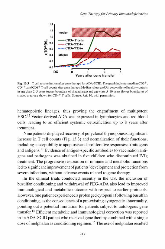
May 4, 2010 11:0 SPI-B903 9in x 6in b903-ch13
Gene Therapy for Primary Immunodeficiencies
Fig. 13.3 T cell reconstitution after gene therapy for ADA-SCID. The graph indicates median CD3+,CD4+, and CD8+ T cell counts after gene therapy. Median values and 5th percentiles of healthy controlsin age class 2–5 years (upper boundary of shaded area) and age class 5–10 years (lower boundaries ofshaded area) are shown for CD4+ T cells. Source: Ref. 10, with permission.
hematopoietic lineages, thus proving the engraftment of multipotentHSC.12 Vector-derived ADA was expressed in lymphocytes and red bloodcells, leading to an efficient systemic detoxification up to 8 years aftertreatment.
Nine patients displayed recovery of polyclonal thymopoiesis, significantincrease in T cell counts (Fig. 13.3) and normalization of their functions,including susceptibility to apoptosis and proliferative responses to mitogensand antigens.13 Evidence of antigen-specific antibodies to vaccination anti-gens and pathogens was obtained in five children who discontinued IVIgtreatment. The progressive restoration of immune and metabolic functionsled to significant improvement of patients’development and protection fromsevere infections, without adverse events related to gene therapy.
In the clinical trials conducted recently in the US, the inclusion ofbusulfan conditioning and withdrawal of PEG-ADA also lead to improvedimmunological and metabolic outcome with respect to earlier protocols.However, one patient experienced a prolonged cytopenia following busulfanconditioning, as the consequence of a pre-existing cytogenetic abnormality,pointing out a potential limitation for patients subject to autologous genetransfer.14 Efficient metabolic and immunological correction was reportedin an ADA-SCID patient who received gene therapy combined with a singledose of melphalan as conditioning regimen.15 The use of melphalan resulted
217
May 4, 2010 11:0 SPI-B903 9in x 6in b903-ch13
A Guide to Human Gene Therapy
in a lower proportion of transduced granulocytes and B cells, suggestingthat type of chemotherapy may influence HSC engraftment.
Results of the above clinical trials indicate that gene therapy with non-myeloablative conditioning is associated with clinical benefit and is now anoption to be considered for allADA-SCID patients lacking an HLA-identicalsibling donor. None of the patients enrolled in the different clinical trialsworldwide showed clonal proliferation or adverse events related to genetransfer, indicating that, as opposed to SCID-X1 (see below), gene therapyfor ADA-SCID has a favorable risk-benefit profile.16,17 The development ofself-inactivating vectors, such as lentiviral vectors might further improvethe safety of this approach.18,19
3. X-linked Severe Combined Immunodeficiency(SCID-X1)
X-linked SCID (SCID-X1) is the most common form of severe combinedimmunodeficiency, accounting for around 60% of all cases. SCID-X1 iscaused by mutations in the common cytokine receptor gamma chain (γc)gene, which forms part of the receptors for IL-4, IL-7, IL-9, IL-15 and IL-21.In the absence of γc-mediated signaling, the differentiation and activationof immune cells is compromised, resulting in lack of T and NK cells as wellas functionally impaired B cells.1
The successful correction of γc-deficient mice by gene therapy lead A.Fischer and colleagues at Hôpital Necker (Paris) to assess the potentialof gene therapy for the treatment of this disease. The protocol was basedon ex vivo retrovirally-mediated γc gene transfer into autologous CD34+
bone marrow cells.20 Progenitor cells were then re-infused into patients,who lacked a matched sibling or unrelated donors, in the absence of anypreparative conditioning. A similar study was initiated by A. Thrasher andcolleagues at Great Hormond Street Hospital (London).21 This protocolvaried from the French study essentially for the use of GALV-pseudotypedinstead of a conventional amphotropic MLV-based vector. Overall, 18 out ofthe 20 SCID-X1 patients enrolled in both clinical trials benefited from genetherapy. Within 3 months after gene therapy, T cell counts reached normallevel in nearly all the patients. Around 10 years of follow-up available forthe first treated patients has provided evidence for still active thymopoiesis,
218

May 4, 2010 11:0 SPI-B903 9in x 6in b903-ch13
Gene Therapy for Primary Immunodeficiencies
with broadly diversified TCR repertoire.16 The evidence that virtually allT and NK cells but fewer B cells and myeloid cells carried the transgene(1–2 trangene copies per cell) clearly demonstrates that γc expression con-fers on T and NK progenitors a strong selective growth advantage. Neverthe-less, several patients discontinued immunoglobulin infusions and showedantigen specific responses following vaccination.
When gene therapy was attempted in older SCID-X1 patients, thisresulted in partial efficacy or inability to recover T cell immunity dueto failure of re-activating thymopoiesis.22,23 The decline of thymic outputcould be likely related to the clinical history of chronic infection, GVHDor to the age of the patients at the time of intervention. This most prob-ably reflects a crucial time-dependency of the capacity to reinitiate thy-mopoiesis, since the prolonged absence of thymocyte and thymic epithelialcell interaction can cause irreversible thymic hypoplasia and disaggrega-tion. Thus, gene therapy should be considered as early as possible afterdiagnosis.
From a safety point of view, the trials for SCID-X1 represented a majorsetback for the safety of gene therapy. Four patients in the French trial and1 in the English trial developed leukemia 3–6 years after treatment. Thisleukemia-like disease was a result of vector-mediated up-regulation of hostcellular oncogenes by the MLV LTR.16,17,24 Gamma-retroviral vectors natu-rally insert into active genes and detailed molecular analysis demonstrates apreference to insert into and around the transcriptional start site. By insertinginto such regions, the strong enhancer elements found in the viral LTR cantransactivate nearby and neighboring promoters leading to aberrant geneexpression. In the SCID-X1 clinical trials the development of leukemia orgenomic instability are directly related to such mechanisms.16,17,24 In thesecases initial aberrant expression of an oncogene (e.g. mainly LMO2) ledto proliferation of specific clones, whereas additional genetic events even-tually resulted in leukemic transformation. Chemotherapy led to sustainedremission in 4 of the 5 cases of T cell leukemia, but one patient died likelydue to refractory leukemia.16,17
Several possible explanations may account for the differences betweenthe ADA-SCID and the SCID-X1 GT trials. First, ADA is a constitutivelyexpressed enzyme of purine metabolism while IL2RG is a signal trans-ducing receptor chain which is upregulated upon T cell activation and
219
May 4, 2010 11:0 SPI-B903 9in x 6in b903-ch13
A Guide to Human Gene Therapy
induces cell proliferation. Transgenic IL2RG may be expressed at inap-propriate levels in different stages of differentiation. It should be notedthat the kinetics of T cell reconstitution was substantially different, beingthe one of ADA-SCID more slow and progressive than the one observedfor the SCID-X1 trials. Furthermore, the SCID-X1 background may favorthe accumulation of mutations in progenitor cells blocked in their devel-opment or in gene corrected thymocytes following their rapid expansionoccurring after restoration of IL2RG expression. Finally, combinatorialmechanisms leading to leukemogenesis may result from the interaction ofIL2RG with cellular proto-oncogenes activated by vector insertions, suchas LMO2.
The occurrence of these severe adverse events lead to the develop-ment of a new area of research focused on virus-mediated oncogenesis andsignificantly contributed to the current safety standards for gene therapyvectors. Most efforts to improve safety and efficiency for SCID GT havefocused on the development of new self-inactivated (SIN) gammaretrovi-ral and lentiviral vectors, which carry a reduced intrinsic risk. Potentialcandidates to future clinical trials include SIN-gammaretroviral vectorsdriven by the human EF1-alpha promoter,25 and SIN-Lentiviral Vectors(SIN-LV) based on HIV-1 incorporating the ubiquitously acting chromatinopening element (UCOE).26 These vectors induced stable γc gene expres-sion and fully restored lymphoid differentiation and functions. In summary,SCID-X1 clinical trials were of major importance to verify the potential ofgene therapy as a real alternative for PID therapy and to define the require-ment of more efficient and safer vectors.
4. Gene Therapy for Other SCIDs
Here we will discuss the most recent advances in the application of somaticcell gene therapy to the correction of various molecular defects responsiblefor other types of SCIDs.
4.1 V(D)J Recombination Defects
To obtain the necessary level of diversity, B and T cell receptors are createdby recombining preexisting gene segments, a process calledV(D)J recombi-nation. In the first steps of the process, the two lymphocyte-specific proteins
220

May 4, 2010 11:0 SPI-B903 9in x 6in b903-ch13
Gene Therapy for Primary Immunodeficiencies
RAG1 and RAG2, collaborate to join the various variable (V), diversity (D),and joining (J) segments.Additionally, a large number of proteins participatein DNA repair during the late phases of V(D)J recombination, such asKu70, Ku80, DNA Ligase IV,Artemis, XRCC4, TdT or the newly identifiedCernunnos/XLF.27 V(D)J recombination defects comprise 30% of humanT−B−NK+ SCIDs. They are caused by mutations in the RAG1 or RAG2genes resulting in an early block of V(D)J recombination or mutations inthe Artemis gene leading to a general defect of nonhomologous end-joiningrepair.28 The latter defect, referred to as radiosensitive-SCID (RS-SCID),is characterized, in addition to the absence of mature T and B lympho-cytes, by an increased cellular sensitivity to ionizing radiation. Moreover,hypomorphic mutations in the RAG1/2 or Artemis genes may generate adifferent phenotype, named Omenn syndrome (OS), which is characterizedby residual T and B cell differentiation and function.29 Due to overwhelminginfections and organ damage, if untreated, patients have a dismal progno-sis. Current treatment requires both immune suppression and allogeneicHSC transplantation. However this is only partially successful, particu-larly for RS-SCID, in which the radiosensitive characteristic of the diseasemay preclude the use of standard conditioning regimens.2 Consequently,gene therapy is considered a promising alternative strategy for thesediseases.
Gene transfer approaches to correct V(D)J recombination defects havebeen initially explored in RAG1 or RAG2-deficient murine models. MLV-based gammaretroviral vectors have been shown to reconstitute RAG2-deficient mice effectively in the absence of detectable toxicity.30 Similarefforts to correct RAG1 deficiency were successful but required high vectorcopy numbers to be efficient.31
More recently, studies have employed SIN-LVs in order to avoid poten-tial side effects associated with the use of LTR-driven RVs. A single copyof an Artemis-expressing LV enabled long-term correction of both T- andB-cell deficiencies in recipient mice.32 As a further step towards clini-cal application, it has been demonstrated that LV-corrected BM CD34+
cells isolated from Artemis-deficient patients are able to promote func-tional B-cell development in SCID mice. Using the same transduction pro-cedure but a slightly different vector, correction of the B-cell defect inRAG1-deficient patients was much less efficient, suggesting that further
221

May 4, 2010 11:0 SPI-B903 9in x 6in b903-ch13
A Guide to Human Gene Therapy
improvements in transgene expression via modified vector design must beconsidered for this pathology.33
4.2 Purine Nucleoside Phosphorylase (PNP) Deficiency
Like ADA, Purine nucleoside phosphorylase (PNP) is an important enzymefor the degradation of purine nucleosides into uric acid or their salvage intonucleic acids.5 PNP-deficient patients suffer from severe T cell immunedysfunction, increased susceptibility to infections, failure to thrive and deathin the first years of life, which may be delayed with supportive treatments.5
B cells are less sensitive than T cells to the metabolic abnormalities becauseof the different metabolic pathway utilized.5 HSC transplants from closelymatched donors have corrected the immune abnormalities in some PNPdeficient patients. However, HLA-matched donors are not readily available,and transplants using other donors are frequently complicated by procedure-related morbidity, development of graft-versus-host disease or graft loss.34
Hopes for successful gene therapy for PNP deficiency have been sup-ported by the success of gene therapy for ADA-SCID. Promising resultshave recently been achieved using a SIN HIV-1-based LV vector encod-ing for human PNP under the control of the human EF1-alpha promoter,which provided stable transgene expression in both human and murine cells.Importantly, after HSC gene therapy in the murine model, PNP activityincreased over time suggesting a selective advantage for gene correctedcells. Although this study provided proof-of-principle that LVs could beused for PNP gene therapy, the observed effect was only transient, so thatbetter BM engraftment and improved gene expression will be required tofulfill the potential of gene therapy for PNP deficiency in the future.35
4.3 Janus Kinase 3 (Jak3) Deficiency
Jak3 belongs to a family of signal transducing tyrosine kinases primarilyexpressed in hematopoietic cells, it associates with γc and is required forsignal transduction by γc-containing receptors.36 Therefore, the phenotypearising from rare mutations in the Jak3 gene is nearly identical to that ofSCID-X1,37 which is characterized by absence of circulating T and nat-ural killer (NK) cells with normal numbers of poorly functioning B cells
222
May 4, 2010 11:0 SPI-B903 9in x 6in b903-ch13
Gene Therapy for Primary Immunodeficiencies
(T-B+NK-). Different in vitro and in vivo preclinical studies have demon-strated that retroviral Jak3 gene transfer can correct developmental andfunctional defects in hematopoietic cells.38 Jak3 gene correction of bonemarrow CD34+ cells was attempted in a single Jak3-SCID patient, whofailed HSC transplantation, without achieving immune reconstitution.39 InJak3 deficient mice, direct intrafemoral injection of retroviral vectors hasbeen postulated as an alternative to avoid deleterious effects of ex-vivomanipulation.40 Although not all treated mice were fully reconstituted, thisstudy demonstrated the feasibility of direct vector injection.
4.4 IL-7R Deficiency
The finding that mutations of γc and Jak3 led to SCID suggested thatother disruptions in this pathway might result in similar disease pheno-types. Cytokines that utilize γc were logical candidates, such as mutationsof IL-7 or the IL-7 receptor α chain (IL-7Rα). Mutations in IL-7Rα weresubsequently identified in patients with autosomal recessive SCID, under-lying about 10% of SCIDs.1 IL-7Rα deficiency in humans lead to SCIDmanifested by isolated absence of T cells (T-B+NK+), underlining theimportance of IL-7Rα-dependent signaling for T cell, but not B or NK cell,development.1 IL-7 plays a critical role in the development and differentia-tion of thymocytes, and survival of both naïve and mature T cells.41 Unlikeits human counter-part, mouse IL-7 is also required for the development ofB lymphocytes.41 In IL-7Rα deficient mice gene transfer using a retroviralvector encoding for the murine IL-Rα gene allowed the reconstitution ofT cell lineage in the thymus and periphery, while B cell recovery remainedpartial.42 Remarkably, upon IL-7 stimulation, the ectopic expression of IL-7Rα on multipotent hematopoietic progenitors has been shown to inducea striking in vitro expansion of myeloid cells.42 This evidence would sug-gest the requirement for more lineage-specific or autologous promoters forfuture clinical applications.
4.5 Zeta Associated 70 kDa Phosphoprotein(ZAP-70) Deficiency
Zap-70 deficiency is a rare autosomal recessive form of SCID, characterizedby a selective inability to produce CD8+ T cells and a signal transduction
223
May 4, 2010 11:0 SPI-B903 9in x 6in b903-ch13
A Guide to Human Gene Therapy
defect in peripheral CD4+ cells. Peripheral T cells from affected patientsdemonstrate defective T cell signaling and abnormal thymic ontogenycaused by inherited mutations in the TCR-associated protein tyrosinekinase (PTK) ZAP-70. SCID due to ZAP-70 deficiency contributed the firstevidence, that PTKs, particularly ZAP-70, are required for normal humanT cell development and function.1
The feasibility of a HSC-based gene therapy strategy has been assessedonly in preclinical mouse models, in which the engraftment of retrovirallytransduced bone marrow progenitor cells allowed the correction of T cell-defect without affecting the differentiation and function of B cells.43 Morerecently alternative gene delivery approaches were tested in order to lowerthe risk of insertional mutagenesis in HSCs and other hematopoietic pre-cursors that physiologically do not express and thus require ZAP-70. Newdelivery approaches were based on direct intrathymic injection of a T cell-specific LV or thymic electro-gene transfer of plasmid DNA both encodingfor ZAP-70 into deficient mice.44,45 Preliminary results indicated long-termreconstitution of T cell differentiation and function with a diversified T cellreceptor repertoire.
5. Wiskott-Aldrich-Syndrome (WAS)
WAS is a severe X-linked immunodeficiency caused by mutations in thegene encoding for WASP, a key regulator of signaling and cytoskele-tal reorganization in hematopoietic cells. Mutations in WASP result in awide spectrum of clinical manifestations ranging from the relatively mildX-linked thrombocytopenia (XLT) to the classic full-blown WAS phenotypecharacterized by thrombocytopenia, immunodeficiency, eczema, and highsusceptibility to developing tumors and autoimmune manifestations. Thelife expectancy of patients affected by severe WAS is reduced, unless theyare successfully cured by HSC transplantation. Since many patients lack acompatible bone marrow donor, the administration of WAS gene correctedautologous HSCs was considered an alternative therapeutic approach.46
Increasing evidence suggests that WASP-expressing cells have a prolif-erative or survival advantage over their WASP-deficient counterparts. Aselective accumulation of revertant T cells has been observed in severalWAS patients who underwent spontaneous somatic reversion in a lymphoid
224

May 4, 2010 11:0 SPI-B903 9in x 6in b903-ch13
Gene Therapy for Primary Immunodeficiencies
progenitor. Ideally, this selective advantage will facilitate the efficacy ofgene therapy.46
Preclinical studies for WAS gene therapy were initially based on gam-maretroviruses and then moved to LV vector approaches in order to increaseefficacy and safety. Protocols for the application of gene therapy for WASare currently under development at several centers in Europe. The first genetherapy study has been initiated in 2007 in Germany to assess the fea-sibility, toxicity, and potential benefit of HSC gene therapy with MLV-derived retroviral vector encoding WASp under the control of a viralpromoter.47 Preliminary results from the German trial seem to be encour-aging, but the safety profile of the retroviral vector still has to be carefullyevaluated.
The LV-based approach included native regulatory sequences to targetthe expression of the therapeutic WAS transgene physiologically to thehematopoietic system. This vector was shown to be effective for correctionof multiple cellular defects in vitro and in vivo.48,49 In theWAS mouse model,engraftment of gene corrected bone marrow HSC yielded long-term WASPexpression and the correction of immune, inflammatory and cytoskeletaldefects,49,50 without adverse events linked to gene transfer. This vector is indevelopment for clinical studies that are planned in several European coun-tries. Due to its design, the WAS-LV vector may have significant advantagesfor clinical application in humans in terms of natural gene regulation, andreduction in the potential for adverse mutagenic events.51
6. Chronic Granulomatous Disease
The encouraging results of the gene therapy trials for SCID demonstratedthe curative potential of gene transfer and represented a strong rationale forthe development of gene therapy protocols for other immunodeficiencies,such as CGD. This PID is an inherited disorder of innate immunity in whichphagocytic leukocytes are unable to generate microbicidal oxidants. Conse-quently, affected patients are susceptible to recurrent opportunistic bacterialand fungal infections, leading to the formation of chronic granulomas. CGDis caused by mutation or deletions in any of four genes encoding for essentialsubunits of the phagocytic nicotinamide dinucleotide phosphate (NADPH)oxidase complex (gp91phox, p22phox, p47phox and p67phox). NADPH
225

May 4, 2010 11:0 SPI-B903 9in x 6in b903-ch13
A Guide to Human Gene Therapy
oxidase, which is localized in the phagosomal and plasma membrane ofphagocytes, generates superoxide and related toxic oxygen metabolites tokill invading bacteria and fungi by respiratory burst.
Approximately 70% of CGD patients carry defects in the X-linked geneencoding for gp91phox (X-CGD).52 Although lifelong prophylaxis reducedthe incidence of infection in CGD patients, the overall annual mortalityis still high (2–5%) and the success rate of HSC transplant is limited bygraft-versus-host disease (GVHD) and inflammatory flare-ups at infectioussites.52 Therefore CGD has been proposed as an ideal candidate for genetherapy, also considering that low levels of gene correction may be sufficientfor curative effects, since healthy female CGD carriers can have <10% ofthe normal levels of neutrophils.53
Although significant long-term correction has been obtained in murinemodels by retroviral-mediated gene transfer into HSC in combination withnonmyeloablative conditioning or bone marrow ablation,54,55 similar resultshave not been achieved in unconditioned human CGD patients.56 In the USclinical trial, a total of ten patients were enrolled, five with a p47phoxdeficiency and five with gp91phox-deficient CGD. The autologous CD34+
cells mobilized from peripheral blood were transduced with a pseudotypedMLV retrovirus encoding for p47phox or gp91phox, respectively. Eightpatients demonstrated very low levels of reconstituted neutrophils for up to14 months after treatment.56
A clinical trial for X-CGD using a RV has recently been reported byM. Grez and colleagues. Two X-CGD patients were treated with ex vivoRV gene transfer of gp91phox into mobilized peripheral blood. Importantlynon-myeloablative conditioning was administered before the infusion ofgenetically modified HSCs. After a period of myelosuppression, cell countsrecovered gradually and patients recovered from severe infections.57 How-ever, an unexpected clonal expansion of transduced myeloid cells, occurredat approximately 5 months post-treatment. Vector transduced myelopoiesisexpanded 3 to 5 fold in both patients due to activating integrations in the zincfinger transcription factor homologs MDS1/EVI1, PRDM16 or SETBP1.Both patients subsequently developed myelodysplasia with Monosomy 7.One patient died 2.5 years after gene therapy as a result of a severe bac-terial infection.57 Though the number of transduced cells remained high atthe time of death, gp91phox expression was almost completely diminished
226
May 4, 2010 11:0 SPI-B903 9in x 6in b903-ch13
Gene Therapy for Primary Immunodeficiencies
due to the predominance of a new myeloid clone with very little oxidasefunction. Subsequent studies of the proviral SFFV LTR demonstrated themethylation of CpG dinucleotides within the promoter but not within theSFFV enhancer sequence. Despite these adverse events, it is clear that ther-apeutic benefit, although transient, can be achieved by gene therapy forCGD.4
Several factors could account for the clinical outcome observed in theCGD trial. The use of a vector with SFFV LTR sequences, which containspotent enhancer elements for gene expression, favored the activation ofspecific genes and led to the observed clonal expansion. The case for CGDis different from SCID due to the absence of a selective advantage for genecorrected cells, which facilitates the progressive loss of transgene expressiondue to promoter methylation.
Future clinical trials will require superior vector design to improve safetyof gene transfer into HSC while ensuring adequate transgene expression inmyeloid lineages. SIN lentiviral vectors encoding gp91phox under constitu-tive promoters have been generated, achieving significant restoration of theoxidase enzyme activity and neutrophil counts in a human/mouse xenograftmodel.58
7. Conclusions and Outlook
Allogeneic HSC transplantation has proved to be a successful treatmentfor PID but it is limited by complications in mismatched donor settings. Inthe last decade, gene therapy has been developed as a successful alternativestrategy. Results from clinical trials employing gene corrected HSC showedimmunological improvement and clinical benefit in patients affected byADA-deficient SCID, SCID-X1, and CGD. The introduction of a low inten-sity conditioning regimen has been identified as a crucial factor in achievingadequate engraftment of HSC and therapeutic levels of transgenes. How-ever, the occurrence of serious complications in SCID-X1 and CGD trialshave highlighted the risks of insertional mutagenesis with the retroviral vec-tor technology. These findings have stimulated a large number of studiesinto mechanisms and ways to prevent this activity by the development ofnovel vector technology. Several clinical trials are being developed to treatvarious forms of PID including SCID and WAS.
227
May 4, 2010 11:0 SPI-B903 9in x 6in b903-ch13
A Guide to Human Gene Therapy
References
1. Geha RS, et al. (2007). Primary immunodeficiency diseases: An update from the Inter-national Union of Immunological Societies Primary Immunodeficiency Diseases Clas-sification Committee. J Allergy Clin Immunol 120: 776–794.
2. Neven B, et al. (2009). Long-term outcome after haematopoietic stem cell transplanta-tion of a single-centre cohort of 90 patients with severe combined immunodeficiency:Long-term outcome of HSCT in SCID. Blood 113: 4114–4124.
3. Antoine C, et al. (2003). Long-term survival and transplantation of haemopoietic stemcells for immunodeficiencies: Report of the European experience 1968–1999. Lancet361: 553–560.
4. Kohn, DB (2008). Gene therapy for childhood immunological diseases. Bone MarrowTransplant 41: 199–205.
5. Hirschorn R, Candotti, F (2006). Immunodeficiency Due to Defects of PurineMetabolism In: Ochs H, C Smith, J Puck (eds). Primary immunodeficiency diseases.Oxford University Press: Oxford. pp. 169–196.
6. Booth C, et al. (2007). Management options for adenosine deaminase deficiency:Proceedings of the EBMT satellite workshop (Hamburg, March 2006). Clin Immunol123: 139–147.
7. Kohn DB, et al. (1995). Engraftment of gene-modified umbilical cord blood cells inneonates with adenosine deaminase deficiency. Nat Med 1: 1017–1023.
8. Bordignon C, et al. (1995). Gene therapy in peripheral blood lymphocytes and bonemarrow for ADA- immunodeficient patients. Science 270: 470–475.
9. Aiuti A, et al. (2002). Correction of ADA-SCID by stem cell gene therapy combinedwith nonmyeloablative conditioning. Science 296: 2410–2413.
10. Aiuti A, et al. (2009). Gene therapy for immunodeficiency due to adenosine deaminasedeficiency. N Engl J Med 360: 447–458.
11. Aiuti A, et al. (2009). Hematopoietic stem cell gene therapy for adenosine deaminasedeficient-SCID. Immunol Res. 4: 150–159.
12. Aiuti A, et al. (2007). Multilineage hematopoietic reconstitution without clonal selec-tion in ADA-SCID patients treated with stem cell gene therapy. J Clin Invest 117:2233–2240.
13. Cassani B, et al. (2008). Altered intracellular and extracellular signaling leads toimpaired T-cell functions in ADA-SCID patients. Blood 111: 4209–4219.
14. Engel BC, et al. (2007). Prolonged pancytopenia in a gene therapy patient with ADA-deficient SCID and trisomy 8 mosaicism: A case report. Blood 109: 503–506.
15. Gaspar HB, et al. (2006). Successful reconstitution of immunity in ADA-SCID by stemcell gene therapy following cessation of PEG-ADA and use of mild preconditioning.Mol Ther 14: 505–513.
16. Hacein-Bey-Abina S, et al. (2008). Insertional oncogenesis in 4 patients after retrovirus-mediated gene therapy of SCID-X1. J Clin Invest 118: 3132–3142.
17. Howe SJ, et al. (2008). Insertional mutagenesis combined with acquired somatic muta-tions causes leukemogenesis following gene therapy of SCID-X1 patients. J Clin Invest118: 3143–3150.
228

May 4, 2010 11:0 SPI-B903 9in x 6in b903-ch13
Gene Therapy for Primary Immunodeficiencies
18. Mortellaro A, et al. (2006). Ex vivo gene therapy with lentiviral vectors rescues adeno-sine deaminase (ADA)-deficient mice and corrects their immune and metabolic defects.Blood 108: 2979–2988.
19. Carbonaro DA, et al. (2008). Neonatal bone marrow transplantation of ADA-deficientSCID mice results in immunological reconstitution despite low levels of engraftmentand an absence of selective donor T lymphoid expansion. Blood 111: 5745–5754.
20. Hacein-Bey-Abina S, et al. (2002). Sustained correction of X-linked severe combinedimmunodeficiency by ex vivo gene therapy. N Engl J Med 346: 1185–1193.
21. Gaspar HB, et al. (2004). Gene therapy of X-linked severe combined immunodeficiencyby use of a pseudotyped gammaretroviral vector. Lancet 364: 2181–2187.
22. Chinen J, et al. (2007). Gene therapy improves immune function in preadolescents withX-linked severe combined immunodeficiency. Blood 110: 67–73.
23. Thrasher AJ, et al. (2005). Failure of SCID-X1 gene therapy in older patients. Blood105: 4255–4257.
24. Hacein-Bey-Abina S, et al. (2003).A serious adverse event after successful gene therapyfor X-linked severe combined immunodeficiency. N Engl J Med 348: 255–256.
25. Thornhill SI, et al. (2008). Self-inactivating gammaretroviral vectors for gene therapyof X-linked severe combined immunodeficiency. Mol Ther 16: 590–598.
26. Zhang F, et al. (2007). Lentiviral vectors containing an enhancer-less ubiquitouslyacting chromatin opening element (UCOE) provide highly reproducible and stabletransgene expression in hematopoietic cells. Blood 110: 1448–1457.
27. Sobacchi C, Marrella V, Rucci F, Vezzoni P, Villa, A (2006). RAG-dependent primaryimmunodeficiencies. Hum Mutat 27: 1174–1184.
28. Moshous D, et al. (2001). Artemis, a novel DNA double-strand break repair/V(D)Jrecombination protein, is mutated in human severe combined immune deficiency. Cell105: 177–186.
29. Villa A, Notarangelo LD, Roifman, CM (2008). Omenn syndrome: Inflammationin leaky severe combined immunodeficiency. J Allergy Clin Immunol 122: 1082–1086.
30. Yates F, et al. (2002). Gene therapy of RAG-2-/- mice: sustained correction of theimmunodeficiency. Blood 100: 3942–3949.
31. Lagresle-Peyrou C, et al. (2006). Long-term immune reconstitution in RAG-1-deficientmice treated by retroviral gene therapy:A balance between efficiency and toxicity. Blood107: 63–72.
32. Mostoslavsky G, FabianAJ, Rooney S,Alt FW, Mulligan, RC (2006). Complete correc-tion of murine Artemis immunodeficiency by lentiviral vector-mediated gene transfer.Proc Natl Acad Sci USA 103: 16406–16411.
33. Lagresle-Peyrou C, et al. (2008). Restoration of human B-cell differentiation into NOD-SCID mice engrafted with gene-corrected CD34+ cells isolated fromArtemis or RAG1-deficient patients. Mol Ther 16: 396–403.
34. Grunebaum E, et al. (2006). Bone marrow transplantation for severe combined immunedeficiency. Jama 295: 508–518.
35. Liao P, Toro A, Min W, Lee S, Roifman CM, Grunebaum E (2008). Lentivirus genetherapy for purine nucleoside phosphorylase deficiency. J Gene Med 10: 1282–1293.
229
May 4, 2010 11:0 SPI-B903 9in x 6in b903-ch13
A Guide to Human Gene Therapy
36. Russell SM, et al. (1995). Mutation of Jak3 in a patient with SCID: essential role ofJak3 in lymphoid development. Science 270: 797–800.
37. Macchi P, et al. (1995). Mutations of Jak-3 gene in patients with autosomal severecombined immune deficiency (SCID). Nature 377: 65–68.
38. Bunting KD, Lu T, Kelly PF, Sorrentino BP (2000). Self-selection by genetically mod-ified committed lymphocyte precursors reverses the phenotype of JAK3-deficient micewithout myeloablation. Hum Gene Ther 11: 2353–2364.
39. Gaspar HB, Thrasher AJ (2005). Gene therapy for severe combined immunodeficien-cies. Expert Opin Biol Ther 5: 1175–1182.
40. McCauslin CS, et al. (2003). In vivo retroviral gene transfer by direct intrafemoralinjection results in correction of the SCID phenotype in Jak3 knock-out animals. Blood102: 843–848.
41. Hofmeister R, KhaledAR, Benbernou N, Rajnavolgyi E, Muegge K, Durum, SK (1999).Interleukin-7: physiological roles and mechanisms of action. Cytokine Growth FactorRev 10: 41–60.
42. Jiang Q, Li WQ, Aiello FB, Klarmann KD, Keller JR, Durum SK (2005). Retroviraltransduction of IL-7Ralpha into IL-7Ralpha-/- bone marrow progenitors: Correction oflymphoid deficiency and induction of neutrophilia. Gene Ther 12: 1761–1768.
43. Otsu M, et al. (2002). Reconstitution of lymphoid development and function in ZAP-70-deficient mice following gene transfer into bone marrow cells. Blood 100: 1248–1256.
44. Adjali O, et al. (2005). In vivo correction of ZAP-70 immunodeficiency by intrathymicgene transfer. J Clin Invest 115: 2287–2295.
45. Irla M, et al. (2008). ZAP-70 restoration in mice by in vivo thymic electroporation.PLoS ONE 3: e2059.
46. Bosticardo M, Marangoni F, Aiuti A, Villa A, Roncarolo MG (2009). Recent advancesin understanding the pathophysiology of Wiskott-Aldrich syndrome. Blood 113: 6288–6295.
47. Boztug K, Dewey RA, Klein C (2006). Development of hematopoietic stem cell genetherapy for Wiskott-Aldrich syndrome. Curr Opin Mol Ther 8: 390–395.
48. Charrier S, et al. (2007). Lentiviral vectors targeting WASp expression to hematopoieticcells, efficiently transduce and correct cells fromWAS patients. Gene Ther 14: 415–428.
49. Marangoni F, et al. (2009). Evidence for Long-term Efficacy and Safety of Gene Ther-apy for Wiskott-Aldrich Syndrome in Preclinical Models. Mol Ther 17: 1073–1082.
50. Dupre L, et al. (2006). Efficacy of gene therapy for Wiskott-Aldrich syndrome using aWAS promoter/cDNA-containing lentiviral vector and nonlethal irradiation. Hum GeneTher 17: 303–313.
51. Galy A, Roncarolo MG, Thrasher AJ (2008). Development of lentiviral gene therapyfor Wiskott Aldrich syndrome. Expert Opin Biol Ther 8: 181–190.
52. Stasia MJ, Li XJ (2008). Genetics and immunopathology of chronic granulomatousdisease. Semin Immunopathol 30: 209–235.
53. Woodman RC, et al. (1995). A new X-linked variant of chronic granulomatous dis-ease characterized by the existence of a normal clone of respiratory burst-competentphagocytic cells. Blood 85: 231–241.
54. Dinauer MC, Li LL, Bjorgvinsdottir H, Ding C, Pech N (1999). Long-term correctionof phagocyte NADPH oxidase activity by retroviral-mediated gene transfer in murineX-linked chronic granulomatous disease. Blood 94: 914–922.
230

May 4, 2010 11:0 SPI-B903 9in x 6in b903-ch13
Gene Therapy for Primary Immunodeficiencies
55. Sadat MA, et al. (2003). Long-term high-level reconstitution of NADPH oxidaseactivity in murine X-linked chronic granulomatous disease using a bicistronic vectorexpressing gp91phox and a Delta LNGFR cell surface marker. Hum Gene Ther 14:651–666.
56. Malech HL, et al. (1997). Prolonged production of NADPH oxidase-corrected granu-locytes after gene therapy of chronic granulomatous disease. Proc Natl Acad Sci USA94: 12133–12138.
57. Ott MG, et al. (2006). Correction of X-linked chronic granulomatous disease by genetherapy, augmented by insertional activation of MDS1-EVI1, PRDM16 or SETBP1.Nat Med 12: 401–409.
58. Naumann N, et al. (2007). Simian immunodeficiency virus lentivector corrects humanX-linked chronic granulomatous disease in the NOD/SCID mouse xenograft. GeneTher 14: 1513–1524.
231

May 4, 2010 11:0 SPI-B903 9in x 6in b903-ch13
This page intentionally left blankThis page intentionally left blank
May 4, 2010 11:0 SPI-B903 9in x 6in b903-ch14
Chapter 14
Gene Therapy for Hemophilia
David Markusic, Babak Moghimi and Roland Herzog∗
HemophiliaA and B are X-linked bleeding disorders resulting from a defi-ciency in coagulation factor VIII (F.VIII) or factor IX (F.IX), respectively.Patients experience spontaneous bleeding episodes into soft tissues andjoints, which is managed by intravenous administration of recombinantcoagulation factor protein. Hemophilia is well suited for correction bygene transfer, providing prophylactic treatment. As little as 1-2% of nor-mal circulating levels of coagulation factor can provide partial correctionof disease, no regulation of gene expression is required, and therapeu-tic efficacy is easily evaluated. Although F.VIII and F.IX are naturallysynthesized in the liver, many cell types are capable of synthesizing bio-logically active factor. Numerous animal models have demonstrated long-term expression and disease correction using different target tissues andvectors. Viral gene transfer in hemophilia B dogs resulted in therapeuticexpression for at least 8 years, while transient therapeutic expression hasbeen demonstrated in clinical trials. Future investigations will attempt toachieve long-term expression in humans using modified protocols.
1. Introduction
Hemophilia A and B are X-linked bleeding disorders, which result froma deficiency of F.VIII or F.IX, respectively, in the coagulation cascade(Fig. 14.1). Severe disease is defined as <1% factor activity, whereas1 to 5% and >5% of normal are defined as moderate and mild disease,respectively.1 The incidence of hemophilia A is 1 in 5,000 live male births
∗Correspondence: Department of Pediatrics, Div. Cellular and Molecular Therapy, University of Florida,Gainesville, Florida.E-mail: [email protected]
233

May 4, 2010 11:0 SPI-B903 9in x 6in b903-ch14
A Guide to Human Gene Therapy
Fig. 14.1 Coagulation cascade.
with two-thirds of affected patients having the severe form of the disease.In contrast, almost one-half of individuals with hemophilia B (1 in 25,000–30,000 births) have F.IX levels above 1 percent (moderate or mild disease).The most common sites of bleeding are into joints, muscles, and the gas-trointestinal tract. Hemarthrosis–bleeding into the joints — is the primarycause of morbidity in hemophilic patients resulting in chronic pain and phys-ical debilitation.2 Intracranial hemorrhage is a leading cause of mortality inhemophilic patients.
Standard treatment is periodical administration of recombinant coagu-lation factors to maintain therapeutic levels in moderate and severe patient’splasma. Patients have a risk of forming inhibitory antibodies (inhibitors),which occur in approximately 25 percent of patients with hemophilia Aand 3 to 5 percent of those with hemophilia B. These inhibitors com-plicate bleeding episodes because of diminished responsiveness to factorconcentrates.3,4 The predilection for patients with severe disease is con-sistent with observations that inhibitors primarily occur in patients with
234
May 4, 2010 11:0 SPI-B903 9in x 6in b903-ch14
Gene Therapy for Hemophilia
large deletions and stop mutations, compared to small deletions or mis-sense mutations.5 Hemophilia patients with high levels of inhibitor requiretreatment with agents such as prothrombin complex concentrates or recom-binant factor VIIa, which bypass the intrinsic pathway (Fig. 14.1). Long-term management of hemophilic patients with inhibitors requires immunetolerance induction (ITI) via the administration of repetitive high doses ofF.VIII or F.IX with or without immunosuppressive therapy, which are highlyexpensive protocols.
2. Limitations of Hemophilia Treatment WithCoagulation Factor Concentrates or RecombinantCoagulation Factors
Treatment regimens for hemophilic patients consist of either prophylaxis ortreatment on demand, where the former is recommended to be the standardof care by the National Hemophilia Foundation, the World Federation ofHemophilia, and theWorld Health Organization.6 Due to the high costs asso-ciated with prophylactic treatment, however, many patients are treated ondemand, which can result in progressive joint damage over time. In the past,there was a significant risk for the transmission of blood borne pathogens(HBV, HCV, and HIV) in contaminated plasma derived factor concentrates.Careful screening and preparation of plasma derived factor concentratesand the introduction of recombinant protein coagulation factors have virtu-ally eliminated the risk of blood borne pathogen transmission. Both F.VIIIand F.IX have a short biologically active half-life, which requires frequentinfusions to maintain therapeutic levels.
Hemophilia is a monogenic recessive disorder, caused by the loss offunction of a single gene and is well suited for correction by transfer of afunctional copy of the genes encoding F.VIII or F.IX. Both F.VIII and F.IXare secreted into the plasma and circulate in an inactive form (zymogen),and therefore do not require strict regulation for expression. Based on clin-ical data from patients treated with plasma derived or recombinant protein,circulating levels as low as 1% of normal promote a transition from severeto moderate disease, and levels reaching 5% of normal can lead to completeelimination of spontaneous bleeding episodes. Although F.VIII and F.IXare naturally expressed from the liver, other tissues can be used to express
235
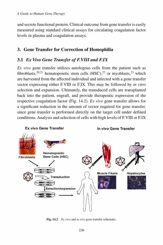
May 4, 2010 11:0 SPI-B903 9in x 6in b903-ch14
A Guide to Human Gene Therapy
and secrete functional protein. Clinical outcome from gene transfer is easilymeasured using standard clinical assays for circulating coagulation factorlevels in plasma and coagulation assays.
3. Gene Transfer for Correction of Hemophilia
3.1 Ex Vivo Gene Transfer of F.VIII and F.IX
Ex vivo gene transfer utilizes autologous cells from the patient such asfibroblasts,20,21 hematopoietic stem cells (HSC),22 or myoblasts,23 whichare harvested from the affected individual and infected with a gene transfervector expressing either F.VIII or F.IX. This may be followed by in vitroselection and expansion. Ultimately, the transduced cells are transplantedback into the patient, engraft, and provide therapeutic expression of therespective coagulation factor (Fig. 14.2). Ex vivo gene transfer allows fora significant reduction in the amount of vector required for gene transfer,since gene transfer is performed directly on the target cell under definedconditions. Analysis and selection of cells with high levels of F.VIII or F.IX
Fig. 14.2 Ex vivo and in vivo gene transfer schematic.
236
May 4, 2010 11:0 SPI-B903 9in x 6in b903-ch14
Gene Therapy for Hemophilia
expression allows for optimal therapeutic benefit following transplantation.From an immunological standpoint, gene transfer to off-target cells, suchas professional antigen presenting cells (APC), is prevented, thus greatlyreducing the risk of inducing a humoral and/or cytotoxic T lymphocyte(CTL) responses against F.VIII or F.IX. Additionally, complications fromactivation of an innate immune response to the vector may be reduced,thereby reducing the risk of inflammation directed against the site of genetransfer. Lastly, ex vivo gene transfer allows for efficient gene transfer if thepatient has neutralizing antibodies against the viral vector,24 or if the viralvector is inactivated in human sera.25
The choice of target cells is limited by the ability to culture the cells invitro while maintaining viability and functionality. Target cells should ide-ally be long-lived stem cells (e.g. hematopoietic stem cells) that can provideexpression in differentiated progeny, or differentiated cells such as fibrob-lasts, which can be efficiently and stably transduced and expanded in vitro.And lastly, the protocol requires efficient engraftment of gene-modified tar-get cells following transplantation in order to provide long-term therapeuticbenefit. From a practical standpoint, ex vivo gene transfer can be more laborintensive, require specialized facilities for the culturing and manipulationof the patient’s cells, and may subject patients to multiple minor surgicalprocedures, which poses a risk for patients with severe hemophilia.
3.2 In Vivo Gene Transfer of F.VIII and F.IX
The liver is a preferred target for gene transfer, because it represents thenatural site of F.VIII and F.IX biosynthesis (Fig. 14.2). However, sinceboth F.VIII and F.IX are secreted into the circulation, liver specific expres-sion is not required. The main limitation is that the intended target tis-sue should be capable of performing the post-translational modificationsrequired for expression of biologically active F.VIII and F.IX protein.26
Adult hemophilia patients with liver disease (e.g. hepatitis B and C fromprior exposure to contaminated coagulation factor concentrates) requirealternative tissues for gene transfer. Skeletal muscle has been extensivelyinvestigated as an alternative target tissue for adeno-associated viral (AAV)gene transfer of F.IX, and has been shown to be capable of expressingbiologically active protein (Fig. 14.2). The poor stability of F.VIII andpotential difficulties with reaching the circulation has been prohibitive
237

May 4, 2010 11:0 SPI-B903 9in x 6in b903-ch14
A Guide to Human Gene Therapy
in obtaining therapeutic levels of F.VIII from gene transfer to skeletalmuscle.27
Liver gene transfer is achieved by intravascular (IV) delivery via theperipheral circulation, or by infusion into the portal vein or hepatic artery.Ideally, the vector administration procedure should be minimally invasive,such as IV administration to a peripheral vein or an angiographic proce-dure for vector infusion into the hepatic artery. Hepatic gene transfer usingan AAV serotype 2 vector has led to long-term therapeutic expression ofF.VIII and F.IX in both murine and canine models of hemophilia. One drawback for administration of AAV2 vectors into a blood vessel is that humansare a natural host for AAV2, and there is a high incidence of neutralizingantibodies, which can block gene transfer.28
Intramuscular (IM) delivery of a gene transfer vector expressing F.IXhas the advantage of being minimally invasive to the patient, and localadministration limits biodistribution to other tissues. Although long-termF.IX expression has been obtained by IM injection of an AAV-F.IX vectorin murine and canine hemophilia models, this was limited to animal modelswith missense mutations.29,30 Additionally, a higher incidence of developinginhibitors was observed when using a high dose per injection site.31 Athigher doses, this may require too many injections to be practically appliedin a clinical treatment protocol. Because of these practical restrictions onIM viral administration in humans, vascular delivery techniques have beendeveloped to substantially enhance gene transfer to skeletal muscle, e.g.by performing isolated limb perfusion.32 This results in wide spread genetransfer to skeletal muscle from a single bolus injection of viral vector.Additionally, alternative AAV serotypes (AAV-1, -6, -7, 8-, and -9)33,34 witha higher tropism for muscle are currently being evaluated in animal modelsand human clinical trials for improved gene transfer efficiency.
4. AAV is a Preferred Gene Therapy Vector for In VivoGene Transfer to Correct of Hemophilia
For in vivo gene transfer, an ideal vector would provide stable long-termexpression in terminally differentiated cells of the liver or skeletal muscle.Of the commonly used gene transfer vectors, adenoviral, lentiviral vec-tors, and AAV vectors are capable of efficient and stable gene transfer to
238

May 4, 2010 11:0 SPI-B903 9in x 6in b903-ch14
Gene Therapy for Hemophilia
non-dividing cells. Early versions of adenoviral vectors and later guttedadenoviral vectors, which do not express any adenoviral proteins, proved tobe too immunogenic for in vivo gene transfer, excluding these vectors foruse in humans. Murine retroviral vectors require cell cycling for efficientgene transfer and have only shown practical use for ex vivo gene transferand administration to neonatal animals, where hepatocytes are still divid-ing. In addition, these vectors pose a higher risk for insertional mutagenesis.Lentiviral vectors — retroviral vectors that efficiently transduce nondivid-ing cells — are currently being evaluated in small and large animal modelswith some promising results. Safety concerns regarding insertional muta-genesis and feasibility of scaling up vector production to therapeutic dosesstill have to be addressed.
In contrast, AAV based vectors have led to long-term expression inmurine and canine hemophilia models and are considered to be lessimmunogenic with regard to innate immune responses, and these vectorsare comparatively inefficient in transducing antigen presenting cells (APC)compared to adeno- or lentiviral vectors. Different serotypes of AAV areavailable with different tissue tropism. The AAV2 serotype has been themost extensively studied for safety and efficacy in pre-clinical and clinicaltrials. The AAV vector genome predominantly remains in an episomal formin hepatocytes and muscle fibers, reducing the risk of insertional mutagene-sis. The main limitation of AAV vectors is a reduced packaging capacity ascompared to adenoviral or retroviral vectors. This has significantly limitedAAV based studies for F.VIII gene transfer due to the large cDNA cod-ing for the full length or B domain deleted (BDD) F.VIII variant.35 Thecoding sequence for BDD deleted F.VIII is ∼4.3 kb compared to 1.4 kbfor F.IX.
5. Immunological Considerations for Efficient F.IXGene Transfer
Gene transfer to an immunocompetent recipient can cause a variety ofimmune responses including an innate and adaptive immune response toviral structural proteins and the transgene product. Factors affecting theimmune response to gene transfer in hemophilia include host factors, genetransfer protocol/route of administration, and vector design and dosing.
239

May 4, 2010 11:0 SPI-B903 9in x 6in b903-ch14
A Guide to Human Gene Therapy
Some examples of host factors effecting gene transfer are immuno-profile/genetic modifiers, prior exposure to virus, nature of the underlying mutation,and coexisting inflammation.
The immune system detects viral gene transfer at different stages. Forexample, Toll-like receptors (TLRs) specifically activate downstream innateimmune response against both single and double stranded viral DNA andRNA. The ability of viral vectors to transduce APCs increases the chanceof being presented by MHC class I to cytotoxic T lymphocytes (CTLs),which can lead to elimination of transduced cells. Finally, the transgeneproduct can be presented on MHC class II and activate further adaptiveimmune responses, including antibody formation. Although many of theimmunogenic components of viral vectors have been removed to reducethese immune responses, fundamental innate responses to viral infectionstill exist and can prime the immune system for adaptive responses. Anotherimportant factor modulating immune responses to the transgene product isthe transfer protocol. Extensive investigations performed in animal modelshave indicated that vehicle characteristics such as route of administration,vector dose, transgene promoter, and preparation contaminants can influ-ence the type and potency of the immune reaction. Hepatic administrationof an AAV2 F.IX vector results in sustained expression and absence ofinhibitors to F.IX protein in hemophilic mice deficient in circulating F.IXprotein suggesting that expression in the liver may lead to tolerance.12 Stud-ies in different strains of mice showed that hepatic gene transfer of AAVvectors induced antigen specific CD4+CD25+ regulatory T cells, whichare both necessary and sufficient for tolerance induction to the transgeneproduct.36
In addition to potential immune responses to transgene product (F.VIIIor F.IX), two clinical trials with an AAV2 vector demonstrate that immuneresponses to the viral vector can affect long-term efficacy of gene ther-apy. AAV2 mediated gene transfer to skeletal muscle in mouse and doghemophilia models demonstrated both safety and efficacy for F.IX expres-sion, leading to the initiation of a human clinical trial for IM AAV2F.IX gene transfer.37 In the human trial, circulating levels of F.IX werebelow 1 percent due to restrictions on total administered viral dose. Allpatients receiving an IM injection experienced an increase of 2-3 logsin neutralizing antibody titer to viral capsid after vector administration,
240

May 4, 2010 11:0 SPI-B903 9in x 6in b903-ch14
Gene Therapy for Hemophilia
effectively preventing re-administration of virus to boost F.IX expressionlevels.38
Based on success in pre-clinical studies, a human trial with hepaticartery delivery of an AAV2 F.IX vector under the control of a liver specificpromoter was initiated. One subject in the highest dose cohort reached thera-peutic levels of F.IX (10–12%). However, 4 weeks after vector infusion, F.IXlevels began to fall, gradually returning to baseline (<1%) at 10 weeks.39
ELISPOT analysis of T cell responses in peripheral blood mononuclear cells(PBMC) isolated from a subsequently treated subject demonstrated IFN-γproduction in response to AAV2 capsid peptides, but not to F.IX peptides.39
The identified AAV2 capsid peptide epitopes were used to synthesize MHCclass I pentamers for the direct detection of capsid-specific CD8+ T cellpopulations in peripheral blood. Expansion of AAV capsid-specific CD8+
T cells in vitro and in vivo was demonstrated.40 These experimental resultsprovided further evidence that a CTL response directed against AAV capsid(presented on transduced hepatocytes) may have resulted in a decline of F.IXlevels. The time course of the observed spike in liver enzymes correlatedwith the time required for expansion and contraction of a capsid-specificCD8+ T cell population. The inconsistency between animal models andhumans in duration of expression may be explained by the fact that humansare natural hosts for AAV infection; and co-infection with immunogenichelper viruses, such as adenovirus, may generate memory T cells, thusleading to concomitant immune response after renewed exposure by genetransfer.
Several strategies are currently being evaluated to address immunologi-cal issues raised from the twoAAV2 clinical trials. Exon-shuffledAAV vari-ants or variants isolated from non human-primate species may avoid bothneutralizing antibodies against viral capsid and potential CTL responsesagainst viral capsid. Pre-treatment with transient immune suppression (IS)has been studied in rodents and primates. Regimens based on mycophe-nolate mofetil (MMF) and rapamycin resulted in long-term expression oftransgene without antibody formation against F.IX, confirming the safetyof AAV-2 gene transfer in the context of an IS regimen. There is increas-ing evidence that pre-treatment with rapamycin provides selective expan-sion of CD4+CD25+Foxp3+ regulatory T cells, and finally blocking ofco-stimulatory signals on T cells could lead tolerance in hemophilia gene
241
May 4, 2010 11:0 SPI-B903 9in x 6in b903-ch14
A Guide to Human Gene Therapy
transfer, pointing toward novel strategies for tolerance to the transgene andblockage of responses to the vector.41
6. Advancements from Small and Large Animal Modelsof Hemophilia
The availability of small murine and large canine animal models ofhemophilia (Table 14.1) have greatly facilitated the development and testingof gene transfer vectors. These animal models mimic some of the diseasephenotypes observed in human hemophilia patients and therefore are idealfor the evaluation of optimal gene transfer vectors, routes of administra-tion, and target tissues for correction of disease phenotype. Additionally,the murine and canine hemophilia disease models have provided a means toassess potential immunological responses to the transgene (F.VIII or F.IX)and viral vector, and develop methodologies to allow for long-term thera-peutic expression of F.VIII and F.IX.
6.1 Murine Hemophilia Models
Murine models for hemophilia A and B have allowed for the demonstrationefficacy of gene transfer both ex vivo and in vivo from retroviral, adenoviral,and AAV vectors and naked DNA for the correction of hemophilia. Genetransfer studies in mice showed that F.IX could be efficiently expressed fromskeletal muscle, providing evidence for the production of biologically activeF.IX protein from a tissue that does not normally synthesize F.IX. Restrictingexpression of F.IX to hepatocytes using an AAV vector with a liver specificpromoter results in induction of tolerance to F.IX in murine hemophiliamodels.12,42 The generation of murine F.IX knockout mice transgenic forcommonly observed human F.IX mutations has allowed for the investigationof the relative risk of inhibitor formation with CRIM — and CRIM + F.IXmutants following gene transfer.11,13
6.2 Canine Hemophilia Models
Canine models of hemophilia have been instrumental in the implementa-tion of human gene therapy clinical trials. Therapeutic gene transfer that iseffective in inbred mice often fails to be reproducible in humans. Dogs are
242
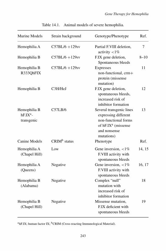
May 4, 2010 11:0 SPI-B903 9in x 6in b903-ch14
Gene Therapy for Hemophilia
Table 14.1. Animal models of severe hemophilia.
Murine Models Strain background Genotype/Phenotype Ref.
Hemophilia A C57BL/6 +129sv Partial F.VIII deletion,activity <1%
7
Hemophilia B C57BL/6 +129sv F.IX gene deletion,Spontaneous bleeds
8–10
Hemophilia BR333QhFIX
C57BL/6 +129sv Expressesnon-functional, crm+protein (missensemutation)
11
Hemophilia B C3H/HeJ F.IX gene deletion,spontaneous bleeds,increased risk ofinhibitor formation
12
Hemophilia BhF.IXa-transgenic
C57LB/6 Several transgenic linesexpressing differentnon-functional formsof hF.IXa (missenseand nonsensemutations)
13
Canine Models CRIMb status Phenotype Ref.
Hemophilia A(Chapel Hill)
Low Gene inversion, <1%F.VIII activity withspontaneous bleeds
14, 15
Hemophilia A(Queens)
Negative Gene inversion, <1%F.VIII activity withspontaneous bleeds
16, 17
Hemophilia B(Alabama)
Negative Complex “null”mutation withincreased risk ofinhibitor formation
18
Hemophilia B(Chapel Hill)
Negative Missense mutation,F.IX deficient withspontaneous bleeds
19
ahF.IX; human factor IX; bCRIM (Cross-reacting Immunological Material).
243
May 4, 2010 11:0 SPI-B903 9in x 6in b903-ch14
A Guide to Human Gene Therapy
much closer in terms of size to humans and provide a more realistic evalua-tion for the feasibility of scaling up vector production to provide therapeuticexpression. Therapeutic doses of AAV2 vectors obtained in canines closelypredicted doses that lead to transient therapeutic expression in humans.39
Since dogs have a longer lifespan then mice, long-term studies for efficacyand safety from gene transfer can be performed. Indeed, a single gene trans-fer of an AAV2 vector expressing canine F.IX to the liver has resulted instable expression and correction of major complications from hemophiliafor at least eight years,43 demonstrating the feasibility of long-term, per-haps even life-long correction from a single AAV vector administration inhumans.
7. Gene Therapy Trials for Hemophilia Past,Present, and Future
There have been six clinical gene therapy trials for the treatment of eitherhemophilia A or hemophilia B, summarized in Table 14.2. Ex vivo genetransfer to autologous fibroblasts with plasmid transfection of F.VIII21 orretroviral vector transduction of F.IX respectively45 followed by transplan-tation lead to a transient increase in F.VIII and F.IX levels to approxi-mately 4% of normal levels followed by a decline to pre-treatment levels.In vivo gene transfer of a retroviral vector44 or a gutless adenoviral vec-tor expressing F.VIII by peripheral intravenous delivery was not furtherpursued. Manno et al. investigated IM46 and hepatic artery39 delivery ofAAV2 vectors expressing F.IX. Long-term expression of F.IX was doc-umented in muscle biopsy samples from treated patients, but circulatinglevels of F.IX were sub-therapeutic. One patient in the high dose cohort ofthe hepatic artery trial had a transient therapeutic increase in F.IX expres-sion as discussed above40 Based on the data accumulated from the liverAAV2 clinical trial, new trials are designed using AAV-F.IX gene transferto liver. A regimen of immune suppression prior to and briefly followingvector injection may prevent activation of a memory CTL response duringthe time that capsid protein antigen will be presented to the immune sys-tem. Self-complementary and alternate serotype vectors may be effectiveat lower vector doses and may help circumvent immune responses to vec-tor antigens. For example, the AAV8 serotype, derived from non-human
244

May 4, 2010 11:0 SPI-B903 9in x 6in b903-ch14
Gene Therapy for Hemophilia
Table 14.2. Hemophilia A and B gene therapy clinical trials.
Trial Endpoint Status Reference
Ex vivo transfectionautologous fibroblastswith F.VIII plasmid
Transient increase inF.VIII activity up to4% normal
Closed Roth et al.21
Intravenous delivery ofF.VIII retroviralvector
Some persistence ofvector genome inPBMC. Intermittentdetection of F.VIIIlevels >1%.
Closed Powell et al.44
Intravenous delivery ofa F.VIII gutlessadenoviral vector
Transient elevation inF.VIII activity withelevation in liverenzymes
Closed Not published
Ex vivo transduction ofautologous fibroblastswith F.IX retroviralvector
Transient increase inF.IX levels toapproximately 4%normal
Closed Qiu et al.45
Intramuscular deliveryof a F.IX AAV vector
Persistent F.IXexpression in muscle.No long-term riseabove 1% normal
Closed Manno et al.46
Hepatic artery deliveryof a F.IX AAV vector
F.IX levels peakedaround 10% in highdose cohort followedby return to baseline
Closed Manno et al.39
primates,47 has shown superior gene transfer efficiency in murine liver andreduced activation of capsid-specific T cells.
8. Conclusions
Pre-clinical studies in animals have allowed for optimized protocols, includ-ing viral vector selection, route of administration, and dosing for initiatingclinical trials. Of the different viral vectors being evaluated, AAV gene
245
May 4, 2010 11:0 SPI-B903 9in x 6in b903-ch14
A Guide to Human Gene Therapy
transfer has shown the greatest efficacy in both animal and human trials.Recent developments with a lentiviral vector, incorporating a miRNA targetto suppress F.IX gene expression inAPCs, have resulted in long-term correc-tion in a murine model of hemophilia following liver gene transfer.48 Humanclinical trials using AAV gene transfer of F.IX for hemophilia B gene ther-apy have shown much promise, but also encountered hurdles. Refinementsin gene transfer protocols, including the use of alternative viral serotypes,routes of delivery, and transient immune suppression may be required toobtain long-term correction of this bleeding disorder in humans. Improvedunderstanding of vector biology will likely result in the production of supe-rior vectors that can express F.VIII at therapeutic levels and lead to thedevelopment of human clinical trials for hemophilia A.
References
1. White GC 2nd, Rosendaal F, Aledort LM, Lusher JM, Rothschild C, Ingerslev J(2001). Definitions in hemophilia. Recommendation of the scientific subcommit-tee on factor VIII and factor IX of the scientific and standardization commit-tee of the International Society on Thrombosis and Haemostasis. Thromb Haemost85: 560.
2. Avina-Zubieta JA, Galindo-Rodriguez G, Lavalle C (1998). Rheumatic manifestationsof hematologic disorders. Curr Opin Rheumatol 10: 86–90.
3. Hoyer LW, Scandella D (1994). Factor VIII inhibitors: structure and function in autoan-tibody and hemophilia A patients. Semin Hematol 31: 1–5.
4. Hay CR (1998). Factor VIII inhibitors in mild and moderate-severity haemophilia A.Haemophilia 4: 558–563.
5. Gouw SC, van der Bom JG, van den Berg Marijke H (2007). Treatment-related riskfactors of inhibitor development in previously untreated patients with hemophilia A:the CANAL cohort study. Blood 109: 4648–4654.
6. Berntorp E et al. (2003). Consensus perspectives on prophylactic therapy forhaemophilia: summary statement. Haemophilia 9 Suppl 1: 1–4.
7. Bi L, Lawler AM, Antonarakis SE, High KA, Gearhart JD, Kazazian HH Jr. (1995).Targeted disruption of the mouse factor VIII gene produces a model of haemophilia A.Nat Genet 10: 119–121.
8. Lin HF, Maeda N, Smithies O, Straight DL, Stafford DW (1997). A coagulation factorIX-deficient mouse model for human hemophilia B. Blood 90: 3962–3966.
9. Wang L, Zoppe M, Hackeng TM, Griffin JH, Lee KF, Verma IM (1997). A factor IX-deficient mouse model for hemophilia B gene therapy. Proc Natl Acad Sci USA 94:11563–11566.
10. Kundu RK, et al. (1998). Targeted inactivation of the coagulation factor IX gene causeshemophilia B in mice. Blood 92: 168–174.
246

May 4, 2010 11:0 SPI-B903 9in x 6in b903-ch14
Gene Therapy for Hemophilia
11. Jin DY, Zhang TP, Gui T, Stafford DW, Monahan PE (2004). Creation of a mouseexpressing defective human factor IX. Blood 104: 1733–1739.
12. Mingozzi F, et al. (2003). Induction of immune tolerance to coagulation factor IXantigen by in vivo hepatic gene transfer. J Clin Invest 111: 1347–1356.
13. Sabatino DE, et al. (2004). Novel hemophilia B mouse models exhibiting a range ofmutations in the Factor IX gene. Blood 104: 2767–2774.
14. Graham JB, Buckwalter JA, et al. (1949). Canine hemophilia; observations on thecourse, the clotting anomaly, and the effect of blood transfusions. J Exp Med 90: 97–111.
15. Lozier JN, et al. (2002). The Chapel Hill hemophilia A dog colony exhibits a factorVIII gene inversion. Proc Natl Acad Sci USA 99: 12991–12996.
16. Hough C, et al. (2002). Aberrant splicing and premature termination of transcriptionof the FVIII gene as a cause of severe canine hemophilia A: similarities with the intron22 inversion mutation in human hemophilia. Thromb Haemost 87: 659–665.
17. Giles AR, Tinlin S, Greenwood R (1982). A canine model of hemophilic (factor VIII:Cdeficiency) bleeding. Blood 60: 727–730.
18. Mauser AE, Whitlark J, Whitney KM, Lothrop CD Jr. (1996). A deletion mutationcauses hemophilia B in Lhasa Apso dogs. Blood 88: 3451–3455.
19. Evans JP, Brinkhous KM, Brayer GD, Reisner HM, High, KA (1989). Caninehemophilia B resulting from a point mutation with unusual consequences. Proc NatlAcad Sci USA 86: 10095–10099.
20. Hansen J, Qing K, Srivastava A (2001). Adeno-associated virus type 2-mediatedgene transfer: altered endocytic processing enhances transduction efficiency in murinefibroblasts. J Virol 75: 4080–4090.
21. Roth DA, Tawa NE Jr., O’Brien JM, Treco DA, Selden RF (2001). Nonviral transfer ofthe gene encoding coagulation factor VIII in patients with severe hemophilia A. N EnglJ Med 344: 1735–1742.
22. Chang AH, Stephan MT, Lisowski L, Sadelain M (2008). Erythroid-specific HumanFactor IX Delivery From In Vivo Selected Hematopoietic Stem Cells Following Non-myeloablative Conditioning in Hemophilia B Mice. Mol Ther 16: 1745–1752.
23. Yao SN, Kurachi K (1992). Expression of human factor IX in mice after injection ofgenetically modified myoblasts. Proc Natl Acad Sci USA 89: 3357–3361.
24. Halbert CL, et al. (2006). Prevalence of neutralizing antibodies against adeno-associated virus (AAV) types 2, 5, and 6 in cystic fibrosis and normal populations:Implications for gene therapy using AAV vectors. Hum Gene Ther 17: 440–447.
25. DePolo NJ, et al. (2000). VSV-G pseudotyped lentiviral vector particles produced inhuman cells are inactivated by human serum. Mol Ther 2: 218–222.
26. Kaufman RJ (1998). Post-translational modifications required for coagulation factorsecretion and function. Thromb Haemost 79: 1068–1079.
27. Rosenberg JB, et al. (1998). Intracellular trafficking of factor VIII to von Willebrandfactor storage granules. J Clin Invest 101: 613–624.
28. Scallan CD, et al. (2006). Human immunoglobulin inhibits liver transduction by AAVvectors at low AAV2 neutralizing titers in SCID mice. Blood 107: 1810–1817.
29. Nathwani AC, Davidoff A, Hanawa H, Zhou JF, Vanin EF, Nienhuis AW (2001). Fac-tors influencing in vivo transduction by recombinant adeno-associated viral vectorsexpressing the human factor IX cDNA. Blood 97: 1258–1265.
247

May 4, 2010 11:0 SPI-B903 9in x 6in b903-ch14
A Guide to Human Gene Therapy
30. Fields PA, et al. (2001). Risk and prevention of anti-factor IX formation in AAV-mediated gene transfer in the context of a large deletion of F9. Mol Ther 4:201–210.
31. Herzog RW, et al. (2002). Influence of vector dose on factor IX-specific T and B cellresponses in muscle-directed gene therapy. Hum Gene Ther 13: 1281–1291.
32. Arruda VR, et al. (2005). Regional intravascular delivery of AAV-2-F.IX to skeletalmuscle achieves long-term correction of hemophilia B in a large animal model. Blood105: 3458–3464.
33. Arruda VR, et al. (2004). Safety and efficacy of factor IX gene transfer to skeletalmuscle in murine and canine hemophilia B models by adeno-associated viral vectorserotype 1. Blood 103: 85–92.
34. Hauck B, Xiao W (2003). Characterization of tissue tropism determinants of adeno-associated virus type 1. J Virol 77: 2768–2774.
35. Pittman DD, Alderman EM, Tomkinson KN, Wang JH, Giles AR, Kaufman RJ(1993). Biochemical, immunological, and in vivo functional characterization ofB-domain-deleted factor VIII. Blood 81: 2925–2935.
36. Cao O, et al. (2007). Induction and role of regulatory CD4+CD25+ T cells in toleranceto the transgene product following hepatic in vivo gene transfer. Blood 110: 1132–1140.
37. Herzog RW, et al. (1997). Stable gene transfer and expression of human blood coag-ulation factor IX after intramuscular injection of recombinant adeno-associated virus.Proc Natl Acad Sci USA 94: 5804–5809.
38. Jiang H, et al. (2006). Evidence of multiyear factor IX expression by AAV-mediatedgene transfer to skeletal muscle in an individual with severe hemophilia B. Mol Ther14: 452–455.
39. Manno CS, et al. (2006). Successful transduction of liver in hemophilia by AAV-FactorIX and limitations imposed by the host immune response. Nat Med 12: 342–347.
40. Mingozzi F, et al. (2007). CD8(+) T-cell responses to adeno-associated virus capsid inhumans. Nat Med 13: 419–422.
41. Peng B, et al. (2008). Transient blockade of the inducible costimulator pathway gen-erates long-term tolerance to factor VIII after nonviral gene transfer into hemophilia Amice. Blood 112: 1662–1672.
42. Dobrzynski E, Fitzgerald JC, Cao O, Mingozzi F, Wang L, Herzog, RW (2006).Prevention of cytotoxic T lymphocyte responses to factor IX-expressing hepato-cytes by gene transfer-induced regulatory T cells. Proc Natl Acad Sci USA 103:4592–4597.
43. Niemeyer GP, et al. (2008). Long term correction of inhibitor prone hemophilia B dogstreated with liver-directedAAV2 mediated factor IX gene therapy. Blood 113: 797–806.
44. Powell JS, et al. (2003). Phase 1 trial of FVIII gene transfer for severe hemophiliaA using a retroviral construct administered by peripheral intravenous infusion. Blood102: 2038–2045.
45. Qiu X, et al. (1996). Implantation of autologous skin fibroblast genetically modi-fied to secrete clotting factor IX partially corrects the hemorrhagic tendencies in twohemophilia B patients. Chin Med J (Engl) 109: 832–839.
46. Manno CS, et al. (2003). AAV-mediated factor IX gene transfer to skeletal muscle inpatients with severe hemophilia B. Blood 101: 2963–2972.
248

May 4, 2010 11:0 SPI-B903 9in x 6in b903-ch14
Gene Therapy for Hemophilia
47. Gao GP, Alvira MR, Wang L, Calcedo R, Johnston J, Wilson JM (2002). Novel adeno-associated viruses from rhesus monkeys as vectors for human gene therapy. Proc NatlAcad Sci USA 99: 11854–11859.
48. Brown BD, et al. (2007). A microRNA-regulated lentiviral vector mediates stable cor-rection of hemophilia B mice. Blood 110: 4144–4152.
249

May 4, 2010 11:0 SPI-B903 9in x 6in b903-ch14
This page intentionally left blankThis page intentionally left blank
May 4, 2010 11:0 SPI-B903 9in x 6in b903-ch15
Chapter 15
Gene Therapy for Obesityand Diabetes
Sergei Zolotukhin∗ and Clive H. Wasserfall
In spite of a complex multitrait etiology of diet-induced obesity, the case ismade for supporting genetic approach in treating this chronic condition.With the recent progress in characterizing hundreds of obesity-relatedgenes, and due to the development of comprehensive computational algo-rithms, a number of primary candidate target genes have been identified.The results of several in vivo studies supporting the notion of successfulgene therapy for obesity are briefly described.
1. Introduction
Obesity is an important health issue worldwide, particularly in the UnitedStates where the prevalence of obesity has increased substantially over thelast 2 decades. In 2005, among the total U.S. adult population surveyed,60.5% were overweight, 23.9% were obese, and 3.0% were extremelyobese. Research has shown that obesity increases the risk of developinga number of conditions including type 2 diabetes (T2D), hypertension,coronary heart disease, ischemic stroke, colon cancer, post-menopausalbreast cancer, endometrial cancer, gall bladder-disease, osteoarthritis, andobstructive sleep apnea.
In spite of a heavy burden imposed by the obesity epidemic, and in theface of a massive effort from various pharmaceutical companies, no reliableweight-reducing drug is yet available. While many studies have highlighted
∗Correspondence: Division of Cellular and Molecular Therapy, Dept. of Pediatrics, Cancer & GeneticsResearch Complex, University of Florida, 1376 Mowry Rd, PO Box 103610.E-mail: [email protected]
251
May 4, 2010 11:0 SPI-B903 9in x 6in b903-ch15
A Guide to Human Gene Therapy
the importance of environmental and behavioral modifications to treat obe-sity, little has been written about the possibility of genetic approaches.
2. Understanding Obesity: Why We Get Fat
At first glance, the rising rate of obesity seems to be simply a consequenceof modern life’s access to large amounts of palatable, high calorie food withlimited physical activity. On the other hand, the less susceptible part of popu-lace, exposed to the same environmental factors, remains lean and metabol-ically healthy therefore implicating genetic makeup as essential componentof body weight (BW) maintenance. So what ‘bears more weight’: natureor nurture, genes or environment? Numerous studies have described com-plex environmental, behavioral, and genetic influences leading to a chronicimbalance favoring energy accumulation and excessive weight gain. Butthere is also growing evidence that suggests a significant contribution ofepigenetic factors into the development of insulin resistance and obesity inchildhood through adult life.
2.1 Genetic Factors: Human Obesity Gene Map
Progress in the human genome sequencing project and mapping of obe-sity genes have raised the possibility of selectively targeting this diseaseat the nucleic acid level. As of October 2005, 176 human obesity casesdue to single-gene mutations in 11 different genes have been reported, 50loci related to Mendelian syndromes relevant to human obesity have beenmapped to a genomic region, and causal genes or strong candidates havebeen identified for most of these syndromes.1 There are 244 genes that,when mutated or expressed as transgenes in the mouse, result in phenotypesthat affect body weight and adiposity. The number of quantitative trait loci(QTLs) reported from animal models currently reaches 408. The numberof human obesity QTLs derived from genome scans continues to grow, andthere are 253 QTLs for obesity-related phenotypes from 61 genome-widescans. A total of 52 genomic regions harbor QTLs supported by two ormore studies. The obesity gene map shows putative loci on all chromo-somes except Y. Recent progress in characterizing obesity-related genes,in and of itself, does not explain epidemic trends in recent history, for it
252
May 4, 2010 11:0 SPI-B903 9in x 6in b903-ch15
Gene Therapy for Obesity and Diabetes
is the environment, not the human genome that has undergone dramaticchanges.
2.2 Environmental Factors: The Big Two and OtherCausal Contributors
There is a consensus that recent environmental changes are almost cer-tainly responsible for the obesity epidemic. The most frequently mentionedfactors include (1) food marketing practices technology, and (2) institution-driven reductions in physical activity — the “Big Two”.2 A multitude ofother environmental factors, coming into play during the last 3 decades,apparently contributed considerably into this trend. These factors include:(1) sleep deprivation; (2) endocrine disruptors (lipophilic, environmentallystable, industrially produced substances that can affect endocrine functions);(3) reduction in variability in ambient temperature; (4) decreased smok-ing; (5) pharmaceutical iatrogenesis (weight gain induced by many psy-chotropic medications, anticonvulsants, antidiabetics, antihypertensives,steroid hormones contraceptives, antihistamines, protease inhibitors, andselective antidepressants); (6) changes in distribution of ethnicity and age;(7) increasing gravida age (age of the first pregnancy); (8) intrauterineand intergenerational effects; (9) greater body-mass index (BMI) that isassociated with greater reproductive fitness yielding selection for obesity-predisposing genotypes; (10) assortative, non-random mating for adipositytrait; (11) composition of gut microbiom. Although the effect of any one ofthe above factors may be small, the combined effects appear to be signifi-cant. Of course, considering any environmental factor, it is important to beaware that such factors act in concert with individual genetic susceptibilitiesdescribed in the sections above.
3. General Strategies in Gene Therapy for Obesity
In the age of biotechnology we have experienced un-paralleled progress ingene delivery techniques and control of gene expression, but can we suc-cessfully apply these technologies to treat obesity? The general perceptionis that conventional gene therapy is not appropriate for multi-trait disor-der such as obesity. The argument is that even if one can achieve technical
253

May 4, 2010 11:0 SPI-B903 9in x 6in b903-ch15
A Guide to Human Gene Therapy
excellence in delivering and regulating genetic information in particular celltypes or tissues, there are too many deregulated genes to balance, and thatthe subset of these genes are going to be different in each particular caseof obesity anyway. Yet, a counter-argument could be made that preciselybecause of the complex cumulative nature of this disorder manifesting invariable expressivity and variegation of many genes, one needs to modulateonly a limited number of shared pathways, or maybe even a single gene, aslong as this particular gene occupies a key position in a crucial metabolicpathway. One might picture a recognizable energy scale balancing two cupswith hundreds of genes combining for energy intake in one cup while asmany genes acting together for energy expenditure in the opposite cup. Toshift a balance in the desired direction one needs to either ‘reduce the weight’in one cup by inhibiting the action of any gene in this pool or, conversely,induce the expression of any other gene in the opposite pool. In other words,regardless of the underlying causes, the treatment (gene target) could be ageneral one. In fact, a short list of such genes has been recently described byTiffin et al.3 The investigators reviewed seven independent computationaldisease gene prioritization methods, and then applied them in concert to theanalysis of 9556 positional candidate genes for T2DM and the related traitobesity.As a result, a list of nine primary candidate genes for T2DM and fivegenes for obesity had been generated. Two genes, lipoprotein lipase precur-sor (LPL) and branched-chain α-keto acid dehydrogenase (BCKDHA), arecommon to these two sets.
What are the best gene targets among various genes associated with thiscomplex disorder? In general, genes regulating anabolic (inducing conser-vation and uptake of energy) and catabolic (promoting energy expenditureand decreasing FI) pathways are legitimate targets. For example, to reduceBW one would aim to downregulate the activity of the former (e.g. by utiliz-ing siRNAs) and/or upregulate the latter (either by exogenous gene deliveryor by control of endogenous gene). Furthermore, the mechanism of actionof the targeted gene may require central (brain) or peripheral administra-tion of a vector. The hypothalamus, a satiety center within the brain, is anattractive target because of its involvement in the integration of peripheralmetabolic signals. In a clinical setting, however, the choice will most likelyinvolve targeting peripheral organs involved in energy metabolism (gut,liver, muscle, or fat).
254
May 4, 2010 11:0 SPI-B903 9in x 6in b903-ch15
Gene Therapy for Obesity and Diabetes
4. Gene Delivery Vehicles
Excess adipose tissue accumulates over long periods of time, making obesitya chronic condition that discourages physical activity and energy expendi-ture further promoting the vicious cycle of weight gain. If gene therapy is tobe successful, the expression of a therapeutic transgene should be persistentfor as long as the condition persists. Hence the treatment should provide along-term mode of transgene expression either through the integration intohost cell chromosome, or, preferably, via persistent episomal expression.Among current gene delivery systems, only viral vectors can fulfill thisrequirement.
Viruses are naturally evolved vehicles which efficiently transfer theirgenes into host cells. This ability makes them particularly useful for thedelivery of therapeutic genes as engineered vectors. Viral vectors for genetransfer are the topic of several chapters in this book, so the experimentalexamples provided here will highlight only recombinant Adeno-associatedvirus (rAAV) vectors for the treatment of obesity and provide a rationalefor the use of rAAV.
5. Gene Targets for Obesity
The following selected list of genes does not represent a comprehensivecatalog of potential targets, but reflects only the contributing author’s exper-imental data and bias. It is safe to assume that many more gene targets willbe tested and, hopefully, applied in clinical practice in the future.
5.1 Leptin
Despite the problematic issues related to leptin resistance, two applica-tions may be appropriate for peripheral administration of leptin gene ther-apy: (1) congenital leptin deficiency, a rare disorder of morbid obesity;(2) lipodystrophic syndrome, an adiposopathy associated with low leptinlevels. In both cases, clinical trials of leptin protein replacement therapy havebeen very successful. Leptin treatment of these patients improved metabolicabnormalities such as insulin resistance, hyperglycemia, hyperinsulinemia,dyslipidemia and hepatic steatosis.4 Intramuscular administration of rAAV
255
May 4, 2010 11:0 SPI-B903 9in x 6in b903-ch15
A Guide to Human Gene Therapy
encoding the leptin gene could provide long-term regulation of leptinexpression5 and replace daily injectable drug regimens. For most obesepatients, however, leptin therapy does not represent a viable option requir-ing the development of alternative strategies.
5.2 Neurocytokines
One strategy is to by-pass the leptin signaling pathway altogether. This couldbe accomplished utilizing CNTF, a cytokine belonging to the Interleukin 6(IL6) family. Acute treatment with CNTF reduced the obesity-relatedphenotype of ob/ob and db/db mice which lack functional leptin and leptinreceptor, respectively.
Since CNTF is under investigation in clinical trials, it is critical to studythe long term effects of its action on the brain where it exerts anorexigeniceffect. This was accomplished in DIO rat model using centrally administeredrAAV encoding CNTF, or leukemia inhibitory factor (LIF), another neuro-cytokine belonging to the IL6 family.6 Using DNA microarray analysis ofgene expression in the hypothalamus, the authors presented evidence thatconstitutive expression of cytokines in the brain evokes a state of perceivedchronic inflammation leading to either temporal weight reduction (CNTF)or severe cachexia (LIF). Neither of these neurocytokines appears to ful-fill the requirements of a sustained (CNTF) and safe (LIF) therapy. Theseresults convey a cautionary note regarding long-term use of neurocytokinesin therapeutic applications.
5.3 AMP-Activated Protein Kinase (AMPK)
One of the genes activated downstream of leptin signaling in skeletal muscleis AMPK, a “master switch” that mediates a majority of metabolic func-tions.AMPK is also the downstream effector of a protein kinase cascade thatis switched on by increases in the AMP:ATP ratio. Once activated, AMPKswitches on catabolic pathways that generate ATP while switching off ATP-consuming processes. AMPK appears to be a key regulator in controllingmetabolism in response to diet and exercise. This enzyme also regulatesfood intake and energy expenditure at the whole body level by mediatingthe effects of hormones and cytokines such as leptin, adiponectin and ghre-lin. Complex multi-domain protein structure precludes its upregulation via
256
May 4, 2010 11:0 SPI-B903 9in x 6in b903-ch15
Gene Therapy for Obesity and Diabetes
transgene vector delivery making AMPK unlikely as a direct gene therapytarget. Modifying the upstream steps of the signaling cascade could providean alternative approach.
5.4 Adiponectin
One example of upstream targeting of the AMPK signaling cascade wasdemonstrated in the case of gene adiponectin. This hormone, also knownas Acrp30, is a polypeptide hormone secreted by adipose tissue that showsinsulin-sensitizing, anti-inflammatory, and anti-atherogenic properties.7,8
In contrast to leptin, the expression of adiponectin is reduced in obese anddiabetic mice, and plasma levels of Acrp30 are lower in obese comparedto lean humans. Similarly, Acrp30 replenishment decreases body adiposityand improves insulin sensitivity in various models of obesity. The mecha-nism by which Acrp30 ameliorates insulin sensitivity and improves glucosemetabolism remains obscure, but evidence suggests that Acrp30 increasesfatty acid oxidation in muscle and liver, and decreases hepatic glucoseproduction. When rAAV vector harboring Acrp30 gene was administeredintraportally, ectopically expressed adiponectin counteracted the develop-ment of diet-induced obesity (DIO) and ameliorated insulin sensitivity for41 weeks.9 Experiments elucidating the mechanism of this sustained weightloss suggested that transgene adiponectin regulated hepatic lipogenesis andgluconeogenesis, dichotomizing downstream at the point of AMPK.
5.5 Wnt-10b
Adipose tissue is derived from the embryonic mesenchyme. In adult organ-isms, a population of mesenchymal progenitor stem cells (MSCs) can beinduced to multilineage differentiation including adipo-, osteo-, neuro-, andmyogenesis. Surprisingly, even mature human adipocytes, when culturedin vitro, can de-differentiate into fibroblasts, which, upon expansion, canbe turned into lipid-synthesizing adipocytes again. These facts demonstrateamazing flexibility and plasticity of adipose tissue providing hope for atherapeutic, rather than surgical, reduction in fat mass.
At first glance inhibition of adipogenesis is an inappropriate approach toanti-obesity therapy. Indeed, enhanced adipogenesis cannot cause obesity.Adiposity represents increased energy storage, the result of an imbalance
257

May 4, 2010 11:0 SPI-B903 9in x 6in b903-ch15
A Guide to Human Gene Therapy
between energy intake and energy output. Blocking differentiation ofpre-adipocytes into adipocytes does not alter this fundamental equation.Enhanced adipogenesis in obesity is the result of energy imbalance, not thecause. Adipocytes provide a safe place to store lipids; when these cells areabsent, as in lipodystrophy, lipids accumulate in muscle, liver and other loca-tions. This is believed to cause significant metabolic derangement, includinginsulin resistance and hepatosteatosis that leads to cirrhosis.
Following this rationale, it appears that any therapy directed towardsblocking adipocyte differentiation will be counterproductive. And yet, thein vivo experimental data obtained in this author’s laboratory contradictsconventional wisdom. In one study, the long-term metabolic consequencesof the upregulated Wnt/β-catenin signaling in skeletal muscles of adult, DIOrats has been investigated.10 The long-term expression of rAAV1-Wnt10bwas tested after intramuscular injection in female DIO rat. Animals fedhigh-fat diet and treated with rAAV1-Wnt10b showed a sustained reduc-tion of 16% in BW accumulation compared to controls, and expression ofWnt10b was accompanied by a reduction in hyperinsulinemia and triglyc-erides plasma levels, as well as improved glucose homeostasis.
5.6 Obesity Gene Menu à la Carte
Considering the polygenic character of obesity, the task of picking the mostpromising and efficient candidate target gene appears to be overwhelm-ing. Nevertheless, the contributing author’s experimental data points at thefollowing list of potential promising gene targets: (i) Stearoyl-CoA Desat-urase (SCD1); (ii) Anorexigenic gut peptides Oxyntomodulin and peptidetyrosine–tyrosine (PYY); and (iii) Fibroblast Growth Factors (FGF-19 andFGF-21). In addition, computational methods employed data from a vari-ety of sources helped to identify the most likely candidate disease genesfrom vast gene sets. For example, Tiffin et al. utilized seven independentcomputational disease gene prioritization methods, and then applied themin concert to the analysis of 9556 positional candidate genes for type 2 dia-betes (T2D) and the related trait obesity.3 The analysis exploited the premisethat genes selected by the most independent methods are least likely to befalse positives or artefacts of the type of approach used. This study gen-erated a list of 9 genes selected as potential T2D genes: four of the ninegenes located in the mitochondrion (BCKDHA, OAT, ACAA2, ECHS1);
258

May 4, 2010 11:0 SPI-B903 9in x 6in b903-ch15
Gene Therapy for Obesity and Diabetes
some were involved in the metabolism of fatty acids (ACAA2, ECHS1,LPL), lipids (LPL and ACAA2), amino acids (BCKDHA and OAT), andglycogen and glucose (PRKCSH and PGM1). Using the same approach, atotal of five genes were selected as most likely candidate genes for obesity.Two of these, LPL and BCKDHA, overlap with the set of most likely T2Dcandidates, and the additional three selected as candidates for obesity onlyare CAT, NEU1 and VLDLR.
The described list of potential obesity related gene targets is not completeand will be undoubtedly expand in the near future. Embracing gene-basedtherapeutics will require overcoming many obstacles. It will take a signifi-cant effort from the scientific community to educate public and physiciansalike on the molecular mechanisms of obesity and diabetes while encour-aging behavioral compliance with healthier food choices, and promotingroutine physical exercise.
5.7 Obesity and Diabetes
This chapter has discussed obesity and described the disease diabetes as arelated consequence. Chapter XVIII on autoimmune diseases also describesa form of diabetes. It is important to put these two forms of diabetes in con-text. The word diabetes comes from ancient Greeks and means to “passthrough”, referring to the symptom of excessive urination. This is then sub-categorized into two forms of diabetes, firstly diabetes insipidus, which asthe name implies refers to “insipid” urine, and this is caused by an imbalancein antidiuretic hormone and not part of this discussion. Secondly we havediabetes mellitus which has many underlying causes.11 Mellitus comes fromLatin meaning “honey” and hence refers to glucose in the urine. The twomajor forms of diabetes mellitus are now referred to as Type 1 Diabetes andType 2 diabetes (referred to earlier in the Chapter XVIII and this chapter asT1D and T2D). The other forms are rarer and related to either complicationsof other diseases (e.g. cystic fibrosis-related diabetes) or to specific geneticdefects (e.g. mitochondrial genes in maturity onset diabetes of youth). Theseare all covered in the American Diabetes Associations publications.11
So while the diagnosis of diabetes mellitus essentially means that bloodglucose levels are elevated, for T1D insulin is an absolute requirementbecause the underlying autoimmune destruction has all but eliminated thebody’s ability to produce insulin and for T2D there is effectively a resistance
259

May 4, 2010 11:0 SPI-B903 9in x 6in b903-ch15
A Guide to Human Gene Therapy
to the action of insulin and insulin may only be required later in the diseasewhen other agents are failing.
Some of the long term complications for bothT1D andT2D as mentionedbefore may be amenable to similar therapeutic approaches including genetherapy, but either preventing or treating the underlying causes will be quitedifferent. In summary T1D is an autoimmune disease and this aspect has tobe an integral part of therapy as discussed in Chapter XVIII. T2D has obesityand genetics as the central cause and these aspects need to be addressed asoutlined earlier in this chapter.
References
1. Rankinen T, et al. (2006). The human obesity gene map: The 2005 update. Obesity(Silver Spring) 14: 529–644.
2. Keith SW, et al. (2006). Putative contributors to the secular increase in obesity: Explor-ing the roads less traveled. Int J Obes (Lond).
3. Tiffin N, et al. (2006). Computational disease gene identification: A concert ofmethods prioritizes type 2 diabetes and obesity candidate genes. Nucleic Acids Res 34:3067–3081.
4. Gorden P, Gavrilova O (2003). The clinical uses of leptin. Curr Opin Pharmacol 3: 655–659.
5. Murphy JE, Zhou S, Giese K, Williams LT, Escobedo JA, DwarkiVJ (1997). Long-termcorrection of obesity and diabetes in genetically obese mice by a single intramuscularinjection of recombinant adeno-associated virus encoding mouse leptin. Proc Natl AcadSci USA 94: 13921–13926.
6. Prima V, Tennant M, Gorbatyuk OS, Muzyczka N, Scarpace PJ, Zolotukhin S (2004).Differential modulation of energy balance by leptin, ciliary neurotrophic factor, andleukemia inhibitory factor gene delivery: Microarray deoxyribonucleic acid-chip anal-ysis of gene expression. Endocrinology 145: 2035–2045.
7. Scherer PE, Williams S, Fogliano M, Baldini G, Lodish HF (1995). A novel serumprotein similar to C1q, produced exclusively in adipocytes. J Biol Chem 270: 26746–26749.
8. Tsao TS, Lodish HF, Fruebis J (2002). ACRP30, a new hormone controlling fat andglucose metabolism. Eur J Pharmacol 440: 213-221.
9. Shklyaev S, et al. (2003). Sustained peripheral expression of transgene adiponectinoffsets the development of diet-induced obesity in rats. Proc Natl Acad Sci USA 100:14217–14222.
10. Aslanidi G, et al. (2007). Ectopic expression of Wnt10b decreases adiposity andimproves glucose homeostasis in obese rats. Am J Physiol (in press).
11. (2009). Diagnosis and classification of diabetes mellitus. Diabetes Care 32(Suppl 1):S62–S67.
260
May 4, 2010 11:0 SPI-B903 9in x 6in b903-ch16
Chapter 16
Gene Therapy for DuchenneMuscular Dystrophy
Takashi Okada∗ and Shin’ichi Takeda
Duchenne muscular dystrophy (DMD) is caused by various distinct muta-tions, ranging from point mutations to large deletions, in the dystrophingene. These mutations have led to a variety of therapeutic modalities formuscular dystrophy, including gene replacement, gene correction, andmodification of the gene product. Gene replacement therapy provides animpersonal approach for treating DMD. Adeno-associated virus (AAV)vector-mediated truncated micro-dystrophin gene delivery has been suc-cessful in some animal models of DMD. However, recent evidence ofimmune-mediated loss of vector persistence in dogs and humans sug-gests that immune modulation might be necessary to achieve successfullong-term transgene expression in these species. In this chapter, we focuson the methods that have been developed for gene replacement therapyusing vectors based on the AAV.
1. Introduction
1.1 Background of Duchenne Muscular Dystrophy
Duchenne muscular dystrophy (DMD) is the most common form of child-hood muscular dystrophy. While the focus of this chapter is gene therapyfor DMD, it should be pointed out that substantial progress has also beenmade toward gene therapy for other muscular dystrophies. For example,
∗Correspondence: Department of Molecular Therapy, National Institute of Neuroscience, NationalCenter of Neurology and Psychiatry, 4-1-1 Ogawa-Higashi, Kodaira, Tokyo 187-8502, Japan.E-mail: [email protected]
261

May 4, 2010 11:0 SPI-B903 9in x 6in b903-ch16
A Guide to Human Gene Therapy
sustained partial restoration of α-sarcoglycan deficiency in skeletal musclehas been reported in patients with a severe form of this disorder.1
DMD is an X-linked recessive disorder with an incidence of 1 in 3500live male births.2 DMD causes progressive degeneration and regenerationof skeletal and cardiac muscles due to mutations in the dystrophin gene,which encodes a 427-kDa subsarcolemmal cytoskeletal protein.3 DMD isassociated with severe progressive muscle weakness and typically leadsto death between the ages of 20 and 35 years. Due to recent advancesin respiratory care, much attention is now focused on treating the cardiacconditions suffered by DMD patients.
The approximately 2.5-megabase dystrophin gene is the largest geneidentified to date, and because of its size, it is susceptible to a high sporadicmutation rate. Absence of dystrophin and the dystrophin-glycoprotein com-plex (DGC) from the sarcolemma leads to severe muscle wasting (Fig. 16.1).DMD is characterized by the absence of functional protein (in contrast toBecker muscular dystrophy, which is commonly caused by in-frame dele-tions of the dystrophin gene resulting in the synthesis of a partially functionalprotein).
2. Gene-replacement Strategies using Virus Vectors
2.1 Choice of Vector
(i)AAV vectors: Successful therapy for DMD requires the restoration of dys-trophin protein in skeletal and cardiac muscles. While various viral vectorshave been considered for the delivery of genes to muscle fibers, the adeno-associated virus (AAV)-based vector is emerging as the gene transfer vehi-cle with the most potential for use in DMD gene therapies. The advantagesof the AAV vector include the lack of disease associated with a wild-typevirus, the ability to transduce non-dividing cells, and the long-term expres-sion of the delivered transgenes.5 Serotypes 1, 6, 8 and 9 of recombinantAAV (rAAV) exhibit a potent tropism for striated muscles.6 Since a 5 kbgenome is considered to be the upper limit for a single AAV virion, a seriesof rod-truncated micro-dystrophin genes is used in this treatment.7
(ii) Adenovirus vectors are efficient delivery systems of episomal DNAinto eukaryotic cell nuclei.8 The utility of adenovirus vectors has beenincreased by capsid modifications that alter tropism, and by the generation
262
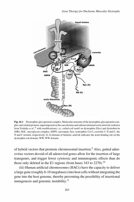
May 4, 2010 11:0 SPI-B903 9in x 6in b903-ch16
Gene Therapy for Duchenne Muscular Dystrophy
Fig. 16.1 Dystrophin-glycoprotein complex. Molecular structure of the dystrophin-glycoprotein com-plex and related proteins superimposed on the sarcolemma and subsarcolemmal actin network (redrawnfrom Yoshida et al.,4 with modifications). cc, coiled-coil motif on dystrophin (Dys) and dystrobrevin(DB); SGC, sarcoglycan complex; SSPN, sarcospan; Syn, syntrophin; Cav3, caveolin-3; N and C, theN and C termini, respectively; G, G-domain of laminin; asterisk indicates the actin-binding site on thedystrophin rod domain; WW, WW domain.
of hybrid vectors that promote chromosomal insertion.9 Also, gutted aden-ovirus vectors devoid of all adenoviral genes allow for the insertion of largetransgenes, and trigger fewer cytotoxic and immunogenic effects than dothose only deleted in the E1 regions (from bases 343 to 2270).10
(iii) Human artificial chromosomes (HACs) have the capacity to delivera large gene (roughly 6-10 megabases) into host cells without integrating thegene into the host genome, thereby preventing the possibility of insertionalmutagenesis and genomic instability.11
263

May 4, 2010 11:0 SPI-B903 9in x 6in b903-ch16
A Guide to Human Gene Therapy
(iv) Vectors for targeted integration: A goal in clinical gene therapy isto develop gene transfer vehicles that can integrate exogenous therapeuticgenes at specific chromosomal loci, so that insertional oncogenesis is pre-vented. AAV can insert its genome into a specific locus, designated AAVS1,on chromosome 19 of the human genome.12 The AAV Rep78/68 proteinsand the Rep78/68-binding sequences are the trans- and cis-acting elementsneeded for this reaction. A dual high-capacity adenovirus-AAV hybrid vec-tor with full-length human dystrophin-coding sequences flanked by AAVintegration-enhancing elements was tested for targeted integration.13
(v) Gene correction is a process whereby sequence alterations in genescan be corrected by homologous recombination-mediated gene conver-sion between the recipient target locus and a donor construct encodingthe correct sequence.14 The introduction of a corrective sequence togetherwith a site-specific nuclease to induce a double-stranded break (DSB) atsites responsible for monogenic disorders would activate gene correction.Pairs of designated zinc-finger protein with tandem DNA binding sitesfused to the cleavage domain of the Fok1 protein were introduced intomodel systems or cell lines and produced corrections in 10–30% of casestested.15
2.2 Modification of the Dystrophin Gene and Promoter
Due to the large deletion in its genome, the gutted adenovirus vector canpackage 14 kb of full-length dystrophin cDNA. Multiple proximal musclesof seven-day-old utrophin/dystrophin double knockout mice (dko mice),which typically show symptoms similar to human DMD, were effectivelytransduced with the gutted adenovirus bearing full-length murine dys-trophin cDNA.16 However, further improvements are needed to regulatethe virus-associated host immune response before clinical trials can beperformed.
A series of truncated dystrophin cDNAs containing rod repeats withhinge 1, 2, and 4 were constructed (Fig. 16.2).7 Although AAV vectors aretoo small to package the full-length dystrophin cDNA,AAV vector-mediatedgene therapy using a rod-truncated dystrophin gene provides a promisingapproach.17 The structure and, particularly, the length of the rod are crucialfor the function of micro-dystrophin.18 An AAV type 2 vector expressing
264

May 4, 2010 11:0 SPI-B903 9in x 6in b903-ch16
Gene Therapy for Duchenne Muscular Dystrophy
Fig. 16.2 Structures of full-length and truncated dystrophin. Helper-dependent adenovirus vector canpackage 14 kb of full-length dystrophin cDNA because of the large-sized deletion in its genome. Amini-dystrophin is cloned from a patient with Becker muscular dystrophy, which is caused by in-framedeletions resulting in the synthesis of partially functional protein.A series of truncated micro-dystrophincDNAs harboring only four rod repeats with hinge 1, 2, and 4 (CS1); the same components, except thatthe C-terminal domain is deleted (delta CS1); or one rod repeat with hinge 1 and 4 (M3), are constructedto be packaged in the AAV vector.
micro-dystrophin (DeltaCS1) under the control of a muscle-specific MCKpromoter was injected into the tibialis anterior (TA) muscles of dystrophin-deficient mdx mice,19 and resulted in extensive and long-term expression ofmicro-dystrophin that exhibited improved force generation.
Codon usage may impact transgene expression. For example, expressionand function of micro-dystrophin was investigated in the mdx mouse usingtwo different configurations of the gene under the control of a muscle-restrictive promoter (Spc5-12) and optimized for codon usage.20 Codonoptimization of micro-dystrophin significantly increased micro-dystrophinmRNA and protein levels after intramuscular and systemic administrationof plasmid DNA or rAAV8. By randomly assembling myogenic regula-tory elements into synthetic promoter recombinant libraries, several artifi-cial promoters were isolated whose transcriptional potencies greatly exceedthose of natural myogenic and viral gene promoters.21
265

May 4, 2010 11:0 SPI-B903 9in x 6in b903-ch16
A Guide to Human Gene Therapy
2.3 Use of Surrogate Genes
An approach using a surrogate gene would bypass the potential immuneresponses associated with the delivery of exogenous dystrophin. Methodsto increase expression of utrophin, a dystrophin paralog, show promise asa treatment for DMD. AAV-6 vector harboring a murine codon-optimizedmicro-utrophin transgene was intravenously administered into adult dkomice to alleviate the pathophysiological abnormalities.22 The paralogousgene efficiently acted as a surrogate for dystrophin. Myostatin has beenextensively documented as being a negative regulator of muscle growth.Systemic gene delivery of myostatin propeptide, a natural inhibitor of myo-statin, enhanced body-wide skeletal muscle growth in both normal and mdxmice.23 The delivery of various growth factors, such as insulin-like growthfactor-I (IGF-I), has been successful in promoting skeletal muscle regener-ation after injury.24
Matrix metalloproteinases (MMPs) may also be useful in improvingmuscle regeneration. MMPs are key regulatory molecules in the forma-tion, remodeling and degradation of all extracellular matrix (ECM) compo-nents in pathological processes. MMP-9 is involved predominantly in theinflammatory process during muscle degeneration.25 In contrast, MMP-2is associated with ECM remodeling during muscle regeneration and fibergrowth.
3. AAV-Mediated Transduction of Animal Models
3.1 Vector Production
A potential hurdle for treatment of multiple muscle groups using viral genetransfer is the required large amount of vector and the related task of scalingup production. To gain acceptance as a medical treatment with a dose ofover 1 × 10¹³ genome copies (g.c.)/kg body weight, AAV vectors requirea scalable and economical production method. An example for a currentproduction method using a helper virus-free transfection system is shownin Fig. 16.3 (see also Chapter 6). Alternatives include packaging cell linesexpressing Rep/Cap with regulated Rep/Cap expression, which is compli-cated by toxic effects of early expression of Rep proteins.26 A scalablemethod, using active gassing and large culture vessels, was developed to
266
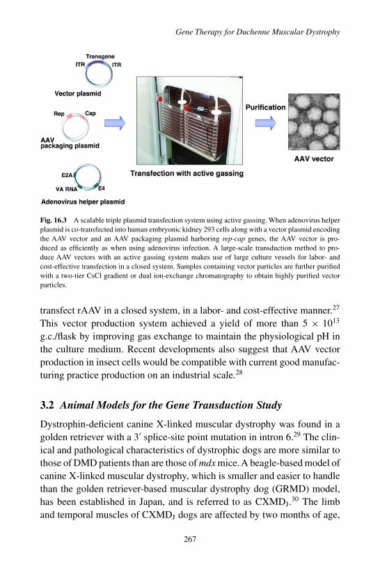
May 4, 2010 11:0 SPI-B903 9in x 6in b903-ch16
Gene Therapy for Duchenne Muscular Dystrophy
Fig. 16.3 A scalable triple plasmid transfection system using active gassing. When adenovirus helperplasmid is co-transfected into human embryonic kidney 293 cells along with a vector plasmid encodingthe AAV vector and an AAV packaging plasmid harboring rep-cap genes, the AAV vector is pro-duced as efficiently as when using adenovirus infection. A large-scale transduction method to pro-duce AAV vectors with an active gassing system makes use of large culture vessels for labor- andcost-effective transfection in a closed system. Samples containing vector particles are further purifiedwith a two-tier CsCl gradient or dual ion-exchange chromatography to obtain highly purified vectorparticles.
transfect rAAV in a closed system, in a labor- and cost-effective manner.27
This vector production system achieved a yield of more than 5 × 10¹³
g.c./flask by improving gas exchange to maintain the physiological pH inthe culture medium. Recent developments also suggest that AAV vectorproduction in insect cells would be compatible with current good manufac-turing practice production on an industrial scale.28
3.2 Animal Models for the Gene Transduction Study
Dystrophin-deficient canine X-linked muscular dystrophy was found in agolden retriever with a 3′ splice-site point mutation in intron 6.29 The clin-ical and pathological characteristics of dystrophic dogs are more similar tothose of DMD patients than are those of mdx mice.A beagle-based model ofcanine X-linked muscular dystrophy, which is smaller and easier to handlethan the golden retriever-based muscular dystrophy dog (GRMD) model,has been established in Japan, and is referred to as CXMDJ.30 The limband temporal muscles of CXMDJ dogs are affected by two months of age,
267

May 4, 2010 11:0 SPI-B903 9in x 6in b903-ch16
A Guide to Human Gene Therapy
corresponding to the second peak of serum creatine kinase. Interestingly,extensive lymphocyte-mediated immune responses to rAAV2-lacZ occurredafter direct intramuscular injection into CXMDJ dogs, despite successfuldelivery of the same viral construct into mouse skeletal muscle.31 In con-trast to rAAV2, rAAV8-mediated transduction of canine skeletal musclesproduced significantly higher transgene expression with less lymphocyteproliferation than rAAV2.32
It is increasingly important to develop strategies to treat DMD that con-sider the effect on cardiac muscle. The pathology of the conduction systemin CXMDJ was analyzed to establish the therapeutic target for DMD.33
Although dystrophic changes of the ventricular myocardium were not evi-dent at the age of 1 to 13 months, Purkinje fibers showed remarkable vacuo-lar degeneration when dogs were as young as four-months-old. Furthermore,degeneration of Purkinje fibers was coincident with overexpression of Dp71at the sarcolemma. The degeneration of Purkinje fibers could be associatedwith the distinct deep Q waves present in ECGs and the fatal arrhythmiasseen in cases of dystrophin deficiency.33
3.3 Immunological Issues of rAAV
Neo-antigens introduced by AAV vectors evoke significant immune reac-tions in DMD muscle, since increased permeability of sarcolemma allowsleakage of the transgene products from the dystrophin-deficient musclefibers.34 AAV2-mediated transfer into skeletal muscles of normal dogsresulted in low and transient expression, together with intense cellularinfiltration, and the marked activation of cellular and humoral immuneresponses.31 Furthermore, an in vitro interferon-gamma release assayshowed that canine splenocytes respond to immunogens or mitogensmore strongly than do murine splenocytes. In fact, co-administrationof immunosuppressants, cyclosporine (CSP) and mycophenolate mofetil(MMF) improved rAAV2 transduction. The AAV2 capsids can induce acellular immune response via MHC class I antigen presentation with across-presentation pathway, and rAAV2 has also been proposed to have aneffect on human dendritic cells (DCs). In contrast, other serotypes, such asrAAV8, induced T cell activation to a lesser degree.32 Immunohistochemi-cal analysis revealed that the rAAV2-injected muscles showed higher rates
268

May 4, 2010 11:0 SPI-B903 9in x 6in b903-ch16
Gene Therapy for Duchenne Muscular Dystrophy
of infiltration of CD4+ and CD8+ T lymphocytes in the endomysium thanthe rAAV8-injected muscles.32
Resident antigen-presenting cells, such as DCs, myoblasts, myotubesand regenerating immature myofibers, might play a role in the immuneresponse. A recent study also showed that mRNA levels of MyD88 and co-stimulating factors, such as CD80, CD86 and type I interferon, are elevatedin both rAAV2- and rAAV8-transduced dog DCs in vitro.32 A brief course ofimmunosuppression with a combination of anti-thymocyte globulin (ATG),CSP and MMF was effective in permitting AAV6-mediated, long-term androbust expression of a canine micro-dystrophin in the skeletal muscle of adog DMD model.35
3.4 Intravascular Vector Administration by Limb Perfusion
Although recent studies suggest that vectors based on AAV are capableof body-wide transduction in rodents, translating this finding into largeanimals remains a challenge. Intravascular delivery can be performed as aform of limb perfusion, which might bypass the immune activation of DCsin the injected muscle.32 Our laboratory performed limb perfusion-assistedintravenous administration of rAAV8-micro-dystrophin into the hind limbof CXMDJ dogs resulting in extensive transgene expression in the distallimb muscles without obvious immune responses for as long as eight weeksafter injection (Fig. 16.4).32
3.5 Global Muscle Therapies
In comparison with fully dystrophin-deficient animals, targeted transgenicrepair of skeletal muscle, but not cardiac muscle, paradoxically elicits a five-fold increase in cardiac injury and dilated cardiomyopathy.36 Because thedystrophin-deficient heart is highly sensitive to increased stress, increasedactivity by the repaired skeletal muscle provides the stimulus for heightenedcardiac injury and heart remodeling. In contrast, a single intravenous injec-tion of AAV9 vector expressing micro-dystrophin efficiently transduces theentire heart in neonatal mdx mice, thereby ameliorating cardiomyopathy.37
Since a number of muscular dystrophy patients can be identified throughnewborn screening, neonatal transduction may lead to an effective earlyintervention in DMD patients. After a single intravenous injection, robust
269

May 4, 2010 11:0 SPI-B903 9in x 6in b903-ch16
A Guide to Human Gene Therapy
Fig. 16.4 Intravascular vector administration by limb perfusion. (A) A blood pressure cuff is appliedjust above the knee of an anesthetized CXMDJ dog. A 24-gauge catheter is inserted into the lateralsaphenous vein, connected to a three-way stopcock, and flushed with saline. With a blood pressurecuff inflated to over 300 mmHg, saline (2.6 ml/kg) containing papaverine (0.44 mg/kg) and heparin(16 U/kg) is injected by hand over a 10 second period. The three-way stopcock is connected to a syringecontaining rAAV8 expressing micro-dystrophin (1 × 10¹⁴ vg/kg, 3.8 ml/kg). The syringe is placed in aPHD 2000 syringe pump. Five minutes after the papaverine/heparin injection, rAAV8 is injected at a rateof 0.6 ml/sec. (B) Administration of rAAV8-micro-dystrophin by limb perfusion produces extensivetransgene expression in the distal limb muscles of CXMDJ dogs without obvious immune responses atfour weeks after injection. Modified from Mol Ther 17: 73–80, 2008.
skeletal muscle transduction with AAV9 vector throughout the body wasobserved in neonatal dogs.38 Systemic transduction was achieved in theabsence of pharmacological intervention or immune suppression and lastedfor at least six months, whereas cardiac muscle was barely transduced inthe dogs.
4. Safety and Potential Impact of Clinical Trials
The initial clinical studies have laid the foundation for future studies, pro-viding important information about vector dose, viral serotype selection,and immunogenicity in humans. The first virus-mediated gene transfer formuscle disease was carried out for limb-girdle muscular dystrophy type 2Dusing rAAV1. The study, consisting of intramuscular injection of virus intoa single muscle, was discharged to establish the safety of this procedure inphase I clinical trials.39 The first clinical gene therapy trial for DMD beganin March 2006.39 This was a phase I/IIa study in which an AAV vectorwas used to deliver micro-dystrophin to the biceps of boys with DMD. Thestudy was conducted on six boys with DMD, each of whom was transduced
270

May 4, 2010 11:0 SPI-B903 9in x 6in b903-ch16
Gene Therapy for Duchenne Muscular Dystrophy
with mini-dystrophin genes in a muscle of one arm in the absence of seriousadverse events.
While low immunogenicity was considered a major strength supportingthe use of rAAV in clinical trials, a number of observations have recentlyprovided a more balanced view of this procedure.40 An obvious barrierto AAV transduction is the presence of circulating neutralizing antibodiesthat prevent the virion from binding to its cellular receptor.41 This potentialthreat can be reduced by prescreening patients for AAV serotype-specificneutralizing antibodies or by performing procedures such as plasmapheresisbefore gene transfer.Another challenge recently revealed is the developmentof a cytotoxic T cell (CTL) response to AAV capsid peptides. In the humanfactor IX gene therapy trial in which rAAV was delivered to the liver, onlyshort-term transgene expression was achieved and levels of therapeutic pro-tein declined to baseline levels 10 weeks after vector infusion.40 This wasaccompanied by elevation of serum transaminase levels and a CTL responsetoward specific AAV capsid peptides. To overcome this response, transientimmunosuppression may be required until AAV capsids are completelycleared. Additional findings suggest that T cell activation requires AAV2capsid binding to the heparan sulfate proteoglycan (HSPG) receptor, whichwould permit virion shuttling into a DC pathway, as cross-presentation.42
Exposure to vectors from other AAV clades, such as AAV8, did not activatecapsid-specific T cells.
5. Development of Alternative Strategies
5.1 Design of Read-through Drugs
To suppress premature stop codon mutations, treatments involving amino-glycosides and other agents have been attempted. PTC124, a novel drugcapable of suppressing premature termination and selectively inducing ribo-somal read-through of premature, but not normal, termination codons, wasrecently identified using nonsense-containing reporters.43 The selectivity ofPTC124 for premature termination codons, its oral bioavailability and itspharmacological properties indicate that this drug may have broad clinicalpotential for the treatment of a large group of genetic disorders with limitedor no therapeutic options.
271
May 4, 2010 11:0 SPI-B903 9in x 6in b903-ch16
A Guide to Human Gene Therapy
5.2 Modification of mRNA Splicing
By inducing the skipping of specific exons during mRNA splicing, antisensecompounds against exonic and intronic splicing regulatory sequences wereshown to correct the open reading frame of the DMD gene and thus to restoretruncated yet functional dystrophin expression in vitro.44 Intravenous infu-sion of an antisense phosphorothioate oligonucleotide created an in-framedystrophin mRNA via exon skipping in a 10-year-old DMD patient possess-ing an out-of-frame exon 20 deletion of the dystrophin gene.45 Moreover, theadverse-event profile and local dystrophin-restoring effect of a single intra-muscular injection of an antisense 2′-O-methyl phosphorothioate oligonu-cleotide, PRO051, in patients with DMD were explored.46 Four patientsreceived a dose of 0.8 mg of PRO051 in the TA muscle. Each patient showedspecific skipping of exon 51 of dystrophin in 64 to 97% of myofibers,without clinically apparent adverse side effects.
The efficacy and toxicity of intravenous injection of stable morpholinophosphorodiamidate (morpholino)-induced exon skipping were tested usingCXMDJ dogs, and widespread rescue of dystrophin expression to thera-peutic levels was observed.47 Furthermore, a morpholino oligomer with adesigned cell-penetrating peptide can efficiently target a mutated dystrophinexon in cardiac muscles.48
Long-term benefits can be obtained through the use of viral vectorsexpressing antisense sequences against regions within the dystrophin gene.The sustained production of dystrophin at physiological levels in entiregroups of muscles as well as the correction of muscular dystrophy wereachieved by treatment with exon-skipping AAV1-U7.49
5.3 Ex Vivo Gene Therapy
Transplantation of genetically corrected autologous myogenic cells is apossible treatment for DMD. Freshly isolated satellite cells transduced withlentiviral vectors expressing micro-dystrophin were transplanted into the TAmuscles of mdx mice, and these cells efficiently contributed to the regener-ation of muscles with micro-dystrophin expression at the sarcolemma.50
Mesoangioblasts are vessel-associated stem cells and might be candi-dates for future stem cell therapy for DMD.51 Intra-arterial delivery ofwild-type canine mesoangioblasts resulted in the extensive recovery of
272
May 4, 2010 11:0 SPI-B903 9in x 6in b903-ch16
Gene Therapy for Duchenne Muscular Dystrophy
dystrophin expression, normal muscle morphology and function in theGRMD. Multipotent mesenchymal stromal cells (MSCs) are less immuno-genic and have the potential to differentiate and display a myogenicphenotype.52
6. Future Perspectives
6.1 Pharmacological Intervention
The use of a histone deacetylase (HDAC) inhibitor would likely enhance theutility of rAAV-mediated transduction strategies in the clinic.53 In contrastto adenovirus-mediated transduction, the improved transduction with rAAVinduced by the HDAC inhibitor is due to enhanced transgene expressionrather than to increased viral entry. Enhanced transduction may be relatedto the histone-associated chromatin form of the rAAV concatemer in trans-duced cells. Since various HDAC inhibitors are currently being tested inclinical trials for many diseases, the use of such agents in rAAV-mediatedDMD gene therapy is theoretically and practically reasonable.
6.2 Capsid Modification
A DNA shuffling-based approach for developing cell type-specific vectorsis an intriguing possibility to achieve altered tropism. Capsid genomes ofAAV serotypes 1-9 were randomly reassembled using PCR to generate achimeric capsid library.54 A single infectious clone (chimeric-1829) con-taining genome fragments from AAV1, 2, 8, and 9 was isolated from anintegrin minus hamster melanoma cell line previously shown to have lowpermissiveness to AAV.
7. Conclusions and Outlook
DMD remains an untreatable genetic disease that severely limits motilityand life expectancy in affected children. The systemic delivery of rAAV totransduce truncated dystrophin is predicted to ameliorate the symptoms ofDMD patients in the future. To translate gene transduction technologies intoclinical practice, development of an effective delivery system with improvedvector constructs as well as efficient immunological modulation must be
273
May 4, 2010 11:0 SPI-B903 9in x 6in b903-ch16
A Guide to Human Gene Therapy
established. A novel protocol that considers all of these issues would helpimprove the therapeutic benefits of DMD gene therapy.
References
1. Mendell JR, et al. (2009). LGMD 2D gene therapy restores alpha-sarcoglycan andassociated proteins. Annals Neurol, in press.
2. EmeryAE (1991). Population frequencies of inherited neuromuscular diseases–a worldsurvey. Neuromuscul Disord 1: 19–29.
3. Hoffman EP, Brown RH Jr., Kunkel LM (1987). Dystrophin: the protein product of theDuchenne muscular dystrophy locus. Cell 51: 919–928.
4. Yoshida M, et al. (2000). Biochemical evidence for association of dystrobrevin with thesarcoglycan-sarcospan complex as a basis for understanding sarcoglycanopathy. HumMol Genet 9: 1033–1040.
5. Okada T, et al. (2002). Adeno-associated viral vector-mediated gene therapy ofischemia-induced neuronal death. Methods Enzymol 346: 378–393.
6. Inagaki K, et al. (2006). Robust systemic transduction with AAV9 vectors in mice:efficient global cardiac gene transfer superior to that of AAV8. Mol Ther 14: 45–53.
7. Yuasa K, MiyagoeY,Yamamoto K, NabeshimaY, Dickson G, Takeda S (1998). Effectiverestoration of dystrophin-associated proteins in vivo by adenovirus-mediated transferof truncated dystrophin cDNAs. FEBS Lett 425: 329–336.
8. Okada T, Ramsey J, Munir J, Wildner O, Blaese M (1998). Efficient directional cloningof recombinant adenovirus vectors using DNA-protein complex. Nucleic Acids Res 26:1947–1950.
9. Okada T, et al. (2004). In situ generation of pseudotyped retroviral progeny byadenovirus-mediated transduction of tumor cells enhances the killing effect of HSV-tksuicide gene therapy in vitro and in vivo. J Gene Med 6: 288–299.
10. Hammerschmidt DE (1999). Development of a gutless vector. J Lab Clin Med134: C3.
11. Hoshiya, H, et al. (2008). A highly Stable and Nonintegrated Human Artificial Chro-mosome (HAC) Containing the 2.4 Mb Entire Human Dystrophin Gene. Mol Ther.
12. Kotin RM, Linden RM, Berns KI (1992). Characterization of a preferred site on humanchromosome 19q for integration of adeno-associated virus DNA by non-homologousrecombination. Embo J 11: 5071–5078.
13. Goncalves MA, et al. (2005). Transfer of the full-length dystrophin-coding sequenceinto muscle cells by a dual high-capacity hybrid viral vector with site-specific integra-tion ability. J Virol 79: 3146–3162.
14. Klug, A (2005). Towards therapeutic applications of engineered zinc finger proteins.FEBS Lett 579: 892–894.
15. Porteus MH, Baltimore, D (2003). Chimeric nucleases stimulate gene targeting inhuman cells. Science 300: 763.
16. Kawano R, Ishizaki M, MaedaY, UchidaY, Kimura E, Uchino M (2008). Transductionof full-length dystrophin to multiple skeletal muscles improves motor performance andlife span in utrophin/dystrophin double knockout mice. Mol Ther 16: 825–831.
274

May 4, 2010 11:0 SPI-B903 9in x 6in b903-ch16
Gene Therapy for Duchenne Muscular Dystrophy
17. Wang B, Li J, Xiao X (2000). Adeno-associated virus vector carrying human minidys-trophin genes effectively ameliorates muscular dystrophy in mdx mouse model. ProcNatl Acad Sci USA 97: 13714–13719.
18. Sakamoto M, et al. (2002). Micro-dystrophin cDNA ameliorates dystrophic phenotypeswhen introduced into mdx mice as a transgene. Biochem Biophys Res Commun 293:1265–1272.
19. Yoshimura M, et al. (2004). AAV vector-mediated microdystrophin expression in arelatively small percentage of mdx myofibers improved the mdx phenotype. Mol Ther10: 821–828.
20. Foster H, et al. (2008). Codon and mRNA sequence optimization of microdystrophintransgenes improves expression and physiological outcome in dystrophic mdx micefollowing AAV2/8 gene transfer. Mol Ther 16: 1825–1832.
21. Li X, Eastman EM, Schwartz RJ, Draghia-Akli, R (1999). Synthetic muscle promot-ers: activities exceeding naturally occurring regulatory sequences. Nat Biotechnol 17:241–245.
22. Odom GL, Gregorevic P, Allen JM, Finn E, Chamberlain JS (2008). Microutrophindelivery through rAAV6 increases lifespan and improves muscle function in dystrophicdystrophin/utrophin-deficient mice. Mol Ther 16: 1539–1545.
23. Qiao C, et al. (2008). Myostatin propeptide gene delivery by adeno-associated virusserotype 8 vectors enhances muscle growth and ameliorates dystrophic phenotypes inmdx mice. Hum Gene Ther 19: 241–254.
24. Schertzer JD, Lynch GS (2006). Comparative evaluation of IGF-I gene transfer andIGF-I protein administration for enhancing skeletal muscle regeneration after injury.Gene Ther 13: 1657–1664.
25. Fukushima K, et al. (2007). Activation and localization of matrix metalloproteinase-2and -9 in the skeletal muscle of the muscular dystrophy dog (CXMDJ). BMC Muscu-loskelet Disord 8: 54.
26. Okada T, et al. (2001). Development and characterization of an antisense-mediatedprepackaging cell line for adeno-associated virus vector production. Biochem BiophysRes Commun 288: 62–68.
27. Okada T, et al. (2005). Large-scale production of recombinant viruses by use of a largeculture vessel with active gassing. Hum Gene Ther 16: 1212–1218.
28. Cecchini S, Negrete A, Kotin RM (2008). Toward exascale production of recombinantadeno-associated virus for gene transfer applications. Gene Ther 15: 823–830.
29. Valentine BA, Cooper BJ, de Lahunta A, O’Quinn R, Blue JT (1988). Canine X-linkedmuscular dystrophy. An animal model of Duchenne muscular dystrophy: clinical stud-ies. J Neurol Sci 88: 69–81.
30. ShimatsuY, et al. (2005). Major clinical and histopathological characteristics of canineX-linked muscular dystrophy in Japan, CXMDJ. Acta Myol 24: 145–154.
31. Yuasa K, et al. (2007). Injection of a recombinant AAV serotype 2 into canine skeletalmuscles evokes strong immune responses against transgene products. Gene Ther.
32. Ohshima S, et al. (2008). Transduction Efficiency and Immune Response AssociatedWith the Administration of AAV8 Vector Into Dog Skeletal Muscle. Mol Ther.
33. Urasawa N, et al. (2008). Selective vacuolar degeneration in dystrophin-deficient caninePurkinje fibers despite preservation of dystrophin-associated proteins with overexpres-sion of Dp71. Circulation 117: 2437–2448.
275

May 4, 2010 11:0 SPI-B903 9in x 6in b903-ch16
A Guide to Human Gene Therapy
34. Yuasa K, et al. (2002). Adeno-associated virus vector-mediated gene transfer intodystrophin-deficient skeletal muscles evokes enhanced immune response against thetransgene product. Gene Ther 9: 1576–1588.
35. Wang Z, et al. (2007). Sustained AAV-mediated Dystrophin Expression in a CanineModel of Duchenne Muscular Dystrophy with a Brief Course of Immunosuppression.Mol Ther 15: 1160–1166.
36. Townsend D, Yasuda S, Li S, Chamberlain JS, Metzger JM (2008). Emergent dilatedcardiomyopathy caused by targeted repair of dystrophic skeletal muscle. Mol Ther 16:832–835.
37. Bostick B, Yue Y, Lai Y, Long C, Li D, Duan D (2008). Adeno-associated virusserotype-9 microdystrophin gene therapy ameliorates electrocardiographic abnormal-ities in mdx mice. Hum Gene Ther 19: 851–856.
38. YueY, et al. (2008). A single intravenous injection of adeno-associated virus serotype-9leads to whole body skeletal muscle transduction in dogs. Mol Ther 16: 1944–1952.
39. Rodino-Klapac LR, Chicoine LG, Kaspar BK, Mendell JR (2007). Gene therapy forduchenne muscular dystrophy: expectations and challenges. Arch Neurol 64: 1236–1241.
40. Manno CS, et al. (2006). Successful transduction of liver in hemophilia by AAV-FactorIX and limitations imposed by the host immune response. Nat Med 12: 342–347.
41. Scallan CD, et al. (2006). Human immunoglobulin inhibits liver transduction by AAVvectors at low AAV2 neutralizing titers in SCID mice. Blood 107: 1810–1817.
42. Vandenberghe LH, et al. (2006). Heparin binding directs activation of T cells againstadeno-associated virus serotype 2 capsid. Nat Med 12: 967–971.
43. Welch EM, et al. (2007). PTC124 targets genetic disorders caused by nonsense muta-tions. Nature 447: 87–91.
44. Takeshima Y, Nishio H, Sakamoto H, Nakamura H, Matsuo M (1995). Modulation ofin vitro splicing of the upstream intron by modifying an intra-exon sequence which isdeleted from the dystrophin gene in dystrophin Kobe. J Clin Invest 95: 515–520.
45. TakeshimaY, et al. (2006). Intravenous infusion of an antisense oligonucleotide resultsin exon skipping in muscle dystrophin mRNA of Duchenne muscular dystrophy. PediatrRes 59: 690–694.
46. van Deutekom JC, et al. (2007). Local dystrophin restoration with antisense oligonu-cleotide PRO051. N Engl J Med 357: 2677–2686.
47. Yokota T, et al. (2009). Efficacy of systemic morpholino exon-skipping in Duchennedystrophy dogs. Annals Neurol.
48. Wu B, et al. (2008). Effective rescue of dystrophin improves cardiac function indystrophin-deficient mice by a modified morpholino oligomer. Proc Natl Acad SciUSA 105: 14814–14819.
49. Goyenvalle A, et al. (2004). Rescue of dystrophic muscle through U7 snRNA-mediatedexon skipping. Science 306: 1796–1799.
50. Ikemoto M, et al. (2007). Autologous transplantation of SM/C-2.6(+) satellite cellstransduced with micro-dystrophin CS1 cDNA by lentiviral vector into mdx mice. MolTher 15: 2178–2185.
51. Sampaolesi M, et al. (2006). Mesoangioblast stem cells ameliorate muscle function indystrophic dogs. Nature 444: 574–579.
276

May 4, 2010 11:0 SPI-B903 9in x 6in b903-ch16
Gene Therapy for Duchenne Muscular Dystrophy
52. Dezawa M, et al. (2005). Bone marrow stromal cells generate muscle cells and repairmuscle degeneration. Science 309: 314–317.
53. Okada T, et al. (2006). A histone deacetylase inhibitor enhances recombinant adeno-associated virus-mediated gene expression in tumor cells. Mol Ther 13: 738–746.
54. Li W, et al. (2008). Engineering and selection of shuffled AAV genomes: a new strategyfor producing targeted biological nanoparticles. Mol Ther 16: 1252–1260.
277

May 4, 2010 11:0 SPI-B903 9in x 6in b903-ch16
This page intentionally left blankThis page intentionally left blank
May 4, 2010 11:0 SPI-B903 9in x 6in b903-ch17
Chapter 17
Cancer Gene Therapy
Kirsten A.K. Weigel-Van Aken∗
Cancer gene therapy, initially designed as the transfer of a single gene intoa tumor, has long been viewed with skepticism, and ultimately has failedclinically. Gene transfer in the context of additional treatment modali-ties, such as radiation/chemotherapy, oncolytic activity of a replicatingvirus, anti-angiogenesis, or immune stimulation, however, has the poten-tial to have a significant impact on current cancer treatment. Primarytumor destruction has been one of the most successful endeavors ofmodern oncology. The re-occurrence of disease due to dormant tumorcells that evade the primary therapy, however, still constitutes a deathsentence for many patients. Due to their inherent resistance to conven-tional chemo- and radiation therapies, these relapsed tumor cells warrantextensive exploration of alternative therapeutic approaches. Localizedtreatments of metastases such as direct injection or electroporation ofplasmid DNA or injection of oncolytic or gene transfer viruses have onlyresulted in localized successes, because the occult and multifocal nature ofthe metastatic process still leads to evasion from therapeutic intervention.Novel strategies of cancer gene therapy therefore focus on means toprovoke an antitumor immune response in a tumor-tolerant host usinga variety of methods for tumor antigen and costimulatory signal deliveryand means to improve tumor-targeting and prevent immune clearance oftherapeutic viruses.
∗Correspondence: Department of Pediatrics, Division of Cellular and Molecular Therapy, University ofFlorida College of Medicine, Cancer & Genetics Research Complex, 1376 Mowry Road, Gainesville,FL 32610-3610.E-mail: [email protected]
279
May 4, 2010 11:0 SPI-B903 9in x 6in b903-ch17
A Guide to Human Gene Therapy
1. Introduction
Gene therapy involves the transfer of genetic information into a patient’scells to replace, repair, or turn off an abnormal gene, a strategy originallydeveloped for single-gene diseases and subsequently extended to complex,multigenic disease such as cancer. Some common strategies used for cancergene therapy are:
1. Suppression of oncogene expression or transfer of a tumor suppressorgene. Examples include transfer of a normal p53 gene, suppression of theErbB2/HER2 gene, suppression of c-myc and c-fos genes, and transferof melanoma differentiation associated protein 7.
2. Augmentation of immunological responses to cancer cells through trans-fer of cytokine genes (IL-2, IL-12, GM-CSF, TNF-α), co-stimulatorymolecules (CD80), and antigen genes (CA15-3, HER2)
3. Suicide gene therapy by transfer of prodrug-activating enzymes intotumor cells followed by treatment with the prodrug, resulting in a highconcentration of the activated drug in the tumor tissues: for example,herpes simplex virus thymidine kinase (HSV-TK) gene and gancyclovir;cytosine deaminase gene and fluorocytosine; and cytochrome P450 2B6gene and cyclophosphamide.
2. Targeting the Tumor Cell
2.1 DNA Electroporation
Electroporation, or electric-pulse-provoked permeabilization of the plasmamembrane, has been defined as the modification of the membrane imper-meability to ions and hydrophilic and/or charged molecules as a conse-quence of exposure of cells to appropriate electric pulses. Transfer of DNAacross the cell membrane requires electrophoretic acceleration of the DNAmolecules within the electric field. Tumor electroporation differs from othertissues such as skeletal muscle, in the duration of the physiological local,histamine-dependent hypoperfusion (vascular lock) that can last up to sev-eral hours in the former and only a few minutes in the latter. In 2008, thefirst human trial of gene transfer utilizing in vivo DNA electroporation wasreported. Of 24 melanoma patients treated with electroporation after IL-12
280

May 4, 2010 11:0 SPI-B903 9in x 6in b903-ch17
Cancer Gene Therapy
DNA injections, 10% showed complete regression of treated lesions and42% experienced either disease stabilization or partial responses.1
2.2 Non-Oncolytic Viral Vectors
2.2.1 Retrovirus
Retroviral vectors derived from the murine retrovirus Moloney MurineLeukemia Virus (MMLV) were the first and most extensively used vectorsin gene therapy. They have a relatively limited size capacity for foreignDNA (up to 8 kb) and require a cell to go through mitosis with the con-sequent disintegration of the nuclear membrane to allow transduction tooccur. The viral DNA randomly integrates into the cellular genome, a pro-cess that led to severe side effects in a human sever combined immunedeficiency (SCID) trial in France.2 Since the tumor selectivity of retroviralvectors solely relies on the dependence of transduction on the active pro-liferation of target cells, retroviral vectors have been preferentially used intumors that reside within a tissue that itself has a low proliferative index,such as the brain. Clinical trial results from patients with malignant gliomathat received stereotactic injections of either retroviral vectors encoding aprodrug-converting enzyme (HSV-TK) or vector-producing cells generatingthe vectors in situ reported extremely low levels of transduced tumor cells,less than 0.03%, while the threshold transduction level for clinical benefitwas estimated to be around 10%.3 The first clinical trial in the US using sys-temic, intravenous (i.v.) administration of a retroviral vector was done witha non-replicating retroviral vector that displayed a collagen-binding domainof vonWillebrandt factor on its envelope and encoded a mutant human cyclinG1 gene (Rexin-G vector).Although this vector had demonstrated inhibitionof pancreatic cancer cell proliferation in vitro, the results in pancreatic can-cer patients were disappointing with no clinical tumor response detected.4
Retroviral vectors have also been used to transduce hematopoietic stem cellswith drug resistance genes to prevent bone marrow suppression followinghigh dose chemotherapy. Clinical trials, however, demonstrated only a rela-tively modest protection, and alternative vector systems are currently beingevaluated.
Replication-competent retrovirus vectors based on amphotropic MLVwere shown to be capable of achieving highly efficient replicative spread
281

May 4, 2010 11:0 SPI-B903 9in x 6in b903-ch17
A Guide to Human Gene Therapy
and gene delivery through solid tumors in mice. However, the use of sucha vector in humans needs to be critically evaluated.
2.2.2 Lentivirus
Lentiviruses, such as human and simian immunodeficiency viruses (HIV,SIV), were developed as gene therapy vectors to overcome some of theshortcomings of retroviruses, including viral instability and the inability totransduce nondividing cells. Lentiviral vectors transduce quiescent cells andare engineered as self-inactivating (SIN) transfer vectors to minimize therisk of emergence of replication-competent virions. The tropism of lentiviralvectors is modified through the use of the promiscuous vesicular stomatitisvirus (VSV) glycoprotein as a heterologous envelope. Although lentiviralvectors have been employed for gene transfer to cancer cells in mousemodels, such as hepatocellular carcinoma and metastatic prostate cancer,their main use in cancer therapy is in the development of antitumor vaccines.
2.3 Oncolytic Viruses
It has long been known that viremia, following infection or vaccination,can result in tumor responses in patients with lymphoma or leukemia. Inorder to address some of the limitations of non-replicating viral therapeutics,over a dozen distinct families of viruses have been engineered to optimizetheir “oncolytic” potential — i.e. their ability to replicate in and kill tumorcells without harming normal tissue. Viruses recognized as oncolytic agentscan be divided into three categories: (i) naturally occurring viruses (e.g.New Castle Disease virus, VSV, autonomous parvoviruses, some measlesvirus strains, and reovirus) that selectively replicate in tumor cells, in someinstances owing to their dependence on an activated Ras pathway (reovirus)or to their relative resistance to interferon action; (ii) virus mutants in whichsome genes essential for replication in normal cells but evitable in cancercells have been deleted (e.g. adenovirus ONYX-015 that replicates only incells with mutant p53); and (iii) virus mutants modified by the introductionof tissue-specific transcriptional elements that drive viral genes (e.g. aden-ovirus CV706 that expresses E1A and E1B under the control of a prostate-specific antigen (PSA) promoter and adenovirus adMyc-TK that expressesa thymidine kinase gene under the control of a Myc-Max response element).
282
May 4, 2010 11:0 SPI-B903 9in x 6in b903-ch17
Cancer Gene Therapy
2.3.1 Herpesvirus
HSV-1 is an enveloped, double-stranded linear DNA virus that can infectreplicating and quiescent cells, and infections can be either lytic or latent.The advantages of HSV-1 vectors over other vectors used in cancer genetherapy include (i) potential for incorporation of large amounts of foreignDNA (∼30 kb); (ii) neurotropism; (iii) sensitivity to antiherpetic agents;and (iv) the fact that HSV-1 does not integrate. Recombinant HSV thatare used as anti-cancer agents are viruses with mutations in two or moredifferent genes to reduce the potential risk of restoring a wild-type pheno-type via recombination with latent host virus. Results from clinical trialsusing HSV (1716) locally in glioblastoma patients showed viral replica-tion in the tumor site and subtle changes in tumor morphology but noclinical improvement.5 Injection of the same HSV (1716) in melanomapatients exhibited microscopic tumor necrosis but no systemic efficacy.Intratumor (i.t.) injection of a GM-CSF-armed HSV (OncovexGM−CSF,Biovex, Cambridge MA) into cutaneous metastases also did not show sys-temic efficacy although necrosis in multiple tumor nodules was observed.Intrahepatic artery delivery of a replication-competent, attenuated, geneti-cally engineered HSV-1 vector into patients with hepatic colorectal metas-tases was well tolerated, but demonstrated only minor effects in 2 outof 12 patients and either stable disease or disease progression in theremainder.6
2.3.2 Adenovirus
Adenoviruses are double-stranded DNA viruses that have been extensivelyused in gene therapy studies. Since adenoviruses typically infect cellsexpressing coxsackie and adenovirus receptors (CARs) and CARs are onlyexpressed by some human tumor cells, such as prostate cancer,7 severalfiber mutants have been generated to overcome the limited tropism. Theadenoviral genome is maintained as an extra-chromosomal element that israpidly lost in dividing cells. Oncolytic adenoviruses are highly immuno-genic, which can be advantageous if an antitumor immune response isachieved, but disadvantageous if the immune response blocks viral propa-gation or leads to toxicity. One modification that confers tumor selectivityis deletion of the E1B region, found in the vector Onyx-015, which restricts
283

May 4, 2010 11:0 SPI-B903 9in x 6in b903-ch17
A Guide to Human Gene Therapy
viral replication to cells lacking a normal p53 protein (∼40% of humantumors), since E1B’s ability to sequester p53 is essential to viral replica-tion. The uses of Onyx-015 include i.t. injections into head and neck cancerlesions, i.t. injections into pancreatic cancers and recurrent glioblastomas,intraperitoneal (i.p.) injections into ovarian carcinoma patients, injectionsinto the hepatic artery of colorectal cancer patients and i.v. injections intolung cancer patients, all of which demonstrated local oncolytic viral repli-cation, but no clinical responses.5
2.3.3 Poxvirus
Poxviruses are double-stranded enveloped DNA viruses. Vaccinia virus hashad a critical role in the eradication of small pox and its highly immuno-genic nature has led to its continued use in immunotherapy. Vaccinia viruscan accommodate up to 25 kb of foreign DNA, causes timely lysis ofinfected cells (48–72 hours after infection), and has a broad tumor tis-sue tropism due to its ability to infect through several membrane fusionpathways. Vaccinia virus DNA does not integrate into the host chromo-some but replicates in mini-nuclear structures in the cytoplasm. Poxvirusesare highly efficient in spreading to distant tumors within a host and directinfection of cancer cells results in efficient lysis. Furthermore, immune-mediated cell death through release of cellular and viral danger signalsand release of virus- and tumor-associated antigens at the site of infec-tion as well as vascular collapse, due to intravascular thrombosis followingexcessive neutrophil attraction, and direct infection and lysis of tumor-associated endothelial cells contribute to its antitumor efficiency. In addi-tion, the potent inflammatory response initiated by release of virus- andtumor-associated antigens has been augmented by the expression of trans-genes that encode relevant cytokines.8,9 The tumor selectivity of myxomavirus, for example, relies on disrupted type I interferon (IFN) inductionin murine tumor cells,10 and increased Akt levels in human tumor cells.11
Non-engineered live vaccinia vaccine strains were used in early trials inthe 1970s and 1990s, primarily by superficial tumor injection in melanomapatients and tumor responses at the injection site, but no distant responseswere observed.12 Clinical trial results on a targeted and armed oncolyticpoxvirus (JX-594) expressing GM-CSF demonstrated local efficacy in
284
May 4, 2010 11:0 SPI-B903 9in x 6in b903-ch17
Cancer Gene Therapy
injected melanomas, including some cases of complete responses, andoccasional responses in distant skin but not visceral metastases. A secondtrial in patients with advanced hepatocellular carcinoma demonstratedreplication-dependent dissemination in the blood and infection of non-injected tumor sites, significant increases in blood neutrophil concentra-tion due to GM-CSF expression, and tumor cell and tumor vasculaturedestruction.13,14
2.3.4 Measles virus
Measles virus (MV) is a negative sense single-stranded RNA paramyx-ovirus that has oncolytic potential in several types of cancer.15 MV usesCD46 and signaling-activation molecule (SLAM), which is predominantlyfound on activated B and T lymphocytes, as receptors for entry into humancells. The tumor selectivity of the attenuated measles vaccine strain usedin oncolytic approaches is based on its preferential use of the CD46 recep-tor, which is overexpressed in tumor cells. In animal studies, an attenu-ated MV strain engineered to express the human carcinoembryonic antigen(CEA) has demonstrated significant antitumor activity against glioma.16
A clinical protocol has been developed where MV-CEA is installed intothe resection cavity of glioma multiforme and CEA is used to moni-tor viral gene expression.17 MVs were also engineered to express thehuman sodium-iodide symporter, which enhances iodide uptake and allowsmonitoring of radioiodine concentration in MV-infected cells. In addi-tion, combination with therapeutic doses of radioiodine proved effective ininducing a complete regression in a measles-resistant myeloma tumor modelin mice.18
2.3.5 Vesicular stomatitis virus
Vesicular stomatitis virus (VSV) is a negative sense single-stranded RNAvirus that has oncolytic activity when administered systemically.19 VSVis highly sensitive to the antiviral effects of the interferon system and itspropagation is hindered in cells that contain a functional interferon sig-naling system. Mutant VSVs have been engineered that carry a deletionin the matrix protein therefore blocking the nuclear export of interferonRNA. These variants have been demonstrated to have superior antitumor
285
May 4, 2010 11:0 SPI-B903 9in x 6in b903-ch17
A Guide to Human Gene Therapy
effects.20 VSV engineered to express the human sodium-iodide sym-porter demonstrated increased oncolytic activity in a mouse myelomamodel following i.t. or i.v. administration in combination with radioiodinetherapy.21
3. Targeting the Immune System
A ‘tumor surveillance hypothesis’ was originally formulated by Burnet in1970, stating that tumor cells are recognized as foreign by the immunesystem and subsequently specifically eliminated without damaging theirhealthy counterparts, much in the same way as for virus infected cells,thus avoiding autoimmunity.22 Dendritic cells (DC) are considered themost potent antigen-presenting cells (APC) of the immune system. In ani-mal models, vaccination with DCs pulsed with tumor peptides, lysates orRNA or loaded with apoptotic/necrotic tumor cells could induce signif-icant antitumor cytotoxic T lymphocyte (CTL) responses and antitumorimmunity. However, results from early clinical trials pointed to a need foradditional improvement of DC-based vaccines. In subsequent studies, DCsthat expressed transgenes encoding tumor antigens proved to be more potentprimers of antitumor immunity both in vitro and in vivo. Although cancercells can potentially be distinguished from normal cells through “tumorantigens”, they are still considered to be self-antigens by the immune sys-tem. Thus, several tolerogenic mechanisms and different modes of activeinhibition impede a productive antitumor immune response as exemplifiedby the presence of high numbers of CTL in a tumor and the periphery with-out measurable tumor regression.23 A productive T cell immune responserequires specific recognition of an MHC-peptide complex by the T cellreceptor (signal 1) along with adequate signaling through co-stimulatorymolecules (signal 2). Recently, more and more data indicate that to estab-lish strong antitumor responses, an inflammatory environment (signal 3) isrequired alongside functional recognition of T cells by APC. This can beachieved by strong activation of the innate arm of the immune system, in par-ticular through toll-like receptors (TLR).24 In recent years, CD4+CD25+
regulatory T cells have been discovered and found to be potent suppres-sors of effector T cells. Importantly, depletion of regulatory T cells greatlyenhances antitumor responses.25,26
286
May 4, 2010 11:0 SPI-B903 9in x 6in b903-ch17
Cancer Gene Therapy
3.1 Cancer Vaccines
Plasmid DNA vaccines and recombinant viral vectors are particularly suitedto induce CD8+ T cell responses because they express antigens intra-cellularly, introducing them directly into the MHC class I antigen pro-cessing and presentation pathway. In 2005, a clinical trial with plasmidDNA encoding IL-12 injected into one melanoma lesion per patient over a10-month period demonstrated local responses in 8 out of 9, and substan-tial tumor reductions in 5 out of 9 patients.27 These results were superiorto i.t. injection of virally transduced autologous cells expressing IL-12,28
and i.v. injected recombinant IL-12 protein.29 Clinical results with plasmid-DNA vaccines expressing CEA and hepatitis B virus surface antigen as wellas the melanoma antigens Melan-A/MART and gp 100, however, demon-strated poor immunogenicity in cancer patients,30 even in the presence ofadjuvant. Some improvement has been made using more immunogenicapproaches such as electroporation and particle-mediated delivery (‘genegun) for epidermal transduction of Langerhans cells.
3.1.1 Vaccinia virus
The non-replicating, modified vaccinia virus Ankara (MVA) has demon-strated favorable results upon co-expression of MUC-1 and IL-2 in lungand prostate cancer, HPV type 16 E6 and E7 antigens and IL2 in cervi-cal cancer, and the oncofetal antigen 5T4 in colon, breast, ovarian andrenal cancers, respectively. Weaker immune responses were observed witha non-replicating avipoxvirus containing MAGE-1 and MAGE-3 melanomaantigen peptides.31
3.1.2 Lentivirus
Since the first successful transduction of human monocyte-derived DCwith lentivirus vectors in 1999, several groups reported in vitro priming ofnaïve T cells against tumor-associated antigens using lentivirus-transducedhuman DC. Strong protective immunity against subsequent tumor chal-lenges as well as regression of pre-established tumors was documented invivo. Compared to vaccination with ex vivo lentivirus-transduced DC, supe-rior immune responses were generated by direct in vivo administration ofthe virus.32
287
May 4, 2010 11:0 SPI-B903 9in x 6in b903-ch17
A Guide to Human Gene Therapy
3.1.3 Adenovirus
Although recombinant adenovirus has been widely tested as a delivery vec-tor for gene therapy, only a few cancer vaccine studies have been published.Adenoviral vectors expressing the melanoma antigens MART-1 or gp100 orthe lung cancer antigen L523S have demonstrated limited immunogenicity,which might be related to high-levels of pre-existing neutralizing antibodiesagainst the adenovirus seroptype used.
3.1.4 Parvoviruses
Parvoviruses, such as rat H-1 virus and minute virus of mice (MVM), arelytic viruses that exert their cytotoxic activity preferentially in transformedcells, which is mediated largely by the non-structural protein NS-1. Recom-binant MVMp vectors harboring cytokines such as IL2 and IP-10 have beendemonstrated to exert moderate antitumor activity in tumor-bearing mice.Recently, novel AAV serotypes have been evaluated for their efficiency indelivering cytokine genes to intracerebral tumors. In a glioblastoma mousemodel, AAV.rh8-hIFN-β vectors demonstrated prolonged survival but nocure. Transduction of DC with the human CEA antigen using an AAV-6vector was shown to result in CEA-specific antibodies and a Th1 immuneresponse in mice.33
3.2 Mesenchymal Stem Cells (MSC) as Delivery Vehicles
Adenoviruses as well asAAV vectors have been used to transduce MSC withcytokine genes such as IFN-β and IFN-α. Injection into tumor bearing micedemonstrated efficient localization of the MSC to tumors and metastasesand efficient secretion of these cytokines. Moderate to strong reductionsin tumor cell growth rates in vitro and in vivo and a moderate increase inanimal survival were reported.34
3.3 Adoptive T Cell Transfer
Studies in murine models demonstrated that CD8+ T cells can medi-ate human tumor rejection. Expansion of autologous antigen-specificCD8+ T cells in vitro and subsequent transfer into patients can elicittumor regression.35 Adoptive immunotherapy with T cells expressing a
288
May 4, 2010 11:0 SPI-B903 9in x 6in b903-ch17
Cancer Gene Therapy
tumor-specific chimeric T cell receptor (TCR) has been explored for thetreatment of non-Hodgkin and mantle cell lymphoma. Autologous T cellswere transduced ex vivo using electroporation with a plasmid encoding aCD20-specific chimeric TCR and reinfused followed by s.c. injections ofIL-2. However, clinical responses were modest which was attributed toinadequate chimeric TCR surface expression and lack of co-stimulatorysignaling from the chimeric TCR construct.36
4. Targeting the Tumor Microenvironment
The promising results of antiangiogenesis therapy using drugs that are ableto control solid tumor and metastasis growth are dampened by the sideeffects of constant drug administration and limited half-life of antiangio-genic peptides. However, it is encouraging that i.m. injected viral vectorsthat encoded secretable forms of human angiostatin and endostatin demon-strated strong antiproliferative effects on human angiogenesis-dependenttumor xenografts in mice.37
5. Challenges and Risks of Cancer Gene Therapy
Many safety issues, such as the dissemination of vectors to remote, non-targeted tissues (even upon local administration), the potential inductionof an exaggerated immune response by improperly disabled viruses, andthe insertion of viral genomes into sites within the human genome leadingto oncogene activation or tumor suppressor gene inactivation, need to beaddressed. One of the major problems with viral gene therapy of cancersis that it is not possible at this time to introduce sufficient quantities ofthe virus into every cell of every tumor a patient might carry. Thus, moreefficient delivery systems are needed. In highly invasive cancers, such asglioblastoma, it was estimated that the replicating viral infection travels at aspeed about 5-10 times slower than the tumor wave front invasive velocity.38
Replication of oncolytic viruses might be restricted by immunity, whetherinnate or acquired. Depending on the route of administration, the immunesystem can either antagonize the efficacy of intravascular injected oncolyticviruses by limiting viral delivery to the tumor, or augment tumor reductionby redirecting the CTL response from viral antigens to tumor antigens, once
289
May 4, 2010 11:0 SPI-B903 9in x 6in b903-ch17
A Guide to Human Gene Therapy
the virus reaches the tumor cells. The anti-tumor effects of oncolytic virusesarmed with therapeutic transgenes have had limited effects in clinical trialsso far. This might be due to the local immunosuppressive nature of thetumor microenvironment or immune editing, which can lead to the escapeof tumor cell subpopulations that do not express the target antigens.
The risk of dissemination of a viral vector into the environment viaexcreta from the treated patient, a phenomenon called shedding, is also asafety concern. The collected data (1619 patient from 100 publications)from trial using retroviral, adenoviral, AAV and pox viral vectors documentthat shedding of viral vectors occurs in practice, mainly determined by thetype of vector and the route of administration.
6. Novel Strategies
6.1 Prime/Boost Regimens
After a priming immunization, the antigen-specific T cell populationexpands to a modest level and then contracts. Over time, a proportion ofthese cells transform into antigen-specific memory T cells, which have theability to expand rapidly upon subsequent encounter with the same antigen.In order to circumvent pre-existing immunity to the vector, heterologousprime-boost immunizations were tested using the same antigen deliveredin sequence by different vectors. Thus, T cells that specifically target theviral vectors are not boosted and do not activate cell number control mech-anisms, therefore allowing for greater expansion of the disease antigen-specific T cell population.39 The results obtained from studies using plas-mid DNA and recombinant MVA, or replicating vaccinia virus followedby a fowlpox virus vector, indicate that enhanced antigen-specific T cellresponses are observed in the majority of patients.
6.2 Immune Cells as Carriers for Viruses
Neutralizing antiviral antibodies can hinder systemic virotherapy. One excit-ing development in the field has been the combination of utilizing cytokineinduced killer (CIK) cells as carrier vehicles to deliver oncolytic viruses,such as vaccinia virus, to the tumor. This approach potentially eliminates themajor limitations of each single-agent approach: inefficient tumor delivery
290
May 4, 2010 11:0 SPI-B903 9in x 6in b903-ch17
Cancer Gene Therapy
of vaccinia virus and limited tumor cell killing by CIK cells. CIK cells havebeen demonstrated to display impressive tumor-trafficking potential follow-ing systemic delivery. However, large numbers (effector to target ratios of10 or 20 to 1) are typically required to observe tumor clearance. Other celltypes have been explored for their use as carriers for oncolytic viruses suchas mesenchymal progenitors, monocytic cell lines, T lymphocytes, and evenirradiated tumor cells.40
7. Conclusions
So far, gene therapy has failed in cancer patients owing to inefficient deliveryof the gene to sufficient numbers of cancer cells locally and systemically, andvaccines have been limited by the immune evasion of tumors and the exclu-sive reliance on host factors for vaccine efficacy in patients with advancedcancers. However, combinations of viro- and chemo/radiation therapy andnovel strategies, such as cell carrier-mediated oncolytic virus delivery, bearthe promise of gene therapy becoming an effective tool in the fight againstcancer.
References
1. DaudAI, et al. (2008). Phase I trial of interleukin-12 plasmid electroporation in patientswith metastatic melanoma. J Clin Oncol 26: 5896–5903.
2. Hacein-Bey-Abina S, et al. (2003).A serious adverse event after successful gene therapyfor X-linked severe combined immunodeficiency. N Engl J Med 348: 255–256.
3. Harsh GR, et al. (2000). Thymidine kinase activation of ganciclovir in recurrentmalignant gliomas: a gene-marking and neuropathological study. J Neurosurg 92:804–811.
4. Galanis E, et al. (2008). Phase I trial of a pathotropic retroviral vector expressing acytocidal cyclin G1 construct (Rexin-G) in patients with advanced pancreatic cancer.Mol Ther 16: 979–984.
5. Aghi M, Martuza RL (2005). Oncolytic viral therapies - the clinical experience. Onco-gene 24: 7802–7816.
6. Kemeny N, et al. (2006). Phase I, open-label, dose-escalating study of a geneticallyengineered herpes simplex virus, NV1020, in subjects with metastatic colorectal car-cinoma to the liver. Hum Gene Ther 17: 1214–1224.
7. Rauen KA, et al. (2002). Expression of the coxsackie adenovirus receptor in normalprostate and in primary and metastatic prostate carcinoma: potential relevance to genetherapy. Cancer Res 62: 3812–3818.
291
May 4, 2010 11:0 SPI-B903 9in x 6in b903-ch17
A Guide to Human Gene Therapy
8. Kirn DH, Wang Y, Le Boeuf F, Bell J, Thorne, SH (2007). Targeting of interferon-betato produce a specific, multi-mechanistic oncolytic vaccinia virus. PLoS Med 4: e353.
9. Kim JH, et al. (2006). Systemic armed oncolytic and immunologic therapy for cancerwith JX-594, a targeted poxvirus expressing GM-CSF. Mol Ther 14: 361–370.
10. Wang F, et al. (2004). Disruption of Erk-dependent type I interferon induction breaksthe myxoma virus species barrier. Nat Immunol 5: 1266–1274.
11. Wang G, et al. (2006). Infection of human cancer cells with myxoma virus requires Aktactivation via interaction with a viral ankyrin-repeat host range factor. Proc Natl AcadSci USA 103: 4640–4645.
12. Mastrangelo MJ, Maguire HC, Lattime EC (2000). Intralesional vaccinia/GM-CSFrecombinant virus in the treatment of metastatic melanoma. Adv Exp Med Biol 465:391–400.
13. Liu TC, Hwang T, Park BH, Bell J, Kirn DH (2008). The targeted oncolytic poxvirusJX-594 demonstrates antitumoral, antivascular, and anti-HBV activities in patients withhepatocellular carcinoma. Mol Ther 16: 1637–1642.
14. Park BH, et al. (2008). Use of a targeted oncolytic poxvirus, JX-594, in patients withrefractory primary or metastatic liver cancer: a phase I trial. Lancet Oncol 9: 533–542.
15. Nakamura T, Russell, SJ (2004). Oncolytic measles viruses for cancer therapy. ExpertOpin Biol Ther 4: 1685–1692.
16. Phuong LK, et al. (2003). Use of a vaccine strain of measles virus genetically engineeredto produce carcinoembryonic antigen as a novel therapeutic agent against glioblastomamultiforme. Cancer Res 63: 2462–2469.
17. Myers R, et al. (2008). Toxicology study of repeat intracerebral administration of ameasles virus derivative producing carcinoembryonic antigen in rhesus macaques insupport of a phase I/II clinical trial for patients with recurrent gliomas. Hum Gene Ther19: 690–698.
18. Dingli D, et al. (2004). Image-guided radiovirotherapy for multiple myeloma using arecombinant measles virus expressing the thyroidal sodium iodide symporter. Blood103: 1641–1646.
19. Stojdl DF, et al. (2003). VSV strains with defects in their ability to shutdown innateimmunity are potent systemic anti-cancer agents. Cancer Cell 4: 263–275.
20. Stojdl DF, et al. (2000). Exploiting tumor-specific defects in the interferon pathwaywith a previously unknown oncolytic virus. Nat Med 6: 821–825.
21. Goel A, et al. (2007). Radioiodide imaging and radiovirotherapy of multiple myelomausing VSV(Delta51)-NIS, an attenuated vesicular stomatitis virus encoding the sodiumiodide symporter gene. Blood 110: 2342–2350.
22. Burnet FM (1970). The concept of immunological surveillance. Prog Exp Tumor Res13: 1–27.
23. Lurquin C, et al. (2005). Contrasting frequencies of antitumor and anti-vaccine T cellsin metastases of a melanoma patient vaccinated with a MAGE tumor antigen. J ExpMed 201: 249–257.
24. Yang Y, Huang CT, Huang X, Pardoll DM (2004). Persistent Toll-like receptor signalsare required for reversal of regulatory T cell-mediated CD8 tolerance. Nat Immunol 5:508–515.
25. Fehervari Z, Sakaguchi S (2004). CD4+ Tregs and immune control. J Clin Invest 114:1209–1217.
292

May 4, 2010 11:0 SPI-B903 9in x 6in b903-ch17
Cancer Gene Therapy
26. Van Meirvenne S, Dullaers M, Heirman C, Straetman L, Michiels A, Thielemans K(2005). In vivo depletion of CD4+CD25+ regulatory T cells enhances the antigen-specific primary and memory CTL response elicited by mature mRNA-electroporateddendritic cells. Mol Ther 12: 922–932.
27. Heinzerling L, et al. (2005). Intratumoral injection of DNA encoding human interleukin12 into patients with metastatic melanoma: clinical efficacy. Hum Gene Ther 16: 35–48.
28. Kang WK, et al. (2001). Interleukin 12 gene therapy of cancer by peritumoral injectionof transduced autologous fibroblasts: outcome of a phase I study. Hum Gene Ther 12:671–684.
29. Atkins MB, et al. (1997). Phase I evaluation of intravenous recombinant human inter-leukin 12 in patients with advanced malignancies. Clin Cancer Res 3: 409–417.
30. Triozzi PL, Aldrich W, Allen KO, Carlisle RR, LoBuglio AF, Conry RM (2005). Phase Istudy of a plasmid DNA vaccine encoding MART-1 in patients with resected melanomaat risk for relapse. J Immunother 28: 382–388.
31. van Baren N, et al. (2005). Tumoral and immunologic response after vaccination ofmelanoma patients with an ALVAC virus encoding MAGE antigens recognized by Tcells. J Clin Oncol 23: 9008–9021.
32. Breckpot K, Aerts JL, Thielemans K (2007). Lentiviral vectors for cancer immunother-apy: transforming infectious particles into therapeutics. Gene Ther 14: 847–862.
33. Aldrich WA, et al. (2006). Enhanced transduction of mouse bone marrow-derived den-dritic cells by repetitive infection with self-complementary adeno-associated virus 6combined with immunostimulatory ligands. Gene Ther 13: 29–39.
34. Ren C, Kumar S, Chanda D, Chen J, Mountz JD, Ponnazhagan S (2008). Therapeuticpotential of mesenchymal stem cells producing interferon-alpha in a mouse melanomalung metastasis model. Stem Cells 26: 2332–2338.
35. Powell DJ Jr., Dudley ME, Hogan KA, Wunderlich JR, Rosenberg SA (2006). Adop-tive transfer of vaccine-induced peripheral blood mononuclear cells to patients withmetastatic melanoma following lymphodepletion. J Immunol 177: 6527–6539.
36. Till BG, et al. (2008). Adoptive immunotherapy for indolent non-Hodgkin lymphomaand mantle cell lymphoma using genetically modified autologous CD20-specific Tcells. Blood 112: 2261–2271.
37. Ponnazhagan S, et al. (2004).Adeno-associated virus 2-mediated antiangiogenic cancergene therapy: long-term efficacy of a vector encoding angiostatin and endostatin overvectors encoding a single factor. Cancer Res 64: 1781–1787.
38. Wu JT, Kirn DH, Wein LM (2004). Analysis of a three-way race between tumor growth,a replication-competent virus and an immune response. Bull Math Biol 66: 605–625.
39. Schneider J, et al. (1998). Enhanced immunogenicity for CD8+ T cell induction andcomplete protective efficacy of malaria DNA vaccination by boosting with modifiedvaccinia virus Ankara. Nat Med 4: 397–402.
40. Ong HT, Hasegawa K, Dietz AB, Russell SJ, Peng KW (2007). Evaluation of T cells ascarriers for systemic measles virotherapy in the presence of antiviral antibodies. GeneTher 14: 324–333.
293

May 4, 2010 11:0 SPI-B903 9in x 6in b903-ch17
This page intentionally left blankThis page intentionally left blank
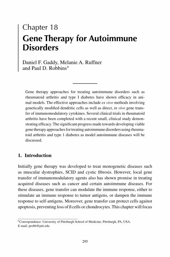
May 4, 2010 11:0 SPI-B903 9in x 6in b903-ch18
Chapter 18
Gene Therapy for AutoimmuneDisorders
Daniel F. Gaddy, Melanie A. Ruffnerand Paul D. Robbins∗
Gene therapy approaches for treating autoimmune disorders such asrheumatoid arthritis and type I diabetes have shown efficacy in ani-mal models. The effective approaches include ex vivo methods involvinggenetically modified dendritic cells as well as direct, in vivo gene trans-fer of immunomodulatory cytokines. Several clinical trials in rheumatoidarthritis have been completed with a recent small, clinical study demon-strating efficacy. The significant progress made towards developing viablegene therapy approaches for treating autoimmune disorders using rheuma-toid arthritis and type 1 diabetes as model autoimmune diseases will bediscussed.
1. Introduction
Initially gene therapy was developed to treat monogenetic diseases suchas muscular dystrophies, SCID and cystic fibrosis. However, local genetransfer of immunomodulatory agents also has shown promise in treatingacquired diseases such as cancer and certain autoimmune diseases. Forthese diseases, gene transfer can modulate the immune response, either tostimulate an immune response to tumor antigens, or dampen the immuneresponse to self-antigens. Moreover, gene transfer can protect cells againstapoptosis, preventing loss of ß cells or chondrocytes. This chapter will focus
∗Correspondence: University of Pittsburgh School of Medicine, Pittsburgh, PA, USA.E-mail: [email protected]
295
May 4, 2010 11:0 SPI-B903 9in x 6in b903-ch18
A Guide to Human Gene Therapy
on the significant progress that has been made towards developing clinicallyuseful approaches to treat two types of autoimmune diseases: rheumatoidarthritis and type I diabetes.
2. Rheumatoid Arthritis
2.1 Background
Arthritis is the leading cause of disability among adults in the United States,affecting approximately 21% (more than 46 million) adults. Rheumatoidarthritis (RA) affects approximately 1.3 million adults in the United States,and more than 60 million people worldwide.1 RA is characterized by inflam-mation of the synovial lining and destruction of extra-articular bone andcartilage.
It is believed that arthritogenic peptides, either from foreign or self-proteins, are presented to T cells preferentially by MHC molecules onantigen-presenting cells. Activated T cells produce a variety of proinflam-matory cytokines, including tumor necrosis factor (TNF)-α, interleukin(IL)-1 and IL-6. The inflammatory response induced by these cytokinesis directly responsible for the overt RA symptoms, including joint pain,swelling, effusion and stiffness.
2.2 Existing Therapies
Despite the prevalence and rising economic burden of RA, effective thera-pies for this disease remain limited, and there is no cure. Current therapiesconsist of early, aggressive and continuous treatment with non-steroidalanti-inflammatory drugs (NSAIDs) and disease-modifying antirheumaticdrugs (DMARDs). Among the DMARDs, methotrexate has long been thedrug of choice for RA, but newer biologic targeted therapies have emerged inrecent years. These targeted therapies include abatacept (Orencia®), a fusionprotein composed of the extra-cellular domain of cytotoxic T lymphocyteantigen-4 fused to an immunoglobulin (CTLA4-Ig); anakinra (Kineret®),an interleukin-1 receptor antagonist (IL-1Ra); infliximab (Remicade®), achimeric anti–TNFα antibody; adalimumab (Humira®), a fully human anti–TNFα antibody; and etanercept (Enbrel®), a soluble TNF-α receptor.2
296
May 4, 2010 11:0 SPI-B903 9in x 6in b903-ch18
Gene Therapy for Autoimmune Disorders
2.3 Target Tissues and Routes of Delivery
2.3.1 Local RA Gene Therapy
Synovium, the soft tissue that lines non-cartilaginous surfaces of joints, isthe primary target for intra-articular gene therapy because synovium hasa large surface area and direct access to the joint space. This is achievedby directly injecting vectors or cells expressing the therapeutic gene ofinterest into individual diseased joints, which limits the amount of vectorneeded compared to systemic injections. The drawbacks of this approachare that multiple joints may require injections in patients with polyarticulardisease, and that extra-articular manifestations of the disease may not beaffected.
One method to overcome this drawback is the use of genetically modi-fied dendritic cells3 or T cells.4 When transduced with anti-arthritic genesusing viral vectors, these cells demonstrate the ability to home in on mul-tiple joints or extra-articular tissues affected by RA, such as lymph nodes.Similarly, vesicles, known as exosomes, derived from genetically modifieddendritic cells, or exosomes from immature dendritic cells that are subse-quently treated with immunomodulatory factors, reduce inflammation andseverity of disease in arthritis mouse models.5
2.3.2 Systemic RA Gene Therapy
Systemic delivery, in which vectors are administered extra-articularly, suchas via intramuscular or intravenous injections, has been attempted in mul-tiple studies (Fig. 18.1). Systemic injection will result in relatively highcirculating levels of therapeutic proteins and lower concentrations of ther-apeutic proteins in the joints. It is possible that this method will improvethe ability of gene therapy to treat polyarticular or extra-articular mani-festations of RA, and do so in a manner that improves upon conventionalbiologic therapies that require frequent injections or infusions. While thisstrategy has demonstrated promising results in animal models, it has mul-tiple drawbacks, including the requirement for large doses of vectors tofacilitate systemic infection, and the observation that the majority of viralvectors, when administered systemically, will home in primarily on the liver,as opposed to joints or other sites of disease.6
297
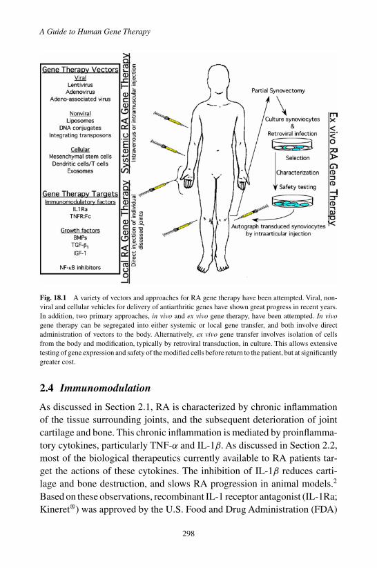
May 4, 2010 11:0 SPI-B903 9in x 6in b903-ch18
A Guide to Human Gene Therapy
Fig. 18.1 A variety of vectors and approaches for RA gene therapy have been attempted. Viral, non-viral and cellular vehicles for delivery of antiarthritic genes have shown great progress in recent years.In addition, two primary approaches, in vivo and ex vivo gene therapy, have been attempted. In vivogene therapy can be segregated into either systemic or local gene transfer, and both involve directadministration of vectors to the body. Alternatively, ex vivo gene transfer involves isolation of cellsfrom the body and modification, typically by retroviral transduction, in culture. This allows extensivetesting of gene expression and safety of the modified cells before return to the patient, but at significantlygreater cost.
2.4 Immunomodulation
As discussed in Section 2.1, RA is characterized by chronic inflammationof the tissue surrounding joints, and the subsequent deterioration of jointcartilage and bone. This chronic inflammation is mediated by proinflamma-tory cytokines, particularly TNF-α and IL-1β. As discussed in Section 2.2,most of the biological therapeutics currently available to RA patients tar-get the actions of these cytokines. The inhibition of IL-1β reduces carti-lage and bone destruction, and slows RA progression in animal models.2
Based on these observations, recombinant IL-1 receptor antagonist (IL-1Ra;Kineret®) was approved by the U.S. Food and Drug Administration (FDA)
298

May 4, 2010 11:0 SPI-B903 9in x 6in b903-ch18
Gene Therapy for Autoimmune Disorders
for RA therapy in 2001. Similarly, inhibition of TNF-α activity amelioratesjoint disease in animal models.2 Three drugs, Humira®, Remicade®, andEnbrel®, have been licensed by the FDA for use as TNFα inhibitors for thetreatment of RA and other inflammatory diseases.
Other strategies to treat RA via immunomodulation include blocking theactivity of IL-6 and NF-κB. As of this writing, several clinical trials, includ-ing 5 phase III trials, are underway and indicate that an antibody againstthe IL-6 receptor shows some therapeutic benefit in patients with RA andsystemic-onset juvenile idiopathic arthritis.7 In addition, a growing body ofevidence indicates that NF-κB may be a master regulator of RA. Numer-ous inhibitors of the IKK complex, the enzymatically active component ofthe NF-κB complex, have been developed, several of which have enteredclinical trials for the treatment of RA.8
2.5 Overview of Preclinical Gene Therapy Studies
Given the success of biologic targeted therapies, various strategies havebeen employed to utilize these immunomodulatory agents in RA gene ther-apy. Multiple preclinical RA gene therapy studies have demonstrated thatgene therapy vectors, including retrovirus and adenovirus vectors, express-ing IL-1Ra produce high levels of the transgene in target tissues and inhibitinflammation and cartilage loss in animal models. Similarly, numerous stud-ies have demonstrated the effectiveness of blocking TNF-α activity via genetherapy, including the suppression of inflammatory cell infiltration, pannusformation, cartilage and bone destruction, and expression of joint proin-flammatory cytokines in animal models.2
Furthermore, adeno-associated virus (AAV) expressing a TNFR:Fcfusion gene under the control of an inflammation-inducible NF-κB pro-moter delayed disease onset and decreased the incidence and sever-ity of joint damage in mouse and rat arthritis models.2 This type ofgene therapy, utilizing a disease-inducible promoter, is of particularinterest in autoimmune diseases like RA, which are characterized byflare-ups followed by periods of disease regression. By utilizing theinflammation-inducible NF-κB promoter, high levels of transgene expres-sion are obtained only during disease flares, preventing unnecessary expo-sure of the patient to immunosuppressive agents during periods of diseaseregression.
299
May 4, 2010 11:0 SPI-B903 9in x 6in b903-ch18
A Guide to Human Gene Therapy
In addition, both adenovirus and AAV have been utilized to deliverNF-κB inhibitors to synovium, successfully preventing expression of proin-flammatory cytokines.9,10 One potential drawback of using non-secretedfactors, such as IKK complex inhibitors, is that only directly transducedcells will express the therapeutic gene, whereas secreted factors, such asIL-1Ra and soluble TNFR, will mediate protection to the entire inflamedenvironment.
A variety of growth factors has also been studied as therapeutics for RA.Bone morphogenetic proteins (BMPs), particularly BMP-2 and BMP-7,have been shown to induce chondrogenesis and osteogenesis when deliv-ered by adenovirus or AAV vectors.2 Additional growth factors that havebeen studied in relation to RA gene therapy include transforming growthfactor (TGF)-β1 and insulin-like growth factor (IGF)-1. Adenovirus or AAVvectors expressing TGF-β1 have been used to transduce mesenchymal stemcells (MSCs) and drive ex vivo differentiation of MSCs into chondrocytes,facilitating cartilage repair in animal models.11 Similarly, ex vivo chon-drocytes transduced with an adenovirus expressing IGF-1 enhanced matrixsynthesis and cartilage repair in horses.12 These studies suggest importantroles for growth factors in RA gene therapy, and indicate that a variety ofgrowth factors may be able to join the list of effective RA therapies.
Non-viral gene transfer systems, including naked DNA, cationic lipo-somes, histones and polymers, have experienced limited use for the treat-ment of RA because RA will necessarily require long-term, high-levelexpression of therapeutic transgenes, which has traditionally not been anoption with non-viral systems.2 However, recent advances may overcomethis issue. For example, transient expression of the phage phiC31 inte-grase allows for efficient, targeted integration of plasmid DNA. Also, plas-mids containing vectors based on transposons such as Sleeping Beautyor Himar1 also can confer efficient integration when delivered in con-junction with the appropriate transposase. While these vectors have notbeen extensively tested for RA gene therapy, phiC31 integrase and Himar1transposase have been shown to confer stable transgene expression in cul-tured human synoviocytes and in the rabbit knee following intra-articulardelivery.2 Thus it is possible that these non-viral, integrating vector sys-tems, when delivered intra-articularly, could be useful for chronic RA genetherapy.
300

May 4, 2010 11:0 SPI-B903 9in x 6in b903-ch18
Gene Therapy for Autoimmune Disorders
2.6 Overview of Clinical Gene Therapy Studies
As illustrated in Fig. 18.1, early arthritis gene transfer trials used retrovirusvectors in ex vivo protocols to deliver IL-1Ra to the metacarpophalangealjoints of RA patients.13 These safety and feasibility studies illustrated thatgene transfer to RA joints could be safe and effective, but the low numbers ofpatients enrolled prevented any conclusions with regard to efficacy.A similarphase I trial has been initiated by TissueGene, Inc utilizing retroviral vectorsexpressing TGF-β1 to transduce human chondrocytes, which are then mixedwith normal human chondrocytes and injected into the knee of patients withdegenerative joint disease. As of this writing, 16 patients have been treatedin the United States and South Korea, with approximately 50% of the treatedpatients demonstrating symptomatic improvement.2,14
Another RA gene therapy trial, sponsored by Targeted Genetics, Incevaluated the safety and efficacy of a single-stranded rAAV2 virus express-ing the complete coding sequence of a TNFR:Fc fusion protein, whichis identical to etanercept. This trial received a great deal of publicity in2007 because of the death of an enrolled patient. Subsequent investigationdetermined that the death of the subject was not likely due to the viralvector.2 Despite the death of this subject, the Targeted Genetics trial hasshown promising results. The AAV vector appears safe, in that there isno evidence of circulating TNFR:Fc or extra-articular over-expression ofTNFR-Fc, which would have indicated vector dissemination and amplifica-tion in extra-articular tissues. Clinical response was assessed using patientreported outcomes, revealing moderate improvement in target joint pain andswelling.15
3. Type I Diabetes Mellitus
3.1 Background
Type 1 diabetes (T1D), also known as insulin-dependent diabetes mellitus,is recognized as a rapidly growing health threat worldwide. The CDC esti-mates that 15,000 young people in the United States per year are diagnosedwith T1D, with 19 new cases per 100,000 youth each year.16 Worldwide,the incidence of diabetes varies greatly by geographical location, rangingfrom 0.1 per 100,000/year in China and Venezuela to 40.9 per 100,000/year
301
May 4, 2010 11:0 SPI-B903 9in x 6in b903-ch18
A Guide to Human Gene Therapy
in Finland, and has been increasing since the 1950s.17 From 1990–1999,the incidence of T1D worldwide increased 2.8%, confirming the need fornew methods to address this growing public health concern.17
T1D is a polygenic autoimmune disease characterized by the T cell-mediated destruction of insulin-producing β cells in the pancreatic isletsof Langerhans, leading to insulin deficiency. T1D is a chronic disease thatis known to exist for years in a preclinical phase before the classic mani-festations are observed. This preclinical phase, known as insulitis, is char-acterized by the infiltration of leukocytes into the islets. The actual onsetof diabetes occurs after most β cells have been killed, resulting in insulindeficiency and hyperglycemia. β cell auto-antigens, such as glutamic aciddecarboxylase (GAD) and insulin, are processed by antigen presenting cells(APC), including macrophages, dendritic cells, and B cells, in the pancreaticislets. APC then present processed peptides to autoreactive CD4+ T cellsin the peripheral lymphoid tissues. Activated autoreactive CD4+ T cellssecrete cytokines and activate β cell-specific cytotoxic CD8+ T cells. Acti-vated CD8+ T cells (CTL) then migrate to the islets and produce cytokines,which activate more CTL, as well as macrophages, further contributing tothe destruction of β cells.18 In general, TH1 cytokines, such as IL-2, inter-feron (IFN)-γ , and TNF-α, promote T1D development, while TH2 or TH3cytokines, such as IL-4, IL-10, and TGF-β, prevent T1D onset. β cell deathis the direct result of these cytotoxic cytokines and other agents, includ-ing oxygen free radicals, granzyme and perforin, released from CTL andmacrophages. Additionally, Fas and TNF receptor-mediated apoptosis playroles in β cell death.18
3.2 Existing Therapies
T1D patients require lifelong insulin replacement therapy, and are at risk ofdeveloping significant complications associated with hyperglycemia, suchas retinopathy, neuropathy, nephropathy, and accelerated peripheral vascu-lar and coronary artery disease. The goal of existing therapy is to providetight glycemic control in all diabetic patients in order to minimize the com-plications associated with hyperglycemia.
Furthermore, while allogeneic islet transplantation has shown some evi-dence of success, the allo- and autoimmune response against the islets often
302

May 4, 2010 11:0 SPI-B903 9in x 6in b903-ch18
Gene Therapy for Autoimmune Disorders
leads to their destruction.18,19 Therefore patients receiving grafted isletsrequire lifelong immunosuppressive therapy, which carries significant sideeffects. Due to the need for lifelong immunosuppression, whole-pancreastransplant is rarely performed alone, and more frequently performed inT1D patients undergoing kidney transplant for diabetic nephropathy. Thewidespread applicability of both of these techniques is limited by the supplyof islets from cadaveric donors, and in general both procedures have beenoffered only to T1D patients who have hypoglycemic unawareness, severemetabolic or secondary complications, or are unable to follow an insulinregimen.20
3.3 Target Tissues and Routes of Delivery
As shown in Fig. 18.2, T1D interventions have taken many forms, and as aresult, many different target tissues have been tried. Chief among the targettissues investigated has been the pancreatic islets. The vast majority of thestudies utilizing gene transfer strategies to enhance islet function or survivalhave used an ex vivo transduction protocol, which follows logically fromthe islet transplant protocols currently in clinical use. In vivo transductionprotocols of β cells are more rare, but have been accomplished using AAVvectors.21
In some cases, systemic expression of protein has been desired for ther-apy of T1D. Examples of this may include expression of insulin to regulatethe blood glucose or systemic expression of an immunomodulatory agent inorder to prevent the onset of diabetes. In these cases, investigators have uti-lized the relative ease of transduction of other tissues, such as liver or muscleto achieve high transduction efficiencies and better systemic expression ofprotein.
3.4 Immunomodulation
Over the past 30 years, significant efforts have been made to test the efficacyof immunomodulating therapies in T1D.An ideal immunomodulating agentfor diabetes would specifically halt β cell destruction without causing sys-temic immunosuppression or inhibiting the process of β cell regeneration.It has been demonstrated in both humans and non-obese diabetic (NOD)mice that at the onset of clinical diabetes there is still significant residual
303

May 4, 2010 11:0 SPI-B903 9in x 6in b903-ch18
A Guide to Human Gene Therapy
Fig. 18.2 A variety of vectors and approaches for T1D gene therapy have been attempted. Viral, non-viral and cellular vehicles for delivery of genes have been utilized. Both in vivo and ex vivo gene therapyhas been attempted. Ex vivo gene transfer involves isolation of cells from the body and modificationin culture as shown for antigen presenting cells in A, and pancreatic islet transplant in D. In vivo genetherapy involves direct administration of vectors to the body, either directly to the pancreas as shownin B, or to another target such as muscle (as in C) or liver for systemic expression of transgene.
β cell mass, and early intervention with an effective immunotherapy duringthis period has the potential to restore tolerance and allow endogenous cellsto regenerate the islets.22 However, some immunosuppressive drugs usedin common islet transplant protocols have been shown to inhibit sponta-neous regeneration of β cell mass in vivo, and therefore drugs intended forthis application must be carefully tested for their effects on β cell growthin vivo.23 Anti-CD3 has shown some promise in clinical trials, but recent dataconcerning potentially serious systemic side effects has dampened enthu-siasm for this molecule.24 Trials are also underway investigating the ther-apeutic potential of rituximab, an anti-CD20 antibody that down regulatesthe B cell signaling of T cells.
In terms of diabetes antigen-specific studies, systemic or oral adminis-tration of insulin given to high-risk relatives had no efficacy in preventing
304
May 4, 2010 11:0 SPI-B903 9in x 6in b903-ch18
Gene Therapy for Autoimmune Disorders
diabetes onset when compared to a matched control group.26 Other trialsexamining the benefits of intranasally administered insulin, as well as anintramuscular alum-Gad65 preparation, have shown some initial promiseand work is ongoing to investigate the ultimate benefit of these methods.25,26
Collectively, these studies illustrate the challenges facing investigatorsin finding appropriate immunotherapies for T1D. At this point, it remainsunclear, which agents are the most beneficial at halting the progression ofautoimmunity as many underlying aspects of the pathogenesis of T1D haveyet to be elucidated. The clinical heterogeneity of human disease when com-pared to the homogeneity of inbred animal models, such as the NOD mouse,remains another challenge, as many interventions that have prevented dia-betes in preclinical models do not have promising results when translatedinto human therapy.
3.5 Overview of Preclinical Gene Therapy Studies
Until recently, gene therapy studies for T1D had focused primarily on ex vivoapproaches to modify islets for transplantation. Although many differentapproaches have been examined, the goals of these modifications are sim-ilar: to transfer a gene encoding a protein which would confer some typeof islet-protective effect to the grafted islets in order to protect them fromallo- and autoimmune attack when transplanted into the patient. When suc-cessful, this prolongs graft survival and may potentially reduce the need forsystematic immunosuppressive therapy to prevent loss of the graft. Somelingering concerns with the use of transduced islets are safety and efficacy.Particular concerns include whether expression of virally encoded proteinsfrom the islets will have some effect on the cellular function of the trans-planted cells or immunological function of the patient receiving the graft.
Another major focus of ex vivo gene therapy strategies for T1D hasbeen in the modification of immunological cells to promote tolerance uponadoptive transfer in vivo. Dendritic cells have been of particular interestfor applications in T1D because of their unique role in regulating T cellresponses. Both adenoviral and lentiviral gene transfer of IL-4 to DC havebeen shown to have protective effects in the NOD model of T1D.27,28
Others have shown that modifying DC with an adenoviral vector encod-ing for galectin-1, a lectin with regulatory effects on T cells, could deletediabetogenic T cells in vivo.29 Lastly, approaches using oligonucleotides
305
May 4, 2010 11:0 SPI-B903 9in x 6in b903-ch18
A Guide to Human Gene Therapy
have been particularly successful. NF-κB decoy oligoneucleotide treatmentof DC was shown to inhibit DC activation and maturation, inhibit the expres-sion of co-stimulatory molecules, and prevent the onset of diabetes in theNOD mouse model. These studies have progressed into clinical trials.30
They are described in further detail in Section 3.6. In addition, retroviraltransduction of genes encoding Gad65-IgG or (pro)Insulin-IgG fusion pro-teins into B cells resulted in a suppressive B cell phenotype which was capa-ble of reducing diabetes incidence in a regulatory T cell-dependent man-ner on adoptive transfer into NOD mice.31 Approaches using the patient’sautologous antigen-presenting cells are particularly attractive because ofthe known importance of these cells in regulating autoimmunity, as well asthe ease and relative safety of ex vivo transduction protocols.
In vivo gene therapy efforts for T1D have primarily focused on address-ing the underlying autoimmune destruction and loss of β cell mass ratherthan on insulin replacement strategies.32 Many in vivo efforts have beendirected towards modifying nonpancreatic tissues, such as liver and muscle,to systemically express insulin or immuno-regulatory compounds in orderto modulate autoimmunity in models of T1D.33
For many years, the potential of gene therapy as a means to treat T1Dwas hindered by the lack of an effective vehicle to target genes specificallyto the pancreas in vivo. However, recent studies have demonstrated that adouble-stranded AAV vector utilizing the insulin promoter provided stable,long-term expression that is specific to β cells. Use of this system to deliverIL-4 was protective against insulitis and hyperglycemia in the NOD model,and was able to maintain levels of regulatory T cells as the mice aged.34
In vivo transduction of islets could be of major benefit in preserving orenhancing the amount of β cell mass remaining in T1D patients at diagnosis.
3.6 Overview of Clinical Gene Therapy Studies
To date, few proposed gene therapy strategies for T1D have progressed to thepoint of clinical trials. A phase I clinical trial to examine safety is currentlyongoing using DC genetically modified using antisense oligonucleotides(AS-ODN) to the co-stimulatory molecules CD40, CD80, and CD86. In pre-clinical studies using the NOD mouse model of T1D, investigators demon-strated that the AS-ODN treated bone-marrow derived DC were able todelay the incidence of diabetes after a single injection, and observed that the
306
May 4, 2010 11:0 SPI-B903 9in x 6in b903-ch18
Gene Therapy for Autoimmune Disorders
AS-ODN treated DC resulted in an expansion of a CD4+CD25+CD62L+
regulatory T cell population.35 It is believed that the lack of co-stimulatorymolecules on the DC result in an anergizing signal to diabetogenic T cells,as well as expansion of regulatory T cells. In the ongoing clinical study,autologous DC are generated from T1D patient’s leukocytes in vitro afterharvest by leukapheresis. The DC are cultured and engineered using theAS-ODN to CD40, CD80, and CD86 and then injected intradermally at asite distal to the pancreas to allow for DC migration into the lymph nodes.At the time of writing, the phase I trial is currently ongoing and is focusedon the safety of this approach in a patient population that is over 18 andhas had insulin-dependent T1D for greater than five years.36 If successful,the next step planned by investigators will be a phase II trial focused onefficacy of treatment in newly-diagnosed T1D patients, in fitting with theparadigm that early intervention with immunotherapies is most effective atsaving any residual β cell mass.
Some additional phase I/II trials have occurred for gene therapy prod-ucts, which are not specifically targeted towards T1D, but rather are aimedat treating hyperglycemic complications that can occur with Type 1 or 2diabetes. For example, Tissue Repair Company has recruited patients fortrials of EXCELLERATE, which is a collagen-matrix gel containing aden-oviral vectors expressing platelet-derived growth factor for the treatment ofchronic lower extremity ulcers in diabetic patients. Another clinical trial isalso ongoing looking at the gene transfer of vascular-endothelium derivedgrowth factor for the treatment of diabetic neuropathy.37 As of this writ-ing, results from these trials are eagerly anticipated, but have not yet beenpublished.
4. Conclusions and Outlook
As outlined in this chapter, considerable progress has been made towardsdeveloping gene therapy approaches for autoimmune disease such as RAand T1D, leading to several phase I clinical trials, and one phase II trial inRA. There is also evidence of clinical responses in certain subjects, suggest-ing that additional trials to establish efficacy are merited. Although not dis-cussed in this chapter, proof of principle also has been established in animalmodels of Sjogren’s syndrome and lupus. Because these diseases, unlike
307

May 4, 2010 11:0 SPI-B903 9in x 6in b903-ch18
A Guide to Human Gene Therapy
RA and T1D, do not respond well to present biologics, the developmentof alternative approaches, such as gene therapy, seems highly appropriate.Furthermore, although osteoarthritis (OA) is not an autoimmune disease, itshares certain pathology with RA. Two clinical trials exploring the use ofgene therapy in OA are underway and another is in the pipeline. Given therecurrently is no effective treatment for OA, gene therapy offers the potentialto improve the quality of life for the 10% of the population that will developthis disease. Overall the technology of gene transfer, along with efficacy ofgene therapy in animal models of autoimmune diseases, has developed tothe point where it is no longer the rate-limiting step for many purposes.Instead, the focus of the field of gene therapy for autoimmune diseases isnow on bringing these approaches into the clinic.
References
1. Lundkvist J, Kastäng F, Kobelt G (2008). The burden of rheumatoid arthritis and accessto treatment: Health burden and costs. The European Journal of Health Economics8: S49–S60.
2. Gaddy DF, Robbins PD (2008). Current status of gene therapy for rheumatoid arthritis.Current Rheumatology Reports 10: 398–404.
3. Kim SH, Kim S, Oligino TJ, Robbins PD (2002). Effective treatment of establishedmouse collagen-induced arthritis by systemic administration of dendritic cells geneti-cally modified to express FasL. Mol Ther 6: 584–590.
4. Nakajima A, et al. (2001). Antigen-specific T cell-mediated gene therapy in collagen-induced arthritis. J Clin Invest 107: 1293–1301.
5. Bianco NR, Kim SH, Morelli AE, Robbins PD (2007). Modulation of the immuneresponse using dendritic cell-derived exosomes. Methods Mol Biol 380: 443–455.
6. Evans C, Ghivizzani S, Robbins P (2006). Gene therapy for arthritis: What next? Arthri-tis and rheumatism 54: 1714–1729.
7. Finckh A, Gabay C (2008). At the horizon of innovative therapy in rheumatology: Newbiologic agents. Current opinion in rheumatology 20: 269–275.
8. Calzado MA, Bacher S, Schmitz ML (2007). NF-kappaB inhibitors for the treatmentof inflammatory diseases and cancer. Current medicinal chemistry 14: 367–376.
9. Amos N, Lauder S, EvansA, Feldmann M, Bondeson J (2006).Adenoviral gene transferinto osteoarthritis synovial cells using the endogenous inhibitor IkappaBalpha revealsthat most, but not all, inflammatory and destructive mediators are NFkappaB dependent.Rheumatology (Oxford, England) 45: 1201–1209.
10. Tas SW, et al. (2006). Amelioration of arthritis by intra-articular dominant nega-tive Ikk beta gene therapy using adeno-associated virus type 5. Human gene therapy17: 821–832.
308

May 4, 2010 11:0 SPI-B903 9in x 6in b903-ch18
Gene Therapy for Autoimmune Disorders
11. Pagnotto M, Wang Z, Karpie J, Ferretti M, Xiao X, Chu C (2007). Adeno-associatedviral gene transfer of transforming growth factor-beta1 to human mesenchymal stemcells improves cartilage repair. Gene therapy 14: 804–813.
12. Goodrich L, Hidaka C, Robbins P, Evans C, Nixon A (2007). Genetic modification ofchondrocytes with insulin-like growth factor-1 enhances cartilage healing in an equinemodel. J Bone Joint Surg Br 89: 672–685.
13. Evans C, et al. (1996). Clinical trial to assess the safety, feasibility, and efficacy oftransferring a potentially anti-arthritic cytokine gene to human joints with rheumatoidarthritis. Human gene therapy 7: 1261–1280.
14. Lee K (2008). Preclincal and early clinical analysis of allogeneic chondrocytes trans-fected retrovirally with TGF-beta1 gene for degenerative arthritis patients. In 5th Inter-national Meeting of Gene Therapy of Arthritis and Related Disorders, Seattle, WA,USA.
15. Mease P, et al. (2008). Safety, Local Tolerability and Clinical Response After Intra-articular Administration of a Recombinand Adeno-associated Vector Containing a TNFAntagonist in Inflammatory Arthritis. In EULAR Congress 2008, Paris, Fance.
16. (2008). Centers for Disease Control and Prevention. National Diabetes Fact Sheet:United States 2007. General Information and National Estimates on Diabetes. Availableat: http://wwwcdcgov/diabetes/pubs/pdf/ndfs_2007pdf.
17. (2006). Incidence and trends of childhood Type 1 diabetes worldwide 1990–1999.Diabet Med 23: 857–866.
18. Marzorati S, Pileggi A, Ricordi C (2007). Allogeneic islet transplantation. Expert OpinBiol Ther 7: 1627–1645.
19. van der Windt DJ, Bottino R, Casu A, Campanile N, Cooper DK (2007). Rapid lossof intraportally transplanted islets: An overview of pathophysiology and preventivestrategies. Xenotransplantation 14: 288–297.
20. Meloche RM (2007). Transplantation for the treatment of type 1 diabetes. World JGastroenterol 13: 6347–6355.
21. Wang Z, et al. (2006). Widespread and stable pancreatic gene transfer by adeno-associated virus vectors via different routes. Diabetes 55: 875–884.
22. Pasquali L, Giannoukakis N, Trucco M (2008). Induction of immune tolerance tofacilitate beta cell regeneration in type 1 diabetes. Adv Drug Deliv Rev 60: 106–113.
23. Nir T, Melton DA, Dor Y (2007). Recovery from diabetes in mice by beta cell regen-eration. J Clin Invest 117: 2553–2561.
24. Gandhi GY, et al. (2008). Immunotherapeutic agents in type 1 diabetes: A systematicreview and meta-analysis of randomized trials. Clin Endocrinol (Oxf) 69: 244–252.
25. Harrison LC, et al. (2004). Pancreatic beta-cell function and immune responses toinsulin after administration of intranasal insulin to humans at risk for type 1 diabetes.Diabetes Care 27: 2348–2355.
26. Ludvigsson J, et al. (2008). GAD treatment and insulin secretion in recent-onset type1 diabetes. N Engl J Med 359: 1909–1920.
27. Creusot RJ, et al. (2008). Tissue-targeted therapy of autoimmune diabetes using den-dritic cells transduced to express IL-4 in NOD mice. Clin Immunol 127: 176–187.
28. Feili-Hariri M, et al. (2003). Dendritic cells transduced to express interleukin-4 pre-vent diabetes in nonobese diabetic mice with advanced insulitis. Hum Gene Ther 14:13–23.
309

May 4, 2010 11:0 SPI-B903 9in x 6in b903-ch18
A Guide to Human Gene Therapy
29. Perone MJ, et al. (2006). Dendritic cells expressing transgenic galectin-1 delay onsetof autoimmune diabetes in mice. J Immunol 177: 5278–5289.
30. Ma L, et al. (2003). Prevention of diabetes in NOD mice by administration of dendriticcells deficient in nuclear transcription factor-kappaB activity. Diabetes 52: 1976–1985.
31. Soukhareva N, Jiang Y, Scott DW (2006). Treatment of diabetes in NOD mice by genetransfer of Ig-fusion proteins into B cells: Role of T regulatory cells. Cell Immunol240: 41–46.
32. Samson SL, Chan L (2006). Gene therapy for diabetes: Reinventing the islet. TrendsEndocrinol Metab 17: 92–100.
33. Siatskas C, et al. (2006). Gene therapy strategies towards immune tolerance to treat theautoimmune diseases. Curr Gene Ther 6: 45–58.
34. Rehman KK, Trucco M, Wang Z, Xiao X, Robbins PD (2008). AAV8-mediated genetransfer of interleukin-4 to endogenous beta-cells prevents the onset of diabetes in NODmice. Mol Ther 16: 1409–1416.
35. Machen J, Harnaha J, Lakomy R, Styche A, Trucco M, Giannoukakis N (2004).Antisense oligonucleotides down-regulating costimulation confer diabetes-preventiveproperties to nonobese diabetic mouse dendritic cells. J Immunol 173: 4331–4341.
36. Giannoukakis N, Phillips B, Trucco M (2008). Toward a cure for type 1 diabetes mel-litus: Diabetes-suppressive dendritic cells and beyond. Pediatr Diabetes 9: 4–13.
37. Prieto J, Fernandez-Ruiz V, Kawa MP, Sarobe P, Qian C (2008). Cells as vehicles fortherapeutic genes to treat liver diseases. Gene Ther 15: 765–771.
310
May 4, 2010 11:0 SPI-B903 9in x 6in b903-ch19
Chapter 19
Gene Therapy for InheritedMetabolic Storage Diseases
Cathryn Mah∗
In this chapter, we will discuss the principles surrounding the study anddevelopment of gene therapy for inherited metabolic storage diseases.These rare diseases are caused primarily by a defect in a single gene andresult in the excess storage of substrates within affected cells, ultimatelyrendering the cells dysfunctional. There exist numerous classifications ofinherited metabolic storage diseases which include two broad categories:lysosomal storage diseases and glycogen storage diseases. No cures existfor any of the diseases and current therapies are palliative. Gene therapyprovides a possible alternative therapy for inherited metabolic storagediseases and here we discuss current gene therapy strategies and consid-erations for strategy design for representative diseases including MPS I,Gaucher disease, Pompe disease, and glycogen storage disease type Ia.
1. Introduction
The majority of gene therapy studies for inherited metabolic storage dis-eases have employed viral-based gene therapy vectors, in particular retro-viral, lentiviral, adenoviral, and adeno-associated viral vectors. Other viraland non-viral vectors have also been explored to a lesser extent. Treatmentstrategies have varied substantially, in that different vectors, differingcis-elements within each vector construct, methods of vector delivery, andthe target tissue(s) have differed, even when approaching the same disease.
∗Correspondence: University of Florida College of Medicine, Dept. Pediatrics, Div. Cellular MolecularTherapy, and Powell Gene Therapy Center, 1376 Mowry Road, Rm 493, Gainesville, FL 32610.E-mail: [email protected]
311

May 4, 2010 11:0 SPI-B903 9in x 6in b903-ch19
A Guide to Human Gene Therapy
However, the primary goals of sufficient transduction of the affected tis-sue(s), sustained expression of the delivered gene at therapeutic levels, andavoidance of untoward or adverse effects such as immune response to vectoror transgene, are common for each.
While at the time of writing of this chapter, a gene therapy strategy to treatan inherited metabolic storage disorder has not yet yielded a complete curefor disease, nor has a gene therapy treatment been approved as a standardof care, data suggest that significantly improved functional outcomes canbe achieved using gene therapy as compared to current therapies. Each newstudy has provided information towards design of improved strategies andfurther research can only move the field of gene therapy closer toward thegoal of a cure.
2. Lysosomal Storage Diseases
Lysosomal storage diseases (LSDs) are a class of inherited metabolic stor-age diseases encompassing greater than 40 distinct diseases. Lysosomesare intracellular organelles that contain enzymes that play important rolesin the break down of complex substrates and macromolecules. Most LSDsare caused by a deficiency in a functional lysosomal enzyme, however defi-ciencies in nonenzymatic activator proteins, proteins required for properintracellular trafficking of lysosomal enzymes, or mutations in integral lyso-somal membrane proteins can also result in disease. LSDs are characterizedby excess storage of undigested or partially digested materials within thelysosomes of affected cells. Accumulation of substrates within lysosomeseventually leads to disruption of normal cellular architecture and ultimatelycellular/tissue function. In some cases, the substrates themselves can becytotoxic. Affected tissues range from skeletal and cardiac muscle to tis-sues comprising the central nervous system.1 While gene therapy strategiesare being developed for numerous LSDs, two LSDs, mucopolysaccharidosisType I and Gaucher disease, will be discussed in this chapter as examples.
Mucopolysaccharidoses (MPS disorders) are a group of LSDs charac-terized by the inability to degrade glycosaminoglycans used to build bone,cartilage, tendons, joint fluid, and other tissues. MucopolysaccharidosisType I (MPS I) is caused by a lack of alpha-L-iduronidase (IDUA), result-ing in the build-up of dermatan and heparan sulfate. MPS I can range from
312
May 4, 2010 11:0 SPI-B903 9in x 6in b903-ch19
Gene Therapy for Inherited Metabolic Storage Diseases
a severe form (also known as Hurler syndrome) to mild (Scheie syndrome)form. Clinical features can vary greatly with the severity of disease, how-ever hallmark complications can include joint stiffness and skeletal dys-plasia, cardiovascular disease, liver and/or spleen enlargement, cloudingof the cornea, developmental delay, and characteristic facial and physicaldysmorphisms.1,2
Gaucher disease is the most common LSD and results in the storage ofthe lipid glucocerebroside primarily in macrophages and monocytes due toa defect in beta-glucosidase enzyme. Clinical symptoms of Gaucher diseaseinclude hepatosplenomegaly, anemia, thrombocytopenia, leukopenia, andsevere bone disease. Like with most other LSDs, there exist subtypes ofdisease that are characterized by severity and differing clinical symptoms.Type 1 Gaucher disease is the most prevalent form of Gaucher disease andis non-neuropathologic. Gaucher disease Types 2 and 3 are characterizedby having acute or chronic neurologic symptoms, respectively.1,2
Current therapies for both MPS I and Gaucher disease include bone mar-row/hematopoietic stem cell transplantation and/or recombinant enzymereplacement therapy (ERT) or substrate reduction therapy (SRT).3−8 Whilethese therapies have been shown to improve survival and quality of life,in most cases, disease is not cured, and complications can arise. Bonemarrow/stem cell transplantation has been employed as blood cells havebeen shown to express and secrete the deficient enzymes in MPS I andGaucher disease. The primary acute complication with this form of ther-apy is rejection of the graft, and while disease progression can be halted ordelayed, bone and neurological components of disease cannot be reversed.ERT requires repeated intravenous dosing for the lifetime of the patient andcan correct non-neuronopathlogies however, due to the blood-brain bar-rier, ERT is unable to correct neurological manifestations. SRT utilizes oraldosing of small molecule compounds to inhibit the formation of glycosphin-golipids. The potential advantage of SRT compounds is the ability to crossthe blood-brain barrier, which theoretically can improve CNS pathology,however safety concerns still exist for current SRT drugs and are used onlyin patients unable to receive ERT. Gene therapy may provide a more effec-tive alterative to conventional therapies for lysosomal storage diseases as asingle administration could lead to expression of the therapeutic enzyme forthe lifetime of the individual. Furthermore it is feasible that gene therapy
313

May 4, 2010 11:0 SPI-B903 9in x 6in b903-ch19
A Guide to Human Gene Therapy
can provide widespread correction in all affected tissues including cells ofthe CNS.9−17
3. Glycogen Storage Diseases
Glycogen storage diseases (GSDs) are a group of nine different inheritedmetabolic storage disorders. As the name implies, GSDs are characterizedby aberrations in normal glycogen metabolism and results in the accumula-tion of glycogen in affected cells (with the exception of GSD Type 0 whichis considered a form of GSD but results in an inability to store glycogen).Much like the lysosomal storage disorders, most GSDs are a result of alack of a functional enzyme in glycogen metabolism, but some types orsubtypes of GSD types can be caused by improper intracellular traffickingof enzymes from proper compartments. Furthermore, GSDs can manifestvery different clinical symptoms and pathologies.2,18 Two example types ofGSD will be discussed in this chapter, GSD Types I and II.
Glycogen storage disease Type II (GSDII; Pompe disease) is also alysosomal storage disorder and results from a lack of lysosomal acid alpha-glucosidase (GAA). GAA is responsible for cleavage of alpha-1,4- andalpha-1,6-glycosidic bonds of glycogen and in Pompe disease, glycogenwill build up within the lysosomes to the point of destroying normal cellstructure and function.19,2 The clinical phenotype of Pompe disease is thatof a muscular dystrophy with severe progressive weakening of cardiacand skeletal muscles as the most outwardly affected tissues, however evi-dence suggests a neurological component exists as well. In 2006, ERT wasapproved for the treatment of Pompe disease. Like with ERT for other lyso-somal storage diseases, therapy significantly improves survival and qualityof life, however, there exist shortcomings and therapy is not curative.20
Gene therapy provides the potential for sustained endogenous expressionof the therapeutic enzyme, thus eliminating the need for serial treatments.Furthermore endogenous enzyme expression may lead to more efficientprocessing, post-translational modification, and intracellular trafficking ofthe enzyme, thereby creating a more potent therapeutic agent.
Glycogen storage disease Type I (GSDI, von Gierke disease) is an inher-ited metabolic storage disorder resulting from the inability to break downglycogen to glucose for energy. As such, affected individuals suffer from
314
May 4, 2010 11:0 SPI-B903 9in x 6in b903-ch19
Gene Therapy for Inherited Metabolic Storage Diseases
severe hypoglycemia, which in turn results in other profound metabolic dis-turbances such as lactic acidemia and hyperlipidemia. There exist subtypesof GSDI (types Ia and Ib) in which the molecular mechanism of diseasediffers. GSDIa is caused by a lack of functional glucose-6-phosphatase(G6Pase) enzyme whereas type Ib is caused by improper transport of theenzyme within the cell. Therapy for GSDI is solely palliative and consists ofstrict dietary supplementation with uncooked cornstarch as a slow-releasesource of glucose. Gene therapy is currently the only option to correct thebasic metabolic defect by providing the necessary G6Pase to the affectedcells.21−23
4. Animal Models
While most inherited metabolic storage diseases are caused primarily by asingle protein defect and may result in biochemical abnormalities in iso-lated tissues, clinical symptoms tend to manifest in more global impairedfunctioning of affected individuals. The metabolic processes of the body arestrongly tied to one another in that changes in one small aspect can cause acascade of downstream events far removed from the original founding prob-lem. Preclinical studies in animal models of disease provide unique complexsystems in which therapeutic success and outcomes of gene therapy can beassessed as a whole.
While animal models do not exist for every form of disease, thereare numerous naturally-occurring as well as genetically engineered animalmodels of inherited metabolic storage disease.14,17 Mouse models of dis-ease, when available, provide an invaluable resource as a system in whichto assess proof-of-principle studies. Most mouse models are inbred withidentical genetically-defined backgrounds, thereby reducing variability inresponse to therapy and outcome measures. In addition, mice are relativelyeasy to breed in that they have multiple pups per litter and gestation rangesfrom approximately 18–22 days, depending on the strain, thus large numbersof animals can be evaluated in a relatively shorter span of time than largeranimal models. Mouse models of disease, however, do not always necessa-rily mimic all aspects of human disease and due to their homogenous geneticbackgrounds, may not accurately represent therapeutic outcomes that willbe seen in the general patient population where the genetic makeup of each
315

May 4, 2010 11:0 SPI-B903 9in x 6in b903-ch19
A Guide to Human Gene Therapy
individual can be vastly different. While the biochemical and even molec-ular basis for most of the inherited metabolic storage diseases remains thesame between individuals with a particular disease, other underlying factors(such as the existence of other congenital abnormalities or other modifyingfactors that are not directly related to disease) may impact the course ofdisease pathology.
Larger animal models of disease tend to be naturally-occurring and areusually originally identified by their clinical symptoms as they relate tohuman disease.14−17 These higher vertebrates tend to be more complex thantheir rodent counterparts with more heterogeneous genetic makeup and insome cases, more accurately represent physiological and pathological phe-notypes seen in the patient population. In addition, more clinically relevantvector delivery methods or functional testing measures can be performedin the larger animal models of disease.
Murine, feline and canine models exist for MPS I.24−25 The MPS Iknockout mouse model was generated by the targeted disruption of themurine Idua gene and exhibits most hallmark clinical features of diseasesuch as altered facial and paw morphology, and abnormal lysosomal storagein most analyzed tissues including liver, spleen, bone, muscle, and brain.Animals exhibit skeletal dysplasia, abnormal cardiac function, and reducedvision and hearing capabilities. While primary complications of MPS I arenoted in these mouse model, they do not precisely mimic human diseaseeither. For example, despite having impaired vision, corneal clouding is notnoted, animals also do not have shortened lifespans, and while MPS I micehave impaired cardiac function, the specific regions of cardiac abnormalitiesdiffer between mice and humans. The feline and canine models of MPS I arecaused by naturally-occurring mutations in the Idua gene. Both exhibit facialdeformities, lysosomal storage of dermatan and heparan sulfates, cardiacmurmurs, corneal clouding, joint disease, and skeletal dysplasia. MPS Idogs have more pronounced skeletal disease than cats with MPS I and thecourse of pathology most closely resembles the moderate Hurler-Scheieform of disease.
Gene therapy studies have been performed in all three animal modelsof MPS I and comparison of a single strategy performed in each revealeddifferent outcomes, thus demonstrating that strategies may not translate ina linear fashion as we move from preclinical to clinical studies. Intravenous
316

May 4, 2010 11:0 SPI-B903 9in x 6in b903-ch19
Gene Therapy for Inherited Metabolic Storage Diseases
delivery of a retroviral vector encoding the canine IDUA gene under the con-trol of liver-specific promoter resulted in very high levels of serum cIDUAthat was sustained throughout the study (8 months) with significant storagereduction and complete correction and normalization of cardiac function,vision, and bone mass density in the mouse model.26 In the MPS I dog, thesame gene therapy strategy still resulted in 25% of the serum cIDUA levelseen in mice and the dogs had only moderate clinical improvement.27 Inthe MPS I cat, gene expression was similar to that in the dogs but expres-sion lasted only 1–3 months as a result of a robust cytotoxic T lympho-cyte response against cIDUA-expressing cells, in the absence of a humoralanti-cIDUA response,14,28 Several factors including the difference of vec-tor transduction (infection and subsequent gene expression) efficiency asit relates to each different species of animal, the maturity and reactivity ofeach species immune system to the vector and the expressed enzyme, andthe subtle differences in disease pathology may have played roles in thedifference responses to the gene therapy.29−31
Several independent mouse models of Gaucher disease have been devel-oped either by point mutation or postconditional knockout of gba.14,32,33
Both models exhibit pathology, however the point mutation model does notdevelop clinical disease whereas the conditional knockout exhibits moreclinical symptoms of disease, including infiltration of Gaucher cells (reticu-loendothelial cells with excess storage of glucocerebroside) in bone marrow,liver, and spleen and anemia, one year post-induction of the knockout con-dition. Up until recently, mouse models of neuronopathic Gaucher disease(Types 2 and 3) resulted in animals with limited viability and usually diedwithin hours after birth. In 2007, a model in which acid beta-glucosidaseenzyme deficiency was restricted to neural and glial cells was generatedand resulted in mice with significant neurological abnormalities similar toTypes 2 and 3 Gaucher. These animals have only slightly longer lifespansof 3-4 weeks of age however; this timeframe does provide opportunities totest some therapies.34
Animal models of Pompe disease include knockout mouse modelsand a naturally-occurring Japanese quail model.14,16,17 Although indepen-dent mouse models of Pompe disease all lack expression of acid alpha-glucosidase (GAA) and present with glycogen storage in skeletal andcardiac muscle, only the exon 6-disrupted model (6neo/6neo) manifests the
317

May 4, 2010 11:0 SPI-B903 9in x 6in b903-ch19
A Guide to Human Gene Therapy
clinical phenotype of progressive skeletal muscle weakening. The differenceis, in part, attributed to the difference of background mouse strains (FVBversus C57BL/6 for 6neo/6neo mice) used to develop the models. There stillexist however, differences between the mouse and human forms of disease,the most striking being that although the mouse model completely lacksGAA enzyme activity, mice survive to adulthood and exhibit progressivedisease similar to juvenile and adult-onset patients, whereas patients wholack functional enzyme normally do not survive past infancy.16
There exist a knockout mouse and naturally-occurring canine model ofglycogen storage disease type Ia. Both models are unable to survive pastweaning age without intense dietary intervention to prevent severe hypo-glycemia and are characterized by massive hepatomegaly, hyperlipidemia,and hyperuricemia, however the canine model also develops lactic acidemiawhich is a primary complication of disease in the patient population.22,23
Animal models of disease are invaluable resources in which to assessnovel therapeutics and when available, preclinical proof-of-concept and tox-icological studies are required prior to moving toward clinical trials. Whendesigning preclinical gene therapy studies, an understanding of the interplayfrom the molecular level to whole animal physiology is important in devel-oping effective treatment strategies. Other factors such as species-relateddifferences and genetic background should also be taken into consideration
Fig. 19.1 Newborn affected and carrier GSDIa dogs. Note the gross hepatomegaly that is evident inthe affected puppy even at birth.
318

May 4, 2010 11:0 SPI-B903 9in x 6in b903-ch19
Gene Therapy for Inherited Metabolic Storage Diseases
when evaluating therapeutic strategies and outcomes. For study end-pointmeasurements, it is important to characterize the immediate biochemicaloutcomes (is the transgene expressed and how much is being expressed)and the effects on the target cells (is substrate storage reduced) and tis-sues (tissue histology/morphology), however improvements in physiologicand/or behavioral function are the true indicators of therapeutic success asthese are the markers that would ultimately translate to improved survivaland quality of life in the patient.
5. Cross-Correction Strategies
For most inherited metabolic storage diseases, levels of expression of thetherapeutic protein, as small as 1% of normal levels, can result in signif-icant benefits and improvement of the clinical phenotype.14,16,17 A uniquecharacteristic of lysosomal storage diseases is that most arise from a defi-ciency of a soluble hydrolase. While the majority of synthesized lysosomalenzymes are targeted to the endosomal system via binding of the enzymeto the mannose-6-phosphate receptor followed by intracellular traffickingof the complex, a small percentage of expressed enzyme does not traf-fic to the endosome/lysosome and instead is secreted from the cell, whichcan be taken up by distal cells and trafficked to the lysosome to performits enzymatic function.16,17 Current ERT and bone marrow transplantationtherapies take advantage of this secretion/uptake process. A challenge forcross-correction-based strategies is the efficient penetration of circulatingenzyme into the affected tissue. This challenge becomes more prominent indiseases that have a neural component, due to the blood-brain barrier. How-ever, gene therapy has the potential to both directly correct affected cellsand tissues and also provide an endogenous system for cross-correction,thus providing correction that is more similar to the native function andtrafficking of the enzyme.
Due to ease of vector delivery by intramuscular (IM) injection, musclehas been one target as an endogenous source of enzyme, however strategieshave been ineffective so far and is thought to be partially attributed to theinefficiency of those secretion processes in muscle.14,16,17 In addition, datafrom multiple gene therapy studies suggests that the site of vector deliveryplays an important role in the individual to mount an immune response
319

May 4, 2010 11:0 SPI-B903 9in x 6in b903-ch19
A Guide to Human Gene Therapy
Fig. 19.2 Schematic of cross-correction. The expressed lysosomal enzyme (green) are glycosylatedin the rough endoplasmic reticulum followed by phosphorylation of the mannose-6-phosphate in theGolgi apparatus (orange circle) where they can bind the mannose-6-phosphate receptor (blue, M6PR).The majority of enzyme is then trafficked within the cell to the mature lysosome. A small percentage ofenzyme is secreted from the cell where it can bind M6PR on distal cells and be endocytosed followedby trafficking to the lysosome.
(either to the vector itself, the therapeutic protein, or the transduced cells)and that muscle tissue may be more prone to the development of humoraland/or cytotoxic immune response.35
In contrast to muscle cells, hepatocytes synthesize and secrete myriadproteins and may provide a more appropriate milieu as a center for therapeu-tic enzyme production. Administration of liver-targeting vectors to neonatalanimals has been quite successful for several diseases including MPS I andMPS VII, however treatment of adult animals has been more challengingdue to the mature immune system being able to mount an immune response.In the mouse model of Pompe disease, studies showed that when antibod-ies were elicited in response to gene therapy, cross-correction was com-pletely abrogated.36,23,35 However, in animals that did not elicit an immuneresponse, cross-correction of skeletal and cardiac muscle could be achieved.
Numerous in vivo studies in varying models of disease ranging frommuscular dystrophy to hemophilia have shown that restriction of gene
320

May 4, 2010 11:0 SPI-B903 9in x 6in b903-ch19
Gene Therapy for Inherited Metabolic Storage Diseases
expression to only target cells can eliminate unwanted immunological reac-tions resulting from expression of the therapeutic protein in cells that donot express the gene in wild-type status or from expression directly in anti-gen presenting cells. Use of tissue-specific promoters does not completelypreclude the possibility of immune reactivity, suggesting that other factorssuch as mechanisms of immune evasion or thresholds of immune tolerance,as well as the nature of the animal model itself likely play a role in theimmune response.
Ex vivo bone marrow or hematopoietic stem cell gene therapy has beenexplored for the treatment of lysosomal storage disease. These strategiesprovide an advantage in which autologous bone marrow or hematopoieticstem cells are transduced with a gene therapy vector to express the therapeu-tic protein and are then introduced to the affected individual. The potentialbenefit of this is a reduction in graft rejection and other complications asso-ciated with the use of allogeneic or unrelated cells. Murine studies haveresulted in varying levels of efficacy and as studies moved to the largeranimal models, transplantation of ex vivo–transduced bone marrow has notbeen successful, suggesting greater complexities. Both preclinical and clin-ical studies have revealed that a degree of immune conditioning (in whichbone marrow cells are ablated or partially eradicated) prior to transplantationis required for the successful engraftment of ex vivo transduced cells.4,10,14,16
Other factors such as the nature of disease itself, animal model, degree ofdisease progression, and the levels of gene expression also play a role withthe trend being that less disease pathology and the greater the levels oftransduction lead to the most clinical improvement.
6. Direct Correction of Target Tissues
Direct delivery of gene therapy vectors to affected tissues is the moststraightforward gene therapy strategy however most inherited metabolicstorage diseases involve pathologies in several tissue or organ systems. Dueto the inherent differences between each affected cell type, it may be nec-essary to treat specific tissue types separately to achieve global correction.Significant correction of the prominently affected tissues could result insubstantial amelioration of clinical symptoms and in some cases, such aswhen both CNS and peripheral organs are affected, separate correction for
321
May 4, 2010 11:0 SPI-B903 9in x 6in b903-ch19
A Guide to Human Gene Therapy
each aspect is probably required. Multiple considerations exist in develop-ing gene therapy strategies for direct correction of target tissues and includevector type, promoter and other cis-elements in the vector construct, andmethod of vector delivery. Selection of each component will vary for eachtissue type and disease as well as the ultimate goals of the study.
The majority of gene therapy studies to-date involving direct correctionof CNS for lysosomal storage diseases have been performed using AAV-based vectors, however other vectors have also shown promise and ther-apeutic strategies will evolve with each subsequent study. Intrathecal andintracranial administration of rAAV vector resulted in widespread biochem-ical correction in the brain with significant histological correction for severallysosomal storage diseases including MPS I, MPS IIIB, MPS VII, Sandhoffdiseases, Niemann-PickA, globoid cell leukodystrophy, and metachromaticleukodystrophy and in some cases, improvement in behavioral symptomswere noted.10,16,37 Differences in success between each study suggest thatinterplay between the specific disease (whether it is the altered cellularstructure or physiology, or basic cellular mechanics or biochemistry) andspecific vector type also affects therapeutic outcomes. While a single or fewinjections in the mouse can result in correction throughout the brain, it islikely that as studies progress towards humans, modifications either to thevector itself or in the basic delivery method may need to be done to ensurewidespread distribution, however the fact that vectors can be transportedalong neuronal connections to distal sites in the CNS and that secretedenzymes can be transported antero- and retrograde to cross-correct cellsaway from the site of injection provide advantages to CNS-targeted genetherapy.
The challenge in gene therapy strategies to directly correct muscle is theability to correct all muscle fibers within the body.As body-wide direct injec-tion is not practical, studies have focused more on specific muscles groupsthat may provide the most clinical benefit. For example, Pompe diseasestrategies have targeted the diaphragm for respiratory function or hindlimbsfor ambulation.23,38 The development of novel vectors with distinct kineticsof expression and in vivo biodistribution studies have resulted in vectors thatcan mediate wide-spread transduction of skeletal and cardiac muscle aftersimple intravenous injection. For example, intravenous injection of a thera-peutic AAV serotype 1 vector to neonate Pompe mice resulted in long-term
322

May 4, 2010 11:0 SPI-B903 9in x 6in b903-ch19
Gene Therapy for Inherited Metabolic Storage Diseases
biochemical and histological correction of skeletal muscle, diaphragm, andcardiac tissues with corresponding functional improvement in each.39
Systemic intravenous delivery of vectors has been explored to treat manyforms of inherited metabolic storage diseases. Ideally, gene therapy strate-gies should be optimized so that a single peripheral injection of vector wouldbe sufficient to cure disease and that the inherent nature of the vector itselfwould selectively infect and completely correct all negatively affected cellsin the body. While that level of correction has not yet been achieved, under-standing of the basic biology of vectors provides avenues for manipulationto design a better vector. The biodistribution of vector transduction is pri-marily centered around the mechanism of transduction; (1) whether it istaken up by a receptor-mediated process or endocytosed, (2) the intracellu-lar fate of the vector genome as it is released in the cell, (3) the availabilityof the vector genome to transcription machinery as well as the presence oftranscription factors specific to each construct, and (4) efficient translation(and when necessary post-translational modification) to a functional prod-uct. These factors can vary due to species, age, gender, and/or other geneticmodifiers of the subject it is also possible that the aberrant functioning of dis-eased cells could affect biodistribution and expression. Furthermore, otherfactors such as immune status with respect to the vector or the transgeneproduct also affect transduction.
Direct correction of liver is the primary strategy for the treatment ofGSDIa as the therapeutic enzyme is a transmembrane protein and there-fore not amenable to cross-correction strategies. To-date all gene therapystudies for GSDIa have involved neonatal delivery of vector due to the veryshort average lifespans (1–4 weeks) for both mouse and dog models ofdisease.22,23,40 Intense dietary therapy has been shown to prolong life inthe GSDIa dog and future studies examining the effects of gene therapyin older animals would be of interest, as much of the current patient pop-ulation approaches adulthood. AAV vectors have mediated expression ofhepatic G6Pase, prolonged survival, and reduction in accumulated glyco-gen and most importantly, the ability to sustain short periods of fastingwithout suffering severe hypoglycemia. Like with the lysosomal storagediseases, only small amounts of enzyme activity (as little as 10–11% wild-type activity in both the GSDIa mouse and dog) resulted in substantialphysiologic correction.22,23,40 Interestingly, despite the use of constitutive
323
May 4, 2010 11:0 SPI-B903 9in x 6in b903-ch19
A Guide to Human Gene Therapy
promoters in some of the aforementioned studies, no untoward effects havebeen noted however this may be due to subphysiologic levels of expression.As such metabolic processes are tightly controlled, regulatable promoterswill likely become important as newer vectors give rise to more efficienttransduction levels.
7. Conclusions and Outlook
There exists minimal clinical experience with gene therapy for inheritedmetabolic storage diseases with only two phase I/II studies that investi-gated the potential of ex vivo retrovirally-transduced autologous cells for thetreatment of MPS II or Gaucher disease (Clinicaltrials.gov NCT00004294,NCT00004454).16 Unfortunately low expression and no improvement indisease phenotype were noted. Since the initiation of those studies, a surgeof improvements in vector design as well as the identification of novel vec-tors occurred and preclinical gene therapy studies have shown very promis-ing results with significant correction of the clinical phenotype of numerousinherited metabolic storage diseases. It is possible that a combination of dif-ferent gene therapy strategies together or with current therapeutic modalitieswill further improve clinical outcomes. Nonetheless, while no one therapyhas been shown to be absolutely perfect so far, significant improvements insurvival and quality of life may be achievable with the current technology.Further investigation and development of vectors as well as a more refinedunderstanding of the host-vector interactions will only lead to better genetherapy approaches.
References
1. Neufeld EF (1991). Lysosomal storage diseases. Annu Rev Biochem 60: 257–280.2. Scriver CR, et al. (2001). The Metabolic and Molecular Basis of Inherited Disease.3. Beck M, (2007). New therapeutic options for lysosomal storage disorders: enzyme
replacement, small molecules and gene therapy. Hum Genet 121: 1–22.4. Boelens JJ (2006). Trends in haematopoietic cell transplantation for inborn errors of
metabolism. J Inherit Metab Dis 29: 413–420.5. Brady RO (2006). Enzyme replacement for lysosomal diseases. Annu Rev Med 57:
283–296.
324
May 4, 2010 11:0 SPI-B903 9in x 6in b903-ch19
Gene Therapy for Inherited Metabolic Storage Diseases
6. Grabowski GA (2008). Treatment perspectives for the lysosomal storage diseases.Expert Opin Emerg Drugs 13: 197–211.
7. Pastores GM, Barnett NL (2005). Current and emerging therapies for the lysosomalstorage disorders. Expert Opin Emerg Drugs 10: 891–902.
8. Winchester B, Vellodi A, Young E, (2000). The molecular basis of lysosomal storagediseases and their treatment. Biochem Soc Trans 28: 150–154.
9. Barranger JM, Novelli EA (2001). Gene therapy for lysosomal storage disorders. ExpertOpin Biol Ther 1: 857–867.
10. Biffi A, Naldini L, (2005). Gene therapy of storage disorders by retroviral and lentiviralvectors. Hum Gene Ther 16: 1133–1142.
11. Cabrera-Salazar MA, Novelli E, Barranger JA (2002). Gene therapy for the lysosomalstorage disorders. Curr Opin Mol Ther 4: 349–358.
12. Caillaud C, Poenaru L, (2000). Gene therapy in lysosomal diseases. Biomed Pharma-cother 54: 505–512.
13. Cheng SH, Smith AE (2003). Gene therapy progress and prospects: gene therapy oflysosomal storage disorders. Gene Ther 10: 1275–1281.
14. Ellinwood NM, Vite CH, Haskins ME (2004). Gene therapy for lysosomal storagediseases: the lessons and promise of animal models. J Gene Med 6: 481–506.
15. Grabowski GA (2003). Perspectives on gene therapy for lysosomal storage diseasesthat affect hematopoiesis. Curr Hematol Rep 2: 356–362.
16. Hodges BL, Cheng SH (2006). Cell and gene-based therapies for the lysosomal storagediseases. Curr Gene Ther 6: 227–241.
17. Sands MS, Davidson BL (2006). Gene therapy for lysosomal storage diseases. MolTher 13: 839–849.
18. Wolfsdorf JI, Weinstein DA (2003). Glycogen storage diseases. Rev Endocr MetabDisord 4: 95–102.
19. Raben N, Plotz P, Byrne BJ (2002). Acid alpha-glucosidase deficiency (glycogenosistype II, Pompe disease). Curr Mol Med 2: 145–166.
20. Kishnani PS, et al. (2007). Recombinant human acid [alpha]-glucosidase: major clinicalbenefits in infantile-onset Pompe disease. Neurology 68: 99–109.
21. Chou JY, Matern D, Mansfield BC, ChenYT (2002). Type I glycogen storage diseases:disorders of the glucose-6-phosphatase complex. Curr Mol Med 2: 121–143.
22. Chou JY, Mansfield BC (2007). Gene therapy for type I glycogen storage diseases.Curr Gene Ther 7: 79–88.
23. Koeberl DD, Kishnani PS, Chen YT (2007). Glycogen storage disease types I and II:treatment updates. J Inherit Metab Dis 30: 159–164.
24. Clarke LA, et al. (1997). Murine mucopolysaccharidosis type I: targeted disruption ofthe murine alpha-L-iduronidase gene. Hum Mol Genet 6: 503–511.
25. Russell C, et al. (1998). Murine MPS I: insights into the pathogenesis of Hurler syn-drome. Clin Genet 53: 349–361.
26. Liu Y, et al. (2005). Liver-directed neonatal gene therapy prevents cardiac, bone, ear,and eye disease in mucopolysaccharidosis I mice. Mol Ther 11: 35–47.
27. Traas AM, et al. (2007). Correction of clinical manifestations of canine mucopolysac-charidosis I with neonatal retroviral vector gene therapy. Mol Ther 15: 1423–1431.
325
May 4, 2010 11:0 SPI-B903 9in x 6in b903-ch19
A Guide to Human Gene Therapy
28. Ponder KP, et al. (2006). Mucopolysaccharidosis I cats mount a cytotoxic T lymphocyteresponse after neonatal gene therapy that can be blocked with CTLA4-Ig. Mol Ther14: 5–13.
29. Chung S, Ma X, Liu Y, Lee D, Tittiger M, Ponder KP (2007). Effect of neonataladministration of a retroviral vector expressing alpha-L-iduronidase upon lysosomalstorage in brain and other organs in mucopolysaccharidosis I mice. Mol Genet Metab90: 181–192.
30. Kobayashi H, et al. (2005). Neonatal gene therapy of MPS I mice by intravenousinjection of a lentiviral vector. Mol Ther 11: 776–789.
31. Ma X, et al. (2007). Improvements in mucopolysaccharidosis I mice after adult retro-viral vector-mediated gene therapy with immunomodulation. Mol Ther 15: 889–902.
32. Enquist IB, et al. (2007). Murine models of acute neuronopathic Gaucher disease. ProcNatl Acad Sci USA 104: 17483–17488.
33. Berglin EI, Nilsson E, Mansson JE, Ehinger M, Richter, J, Karlsson S, (2009). Success-ful low-risk hematopoietic cell therapy in a mouse model of type 1 Gaucher disease.Stem Cells, in press
34. Sinclair GB, et al. Jevon G, Colobong KE, Randall DR, Choy FY, Clarke LA (2007).Generation of a conditional knockout of murine glucocerebrosidase: utility for the studyof Gaucher disease. Mol Genet Metab 90: 148–156.
35. Warrington KH Jr, Herzog RW (2006). Treatment of human disease by adeno-associatedviral gene transfer. Hum Genet 119: 571–603.
36. Cresawn KO, et al. (2005). Impact of humoral immune response on distribution andefficacy of recombinant adeno-associated virus-derived acid alpha-glucosidase in amodel of glycogen storage disease type II. Hum Gene Ther 16: 68–80.
37. Sands MS, Haskins ME (2008). CNS-directed gene therapy for lysosomal storagediseases. Acta Paediatr Suppl 97: 22–27.
38. Mah C, Fraites TJ Jr., Cresawn KO, Zolotukhin I, Lewis MA, Byrne BJ (2004). A newmethod for recombinant adeno-associated virus vector delivery to murine diaphragm.Mol Ther 9: 458–463.
39. Mah C, et al. (2007). Physiological correction of Pompe disease by systemic deliveryof adeno-associated virus serotype 1 vectors. Mol Ther 15: 501–507.
40. Koeberl DD, et al. (2008). AAV vector-mediated reversal of hypoglycemia in canineand murine glycogen storage disease type Ia. Mol Ther 16: 665–672.
326

May 4, 2010 11:0 SPI-B903 9in x 6in b903-ch20
Chapter 20
Retinal Diseases
Shannon E. Boye∗, Sanford L. Boyeand William W. Hauswirth
A variety of retinal diseases can potentially be treated with gene therapy.Because of its safety, long term expression and ability to transduce differ-entiated cells, recombinant Adeno-associated virus (AAV) has emergedas the most optimal gene delivery vehicle to treat retinal disease. Recom-binant AAV can be engineered to have either broad or selective tropismbased on the choice of promoter sequence or serotype used. The ability totarget specific cells affords researchers the opportunity to treat diseasesaffecting specific cells of the retina including rod and cone photorecep-tors, retinal ganglion cells and the retinal pigment epithelium. Proof ofconcept experiments have demonstrated the efficacy of AAV-mediatedtherapy in a variety of animal models of retinal disease. In the case ofone form of inherited retinal dystrophy, Leber’s congenital amaurosis-2(LCA2), these studies laid the groundwork for the first AAV-mediatedocular gene therapy in patients. A summary of events which bridged thegap from bench (pre-clinical, proof-of-principle and safety studies) tobedside (phase I clinical trials for LCA2) are also discussed.
1. Introduction
The use of recombinant viral vectors to treat disease has increased inrecent years. Although of limited impact presently, they have perhaps themost potential to treat inherited and acquired ocular disease. Several fac-tors make the eye a most amenable organ for gene therapy, includingits immune privileged status, physical compartmentalization and ease of
∗Correspondence: University of Florida, Gainesville, Fl.E-mail: [email protected]
327
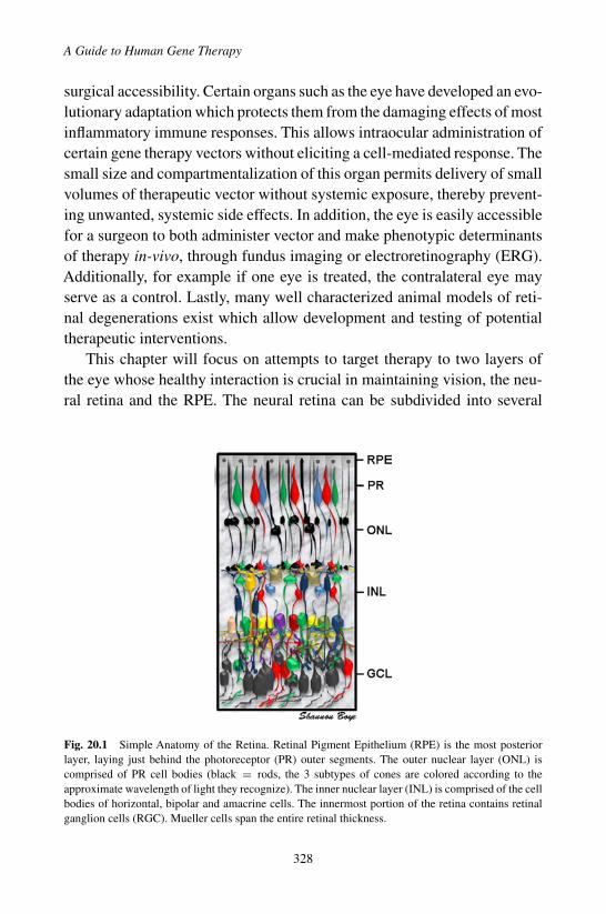
May 4, 2010 11:0 SPI-B903 9in x 6in b903-ch20
A Guide to Human Gene Therapy
surgical accessibility. Certain organs such as the eye have developed an evo-lutionary adaptation which protects them from the damaging effects of mostinflammatory immune responses. This allows intraocular administration ofcertain gene therapy vectors without eliciting a cell-mediated response. Thesmall size and compartmentalization of this organ permits delivery of smallvolumes of therapeutic vector without systemic exposure, thereby prevent-ing unwanted, systemic side effects. In addition, the eye is easily accessiblefor a surgeon to both administer vector and make phenotypic determinantsof therapy in-vivo, through fundus imaging or electroretinography (ERG).Additionally, for example if one eye is treated, the contralateral eye mayserve as a control. Lastly, many well characterized animal models of reti-nal degenerations exist which allow development and testing of potentialtherapeutic interventions.
This chapter will focus on attempts to target therapy to two layers ofthe eye whose healthy interaction is crucial in maintaining vision, the neu-ral retina and the RPE. The neural retina can be subdivided into several
Fig. 20.1 Simple Anatomy of the Retina. Retinal Pigment Epithelium (RPE) is the most posteriorlayer, laying just behind the photoreceptor (PR) outer segments. The outer nuclear layer (ONL) iscomprised of PR cell bodies (black = rods, the 3 subtypes of cones are colored according to theapproximate wavelength of light they recognize). The inner nuclear layer (INL) is comprised of the cellbodies of horizontal, bipolar and amacrine cells. The innermost portion of the retina contains retinalganglion cells (RGC). Mueller cells span the entire retinal thickness.
328
May 4, 2010 11:0 SPI-B903 9in x 6in b903-ch20
Retinal Diseases
classes of neurons including rod and cone photoreceptors, horizontal, bipo-lar, amacrine and ganglion cells.
Rod and cone photoreceptors support the initial event in the phototrans-duction cascade, the conversion of electromagnetic, light energy into elec-trochemical signals. Proper function of both rod and cone photoreceptorcells is dependent on the support of the retinal pigment epithelium. Amongother things, the RPE is involved in the phagocytosis of shed photoreceptorouter segment tips and the replenishment of chromophore (Vitamin A) torods and cones. The neural output originating from the visual signal in rodsand cones is sent through secondary order neurons (horizontal and bipolarcells) to the retinal ganglion cells (RGCs). The process of vision culmi-nates in the retina with transmission of these signals from the RGCs to thevisual cortex. It is therefore not surprising that the health of photoreceptors,RPE and RGCs are often compromised in many types of inherited retinaldegenerations, and, for this reason these three retinal layers are frequentlytargeted by gene therapies.
There are a number of viral vectors capable of delivering therapeu-tic transgenes to mammalian rods, cones, RPE and/or RGCs, includingadenovirus, lentivirus and adeno-associated virus (AAV). Each vectoris unique in its tropism, efficiency, persistence, carrying capacity andimmunogenicity. The occurrence of two high-profile adverse events inhumans involving adenoviral and lentiviral vectors has impacted their util-ity in clinical studies.1,2 Some changes have been made in these vectors toincrease safety. For example, non-integrating HIV lentiviral vectors havebeen developed which are capable of long-term transgene expression withreduced risk of insertional mutagenesis. While these lentiviral vectors havea greater packaging capacity than AAV-based vectors, they are still onlycapable of efficiently transducing the RPE. AAV is the only vector capableof efficiently transducing rods, cones and the RPE and is still considered asafer viral vector. For this reason, it is the most commonly used vector forretinal gene therapy and will be the focus of this chapter.
To date, ocular gene therapy has focused primarily on ‘loss of function’mutations in genes encoding proteins that mediate critical functions of pho-toreceptors, retinal ganglion cells and RPE. The potential for treatment inthese cell types has been aided by the cloning and mapping of more than120 retinal disease genes. The focus of this chapter is AAV-mediated ocular
329
May 4, 2010 11:0 SPI-B903 9in x 6in b903-ch20
A Guide to Human Gene Therapy
gene therapies in animal models which have shown the most potential inbridging the gap from bench to bedside. These therapies will be divided bythe retinal cell type which is targeted for treatment. For each example, AAV-mediated somatic gene transfer resulted in significant functional improve-ment, as assessed by ERG or behavior, and/or regeneration or stabilizationof retinal structure. One study will receive special attention because it laidthe groundwork for ongoing human clinical trials. Analysis of the research,safety studies and regulatory review process leading up to the clinical trialwill also be discussed.
2. Rod and Cone Photoreceptors
One of the most prevalent types of retinal degeneration is X-linkedretinoschisis (XLRS). Affecting between 1 in 15,000 to 1 in 30,000 peopleworldwide, XLRS is the leading cause of juvenile macular degenerationin males. Female carriers are usually unaffected. This retinal dystrophyis caused by mutations in the retinoschisis (RS1) gene on Xp22.2 whichencodes the 224 amino acid, secretable retinal protein retinoschisin (RS1).3
This protein consists of a 23 amino acid N-terminal leader sequence, a 39amino acid Rs1 domain, a 157 amino acid discoidin domain and a fiveamino acid C-terminal segment. Following cleavage of the leader sequencein the ER lumen, the protein is assembled into a homo-octameric complexlinked by disulfide bonds. Subsequently, this complex is secreted from cells,specifically for rod and cone photoreceptors.4 Upon secretion, RS1 is local-ized to the surface of photoreceptors and bipolar cells. Over 130 mutationsin RS1 (intragenic deletions, nonsense and missense mutations, frame shiftinsertions and deletions and splice site mutations) have been associatedwith disease. Molecular and cellular studies highlight three mechanismsthrough which these mutations may lead to XLRS; mutations in the dis-coidin resulting in misfolding and retention in the endoplasmic reticulum,cysteine mutations in the Rs1 domain or C-terminal segment which causedefective octamer assembly, and mutations in the leader sequence whichprevent insertion of the polypeptide into the ER membrane.4 It has beendetermined that it is the lack of functional RS1 protein and not the presenceof a mutated protein that is responsible for XLRS.4
330

May 4, 2010 11:0 SPI-B903 9in x 6in b903-ch20
Retinal Diseases
There is great variation in disease severity and rate of progression amongaffected patients, even in those carrying the same causative mutation oramong those in the same family. While some patients present at school agewith poor vision, the disease can also be detected in infants as young as3 months. Visual impairment is also variable, with best corrected visualacuity from 20/20 to 20/600. Typical features of XLRS are a loss in centralvision in the first decade of life and the presence of retinal schisis (splitting)which appears as ‘spoke wheel-like’cystic cavities radiating from the centralretina. Peripheral schisis is observed in approximately 50% of patients and isoften accompanied by peripheral vision loss. This schisis, or retinal splitting,is caused by cystic cavities or gaps, most often in, but not limited to theinner retina. Blood vessels which traverse these gaps are fragile and at riskof hemorrhage. Visual function in XLRS patients is often stable until thefourth decade of life, but severe complications such as vitreal hemorrhage(up to a third of patients) and retinal detachment (up to 20% or patients)may lead to severe visual impairment.
Diagnosis of XLRS is typically made when specific problems are foundin the patient’s electroretinogram (ERG). Affected patients exhibit a char-acteristic reduction in b-wave amplitudes which is best detected after darkadaptation. These reduced b-wave amplitudes are indicative of a problemwith the inner retina. Up to one third of XLRS patients also exhibit reduc-tions in a-wave amplitudes over time, indicative of a progressive photorecep-tor abnormality. Because these characteristic ERG patterns are not uniqueto XLRS, further diagnostic tests are necessary. Optical coherence tomogra-phy (OCT) is a non-invasive procedure which produces a two dimensional,cross-sectional image of structures in the eye. OCT scans of the centraland peripheral retina can detect splitting and the eventual central retinalthinning due to photoreceptor cell loss. Scanning laser ophthalmoscopy inconjunction with OCT confirms that splitting may occur in any layer of theretina. Currently, there is no treatment for XLRS.
While the exact function of RS1 in the eye remains to be elucidated, itis generally thought to play an important role in the structural integrity andmaintenance of retinal cell adhesion and architecture. Recent studies suggestthat this protein interacts with extracellular β2 laminin, known for its rolein the development and stabilization of neuronal synapses. In fact, deletion
331

May 4, 2010 11:0 SPI-B903 9in x 6in b903-ch20
A Guide to Human Gene Therapy
of the laminin β2 chain leads to reductions in ERG signal amplitudes likethose seen in XLRS.5
Three separate mouse models have been created in which the mousehomolog to human RS1, Rs1h, has been knocked out, and all display mor-phological and functional phenotypes similar to human XLRS.6,7,8 Rs1hknockout mice have highly disorganized retinas, cystic cavities and gapsbetween bipolar cells and disrupted outer nuclear layers that lead to eventualloss of photoreceptors. In addition, they exhibit the characteristic negativeERG response, indicating a disruption in the integrity of photoreceptor-bipolar cell synapses. Because the Rs1h-deficient mouse shares these impor-tant diagnostic features with XLRS patients, it has been a valuable modelfor evaluating potential therapeutic interventions.
Thus far, two groups have attempted to restore retinal structure andfunction in the Rs1h KO mouse.7,9 The most recent study will be high-lighted here as it reported the most complete therapeutic outcome. Severalchoices concerning vector design were made in that study with the goalof targeting expression of human RS1 cDNA to photoreceptors. Of thetwo well characterized serotypes known to efficiently target photorecep-tors (AAV2 and AAV5), AAV5 has demonstrated higher efficiency and amore rapid onset of transgene expression when delivered into the subreti-nal space. Secondly, a photoreceptor-specific opsin promoter was chosenbased on its ability to target transgene expression to rods and cones. Atpostnatal day 15 (P15), the AAV5-opsin promoter-Rs1h vector was deliv-ered subretinally to Rs1h KO mice. Histological analysis showed that RS1expression was restored to both photoreceptors and the inner retina, a pat-tern identical to that seen in wild type mice. Importantly, vector-mediatedgene expression led to a progressive and significant improvement in reti-nal function (ERG).9 Significant improvements were found in both rod-and cone-mediated aspects of the ERG signal. This functional recoverywas evident for at least 5 months, the longest time point evaluated. In vivoimaging and histological analysis revealed that therapy also conferred struc-tural improvements, with treated retinas exhibiting almost a complete lackof retinal schisis. These results are more complete than an earlier studywhich showed only functional improvements.7 This difference is most likelyattributable to the choice of vector serotype (AAV2) and promoter (CMV)used in the earlier study which likely resulted in lower levels of therapeutic
332
May 4, 2010 11:0 SPI-B903 9in x 6in b903-ch20
Retinal Diseases
RS1 expression in treated eyes. These results of successful gene replace-ment therapy in the Rs1h KO mice demonstrate that this approach is capableof preserving retinal structure and visual function in an animal model ofXLRS and provides support for the development of clinical trials for thisdisease.
3. Cone Photoreceptors
The human retina contains approximately 6 million cones. Cone photore-ceptors are responsible for central, high-resolution, color vision. Unlike rodphotoreceptors which support vision in dim, night-time conditions, we relyon cones to perform daily tasks in ambient to bright lighting. Diseases whichcompromise the integrity of this cell type are debilitating because they caninduce daytime blindness, preventing the patient from performing ‘normal’tasks such as driving. One such disease is rod monochromatism, betterknown as complete achromatopsia. Affecting approximately 1 in 30,000individuals worldwide, this recessively inherited disease is characterizedby permanent central vision loss, a lack of cone-mediated ERG signal andcomplete color blindness. Patients afflicted by achromatopsia typically havevisual acuity of approximately 20/200 or less and are extremely sensitive tolight. There is currently no cure for achromatopsia. All that is available forthe management of symptoms are tinted contact lenses or sunglasses whichlimit exposure of the retina to bright light.
The three genes found to be associated with human achromatopsia thusfar are CNGB3, CNGA3 and GNAT2.10,11.12,13 CNGB3 and CNGA3 encodethe β and α subunits of the cone cyclic nucleotide-gated cation channel,respectively. This cone-specific cGMP-gated cation channel plays a pivotalrole in cone phototransduction by opening and closing in response to light.These positions, open or closed, allow for or prevent the flow of ions intothe cell, thereby establishing the electrical state of the cone photorecep-tor, either depolarized or hyperpolarized, respectively. GNAT2 encodes thecone-specific α subunit of transducin, another essential phototransductionprotein. Transducin bridges the gap between photoisomerized opsin pro-teins and the breakdown of cGMP via phosphodiesterase. This ultimatelyleads to the closure of cGMP-gated channels.
333
May 4, 2010 11:0 SPI-B903 9in x 6in b903-ch20
A Guide to Human Gene Therapy
To date, two mouse models and one canine model of achromatopsiahave been characterized. They each have a different gene mutated, GNAT2and CGNA3 individually in mice and CNGB3 in dogs, with all modelsimitating the human form of the disease very well. This chapter will focus ongene therapy in the mouse model of the disease, the Gnat2cpfl3 mouse. Thismouse contains a point mutation in cone α-transducin rendering it inactive.Consequently, this animal has no recordable cone ERG but a normal rodERG.14 Light and electron microscopy reveal that the Gnat2cpfl3 mouseretina appears to have relatively normal structure.
As with other retinal gene therapies, specific choices were made aboutthe nature of the vector, this time with the goal to target expression to conephotoreceptors in the Gnat2cpfl3 mouse. Again, a serotype 5 vector was cho-sen based on its superior ability to target transgene expression in the retina.To specifically target cone photoreceptors, a cone-preferential promoter waschosen. This type of specific targeting is done in order to avoid the poten-tially toxic side effects that expression in the wrong cell type might elicit.A 2.1 kb fragment of the human red/green cone opsin promoter (PR2.1)was chosen based on previous evidence that mice transgenic for sequencesupstream of the red/green opsin gene containing a core promoter and locuscontrol regions directed reporter gene expression to both classes of cones.Additionally, this promoter, in conjunction with a serotype 5 vector wasshown to target reporter gene, green fluorescent protein (GFP), expressionpreferentially to cones in squirrel monkeys, mice, rats, ferrets, guinea pigs,dogs and monkeys. A serotype 5 vector containing the PR2.1 promoter andthe wild type, human Gnat2 cDNA (AAV5-PR2.1-hGnat2) was adminis-tered to the subretinal space of the Gnat2cpfl3 mice, a delivery route intendedto optimize infection of photoreceptors. Following treatment, cone func-tion in treated and untreated eyes were assessed by ERG assays specificfor cone function. Additionally, visually evoked behavior was assessed bydetermining visual acuity through optomotor (head movement) responsesto a rotating sine-wave grating. The results of both tests showed that AAV5-PR2.1-hGnat2 treated eyes responded significantly better than untreatedeyes in the Gnat2cpfl3 mice. Responses in treated eyes were equivalent tothose seen in normal sighted, wild type mice.14 This study was the first suc-cessful attempt demonstrating that cone-targeted AAV therapy is capable ofrestoring cone-mediated function and visual acuity in an animal model of
334

May 4, 2010 11:0 SPI-B903 9in x 6in b903-ch20
Retinal Diseases
achromatopsia. Currently, an analogous approach is being taken to correctvision in another mouse model with the CGNA3 form as well as a caninemodel with the CNGB3 form of this disease.
More recently, investigators have reported success in another cone-based disorder, red-green color blindness.15 This disease is the result ofthe absence of either the long- (L) or middle- (M) wavelength-sensitivevisual photopigments and is the most common single locus human geneticdisorder. AAV containing a human L-opsin gene under the control of theL/M-opsin enhancer and promoter was delivered to the photoreceptors ofadult squirrel monkeys in three separate subretinal injections (100 µl each).Behavior-based color vision tests were used to compare subjects before andafter treatment. In spite of doubts that the absence of neural connectionsnormally established during development would prevent appropriate pro-cessing post-treatment, investigators showed that addition of a third opsinto these adult red-green colour deficient primates was sufficient to restoretrichromatic color vision behavior.15 This result suggests that gene-basedtherapy in the retina has the potential to cure adult cone vision disorders.
The success of these studies supports the development of other cone-targeted therapies in which cures for debilitating human conditions suchas late stage retinitis pigmentosa, age-related macular degeneration anddiabetic retinopathy are needed. While more efficacy and safety data isnecessary, cone-targeting clinical trials for human achromatopsia and red-green color blindness may be reasonably considered.
4. Retinal Ganglion Cells
Retinal ganglion cells (RGC) are located in the innermost portion of theretina and are, on average, larger than most other retinal neurons. They areresponsible for receiving electrical signals originally generated by photore-ceptors and second order neurons and passing that information through theoptic nerve to the brain. Diseases which affect the health of RGC can havedeleterious effects on vision. One such disease is glaucoma, the secondmost common cause of blindness in the world.16 A major component of thisdisease is apoptotic death of RGCs. It was once thought that this neuronalloss in glaucoma patients was attributed solely to the long term elevation ofintraocular pressure (IOP). However, clinical trials revealed that when the
335

May 4, 2010 11:0 SPI-B903 9in x 6in b903-ch20
A Guide to Human Gene Therapy
IOP was normalized, some patients still continued to lose RGCs. Currently,two main hypotheses are commonly accepted as explanations for glauco-matous RGC death; long term elevation in intraocular pressure (IOP) orthe presence of genetic factors which predispose a patient to RGC loss inthe absence of elevated IOP. Regardless of the cause of initial damage, itresults in a cascade of biochemical events which can further affect RGCs.Such secondary events include uncontrollable increases in levels of bio-chemical compounds such as glutamate and nitric oxide that are releasedby dying neurons. These compounds can eventually reach concentrationsthat are neurotoxic. Additionally, the initial insult may cause an interrup-tion in normal axonal transport which results in deprivation of target-derivedtrophic factors and consequent triggering of intracellular changes that maylead to apoptotic death of affected RGCs.
One neurotrophic factor that is especially important for RGC health,both during development and in adult life, is brain-derived neurotrophicfactor (BDNF). It is produced in the brain and is retrogradely transportedfrom RGC axons to cell bodies in microsomal vesicles. It was found thatwhen IOP is elevated in rats and monkeys that retrograde transport of BDNFis obstructed at the optic nerve head.17 This finding led to the hypothesis thatBDNF deprivation plays a role in glaucomatous RGC death. This theorywas further supported by evidence that intravitreal administration of BDNFin a rat model of glaucoma temporarily slowed RGC loss. However, theneed for repeated administration of BDNF protein limits its clinical utilityfor persistent diseases like glaucoma.
Multiple viral vector systems have been employed in attempts to achievelong term expression of BDNF in RGCs. Initial studies using adenoviralvectors to deliver the BDNF cDNA to the intravitreal space of rats withaxotomized RGCs revealed that expression of the therapeutic transgenewas capable of temporarily promoting RGC survival.18 However, due tolimitations of adenoviral gene therapy, including the inability to elicit longterm transgene expression and the tendency to promote inflammation, amore optimal vector system was necessary. More recently an AAV vec-tor was used to deliver the BDNF cDNA to the intravitreal space of anexperimental rat model of glaucoma.19 Two weeks later, glaucoma wasinduced in this model through an elevation in IOP via laser treatment ofthe trabecular meshwork (the mesh-like structure near the cornea and iris
336

May 4, 2010 11:0 SPI-B903 9in x 6in b903-ch20
Retinal Diseases
which facilitates removal of aqueous humor from the eye into the bloodstream). At four weeks post treatment, AAV-mediated BDNF therapy pro-moted survival of a statistically significant fraction of RGCs (as estimatedby RGC axon counts) in treated eyes relative to untreated controls. Trans-gene expression was found predominantly in the desired target cell, theRGC. One limitation of this therapy however was that RGC survival wasnot quantified beyond 4 weeks. In addition, BDNF is known to have highaffinity for its receptor TrkB, an antigen expressed on multiple retinal celltypes, not just RGCs. This in theory permits the AAV-delivered transgeneto exert its effects in a broad fashion which could have implications forother aspects of retinal structure and/or function. In fact, it is known thatapplication of neurotrophic factors in the retina can cause upregulation ofnitric oxide synthase activity and suppression of heat shock proteins, bothof which could have detrimental effects. In addition, it has been shownthat delivery of a related molecule, ciliary neurotrophic factor (CNTF), hasdetrimental effects on retinal function.20 With this in mind, it was thoughtthat intervening in pathways downstream of neurotrophin administrationmight have ‘cleaner’ therapeutic effects. It is known that BDNF bindingto its receptor, TrkB, stimulates multiple signaling pathways including theextracellular signal-related kinase 1/2 (ERK1/2) which has been implicatedin RGC survival. Using an RGC preferential serotype 2 AAV vector todeliver MEK1 (the upstream activator of ERK1/2) to the intravitreal spaceof an experimental rat model of glaucoma, researchers were able to stim-ulate the ERK1/2 pathway in cells transduced with vector.21 At five weekspost treatment, it was found that MEK1 gene transfer markedly increasedRGC survival in this model. Through the use of a serotype 2 AAV vectorand delivery to the vitreous cavity, this therapy specifically targeted RGCs.This study is further proof that gene therapy is capable of promoting RGCsurvival in animal models of glaucoma.
5. Retinal Pigment Epithelium
The retinal pigment epithelium (RPE) is the pigmented cell monolayer posi-tioned between the neurosensory retina and the choroidal blood supply. TheRPE sustains the metabolic needs of the underlying neural retina by con-trolling the transfer of small molecules between the blood stream and the
337

May 4, 2010 11:0 SPI-B903 9in x 6in b903-ch20
A Guide to Human Gene Therapy
Fig. 20.2 The Vitamin A Cycle: all-trans-retinol (vitamin A) is transported from the blood streamto the retinal pigment epithelium (RPE). There it is esterified into all-trans-retinyl esters. On demand,these esters can be hydrolyzed and isomerized (by RPE65) to form 11-cis-retinol which is oxidized toform 11-cis-retinal. 11-cis-retinal is transported to photoreceptor outer segments where it binds withopsin to form the visual pigment, rhodopsin in rods and cone opsin in cones. Absorption of a photon oflight catalyzes the isomerization of 11-cis-retinal to all-trans-retinal which releases it from the opsinmolecule. This photo-isomerization triggers a biochemical cascade in photoreceptors which eventuallyleads to the generation of a neural signal in RGCs that is interpreted in the brain as vision.
retina. It is also responsible for the phagocytosis of shed photoreceptor outersegment tips and plays a crucial role in the Vitamin A cycle by isomerizingall trans-retinol to 11-cis retinal (Fig. 20.2).
RPE65 (retinal pigment epithelium-specific 65 kDa protein), expressedpredominantly in RPE cells, has been identified as the all-trans to 11-cisisomerase.22 Improper functioning of RPE65 results in a lack of 11-cisretinal production and an inability to form visual pigment (rhodopsin andcone opsin). Without these light-sensitive opsins, photoreceptor cells of theretina lack the ability to usefully absorb photons and initiate the conversionof light into a visual signal. Mutations in Rpe65 are associated with Leber’scongenital amaurosis type 2 (LCA2), one of the earliest and most severeforms of inherited retinal dystrophies.23 This disease, which is most ofteninherited in an autosomal recessive fashion, is usually diagnosed at birthor within the first few months of life when patients present with severelyimpaired vision, pendular nystagmus and abnormal ERG and pupillary lightreflexes. Patients progress to near total blindness by adolescence. Despite
338

May 4, 2010 11:0 SPI-B903 9in x 6in b903-ch20
Retinal Diseases
these severe, early symptoms there are often not any obvious ophthalmo-scopic abnormalities present.
In order to circumvent the underlying defect in the retinoid cycle (the lackof 11-cis retinal and failure to regenerate visual pigment), three therapieshave been evaluated; RPE cell transplantation, retinoid delivery and genebased intervention. RPE cell transplantation is mechanically difficult andinvasive. Oral administration of retinoid is not optimal because it requiresrepeated administration of drug. Of the three, gene-based intervention hashad the most success. AAV was used to deliver functional copies of RPE65to the naturally occurring Rpe65 mutant Briard dogs, a naturally occurringRPE65 mutant (rd12) mouse and the transgenic knockout Rpe65−/− mouse.Taken together, these studies showed that AAV-mediated RPE65 expressionconfers partial restoration of ERG, retinal structure, and visually evokedbehaviors in these animals.24,25,26 These studies laid the groundwork forPhase I Clinical trials for LCA2.
6. LCA2 Gene Therapy, a Perspective on TranslationalResearch
The University of Florida — University of Pennsylvania LCA Consortium,established through a NEI Cooperative Clinical Research Grant, had thegoal of establishing a safe and efficacious AAV gene therapy vector andemploying it in a clinical trial for LCA2 within 5 years. The Consortium’sfirst step was to expand on the proof of concept work that began with AAV-RPE65 treatments of Briard dogs.24 The first of these dogs to be treated andsubsequently shown to have restored vision, ‘Lancelot’, was instrumental inraising public awareness of vision research. EventuallyAAV-RPE65 vectorswere evaluated in two other animal models of LCA2, the transgenic knock-out Rpe65−/− mouse and rd12 mutant mouse.26,25 Restoration of visualresponses was achieved by subretinal injection of AAV-RPE65 vector inboth mouse models. In addition, in the Briard dogs vision-evoked corticalresponses had been restored similar to behavioral characteristics correlatingwith the functional vision that were documented in treated rd12 mice. Theseexperiments provide further evidence that therapy could be successful inrestoring ‘functional’vision.27,25 Vision restoration has persisted unchangedfor more than 8 years after a single treatment in a Briard dog (Lancelot).
339
May 4, 2010 11:0 SPI-B903 9in x 6in b903-ch20
A Guide to Human Gene Therapy
A dose response study in affected Briard dogs revealed the therapeuti-cally effective dose range of the vector. Safety within this range was furthervalidated in Cynomolgous monkeys. Both short term and long term biodis-tribution and pathology studies were performed in vector-treated normalvision rats and monkeys. In order to ensure that the monkey data was robustand conclusions impartial, it was done by a contract research organizationin accordance with Good Laboratory Practices (GLP).28 Ophthalmologicalexaminations carried out in dogs and monkeys post vector administrationindicated that there were some minor to moderate post surgical compli-cations, most of which resolved within a week post surgery and, asidefrom anterior chamber flare, were not related to vector treatment (i.e. alsooccurred in vehicle treated animals). Signs of retinal thinning localized tothe injection site of treated dogs and monkeys was likely a consequence ofthe surgical procedure, and have been previously documented. Other mea-sures of ocular health such as visual function as assessed by ERG did notindicate a loss of visual function in monkeys when comparing pre and postvector treatment time points. In affected Briard dogs, vector treatment wascorrelated with improved ERG responses, as had already been establishedin prior proof of concept experiments.
Pathological examinations of all non-ocular tissues were unremarkablein all of the studies. Immunology experiments were carried out in the mon-key studies. Circulating antibodies to AAV2 capsid protein were evaluatedby ELISA. For the 7 day monkey study, antibody titers were low beforetreatment and remained so. However, given the short duration of this exper-iment and the time necessary to mount a humoral response, these resultsare inconclusive. In the 3 month monkey study, despite some variation inpre-treatment antibody titers, there were no consistent changes in antibodylevels over the 3 month period following vector administration.
Biodistribution studies in dogs, rats and monkeys looked for the presenceof vector genomes in specific tissues. Tissues of particular interest includedthe injected eye (where vector DNA is expected), tissues of the visual path-way and brain (to determine whether retrograde vector transport along theoptic nerve occurred), and gonad (necessary to rule out the possibility ofgerm-line vector transmission). All studies indicated vector was rarely andonly sporadically found outside of the injection site. Importantly, vector wasgenerally not found in the optic nerve or other tissues of the optic pathway
340
May 4, 2010 11:0 SPI-B903 9in x 6in b903-ch20
Retinal Diseases
leading to the brain, and never in the brain itself. Gonads, both male andfemale, were found to be negative indicating that the potential for germ-linetransfer of vector is minimal.
Taking the conclusions from the safety studies and translating them intoa protocol for a LCA2 clinical trial highlighted several key points: 1) Detailsof the surgical technique are of critical importance. Because limited injec-tion site damage was noted proximal to the retinotomy, vector should bedelivered adjacent to target areas of best retained retina, thereby allowingvector to spread into them by flow into the subretinal space. 2) The vectordose range would be defined by the results in dogs which provided efficacybut no established toxicity. Based on the efficacy and safety studies summa-rized above, approval to proceed with a clinical trial was given by the USFDA, the US Recombinant DNA Advisory Committee, an NIH/NEI con-stituted Data Safety and Monitoring Committee, the Institutional BiosafetyCommittees of the University of Florida and the University of Pennsylvaniaand the Institutional Review Boards of the University of Florida (throughthe Western IRB) and the University of Pennsylvania.
Presently, 3–12 month results from three separate clinical studies(including the aforementioned University of Florida and University of Penn-sylvania, NEI funded trial) have been reported involving a total of nineyoung adults with LCA2.29,30,31,32,33 Although the primary purpose of allthree studies was to determine safety, each trial reported some measure oftherapy in at least one of their patients. Neither humoral nor T-cell medi-ated immune responses to vector were detected in any patient. The factthat LCA2 patients received 2 to 4 orders of magnitude lower AAV2 vectordoses than had been reported for patients in other non-ocular clinical trialstogether with the relative immune privileged status of the eye makes thislack of immune response understandable. One patient from the Bainbridgeet al. (2008) study and all three patients from the Hauswirth et al. (2008)and Cideciyan et al. (2008) study experienced significant increases in lightsensitivity within the retinal area treated, with the best response being a63,000-fold improvement over pre-treatment levels.32 A key conclusion ofall three trials was that the subretinal vector delivery procedure and anytransient post-surgical complications did not promote vision loss. A strik-ing observation was noted in one patient one year post treatment.33 For thefirst time in her life, the patient reported an ability to read the illuminated
341
May 4, 2010 11:0 SPI-B903 9in x 6in b903-ch20
A Guide to Human Gene Therapy
numerical clock display on the dashboard of the family vehicle. This newperception was accompanied by a shift in fixation into the treated supero-temporal retina. In other words, the patient had developed the ability tofixate on an image with a treatment-created pseudo-fovea, an area whichnow had better sensitivity than the untreated foveal region. The slow devel-opment of this pseudo-fovea suggests an underlying experience-dependentplasticity in the adult visual system. Put simply, this patient’s brain had‘learned’ how to use the treated pseudo-foveal retinal region for fixationover the 12 months post treatment. This result raises the possibility thatother retinal gene-based therapies have the potential to improve vision inpreviously unexpected ways. In summary, early results suggesting efficacyand safety in three AAV2-mediated gene therapy trials for RPE65 LCAportend a hopeful future for retinal gene therapy with AAV vectors.
References
1. Raper SE, Chirmule N, Lee FS, Wivel NA, Bagg A, Gao GP, et al. (2003). Fatalsystemic inflammatory response syndrome in a ornithinetranscarbamylase deficientpatient following adenoviral gene transfer. Mol Genet Metab 80: 148–158.
2. Williams DA (2006). Vector insertion, mutagenesis and transgene toxicity. Mol Ther14: 457.
3. Sauer CG, Gehrig A, Warneke-Wittstock R, Marquardt A, Ewing CC, Gibson A, et al.(1997). Positional cloning of the gene associated with X-linked juvenile retinoschisis.Nat Genet 17: 164–170.
4. Wu WW, Molday RS (2003). Defective discoidin domain structure, subunit assem-bly, and endoplasmic reticulum processing of retinoschisin are primary mechanismsresponsible for X-linked retinoschisis. J Biol Chem 278: 28139–28146.
5. Libby RT, Lavallee CR, Balkema GW, Brunken WJ, Hunter DD (1999). Disruption oflaminin beta2 chain production causes alterations in morphology and function in theCNS. J Neurosci 19: 9399–9411.
6. Weber BH, Schrewe H, Molday LL, Gehrig A, White KL, Seeliger MW, et al. (2002).Inactivation of the murine X-linked juvenile retinoschisis gene, Rs1h, suggests a roleof retinoschisin in retinal cell layer organization and synaptic structure. Proc Natl AcadSci USA 99: 6222–6227.
7. ZengY, TakadaY, Kjellstrom S, Hiriyanna K, Tanikawa A, Wawrousek E, et al. (2004).RS-1 Gene Delivery to an Adult Rs1h Knockout Mouse Model Restores ERG b-Wavewith Reversal of the Electronegative Waveform of X-Linked Retinoschisis. Invest Oph-thalmol Vis Sci 45: 3279–3285.
8. Jablonski MM, Dalke C, Wang X, Lu L, Manly KF, Pretsch W, et al. (2005). An ENU-induced mutation in Rs1h causes disruption of retinal structure and function. Mol Vis11: 569–581.
342

May 4, 2010 11:0 SPI-B903 9in x 6in b903-ch20
Retinal Diseases
9. Min SH, Molday LL, Seeliger MW, Dinculescu A, Timmers AM, Janssen A,et al. (2005). Prolonged recovery of retinal structure/function after gene therapyin an Rs1h-deficient mouse model of x-linked juvenile retinoschisis. Mol Ther12: 644–651.
10. Kohl S, Baumann B, Rosenberg T, Kellner U, Lorenz B, Vadala M, et al. (2002).Mutations in the cone photoreceptor G-protein alpha-subunit gene GNAT2 in patientswith achromatopsia. Am J Hum Genet 71: 422–425.
11. Kohl S,Varsanyi B,Antunes GA, Baumann B, Hoyng CB, Jagle H, et al. (2005). CNGB3mutations account for 50% of all cases with autosomal recessive achromatopsia. Eur JHum Genet 13: 302–308.
12. Kohl S, Marx T, Giddings I, Jagle H, Jacobson SG, Apfelstedt-Sylla E, et al. (1998).Total colourblindness is caused by mutations in the gene encoding the alpha-subunit ofthe cone photoreceptor cGMP-gated cation channel. Nat Genet 19: 257–259.
13. Sundin OH, Yang JM, Li Y, Zhu D, Hurd JN, Mitchell TN, et al. (2000). Genetic basisof total colourblindness amoung Pingelapese islanders. Nat Genet 25: 289–293.
14. Alexander JJ, Umino Y, Everhart D, Chang B, Min SH, Li Q, et al. (2007). Restorationof cone vision in a mouse model of achromatopsia. Nat Med 13: 685–687.
15. Mancuso K, Hauswirth WW, Li Q, Connor TB, Kuchenbecker JA, Mauck MC, et al.(2009). Gene therapy for red-green colour blindness in adult primates. Nature, Sep 16(Epub ahead of print).
16. Quigley HA (1996). Number of people with glaucoma worldwide. Br J Ophthalmol.80: 389–393.
17. Pease ME, McKinnon SJ, Quigley HA, Kerrigan-Baumrind LA, Zack DJ (2000).Obstructed axonal transport of BDNF and its receptor TrkB in experimental glaucoma.Invest Ophthalmol Vis Sci 41: 764–774.
18. Di Polo A, Aigner LJ, Dunn RJ, Bray GM, Aguayo AJ (1998). Prolonged deliv-ery of brain-derived neurotrophic factor by adenovirus-infected Muller cells tem-porarily rescues injured retinal ganglion cells. Proc Natl Acad Sci USA 95:3978–3983.
19. Martin KR, Quigley HA, Zack DJ, Levkovitch-Verbin H, Kielczewski J, Valenta D,et al. (2003). Gene therapy with brain-derived neurotrophic factor as a protection:retinal ganglion cells in a rat glaucoma model. Invest Ophthalmol Vis Sci 44: 4357–4365.
20. McGill TJ, Prusky, GT, Douglas, RM, Yasumura, D, Matthes, MT, Nune, G, et al.(2007). Intraocular CNTF Reduces Vision in Normal Rats. Invest Ophthalmol Vis Sci48: 5756–5766.
21. Zhou Y, Pernet V, Hauswirth WW, Di Polo A (2005). Activation of the extracellularsignal-regulated kinase 1/2 pathway by AAV gene transfer protects retinal ganglioncells in glaucoma. Mol Ther 12: 402–412.
22. Moiseyev G, Chen Y, Takahashi Y, Wu BX, Ma JX (2005). RPE65 is the iosmerohy-drolase in the retinoid visual cycle. Proc Natl Acad Sci USA. 102: 12413–12418.
23. Marlhens F, Bareil C, Griffoin JM, Zrenner E, Amalric P, Eliaou C, et al. (1997).Mutations in RPE65 cause Leber’s congenital amaurosis. Nat Genet. 17: 139–141.
24. Acland GM, Aguirre GD, Ray J, Zhang Q, Aleman TS, Cideciyan AV, et al. (2001).Gene therapy restores vision in a canine model of childhood blindness. Nat Genet28: 92–95.
343
May 4, 2010 11:0 SPI-B903 9in x 6in b903-ch20
A Guide to Human Gene Therapy
25. Pang JJ, Chang B, Kumar A, Nusinowitz S, Noorwez SM, Li J, et al. (2006). Genetherapy restores vision-dependent behavior as well as retinal structure and function ina mouse model of RPE65 Leber congential amaurosis. Mol Ther 13: 565–572.
26. Dejneka NS, Surace EM, Aleman TS, Cideciyan AV, Lyubarsky A, Savchenko A, et al.(2004). In utero gene therapy rescues vision in a murine model of congenital blindness.Mol Ther 9: 182–188.
27. Aguirre GK, Komáromy AM, Cideciyan AV, Brainard DH, Aleman TS, Roman AJ,et al. (2007). Canine and human visual cortex intact and responsive despite early retinalblindness from RPE65 mutation. PLoS Med. 4: e230.
28. Jacobson SG, Boye SL, Aleman TS, Conlon TJ, Zeiss CJ, Roman AJ, et al. (2006).Safety in nonhuman primates of ocular AAV2-RPE65, a candidate treatment for blind-ness in Leber congenital amaurosis. Hum Gene Ther 17: 845–858.
29. Bainbridge JW, SmithAJ, Barker SS, Robbie S, Henderson R, Balaggan K, et al. (2008).Effect of gene therapy on visual function in Leber’s congenital amaurosis. N Engl JMed 358: 2231–2239.
30. Maguire AM, Simonelli F, Pierce EA, Pugh EN Jr, Mingozzi F, Bennicelli J, et al.(2008). Safety and efficacy of gene transfer for Leber’s congenital amaurosis. N EnglJ Med 358: 2240–2248.
31. Hauswirth W, Aleman TS, Kaushal S, Cideciyan AV, Schwartz SB, Wang L, et al.(2008). Phase I Trial of Leber CongenitalAmaurosis due to RPE65 Mutations by OcularSubretinal Injection ofAdeno-Associated Virus Gene Vector: Short-Term Results. HumGene Ther, 2008 Sep 7 [Epub ahead of print].
32. Cideciyan AV, Aleman TS, Boye SL, Schwartz SB, Kaushal S, Roman AJ, et al. (2008).Human gene therapy for RPE65 isomerase deficiency activates the retinoid cycle ofvision but with slow rod kinetics. Proc Natl Acad Sci USA 105: 15112–15117.
33. Cideciyan AV, Hauswirth WW, Aleman TS, Kaushal S, Schwartz SB, Boye SL, et al.(2009). Vision 1 year after gene therapy for Leber’s congenital amaurosis. N Engl JMed 361: 725–727.
344
May 4, 2010 11:0 SPI-B903 9in x 6in b903-ch21
Chapter 21
A Brief Guide to Gene TherapyTreatments for PulmonaryDiseases
Ashley T. Martino,∗ Christian Muellerand Terence R. Flotte
Pioneering studies in the field of gene therapy were focused on thecommon monogenic lung disease, cystic fibrosis. These efforts werelargely unsuccessful. Subsequent to these early studies gene therapyfor pulmonary disorders has expanded well beyond the initial targetsinto diseases such as lung cancer, infectious diseases, and asthma. Theemergence of new vectors and concepts of preparative treatment haveallowed gene correction to advance despite initial setbacks. Moreoverhybrid technologies such as vector-mediated RNAi therapy and cell-based genetic manipulation have added to the therapeutic armamentarium.Overall, the various advances increased the number of potential drug can-didates that might ultimately have a lasting impact on outcomes of patientswith lung diseases.
1. Introduction
Initially pulmonary gene therapy studies involved cystic fibrosis (CF) andbrought gene therapy into the spotlight. There have been 25 phase I/IIclinical trials for CF using viral and non-viral gene therapy methods.1
Unfortunately, the goal of effective, long-term cystic fibrosis transmem-brane conductance regulator (CFTR) gene correction was not achieved with
∗Correspondence: University of Florida, Dept. Pediatrics, Gainesville, FL.E-mail: [email protected]
345

May 4, 2010 11:0 SPI-B903 9in x 6in b903-ch21
A Guide to Human Gene Therapy
the initial trials. Human trials have also been pursued for the genetic disease:Alpha-1 anti-trypsin (A1AT), with no significant therapeutic benefit. Thechallenges encountered in these first clinical trials were critical in designingstrategies to overcome the early setbacks. Data from these studies promptedimprovements in the technology that yielded new generations of viral andnon-viral delivery systems and provided insight into how to improve effi-ciency, many of which are currently either moving towards or are beingused in clinical studies.
The transformation of gene therapy for genetic lung diseases bolsteredthe research potential of genetic manipulation for lung disorders. The fullspectrum of gene therapy based trials has expanded and now includes mul-tiple genetic diseases and non-hereditary conditions like lung cancer andasthma. In addition, multiple strategies, such as cell-based therapy and RNAimediated gene silencing, have emerged.
Although many critical advances have been made, there are still issues tosolve and new avenues to pursue before gene therapy becomes an acceptedtreatment for lung diseases. This chapter will review common diseases,evolution of vectors and strategies for their effective delivery, hurdles toovercome, successes and the foreseeable future.
2. Common Disorders
2.1 Cystic Fibrosis
Cystic fibrosis (CF) is the most common genetic disease among Caucasians.Interest in CF gene therapy treatments materialized after the 1989 publi-cation by Riordan et al. revealed that CF was linked to a homozygousgene defect in the Cystic Fibrosis Transmembrane Conductance Regulator(CFTR) gene.2 The CFTR gene encodes a membrane-bound chloride chan-nel protein and regulates the depth and composition of the airway surfaceliquid (ASL), in which the cilia of the respiratory epithelium are immersed.3
It is widely accepted that a CFTR deficiency induces an alteration inthe ASL.4 This alteration leads to a build up of mucus, which impairs cil-iary function and compromises pathogen clearance from the lung. Defectivebacterial clearance results in the colonization of Pseudomonas aeruginosa,as well as other pathogens. This chronic state of persistent lung infection
346

May 4, 2010 11:0 SPI-B903 9in x 6in b903-ch21
A Brief Guide to Gene Therapy Treatments for Pulmonary Diseases
Table 21.1. Pathological consequences related to cystic fibrosis.
Organs/System Affected Pathological Condition
Lung/respiratory system Chronic endobronchial infection withStaphylococcus aureus, Pseudomonas aeruginosa,Burkholderia cepacia, and other specificorganisms, small and large airways obstruction,bronchiectasis, allergic bronchopulmonaryaspergillosis, hyper innate immune response,pneumonia, hemoptysis, fibrosis, pulmonaryhypertension, heart failure, respiratory failure;chronic rhinosinusitis, nasal polyposis
Sweat gland Increased chloride and sodium loss due toimpairment of ductal reabsorption
Liver Ductal obstruction, biliary cirrhosis, multi-lobularcirrhosis, metabolic complications, fibrosis
Pancreas Pancreatic insufficiency, ductal obstruction, loss ofinsulin producing beta-cells, cystic fibrosis relateddiabetes (CFRD)
Digestive System Gut obstruction, malabsorption, malnutrition
Reproductive system Infertility, absence of vas deferens (99% of men),thickened cervical mucus (minor in women)
promotes a constant influx of cellular infiltrates from the immune system,which ultimately leads to lung tissue destruction that may progress to res-piratory failure and premature death. This severe pathology is complicatedby a collection of consequences in other organs and biological systems(Table 1).
Why did CF become a popular candidate for gene therapy? Forty yearsago cystic fibrosis patients died in early childhood. There have been manyadvances in treatments unrelated to gene therapy that have dramaticallyincreased life expectancy, but premature death still occurs. Correcting theCFTR gene deficiency is of great interest due to the potential to completelyreverse the lung pathology.
347
May 4, 2010 11:0 SPI-B903 9in x 6in b903-ch21
A Guide to Human Gene Therapy
The bulk of the mortality comes from the lung condition and it hasbeen generally accepted that a minor CFTR correction in the deficient lung,6–10% of normal levels, will be effective in reversing the pathophysiology.5
There is also the potential that correction of a single organ, the lung, willlead to long-term benefit.6 Since newborns with CF usually have healthylungs, there may be an opportunity to prevent symptoms before they begin.Furthermore, the lungs are relatively accessible as compared with otherorgans, since the airway may serve as a route of vector delivery. Mostvectors can be readily aerosolized for inhaled delivery to the lung, anoption not available to other target organs, such as the brain, pancreas,liver, etc.
2.2 Alpha-1 Antitrypsin (A1AT)
A1AT is caused by a homozygous gene defect in the SERPINA1 gene.The encoded protein, alpha-1 antitrypsin (A1AT), is a protease inhibitorpredominately secreted by the liver. This protein is critical in regulatingthe levels of pro-inflammatory elastases (i.e. neutrophil elastase) that arepresent at sites of inflammation by degranulation of polymorphonuclearcells such as neutrophil granulocytes.7
The common lung conditions that develop from the decrease in sys-temic A1AT levels are chronic obstructive pulmonary disease (COPD) andemphysema, both of which lead to respiratory insufficiency. The progres-sion to these conditions is linked to the secretion of elastases in the lungenvironment without proper regulation offered by A1AT. Unchecked, theelastases linger and eventually lead to cellular damage. The lung is notablyvulnerable to the deficiency in circulating A1AT levels and is augmentedby smoking.
3. Development of Viral Vectors for Lung Disease
The evolution of viral vectors for genetic manipulation in the lung hasprogressed via the early clinical trials related to CFTR gene correction forcystic fibrosis treatment. These trials involved adenovirus (Ad) and a non-pathogenic, helper-dependent adeno-associated virus (AAV2) administeredby nasal installation, lung installation and aerosolization.
348
May 4, 2010 11:0 SPI-B903 9in x 6in b903-ch21
A Brief Guide to Gene Therapy Treatments for Pulmonary Diseases
3.1 Adenoviral Vectors
Adenovirus was chosen because infections occur naturally in the lung andintestines. Unfortunately, these infections lead to a host of conditions includ-ing pneumonia, diarrhea and vomiting. Steps were taken to ensure safetywhen using Ad vectors. With early indication showing that the E1 geneticregion was responsible for the cytotoxicity of Ad, the first clinical trialinvolved an E1 deficient Ad vector expressing full length CFTR.8 This ini-tial adenoviral trial revealed a transient pulmonary condition categorizedas pneumonia. Later efforts to address these safety issues by deleting theE2 and E4 regions along with the E1 region did not prevent this pulmonarycondition.9
3.2 Adeno-Associated Viral Vectors
The AAV2 trials utilized the full-length CFTR coding region flanked bythe AAV2 viral integrated terminal repeats (ITRs). Since the CFTR codingregion is 4.4 kb, the small genome restraints (4.7 kb maximum capacity) ofthe AAV2 viral vector prevented the use of expression enhancing elements.Therefore transcription was relegated to the minor innate ability of theviral ITRs. Wild-type AAV infections occur without pathology, and withsafety being a primary concern, the AAV viral vector delivery system waspreferable to the Ad delivery system.
3.3 Early Conclusions
Unfortunately, the initial trials of CF gene therapy fell short of expectations.Viral vector transduction was not as efficient in the lungs of CF patients aswas predicted from the preliminary cell culture and animal models.10,11
However, safe administration of rAAV vectors was promising, and therewas correlation between presence of vector-derived CFTR cDNA and phys-iologic correction of the CFTR chloride channel defect in cells recoveredfrom patients after rAAV gene therapy.12 The most difficult issues remain-ing were efficiency and duration of gene transfer and the inability to redosevectors after neutralizing antibody responses were elicited.
Therefore, to increase efficiency, alternate forms of these viral vectorswere created.
349
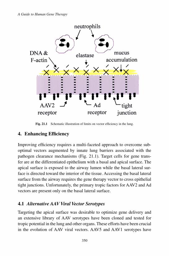
May 4, 2010 11:0 SPI-B903 9in x 6in b903-ch21
A Guide to Human Gene Therapy
Fig. 21.1 Schematic illustration of limits on vector efficiency in the lung.
4. Enhancing Efficiency
Improving efficiency requires a multi-faceted approach to overcome sub-optimal vectors augmented by innate lung barriers associated with thepathogen clearance mechanisms (Fig. 21.1). Target cells for gene trans-fer are at the differentiated epithelium with a basal and apical surface. Theapical surface is exposed to the airway lumen while the basal lateral sur-face is directed toward the interior of the tissue. Accessing the basal lateralsurface from the airway requires the gene therapy vector to cross epithelialtight junctions. Unfortunately, the primary tropic factors for AAV2 and Advectors are present only on the basal lateral surface.
4.1 Alternative AAV Viral Vector Serotypes
Targeting the apical surface was desirable to optimize gene delivery andan extensive library of AAV serotypes have been cloned and tested fortropic potential in the lung and other organs. These efforts have been crucialin the evolution of AAV viral vectors. AAV5 and AAV1 serotypes have
350

May 4, 2010 11:0 SPI-B903 9in x 6in b903-ch21
A Brief Guide to Gene Therapy Treatments for Pulmonary Diseases
been chosen in the newest generations of AAV viral vectors to target theapical surface of the lung. It was discovered that the receptors for AAV5serotype are the platelet derived growth factor (PDGF) and 2,3 sialic acidresidues. Both these receptors are located on the apical surface of airwayepithelial cells.13,14 Notably, AAV1 is more efficient than AAV2 and AAV5at transducing human airway cultures and also enters through the apicalsurface of the cell.15−18
4.1.1 Addition of Expression Enhancing Elements
Adding promoters to enhance expression of the packaged CFTR transgenewas envisioned but due to packaging constraints would be problematic usingthe AAV vector delivery system. To bypass this problem the idea of packag-ing a functional CFTR mini-gene along with a continuative promoter wasinvestigated.
In 1995 it was discovered that the removal of a large portion of thefirst trans-membrane domain of the CFTR protein (TMD1) could poten-tially yield a functional protein. The truncation had negligible effect on thestability and function of the CFTR protein.19 A more focused study delet-ing the entire TMD1 and the deletion of the first 264 amino acids in 2004produced a viable truncated CFTR protein.17 This deletion facilitated theincorporation of an efficient universal, hybrid promoter using the constitu-tive chicken beta-actin promoter (CBA), and the cytomegalovirus (CMV)enhancer downstream of the �264 CFTR mini-gene flanked by the twoviral ITRs. The result was a very effective CFTR expression cassette thatwas packaged into the AAV5 capsid. Further tests in non-human primatesand CF disease mouse models confirmed the improved efficiency with noevidence of lung inflammation.20
4.2 Adenoviral Vectors
Discovering an alternative adenoviral vector that transduces through the api-cal surface has been unsuccessful. Alternate strategies have been pursued toenhance basal lateral transfer by temporarily compromising the tight junc-tions using a pretreatment of ethylene glycol tetraacetic acid (EGTA). Tocomplement this strategy, less immunogenic second and third generationAdviral vectors were developed. The second contained deletions or mutations
351

May 4, 2010 11:0 SPI-B903 9in x 6in b903-ch21
A Guide to Human Gene Therapy
of the E2a, E4 and E3 regions, while the third, helper-dependent (HD-Ad),had all Ad coding sequences deleted.21
Although efficiency was improved, obvious focal pulmonary inflamma-tion occurred, which included high levels of lymphocyte and macrophageinfiltrates.22 Such inflammation is problematic to a CF lung already heav-ily burdened by elevated cellular infiltrates and bacterial colonization andcould potentially increase lung exacerbations. Moreover, pulmonary edemaby compromised tight junctions or sepsis by bacteria crossing and enteringthe vascular system is a legitimate concern.23 Unfortunately, these concernshave marginalized the role of Ad vectors for clinically relevant CFTR genereplacement.
4.3 Physiological Hurdles in the Lung Environment
Innate physiological impediments within the lung environment provide apotent barrier to vector transmission. The mucus of a CF patient, charac-terized as extremely thick and sticky, is extremely obstructive comparedto the sputum of a healthy subject. Additionally, chronic innate immuneresponses burden the CF airways with neutrophils that release large quan-tities of elastase, DNA and F-actin. This extensive web of overly viscousmucus laden with DNA, F-actin and mucus glycoproteins causes a physi-cal barrier that limits the transduction of the epithelium. Pretreatment withDnases, mucolytic enzymes, and the elastase inhibitor, Alpha-1 antitrypsin(A1AT) have shown promising results in breaking down this barrier.24
5. Non-Viral Vectors
5.1 Cationic Liposomes
Cationic lipid delivery has been aggressively pursued in CFTR gene replace-ment studies and has led to many clinical trials. Cationic liposomes haveproportionately lower efficiency and elicit innate immune responses. Yet,cationic liposomes are unlikely to produce an adaptive immune response,providing an advantage over viral vectors.
The absence of an adaptive response perpetuated repetitive lung deliveryof the CFTR cDNA as a viable option to improve efficiency. This strategyis less likely using viral vectors. Unfortunately, the inefficiency of cationic
352
May 4, 2010 11:0 SPI-B903 9in x 6in b903-ch21
A Brief Guide to Gene Therapy Treatments for Pulmonary Diseases
liposome gene therapy could not be overcome by compounding gene cor-rection with repetitive delivery.25
Improvements in cationic liposomes to increase efficiency have alsobeen pursued. Variations in the hydrophobic body as well as additionsto increase the positive charge of the hydrophilic head and the use ofdifferent neutral co-lipids have been tested. Although an improved lipidcation and co-lipid combination has surfaced (DC-Chol ((3b[N-1(N_,N_-dimethylaminoethane)-carbamoyl]cholesterol)) with DOPE), no significantincrease to overall efficiency has been achieved.26
5.2 Compacted DNA Nanoparticles
Compacted DNA nanoparticle delivery is a new player in CFTR gene cor-rection. Briefly, polylysine or polyethylenimine can compact DNA intonanoparticles. These vectors have the capability of transducing non-dividingcells and have shown encouraging results in delivery to the lung epitheliumin animal models.27
The results from the only phase I clinical trial, using nasal installationwith the CMV enhancer to induce expression, were published in 2004.28
Although there were no detectable levels of vector-derived CFTR mRNA,the overall prognosis was positive. There was no evidence of clinical toxicityand partial correction of ion transport occurred in some patients. Still, itseems unlikely that this delivery system will overcome the lung barriersthat have obstructed the success of other vectors. Improvements to increaseexpression and transduction efficiency will surely have to been considered.
6. Gene Therapy Development for Alpha-1 Anti-trypsin
The first human trial for A1AT involved nasal delivery of A1AT usingcationic liposomes. Although, nasal delivery showed an increase of A1AT,the levels were not significant enough to be of any therapeutic benefit.29
Early results redirected studies towards reversing the global deficiency inA1AT levels. The protective threshold of circulatingA1AT is 600–800 ug/mland the primary goal clinical of studies has been to achieve this level.30 Usingthe skeletal muscle as a protein factory for recombinant A1AT is a suitablemodel for elevating levels of secreted protein in serum. The intramuscular
353
May 4, 2010 11:0 SPI-B903 9in x 6in b903-ch21
A Guide to Human Gene Therapy
delivery approach has minimal invasive properties and avoids the physio-logical lung barriers that limit local gene delivery to the airway epithelium.This and other strategies have been effective in augmenting systemic levelsof the transgene product in a host of animal studies, which include directliver injections, intramuscular injections and intravenous injections usingAAV2, AAV1, AAV8, retrovirus, adenovirus and naked DNA.
Since the first human trial involving lung delivery of A1AT, there havebeen two clinical trials, one completed and one ongoing, both targetingA1AT delivery to skeletal muscle. The completed phase 1 study utilized anAAV2-A1AT viral vector.31 Elevated antibody titers against the AAV2 cap-sid were the only observed immune response and detection of recombinantA1AT protein was complicated by the protein replacement therapy many ofthe patients were on prior to entering the trial. To bypass this, protein ther-apy was suspended prior to vector instillation but the washout period for therecombinant protein was longer than anticipated. Consequently, the A1ATbaseline levels where elevated by the residue protein. The active phase 1study is a follow up to the first study employing an AAV1 vector. A host ofstudies have confirmed that AAV1 transduces skeletal muscle substantiallybetter than AAV2. AAV1 coupled with CBA/CMV hybrid promoter rep-resents the most advanced AAV vector for muscle expression. Despite thedevelopment of neutralizing immune responses to the AAV1 viral vectorin this trial, there was sustained vector specific A1AT expression, 0.1% ofnormal, for one year in the highest-dose patient group.32
An added complication occurs in A1AT patients with a PiZ genotype.Secretion of the PiZ protein from liver hepatocytes is severely impaired.The PiZ protein fails to be degraded and aggregate lesions develop in thehepatocytes. This pathology progresses to liver disease and cannot be treatedby A1AT gene correction. This condition requires an additional strategy toreduce the PiZ proteins levels which can also be achieved by RNAI usingAAV to deliver shRNA or miRNAs that specifically target these transcripts.
7. Lung Cancer Gene Therapy Development
The development of gene therapy has added another alternative for theaggressive push to treat lung cancer. Assisted by the dissection of cancergenetics, gene delivery offers the development of gene-based vaccines
354

May 4, 2010 11:0 SPI-B903 9in x 6in b903-ch21
A Brief Guide to Gene Therapy Treatments for Pulmonary Diseases
against tumor cells, oncogene inhibition by RNAi delivery and genecorrection of tumor suppressor genes. This has been a welcome advance-ment due to resistance against current chemotherapies in some types of lungcancer like esophageal squamous cell carcinoma (SCC).
Preferred candidate genes have been elucidated, which include ras andmyc as oncogenes to inhibit and p53 and FUS1 as suppressor genes toenhance. Both p53 and FUS1 gene replacement strategies have been usedin several clinical trials using Ad vectors, lentiviral vectors and cationicliposomes with encouraging results.32 In addition, combined therapies arebeing pursued and p53/FUS1 dual gene therapy has shown improved successin tumor regression in preclinical animal studies and is moving towards aclinical trial.33
The collection of results from the early body of work related to genecorrection (including p53 and FUS1) has demonstrated success in tumorregression and increased apoptosis in tumor cells. This area of gene therapycontinues to evolve with strong enthusiasm. Efforts towards materializingthe potential of RNAi gene therapy targeted at oncogenes (ras and myc) forlung cancer are underway.34 More time is necessary to build a strong bodyof work in order to make this field more concrete.
8. Cystic Fibrosis Animal Models
Developing a confident animal model that presents with the entire panelof disease manifestations is a critical step to developing effective treat-ments. The absence of a reliable, living model can severely retard treatmentprogress. This hurdle has been a long-standing obstacle for developing cys-tic fibrosis treatments.
The first CFTR null mouse was developed after the discovery of theCFTR gene.35 Unfortunately, this mouse and subsequent CFTR deficientmice strains do not present with the characteristic CF lung pathologiesincluding chronic bacterial colonization, which is the center of lung diseasein cystic fibrosis. Although progress has been made using these cftr micestrains, the development of a more comprehensive model is crucial.
Efforts are ongoing to generate CFTR deficiencies in animals that haveanatomies that strongly resemble that of humans. These advances includeferrets and pig models. The CFTR null ferrets are still under development,
355
May 4, 2010 11:0 SPI-B903 9in x 6in b903-ch21
A Guide to Human Gene Therapy
while data from the initial study to test whether CFTR deficient newbornpigs develop CF pathology has recently been published.36 Although theCFTR defective newborn pigs present with several comparable manifesta-tions in other organs (pancreas and intestine), no signs of lung inflammationwere evident. It remains to be seen whether lung disease occurs in agedCFTR null pigs. This may seem discouraging, but a long-standing, unan-swered question in cystic fibrosis lung pathology has been whether thereis an increased inflammatory condition in the absence of lung pathogens.Controlled bacterial challenging of these newborn pigs may help unmaskthis perennial enigma.
9. Cell-Based Therapy for Cystic Fibrosis
Genetic manipulation of pluripotent stem cells is the focus of a new fieldof study to treat lung disease in cystic fibrosis. Isolated stem cells can beexpanded and infected for genetic manipulation by in vitro experimentsand can be transferred back to the original donor. This strategy requiressuccessful in vivo transformation of these manipulated stem cells so lungcell repopulation can occur (Fig. 21.2). The ease of ex vivo gene transfer is awelcome departure from the complications involving in vivo transduction.Unfortunately, differentiation of plastic cells to fully functional epithelialcells and successful engraftment into the lung is not without unique chal-lenges.
Progressive studies have led to some progress towards cellular basedtherapy that encompasses a range of stem cells. ES cells have demonstratedthe potential to differentiate into a class of airway cells called Clara Cellsunder normal tissue culture conditions.37 An early animal study revealed thatsystemic transplantation of hematopoietic stem cells (HSCs) could engraftinto the lung epithelium.38 Unfortunately, subsequent studies from othergroups failed to replicate these results. Mesenchymal stem cells (MSCs)have been successfully directed to repopulate the lung environment, whichresulted in improvement of lung function after an induced lung injury.39
Finally, with specific emphasis on CFTR gene correction, MSCs from CFpatients were successfully isolated, expanded and CFTR corrected. TheseMSCs possess the capability to differentiate into airway epithelial cells andcombat CF lung pathology.40
356

May 4, 2010 11:0 SPI-B903 9in x 6in b903-ch21
A Brief Guide to Gene Therapy Treatments for Pulmonary Diseases
Fig. 21.2 Schematic overview of cell-based therapy.
Initial studies have generated a favorable opinion about the potentialof cell-based therapy for lung disease. This promising new field has thepotential to restructure the field of genetic manipulation. Unfortunately,ethical issues involving stem cell therapies have slowed progress.
10. Conclusion and Outlooks
Twenty years of clinically relevant gene therapy advancement for lung dis-ease has compiled a diverse collection of studies that expanded from single-gene, hereditary diseases into non-hereditary disorders, multi-gene combi-nation therapy and the merger of stem cell and genetic manipulation studies.During this tenure, gene therapy vectors have evolved along with strategiesfor genetic modification. Favorable vectors and approaches have emergedspecific to the disorder being treated, while some have been precluded. Itis definite that unique tailoring of gene therapy approaches is necessary toaccommodate the diverse group of lung diseases that are targets for genecorrection. The adaptive response of the gene therapy field to these new chal-lenges has been slow but productive. With many of the improved strategies
357
May 4, 2010 11:0 SPI-B903 9in x 6in b903-ch21
A Guide to Human Gene Therapy
moving towards or being used in clinical studies, this is a dynamic time thatmay define gene correction for lung diseases.
References
1. Bush AA (2006). Cystic fibrosis in the 21st century, Karger: Basel; New York.2. Riordan JR, Rommens JM, Kerem B, Alon N, Rozmahel R, Grzelczak Z, et al. (1989).
Identification of the cystic fibrosis gene: Cloning and characterization of complemen-tary DNA. Science 245: 1066–1073.
3. Welsh MJ, Smith AE (1993). Molecular mechanisms of CFTR chloride channel dys-function in cystic fibrosis. Cell 73: 1251–1254.
4. Boucher RC (2003). Regulation of airway surface liquid volume by human airwayepithelia. Pflugers Arch 445: 495–498.
5. Johnson LG, Olsen JC, Sarkadi B, Moore KL, Swanstrom R, Boucher RC (1992).Efficiency of gene transfer for restoration of normal airway epithelial function in cysticfibrosis. Nat Genet 2: 21–25.
6. Wilson JM (1995). Gene therapy for cystic fibrosis: Challenges and future directions.J Clin Invest 96: 2547–2554.
7. Cruz PE, Mueller C, Flotte TR (2007). The promise of gene therapy for the treatmentof alpha-1 antitrypsin deficiency. Pharmacogenomics 8: 1191–1198.
8. Boucher RC, Knowles MR, Johnson LG, Olsen JC, Pickles R, Wilson JM, et al. (1994).Gene therapy for cystic fibrosis using E1-deleted adenovirus: A phase I trial in thenasal cavity. The University of North Carolina at Chapel Hill. Hum Gene Ther 5:615–639.
9. Zuckerman JB, Robinson CB, McCoy KS, Shell R, Sferra TJ, Chirmule N, et al.(1999). A phase I study of adenovirus-mediated transfer of the human cystic fibrosistransmembrane conductance regulator gene to a lung segment of individuals with cysticfibrosis. Hum Gene Ther 10: 2973–2985.
10. Flotte T, Carter B, Conrad C, Guggino W, Reynolds T, Rosenstein B, et al. (1996). Aphase I study of an adeno-associated virus-CFTR gene vector in adult CF patients withmild lung disease. Hum Gene Ther 7: 1145–1159.
11. Conrad CK, Allen SS, Afione SA, Reynolds TC, Beck SE, Fee-Maki M, et al. (1996).Safety of single-dose administration of an adeno-associated virus (AAV)-CFTR vectorin the primate lung. Gene Ther 3: 658–668.
12. Flotte TR, Schwiebert EM, Zeitlin PL, Carter BJ, Guggino WB (2005). Correla-tion between DNA transfer and cystic fibrosis airway epithelial cell correction afterrecombinant adeno-associated virus serotype 2 gene therapy. Hum Gene Ther 16:921–928.
13. Di Pasquale G, Davidson BL, Stein CS, Martins I, Scudiero D, Monks A, et al.(2003). Identification of PDGFR as a receptor for AAV-5 transduction. Nat Med 9:1306–1312.
14. Walters RW, Yi SM, Keshavjee S, Brown KE, Welsh MJ, Chiorini JA, et al. (2001).Binding of adeno-associated virus type 5 to 2,3-linked sialic acid is required for genetransfer. J Biol Chem 276: 20610–20616.
358
May 4, 2010 11:0 SPI-B903 9in x 6in b903-ch21
A Brief Guide to Gene Therapy Treatments for Pulmonary Diseases
15. Virella-Lowell I, Zusman B, Foust K, Loiler S, Conlon T, Song S, et al. (2005). Enhanc-ing rAAV vector expression in the lung. J Gene Med 7: 842–850.
16. Liu X, Luo M, Trygg C, Yan Z, Lei-Butters DC, Smith CI, et al. (2007). BiologicalDifferences in rAAV Transduction of Airway Epithelia in Humans and in Old WorldNon-human Primates. Mol Ther 15: 2114–2123.
17. Sirninger J, Muller C, Braag S, Tang Q, Yue H, Detrisac C, et al. (2004). Functionalcharacterization of a recombinant adeno-associated virus 5-pseudotyped cystic fibrosistransmembrane conductance regulator vector. Human Gene Therapy 15: 832–841.
18. Muller C, Braag SA, Herlihy JD, Wasserfall CH, Chesrown SE, Nick HS, et al. (2006).Enhanced IgE allergic response to Aspergillus fumigatus in CFTR-/- mice. Lab Invest86: 130–140.
19. Carroll TP, Morales MM, Fulmer SB, Allen SS, Flotte TR, Cutting GR, et al. (1995).Alternate translation initiation codons can create functional forms of cystic fibrosistransmembrane conductance regulator. J Biol Chem 270: 11941–11946.
20. Fischer AC, Smith CI, Cebotaru L, Zhang X, Askin FB, Wright J, et al. (2007). Expres-sion of a truncated cystic fibrosis transmembrane conductance regulator with an AAV5-pseudotyped vector in primates. Mol Ther 15: 756–763.
21. Palmer DJ, Ng P (2005). Helper-dependent adenoviral vectors for gene therapy. HumGene Ther 16: 1–16.
22. Chu Q, St George JA, Lukason M, Cheng SH, Scheule RK, Eastman SJ (2001).EGTA enhancement of adenovirus-mediated gene transfer to mouse tracheal epitheliumin vivo. Hum Gene Ther 12: 455–467.
23. Kitson C, Angel B, Judd D, Rothery S, Severs NJ, Dewar A, et al. (1999). The extra-and intracellular barriers to lipid and adenovirus-mediated pulmonary gene transfer innative sheep airway epithelium. Gene Ther 6: 534–546.
24. Virella-Lowell I, Poirier A, Chesnut KA, Brantly M, Flotte TR (2000). Inhibition ofrecombinant adeno-associated virus (rAAV) transduction by bronchial secretions fromcystic fibrosis patients. Gene Ther 7: 1783–1789.
25. Hyde SC, Southern KW, Gileadi U, Fitzjohn EM, Mofford KA, Waddell BE, et al.(2000). Repeat administration of DNA/liposomes to the nasal epithelium of patientswith cystic fibrosis. Gene Ther 7: 1156–1165.
26. Caplen NJ, Alton EW, Middleton PG, Dorin JR, Stevenson BJ, Gao X, et al. (1995).Liposome-mediated CFTR gene transfer to the nasal epithelium of patients with cysticfibrosis. Nat Med 1: 39–46.
27. Gautam A, Densmore CL, Golunski E, Xu B, Waldrep JC (2001). Transgene expressionin mouse airway epithelium by aerosol gene therapy with PEI-DNA complexes. MolTher 3: 551–556.
28. Konstan MW, Davis PB, Wagener JS, Hilliard KA, Stern RC, Milgram LJ, et al. (2004).Compacted DNA nanoparticles administered to the nasal mucosa of cystic fibrosissubjects are safe and demonstrate partial to complete cystic fibrosis transmembraneregulator reconstitution. Hum Gene Ther 15: 1255–1269.
29. Brigham KL, Lane KB, Meyrick B, Stecenko AA, Strack S, Cannon DR, et al.(2000). Transfection of nasal mucosa with a normal alpha1-antitrypsin gene in alpha1-antitrypsin-deficient subjects: comparison with protein therapy. Hum Gene Ther 11:1023–1032.
359

May 4, 2010 11:0 SPI-B903 9in x 6in b903-ch21
A Guide to Human Gene Therapy
30. American Thoracic Society and the European Respiratory Society (2003). AmericanThoracic Society/European Respiratory Society statement: Standards for the diagnosisand management of individuals with alpha-1 antitrypsin deficiency. Am J Respir CritCare Med 168: 818–900.
31. Brantly ML, Spencer LT, Humphries M, Conlon TJ, Spencer CT, Poirier A, et al.(2006). Phase I trial of intramuscular injection of a recombinant adeno-associated virusserotype 2 alphal-antitrypsin (AAT) vector in AAT-deficient adults. Hum Gene Ther17: 1177–1186.
32. Brantly ML, Chulay JD, Wang L, Mueller C, Humphries M, Spencer LT, et al. (2009).Sustained transgene expression despite T lymphocyte responses in a clinical trial ofrAAV1-AAT gene therapy. Proc Natl Acad Sci USA 106: 16363–16368.
33. Toloza EM, Morse MA, Lyerly HK (2006). Gene therapy for lung cancer. J Cell Biochem99: 1–22.
34. Deng WG, Kawashima H, Wu G, Jayachandran G, Xu K, Minna JD, et al. (2007).Synergistic tumor suppression by coexpression of FUS1 and p53 is associated withdown-regulation of murine double minute-2 and activation of the apoptotic protease-activating factor 1-dependent apoptotic pathway in human non-small cell lung cancercells. Cancer Res 67: 709–717.
35. Yin JQ, Gao J, Shao R, Tian WN, Wang J, WanY (2003). siRNA agents inhibit oncogeneexpression and attenuate human tumor cell growth. J Exp Ther Oncol 3: 194–204.
36. Collins FS, Wilson JM (1992). Cystic fibrosis. A welcome animal model. Nature 358:708–709.
37. Rogers CS, Stoltz DA, Meyerholz DK, Ostedgaard LS, Rokhlina T, Taft PJ, et al.(2008). Disruption of the CFTR gene produces a model of cystic fibrosis in newbornpigs. Science 321: 1837–1841.
38. Ali NN, Edgar AJ, Samadikuchaksaraei A, Timson CM, Romanska HM, Polak JM,et al. (2002). Derivation of type II alveolar epithelial cells from murine embryonicstem cells. Tissue Eng 8: 541–550.
39. Krause DS, Theise ND, Collector MI, Henegariu O, Hwang S, Gardner R, et al. (2001).Multi-organ, multi-lineage engraftment by a single bone marrow-derived stem cell. Cell105: 369–377.
40. Ortiz LA, Gambelli F, McBride C, Gaupp D, Baddoo M, Kaminski N, et al. (2003).Mesenchymal stem cell engraftment in lung is enhanced in response to bleomycinexposure and ameliorates its fibrotic effects. Proc Natl Acad Sci USA 100: 8407–8411.
41. Wang G, Bunnell BA, Painter RG, Quiniones BC, Tom S, Lanson NA Jr., et al. (2005).Adult stem cells from bone marrow stroma differentiate into airway epithelial cells:potential therapy for cystic fibrosis. Proc Natl Acad Sci USA 102: 186–191.
360
May 4, 2010 11:0 SPI-B903 9in x 6in b903-ch22
Chapter 22
Cardiovascular Disease
Darin J. Falk, Cathryn S. Mah and Barry J. Byrne∗
In this chapter we will discuss gene therapy-based applications for thetreatment of cardiovascular disease. Although much progress has beenmade in tackling the disease, it still remains the number one cause ofdeath and disability in the United States and the majority of Europeancountries. It is estimated that 80 million Americans alone have one ormore types of cardiovascular disease. Given the worldwide prevalence,the development of an effective strategy for cardiac repair and protection iscritical. Numerous approaches have been investigated and to date, thereis no single treatment that has proven to be successful. Inevitably, theoverall goal is to improve our understanding of the molecular basis ofmyocardial injury in order to develop treatment paradigms to improvecardiovascular disease related outcomes. Gene therapy based applicationsprovide a unique tool to understand and elucidate the basic underlyingcauses of disease and presents a promising avenue to circumvent theprevalence of cardiovascular related death and disability.
1. Introduction
Cardiovascular disease (CVD) accounts for ∼35% of all deaths and hasbeen the primary cause of death in the United States in each year since1900 (with the exception of 1918).1 Much progress has been made duringthis time with the advent of new technologies and innovative research whichhas led to rapid diagnosis and treatment for CVD. However, the rank of CVDhas remained unchanged and a rapid increase in previously unconventional
∗Correspondence: University of Florida College of Medicine, Dept. Pediatrics, Div., Cellular MolecularTherapy, and Powell Gene Therapy Center, 1600 SW Archer Road, ARB-165, Gainesville, FL 32610.E-mail: [email protected]
361
May 4, 2010 11:0 SPI-B903 9in x 6in b903-ch22
A Guide to Human Gene Therapy
diseases (Type II diabetes, obesity, etc.) will invariably lead to increasedincidence of CVD disease. The financial impact of CVD constitutes anoverwhelming share of burden on the health care system and the estimatefor the US alone in 2009 stands at over $475 billion. Despite the significantresources expended on the treatment of this disease, outcomes still remainpoor. The five-year survival rate for individuals diagnosed with heart failureis less than 50%, and in end-stage heart failure, the one-year survival ratemay be as low as 25% regardless of medical therapy.1
A vast amount of preclinical work has laid the foundation for the treat-ment of CVD disease. Uncovering the cascade of events that occur duringmyocardial insult has identified the potential targets and timing for inter-vention. The majority of treatments prescribed for CVD is pharmacolog-ically based and restricts therapeutic options for patients. The recurrenceof myocardial injury and lack of regenerative capacity in cardiac tissue hashighlighted the limitations of this approach. Patients are required to followlifelong dosing regimens with repeated visits and monitoring. Therefore,the development of alternative therapies will inevitably enhance outcomeof diagnosis and improve the overall quality of life in the affected population.
2. Therapeutic Targets
Presently, there is no effective treatment for these disorders but several novelstrategies for replacing or enhancing the defective gene are in development.An overwhelming number of preclinical studies have shown positive out-comes in conjunction with a genetic based approach. At the same time,some investigations are hampered by several limitations but in the end haveled to the optimization of applications. Still, we should move forward cau-tiously to ensure the optimal therapy for affected individuals is prescribedappropriately.
2.1 Congenital Heart Disease
The muscular dystrophies consist of a vast group of genetically inherita-ble diseases characterized by progressive and debilitating muscle atrophy.Over 40 diseases bear similarities to muscular dystrophy and are describedby a manifestation of a single or combinatorial defect typically found in
362

May 4, 2010 11:0 SPI-B903 9in x 6in b903-ch22
Cardiovascular Disease
structural muscle-membrane associated proteins, ranking them as the mostcommon inheritable childhood muscle disease.2 One of the hallmarks ofthe most prevalent form is dilated cardiomyopathy making it one of the twoleading causes of death in Duchenne Muscular Dystrophy (DMD) patients.Currently the prognosis is grim for patients and is based upon the type andprogression of the disorder. Clinical trials have gained momentum recentlyalthough most regimens are pharmaceutical based and only cause a delayin the progression and materialization of pathogenic abnormalities (clini-caltrials.gov).
Accordingly, much interest has been shifted to gene therapy where awide range of vectors have been employed (plasmid DNA, Ad, lentiviral,antisense oligonucleotides, and AAV) and have shown promise in circum-venting the cascade of events defining the dystrophic process.3 The major-ity of pre-clinical models show restoration of the dystrophic cardiomyopicabnormalities associated with the disease (Table 1). Recently, stop codonsuppression with antisense oligonucleotides has shown substantial successin mdx mice, dystrophic dogs and isolated cells from DMD patients andmay move to a clinical trial in the near future.4−6
Ongoing clinical trials are investigating AAV-mediated transgeneexpression for DMD (rAAV2.5-CMV-Mini-Dystrophin) and LGMD-2(rAAV1.tMCK.hαSG) (clinicaltrials.gov) and while both of these dystro-phies affect cardiac phenotype and function, neither treatment is currentlyaddressing cardiac parameters. However, these trials will generate impor-tant findings by determining the safety and efficacy of AAV-mediated genereplacement in skeletal muscle for both dystrophies.
While the most commonly known muscular dystrophies are caused byabnormalities in proteins involved in tissue structure and function, inheritedmetabolic diseases can lead to cardiac pathologies as well. Two examples ofthis include Fabry disease, an X-linked recessive disorder due to a deficiencyof alpha-galactosidaseA (alpha-galA), and Pompe disease, a glycogen stor-age disease caused by a lack of functional lysosomal acid alpha-glucosidase(GAA).7,8 Pathology in both diseases results from progressive accumulationof cellular products. The storage of excess glycosphingolipids in Fabry canlead to impaired arterial circulation whereas the storage of lysosomal glyco-gen in Pompe disease can result in physical damage to heart and musclecells. In either disease, patients often succumb to cardiac complications.7,9
363
May 4, 2010 11:0 SPI-B903 9in x 6in b903-ch22
A Guide to Human Gene Therapy
Currently, only enzyme replacement therapy (ERT) is available for both dis-eases, however there is a short half-life requiring repeated administrationwhich incur substantial health care costs. These qualities have invoked theneed for a long-term correction of the enzyme deficiency which has gainedthe attention of gene therapy.
Work by Ogawa et al. investigated the effects of AAV delivery of humanalpha-gal A to a neonatal Fabry mouse model. Activities of alpha-gal A 25weeks after a single injection produced a 45 fold increase above wild-typebasal levels and that the high level of cardiac alpha-gal A was attributedto direct transduction of the heart rather than uptake of secreted protein.10
Interestingly, the authors also noted that enzyme levels varied with respectto age at administration and/or gender of the animals, suggesting that theseattributes are also major factors in the degree of success garnered by genetherapy.
Clinical studies of ERT for Pompe disease have shown marked improve-ment in myocardial pathology and prognosis. However, Pompe disease isa multi-system disorder and improvements in skeletal muscle function andphenotype have been met with less success. Patients initially appear to wardoff the rapid progression of the disease but eventually succumb to the cardiacand skeletal pathogenesis.
Recent attempts have focused on improving the delivery of hGAA toaffected tissues where the incorporation of enzyme enhancement (chaper-one) and protein modification (increased affinity for M6P receptor) haveincreased the payload to affected tissues.8 These studies are ongoing andclinical trials are currently enrolling patients to establish the safety andefficacy of these options.
Ideally, treatment for Pompe disease would consist of a single treatmentthat would reverse and restore normal phenotype and function of organsystems. Gene therapy has consistently shown these characteristics whilereceiving enhanced development and refinement. Our laboratory and oth-ers have repeatedly shown that administration of GAA via AAV or Ad canpromote regression of cardiomyopathy.8 Specifically AAV-GAA has beenshown to increase cardiac function (i.e. PR interval, cardiac output, ejec-tion fraction, end diastolic mass), decrease left ventricular mass and reducemyocardial glycogen accumulation. The preclinical evidence for a potential
364
May 4, 2010 11:0 SPI-B903 9in x 6in b903-ch22
Cardiovascular Disease
curative option is mounting; however there are still additional hurdles (i.e.immunotolerance) to overcome.
2.2 Coronary Artery Disease and Ischemia/Reperfusion Injury
Myocardial ischemia/reperfusion injury and infarction originally resultfrom inefficient bloodflow to the heart, with myocardial ischemia gener-ally occurring first. Repeated and/or prolonged periods of ischemia canresult in injury, and, if allowed to progress, ultimately cause myocardialinfarction, or permanent damage, death, and necrosis of cardiomyocytesand scarring of tissue. Atherosclerosis of the coronary arteries is the mostcommon cause of myocardial ischemia. The majority of gene therapy stud-ies for the treatment of CVD have focused on correction of myocardialischemia by therapeutic angiogenesis in which new vessels are promotedto form, creating a bypass around the restricted or occluded vessels andallowing for continued efficient blood supply to the heart.11,12 Angiogene-sis is the natural response to ischemia, however, in many cases, the nativeresponse is inadequate to overcome progressive disease. Clinical studies inwhich recombinant angiogenic factors were administered to treat coronaryor peripheral artery disease have not been successful and are attributed inpart to the short half-life of the recombinant proteins. Gene therapy is anattractive alternative mode of therapy, offering the potential for providing asustained source of therapeutic protein expression. Preclinical gene transferstudies utilizing plasmid DNA vector or adenovirus-based vectors encod-ing proangiogenic factors including isoforms of vascular endothelial growthfactor (VEGF), fibroblast growth factor (FGF), and hepatocyte growth fac-tor (HGF) have resulted in neovascularization and enhanced bloodflow inlocalized areas of vector delivery.11 The success of these preclinical studieswarranted testing in clinical trials (Table 2), some studies of which are stillcurrently active.
2.3 Oxidative Stress
Myocardial ischemic events lasting beyond 20 minutes result in irreversibledamage and death to cardiomyocytes. During such ischemic periods there is
365

May 4, 2010 11:0 SPI-B903 9in x 6in b903-ch22
A Guide to Human Gene Therapy
a substantial increase in the levels of free radicals, reactive oxygen/nitrogenspecies and in the rise and duration of intracellular calcium concentra-tion. Depending on the extent and severity of ischemia and reperfusion(IR) events puts the myocardium at risk for oxidative-mediated damage.The concomitant production of these reactive species overwhelms cellulardefense mechanisms and the subsequent cascade of events may diminishcontractility and cardiac pump function. Fortunately there are innate mecha-nisms within cardiomyocytes reducing the susceptibility to ischemia-relatedinjury. The presence of cytoprotective proteins involved with calcium han-dling and antioxidant buffering capacity allow for the maintenance of cel-lular homeostasis.
2.4 Antioxidants
As shown by Sweeney and colleagues, early gene therapy studies manipu-lating cellular antioxidant defense mechanisms were effective in reducingand minimizing infarct size and cardiac contractile dysfunction during IRinjury.13 Since this time there has been an expansion on initial studies and theoutcome for vector delivery of antioxidant species has consistently agreedupon previous findings.14−16 In a rabbit model of IR injury, extracellular-SOD (Ad5 with and without heparin) reduced the size and area at risk ofinfarction with a ∼5 fold increase in cardiac SOD (total) activity.15,16
Modulation of HO-1, and to some extent HO-2, has been under intensestudy for potential in mediating or preventing the progression of a wide rangeof CVD (I/R, cardiomyopathy, hypertension, organ transplantation). Uponthe degradation of heme, HO-1 exerts antioxidant properties with the releaseof biliverdin, bilirubin and carbon monoxide. These moieties have shownvaluable antioxidant properties in various models of CVD while inhibitionof HO-1 promotes lesion formation.18 Direct augmentation of HO-1 or itsbreakdown products confers cardioprotection after intramyocardial deliveryof HO-1 against IR injury.Ad-mediated transfer (vascular delivery) of HO-1was shown to maintain arterial vasodilation and exhibited antiproliferativeeffects in a swine model of vascular injury.19 Administration of rAAV-HO-1 to rodents prior to I/R injury was associated with significant decreasesin lipid peroxidation and apoptotic/inflammatory markers resulting in 70–80% reduction in infarct size.20 These studies shed light not only on the
366

May 4, 2010 11:0 SPI-B903 9in x 6in b903-ch22
Cardiovascular Disease
effectiveness of HO-1 therapy but help delineate the mechanism responsiblefor IR injury as well.
Preferably, an approach similar to Phillips and colleagues would allowinvestigators to express the transgene of choice under conditions of cellularstress.21,22 Using novel responsive promoter and transactivator elements ingene therapy strategies, they demonstrated the feasibility of switching ona transgene only during bouts of IR injury in vivo showing preservation ofmyocardial function and integrity, decreases in oxidative stress, inflamma-tion and apoptotic markers under periods of prolonged hypoxia.
2.5 Cardiac Contractility
Calcium dysregulation is considered a primary factor of myocardial patho-genesis. Loss of calcium homeostasis can be attributed to dysfunction of thesarcoplasmic endoplasmic reticulum Ca2+ATPase (SERCA) in cardiomy-ocytes and directly effects cardiac contractility and relaxation properties.Importantly, phospholamban, the endogenous inhibitor of SERCA pumpactivity, has also shown to be amenable to gene therapy intervention andimprovement of cardiac contractility and calcium handling in vivo.23 Alter-natively, modulation of the calcium binding proteins S100A1 and parvalbu-min have shown similar results in augmenting cardiac function in senescentand aortic banding rodent models.24,25 These studies highlight the criticalnature of restoring calcium homeostasis in vivo where prolonged and sus-tained damage to the myocardium may result in a rapid upregulation ofcalcium activated proteases (i.e. calpain), ubiquitin-proteasome machineryand apoptotic signaling. Pharmacological inhibition of these mechanismsresults in preservation of cardiac function and reduction in area of infarc-tion. A plausible solution may include siRNA mediated knockdown of keyregulators within these signaling cascades or upregulation of chaperone andanti-apoptotic mediators to lower myocardial injury.
3. Animal Models
There exist several animal models of CVD. Progressive CVD manifestationscan be observed in some models of congenital disease such as the musculardystrophies. However, many models require induced physical damage such
367
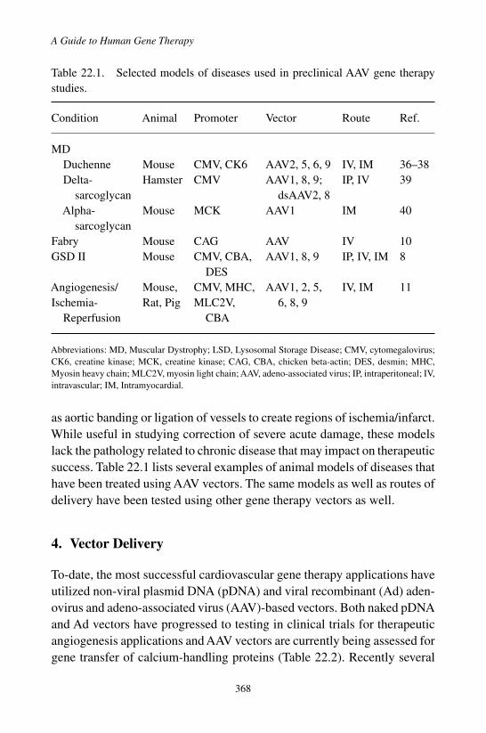
May 4, 2010 11:0 SPI-B903 9in x 6in b903-ch22
A Guide to Human Gene Therapy
Table 22.1. Selected models of diseases used in preclinical AAV gene therapystudies.
Condition Animal Promoter Vector Route Ref.
MDDuchenne Mouse CMV, CK6 AAV2, 5, 6, 9 IV, IM 36–38Delta- Hamster CMV AAV1, 8, 9; IP, IV 39
sarcoglycan dsAAV2, 8Alpha- Mouse MCK AAV1 IM 40
sarcoglycanFabry Mouse CAG AAV IV 10GSD II Mouse CMV, CBA, AAV1, 8, 9 IP, IV, IM 8
DESAngiogenesis/ Mouse, CMV, MHC, AAV1, 2, 5, IV, IM 11Ischemia- Rat, Pig MLC2V, 6, 8, 9
Reperfusion CBA
Abbreviations: MD, Muscular Dystrophy; LSD, Lysosomal Storage Disease; CMV, cytomegalovirus;CK6, creatine kinase; MCK, creatine kinase; CAG, CBA, chicken beta-actin; DES, desmin; MHC,Myosin heavy chain; MLC2V, myosin light chain; AAV, adeno-associated virus; IP, intraperitoneal; IV,intravascular; IM, Intramyocardial.
as aortic banding or ligation of vessels to create regions of ischemia/infarct.While useful in studying correction of severe acute damage, these modelslack the pathology related to chronic disease that may impact on therapeuticsuccess. Table 22.1 lists several examples of animal models of diseases thathave been treated using AAV vectors. The same models as well as routes ofdelivery have been tested using other gene therapy vectors as well.
4. Vector Delivery
To-date, the most successful cardiovascular gene therapy applications haveutilized non-viral plasmid DNA (pDNA) and viral recombinant (Ad) aden-ovirus and adeno-associated virus (AAV)-based vectors. Both naked pDNAand Ad vectors have progressed to testing in clinical trials for therapeuticangiogenesis applications and AAV vectors are currently being assessed forgene transfer of calcium-handling proteins (Table 22.2). Recently several
368

May 4, 2010 11:0 SPI-B903 9in x 6in b903-ch22
Cardiovascular Disease
Table 22.2. Gene therapy clinical trials for CVD.
Target Delivery Vector Trial
Angiogenesis Intracoronary Ad-VEGF165 KATAd-FGF-4 AGENT-2,3,4Ad-FGF-4 AWAREAd-AC6 Adenylyl cyclase
Intramyocardial Ad-VEGF161 REVASCpDNA VEGF165 Euroinject onepDNA VEGF-2 GenasisAd-VEGF121 NorthernAd-VEGF121 NOVA
Heart Failure Intracoronary AAV-SERCA2a SERCA Gene TherapyAAV-SERCA2a CUPID
Angina Intramyocardial pDNA VEGF-2 VEGF therapyIn-stent Local delivery after Anti-sense oligo
Italicsrestenosis stent implantation against c-myc
Abbreviations: Ad, Adenovirus; VEGF, Vascular endothelial growth factor; FGF, Fibroblast growthfactor; AC, Adenyl Cyclase; pDNA, plasmid DNA; AAV, Adeno-associated virus; SERCA, Sarco-plasmic endoplasmic reticulum calcium ATPase; oligo, oligonucleotide.
lentiviral vectors studies have also shown potential, thus warranting contin-ued investigation of these vectors for cardiovascular applications.26
In vivo delivery methods for cardiovascular gene therapy broadly includedirect delivery of vector to the target tissue, local intravascular delivery, andsystemic delivery of vector. Briefly, direct delivery methods entail adminis-tration of vector directly into the heart musculature or onto walls of the vas-culature, most commonly with a needle or other device. Coronary delivery isthe most common form of local intravascular delivery and requires transientisolation of the circulation by cross-clamping or balloon angioplasty fol-lowed by injection of vector into the coronary circulation. Systemic deliveryusually entails simple IV injection of vector at a peripheral site.
From a theoretical, but not necessarily technical, standpoint, the sim-plest mode of delivery is direct injection of vector into the target tissue,
369

May 4, 2010 11:0 SPI-B903 9in x 6in b903-ch22
A Guide to Human Gene Therapy
however specific techniques can vary. For example, in rodents, blind injec-tions through the chest wall or through the diaphragm from the abdomi-nal side have been used successfully to transduce myocardium in variousdisease models. In larger animal models, ultrasound or electromechanicalmapping technologies to visualize the heart have allowed for more accuratetargeted delivery to the affected tissue. Following injection, overall trans-duction efficiencies then become dependent on the ability of the vector toefficiently enter the target cell and subsequently, cellular expression of thetherapeutic transgene. The obvious advantage of this method is the directexposure of target cells to the vector, thereby maximizing the potential forefficient transduction in those cells. Furthermore, restriction of vector tothe sites of injection reduces the potential for spread of vector to non-targettissues and organs thereby reducing the potential of adverse events resultingfrom inappropriate transgene expression. A limitation of direct injection isthe technical difficulty of the procedure, as not all regions of the heart areeasily accessible for direct injection. As such, procedure-related adverseevents are of concern, especially for highly invasive methods. Expressionfrom direct injection is generally limited to regions of delivery which maynot be as effective in diseases that may benefit from more global cardiactransduction; however, applications better suited for localized transductionof the heart do exist.
Direct injection in the myocardium has been shown to be effective inanimal models of disease and as such has been utilized in several phase II/IIIrandomized control clinical trials. Direct intramyocardial injection of Advectors encoding for VEGF121 (REVASC trial) or naked pDNA vectorsencoding for VEGF-2 or VEGF165 (Genasis and Euroinject One trials,respectively) are currently being assessed for the treatment of coronaryartery disease. Despite impressive results in animal models with pDNAvectors, current results in clinical trials thus far have been disappointing.The REVASC trial had more successful results with improvement in timeto 1mm ST-segment depression and time to level 2 angina in treated groups,as well as first anatomical evidence that AdVEGF121 could mediate neovas-cularization in humans.1,11,27−29
Gene gun, or particle bombardment technology, is a modified form ofdirect injection in which vector is linked to micron-sized heavy metal par-ticles. The particles are then propelled under high pressure into targeted
370
May 4, 2010 11:0 SPI-B903 9in x 6in b903-ch22
Cardiovascular Disease
tissue. This method has been used with pDNA vector for cardiac applica-tions in several preclinical studies but has not been shown to be significantlybetter than traditional direct injection techniques.26
The use of solid support structures to mediate local direct delivery hasbeen investigated, and in particular the potential of vector-eluting stents.Over 1 million stents were placed in patients in the United States in 2006and restenosis can occur in up to 30% of patients.27,30 Local gene transfermay provide a sustained supply of therapeutic protein to prevent restenosisand may have the additional advantage of being able to correct the localunderlying molecular defect(s). Preclinical studies in which pDNA, Ad,or AAV vectors are tethered to stents have shown promise, giving rise toefficient local transduction.27,31
Injection of vector into the pericardial space, rather than directly into theheart musculature has also been employed in preclinical studies utilizingpDNA, Ad, and AAV vectors, leading to moderate transduction in regionsadjacent to the pericardial space. Transmural transduction however wasinefficient and could thus far be achieved only with concomitant treatmentwith enzymes such as collagenase or proteases to disrupt the extracellularmatrix.26,29
For some CVD models, direct delivery of vector to tissue distal fromthe heart (such as skeletal muscle or liver) has been utilized. This strategyrelies upon secretion of the therapeutic protein from the transduced tissueand uptake into the affected myocardium. For example, for Pompe disease,GAA expressed in liver or skeletal muscle can be secreted, then taken upby the heart, resulting in correction of cardiac biochemical abnormalities ina mouse model of disease.32
Local intravascular delivery in which vector delivery is isolated to theheart circulation is attractive as the vasculature structure and dense cap-illary network allows for improved dispersion of vector and access totarget cells.1,26,33 Restriction of delivery to the local vasculature can beachieved by injection into heart ex vivo (for transplanted hearts), injectioninto the vasculature after cardiac bypass with induced cardioplegia andcross-clamping, where blood flow into and/or out of the heart is blockedby clamping or balloon angioplasty, or cross-clamping followed by vec-tor delivery into the left ventricle of the beating heart. These methodshave been shown to be very effective in achieving efficient global cardiac
371
May 4, 2010 11:0 SPI-B903 9in x 6in b903-ch22
A Guide to Human Gene Therapy
transduction in preclinical studies; however the technical difficulty andinvasive nature of the procedures may limit clinical use. The most suc-cessful application of local intravascular delivery thus far has been theuse of intracoronary catheters. In the phase II Kuopio Angiogenesis Trial(KAT), pDNA and Ad vectors encoding VEGF165 were delivered locallyvia an infusion-perfusion catheter during percutaneous coronary interven-tion procedures. At 6 months post-treatment, there was significant increasein myocardial perfusion in the group treated with Ad vector. No differ-ence was seen with the pDNA vector. The Angiogenic Gene Therapy Trials(AGENT) assessed intracoronary injection of AdFGF-4 vectors and whilesmaller cohort studies demonstrated some improvement in exercise time andreduction of ischemic defect size, differences were not noted in the largerstudies. Post-hoc analysis, however, did reveal that when subject age orgender was a factor, significant improvements in response to therapy wereevident and as such, a study assessing a more focused patient population(Angiogenesis in women with angina pectoris who are not candidates forrevascularization; AWARE) is currently ongoing.1,26,28,34 In addition to Advectors, recombinant AAV vectors have recently shown great promise asvectors for CVD in preclinical studies and the first AAV trial for car-diac failure entailing delivery of vector encoding SERCA2a via percuta-neous intracoronary injection (Calcium Up-Regulation by PercutaneousAdministration of Gene Therapy in Cardiac Disease; CUPID) has beeninitiated and results presented at the 2009 American Society of Gene Ther-apy meeting so far suggest evidence of biological activity and safety of thevector.28
Improvements to current local intravascular delivery methods are beinginvestigated in preclinical animal studies and include modification of physi-cal delivery means such as increasing delivery/injection pressure and vectordwell times, inclusion of adjuvants to vector preparations which increasemicrovascular permeability, or use of electroporation or sonoporation toincrease cell membrane permeability.35 Vector modifications are also beinginvestigated as potential means to improve uptake into target cells. Such vec-tor modifications also lend themselves well to systemic delivery strategiesand will be discussed further below.
Simple intravenous (IV) injection for systemic delivery of vector is theidealized mode of gene therapy in which the vector itself provides all the
372
May 4, 2010 11:0 SPI-B903 9in x 6in b903-ch22
Cardiovascular Disease
means to transduce only the affected target cells and mediate correction. Theclear advantage to such methods is the ease of vector delivery and avoidanceof procedure-related complications. Current disadvantages however includethe probable transduction of non-target tissues which could result in severeimmune response to the vector itself, the therapeutic transgene product,and the transduced cells themselves. As such, control of each step of vectortransduction is likely necessary, beginning from initial attachment to targetcells, to tissue/cell-type specific transgene expression, and regulation of saidexpression.
Systemic delivery of naked pDNA vectors has met with little success forcardiac transduction, however pDNA preparations utilizing lipid or otherpolymer complexes/coatings and in particular, coatings in which specificpeptide ligands or antibodies have been incorporated, have been shownto enhance uptake in target tissues. A physical method of targeted deliv-ery has used microbubble technology in which vectors (pDNA or viral)have been encapsulated in gas microbubbles which can then be deliv-ered systemically. Ultrasound in the heart or vasculature will then resultin destruction of the microbubbles in the local area, releasing the vectorsin the targeted region. For enveloped viral vectors, pseudotyping envelopesto include targeting moieties have also been employed. For non-envelopedviruses, molecular adapters (cell-specific antibodies, ligands, targeting pep-tides, etc.) have been either physically conjugated to the viral capsidto allow for re-direction of the vector to target cells, or, in some casessuch as with AAV vectors, directly incorporated into the capsid structureitself.
Recently, natural tissue tropism of novel vectors has been exploitedin gene therapy strategies. For example, recombinant AAV pseudotype 9(rAAV2/9) vectors have been shown to be naturally tropic for the heartand globally transduce myocardium after IV delivery and have shown greatpromise in models of congenital cardiac disease, IR injury, and calcium dys-regulation. Several studies have investigated the potential of directed evolu-tion of AAV vectors in which viral capsid proteins are randomly mutated orcapsid proteins from different serotypes are mixed together to create novelvectors with different bioactivities and tissue tropisms. These novel vectorscan then be screened for tissue tropism for the cell-types of choice and couldyield vectors with stringent tropisms.
373

May 4, 2010 11:0 SPI-B903 9in x 6in b903-ch22
A Guide to Human Gene Therapy
5. Conclusions and Outlook
Since the first preclinical CVD gene therapy studies in the late 1980’s,the development of improved vector systems, cell-specific promoters, androutes of delivery have enabled investigators to target a myriad of disease-associated factors affecting the myocardium and the translational nature isnow being realized. Initial clinical studies have been promising however it isclear that continued development and understanding of the molecular basisfor disease as well as vector biology is necessary to develop effective genetherapies. Questions with regard to dosing, timing of delivery and long-term safety remain. Well-designed clinical trials have the potential to com-plement and even stimulate basic science and preclinical research. In mostcases there likely will not be a single target that may provide an all end cure,and refinement of treatments may likely include a multifactorial approachincluding gene therapy and additional strategies. Better understanding ofthe molecular mechanisms will invariably provide platforms on which todevelop effective countermeasures for this devastating disease. Gene ther-apy provides an attractive alternative or complement to current therapies forefficient and effective treatment for CVD. The examples provided through-out this chapter represent some of the many pioneering studies contributingto the characterization, development and implementation of therapy forCVD and the authors regret that only a minor portion could be included.
Acknowledgments
This work was supported in part by NIH PO1 HL059412 and NIH PO1DK058327 (B.J.B., C.S.M., D.J.F.), American Heart Association, NationalCenter (C.S.M.), NRSA FHL095282A (D.J.F).
References
1. Lloyd-Jones D, et al. (2009). Heart disease and stroke statistics–2009 update: A reportfrom the American Heart Association Statistics Committee and Stroke Statistics Sub-committee. Circulation 119: 480–486.
2. Emery AEH (2001). The Muscular Dystrophies, Oxford University Press: Oxford.3. Muir LA, Chamberlain JS (2009). Emerging strategies for cell and gene therapy of the
muscular dystrophies. Expert Rev Mol Med 11: e18.
374
May 4, 2010 11:0 SPI-B903 9in x 6in b903-ch22
Cardiovascular Disease
4. Mann CJ, et al. (2001). Antisense-induced exon skipping and synthesis of dystrophinin the mdx mouse. Proc Natl Acad Sci USA 98: 42–47.
5. McClorey G, Moulton HM, Iversen PL, Fletcher S, Wilton SD (2006). Antisenseoligonucleotide-induced exon skipping restores dystrophin expression in vitro in acanine model of DMD. Gene Ther 13: 1373–1381.
6. van Deutekom JC, et al. (2001). Antisense-induced exon skipping restores dys-trophin expression in DMD patient derived muscle cells. Hum Mol Genet 10: 1547–1554.
7. Clarke JT, Iwanochko RM (2005). Enzyme replacement therapy of Fabry disease. MolNeurobiol 32: 43–50.
8. Schoser B, Hill V, Raben N (2008). Therapeutic approaches in glycogen storage diseasetype II/Pompe Disease. Neurotherapeutics 5: 569–578.
9. van der Ploeg AT, Reuser AJ (2008). Pompe’s disease. Lancet 372: 1342–1353.10. Ogawa K, et al. (2009). Long-term inhibition of glycosphingolipid accumulation in
Fabry model mice by a single systemic injection of AAV1 vector in the neonatal period.Mol Genet Metab 96: 91–96.
11. Rissanen TT, Yla-Herttuala S (2007). Current status of cardiovascular gene therapy.Mol Ther 15: 1233–1247.
12. Yla-Herttuala S, Rissanen TT, Vajanto I, Hartikainen J (2007). Vascular endothelialgrowth factors: Biology and current status of clinical applications in cardiovascularmedicine. J Am Coll Cardiol 49: 1015–1026.
13. Zhu HL, Stewart AS, Taylor MD, Vijayasarathy C, Gardner TJ, Sweeney HL (2000).Blocking free radical production via adenoviral gene transfer decreases cardiacischemia-reperfusion injury. Mol Ther 2: 470–475.
14. WooYJ, et al. (1998). Recombinant adenovirus-mediated cardiac gene transfer of super-oxide dismutase and catalase attenuates postischemic contractile dysfunction. Circu-lation 98: II255–260; discussion II260–251.
15. Li Q, Bolli R, Qiu Y, Tang XL, Guo Y, French BA (2001). Gene therapy with extra-cellular superoxide dismutase protects conscious rabbits against myocardial infarction.Circulation 103: 1893–1898.
16. Li Q, Bolli R, Qiu Y, Tang XL, Murphree SS, French BA (1998). Gene therapy withextracellular superoxide dismutase attenuates myocardial stunning in conscious rabbits.Circulation 98: 1438–1448.
17. Peterson SJ, Frishman WH (2009). Targeting heme oxygenase: Therapeutic implica-tions for diseases of the cardiovascular system. Cardiol Rev 17: 99–111.
18. Juan SH, et al. (2001). Adenovirus-mediated heme oxygenase-1 gene transfer inhibitsthe development of atherosclerosis in apolipoprotein E-deficient mice. Circulation 104:1519–1525.
19. Duckers HJ, et al. (2001). Heme oxygenase-1 protects against vascular constrictionand proliferation. Nat Med 7: 693–698.
20. Melo LG, et al. (2002). Gene therapy strategy for long-term myocardial protectionusing adeno-associated virus-mediated delivery of heme oxygenase gene. Circulation105: 602–607.
21. TangYL, Qian K, ZhangYC, Shen L, Phillips MI (2005). A vigilant, hypoxia-regulatedheme oxygenase-1 gene vector in the heart limits cardiac injury after ischemia-reperfusion in vivo. J Cardiovasc Pharmacol Ther 10: 251–263.
375
May 4, 2010 11:0 SPI-B903 9in x 6in b903-ch22
A Guide to Human Gene Therapy
22. Tang Y, Schmitt-Ott K, Qian K, Kagiyama S, Phillips MI (2002). Vigilant vectors:Adeno-associated virus with a biosensor to switch on amplified therapeutic genes inspecific tissues in life-threatening diseases. Methods 28: 259–266.
23. Andino LM, Takeda M, Kasahara H, Jakymiw A, Byrne BJ, Lewin AS (2008). AAV-mediated knockdown of phospholamban leads to improved contractility and calciumhandling in cardiomyocytes. J Gene Med 10: 132–142.
24. Schmidt U, Zhu X, Lebeche D, Huq F, Guerrero JL, Hajjar RJ (2005). In vivo genetransfer of parvalbumin improves diastolic function in aged rat hearts. Cardiovasc Res66: 318–323.
25. Sakata S, et al. (2007). Restoration of mechanical and energetic function in failingaortic-banded rat hearts by gene transfer of calcium cycling proteins. J Mol Cell Cardiol42: 852–861.
26. Gaffney MM, Hynes SO, Barry F, O’Brien T (2007). Cardiovascular gene therapy:Current status and therapeutic potential. Br J Pharmacol 152: 175–188.
27. LyonAR, Sato M, Hajjar RJ, Samulski RJ, Harding SE (2008). Gene therapy: Targetingthe myocardium. Heart 94: 89–99.
28. Nordlie MA, Wold LE, Simkhovich BZ, Sesti C, Kloner RA (2006). Molecular aspectsof ischemic heart disease: Ischemia/reperfusion-induced genetic changes and potentialapplications of gene and RNA interference therapy. J Cardiovasc Pharmacol Ther 11:17–30.
29. Yla-Herttuala S (2006). An update on angiogenic gene therapy: Vascular endothelialgrowth factor and other directions. Curr Opin Mol Ther 8: 295–300.
30. Davis J, et al. (2008). Designing heart performance by gene transfer. Physiol Rev 88:1567–1651.
31. Fishbein I, Stachelek SJ, Connolly JM, Wilensky RL, Alferiev I, Levy RJ (2005).Site specific gene delivery in the cardiovascular system. J Control Release 109:37–48.
32. Raben N, Plotz P, Byrne BJ (2002). Acid alpha-glucosidase deficiency (glycogenosistype II, Pompe disease). Curr Mol Med 2: 145–166.
33. Williams ML, Koch WJ (2004). Viral-based myocardial gene therapy approaches toalter cardiac function. Annu Rev Physiol 66: 49–75.
34. Giacca M (2007). Virus-mediated gene transfer to induce therapeutic angiogenesis:Where do we stand? Int J Nanomedicine 2: 527–540.
35. Mayer CR, Bekeredjian R (2008). Ultrasonic gene and drug delivery to the cardiovas-cular system. Adv Drug Deliv Rev 60: 1177–1192.
36. Townsend D, Blankinship MJ, Allen JM, Gregorevic P, Chamberlain JS, MetzgerJM (2007). Systemic administration of micro-dystrophin restores cardiac geome-try and prevents dobutamine-induced cardiac pump failure. Mol Ther 15: 1086–1092.
37. Bostick B,YueY, LaiY, Long C, Li D, Duan D (2008).Adeno-associated virus serotype-9 microdystrophin gene therapy ameliorates electrocardiographic abnormalities in mdxmice. Hum Gene Ther 19: 851–856.
38. Athanasopoulos T, Graham IR, Foster H, Dickson G (2004). Recombinant adeno-associated viral (rAAV) vectors as therapeutic tools for Duchenne muscular dystrophy(DMD). Gene Ther 11 Suppl 1: S109–S121.
376

May 4, 2010 11:0 SPI-B903 9in x 6in b903-ch22
Cardiovascular Disease
39. Vitiello C, et al. (2009). Disease rescue and increased lifespan in a model ofcardiomyopathy and muscular dystrophy by combined AAV treatments. PLoS One 4:e5051.
40. Pacak CA, Conlon T, Mah CS, Byrne BJ (2008). Relative persistence of AAV serotype1 vector genomes in dystrophic muscle. Genet Vaccines Ther 6: 14.
377

May 4, 2010 11:0 SPI-B903 9in x 6in b903-ch22
This page intentionally left blankThis page intentionally left blank
May 4, 2010 15:11 SPI-B903 9in x 6in b903-index
Index
β cells, 302–304, 306, 307β-globin, 197–200, 202–208β-thalassemia, 197–199, 201, 203–205,
208φC31 integrase, 123, 126, 127, 129–134,
300
arthritogenic peptides, 296asthma, 345, 346atherosclerosis, 30, 365autoreactive CD4+ T cells, 302
bacterial artificial chromosomes, 73, 129bacteriophage, 126BCKDHA, 254, 258, 259binding domain, 147, 148, 151, 153, 154,
165, 170, 175, 176, 281bioreactor, 94biosafety, 47, 171, 341bone marrow, 43, 46, 48, 177, 198–201,
205, 206, 215, 218, 223–226, 281, 306,313, 317, 319, 321
Bone Marrow Transplantation (BMT),200, 201, 319
bone morphogenetic proteins (BMPs), 300brain, 12, 53, 59, 61, 69, 93, 96, 97, 117,
131, 171, 175, 181–187, 191–194, 254,256, 281, 313, 316, 319, 322, 335, 336,338, 340–342, 348
brain-derived neurotrophic factor, 184,336
calcium, 2, 366–369, 372, 373Canavan disease, 190cancer gene discovery, 134cancer gene therapy, 21, 28, 132, 279, 280,
283, 289, 354cancer vaccines, 1, 2, 287, 288canine disease model, 242canine X-linked muscular dystrophy in
Japan (CXMDJ), 267cap, 88, 267capsid, 21–24, 26, 27, 30, 31, 54, 55, 58,
71, 72, 74, 89, 91–93, 95–99, 123, 240,241, 244, 245, 262, 268, 271, 273, 340,351, 354, 373
carbamate linkage, 9cardiomyopathy, 199, 269, 363, 364, 366cardiovascular, 15–17, 21, 29, 313, 361,
368, 369cardiovascular gene therapy, 368, 369cationic lipids, 1, 2, 6, 9–15, 352cationic liposomes, 2, 3, 9, 13, 17, 300,
352, 353, 355cationic polymers, 1, 14, 17CCR5 gene, 150, 151CD34+ cells, 42, 45, 204, 215, 216, 221,
223, 226cell-based therapy, 346, 356, 357central nervous system (CNS), 69, 70, 79,
181–183, 185, 190, 193, 214, 312–314,321, 322
central polypurine tract (cPPT), 56, 57Cernunnos, 221
379

May 4, 2010 15:11 SPI-B903 9in x 6in b903-index
A Guide to Human Gene Therapy
CFTR, 201, 345–349, 351–353, 355, 356chondrocytes, 295, 300, 301chondrogenesis, 300
chromatography, 6, 95, 130, 267Chronic Granulomatous Disease (CGD),
41, 213, 214, 225–227cis-acting sequences, 106, 117clinical gene therapy, 37–39, 47, 104, 176,
244, 264, 270, 301, 306clinical trial, 2, 3, 6–10, 12, 13, 15–17, 21,
29, 32, 41, 58, 63, 64, 69, 73, 79–81,87, 88, 97, 99, 110, 135, 142, 150, 151,156, 163, 166, 185, 190, 191, 193, 194,200, 204, 217–220, 226, 227, 233,238–242, 244–246, 255, 256, 264, 270,271, 273, 281, 283, 284, 286, 287, 290,292, 295, 299, 304, 306–308, 318, 327,330, 333, 335, 339, 341, 345, 346, 348,349, 352–355, 363–365, 368–370, 374
clonal dominance, 37, 42, 46, 47CNTF, 189, 190, 256, 337collagen-matrix, 307compacted DNA nanoparticles
concatemers, 353cone opsin, 334, 338cone photoreceptors, 327, 329, 330, 333,
334coxsackie and adenovirus receptors, 283cross-correction, 319, 320, 323cystic fibrosis, 14, 96, 201, 259, 295,
345–348, 355, 356cytokines, 13–15, 25, 30, 31, 40, 60, 62,
97, 218, 223, 256, 280, 284, 288, 290,295, 296, 298–300, 302
cytotoxic T lymphocyte (CTL), 97, 237,240, 286, 296, 317
degeneration, 49, 104, 113, 115, 182,190–192, 262, 266, 268, 328–330, 335
delivery, 2, 6–8, 13, 14, 17, 21, 23, 26–31,37, 38, 53, 61, 64, 69, 72, 75, 77, 79, 82,87, 96, 103, 105–108, 114, 119, 130,131, 150, 156, 163, 167, 172–174,181–184, 187, 188, 190, 192, 193, 224,238, 241, 244–246, 253–255, 257, 261,262, 266, 268, 269, 272, 273, 279, 282,
283, 287–291, 297, 298, 300, 303, 304,311, 316, 317, 319, 321–323, 327, 328,334, 337, 339, 341, 346, 348–355,364–366, 368–374
dendritic cells (DCs), 12, 268, 269, 286,295, 297, 302, 305
density gradient centrifugation, 6dermatan sulfate, 312, 316diabetes, 79, 199, 251, 258, 259, 295, 296,
301–307, 347, 362diabetogenic T cells, 305, 307diacyl cationic lipids, 9Dicer, 105, 107, 112diphytanolylphosphatidylethanolamine, 13DNA methylation, 96DNA polymerase, 24, 90, 170DNA transposons, 124dopamine, 182–186double-strand break (DSB), 49, 139, 141,
144–147, 149, 150, 154–156, 264doxycycline, 168, 171Dp71, 268Drosha, 109, 112Duchenne, 201, 261, 363, 368Duchenne’s muscular dystrophy (DMD),
201, 261, 262, 264, 266–270, 272–274,363
dystrophin, 131, 132, 261–266, 268–273,363
dystrophin-glycoprotein complex (DGC),262, 263
ecdysteroid receptor, 164, 176efficiency, 1, 8, 9, 12, 13, 17, 21, 26, 41,
42, 53, 57, 77, 89, 98, 99, 128, 129,148, 175, 205–208, 215, 216, 220, 238,245, 284, 288, 303, 317, 319, 329, 332,346, 349–353, 370
electroporation, 1, 2, 8, 9, 131, 279, 280,287, 289, 372
electroretinography (ERG), 328, 331embryonic stem cells, 132, 140–142, 157endocytosis, 22, 23, 92, 99endosome, 12, 15, 23, 72, 92, 93, 107, 319entry, 4, 9, 13, 22, 23, 55, 58, 60, 71, 72,
93, 99, 171, 206, 273, 285
380

May 4, 2010 15:11 SPI-B903 9in x 6in b903-index
Index
enveloped RNA virus, 53enzyme replacement therapy, 215, 313,
364epidermolysis bullosa, 132epilepsy, 181, 186, 187episomal, 49, 61, 75, 239, 255, 262episome, 61, 88, 96equilibrium centrifugation, 5erythroid lineage-restricted transgene
expression, 206erythroid progenitor cells, 197, 200, 206,
208erythropoietin, 166, 207ES cells, 142, 143, 356EXCELLERATE, 307exosomes, 297extra-articular, 296, 297, 301ex vivo gene therapy, 131, 187, 272, 298,
304, 305ex vivo gene transfer, 53, 214, 226, 236,
237, 239, 244, 298, 304, 356eye, 93, 96, 97, 111, 149, 166, 174, 327,
328, 331, 333, 334, 337, 340, 341
Fabry disease, 363factor IX (F.IX), 23, 30, 97, 118, 201, 233,
243, 271factor VIII (F.VIII), 201, 233factor X, 22, 23, 27, 31Fas, 302
fermentor. See bioreactor, 94fiber, 22, 23, 26–28, 31, 131, 191, 239,
262, 266, 268, 283, 322fibroblast growth factor (FGF), 6, 29, 92,
258, 365, 369fibroblasts, 6, 29, 92, 116, 192, 193, 201,
236, 237, 244, 245, 257, 258, 365, 369first generation, 24, 25, 28, 29, 32, 77, 197
FKBP52, 98
galectin-1, 305ganglion cells, 327–329, 335Gaucher disease, 311–313, 317, 324gene switch, 163, 177gene therapy, 1, 2, 6–10, 13–15, 17, 21–23,
25, 28, 29, 32, 33, 37–39, 41, 42, 44–47,
49, 53, 59, 64, 87, 97, 103–106, 108,110–112, 114, 115, 117–119, 123–127,129–132, 134, 139, 141, 142, 144, 146,147, 149–151, 154–157, 163, 164, 166,176, 177, 181–188, 190–193, 197–201,203, 204, 208, 213–222, 224–227, 233,238, 240, 242, 244–246, 251, 253, 255,257, 260, 261, 264, 270–274, 279–283,288, 289, 291, 295, 297–301, 304–308,311–324, 327–329, 334, 336, 337, 339,342, 345–347, 349, 350, 353–355, 357,361, 363–369, 372–374
gene transfer, 1–3, 6, 8, 9, 11, 12, 17, 21,23, 24, 28, 37, 39, 41, 45–48, 53, 56–60,62–64, 79, 80, 87, 130, 141, 143, 157,172, 174, 182, 184, 187, 191–193,201–203, 214–218, 221, 223–227, 233,236–242, 244–246, 255, 262, 264, 266,270, 271, 279, 280, 282, 295, 298, 300,301, 303–305, 307, 308, 330, 337, 342,349, 350, 356, 365, 368, 371
genotoxicity, 48, 63, 147glaucoma, 335–337glial-derived neurotrophic factor, 184glioblastoma, 69, 131, 283, 284, 288, 289glioblastoma multiforme (GBM), 69, 79glutamic acid decarboxylase (GAD), 79,
185, 302glycogen, 259, 311, 314, 317, 318, 323,
363, 364glycogen storage disease, 311, 314, 318,
363glycoproteins, 17, 39, 54, 55, 59, 60, 71,
72, 75, 262, 263, 282, 352glycosphingolipids, 313, 363golden retriever-based muscular dystrophy
dog (GRMD), 267, 273graft-versus-host disease (GvHD), 200,
201, 214, 219, 222, 226granzyme, 302green fluorescent protein (GFP), 11, 99,
112, 132, 147, 148, 152, 334growth factors, 2, 6, 26, 27, 29, 45, 46, 92,
111, 184, 185, 189, 190, 192, 258, 266,300, 307, 351, 365, 369
guide strand, 105, 107, 112
381
May 4, 2010 15:11 SPI-B903 9in x 6in b903-index
A Guide to Human Gene Therapy
hammerhead ribozyme, 108–110, 118, 119helper functions, 25, 73, 88–90, 94
helper-dependent, 25, 265, 348, 352hemagglutination, 21hematopoietic stem cells (HSC), 16, 37,
40, 41, 53, 59–61, 63, 132, 143,197–202, 205, 207, 208, 213–218,221–227, 236, 237, 281, 313, 321, 356
hematopoietic stem cells transduction,200, 207
hematopoietic stem cells transplantation,16, 198, 199, 214, 221–224, 226, 227,313
hemoglobinopathies, 59, 197, 198, 200,201, 203, 204, 208
hemophilia A, 201, 233, 234, 242–246hemophilia B, 30, 63, 118, 201, 233, 234,
242–246heparan, 23, 71, 72, 92, 99, 271, 312, 316heparan sulphate, 23, 71, 72, 92, 99, 271,
312, 316heparan sulphate proteoglycans (HSPG),
23, 71, 72, 271hepatic, 32, 60, 199, 203, 238, 240, 241,
244, 245, 255, 257, 284, 323hepatocyte, 2, 6, 22, 23, 27, 30, 31, 62,
118, 130, 175, 239, 241, 242, 320, 354,365
herpes simplex virus (HSV), 28, 69, 88,168, 175, 183, 280
herpesvirus, 69, 70, 283hexon, 22, 23, 26, 27, 31Himar1, 300hindlimb ischemia, 131hippocampus, 186, 187, 192histone deacetylase inhibitor, 200, 273histones, 96, 170, 200, 273, 300HLA-identical donor, 214, 215, 218homologous recombination, 24, 49, 63,
139–147, 149, 151, 153, 157, 264human alpha-1 antitrypsin, 130human embryonic stem cells, 156human immunodeficiency virus (HIV), 29,
39humoral immune response, 62, 168, 268,
320
Huntington’s disease, 181, 187Hurler Syndrome, 313HVEM, 72hydrodynamic injection, 130, 132, 134hydrophobic interaction chromatography
(HIC), 6hyperglycemia, 255, 302, 306, 307hypervariable regions, 22hypoglycemia, 315, 318, 323
iatrogenesis, 253IFNαβ response, 62IKK complex, 299, 300IL-7R deficiency, 223immune response, 3, 7, 13, 24, 25, 30, 62,
63, 80, 118, 132, 156, 163, 164, 167,168, 172, 175, 176, 201, 237, 239–241,244, 264, 266, 268–270, 279, 283,286–289, 295, 312, 319–321, 328, 341,347, 352, 354, 373
immune suppression, 70, 221, 241, 244,246, 270
immunity, 28, 32, 97, 112, 219, 225, 286,287, 289, 290
immunosuppression, 165, 200, 269, 271,303
immunotherapies, 15, 29, 284, 288, 304,305, 307
immunotoxicity, 165, 177induced pluripotent stem cells, 143inhibitors, 29, 93, 98, 106, 108, 111, 115,
116, 119, 165, 168, 200, 234, 235, 238,240, 242, 243, 253, 266, 273, 299, 300,348, 352, 367
innate immune response, 80, 237, 239,240, 347, 352
insertional mutagenesis, 38, 40, 42, 43,46–49, 53, 197, 204, 214, 224, 227,239, 263, 264, 329
insertional oncogenesis, 61, 63, 64insulin-like growth factor (IGF)-1, 266,
300insulitis, 302, 306integrase, 38, 45, 49, 54, 55, 60, 123, 124,
126–134, 300
382

May 4, 2010 15:11 SPI-B903 9in x 6in b903-index
Index
integrating vectors, 123, 130, 132, 214,300
integration, 37, 38, 40–49, 53, 55, 57, 60,61, 63, 64, 96, 124, 126–129, 131, 133,134, 140, 141, 149, 156, 157, 204, 206,216, 226, 254, 255, 264, 281, 300
integration analysis, 37integration-defective Lentiviral vector
(IDLV), 53, 60, 61integration analysis, 47intein, 96interferon, 8, 15, 17, 113, 114, 268, 269,
282, 284, 285, 302interleukin, 30, 62, 165, 256, 296intramuscular, 6, 7, 12, 163, 166, 167,
173, 190, 238, 245, 255, 258, 265, 268,270, 272, 297, 305, 319, 353, 354
intraocular pressure (IOP), 335, 336intravitreal, 166, 336, 337intravitreous, 167intron, 4, 75, 76, 95, 96, 104, 109, 167,
203, 267in vivo gene transfer, 11, 12, 53, 236, 238,
239, 244, 295iPS cells, 142, 143ischemia, 1, 2, 7, 131, 198, 365, 366, 368islet transplantation, 302–305ITR, 88–92, 94–96, 98, 349, 351
Janus kinase 3 (Jak3) deficiency, 222
K-562, 132, 149, 206keratinocytes, 132kozak sequence, 4Ku, 12, 30Kupffer cell, 12, 30, 31
latency, 74–76, 134latency active promoter (LAP), 76latency-associated transcripts (LAT),
74–76Leber’s congenital amaurosis (LCA), 61,
204, 327, 338lentiviral vector, 40, 41, 46, 53, 56, 60,
118, 184, 191, 197, 203, 204, 213, 218,
220, 227, 238, 239, 246, 272, 282, 311,329, 355, 369
lentivirus, 39, 40, 53, 63, 118, 140, 156,177, 203, 213, 282, 287, 329
leptin, 255–257leukapheresis, 307leukemia, 39, 44, 46–48, 197, 202, 203,
219, 256, 281, 282LIF, 256limb perfusion, 238, 269, 270linear amplification-mediated PCR
(LAM-PCR), 43lipophilic transfection reagents, 131liposome, 2, 3, 9–11, 13, 17, 107, 108,
300, 352, 353, 355liver, 8, 12, 23, 25–28, 30, 32, 33, 34, 53,
59, 93, 96, 97, 106, 108, 118, 129, 130,134, 172, 177, 201, 214, 233, 235, 237,238, 240, 241, 242–246, 254, 257, 258,271, 297, 303, 304, 306, 313, 316, 317,320, 323, 347, 348, 354, 371
liver gene transfer, 238, 246LMO2, 47, 63, 219, 220local delivery, 29, 193, 369locked nucleic acid, 107locus control region, 202, 334long terminal repeats (LTR), 38–40, 43,
44, 46, 48, 49, 55–57, 64, 173, 203,219, 221, 227
LPL, 254, 259luciferase, 12, 112, 117lung, 12, 25, 106, 131, 177, 201, 214, 284,
287, 288, 345–358lung cancer, 284, 288, 345, 346, 354, 355lung diseases, 345, 346, 348, 355–358lupus, 307lymphoproliferative disease, 47lysosomal storage disease, 201, 311–314,
319, 321–323, 368lysosome, 12, 15, 93, 312, 314, 319, 320
macular, 104, 113, 115, 330, 335mannose-6-phosphate, 319, 320MCK promoter, 265measles virus, 59, 60, 282, 285
383
May 4, 2010 15:11 SPI-B903 9in x 6in b903-index
A Guide to Human Gene Therapy
mesenchymal stem cells (MSCs), 144,257, 273, 288, 300, 356
metacarpophalangeal, 301micro RNA (miRNA, microRNA), 5, 63,
103–105, 107, 109, 112–116, 118, 188,246, 354
mifepristone, 164, 175mini-dystrophin, 265, 271, 363miRNA, 103–105, 109, 112–116, 118,
246, 354mobile DNA elements, 124modified oligonucleotides, 103monogenetic, 295monogenic disorder, 264mRNA Splicing, 272mucopolysaccharidosis, 312multiplicity of infection (MOI), 144multipotent mesenchymal stromal cells
(MSCs), 273murine leukemia virus (MLV), 39, 42, 45,
46, 59, 61, 202, 218, 219, 221, 225,226, 281
muscle, 1, 2, 6, 8, 12, 16, 29, 59, 61, 80,93, 96, 131, 166, 172, 174, 175, 177,189, 234, 237–240, 242, 244, 245, 254,256–258, 262, 264–273, 280, 303, 304,306, 312, 314, 316–320, 322, 323, 353,354, 362–364, 371
muscular dystrophy, 201, 261, 262, 265,267, 269, 270, 272, 295, 314, 320, 362,363, 367, 368
myelodysplastic syndrome, 47
Naked DNA, 2, 6, 8, 9, 131, 144, 172, 242,300, 354
nanoparticles, 15, 107, 108, 353nectin-1, 72neuropathy, 81, 302, 307Neuropeptide Y, 187neurotrophic factor, 79, 184, 188–190,
336, 337neurotrophin, 79, 192, 337neutralizing antibodies, 14, 22, 32, 70, 80,
93, 97, 99, 237, 238, 240, 241, 271,288, 349
NF-κB, 165, 175, 299, 300, 306
nicotinamide dinucleotide phosphateoxidase (NADPH), 225
NK cells, 80, 215, 218, 219, 223NLS, 93NOD/SCID mice, 204, 208non-dividing cells, 53, 55, 59, 61, 87, 96,
203, 239, 262, 353non-homologous end joining, 89, 141,
144, 145, 149non-myeloablative
conditioning/Busulfan/Melphalan, 226non-steroidal anti-inflammatory drugs
(NSAIDs), 296non-viral, 1, 2, 6, 8–10, 13, 156, 298, 300,
304, 311, 345, 346, 352, 368non-viral gene therapy, 1, 2, 6, 8–10, 13,
15, 345non-viral gene transfer, 1, 300nonhuman primates, 163, 166nuclear transfer, 142
oligoneucleotide, 306oligonucleotides, 103, 104, 106, 107, 115,
116, 119, 190, 272, 305, 306, 363, 369Omenn syndrome, 221oncogene, 43, 46–48, 63, 64, 110, 111,
123, 133, 140, 143, 219, 220, 280, 289,355
oncogenesis, 32, 37, 42, 61, 63, 220, 264oncolytic, 28, 73, 79–81, 279, 281–286,
289–291oncolytic viruses, 81, 282, 289–291oncoretroviruses, 203open reading frame (ORF), 4, 25, 88, 89,
272origin of replication (ORI), 3, 4, 73, 129osteoarthritis, 251, 308osteogenesis, 144, 300Oxyntomodulin, 258
P2 integrase, 129P3 integrase, 129packaging capacity, 24, 98, 239, 329packaging limit, 95, 117pain, 7, 8, 69, 73, 79, 81, 192, 200, 234,
296, 301
384
May 4, 2010 15:11 SPI-B903 9in x 6in b903-index
Index
palindrome, 89panamdendrimer, 14Parkinson disease, 41Parkinson’s disease, 181, 182parvovirus, 87, 197, 206, 207, 282, 288parvovirus B19, 206, 207passenger strand, 105, 107PEG-ADA, 215–217penton base, 22, 23, 27, 31perforin, 302peripheral blood leukocytes, 43, 132peripheral nervous systems (PNS), 69phase I clinical trial, 17, 185, 200, 204,
270, 307, 339, 353phospholipase, 93phosphorothioate, 107, 108, 110, 272Physiological Hurdles, 352plasmid, 1–9, 11–17, 24, 25, 39, 56–58,
73, 93, 94, 96, 103, 107, 108, 112, 123,124, 126–134, 146, 148, 172, 173, 177,224, 244, 245, 265, 267, 279, 287, 289,290, 300, 363, 365, 368, 369
poloxamer, 14–17poly-adenylation sequence (PolyA), 4poly-L-histidine, 15poly-L-ornithine and poly-L-lysine, 14polyanhydride (PLGA), 15polyarticular, 297polycationic amino acids polymer, 15polyethylene glycol, 12, 14, 15, 27polyethylene oxide, 15polyethyleneimine (PEI), 15polylactide glycolic anhydride (PLGA),
14, 15polylysine, 14, 353polyvinylpyrolidone (PVP), 14, 15pompe disease, 311, 314, 317, 320, 322,
363, 364, 371poxvirus, 284pre-integration complex (PIC), 55, 57preclinical studies, 163, 177, 213, 214,
223, 225, 306, 315, 362, 365, 371, 372Primary Immunodeficiency/
Immunodeficiencies (PID), 214prime/boost regimens, 290
production, 3, 24–26, 30–32, 38, 55–58,63, 70, 73, 81, 87, 89, 93–95, 113, 123,128, 129, 177, 182–184, 188, 191, 205,239, 241, 242, 244, 246, 257, 266, 267,272, 320, 338, 366
promoter, 4, 17, 27, 38, 40, 46–49, 56–59,63, 64, 75–78, 87–91, 93, 94, 98, 104,108–110, 112, 113, 116, 118, 119, 129,131, 133, 134, 149, 165, 169–172, 175,202, 203, 205–208, 219, 220, 222, 223,225, 227, 240–242, 265, 282, 299, 306,317, 321, 322, 324, 327, 332, 334, 335,351, 354, 367, 374
pseudotyping, 27, 28, 58–60, 373purification, 7, 56, 95purine metabolites, 219purine nucleoside phosphorylase (PNP)
deficiency, 222PYY, 258
rapamycin, 163–168, 174, 177, 241RBE, 91, 92receptor, 12, 21, 23, 26–28, 30, 46, 54, 55,
60–62, 71–73, 92, 95, 97, 99, 113, 150,164, 175, 176, 187, 218–220, 222–224,240, 256, 271, 283, 285, 286, 289, 296,298, 299, 302, 319, 320, 323, 337, 351,364
regional intravenous, 172, 173regulatable system, 163–166, 168,
174–177regulation, 63, 103, 104, 106, 111, 113,
114, 118, 155, 163–169, 171–176, 219,225, 233, 235, 256, 348, 373
regulatory T cells, 97, 240, 241, 286, 306,307
rep, 88–92, 94, 267reperfusion, 365, 366, 368replication competent lentivirus (RCL), 63replication factors, 90reticuloendothelium system, 12retina, 53, 61, 131, 166, 168, 172, 174,
177, 178, 327–329, 331–335, 337, 338,341, 342
retinal pigment epithelium (RPE),327–329, 337, 338
385

May 4, 2010 15:11 SPI-B903 9in x 6in b903-index
A Guide to Human Gene Therapy
retinitis pigmentosa, 61, 335retinopathy, 302, 335retinoschisis, 330retrotransposons, 113, 124retroviral vector, 37, 38, 40, 42–45, 47, 48,
57, 59, 63, 64, 96, 155, 197, 203, 213,215, 219, 223, 225, 227, 239, 242, 244,245, 281, 290, 301, 311, 317
Retrovirally Tagged Cancer GeneDatabase” (RTCGD), 48
retrovirus, 37–40, 42, 48, 55, 110, 116,133, 134, 140, 171, 193, 213, 226, 281,282, 299, 301, 354
Rev response element (RRE), 55–57reverse transcriptase, 38, 54, 55, 58rheumatoid arthritis, 295, 296rhodopsin, 338riboswitches, 119, 176ribozyme, 103–105, 108–113, 115, 118,
119, 150, 176RISC, 105, 107, 111, 112, 114rituximab, 304RNA, 2, 4, 6, 14, 38–40, 53–55, 58, 62,
76, 89, 103–117, 119, 120, 176, 188,190, 193, 203, 205, 285, 286
RNA decoys, 103, 104RNA interference, 103, 105, 188, 190, 193RNA pol II, 109, 113, 116RNA pol III, 108, 113RNA polymerase, 4, 105, 108RNAi, 105, 111–113, 150, 345, 346, 355RNAse P, 104, 109Rod photoreceptors, 329, 330route of delivery, 174, 246, 297, 303, 368,
374rpe65, 61, 93, 174, 338, 339, 342
sarcoplasmic endoplasmic reticulumcalcium ATPase (SERCA), 367, 369
SCD1, 258Scheie syndrome, 313second strand synthesis, 97, 98self-complimentary vectors, 205, 244self-inactivating (SIN), 40, 56, 57, 203,
220, 282
self-inactivating lentiviral vectors(SIN-LV), 220, 221
serotype, 21–23, 26–28, 30–32, 92, 95,97–99, 108, 156, 205–208, 238, 239,244, 246, 262, 268, 270, 271, 273, 288,322, 327, 332, 334, 337, 350, 351, 373
severe combined immunodeficiency(SCID), 41, 59, 132, 148, 201, 203, 204,208, 213–221, 223–225, 227, 295
shRNA, 112–114, 354sialic acid, 23, 92, 351
sickle cell disease, 197–201, 204, 208single-stranded vectors, 204siRNA, 5, 103–115, 119, 191, 254, 367Sjogren’s syndrome, 307Sleeping Beauty, 123–125, 300small interfering RNA, 105, 107, 110,
112, 113sonoporation, 1, 8, 9, 372species, 21–23, 27, 28, 30, 31, 38, 87, 88,
95, 166, 177, 241, 261, 317, 318, 323,366
spleen focus forming virus (SFFV), 39splicing, 75, 76, 95, 96, 104, 106,
116, 272stem cells, 16, 37, 40, 41, 53, 61, 63, 132,
140–144, 156, 197, 200, 213, 214, 236,237, 257, 272, 281, 288, 300, 321, 356
stent, 369, 371striatum, 182, 183, 185, 188subretinal, 166, 167, 173, 174, 332, 334,
335, 339, 341substantia nigra, 182suicide gene, 28, 132, 280suicide gene therapy, 280synovium, 297, 300systemic delivery, 273, 291, 297, 369, 372,
373
T-cell, 3, 15, 31, 46, 47, 58, 60, 62, 80, 97,132, 149–151, 156, 165, 215–220,222–224, 240, 241, 245, 268, 271,286–290, 296, 297, 302, 304–307, 341
tamoxifen, 164, 175, 176targeting, 15, 26–28, 60, 61, 72, 73, 88,
98, 108, 110, 117, 139–141, 144–149,
386
May 4, 2010 15:11 SPI-B903 9in x 6in b903-index
Index
152, 154, 156, 157, 177, 185, 188, 190,191, 252, 254, 257, 279, 280, 286, 289,320, 332, 334, 335, 350, 354, 373
Tc1/mariner, 124tegument, 71, 75teratomas, 143tet-dependant system, 171TetOff, 168–170TetOn, 163, 168–175TetR-KRAB, 169–172, 174tetracycline, 163, 164, 168, 173,
176, 177TNFR:Fc, 299, 301transactivator, 165–172, 174–177, 367transcription, 4, 12, 25, 30, 38, 39, 43, 45,
54, 55, 57, 63, 75, 89, 90, 98, 106, 113,115, 116, 119, 132, 142, 151, 163–166,168, 171, 172, 226, 323, 349
transcription start sites, 45, 63transducin, 333, 334transduction, 23, 27, 28, 31, 33, 38, 41, 42,
48, 49, 53, 57, 59–63, 77, 80, 93,95–99, 113, 172, 194, 200, 203,205–208, 221–223, 244, 245, 266–271,273, 281, 287, 288, 298, 303, 306, 312,317, 321–324, 349, 352, 353, 356, 364,370–373
transfection, 2, 5, 9–15, 17, 24, 25, 39, 58,94, 130, 131, 244, 245, 266, 267
transgene, 1, 3, 4, 8, 12, 25, 26, 29, 38–41,46–48, 53, 57, 59, 61–64, 73, 76–81,93, 94, 97, 98, 117, 118, 124, 126–128,130, 131, 139, 141, 149, 157, 163–166,168–172, 174–176, 205–207, 215, 219,222, 225, 227, 239–242, 252, 255, 257,261–263, 265, 266, 268–271, 273, 284,286, 290, 299, 300, 304, 312, 319, 323,329, 332, 334, 336, 337, 351, 354, 363,367, 370, 373
transgene regulation, 163–165, 168, 172,174, 175
transient gene expression, 25, 29transposase, 123–125, 128, 133, 134, 300transposon, 49, 123–126, 128, 140, 300transrepressor, 170, 171
tropism, 26, 27, 53, 56, 59–61, 87, 95, 98,238, 239, 262, 273, 282–284, 327, 329,373, 374
trs, 90–92, 98tumor microenvironment, 289, 290tumor necrosis factor (TNF)-α, 30, 296tumor suppressor, 48, 133, 140, 280, 289,
355type I diabetes, 295, 296, 301type II pneumocyte cells, 131tyrosine hydroxylase, 183
ultrasound, 8, 9, 370, 373uncoating, 93, 95, 97, 99, 206utrophin, 264, 266
V(D)J recombination defects, 220, 221vaccine delivery, 21vaccinia virus, 284, 287, 290, 291variable expressivity, 254variegation, 140, 254vascular endothelial growth factor, 29,
189, 369vascular-endothelium, 307vector, 21, 24–33, 37–49, 53, 56–60,
62–64, 69, 73–82, 87–89, 93–98, 103,105–108, 110, 112, 115, 116, 118, 119,123, 124, 128, 130, 132, 140, 155–157,163, 166–168, 171–175, 177, 182–185,188, 190, 191, 193, 194, 197, 202–208,213–223, 225–227, 233, 236–242,244–246, 254, 255, 257, 261–273,281–283, 287–290, 297–301, 303–307,311, 312, 316, 317, 319–324, 327–329,332, 334, 336, 337, 339–342, 345, 346,348–355, 357, 363, 365, 366, 368–374
VEGF, 2, 29, 104, 111, 113, 131, 189,190, 365, 369, 370, 372
vesicular stomatitis virus, 59, 282, 285,286
Vitamin A, 329, 338von Gierke disease, 314
Wiskott-Aldrich Syndrome (WAS), 213,214, 224, 225, 227
Wnt-10b, 257, 258
387

May 4, 2010 15:11 SPI-B903 9in x 6in b903-index
A Guide to Human Gene Therapy
X-chromosomal linkedadrenoleukodystrophy (X-ALD), 41
X-linked disorder, 233, 262, 363X-linked severe combined
immunodeficiency (SCID-XI), 63, 148,213, 218–220, 222, 227
zeta associated 70 kDa phosphoprotein(ZAP-70) deficiency, 223, 224
zinc finger nuclease (ZFN), 49, 53, 61,139, 141, 146–157
388