Embed Size (px)
Citation preview
CASE REPORT | INFLAMMATORY BOWEL DISEASE
Dissecting Cellulitis of the Scalp: A Rare DermatologicalManifestation of Crohn's DiseaseTaseen A. Syed, MD1, Zain Ul Abideen Asad, MD1, George Salem, MD2, Kanika Garg, MD1,Erin Rubin, MD, FCAP3, and Nelson Agudelo, MD4
1Department of Internal Medicine, University of Oklahoma Health Sciences Center, Oklahoma City, OK2Section of Digestive Diseases & Nutrition, University of Oklahoma Health Sciences Center, Oklahoma City, OK3Section of Pathology, University of Oklahoma Health Sciences Center, Oklahoma City, OK4Section of Infectious Diseases, University of Oklahoma Health Sciences Center, Oklahoma City, OK
ABSTRACTDissecting cellulitis is an inflammatory disease of the skin. We report a case of recurrent dissecting cellulitis in apatient with Crohn’s disease. A 31-year-old man with a history of purulent scalp lesions presented with nightsweats, weight loss, abdominal pain, and hematochezia. Colonoscopy revealed a diffuse friable mucosa withextensive pseudopolyps. Scalp biopsy demonstrated epidermoid inclusion cysts with granulation tissue andchronic inflammatory cell infiltration, indicative of dissecting cellulitis. The incidence of dissecting cellulitis withCrohn’s disease is underreported. This dermatologic condition has a tendency to recur, and considering anunderlying disease is key for its appropriate treatment.
INTRODUCTIONDissecting cellulitis, also known as perifolliculitis capitis abscedens et suffodiens or Hoffman disease, is an inflam-matory disease of the skin. It manifests as perifollicular pustules, keloids, nodules, abscesses, and sinuses thatevolve into scarring alopecia. Dissecting cellulitis is included in the follicular inclusion triad along with hidradenitissuppurativa and acne conglobate. Dissecting cellulitis, the least common of the three, can concurrently presentwith hidradenitis suppurativa or acne conglobate. Dissecting cellulitis has been associated with arthritis, keratitis,and pyoderma gangrenosum.
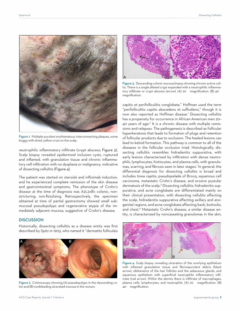
CASE REPORTA 31-year-old man with a history of peptic ulcer disease status post partial gastrectomy 10 years prior and antibi-otic-resistant purulent scalp lesions for 2 years presented with 5 days of night sweats, weight loss, abdominal pain,diarrhea, and hematochezia. On evaluation, he had bilateral lower abdominal tenderness in addition to multiple ery-thematous interconnecting plaques, some boggy with dried yellow crust on the frontal, parietal, and occipital scalpwith scant purulent drainage (Figure 1). Relevant lab results were hemoglobin 8.6 g/dL, platelets 365,000/mm3,erythrocyte sedimentation rate 52 mm/h, and C-reactive protein 138 mg/L.
A comprehensive evaluation for infectious causes of diarrhea was unremarkable, including testing for Clostridiumdifficile. An enhanced abdominal computed tomography showed pancolitis. Colonoscopy disclosed a diffuse areaof severely friable mucosa with contact bleeding and extensive polyps, most likely representing inflammatory pseu-dopolyps in the descending colon along with discontinuous areas of nonbleeding ulcerated mucosa in the rectum(Figure 2). Descending colon biopsy showed chronic active colitis and a single dilated crypt expanded with a
ACG Case Rep J 2018;5:e8. doi:10.14309/crj.2018.8. Published online: January 31, 2018.
Correspondence: Taseen A. Syed, Department of Internal Medicine, University of Oklahoma Health Sciences Center, 500 Central Park Dr, Oklahoma City, OK,73105 ([email protected]).
Copyright: © 2018 Syed et al. This work is licensed under a Creative Commons Attribution-NonCommercial-NoDerivatives 4.0 InternationalLicense. To view a copy of this license, visit http://creativecommons.org/licenses/by-nc-nd/4.0.
ACG Case Reports Journal / Volume 5 acgcasereports.gi.org 1
ACGCASE REPORTS JOURNAL
neutrophilic inflammatory infiltrate (crypt abscess; Figure 3).Scalp biopsy revealed epidermoid inclusion cysts, rupturedand inflamed, with granulation tissue and chronic inflamma-tory cell infiltration with no dysplasia or malignancy, indicativeof dissecting cellulitis (Figure 4).
The patient was started on steroids and infliximab induction,and he experienced complete remission of the skin diseaseand gastrointestinal symptoms. The phenotype of Crohn’sdisease at the time of diagnosis was A2L2xB1: colonic, non-stricturing, non-fistulizing. Retrospectively, the specimenobtained at time of partial gastrectomy showed small sub-mucosal pseudopolyps and regenerative atypia of the im-mediately adjacent mucosa, suggestive of Crohn’s disease.
DISCUSSIONHistorically, dissecting cellulitis as a disease entity was firstdescribed by Spitz in 1903, who named it “dermatits folliculais
capitis et perifolliculitis conglobata.” Hoffman used the term“perifolliculitis capitis abscedens et suffodiens,” though it isnow also reported as Hoffman disease.1 Dissecting cellulitishas a propensity for occurrence in African-American men 20–40 years of age.2 It is a chronic disease with multiple remis-sions and relapses. The pathogenesis is described as follicularhyperkeratosis that leads to formation of plugs and retentionof follicular products due to occlusion. The healed lesions canlead to keloid formation. This pathway is common to all of thediseases in the follicular occlusion triad. Histologically, dis-secting cellulitis resembles hidradenitis suppurativa, withearly lesions characterized by infiltration with dense neutro-phils, lymphocytes, histiocytes, and plasma cells, with granulo-mas, scarring, and fibrosis seen in later stages.1 In general, thedifferential diagnosis for dissecting cellulitis is broad andincludes tinea capitis, pseudopelade of Brocq, squamous cellcarcinoma, metastatic Crohn’s disease, and erosive pustulardermatosis of the scalp.3 Dissecting cellulitis, hidradenitis sup-purativa, and acne conglobate are differentiated mainly ontheir clinical presentation, with dissecting cellulitis affectingthe scalp, hidradenitis suppurativa affecting axillary and ano-genital regions, and acne conglobata affecting back, buttocks,and chest.4 Metastatic Crohn’s disease, a similar disease en-tity, is characterized by noncaseating granulomas in the skin,
Figure 2. Colonoscopy showing (A) pseudopolyps in the descending co-lon and (B) nonbleeding ulcerated mucosa in the rectum.
Figure 1. Multiple purulent erythematous interconnecting plaques, someboggy with dried, yellow crust on the scalp.
Figure 3. Descending colonic mucosa biopsy showing chronic active coli-tis. There is a single dilated crypt expanded with a neutrophilic inflamma-tory infiltrate or crypt abscess (arrow). (A) 20� magnification. (B) 40�magnification.
Figure 4. Scalp biopsy revealing ulceration of the overlying epitheliumwith inflamed granulation tissue and fibrinopurulent debris (blackarrow), obliteration of the hair follicles and the sebaceous glands, andsquamous epithelium with superficial neutrophilic inflammatory infil-trate (red arrow). Within the dermis there is infiltrate of macrophages,plasma cells, lymphocytes, and neutrophils. (A) 20� magnification. (B)40� magnification.
Syed et al Dissecting Cellulitis
ACG Case Reports Journal / Volume 5 acgcasereports.gi.org 2

but its noncontiguous occurrence with the gastrointestinaltract is a rare manifestation. Although the abscesses in dissect-ing cellulitis are sterile, it can be associated with secondarybacterial infection. Reported organisms include Pseudomonasspecies, Staphylococcus epidermidis, Propioni-bacterium acnes,and Prevotella intermedia.4
Dissecting cellulitis has been associated with multiple diseaseentities. In some cases, it may herald the actual clinical mani-festations of an underlying disease process. The well-knownassociations are arthritis, keratitis, pyoderma gangrenosum,keratitis-ichthyosis-deafness syndrome, pilonidal cysts, andosteomyelitis.4 A rare association with Crohn’s disease hasbeen documented; this connection is often missed whenreviewing mucocutaneous manifestations of inflammatorybowel disease due to the low incidence of dissecting cellulitisitself, as well as its nonfulminant clinical course and the widearray of differential diagnoses that can mask the appropriatediagnosis.5,6
Treatment of dissecting cellulitis has changed significantlyover the last decade. Newer treatment options include antibi-otics (first-line therapy: ciprofloxacin, clindamycin, rifampin,and trimethoprim/sulfamethaxole) or tumor necrosis factor(TNF) inhibitors (e.g., adalimumab and infliximab). TNF inhibi-tors can also be used as a bridge to surgery for severe dis-secting cellulitis or perianal fistulas in Crohn’s disease, whichresult in faster healing and delayed relapse when used aftersurgery. Older treatment options included isotretinoin, dap-sone, colchicine, and steroids. Some clinicians prefer usingsteroids (primarily prednisone) as a bridge to success withisotretinoin or other therapies. Zinc sulphate also has docu-mented benefit, but its sole use for the treatment of dissect-ing cellulitis is not well established. Although topical agents inform of antibiotics, isotretinoin, tacrolimus, and steroids cansomewhat control the disease activity, systemic treatmentsshould always be favored for a more therapeutic advantage.If all treatment options fail, more invasive options can beused, such as modern external beam radiation therapy, scalpremoval with graft, full x-ray treatment, or carbon dioxideablation of scalp. Because of their destructive nature and thealternative of newer treatment modalities, invasive proce-dures are avoided.3 Interestingly, the treatment options fordissecting cellulitis and Crohn’s disease have significantoverlap.7
The incidence of dissecting cellulitis is underreported, likelybecause of the diagnostic confusion with other dermatologi-cal conditions. In our patient, a possible diagnosis of Crohn’sdisease was missed at age of 21. Crohn’s disease should beconsidered in patients with dissecting cellulitis. This dermato-logic condition has a tendency for recurrence, and consider-ing an underlying disease is key for its appropriate treatment.Treatment with anti-TNF-a can be beneficial for bothconditions.
DISCLOSURESAuthor contributions: TA Syed and ZUA Asad wrote themanuscript and reviewed the literature. K. Garg reviewed theliterature and collected the images. G. Salem wrote the manu-script. E. Rubin provided the pathology images. N. Aguedeloedited the manuscript and is the article guarantor.
Financial disclosure: None to report.
Informed consent was obtained for this case report.
ReceivedJune27, 2017;AcceptedNovember22, 2017
REFERENCES1. Tchernev G. Folliculitis et perifolliculitis capitis abscedens et suffodiens
controlled with a combination therapy: Systemic antibiosis (metronida-zole plus clindamycin), dermatosurgical approach, and high-dose isotre-tinoin. Indian J Dermatol. 2011;56(3):318–20.
2. Scheinfeld NS. A case of dissecting cellulitis and a review of the litera-ture.Dermatol Online J. 2003;9(1):8.
3. Scheinfeld N. Dissecting cellulitis (perifolliculitis capitis abscedens etsuffodiens): A comprehensive review focusing on new treatments andfindings of the last decade with commentary comparing the therapiesand causes of dissecting cellulitis to hidradenitis suppurativa. DermatolOnline J. 2014;20(5):22692.
4. Gaopande VL, Kulkarni MM, Joshi AR, Dhande AN. Perifolliculitis capitisabscedens et suffodiens in a 7 years male: A case report with review ofliterature. Int J Trichology. 2015;7(4):173–75.
5. Ko JS, Uberti G, Napekoski K, Patil DT, Billings SD. Cutaneous manifes-tations in inflammatory bowel disease: A single institutional study ofnon-neoplastic biopsies over 13 years. J Cutan Pathol. 2016;43(11):946–55.
6. Hindryckx P, Novak G, Costanzo A, Danese S. Disease-related and drug-induced skin manifestations in inflammatory bowel disease. Expert RevGastroenterol Hepatol. 2017;11(3):203–14.
7. Wilkins T, Jarvis K, Patel J. Diagnosis and management of Crohn's dis-ease.Am FamPhysician. 2011;84(12):1365–75.
Syed et al Dissecting Cellulitis
ACG Case Reports Journal / Volume 5 acgcasereports.gi.org 3


![Cellulitis in Adults - WRHN in Adults.pdf• cellulitis is an acute bacterial infection of the dermis and subcutaneous tissue [1]: • Erysipelas is a form of cellulitis caused by](https://img.pdfslide.us/doc/110x75/5e37a2a00fa2bc6b5a461882/cellulitis-in-adults-in-adultspdf-a-cellulitis-is-an-acute-bacterial-infection.jpg)