Embed Size (px)
Citation preview
Case ReportTreatment of Oroantral Communication Usingthe Lateral Palatal Sliding Flap Technique
Fernando Salimon Ribeiro,1 Cassio Torres de Toledo,2
Michele Romero Aleixo,1 Maria Cristina Durigan,1 Willian Corrêa da Silva,1
Samanta Kelen Bueno,1 and Ana Emília Farias Pontes1
1School of Dentistry, Educational Foundation of Barretos, UNIFEB, Avenue Prof. Roberto Frade Monte 389,Bairro Aeroporto, 14783-226 Barretos, SP, Brazil213th Mechanized Cavalry Regiment, Avenue Nilton Prado 2251, Bairro Centro, 13631-900 Pirassununga, SP, Brazil
Correspondence should be addressed to Ana Emılia Farias Pontes; [email protected]
Received 7 April 2015; Accepted 26 May 2015
Academic Editor: FlorianThalhammer
Copyright © 2015 Fernando Salimon Ribeiro et al. This is an open access article distributed under the Creative CommonsAttribution License, which permits unrestricted use, distribution, and reproduction in any medium, provided the original work isproperly cited.
Herein, we present a case of oroantral communication that was to be treated with clinical examination, tomography, andprototyping. A patient presented with oroantral communication with purulent exudation for 4 months, since the displacementof the dental implant and O-ring component to the maxillary sinus. Tomographic examination and prototyping revealed a 5mmbone gap. The patient underwent local washes and antibiotic therapy. After local palpation, a bone defect detected by prototypingwas suspected to be greater than that observed. For the surgery, a communication tunnel was made, and the bone defect was foundto be 12mm in diameter. A pedicle flap was raised on the palate, followed by sliding and suturing. No complications were observedduring the postoperative period, and the suture was removed after a week. Four months later, communication did not resume, andthe patient did not complain of pain, foul smelling, or purulent discharge and was satisfied with the outcome. The findings of thiscase suggest that the lateral sliding flap can be used as an efficient technique for closing oroantral communications. An accurateclinical examination is a critical tool that can be used instead of tomography and prototyping, which can be misleading.
1. Introduction
Oroantral communications are pathologic open connectionsbetween the oral cavity and maxillary sinus and are relativelyfrequent complications observed in dentistry. The highestprevalence is observed inmen, approximately 40 years of age,and complications are normally observed after dental extrac-tion surgery, where the thirdmolars are at a greater risk [1, 2].
Spontaneous closure of defects, smaller than 3mm indiameter, can occur; however, larger communications requiresurgical interventions [3]. Several surgical techniques canbe employed, including slide-in flaps [4], use of pedicled orunpedicled buccal fat pads [5, 6], membranes, autogenous,allogeneic, and xenogeneic grafts, and hemostatic agents [3,7–9].
Scattarella et al. [3] used an autologous bone graft inte-grated with xenologous particulate bone graft, which wascovered with a nonreabsorbable, expandable, polytetrafluor-oethylene titanium-reinforcedmembrane.This guided-tissueregeneration (GTR) technique was successfully employedto optimize the reconstruction of bone tissue and preventepithelial migration to the grafted area. Regenerative tech-niques have better prognosis in cases where resorption of thealveolar wall is low, by providing sufficient bone structuringfor creation of a space for tissue regeneration and stabilizationof the blood clot [10].
Sandhya et al. [7] described a case series of patients withoroantral defects due to dental extractions that were treatedwith a sandwich graft technique. To prepare the sandwich,a freeze dried mineralized bone allograft was sandwiched
Hindawi Publishing CorporationCase Reports in MedicineVolume 2015, Article ID 730623, 5 pageshttp://dx.doi.org/10.1155/2015/730623
2 Case Reports in Medicine
in a collagen membrane, which was closed and suturedwith a resorbable suture. The sandwich was inserted intothe remaining dental alveoli and the remaining granulatedgraft. Successful closure of the communication was observedduring the follow-up period. Hariram et al. [8] performeda comparative study between buccal fat pad grafts andsandwich grafts; the latter was composed of hydroxyapatitecrystals embedded within a collagen sheath and sutured witha polyglactin 910 suture. The findings suggested that thesandwich technique provided a better closure of the oroantralcommunication due to the formation of a layer on the floorof the maxillary sinus that was considered to be suitable forbone regeneration.
In an experimental study by Muglali et al. [9], surgerycreated acute oroantral communications with a 2 mm bur inextraction sockets of rabbits after premolar extraction. Theauthors evaluated bone formation using hemostatic agents,comparing the oxidized regenerated cellulose with bone waxor negative controls without graft. The authors observed thatboth biomaterials did not contribute to the formation offibrous connective tissue and bone in the oroantral region,whichmay have been influenced by the small size of the lesionstudied.
In dental implants, cases of implant displacement intothe sinus after installation of immediate prostheses havebeen reported in the literature, but only a few cases leadto oroantral communications [11]. Some factors may beresponsible for the displacement of the implant into themaxillary sinus, such as low bone quality and bone resorptionafter surgery, causing mucosal thickening and sinusitis, andpotentially lead to chronic infection associated with commu-nication between the oral cavity and maxillary sinus.
This study aimed to present a case of oroantral com-munication, due to the displacement of a dental implantand O-ring component into the maxillary sinus, which wasexamined via clinical examination, tomography, and proto-typing.
2. Case Presentation
A 60-year-old woman with oroantral communication pre-sented for treatment at the Specialization Course in ImplantDentistry of the University Center of the Barretos Educa-tional Foundation (UNIFEB).
The main complaint was foul smelling, purulent dis-charge and intense facial pain, forwhich she continued to takeanalgesic ketorolac trometamol (10mg).The patient reportedto be in good general health, not having undergone radiationtherapy, or consumed drugs, alcohol, or tobacco. On clinicalexamination, total edentulism was observed.
The patient had undergone maxillary sinus lift surgery,and six months later, 6 implants were implanted in the max-illa. Less than a month later, the patient realized that both theimplant and prosthetic component installed in the region oftooth 16 had dislocated and was housed within the maxillarysinus. Oroantral communication was the result. A surgicalprocedurewas performed by an otorhinolaryngologist, whichonly resulted in the removal of the implanted body. Somedays
later, the patient reported that the prosthetic component wasexpelled through the nose. However, the communication andlocal infection persisted, and four months later, the patientsought treatment.
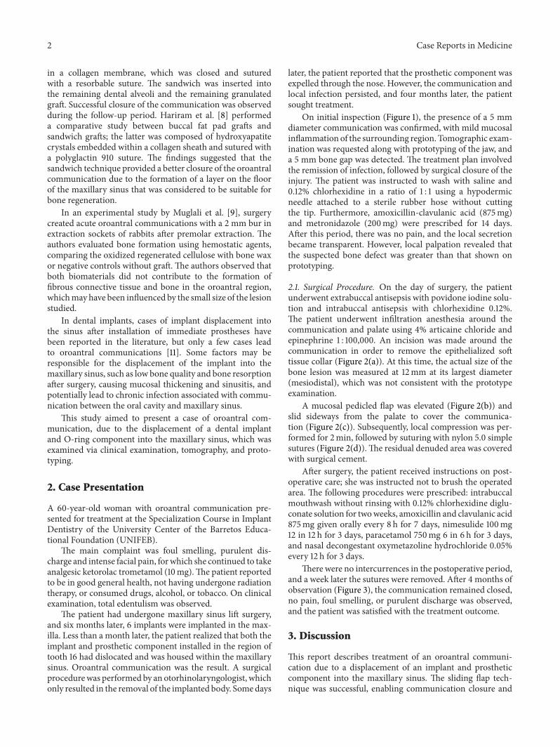
On initial inspection (Figure 1), the presence of a 5 mmdiameter communication was confirmed, with mild mucosalinflammation of the surrounding region. Tomographic exam-ination was requested along with prototyping of the jaw, anda 5 mm bone gap was detected. The treatment plan involvedthe remission of infection, followed by surgical closure of theinjury. The patient was instructed to wash with saline and0.12% chlorhexidine in a ratio of 1 : 1 using a hypodermicneedle attached to a sterile rubber hose without cuttingthe tip. Furthermore, amoxicillin-clavulanic acid (875mg)and metronidazole (200mg) were prescribed for 14 days.After this period, there was no pain, and the local secretionbecame transparent. However, local palpation revealed thatthe suspected bone defect was greater than that shown onprototyping.
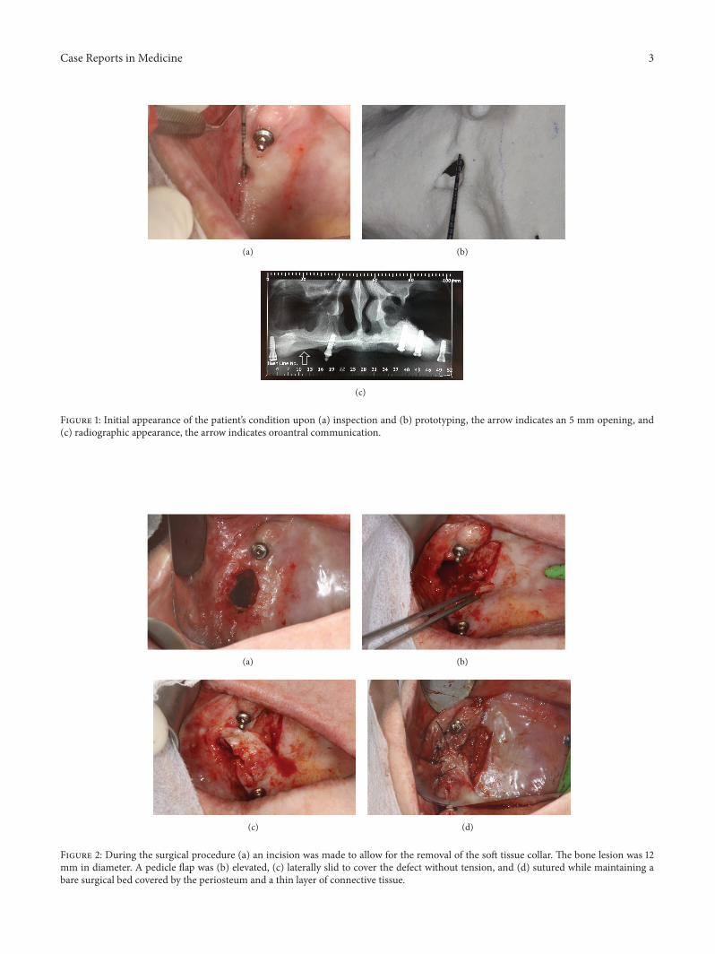
2.1. Surgical Procedure. On the day of surgery, the patientunderwent extrabuccal antisepsis with povidone iodine solu-tion and intrabuccal antisepsis with chlorhexidine 0.12%.The patient underwent infiltration anesthesia around thecommunication and palate using 4% articaine chloride andepinephrine 1 : 100,000. An incision was made around thecommunication in order to remove the epithelialized softtissue collar (Figure 2(a)). At this time, the actual size of thebone lesion was measured at 12mm at its largest diameter(mesiodistal), which was not consistent with the prototypeexamination.
A mucosal pedicled flap was elevated (Figure 2(b)) andslid sideways from the palate to cover the communica-tion (Figure 2(c)). Subsequently, local compression was per-formed for 2min, followed by suturing with nylon 5.0 simplesutures (Figure 2(d)).The residual denuded area was coveredwith surgical cement.
After surgery, the patient received instructions on post-operative care; she was instructed not to brush the operatedarea. The following procedures were prescribed: intrabuccalmouthwash without rinsing with 0.12% chlorhexidine diglu-conate solution for twoweeks, amoxicillin and clavulanic acid875mg given orally every 8 h for 7 days, nimesulide 100mg12 in 12 h for 3 days, paracetamol 750mg 6 in 6 h for 3 days,and nasal decongestant oxymetazoline hydrochloride 0.05%every 12 h for 3 days.
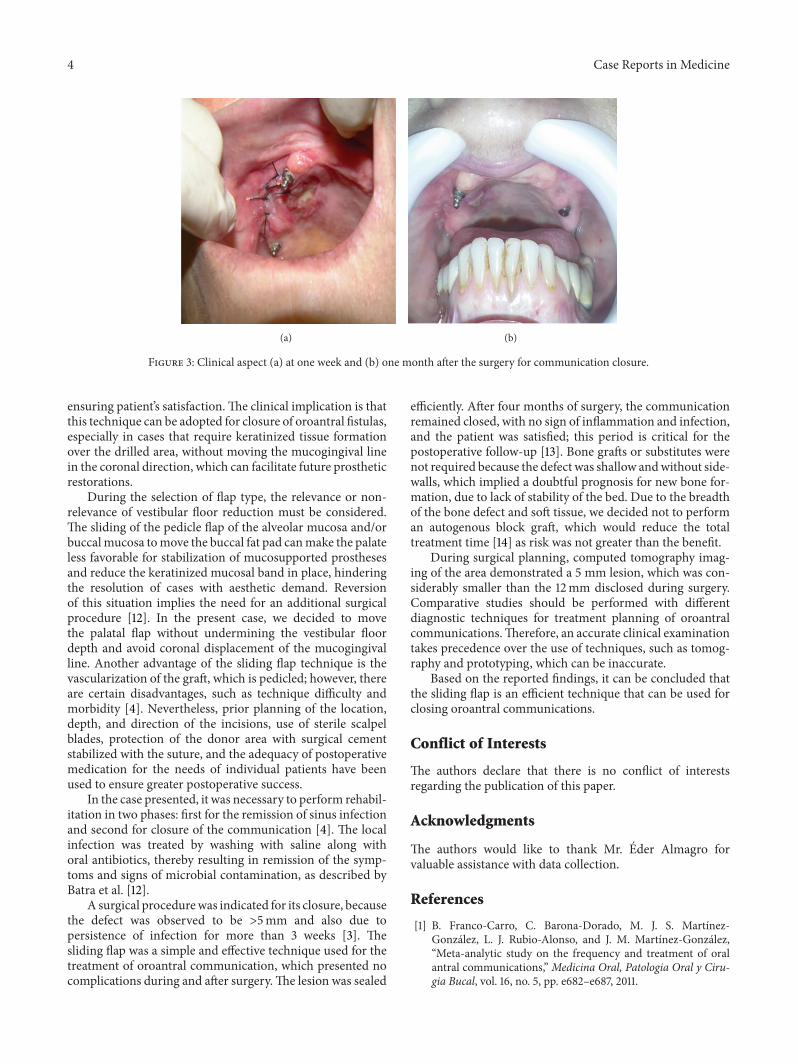
There were no intercurrences in the postoperative period,and a week later the sutures were removed. After 4 months ofobservation (Figure 3), the communication remained closed,no pain, foul smelling, or purulent discharge was observed,and the patient was satisfied with the treatment outcome.
3. Discussion
This report describes treatment of an oroantral communi-cation due to a displacement of an implant and prostheticcomponent into the maxillary sinus. The sliding flap tech-nique was successful, enabling communication closure and
Case Reports in Medicine 3
(a) (b)
(c)
Figure 1: Initial appearance of the patient’s condition upon (a) inspection and (b) prototyping, the arrow indicates an 5 mm opening, and(c) radiographic appearance, the arrow indicates oroantral communication.
(a) (b)
(c) (d)
Figure 2: During the surgical procedure (a) an incision was made to allow for the removal of the soft tissue collar. The bone lesion was 12mm in diameter. A pedicle flap was (b) elevated, (c) laterally slid to cover the defect without tension, and (d) sutured while maintaining abare surgical bed covered by the periosteum and a thin layer of connective tissue.
4 Case Reports in Medicine
(a) (b)
Figure 3: Clinical aspect (a) at one week and (b) one month after the surgery for communication closure.
ensuring patient’s satisfaction.The clinical implication is thatthis technique can be adopted for closure of oroantral fistulas,especially in cases that require keratinized tissue formationover the drilled area, without moving the mucogingival linein the coronal direction, which can facilitate future prostheticrestorations.
During the selection of flap type, the relevance or non-relevance of vestibular floor reduction must be considered.The sliding of the pedicle flap of the alveolar mucosa and/orbuccalmucosa tomove the buccal fat pad canmake the palateless favorable for stabilization of mucosupported prosthesesand reduce the keratinized mucosal band in place, hinderingthe resolution of cases with aesthetic demand. Reversionof this situation implies the need for an additional surgicalprocedure [12]. In the present case, we decided to movethe palatal flap without undermining the vestibular floordepth and avoid coronal displacement of the mucogingivalline. Another advantage of the sliding flap technique is thevascularization of the graft, which is pedicled; however, thereare certain disadvantages, such as technique difficulty andmorbidity [4]. Nevertheless, prior planning of the location,depth, and direction of the incisions, use of sterile scalpelblades, protection of the donor area with surgical cementstabilized with the suture, and the adequacy of postoperativemedication for the needs of individual patients have beenused to ensure greater postoperative success.
In the case presented, it was necessary to perform rehabil-itation in two phases: first for the remission of sinus infectionand second for closure of the communication [4]. The localinfection was treated by washing with saline along withoral antibiotics, thereby resulting in remission of the symp-toms and signs of microbial contamination, as described byBatra et al. [12].
A surgical procedurewas indicated for its closure, becausethe defect was observed to be >5mm and also due topersistence of infection for more than 3 weeks [3]. Thesliding flap was a simple and effective technique used for thetreatment of oroantral communication, which presented nocomplications during and after surgery.The lesion was sealed
efficiently. After four months of surgery, the communicationremained closed, with no sign of inflammation and infection,and the patient was satisfied; this period is critical for thepostoperative follow-up [13]. Bone grafts or substitutes werenot required because the defect was shallow andwithout side-walls, which implied a doubtful prognosis for new bone for-mation, due to lack of stability of the bed. Due to the breadthof the bone defect and soft tissue, we decided not to performan autogenous block graft, which would reduce the totaltreatment time [14] as risk was not greater than the benefit.
During surgical planning, computed tomography imag-ing of the area demonstrated a 5 mm lesion, which was con-siderably smaller than the 12mm disclosed during surgery.Comparative studies should be performed with differentdiagnostic techniques for treatment planning of oroantralcommunications.Therefore, an accurate clinical examinationtakes precedence over the use of techniques, such as tomog-raphy and prototyping, which can be inaccurate.
Based on the reported findings, it can be concluded thatthe sliding flap is an efficient technique that can be used forclosing oroantral communications.
Conflict of Interests
The authors declare that there is no conflict of interestsregarding the publication of this paper.
Acknowledgments
The authors would like to thank Mr. Eder Almagro forvaluable assistance with data collection.
References
[1] B. Franco-Carro, C. Barona-Dorado, M. J. S. Martınez-Gonzalez, L. J. Rubio-Alonso, and J. M. Martınez-Gonzalez,“Meta-analytic study on the frequency and treatment of oralantral communications,” Medicina Oral, Patologia Oral y Ciru-gia Bucal, vol. 16, no. 5, pp. e682–e687, 2011.
Case Reports in Medicine 5
[2] P. P. Pourmand, G. R. Sigron, B. Mache, B. Stadlinger, and M.C. Locher, “The most common complications after wisdom-tooth removal: part 2: a retrospective study of 1,562 cases in themaxilla,” Swiss Dental Journal, vol. 124, no. 10, pp. 1047–1061,2014.
[3] A. Scattarella, A. Ballini, F. R. Grassi et al., “Treatment oforoantral fistula with autologous bone graft and application of anon-reabsorbable membrane,” International Journal of MedicalSciences, vol. 7, no. 5, pp. 267–271, 2010.
[4] R. C. Meirelles and R. M. Neves-Pinto, “Oroantral fistula andgenian mucosal flap: a review of 25 cases,” Brazilian Journal ofOtorhinolaryngology, vol. 74, no. 1, pp. 85–90, 2008.
[5] B. Chaudhary, Z. Gong, Z. Lin, K. Abbas, B. Ling, and H. Liu,“Reconstruction of intraoral maxillary defect with buccal fatpad,”The Journal of Craniofacial Surgery, vol. 25, no. 6, pp. 2174–2177, 2014.
[6] K. Prasad, R. M. Lalitha, K. Ranganath, and J. Singh, “Closureof oroantral communication using buccal pad of fat: report of acase with review of literature,” International Journal of ClinicalDentistry, vol. 4, no. 1, pp. 87–92, 2011.
[7] G. Sandhya, P. B. Reddy, K. A. Kumar, B. Sridhar Reddy, N.Prasad, and G. Kiran, “Surgical management of oro-antralcommunications using resorbable GTR membrane and FDMBsandwich technique: a clinical study,” Journal of Maxillofacialand Oral Surgery, vol. 12, no. 3, pp. 254–259, 2013.
[8] Hariram, U. S. Pal, S. Mohammad, R. K. Singh, G. Singh,and L. R. Malkunje, “Buccal fat pad versus sandwich graft fortreatment of oroantral defects: a comparison,” National Journalof Maxillofacial Surgery, vol. 1, no. 1, pp. 6–14, 2010.
[9] M. Muglali, A. Ozak, M. Yarım, M. S. Nural, N. Celebi, and T.Aksoz, “Assessment of the effects of curacel and bonewax on theacute oroantral opening site bymeans of computer tomographyand histopathology,” Journal of Maxillofacial and Oral Surgery,vol. 11, no. 2, pp. 160–165, 2012.
[10] J. T.Mellonig, “Enamelmatrix derivative for periodontal recon-structive surgery: technique and clinical and histologic casereport,” International Journal of Periodontics and RestorativeDentistry, vol. 19, no. 1, pp. 9–19, 1999.
[11] K. I. Jeong, S. G. Kim, J. S. Oh, and M. A. Jeong, “Displacedimplants into maxillary sinus: report of cases,” Implant Den-tistry, vol. 23, no. 3, pp. 245–249, 2014.
[12] H. Batra, G. Jindal, and S. Kaur, “Evaluation of differenttreatment modalities for closure of oro-antral communicationsand formulation of a rational approach,” Journal ofMaxillofacialand Oral Surgery, vol. 9, no. 1, pp. 13–18, 2010.
[13] J. Hernando, L. Gallego, L. Junquera, and P. Villarreal,“Oroantral communications. A retrospective analysis,” Medic-ina Oral, Patologia Oral y Cirugia Bucal, vol. 15, no. 3, pp. e499–e503, 2010.
[14] M. S. Ahmed and N. A. Askar, “Combined bony closure oforoantral fistula and sinus lift with mandibular bone graftsfor subsequent dental implant placement,” Oral Surgery, OralMedicine, Oral Pathology, Oral Radiology and Endodontology,vol. 111, no. 4, pp. e8–e14, 2011.

Submit your manuscripts athttp://www.hindawi.com
Stem CellsInternational
Hindawi Publishing Corporationhttp://www.hindawi.com Volume 2014
Hindawi Publishing Corporationhttp://www.hindawi.com Volume 2014
MEDIATORSINFLAMMATION
of
Hindawi Publishing Corporationhttp://www.hindawi.com Volume 2014
Behavioural Neurology
EndocrinologyInternational Journal of
Hindawi Publishing Corporationhttp://www.hindawi.com Volume 2014
Hindawi Publishing Corporationhttp://www.hindawi.com Volume 2014
Disease Markers
Hindawi Publishing Corporationhttp://www.hindawi.com Volume 2014
BioMed Research International
OncologyJournal of
Hindawi Publishing Corporationhttp://www.hindawi.com Volume 2014
Hindawi Publishing Corporationhttp://www.hindawi.com Volume 2014
Oxidative Medicine and Cellular Longevity
Hindawi Publishing Corporationhttp://www.hindawi.com Volume 2014
PPAR Research
The Scientific World JournalHindawi Publishing Corporation http://www.hindawi.com Volume 2014
Immunology ResearchHindawi Publishing Corporationhttp://www.hindawi.com Volume 2014
Journal of
ObesityJournal of
Hindawi Publishing Corporationhttp://www.hindawi.com Volume 2014
Hindawi Publishing Corporationhttp://www.hindawi.com Volume 2014
Computational and Mathematical Methods in Medicine
OphthalmologyJournal of
Hindawi Publishing Corporationhttp://www.hindawi.com Volume 2014
Diabetes ResearchJournal of
Hindawi Publishing Corporationhttp://www.hindawi.com Volume 2014
Hindawi Publishing Corporationhttp://www.hindawi.com Volume 2014
Research and TreatmentAIDS
Hindawi Publishing Corporationhttp://www.hindawi.com Volume 2014
Gastroenterology Research and Practice
Hindawi Publishing Corporationhttp://www.hindawi.com Volume 2014
Parkinson’s Disease
Evidence-Based Complementary and Alternative Medicine
Volume 2014Hindawi Publishing Corporationhttp://www.hindawi.com