Embed Size (px)
Citation preview
University of Groningen
Biodegradable polyurethane for closure of oroantral communicationsVisscher, Susan Henrieke
IMPORTANT NOTE: You are advised to consult the publisher's version (publisher's PDF) if you wish to cite fromit. Please check the document version below.
Document VersionPublisher's PDF, also known as Version of record
Publication date:2016
Link to publication in University of Groningen/UMCG research database
Citation for published version (APA):Visscher, S. H. (2016). Biodegradable polyurethane for closure of oroantral communications: Experimentaland clinical evaluation [Groningen]: University of Groningen
CopyrightOther than for strictly personal use, it is not permitted to download or to forward/distribute the text or part of it without the consent of theauthor(s) and/or copyright holder(s), unless the work is under an open content license (like Creative Commons).
Take-down policyIf you believe that this document breaches copyright please contact us providing details, and we will remove access to the work immediatelyand investigate your claim.
Downloaded from the University of Groningen/UMCG research database (Pure): http://www.rug.nl/research/portal. For technical reasons thenumber of authors shown on this cover page is limited to 10 maximum.
Download date: 23-05-2018
Biodegradable polyurethane for closure
of oroantral communications
Experimental and clinical evaluation
The research in this thesis was performed at:
- Department of Oral and Maxillofacial Surgery, University Medical Center Groningen
- Department of Biomedical Engineering, University Medical Center Groningen
- Department of Cell Biology, section Electron Microscopy, University Medical Center
Groningen
- Polyganics BV, Groningen
Bookdesign: Sgaar Groningen
Printed by: Drukkerij van der Eems Heerenveen
ISBN: 978-90-367-8535-8
© Susan Henrieke Visscher, 2015
All rights reserved. No part of this publication may be reported or transmitted, in any
form or by any means, without prior permission of the author.
Biodegradable polyurethane for closure
of oroantral communications
Experimental and clinical evaluation
Proefschrift
ter verkrijging van de graad van doctor in de
Medische Wetenschappen
op gezag van de
rector magnificus prof. dr. E. Sterken
en volgens besluit van het College voor Promoties.
De openbare verdediging zal plaatsvinden op
woensdag 27 januari 2016 om 14.30 uur
door
Susan Henrieke Visscher
geboren op 29 augustus 1980
te Almelo
Printing and distribution of this thesis was kindly supported by:
- Nederlandse Vereniging voor Mondziekten, Kaak- en Aangezichtschirurgie
(NVMKA)
- Rijksuniversiteit Groningen (RUG)
- W.J. Kolff Institute
- Polyganics B.V.
- Mondzorgcentrum Wiranto
- ABN AMRO
Promotor
Prof. dr. R.R.M. Bos
Copromotor
Dr. B. van Minnen
Beoordelingscommissie
Prof. dr. J.P.R. van Merkesteyn
Prof. dr. G.M. Raghoebar
Prof. dr. F.R. Rozema
Paranimfen
Dr G Telleman
Drs E.C. Visscher
Contents
Chapter 1 General introduction and aim of this thesis 9
Chapter 2 Closure of oroantral communications: a review of the literature. 15
Journal of Oral and Maxillofacial Surgery. 2010; 68(6): 1384- 1391
Chapter 3 Retrospective study on the treatment outcome of surgical 33
closure of oroantral communications.
Journal of Oral and Maxillofacial Surgery. 2011; 69(12): 2956-2961
Chapter 4a Closure of oroantral communications using biodegradable 47
polyurethane foam: a long term study in rabbits.
Journal of Biomedical Material Research part B: Applied Biomaterals.
2009; 91(2): 957-63.
Chapter 4b Biodegradable polyurethane foam for closure of oroantral 63
communications in rabbits: a 4 year light- and
electronmicroscopic study.
Submitted
Chapter 5a Closure of oroantral communications using biodegradable 77
polyurethane foam: a feasibility study.
Journal of Oral and Maxillofacial Surgery. 2010; 68(2): 281-286
Chapter 5b Feasibility of conical biodegradable polyurethane foam 91
for closure of oroantral communications.
Journal of Oral and Maxillofacial Surgery. 2011; 69(2): 390-395.
Chapter 6 Is biodegradable PU foam as effective as surgery for 103
closure of oroantral communications? A prospective clinical trial.
Submitted
Chapter 7 General discussion 117
Chapter 8a Summary 125
Chapter 8b Samenvatting (summary in Dutch) 131
Dankwoord 136
Chapter 1
General introduction and aim of this thesis
10 11
analog made of β-tricalciumphosphate (20) have been studied. All of these new tech-
niques claimed to be suitable alternatives but none seem to have gained wide ac-
ceptance because either the costs are too high or because they offer no significant
simplification to the standard surgical treatment. Alternative methods using animal-
derived materials have been described a well. However, nowadays in the develop-
ment of new biomaterials, synthetic materials are preferred over animal-derived ones.
Theoretically there is always the possibility of transmission of pathogens from animal
derived products. More important, the production of synthetic materials is control-
lable, the materials can be produced in any amount, and characteristics of the material
can be changed when necessary.
The biodegradable polyurethane (PU) foam, described in this thesis, also is a fully
synthetic biomaterial. It is a highly porous foam composed of repeating units of hard
urethane segments which give it its strength and soft segments made of D/L lactide
and ε-caprolacton with polyethylene glycol added for hydrofilicity. The foam retains
its mechanical capacities for 2 weeks after which degradation sets in.
Aim of this thesis
The aim of this thesis was the development of a safe, quick, cost-effective and easy
to perform treatment for closure of OACs with predictive results, which may also be
used in general dental practice.
In chapter 2 an overview is presented of the most common surgical treatment
strategies of OACs and the alternative treatment options, including their advantages
and disadvantages.
Little information could be found in literature regarding the (general) complica-
tion rate associated with standard surgical closure of OACs in terms of recurrences of
OACs. To facilitate a comparison between treatment outcomes of conventional surgi-
cal treatment and new and upcoming strategies, a retrospective cohort study on the
treatment outcome of surgical closure of OACs was conducted (chapter 3).
In chapter 4 a long term in vivo degradation study is described. The aim of this
study was to provide conclusive information about the end stage of degradation of
biodegradable PU foam used for closure of OACs.
A human study (chapter 5) was conducted to assess the feasibility of the PU foam,
and to further optimize the treatment strategy.
Lastly, in chapter 6 a clinical study involving a large patient cohort is described.
The aim of this specific study was to answer the question if closure of OACs with PU
foam can be considered a suitable alternative for conventional surgical closure in
terms of recurrences of the OACs.
General introduction and aim of this thesis
An oroantral communication (OAC) is a pathologic connection between the oral cavity
and the maxillary sinus. In most cases OACs are the result of extraction of maxillary
(pre-) molars due to the close relationship between the roots of the molars and the
maxillary sinus floor. Although the incidence is relatively low (5 %) (1), OACs are fre-
quently encountered due to the large number of extractions. It is stated in literature
that OACs smaller than 5 mm heal by themselves (2). However the size of an OAC
is difficult to measure accurately. Therefore, surgical treatment, preferably within 24
hours, is recommended in many cases to minimize the risk of maxillary sinusitis and
subsequent fistula formation (2).
Surgery of OACs by means of a buccal or palatal flap currently still seems the treat-
ment of choice. The buccal advancement flap was first described by Rehrmann in
1928 (3) and involves a broad-based trapezoidal mucoperiosteal flap which is sutured
over the OAC. Its broad base assures adequate blood supply. Success percentages
over 90 % have been reported in literature (4-6). However, the risk of permanent flat-
tening of the buccal sulcus is a disadvantage of this method (4). A different approach
to the buccal flap was described by Môczáir (7). Môczáir described a buccal muco-
periosteal flap that is displaced 1 tooth width distally. The latter has the advantage
that the buccal sulcus remains intact but it may give rise to periodontal disease and is
therefore recommended for edentate patients only (4).
Full- or split-thickness mucoperiosteal palatal flaps in various forms are also often
used for closure of OACs. Many surgeons even prefer the palatal flap over the buccal
flap because it is less vulnerable than a buccal flap, and also has excellent blood sup-
ply. Some suggest the application of palatal flaps especially for large OACs measuring
more than 10 mm (8), were others claim its use for secondary repair of OACs in case
a buccal flap procedure failed.
Other methods for surgical repair of OACs using autologous tissues include use
of the buccal fat pad (9;10), various tongue flaps (11-13) and autogenous bone grafts
from the iliac crest (14), chin (15), zygoma (16) or retromolar area (17).
Surgical treatment of OACs requires surgical equipment and expertise. Even if the
treatment is performed by skilled hands it still leads to postoperative discomfort like
pain and swelling.
For both the patient as well as the general dental practitioner it would be con-
venient if the latter could close an OAC him-/herself instead of having to refer the
patient to a maxillofacial surgeon. It would be interesting, also from an economical
perspective, to develop an alternative method for OAC repair that is easy, quick, has
predictive results and is applicable in general dental practice.
Alternative techniques using synthetic materials for closure of OACs have been
presented in literature. For example the use of hydroxylapatite blocks (18;19) and root

12 13
Radiol Endod 2003 09;96(1079-2104; 3):263-266.
(16) Penarrocha-Diago M, Garcia B, Gomez D, Balaguer J. Zygomatic bone graft for oral-antral
communication closure and implant placement. J Oral Implantol 2007;33(5):305-309.
(17) Watzak G, Tepper G, Zechner W, Monov G, Busenlechner D, Watzek G. Bony press-fit closure of
oro-antral fistulas: a technique for pre-sinus lift repair and secondary closure. J Oral Maxillofac
Surg 2005 09;63(0278-2391; 9):1288-1294.
(18) Zide MF, Karas ND. Hydroxylapatite block closure of oroantral fistulas: report of cases. J Oral
Maxillofac Surg 1992 01;50(1):71-75.
(19) Becker J, Kuntz A, Reichart P. Verschluß von Mund-Antrumperforationen durch Hydroxylapatit-
keramik. Dtsch Z Mund Kiefer Gesichtschir 1987 03;11(2):92-95.
(20) Thoma K, Pajarola GF, Gratz KW, Schmidlin PR. Bioabsorbable root analogue for closure of
oroantral communications after tooth extraction: a prospective case-cohort study. Oral Surg
Oral Med Oral Pathol Oral Radiol Endod 2006 May;101(5):558-564.
Reference List
(1) Bodner L, Gatot A, Bar-Ziv J. Technical note: oroantral fistula: improved imaging with a dental
computed tomography software program. Br J Radiol 1995 11;68(815):1249-1250.
(2) von Wowern N. Correlation between the development of an orantral fistula and the size of the
corresponding bony defect. J Oral Surg 1973;31(2):98-102.
(3) Rehrmann A. Eine methode zur Schliessung von Kieferhohlenperforationen. Deutsche
Zahnarztliche Wochenschrift 1936;48:1136-1138.
(4) von Wowern N. Closure of oroantral fistula with buccal flap: Rehrmann versus Moczar. Int J
Oral Surg 1982 06;11(3):156-165.
(5) Killey HC, Kay LW. Observations based on the surgical closure of 362 oro-antral fistulas. Int
Surg 1972;57(7):545-549.
(6) Killey HC, Kay LW. An analysis of 250 cases of oro-antral fistula treated by the buccal flap
operation. Oral Surg Oral Med Oral Pathol 1967 12;24(0030-4220; 6):726-739.
(7) Môczáir L. Nuovo methodo operatiopela chisura delle fistole del seno mascellase di origina
dentale. Stomatologiia (Roma) 1930;28:1087.
(8) Ehrl PA. Oroantral communication. Epicritical study of 175 patients, with special concern to
secondary operative closure. Int J Oral Surg 1980 10;9(5):351-358.
(9) Egyedi P. Utilization of the buccal fat pad for closure of oroantral and/or oro-nasal
communications. J Maxillofac Surg 1977;5:241.
(10) el Hakim IE, el Fakharany AM. The use of the pedicled buccal fat pad (BFP) and palatal
rotating flaps in closure of oroantral communication and palatal defects. J Laryngol Otol 1999
09;113(9):834-838.
(11) Siegel EB, Bechtold W, Sherman PM, Stoopack JC. Pedicle tongue flap for closure of an
oroantral defect after partial maxillectomy. J Oral Surg 1977 Sep;35(9):746-748.
(12) Kim Y, Yeo H, Kim S. Use of the tongue-flap for intraoral reconstruction: a report of 16 cases.
J Oral Maxillofac Surg 1998;56:716.
(13) Buchbinder D, St Hilaire H. Tongue flaps in maxillofacial surgery. Oral Maxillofac Surg Clin
North Am 2003;15:475.
(14) Proctor B. Bone graft closure of large or persistent oromaxillary fistula. Laryngoscope
1969;79:822.
(15) Haas R, Watzak G, Baron M, Tepper G, Mailath G, Watzek G. A preliminary study of
monocortical bone grafts for oroantral fistula closure. Oral Surg Oral Med Oral Pathol Oral
Chapter 2
Closure of oroantral communications:a review of the literature.
Susan H. Visscher, Baucke van Minnen,
Rudolf R.M. Bos
Edited version of:
Journal of Oral and Maxillofacial Surgery 2010; 68(6): 1384-1391
16 17
Introduction
An oroantral communication is an open connection between the oral cavity and
maxillary sinus. The maxillary sinus occupies a large part of the body of the maxilla,
normally extending into the alveolar process adjacent to the apices of the posterior
teeth.
Oroantral communications (OACs) are usually caused by extraction of maxillary
posterior teeth (1;2). The thinness of the antral floor in that region ranges from 1 tot
7 mm (3). Although the incidence is relatively low (5%) (4;5), OACs are frequently en-
countered due to the high number of extractions.
OACs may close spontaneously especially when the defect has a size below 5
mm (6). Nevertheless, to our knowledge it has never been actually proven that small
OACs (< 5 mm) will heal by itself. Also, it is difficult to determine the size of the OAC
clinically. To prevent chronic sinusitis and the development of fistulas, it is generally
accepted that all of these defects should be closed within 24 to 48 hours (7).
Nowadays, closure of OACs is usually performed by means of a surgical procedure.
In case of a small OAC, suturing the gingiva might be sufficient to close the perfora-
tion. When this does not provide adequate closure, a flap procedure is the treatment
of choice. As Awang (8) suggested, flap procedures can be divided into local flaps and
distant flaps. Local flap procedures include palatal flaps and various buccal flaps, of
which Rehrmann’s and Môczárs techniques are widely known.
When determining how to treat an OAC, several aspects should be taken into ac-
count; the size of the communication, the time of diagnosing and the presence of an
infection (1). Furthermore, the selection of the treatment strategy is influenced by the
amount and condition of the tissue available for repair (8), and the possible placement
of dental implants in the future.
Surgical therapy of OACs has several disadvantages, like the need for surgical ex-
pertise and equipment, postoperative pain and swelling and possibly a permanent
decrease of the buccal sulcus depth (9). Several alternative techniques have been
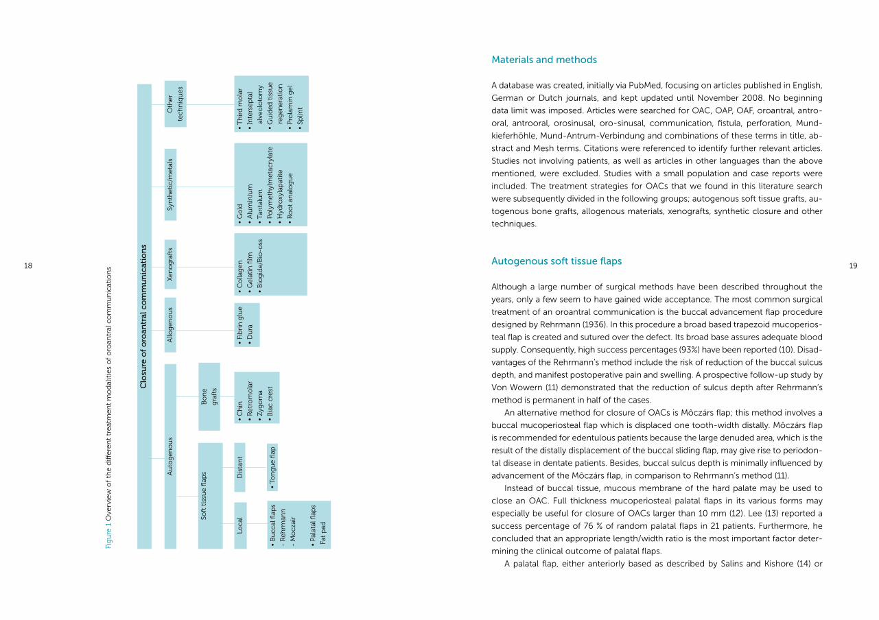
presented throughout the years. An overview of these treatment modalities is given
in Figure 1.
The goal of this literature review was twofold; to answer the question if the buccal
sliding flap still is the treatment of choice 20 years after the last review, and secondly
to provide an overview of the most common surgical treatment strategies of OACs,
as well as the alternative treatment options, including their advantages and disadvan-
tages.
Abstract
Oroantral communications (OACs) are usually caused by extraction of maxillary
posterior teeth. Although the incidence is relatively low, OACs are frequently en-
countered due to the high number of extractions.
This article provides an overview of the most common surgical treatment
strategies of oroantral communications, as well as the alternative treatment op-
tions, including their advantages and disadvantages. The treatment strategies are
divided into techniques using autogenous soft tissue flaps or autogenous bone
grafts, allogenous grafts, xenografts and synthetic materials or metals.
In this literature search no prospective randomized comparative study has
been found with patients groups that are large enough to generate reliable, sta-
tistically significant results on the superiority of one technique compared with
another one.
Furthermore, it can be concluded that a very wide range of treatment tech-
niques has been proposed during the year of which most did not manage to gain
wide acceptance. Nowadays, surgical closure of OACs by means of a buccal or
palatal flap is therefore still the treatment of choice.
18 19
Materials and methods
A database was created, initially via PubMed, focusing on articles published in English,
German or Dutch journals, and kept updated until November 2008. No beginning
data limit was imposed. Articles were searched for OAC, OAP, OAF, oroantral, antro-
oral, antrooral, orosinusal, oro-sinusal, communication, fistula, perforation, Mund-
kieferhöhle, Mund-Antrum-Verbindung and combinations of these terms in title, ab-
stract and Mesh terms. Citations were referenced to identify further relevant articles.
Studies not involving patients, as well as articles in other languages than the above
mentioned, were excluded. Studies with a small population and case reports were
included. The treatment strategies for OACs that we found in this literature search
were subsequently divided in the following groups; autogenous soft tissue grafts, au-
togenous bone grafts, allogenous materials, xenografts, synthetic closure and other
techniques.
Autogenous soft tissue flaps
Although a large number of surgical methods have been described throughout the
years, only a few seem to have gained wide acceptance. The most common surgical
treatment of an oroantral communication is the buccal advancement flap procedure
designed by Rehrmann (1936). In this procedure a broad based trapezoid mucoperios-
teal flap is created and sutured over the defect. Its broad base assures adequate blood
supply. Consequently, high success percentages (93%) have been reported (10). Disad-
vantages of the Rehrmann’s method include the risk of reduction of the buccal sulcus
depth, and manifest postoperative pain and swelling. A prospective follow-up study by
Von Wowern (11) demonstrated that the reduction of sulcus depth after Rehrmann’s
method is permanent in half of the cases.
An alternative method for closure of OACs is Môczárs flap; this method involves a
buccal mucoperiosteal flap which is displaced one tooth-width distally. Môczárs flap
is recommended for edentulous patients because the large denuded area, which is the
result of the distally displacement of the buccal sliding flap, may give rise to periodon-
tal disease in dentate patients. Besides, buccal sulcus depth is minimally influenced by
advancement of the Môczárs flap, in comparison to Rehrmann’s method (11).
Instead of buccal tissue, mucous membrane of the hard palate may be used to
close an OAC. Full thickness mucoperiosteal palatal flaps in its various forms may
especially be useful for closure of OACs larger than 10 mm (12). Lee (13) reported a
success percentage of 76 % of random palatal flaps in 21 patients. Furthermore, he
concluded that an appropriate length/width ratio is the most important factor deter-
mining the clinical outcome of palatal flaps.
A palatal flap, either anteriorly based as described by Salins and Kishore (14) or
Clo
sure
of
oro
antr
al c
om
mu
nic
atio
ns
Au
tog
eno
us
Soft
tis
sue
flap
s
Loca
l•
Ch
in
• R
etro
mo
lar
• Z
ygo
ma
• Ili
ac c
rest
• Fi
bri
n g
lue
• D
ura
• C
olla
gen
• G
elat
in fi
lm
• B
iog
ide/
Bio
-oss
• G
old
• A
lum
iniu
m
• Ta
nta
lum
• Po
lym
eth
ylm
etac
ryla
te
• H
ydro
xyla
pat
ite
• R
oo
t an
alo
gu
e
• T
hir
d m
ola
r
• In
ters
epta
l
al
veo
loto
my
• G
uid
ed t
issu
e
re
gen
erat
ion
• P
rola
min
gel
• Sp
lint
• B
ucc
al fl
aps
- R
ehrm
ann
- M
ocz
air
• Pa
lata
l flap
s
Fat
pad
• To
ng
ue
flap
Fig
ure
1 O
verv
iew
of
the
diff
eren
t tr
eatm
ent
mo
dal
itie
s o
f o
roan
tral
co
mm
un
icat
ion
s
Bo
ne
gra
fts
Allo
gen
ou
sX
eno
gra
fts
Syn
thet
ic/m
etal
sO
ther
tech
niq
ues
Dis
tan
t
20 21
thesia, although the cutting of the pedicle 14 days after attachment may be performed
under local anesthesia (16), and the requirement for a 2-stage or 3-stage procedure
to gain ultimate results.
Autogenous bone grafts
Proctor (27) first suggested bone grafts harvested from the iliac crest for closure of
large oroantral communications in 1969. Nevertheless, bone grafting for closure of
OACs has the disadvantage of requiring a second surgical procedure for bone har-
vesting. This second procedure elongates surgical time and increases patient mor-
bidity. Despite these disadvantages, bone grafting for closure of OACs has gained
attention over the past years, because of the rising demand for implant rehabilitation.
Harvesting bone from the iliac crest involves significant donor site morbidity, like
prolonged post-operative pain and possible sensory disturbance (28). Moreover, har-
vesting bone from intra-oral donor areas significantly reduces the demands made on
the patients postoperatively and can be performed under local anaesthesia (29;30).
Therefore, alternative donor areas have been investigated, including bone grafts from
the retromolar area, zygomatic process and the chin (31-33).
Watzak (31) harvested retromolar bone for press-fitted closure of oroantral com-
munications in 4 patients. After placing the bone graft, soft tissue closure was realised
by means of a Rehrmann buccal flap. No re-opening of the sinus was observed.
A limiting factor of the retromolar donor area is the confined amount of bone
available (29;31). However, in most cases only a small amount of bone will be needed
for closure of OACs. Besides, retromolar bone seems to form a solid base for implant
rehabilitation (31).
Chin bone for oroantral fistula closure was studied in 5 patients by Haas (33). In 3
patients a stable press-fit of the bone graft in the OAC was accomplished. In 2 patients
additional plates and screws were used to obtain a rigid fixation of the graft. Secondly,
a Rehrmann flap was used in all patients for soft tissue closure. Wound dehiscence
occurred in 1 patient, but the sinus remained unaffected. The use of a monocortical
(chin) bone block for closure of an OAC is recommended for patients affected by
maxillary atrophy requiring sinus augmentation before implant placement (39).
Peñarrocha-Diago used zygomatic bone as a bone graft for closure of an OAC in 1
patient. Subsequently 2 dental implants were placed. This technique offers the advan-
tage of the proximity of the donor area to the recipient area, which benefits to mini-
mization of surgical time and patient discomfort (32). As in retromolar bone grafts,
limited bone is obtainable from the zygomatic process. Furthermore, accidental sinus
membrane perforation may occur (32).
posteriorly based, contains a large palatine vessel to ensure adequate blood flow. It
is less vulnerable to rupture than a buccal flap because of the thickness of the palatal
mucosa. Furthermore, the buccal sulcus depth remains intact. Negative aspects of
the palatal flap include the denuded palatal donor area, and a soft tissue bulge at the
axis of rotation. The denuded area remains until secondary epithelialization occurs.
This causes relatively greater discomfort for the patient compared to other soft tissue
techniques. Nevertheless, as Awang (8) mentioned, many surgeons prefer the palatal
flap over the buccal flap procedure.
The buccal fat pad (BFP) is a lobulated mass of fatty tissue surrounded by a slight
capsule, located inside the masticatory spaces (15;16). The size of the BFP has proven
to be constant among individuals, regardless of the body weight and fat distribution
(17). Blood supply to the BFP depends on branches of the superficial temporal, maxil-
lary and facial arteries. Its use as a pedicled graft for reconstruction in oral surgery,
including the closure of oroantral communications, has first been described by Egyedi
(18) in 1977. One of the advantages of the BFP is the proximity of the BFP near the
recipient area, permitting quick grafting. According to Neder (19) this is an important
aspect in successful grafting. Hanazawa (20) used the BFP successfully in 13 of 14
patients for closure of OACs. Clinical findings showed that the BFP, after grafting,
changed into granulation-like tissue over a period of 14 days, followed by complete
epithelialization. These positive findings are in line with other studies (15;16;21;22).
Furthermore, the buccal sulcus depth is not affected by the BFP technique (20-22).
The easy mobilisation, its excellent blood supply and minimal donor site morbidity
are clear advantages of the buccal fat pad as a graft material (16;22;22;23). On the
other hand, the BFP requires very careful manipulation, and although success rates in
literature are high (close to 100%) (16;21-23), closure of large defects could involve
complications like graft necrosis or new fistulae (15). According to several authors,
the indication for use of the BFP lies especially in cases with damage to the alveo-
lar buccal or palatal mucoperiosteum, or cases that have failed with other methods
(16;16;17;20;21).
Tongue flaps are suitable for reconstruction in various areas, including lip, cheek
and palatal or oroantral fistulas, because they offer rich blood supply and pliability
(8;16). Tongue flaps can be created from the ventral, dorsal or lateral part of the tongue
(24). In general, the location of the defect dictates the choice of tongue flap. Especially
the lateral tongue is suitable for closure of oroantral communications (25). Siegel et al.
(26) used a full thickness pedicled flap from the lateral border of the tongue to close
a large oroantral communication after partial maxillectomy. Healing was uneventful
in this patient. The authors (26) stated that the lateral tongue flap is suitable for large
oroantral defects in general, allowing instant repair with rarely failure. Kim et al. (24)
also used a posteriorly based full thickness lateral tongue flap to close an OAC, with
success.
General disadvantages of the tongue flaps are the requirement for general anaes-
22 23
thor used both Bio-Guide® (porcine collagen membrane) and Bio-Oss® (bovine bone
grafting material) to close an OAC in 1 patient. For this purpose the Bio-Oss® granules
were sutured in a prefabricated Bio-Guide® envelope. A full thickness mucoperiosteal
flap was then raised and the Bio-Oss® - Bio-Guide® sandwich placed underneath.
Hereafter the flap was repositioned, resulting in primary closure. Healing was un-
eventful in this patient. According to the author, the radiograph showed bony healing
of the defect 8 months after closure, permitting placement of an endosseous implant.
Nevertheless, bony regeneration has not been objectively quantified in this patient.
Disadvantage of this technique is the need for a mucoperiosteal flap to cover the
sandwich. An advantage is the fact that seemingly both bony and soft closure is ac-
complished, without donor site surgery.
Synthetic closure
Various synthetic materials have been described in literature for closure of oroantral
communications. Several studies report on the use of gold foil or gold plate for clo-
sure of oroantral communications (41-47).
The gold foil is burnished into place with its edges on healthy bone, thus acting as
a bridge for overgrowing sinus mucosa. The mucoperiosteal flaps, that were raised to
expose the bony margins of the defect, are sutured across the gold foil without at-
tempting to realize primary closure. In general, the gold foil exfoliates after a period
of 6 weeks (41;43-45). The value of the gold foil technique seems to lie in the closure
of large OACs that failed in previous attempts, and in the unaltered intra-oral anatomy
(42;43). Disadvantage of this rather expensive technique is the relatively long period
of time needed for complete closure and healing (43).
Steiner (48) proposed 36-gauge pure aluminium plates for closure. In line with
the gold technique, the aluminium plate is used as a protective plate to aid in closure.
Sutures are placed only for approximation of the buccal and palatal tissue; the alu-
minium plate is therefore visible at all times. After 6 weeks, the aluminium is displaced
from its initial position due to the reparative tissue formed underneath. Healing was
uneventful in all 8 patients. Advantages of the aluminium are its malleability and soft-
ness, besides its low cost compared to gold.
In addition, tantalum foil was used by Mc Clung and Chipps (49) for closure of
4 OACs in edentate patients, using the same method as in the gold technique. No
complications were observed. The tantalum foil had been exfoliated after 9 weeks,
revealing new granulation tissue across the defect.
Al-Sibahi et al.(50) described a technique for closure of OACs using self curing
polymethylmethacrylate (PMMA) in 10 patients. The technique resembles the methods
using metals as described above. The PMMA plate is immersed for 24 hours in a steri-
lizing solution, cut to size and placed over the defect. Mucoperiosteal flaps are then
Allogenous materials
Several authors achieved closure of oroantral communications using lyophilized fi-
brin glue of human origin (34;35;36). Kniha (34) and von Gattinger (36) used the fibrin
glue in combination with a collagen sheet, where Stajcic (35) solely used fibrin glue.
Preparation of the fibrin glue takes about 15 to 20 minutes. The glue is then applied in
the socket with a syringe, together with the collagen sheet. Hereafter the oral surface
is sealed with the rest of the fibrin glue. After 2 hours the glue has reached its maxi-
mum strength. Both authors using fibrin glue in combination with collagen reported
high success percentages. An advantage of this strategy is clearly the fact that no
flaps need to be raised. Therefore, intra-oral anatomy remains intact. Furthermore,
the method is straight-forward and gives rise to little post operative complaints (36).
Stajcic (35) reported excellent results with the use of fibrin glue by itself. He stressed
the importance of inserting the syringe above the floor of the antrum to protect the
clot from airflow.
Disadvantages of the method are the, according to the manufacturer, small risk of
transmitting viral hepatitis, and the preparation time needed for the fibrin glue.
Kinner and Frenkel (37) used lyophilized dura to treat OACs in 29 patients. The
sterilized dura is placed in a saline solution to regain its flexibility. Hereafter it is cut to
size to make it cover the bony margins of the defect. Sutures are placed at the cor-
ners of the graft after which it is covered with a plastic plate for protection. The dura
exfoliated after 2 weeks. Uncomplicated healing was observed in 28 of 29 patients.
This successful and simple technique involves no surgical intervention, which makes
it an attractive strategy. However, the small risk of transmitting pathogens can not be
ruled out completely.
Xenografts
Mitchell and Lamb (38) as well as Shaker et al. (39) used lyophilized porcine dermis
(Zenoderm) for closure of oroantral perforations. Mitchell and Lamb (38) left the por-
cine graft exposed to the oral environment. Conversely, Shaker and colleagues (39)
placed both buccal and palatal sliding flaps over the porcine collagen. Both authors
report good results (1 failure of 10 patients and 1 failure of 30 patients, respectively).
The collagen does not have to be removed because it is ultimately replaced by
fibrous tissue. Nevertheless, it remains in place for a sufficient length of time to allow
for mucosal overgrowth across the communication (38;39).
Mitchell and Lamb (38) showed that covering the graft by buccal and palatal flaps
is not necessary to obtain optimal results, apparently offering a far more straightfor-
ward strategy than Shaker and colleagues (39).
A new surgical management of OACs was described by Ogunsalu (40). The au-
24 25
replaced without attempting to cover the acrylic plate. After 3-4 weeks the PMMA plate
becomes visible and is removed as soon as the edges become exposed. Results were
satisfying for all 10 patients. A disadvantage of this method, compared to the use of
gold or aluminium, is the needed preparation in advance; e.g. mixing the power and
liquid, allowing it to set, and sterilizing it for 24 hours.
Dense hydroxyl apatite (HA) has also been used for closure of OAC (51;52). Zide et
al (52) used hydroxyl apatite blocks which were carved to fit the defect, and encircled
with a wire for stability when needed. The authors observed natural extrusion of the
blocks without recurrence of a fistula in all 6 patients.
Becker et al (51) used hydroxyl apatite implants in 5 different sizes for closing oro-
antral defects. Hydroxyl apatite granules were used to fill any remaining space in the
socket. Oral mucosa was approximated without complete closure. Healing was un-
eventful in all 20 patients. By contrast, these authors observed no extrusion of the HA
implants. Due to this, dental implants could not be placed in a later stadium.
Disadvantages of hydroxyl apatite for closure of OAC are the expensiveness of the
material, and the need for a variety of implant sizes to allow for size selection.
Root analogue made of β-tricalcium phosphate was used by Thoma et al (53) in
20 patients with OACs. The root replicas were fabricated chair side, using a mould
of the extracted tooth. Replicas could be placed in only 14 of 20 patients due to the
necessity of a proper recipient socket to ensure tight fitting of the root replica. No
complications were observed. This technique proved to be fast and simple, but can
not be performed in all patients due to technical limitations (53).
Other techniques
Third molar transplantation for closure of oroantral communications has been de-
scribed by Kitagawa et al. (54). The authors successfully used a transplanted upper
and lower third molar for closure of OACs in 2 patients. Donor teeth were placed in
slight infraocclusion, fixed by firm finger pressure and light tapping, without the need
for additional stabilization. Endodontic therapy of the donor teeth was performed
after 3 weeks. The authors concluded that third molar transplantation is a successful
but challenging procedure, depending on a proper recipient socket and perfect fitting
of the donor tooth. Besides the obvious need for a donor tooth, the method is not
recommended when there are space limitations for the donor tooth in the recipient
area, and when mucoperiosteal tissue is damaged.
Hori et al. (55) described the successful application of interseptal alveolotomy for
closure of small oroantral communications in 8 patients. This technique is derived
from Dean’s preprosthetic technique and originally designed for smoothing the al-
veolar ridge. In the extended Dean’s technique the interseptal bone is removed, fol-
lowed by fracturing of the buccal cortex in the direction of the palate. Finally, sutures
are used for soft tissue closure. According to the authors the advantages of the ex-
tended Dean’s technique lie in the fact that a bony base is created for closure, with
less postoperative swelling compared to a flap procedure. Furthermore, the buccal
sulcus depth is not influenced. Nevertheless, this method is restricted to cases with
at least 1 cm of space across the fistula (44). In addition, the required breaking of the
buccal bone carries the risk of inflammation due to formation of bone sequesters and
possible deficient closure of the soft tissue in case the fracture is incomplete.
A technique for the closure of OACs using guided tissue regeneration is described
by Waldrop and Semba (56). The technique involves an absorbable gelatine mem-
brane, allogenic bone graft material, and a non-resorbable polytetrafluoroethylene
(ePTFE) membrane. First a flap is reflected and an absorbable gelatine membrane is
placed over the OAC with its edges on the bony margins of the perforation, to act as
a barrier for the bone graft material. A layer of allogenic bone graft material is put on
the membrane. Hereafter, the non-resorbable ePTFE membrane is used to cover the
bone graft material and the soft tissue flap placed over the membrane. Eight weeks
after placement, the ePTFE membrane is removed, after removal of the inner aspect
of the flap adjacent to the ePTFE membrane, and the mucoperiosteal flap replaced.
Two patients were successfully treated with this technique. Clinically bone formation
was seen by the authors after removal of the ePTFE, although this has not been con-
firmed histologically. Disadvantages of the method are the need for a full thickness
flap, and a second procedure to remove the non-resorbable ePTFE membrane. The
authors did not provide information concerning the tolerance of the patients to the
procedure.
Prolamin occlusion gel is an alkaline alcoholic solution based on corn protein. The
prolamin gel has been used by Götzfried and Kaduk (57) as well as Kinner and Fren-
kel (37) for closure of OACs. The solution is injected in the perforation and hardens
within a few minutes. After a week, granulation tissue is formed and the prolamin gel
completely dissolved after 2-3 weeks (37). According to the authors, the procedure
was well tolerated by the patients (37). This simple treatment strategy results in less
postoperative complaints compared to the standard flap procedure. Besides, it does
not influence the buccal sulcus depth. Disadvantages of this technique are high mate-
rial costs, and the fact that the technique is less suitable for OAC larger then 3 mm, or
shallow OACs (37;57).
Laser light was suggested by Grzesiak-Janas and Janas (58) to establish closure of
OACs without surgical intervention. Laser light in low doses has also been used suc-
cessfully in the prevention and/or healing of chemotherapy induced oral mucositis
(59;60). Grzesiak-Janas and Janas used a biostimulative laser of 30 mW power for 3
cycles of extraoral and intraoral irradiation. In this study, sixty-one patients were ex-
posed to the laser light for 10,5 minutes for 4 consecutive days. patients were treated.
No reopening of the OACs was observed. The technique was well tolerated by the
patients. The elimination of the necessity of a surgical procedure is an obvious advan-
26 27
tage of the laser treatment. Disadvantages seem the costs of the laser therapy, and the
number of visits necessary to accomplish complete closure.
Lastly, Logan and Coates (61) proposed a treatment strategy for OACs in immuno-
compromised patients. A HIV-infected patient was treated with this technique. Firstly,
the OAC was de-epithelialized under local anaesthesia. Secondly, an acrylic surgical
splint was fitted that covered the fistula and the edentulous area including the hard
palate. The patient wore the splint continuously over a period of 8 weeks, removing
it only for cleaning. An oral candidiasis developed, probably in relation to xerostomia,
which was successfully treated with miconazole oral gel.
Complete healing was established after 8 weeks. The technique proved a very useful
option when a surgical intervention is contraindicated because of immunosuppres-
sion. Sokler et al (62) reported that the palatal splint technique in combination with
simultaneous antibiotics is, with success, routinely applied in non-immunocompro-
mised patients in Croatia.
Discussion and conclusions
A literature search of the English, Dutch and German literature concerning closure of
oroantral communications has been performed to provide an overview of the different
treatment options.
Mostly, the studies in this review reporting on a new strategy for closure of OACs
were either case reports, or prospective studies. Unfortunately, none of the authors
have implemented randomized controlled clinical trials allowing for comparison of
the new strategy with, for example, standard surgical closure. Secondly, in a signifi-
cant number of studies the number of patients treated was rather low, and no further
studies were implemented in a larger number of patients.
Thirdly, most studies did not provide information concerning the length of the
proposed procedure, which seems an important aspect to assess its feasibility.
Lastly, in several studies, the description of the treatment strategy did not provide
enough details necessary to gain a complete impression of its quality.
Nevertheless, all of these studies were included in this article to provide a com-
plete overview of the treatment strategies of oroantral communications.
Ideally, a treatment of OACs is quick, safe, straightforward, well tolerated by the
patients, has low costs, and results in both good bony and soft tissue healing with a low
complication rate. However, such a treatment simply does not seem to exist until now.
Therefore, soft tissue closure using a buccal or palatal flap still seems to be the
treatment of choice for OACs, in case primary suturing of the gingiva does not provide
adequate closure of the communication. The buccal flap, despite its risk of reducing
the buccal sulcus depth, appears more popular than the palatal flap, which results
in a denuded palatal donor area requiring secondary epithelialization. Nevertheless,
a number of surgeons seem to prefer the palatal flap because of its excellent blood
supply and the fact that the buccal sulcus remains intact. On the other hand, a re-
duction of the buccal sulcus depth is nowadays becoming less of a problem with the
possibility of implant retained overdentures.
At the present time, bony closure of OACs seems to gain interest. This is probably
as stated before a result of the rising demand for implant rehabilitation. When place-
ment of an endosseous implant is desired, bone grafting for closure of the OAC might
be the best option. Nowadays, intraoral bone harvesting is the strategy of choice for
bone harvesting, reducing patient morbidity compared to extraoral bone harvesting.
Some of the alternative treatment strategies of OACs also claim good bone re-
generation at the site of the perforation. Most of these studies, however, did not as-
sess bone formation objectively. Therefore, strategies that do not involve autogenous
bone grafts like for example the Bio-Gide® – Bio-Oss® technique (40), root analogue
(53) or metals like gold (10;12;49-54) and aluminium (55), might also result in adequate
bone formation for implant rehabilitation, although this has not yet been objectified.
Lastly, there is a tendency in medicine to prefer synthetic materials above materi-
als of animal derived origin. Reason for this is possible transmission of pathogens of
animal derived products.
Based on this review it may be concluded that a wide range of techniques has
been proposed in literature, of which only a few have gained wide acceptance. Rea-
son for this may be found in the costs of the proposed method, where other alter-
native treatments did not offer any simplification compared to the standard closure.
Surgical closure of OACs by means of a buccal or palatal flap therefore remains the
treatment of choice.
28 29
(17) Stuzin JM, Wagstrom L, Kawamoto HK, Baker TJ, Wolfe SA. The anatomy and clinical applica-
tions of the buccal fat pad. Plast Reconstr Surg 1990 Jan;85(1):29-37.
(18) Egyedi P. Utilization of the buccal fat pad for closure of oro-antral and/or oro-nasal communi-
cations. J Maxillofac Surg 1977 Nov;5(4):241-4.
(19) Neder A. Use of buccal fat pad for grafts. Oral Surg Oral Med Oral Pathol 1983 Apr;55(4):349-
50.
(20) Hanazawa Y, Itoh K, Mabashi T, Sato K. Closure of oroantral communications using a pedicled
buccal fat pad graft. J Oral Maxillofac Surg 1995 Jul;53(7):771-5.
(21) Stajcic Z. The buccal fat pad in the closure of oro-antral communications: a study of 56 cases.
J Craniomaxillofac Surg 1992 Jul;20(5):193-7.
(22) Baumann A, Ewers R. Application of the buccal fat pad in oral reconstruction. J Oral Maxillo-
fac Surg 2000 Apr;58(4):389-92.
(23) Rapidis AD, Alexandridis CA, Eleftheriadis E, Angelopoulos AP. The use of the buccal fat pad
for reconstruction of oral defects: review of the literature and report of 15 cases. J Oral Maxil-
lofac Surg 2000 Feb;58(2):158-63.
(24) Kim YK, Yeo HH, Kim SG. Use of the tongue flap for intraoral reconstruction: a report of 16
cases. J Oral Maxillofac Surg 1998 Jun;56(6):716-9.
(25) Buchbinder D, St Hilaire H. Tongue flaps in maxillofacial surgery. Oral Maxillofac Surg Clin
North Am 2003 Nov;15(4):475-86.
(26) Siegel EB, Bechtold W, Sherman PM, Stoopack JC. Pedicle tongue flap for closure of an oroan-
tral defect after partial maxillectomy. J Oral Surg 1977 Sep;35(9):746-8.
(27) Proctor B. Bone graft closure of large or persistent oromaxillary fistula. Laryngoscope 1969
May;79(5):822-6.
(28) Joshi A, Kostakis GC. An investigation of post-operative morbidity following iliac crest graft
harvesting. Br Dent J 2004 Feb 14;196(3):167-71.
(29) Nkenke E, Radespiel-Troger M, Wiltfang J, Schultze-Mosgau S, Winkler G, Neukam FW. Mor-
bidity of harvesting of retromolar bone grafts: a prospective study. Clin Oral Implants Res
2002 Oct;13(5):514-21.
(30) Nkenke E, Schultze-Mosgau S, Radespiel-Troger M, Kloss F, Neukam FW. Morbidity of harvest-
ing of chin grafts: a prospective study. Clin Oral Implants Res 2001 Oct;12(5):495-502.
(31) Watzak G, Tepper G, Zechner W, Monov G, Busenlechner D, Watzek G. Bony press-fit closure
of oro-antral fistulas: a technique for pre-sinus lift repair and secondary closure. J Oral Maxil-
lofac Surg 2005 Sep;63(9):1288-94.
(32) Penarrocha-Diago M, Garcia B, Gomez D, Balaguer J. Zygomatic bone graft for oral-antral
communication closure and implant placement. J Oral Implantol 2007;33(5):305-9.
(33) Haas R, Watzak G, Baron M, Tepper G, Mailath G, Watzek G. A preliminary study of monocorti-
Reference List
(1) Abuabara A, Cortez A.L.V., Passeri L.A., Moraes de M., Moreira R.W.F. evaluation of different treat-
ments for oroantral/oronasal communications. Int J Oral Maxillofac Surg 2006 Feb;35(2):155-8.
(2) Punwutikorn J, Waikakul A, Pairuchvej V. Clinically significant oroantral communications
- a study of incidence and site. Int J Oral Maxillofac Surg 1994 Feb;23(1):19-21.
(3) Skoglund LA, Pedersen SS, Holst E. Surgical management of 85 perforations to the maxillary
sinus. Int J Oral Surg 1983 Feb;12(1):1-5.
(4) del Rey-Santamaria M, Valmaseda CE, Berini AL, Gay EC. Incidence of oral sinus
communications in 389 upper thirmolar extraction. Med Oral Patol Oral Cir Bucal 2006
Jul;11(4):E334-E338.
(5) Bodner L, Gatot A, Bar-Ziv J. Technical note: oroantral fistula: improved imaging with a dental
computed tomography software program. Br J Radiol 1995 Nov;68(815):1249-50.
(6) von Wowern N. Correlation between the development of an orantral fistula and the size of the
corresponding bony defect. J Oral Surg 1973;31(2):98-102.
(7) von Wowern N. Frequency of oro-antral fistulae after perforation to the maxillary sinus. Scand
J Dent Res 1970;78(5):394-6.
(8) Awang MN. Closure of oroantral fistula. Int J Oral Maxillofac Surg 1988 Apr;17(2):110-5.
(9) Obradovic O, Todorovic Lj, Pesic V. Investigations of the buccal sulcus depth after the use
of certain methods of oro-antral communication closure. Bull Group Int Rech Sci Stomatol
Odontol 1981;24(3):209-14.
(10) Killey HC, Kay LW. Observations based on the surgical closure of 362 oro-antral fistulas. Int
Surg 1972;57(7):545-9.
(11) von Wowern N. Closure of oroantral fistula with buccal flap: Rehrmann versus Moczar. Int J
Oral Surg 1982 Jun;11(3):156-65.
(12) Ehrl PA. Oroantral communication. Epicritical study of 175 patients, with special concern to
secondary operative closure. Int J Oral Surg 1980 Oct;9(5):351-8.
(13) Lee J-J, Kok S-H, Chang H-H, Yang P-J, Hahn L-J, Kuo Y-S. Repair of oroantral communications
in the third molar region by random palatal flap. Int J Oral Maxillofac Surg 2002;31:677-80.
(14) Salins PC, Kishore SK. Anteriorly based palatal flap for closure of large oroantral fistula. Oral
Surg Oral Med Oral Pathol Oral Radiol Endod 1996 Sep;82(3):253-6.
(15) Martin-Granizo R, Naval L, Costas A, Goizueta C, Rodriguez F, Monje F, et al. Use of buccal fat
pad to repair intraoral defects: review of 30 cases. Br J Oral Maxillofac Surg 1997 Apr;35(2):81-4.
(16) el Hakim IE, el Fakharany AM. The use of the pedicled buccal fat pad (BFP) and palatal
rotating flaps in closure of oroantral communication and palatal defects. J Laryngol Otol 1999
Sep;113(9):834-8.
30 31
(50) Al Sibahi A, Shanoon A. The use of soft polymethylmethacrylate in the closure of oro-antral
fistula. J Oral Maxillofac Surg 1982 Mar;40(3):165-6.
(51) Becker J, Kuntz A, Reichart P. Verschluß von Mund-Antrumperforationen durch
Hydroxylapatitkeramik. Dtsch Z Mund Kiefer Gesichtschir 1987 Mar;11(2):92-5.
(52) Zide MF, Karas ND. Hydroxylapatite block closure of oroantral fistulas: report of cases. J Oral
Maxillofac Surg 1992 Jan;50(1):71-5.
(53) Thoma K., Pajarola G.F., Gratz K.W., Schmidlin P.R. bioabsorbable root analogue for closure of
oroantral communications after tooth extraction:; a prospective case-cohort study. Oral Surg
Oral Med Oral Pathol Oral Radiol Endod 2006 May;101(5):558-64.
(54) Kitagawa Y, Sano K, Nakamura M, Ogasawara T. Use of third molar transplantation for closure
of the oroantral communication after tooth extraction: a report of 2 cases. Oral Surg Oral Med
Oral Pathol Oral Radiol Endod 2003 Apr;95(4):409-15.
(55) Hori M, Tanaka H, Matsumoto M, Matsunaga S. Application of the interseptal alveolotomy for
closing the oroantral fistula. J Oral Maxillofac Surg 1995 Dec;53(12):1392-6.
(56) Waldrop TC, Semba SE. Closure of oroantral communication using guided tissue regeneration
and an absorbable gelatin membrane. J Periodontol 1993 Nov;64(11):1061-6.
(57) Gotzfried HF, Kaduk B. Okklusion der Mund-Antrum-Verbindung durch eine alkoholische
Prolaminelösung; Tierexperimentelle Studie und erste klinische Erfahrungen. Dtsch Z Mund
Kiefer Gesichtschir 1985 Sep;9(5):390-3.
(58) Grzesiak-Janas G, Janas A. Conservative closure of antro-oral communication stimulated with
laser light. J Clin Laser Med Surg 2001;19(4):181-4.
(59) Wong SF, Wilder-Smith P. Pilot study of laser effects on oral mucositis in patients receiving
chemotherapy. Cancer J 2002 May;8(3):247-54.
(60) Abramoff MM, Lopes NN, Lopes LA, Dib LL, Guilherme A, Caran EM, et al. Low-level laser
therapy in the prevention and treatment of chemotherapy-induced oral mucositis in young
patients. Photomed Laser Surg 2008 Aug;26(4):393-400.
(61) Logan RM, Coates EA. Non-surgical management of an oro-antral fistula in a patient with HIV
infection. Aust Dent J 2003 Dec;48(4):255-8.
(62) Sokler K, Vuksan V, Lauc T. Treatment of oroantral fistula. Acta Stomatol Croat 2002 Jan
16;36:135-40.
cal bone grafts for oroantral fistula closure. Oral Surg Oral Med Oral Pathol Oral Radiol Endod
2003 Sep;96(3):263-6.
(34) Kniha H, Nentwig GH, Bunnag T. Zum Verschluß oroantraler Verbindungen mit dem
Fibrineklebesystem. Dtsch Z Mund Kiefer Gesichtschir 1985 Nov;9(6):431-3.
(35) Stajcic Z, Todorovic LJ, Petrovic V. Tissucol in closure of oroantral communication. A pilot
study. Int J Oral Surg 1985 Oct;14(5):444-6.
(36) Gattinger B. Der verschluß von mund-antrum-verbindungen mit dem lyophilisierten
fibrinklebesystem. Zahnartzl Prax 1984;35(1):8-10.
(37) Kinner U, Frenkel G. Alternative Methoden des Verschlusses von Mund-Antrum-Verbindungen;
Die plastische Deckung der Kieferhöhle mit lyophilisierter Dura mit alkoholischer
Prolaminlösung. ZWR 1990 Nov;99(11):890-6.
(38) Mitchell R, Lamb J. Immediate closure of oro-antral communications with a collagen implant.
A preliminary report. Br Dent J 1983 Mar 19;154(6):171-4.
(39) Shaker MA, Hindy AM, Mounir RM, Geaisa KM. Competent closure of chronic oroantral fistula
with Zenoderm. Egypt Dent J 1995 Jul;41(3):1237-42.
(40) Ogunsalu C. A new surgical management for oro-antral communication: the resorbable
guided tissue regeneration membrane--bone substitute sandwich technique. West Indian Med
J 2005 Sep;54(4):261-3.
(41) Goldman EH, Stratigos GT, Arthur AL. Treatment of oroantral fistula by gold foil closure: report
of case. J Oral Surg 1969 Nov;27(11):875-7.
(42) Mainous EG, Hammer DD. Surgical closure of oroantral fistula using the gold foil technique. J
Oral Surg 1974;32(7):528-30.
(43) Meyerhoff W, Christiansen T, Rontal E, Boerger W. Gold foil closure of oroantral fistulas.
Laryngoscope 1973 Jun;83(6):940-4.
(44) Salman L, Salman SJ. Oro-antral closures using gold plate. N Y State Dent J 1966 Feb;32(2):51-5.
(45) Shapiro DN, Moss M. Gold plate closure of oroantral fistulas. J Prosthet Dent 1972
Feb;27(2):203-8.
(46) Smiler DG, Montemorano P. The gold plate technique for closing oro-antral openings. J South
Calif Dent Assoc 1972 Feb;40(2):122-8.
(47) Rose HP, Allen LJ. Gold plate used for closing a fistula into the maxillary sinus. Dent Dig 1968
Oct;74(10):427-8.
(48) Steiner M, Gould AR, Madion DC, Abraham MS, Loeser JG. Metal plates and foils for closure of
oroantral fistulae. J Oral Maxillofac Surg 2008 Jul;66(7):1551-5.
(49) McClung E, Chipps J. Tantalum foil used in closing antro-oral fistulas. U S Armed Forces Med J
1951 Aug;2(8):1183-6.
Chapter 3
Retrospective study on the treatment outcome of surgical closure of oroantral
communications.
Susan H Visscher, Marije RF van Roon,
Wim J. Sluiter, Baucke van Minnen, Ruud RM Bos
Edited version of:
Journal of Oral and Maxillofacial Surgery 2011 dec; 69(12): 2956-2961.
34 35
Abstract
A retrospective cohort study concerning the surgical closure of oroantral com-
munications (OACs) was carried out to facilitate a comparison between the treat-
ment outcomes of conventional surgical treatment, and new strategies for clo-
sure of OACs. The data were statistically analyzed to gather insight into possible
predictor variables of a recurrent OAC.
A cohort of all patients treated for an OAC in 2004 - 2008 was reviewed. The
recorded data included patient age and gender, location and duration of the OAC,
method of removal of the (pre)molar, presence of maxillary sinusitis, disturbed
wound healing, and the surgical treatment method. Data analysis included de-
scriptive and multivariate logistic regression analysis with recurrence of the OAC
as the outcome variable.
A total of 308 patients were included in the sample, of which 28 patients (9.1%)
required a second intervention to repair the OAC. Of these 28 patients, 4 pa-
tients needed a third intervention, making the total number of recurrent OACs 32
(10.4%). In most cases (60.7%), a buccal advancement flap ad modum Rehr mann
was used to close the perforation. Multivariate regression analysis showed a 15
times higher risk of a recurrence in case of a maxillary sinusitis at the follow-up
appointment.
The overall results of the study showed that in about 1 out of 10 patients the
OAC recurs and requires a second intervention after surgical closure. New strate-
gies should therefore result in an equal or better treatment outcome in order to
be considered a suitable treatment option.
Furthermore, it was demonstrated that the presence of a maxillary sinusitis at the
follow-up appointment is an important determinant of the treatment outcome of
OAC repair.
Introduction
Oroantral communications (OACs) are seen mostly after extraction of the maxillary
first and second molars (1-4). The incidence of OACs reported in the literature varies
to a great extent, probably because some authors report on the number of OACs
asso ciated with the whole upper lateral segments, where others report solely on the
incidence of OACs after third molar removal. For example, Rothamel et al (5) found
an incidence of OACs of 13 % after removal of maxillary third molars. Bodner and co-
authors (6) found an incidence of 5 % after removal of premolars and molars in the
maxilla.
Immediate closure of OACs, preferably within 24 - 48 hours, is recommended to
minimize the risk of maxillary sinusitis and the development of a fistula (7). Surgical clo-
sure still seems to be the treatment of choice to close oroantral communications, al-
though numerous alternative techniques have been proposed (8;9). Primary suture of
the gingiva is used for simple closure of small OACs. However, a study by von Wowern
(1) showed that primary suture resulted in a relatively high number of failures. There-
fore, mucosal closure using a buccal mucoperiosteal flap or a palatal rotational flap
seems preferable, especially for larger OACs. The buccal fat pad (BFP) has also proven
to be suitable for closure of OACs, especially in case of failure of the buccal or palatal
flap. Nevertheless, some authors recommend the use of the BFP as a first option for
closure of (larger) OACs (10;11).
In the literature, success rates for these most commonly used surgical techniques
have been published (4;10;12-15). However, to our knowledge, little information can
be found in the literature concerning the (general) complication rate associated with
surgical closure of OACs, as indicated by recurrence of the OACs. The only informa-
tion on this matter is provided by Abuabara and co-authors (11) and mostly involves
closures by means of primary suture or the buccal fat pad. No statistical analyses were
performed in that study. To facilitate a comparison between the treatment outcomes
of conventional surgical treatment and new and upcoming strategies for closure of
OACs, such information is important. Therefore, we performed a retrospective study
on the complication rate after surgical closure of OACs. Data obtained were statisti-
cally analyzed to gather insight into possible predictor variables of a recurrent OAC.
Patients and methods
Study design and study sample
All records of patients with a documented diagnosis of an OAC, treated in the period
2004-2008 at the department of Oral and Maxillofacial Surgery, University Medical
Centre Groningen, Groningen (the Netherlands) were reviewed in this retrospective
cohort study. This review resulted in a total number of 323 patients. We excluded fif-
36 37
teen patients because of incomplete data making further analysis impossible. The final
study sample included 308 patients.
The OAC was confirmed by inspection and both nose and mouth blowing. We con-
sidered the first treatment the treatment performed at our clinic. In other words, in
case closure of the OAC had been attempted by the general dentist, this was not
counted as the first treatment.
No antibiotics or decongestants were prescribed beforehand. However, OACs
existing > 24 h, or OACs with evident non purulent antral infection, were closed and
additionally treated with antibiotics and decongestives. In case of purulent sinusitis,
antral irrigation with a saline solution was carried out until the fluid draining from the
nose and OAC was clear for at least 2 consecutive days. In addition to closure, both
antibiotics and decongestants were prescribed in these cases. These treatments are in
accordance with the guidelines of the Dutch Society for Oral and Maxillofacial Surgery.
All patients were given instructions to avoid pressure on the OAC, such as nose
blowing, and to take postoperative analgesics (ibuprophen or paracetamol) when
necessary. Each patient was evaluated 10-14 days after closure of the OAC and re-
maining sutures were removed. Further follow-up appointments were scheduled
when necessary.
For all patients the following data was collected: patient gender and age, site of the
OAC, removal method (simple extraction or surgical removal), duration of the OAC,
presence of maxillary sinusitis at the follow-up appointment, disturbed wound heal-
ing, number of recurrences, and the treatment strategy.
In case of a small and deep OAC, primary suture was carried out. In other cases,
methods like a trapezoidal buccal advancement flap ad modum Rehrmann (16), split
thickness palatal rotation flap or buccal fat pad were applied. Spongostan® (Johnson &
Johnson medical bv, Amersfoort, the Netherlands), an absorbable haemostatic gelatin
sponge, in combination with suturing of the gingiva was also used in a few patients.
Statistical analysis
Descriptive statistics were computed for each variable. Univariate logistic regression analy-
ses were performed for the predictor variables on the outcome variable (recurrence of
OAC). Variables with a P-value below 0.15 were used in the further development of the
effect model. Multivariate logistic regression (entry 0.04, removal 0.05, backward condi-
tional) was used to find independent risk factors. All analyses were performed using the
Statistical Package for Social Sciences (SPSS for Windows, version 16.0).
Results
All recorded variables in this study were listed separately for both the patient group
with OACs as a whole (n = 308) and for the patient group with a recurrent OAC.
The first attempt to accomplish closure of the OAC was unsuccessful in 28 patients
(9.1 %), meaning that a second intervention was necessary. However, in four of these
patients, a third attempt was needed to close the perforation, making the total num-
ber of recurrences 32 (10.4 %).
The ratio of men to women in the whole patient cohort was roughly 2/3 to 1/3
(194 men, 114 women). The mean age of the patients was 43 years (range 8 to 78).
The highest incidence of OACs was seen after the third decade of life, with almost
equal numbers for the fourth and fifth decades (Table 1). Most OACs were located
at the first, second and third molars (Table 2), with nearly equivalent numbers for the
left and right maxilla. The (pre)molars were simply extracted by forceps in 137 cases,
whereas surgical removal was needed in 155 cases. In the group with a recurrent OAC,
extraction by forceps and surgical removal were performed on an almost equal basis.
In 50 of the 308 patients (16.2%), maxillary sinusitis was diagnosed clinically and/or
radiographically at the first follow-up appointment (Table 3). Of the 28 patients with a
recurrent OAC, 19 (67.9 %) were diagnosed with a maxillary sinusitis at this stage. Dis-
turbed wound healing was documented in 33 patients (10.7 %), of whom 9 developed
a recurrent OAC.
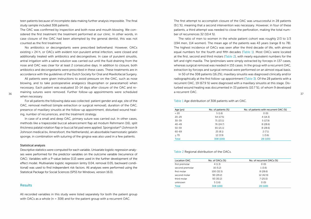
Table 1 Age distribution of 308 patients with an OAC.
Age (yrs) No. of patients (%) No. of patients with recurrent OAC (%)
< 20 5 (1.6) 0 (0)
20-29 54 (17.5) 4 (14.3)
30-39 71 (23.1) 5 (17,9)
40-49 76 (24.7) 8 (28.6)
50-59 65 (21.1) 8 (28.6)
60-69 25 (8.1) 2 (7.1)
≥ 70 12 (3.9) 1 (3.6)
Total 308 (100) 28 (100)
Table 2 Regional distribution of the OACs.
Location OAC No. of OACs (%) No. of recurrent OACs (%)
first premolar 4 (1.3) 0 (0)
second premolar 16 (5.2) 1 (3.6)
first molar 100 (32.5) 8 (28.6)
second molar 90 (29.2) 12 (42.9)
third molar 93 (30.2) 7 (25.0)
unknown 5 (1.6) 0 (0)
Total 308 (100) 28 (100)
38 39
Table 4 Duration of the OACs
Time between occurrence and
closure of OAC
No. of OACs (%) No. of recurrent OACs (%)
≤ 24 hrs 245 (79.5) 18 (64.3)
> 24 hrs
Unknown
52 (16.9)
11 (3.6)
10 (35.7)
0 (0)
Total 308 (100) 28 (100)
Table 5 Overview of the used treatment methods for the patient cohort as a whole.
Treatment method No. of OACs
Buccal flap
Palatal flap
Primary suture
Buccal fat pad
Buccal fat pad and buccal flap
Spongostan®
Unknown
187 (60.7)
3 (1.0)
28 (9.0)
4 (1.3)
5 (1.6)
9 (2.9)
72 (23.4)
Total 308 (100)
Table 6 Overview of the used treatment methods for the patient group with a recur-
rent OAC.
Treatment method recurrent OACs 1st intervention 2nd intervention 3rd intervention
Buccal flap
Palatal flap
Primary suture
Buccal fat pad
Buccal fat pad and buccal flap
Buccal and palatal flap
Acrylic plate
Bone transplant and buccal flap
Unknown
24
0
2
0
0
0
0
2
0
12
1
2
2
7
1
1
2
0
0
2
0
0
0
0
1
1
0
Total 28 28 4
The recurrent OACs were reported mostly within the first 2 weeks after closure (75 %).
In the other 7 patients, the recurrence was diagnosed after a period of 2-4 weeks.
The duration of the OAC was longer than 24 hours in almost 36 % of the patients
with a recurrent OAC, which is twice as many as in the patient cohort as a whole
(Table 4).
The buccal advancement flap was the first choice to accomplish closure of the
OAC in most patients (60.7%), followed by primary suture in case of a small OAC
(Table 5). In 72 patients, it was not exactly clear which treatment method was used.
The OACs that later required a second intervention were all closed with a buccal
flap the first time, except in 2 cases (primary suture). The second attempt to close
these perforations was carried out with either a buccal flap (42.9%), or the buccal fat
pad in combination with a buccal flap (25%) in most cases (Table 6). Strikingly, the
palatal flap was carried out only once after failure of the first intervention, and twice
after failure of the second intervention. In 2 patients, an acrylic plate was fabricated to
cover the OAC, resulting in successful secondary healing.
The overall success percentage of the buccal flap was 87.2%. The palatal flap was
used only in 3 cases, all with success. Closure with primary suture failed twice, result-
ing in a success percentage of 97%.
The univariate regression analysis revealed statistically significant associations be-
tween recurrence of the OAC and closure with a buccal flap (P = 0.008), presence of
maxillary sinusitis (P = 0.000) and disturbed wound healing (p = 0.001) (Table 7). In the
final multivariate model the variable disturbed wound healing was lost because of a
correlation between the presence of maxillary sinusitis and disturbed wound healing
(r = 0.20, P = 0.000). No significant associations were found for the other variables.
Based on the multivariate analysis, the presence of maxillary sinusitis at the follow-
up appointment is associated with a 15 times higher risk of a recurrent OAC (Table 7).
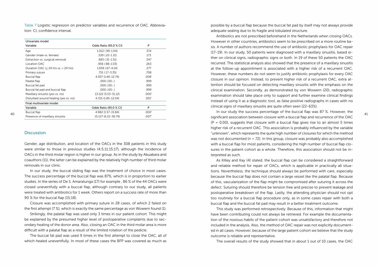
Table 3 Maxillary sinusitis diagnosed at the follow-up appointment, 10-14 days after
closure.
Maxillary sinusitis No. of patients (%) No. of patients with recurrent OAC (%)
Yes 50 (16.2) 19 (76.9)
No 258 (83.8) 9 (32.1)
Total 308 (100) 28 (100)
40 41
possible by a buccal flap because the buccal fat pad by itself may not always provide
adequate sealing due to its fragile and lobulated structure.
Antibiotics are not prescribed beforehand in the Netherlands when closing OACs.
However in other countries, antibiotics seem to be prescribed on a more routine ba-
sis. A number of authors recommend the use of antibiotic prophylaxis for OAC repair
(17-19). In our study, 50 patients were diagnosed with a maxillary sinusitis, based ei-
ther on clinical signs, radiographic signs or both. In 19 of these 50 patients the OAC
recurred. The statistical analysis also showed that the presence of a maxillary sinusitis
at the follow-up appointment is associated with a higher risk of a recurrent OAC.
However, these numbers do not seem to justify antibiotic prophylaxis for every OAC
closure in our opinion. Instead, to prevent higher risk of a recurrent OAC, extra at-
tention should be focused on detecting maxillary sinusitis with the emphasis on the
clinical examination. Secondly, as demonstrated by von Wowern (20), radiographic
examination should take place only to support and further examine clinical findings
instead of using it as a diagnostic tool, as false positive radiographs in cases with no
clinical signs of maxillary sinusitis are quite often seen (22-63%).
In our study the success percentage of the buccal flap was 87 %. However, the
significant association between closure with a buccal flap and recurrence of the OAC
(P = 0.00), suggests that closure with a buccal flap gives rise to an almost 5 times
higher risk of a recurrent OAC. This association is probably influenced by the variable
“unknown”, which represents the quite high number of closures for which the method
was not documented (n = 72). In this group, closure was probably also accomplished
with a buccal flap for most patients, considering the high number of buccal flap clo-
sures in the patient cohort as a whole. Therefore, this association should not be in-
terpreted as such.
As Killey and Kay (4) stated, the buccal flap can be considered a straightforward
and reliable method for repair of OACs, which is applicable in practically all situa-
tions. Nevertheless, the technique should always be performed with care, especially
because the buccal flap does not contain a large vessel like the palatal flap. Because
of this, vascularization of the flap might be compromised after suturing it across the
defect. Suturing should therefore be tension free and precise to prevent leakage and
postoperative breakdown of the flap. Lastly, the attending physician should not opt
too routinely for a buccal flap procedure only, as in some cases repair with both a
buccal flap and the buccal fat pad may result in a better treatment outcome.
This study was performed retrospectively. Because of this, information that might
have been contributing could not always be retrieved. For example the documenta-
tion of the noxious habits of the patient cohort was unsatisfactory and therefore not
included in the analysis. Also, the method of OAC repair was not explicitly document-
ed in all cases. However, because of the large patient cohort we believe that the study
outcome is reliable and representative.
The overall results of the study showed that in about 1 out of 10 cases, the OAC
Table 7 Logistic regression on predictor variables and recurrence of OAC. Abbrevia-
tion: CI, confidence interval.
Univariate model
Variable Odds Ratio (95.0 % CI) P
Age
Gender (male vs. female)
Extraction vs. surgical removal
Location OAC
Duration OAC (≤ 24 hrs vs. > 24 hrs)
Primary suture
Buccal flap
Palatal flap
Buccal fat pad
Buccal fat pad and buccal flap
Maxillary sinusitis (yes vs. no)
Disturbed wound healing (yes vs. no)
1.012 (.99-1.04)
.539 (.22-1.31)
.683 (.31-1.51)
.956 (.88-1.03)
1.659 (.67-4.14)
.751 (.17-3.35)
4.307 (1.46-12.74)
.000 (.00-.)
.000 (.00-.)
.000 (.00-.)
13.122 (5.53-31.12)
4.725 (1.85-12.04)
.374
.173
.347
.263
.277
.708
.008*
.999
.999
.999
.000*
.001*
Final multivariate model
Variable Odds Ratio (95.0 % CI) P
Buccal flap
Presence of maxillary sinusitis
4.982 (1.57-15.84)
15.127 (6.22-36.79)
.000*
.007*
Discussion
Gender, age distribution, and location of the OACs in the 308 patients in this study
were similar to those in previous studies (4;5;11;15;17), although the incidence of
OACs in the third molar region is higher in our group. As in the study by Abuabara and
coauthors (11), the latter can be explained by the relatively high number of third molar
removals in our clinic.
In our study, the buccal sliding flap was the treatment of choice in most cases.
The success percentage of the buccal flap was 87%, which is in proportion to earlier
studies. In the series of De S. Amaratunga (17) for example , 86 % of the 44 OACs were
closed uneventfully with a buccal flap, although contrary to our study, all patients
were treated with antibiotics for 1 week. Others report on a success rate of more than
90 % for the buccal flap (15;18).
Closure was accomplished with primary suture in 28 cases, of which 2 failed on
the first attempt (7 %), which is exactly the same percentage as von Wowern found (1).
Strikingly, the palatal flap was used only 3 times in our patient cohort. This might
be explained by the presumed higher level of postoperative complaints due to sec-
ondary healing of the donor area. Also, closing an OAC in the third molar area is more
difficult with a palatal flap as a result of the limited rotation of the pedicle.
The buccal fat pad was used 9 times in the first attempt to close the OAC, all of
which healed uneventfully. In most of these cases the BFP was covered as much as
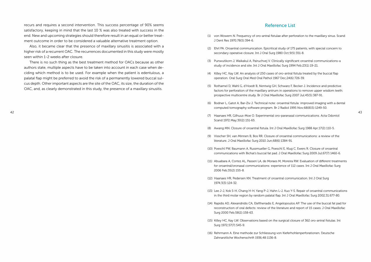
42 43
Reference List
(1) von Wowern N. Frequency of oro-antral fistulae after perforation to the maxillary sinus. Scand
J Dent Res 1970;78(5):394-6.
(2) Ehrl PA. Oroantral communication. Epicritical study of 175 patients, with special concern to
secondary operative closure. Int J Oral Surg 1980 Oct;9(5):351-8.
(3) Punwutikorn J, Waikakul A, Pairuchvej V. Clinically significant oroantral communications-a
study of incidence and site. Int J Oral Maxillofac Surg 1994 Feb;23(1):19-21.
(4) Killey HC, Kay LW. An analysis of 250 cases of oro-antral fistula treated by the buccal flap
operation. Oral Surg Oral Med Oral Pathol 1967 Dec;24(6):726-39.
(5) Rothamel D, Wahl G, d’Hoedt B, Nentwig GH, Schwarz F, Becker J. Incidence and predictive
factors for perforation of the maxillary antrum in operations to remove upper wisdom teeth:
prospective multicentre study. Br J Oral Maxillofac Surg 2007 Jul;45(5):387-91.
(6) Bodner L, Gatot A, Bar-Ziv J. Technical note: oroantral fistula: improved imaging with a dental
computed tomography software program. Br J Radiol 1995 Nov;68(815):1249-50.
(7) Haanaes HR, Gilhuus-Moe O. Experimental oro-paranasal communications. Acta Odontol
Scand 1972 May;30(2):151-65.
(8) Awang MN. Closure of oroantral fistula. Int J Oral Maxillofac Surg 1988 Apr;17(2):110-5.
(9) Visscher SH, van Minnen B, Bos RR. Closure of oroantral communications: a review of the
literature. J Oral Maxillofac Surg 2010 Jun;68(6):1384-91.
(10) Poeschl PW, Baumann A, Russmueller G, Poeschl E, Klug C, Ewers R. Closure of oroantral
communications with Bichat’s buccal fat pad. J Oral Maxillofac Surg 2009 Jul;67(7):1460-6.
(11) Abuabara A, Cortez AL, Passeri LA, de Moraes M, Moreira RW. Evaluation of different treatments
for oroantral/oronasal communications: experience of 112 cases. Int J Oral Maxillofac Surg
2006 Feb;35(2):155-8.
(12) Haanaes HR, Pedersen KN. Treatment of oroantral communication. Int J Oral Surg
1974;3(3):124-32.
(13) Lee J-J, Kok S-H, Chang H-H, Yang P-J, Hahn L-J, Kuo Y-S. Repair of oroantral communications
in the third molar region by random palatal flap. Int J Oral Maxillofac Surg 2002;31:677-80.
(14) Rapidis AD, Alexandridis CA, Eleftheriadis E, Angelopoulos AP. The use of the buccal fat pad for
reconstruction of oral defects: review of the literature and report of 15 cases. J Oral Maxillofac
Surg 2000 Feb;58(2):158-63.
(15) Killey HC, Kay LW. Observations based on the surgical closure of 362 oro-antral fistulas. Int
Surg 1972;57(7):545-9.
(16) Rehrmann A. Eine methode zur Schliessung von Kieferhohlenperforationen. Deutsche
Zahnarztliche Wochenschrift 1936;48:1136-8.
recurs and requires a second intervention. This success percentage of 90% seems
satisfactory, keeping in mind that the last 10 % was also treated with success in the
end. New and upcoming strategies should therefore result in an equal or better treat-
ment outcome in order to be considered a valuable alternative treatment option.
Also, it became clear that the presence of maxillary sinusitis is associated with a
higher risk of a recurrent OAC. The recurrences documented in this study were mostly
seen within 1-2 weeks after closure.
There is no such thing as the best treatment method for OACs because as other
authors state, multiple aspects have to be taken into account in each case when de-
ciding which method is to be used. For example when the patient is edentulous, a
palatal flap might be preferred to avoid the risk of a permanently lowered buccal sul-
cus depth. Other important aspects are the site of the OAC, its size, the duration of the
OAC, and, as clearly demonstrated in this study, the presence of a maxillary sinusitis.
44 45
(17) Amaratunga NA. Oro-antral fistulae-a study of clinical, radiological and treatment aspects.
Br J Oral Maxillofac Surg 1986 Dec;24(6):433-7.
(18) Skoglund LA, Pedersen SS, Holst E. Surgical management of 85 perforations to the maxillary
sinus. Int J Oral Surg 1983 Feb;12(1):1-5.
(19) Poeschl PW. The administration of antibiotics is a crucial point for success and is just
as important as thoroughly rinsing the sinus preoperatively. J Oral Maxillofac Surg 2010
Mar;68(3):707-8.
(20) von Wowern N. Clinical and radiographic findings in maxillary sinus with oro-antral fistula.
Int J Oral Surg 1981;10(Suppl 1):138-42.
Chapter 4a
Closure of oroantral communications using biodegradable polyurethane foam:
a long term study in rabbits.
Susan H Visscher, Baucke van Minnen, M Barbara M van Leeuwen,
Theo G van Kooten, Rudolf RM Bos
Edited version of:
Journal of Biomedical Materials Research
Part B: Applied Biomaterials 2009 Nov; 91(2): 957-963
48 49
Abstract
An oroantral communication (OAC) is an open connection between the oral
cavity and maxillary sinus. Closure of oroantral communications is commonly
performed with a surgical procedure using a mucoperiosteal flap. An alternative
technique using synthetic biodegradable polyurethane (PU) foam for closure of
OACs is presented.
This PU foam is composed of hard urethane segments, and soft segments
made of D/L lactide (50/50), ε-caprolactone and 5% poly ethyleneglycol (PEG).
To evaluate the use of PU foam for this application, OACs were created in the
edentulous part of the maxilla in 21 rabbits, after which PU foams were fitted in
the defects.
Results showed complete healing of the oral mucosa after 4-10 weeks, heal-
ing of the antral mucosal lining after 6 months and complete bony regeneration
after 1 year. No reopening of the defects occurred and no maxillary sinusitis was
observed. Degradation of the PU foam had not yet reached completion 1 year
after implantation.
In conclusion; PU foam with 5% PEG provides adequate closure of an OAC in
the rabbit model to support healing of the oral and maxillary sinus mucosa. Lon-
ger time intervals are needed to assess the complete degradation of the PU foam.
Introduction
An oroantral communication (OAC) is an open connection between the oral cavity and
maxillary sinus. Oroantral communications are most often encountered by extraction
of maxillary (pre)molars with roots extending into the maxillary sinus (1). OACs may
close spontaneously, particularly when the defect is smaller than 5 mm (2). However,
it is difficult to clinically determine the size of the OAC, making it complicated to
predict whether an OAC will heal without (surgical) intervention. Consequently, it is
generally accepted that all OACs should be surgically closed within 24 to 48 hours to
prevent chronic sinusitis and the development of fistulas (3).
Surgical closure of OACs is usually accomplished with a mucoperiosteal flap.
Various forms of buccal flaps, palatal flaps and tongue flaps have been described
in literature for this purpose (4-8). In general, these surgical flap procedures have
high success rates in terms of permanent closure of the OACs (6;9;10). On the other
hand, surgical procedures have several disadvantages including postoperative pain,
swel ling and donor site morbidity. An additional disadvantage of the commonly used
buccal flap procedure is the risk of permanently decreasing the buccal sulcus depth
(11).
Several alternative treatment strategies for closure of OACs have been described
throughout the years. These strategies include the use of gold (12;13), tantalum (14),
aluminium (15) polymethylmethacrylate (16), hydroxylapatite (17) and tissucol (18). To
our knowledge, none of these proposed strategies is currently routinely applied in a
clinical setting, seemingly because either they are too expensive, or no simplification
has been accomplished compared to the standard treatment.
Polyurethane (PU) foam is a fully synthetic biodegradable product that might of-
fer a useful alternative for closure of OACs. This highly porous PU foam has excellent
elastic and mechanical properties, allowing adaptation to the extraction socket. PUs
are polymers of which the polymer chains are built up of two different segments; a
hard urethane segment and the more flexible soft segments. The urethane segments
of the PU in this study are synthesized with 1,4-butanediisocyanate (BDI) and butane-
diol (BDO). When the urethane segments are hydrolyzed, the 1,4 butanediamine is
formed. Butanediamine is normally present in mammalian cells and its catabolites are
excreted in urine (19).
In a previous in vivo study (20), OACs in New Zealand White (NZW) rabbits were
closed with 2 types of PU foam. These 2 foams had identical urethane segments, and
were also built up of BDO and BDI, but their soft segments differed. In the first PU
foam the soft segments were made of only (50/50) DL-lactide and ε-caprolactone.
In the other PU foam, 23 w/w % polyethyleneglycol (PEG) was added to a (50/50)
glycolide/ε-caprolactone soft segment to make the PU more hydrophilic and de-
grade more rapidly. The porosity of these foams was 94 % with pore sizes of 100-300
μm, and interconnected pores of 10-30 μm. Based on the in vivo study (20), it was
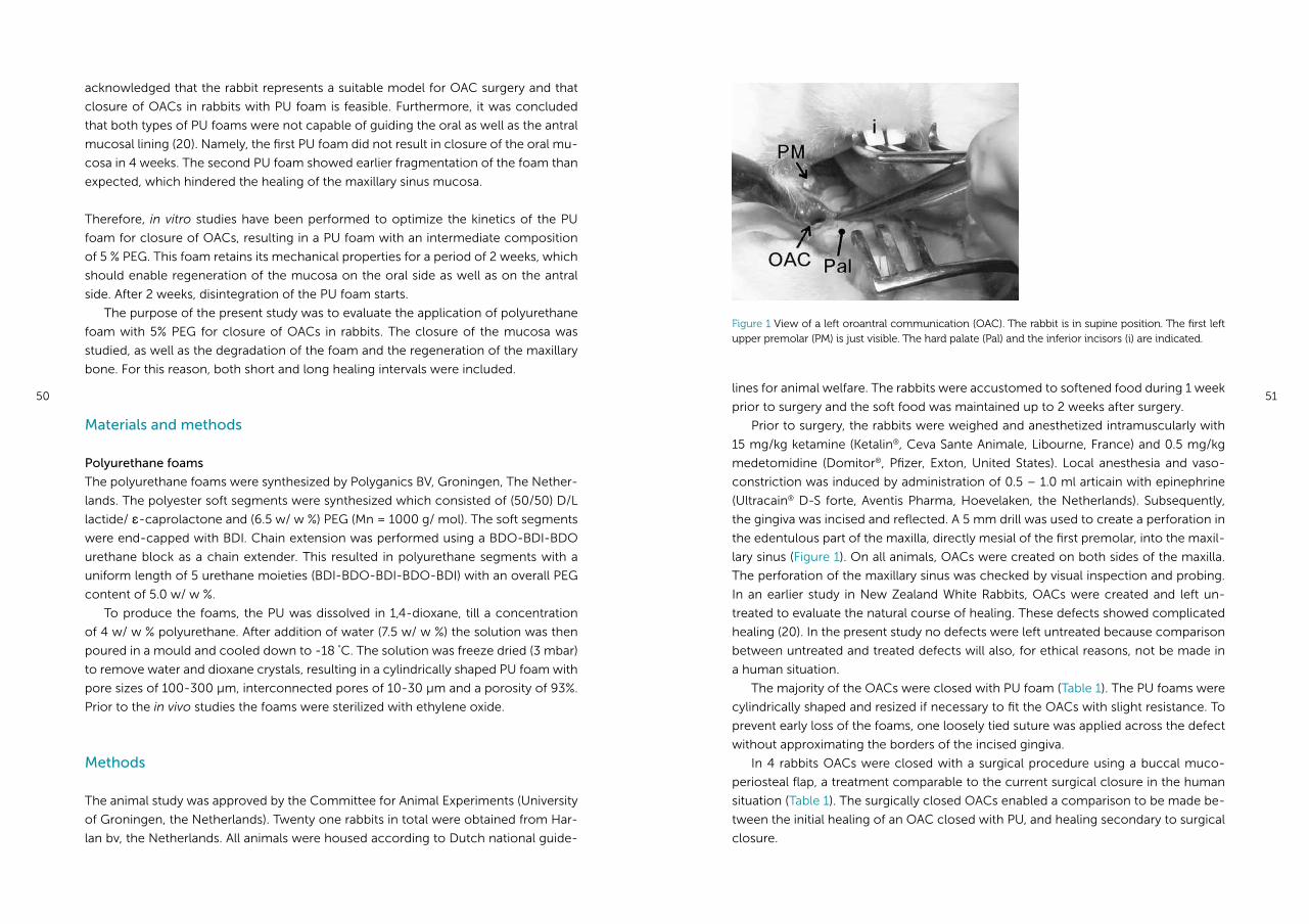
50 51lines for animal welfare. The rabbits were accustomed to softened food during 1 week
prior to surgery and the soft food was maintained up to 2 weeks after surgery.
Prior to surgery, the rabbits were weighed and anesthetized intramuscularly with
15 mg/kg ketamine (Ketalin®, Ceva Sante Animale, Libourne, France) and 0.5 mg/kg
medetomidine (Domitor®, Pfizer, Exton, United States). Local anesthesia and vaso-
constriction was induced by administration of 0.5 – 1.0 ml articain with epinephrine
(Ultracain® D-S forte, Aventis Pharma, Hoevelaken, the Netherlands). Subsequently,
the gingiva was incised and reflected. A 5 mm drill was used to create a perforation in
the edentulous part of the maxilla, directly mesial of the first premolar, into the maxil-
lary sinus (Figure 1). On all animals, OACs were created on both sides of the maxilla.
The perforation of the maxillary sinus was checked by visual inspection and probing.
In an earlier study in New Zealand White Rabbits, OACs were created and left un-
treated to evaluate the natural course of healing. These defects showed complicated
healing (20). In the present study no defects were left untreated because comparison
between untreated and treated defects will also, for ethical reasons, not be made in
a human situation.
The majority of the OACs were closed with PU foam (Table 1). The PU foams were
cylindrically shaped and resized if necessary to fit the OACs with slight resistance. To
prevent early loss of the foams, one loosely tied suture was applied across the defect
without approximating the borders of the incised gingiva.
In 4 rabbits OACs were closed with a surgical procedure using a buccal muco-
periosteal flap, a treatment comparable to the current surgical closure in the human
situation (Table 1). The surgically closed OACs enabled a comparison to be made be-
tween the initial healing of an OAC closed with PU, and healing secondary to surgical
closure.
acknowledged that the rabbit represents a suitable model for OAC surgery and that
closure of OACs in rabbits with PU foam is feasible. Furthermore, it was concluded
that both types of PU foams were not capable of guiding the oral as well as the antral
mucosal lining (20). Namely, the first PU foam did not result in closure of the oral mu-
cosa in 4 weeks. The second PU foam showed earlier fragmentation of the foam than
expected, which hindered the healing of the maxillary sinus mucosa.
Therefore, in vitro studies have been performed to optimize the kinetics of the PU
foam for closure of OACs, resulting in a PU foam with an intermediate composition
of 5 % PEG. This foam retains its mechanical properties for a period of 2 weeks, which
should enable regeneration of the mucosa on the oral side as well as on the antral
side. After 2 weeks, disintegration of the PU foam starts.
The purpose of the present study was to evaluate the application of polyurethane
foam with 5% PEG for closure of OACs in rabbits. The closure of the mucosa was
studied, as well as the degradation of the foam and the regeneration of the maxillary
bone. For this reason, both short and long healing intervals were included.
Materials and methods
Polyurethane foams
The polyurethane foams were synthesized by Polyganics BV, Groningen, The Nether-
lands. The polyester soft segments were synthesized which consisted of (50/50) D/L
lactide/ ε-caprolactone and (6.5 w/ w %) PEG (Mn = 1000 g/ mol). The soft segments
were end-capped with BDI. Chain extension was performed using a BDO-BDI-BDO
urethane block as a chain extender. This resulted in polyurethane segments with a
uniform length of 5 urethane moieties (BDI-BDO-BDI-BDO-BDI) with an overall PEG
content of 5.0 w/ w %.
To produce the foams, the PU was dissolved in 1,4-dioxane, till a concentration
of 4 w/ w % polyurethane. After addition of water (7.5 w/ w %) the solution was then
poured in a mould and cooled down to -18 °C. The solution was freeze dried (3 mbar)
to remove water and dioxane crystals, resulting in a cylindrically shaped PU foam with
pore sizes of 100-300 μm, interconnected pores of 10-30 μm and a porosity of 93%.
Prior to the in vivo studies the foams were sterilized with ethylene oxide.
Methods
The animal study was approved by the Committee for Animal Experiments (University
of Groningen, the Netherlands). Twenty one rabbits in total were obtained from Har-
lan bv, the Netherlands. All animals were housed according to Dutch national guide-
Figure 1 View of a left oroantral communication (OAC). The rabbit is in supine position. The first left upper premolar (PM) is just visible. The hard palate (Pal) and the inferior incisors (i) are indicated.
52 53
Results
Clinical observations
The perforations of the maxillary sinus, surgical closures and placement of the PU
foams all went without complications except for 1 rabbit. In this rabbit (rabbit # 10),
the left OAC had been drilled at a more palatal location than intended. This caused
massive bleeding and peroperative death of the animal. The remaining rabbits had
no postoperative complications, no signs of nasal discharge or sinusitis, and were all
active during the postoperative days. The animals started eating immediately after the
surgical procedure and lost no weight.
Macroscopical observations
After 1 week, the oral side of the foam treated defects were contaminated with food
remnants (Figure 2a). No wound dehiscence was observed of the surgically closed
defect. No inflammation of the oral mucosa was observed neither in both the defects
treated with PU foam nor in the surgically treated defect. During preparation of the
specimens the maxillary sinuses appeared clear.
At the 2 weeks time interval, the defects on the oral side were still visible, but evi-
dently decreasing in size. Healing appeared uneventful at this stage in both the PU
treated defects and the surgically closed defect.
At 4 weeks, all defects had closed macroscopically on the oral side (Figure 2b). No
difference could be observed between the healed mucosa of the surgically closed
defects, and the defects treated with PU foam. In some rabbits, the foams occupied a
relatively large part of the maxillary sinus cavity. However, the remaining space of the
maxillary sinus showed no abnormalities.
At 10 weeks after closure, the foams had a more pulpy aspect compared to the
previous points of evaluation.
Both at 6 months and 1 year after closure, no abnormalities were observed con-
cerning healing. At a time interval of 1 year, in one of the three rabbits PU foam could
still macroscopically be identified in the maxillary sinus.
Microscopical observations
The maxillary sinus mucosa of the surgically closed defect had regenerated into a thin
continuous layer after 1 week. At 2 weeks, the first signs of regeneration of the maxil-
lary bone were observed. At 4 weeks after surgical closure, the mucosa covering the
defect had regained its normal thickness and the defect was almost totally bridged by
newly formed bone. At 10 weeks, the closure of the defect by bone was complete in
the surgically treated defect.
Histology of the foam treated defects showed that the PU foams were fully saturated
with blood. After 1 week, all foams still were rightly positioned with one side partially
introduced into the maxillary sinus. On the antral side, the ciliated epithelium had
In the last two time intervals, both OACs in each rabbit were closed with PU foam to
examine the PU degradation process in the longer term.
After surgery, the animals were given 0.15 mg buprenorfine (Temgesic®, Schering-
Plough, Utrecht, the Netherlands) i.m. as an analgesic. During the first postoperative
weeks, the animals were weighed and observed daily to monitor their appearance,
any nasal discharge, behaviour and eating habits.
The animals were sacrificed by intravenous administration of 2.0 ml embutramide-
mebenzoniumjodide-tetracaine HCL solution (T61®, Intervet, Mechelen, Belgium) and
weighed preceding dissection of the surgical sites and sinuses. All areas of interest
were inspected for abnormalities.
Specimen preparation and analysis
Samples for histological evaluation were taken from both sides of the maxilla. The
samples were fixed in 4% phosphate buffered formalin, and decalcified in RDO for 15-
20 hours. After embedding in GMA, 2 μm sections were cut and stained with Toluidin
Blue or Toluidin Blue/ Basic Fuchsin. The tissue response to the PU foam and the hea-
ling of the oral and antral mucosal lining, as well as the regeneration of the maxillary
bone at the site of the defect were evaluated.
Table 1 Overview of the healing periods and the type of treatment applied to the dif-
ferent animals
Healing periods Rabbit # Type of treatment
Group 1 : 1 week 1
2
L: PU foam
R: Surgical closure
L: PU foam
R: PU foam
Group 2 : 2 weeks 3
4-7
L: PU foam
R: Surgical closure
L: PU foam
R: PU foam
Group 3 : 4 weeks 8
9
L: PU foam
R: Surgical closure
L: PU foam
R: Surgical closure
Group 4 : 10 weeks 10-12
13-15
L: Surgical closure
R: PU foam
L: PU foam
R: PU foam
Group 5 : 6 months 16-18 L: PU foam
R: PU foam
Group 6 : 1 year 19-21 L: PU foam
R: PU foam
54 55
started to grow across the foam. At this stage, the oral defects had not yet been
closed and were contaminated with food remnants.
At 2 weeks, the maxillary sinus mucosa had grown completely across the foam in
seven of nine cases. Under the new antral mucosa, connective tissue had grown into
the foam. This tissue ingrowth was accompanied by vascular ingrowth. The first signs
of degradation of the foam were observed under the surface of this newly formed tis-
sue. The defect on the oral side had grown smaller in eight of nine cases, while one
defect had almost kept its original size.
At 4 weeks, the oral mucosa was restored into a continuous lining in two of the
three cases, while a minor gap was still observed in one case (Figure 3). It is also at
this stage that the first signs of regeneration of the maxillary bone were observed.
The bone grew around the foams, which were situated further into the maxillary sinus
compared to the position of the foams at 1 week and 2 weeks after implantation. Loss
of foam structure was observed in the centre of the foams.
At 10 weeks the oral mucosa had totally closed in all 5 rabbits. The larger part of
the foams was inside the maxillary sinus in some animals (Figure 4a). The remaining
space of the maxillary sinus was clear in all rabbits. Ciliated epithelium supported by
a connective tissue layer was visible on the PU foam (Figure 4b). The epithelial on-
growth had not yet been completed in some cases where the PU foam was located
mainly in the maxillary sinus.
The regeneration of the maxillary bone at the original side of the defect had pro-
gressed further in comparison to the implantation period of 4 weeks, but was not yet
complete. No bone formation was visible within the foams (Figure 4a and c). In one
rabbit the foam had totally lost its architecture, and the whole maxillary sinus was oc-
cupied by the remnants.
In the 6 months group it was observed that both the oral and antral mucosa had
closed completely in all rabbits. In one rabbit, the remains of the PU foam were found
enclosed inside the maxillary sinus.
Bony closure of the defect was totally continuous in all animals except in one
case. In this rabbit, the foam in the left maxilla was situated more on the oral side.
This seemed to have resulted in delayed bony regeneration, because the bone grew
around the PU foam and no bone formation occurred into the foam.
At 1 year, the newly formed antral mucosal lining had further matured (Figure 5b)
as compared to the implantation period of 6 months. In one of three rabbits the re-
maining PU foam was found in both the left and right maxillary sinus. The remaining
PU foams showed degradation round the edges and in the centre. In the other 2 rab-
bits, the PU foam itself was no longer present either inside or below the level of the
maxillary sinus (Figure 5a). Only smaller PU fragments were found in the tissue below
the level of the sinus with newly formed bone visible between the PU fragments.
Figure 3 Frontal view of the left maxillary sinus of rabbit #8 demonstrating the healing of the OAC, 4 weeks after closure with PU foam. The regeneration of the oral mucosa is almost complete (arrow). The antral mucosal lining has grown over the PU foam (double arrow). In the drawing the relatively large size of the PU foam is demonstrated. The foam bulges into the maxillary sinus (MS). The esti-mated original size of the maxillary sinus is indicated by the dotted line. (NC = nasal cavity, O = oral cavity, bar = 1.0 mm).
Figure 2 Occlusal view of the defects after 1 week in rabbit #1 (A) and after 4 weeks in rabbit #5 (B). In both animals the right (R) defects were closed surgically, the left (L) defects were closed with PU foam. The OAC treated with PU foam is filled with food remnants after 1 week. After 4 weeks there is no visible difference between the healed mucosa of the surgically treated defect and the defect treated with PU foam.
56 57
epithelial lining regenerated within a period of 1 week. Bony restoration of the surgi-
cally treated defect was established after 10 weeks.
The results of the closure with PU foam are encouraging, as the application of PU
with 5% PEG leads to an uncomplicated recovery of the oral mucosa. The PU foam
reinforces the coagulum and protects it from displacement. The foam retains its me-
chanical properties for a period of 2 weeks, after which disintegration of the foam sets
in. This period seems long enough to stabilize the blood clot and facilitate mucosal
overgrowth, which is in contrast to the PU foam with 23 % PEG used in the preceding
experiment (20). Namely, the 23 % PEG foam already disintegrated after 24 hours.
For a good reinforcement, the foam had to be saturated with blood. Histology of the
foams in this study showed that this is the case. It is likely that the hydrophilic proper-
ties of the foam in combination with the interconnective pores contribute to this fine
uptake of blood.
The observations of the antral mucosa bring the differences between rabbits and hu-
mans into discussion. Although rabbits have a relatively well developed maxillary si-
nus, the absolute volume of the sinus is small compared to humans.
The suitability of other animal models for this study has also been evaluated; Göt-
Discussion
In this study the closure of OACs in rabbits with highly porous polyurethane foams
was investigated. The study proves that the use of the rabbit model for OAC research
is reproducible. Furthermore, it showes that closure of OACs with 5% PEG PU foam in
New Zealand White rabbits is feasible.
As stated, the healing of OACs depends entirely on the presence of a normal, non-in-
fected blood clot which can serve as support for restoration of the ciliated epithelium
of the maxillary sinus, and squameus epithelium of the oral cavity (9). In non-treated
OACs there is a chance of displacement of the coagulum, and therefore treatment of
OACs is indicated. Surgical closure directly closes the defect on the oral side and will
also support the coagulum. Subsequently, the maxillary sinus mucosa will regenerate.
This has also been confirmed by the results of the surgical closure of the OACs in this
animal study. The surgically closed OACs healed without complications and the antral
Figure 5a Frontal view of the left maxillary sinus of rabbit #20, 1 year after closure with PU foam. The oral mucosa is continuous. The bridging of the defect with immature bone (IB) is complete. Only smaller fragments of PU foam (PU) are visible. (O = oral cavity, MS = maxillary sinus)5b Detailed view of the ciliary epithelium (CE). The epithelium is supported by a connective tissue layer. The regenerated epithelium and connective tissue have normal morphology. No PU fragments can be observed. (O= oral cavity, IB = immature bone, MS = maxillary sinus)
Figure 4a Frontal view of the right maxillary sinus of rabbit #11, 10 weeks after closure of the OAC. The oral mucosa is continuous. The PU foam occupies a large part of the maxillary sinus (MS). The foam has lost its porous structure in the centre. The border of the foam is covered with ciliated epi-thelium. (Bar = 1.0 mm)4b Detailed view on the ciliated epithelium growing on the PU foam. The epithelium is supported by a connective tissue layer. The foam lies near the original wall of the maxillary sinus (MS). The remain-ing space of the sinus is small but clear. (Bar = 25 μm)4c Regeneration of bone at the site of the defect. Immature bone is developing from the more ma-tured bone on the lower right side of the micrograph, at the original site of the defect. The asterisk indicates the border of the degrading foam. No foam structure can be observed. (Bar = 50 μm)
58 59
bony ingrowth into the foam was observed, which might elongate the bony bridging
time across the defect, when compared to the bone regeneration after surgical clo-
sure. This was seen in cases where the PU foam was situated mainly outside the max-
illary sinus, resulting in longer regeneration periods of the maxillary bone, contrary to
cases where the PU foam was located largely into the sinus. Namely, when the PU
foam was placed largely into the sinus, the bony regeneration showed more progress
but consequently the healing of the antral epithelial lining took longer to complete.
However, the extended regeneration of the bone probably does not provide dis-
comfort for the patient, and the non-surgical closure of the OAC is likely to evoke less
discomfort than the surgical closure. Besides, to the best of our knowledge no studies
have been performed concerning how often surgical closure in humans leads to actual
regeneration of the maxillary bone at the site of the defect.
After 1 year, histological sections still showed small fragments of PU foam in 2 rab-
bits. These PU particles were either located inside the maxillary sinus, or in the tissue be-
tween the newly formed bony bridge and the regenerated lining of the maxillary sinus.
It is anticipated that the degradation of these PU fragments will continue further in time.
Both inside and outside the maxillary sinus, no inflammatory or other negative
effects of the PU remnants were observed. Furthermore, the function of the ciliated
epithelium will most probably transport the PU particles out of the maxillary space.
In conclusion, PU foam with 5% PEG provides adequate closure of an OAC in the
rabbit model to support and guide healing of both the oral and antral mucosa. Re-
generation of the maxillary bone is delayed when compared to the surgical closure.
The clinical consequences of this observation seem limited to patients in which dental
implants are planned. Longer time intervals are necessary to document the end stage
of degradation. In addition, human studies will follow for the further development and
evaluation of this new and straightforward method for the treatment of OACs. Lastly,
the cost of the foam for clinical appliance is likely to be between 40 and 50 euro,
which makes closure of OACs with certainty less expensive than surgical closure. This
should make the PU foam also interesting from an socio-economical point of view.
Acknowledgements
We thank Mr A. Nijmeijer and Mr. A. Jansen from the animal laboratory for their assis-
tance. We also would like to thank Polyganics BV for the development and production
of the PU foams.
tingen mini pigs were found unsuitable because of the position of the infraorbital
nerve (21) and extraction of teeth to create an OAC in sheep proved to be too compli-
cated. In the end the rabbit model was chosen, based on its anatomical similarities to
the human; e.g. a well-developed maxillary sinus, and its proven suitability for closure
of OACs in earlier experiments.
The applied oroantral communications measured 5 mm. This is a relatively large
defect size for the rabbit when compared to the size of its maxillary sinus. However,
a previous study showed that a 5 mm defect is applicable in a rabbit model (20). This
previous study also showed delayed and complicated healing when such a 5 mm
defect was left untreated (20).
The PU foam that was needed for closure of the defect may fill a large part of the
relatively small rabbit sinus. This is caused by the fact that the foam easily bulges into
the sinus, because the maxillary sinus floor in rabbits is very thin. On the other hand, it
is essential to push the foam partly through the perforation to ensure adequate fixation.
Once the foam is partly placed in the maxillary sinus, it can still easily be pushed
further into it. A displacement of a foam may also be the result of the forces during
mastication. Furthermore, the rabbits in this study had defects on both sides of the
maxilla which made it difficult for the animals to avoid pressure on the OACs.
The largest displacement of the foams probably already occurred during, or shortly
after surgery. In one of the rabbits in the 1 year group the remaining foam was located
entirely inside the maxillary sinus. However, on account of the rapid degradation on-
set of the foam, it was not expected that free foam would be found in the maxillary
sinus after 6-12 months. The fact that free foam appeared present in this rabbit 1 year
after implantation may be explained by a combination of an early dislocation of the
foam into the sinus shortly after implantation, and a slower degradation inside the
sinus compared to degradation of an incorporated situation. Within the other rabbits
of the 1 year time interval, foam remnants were detectable only in the tissue below
the level of the maxillary sinus.
Dislocation of the foams and the occupation of the sinus will probably occur less
frequently in the human situation. Firstly, many extraction sockets in humans will not
allow dislocation of the PU foam into the sinus, because of their conical shape. Se-
condly, dislocation can be diminished by instructing the patients to avoid forces on
the treated area.
After closure of the OACs with PU foam, the maxillary sinus was in no way affected by
the foam bulging into it. The ciliated epithelial cells of the antral mucosa regenerated
along the surface of the foam, leaving a smaller cavity of the maxillary sinus, which
was totally covered with ciliated epithelium. After 4 weeks the first signs of bone re-
generation were observed and the oral mucosa was almost healed. After 10 weeks,
the oral mucosa was fully restored in all animals. Furthermore, bone regeneration at
this stage was still in progress.
Once the oral mucosa had closed, the maxillary bone started to regenerate. No
60 61
(17) Zide MF, Karas ND. Hydroxylapatite block closure of oroantral fistulas: report of cases. J Oral
Maxillofac Surg 1992 Jan;50(1):71-5.
(18) Stajcic Z, Todorovic LJ, Petrovic V. Tissucol in closure of oroantral communication. A pilot
study. Int J Oral Surg 1985 Oct;14(5):444-6.
(19) Rodwell VW. Conversion of amino acids to specialized products. In: Murray RK, Granner DK,
Mayes PA, Rodwell VW, editors. Harper’s Biochemistry. 25 ed. New York: Appleton & Lange;
2000. p. 347-58.
(20) van Minnen B, Stegenga B, van Leeuwen MB, van Kooten TG, Bos RR. Nonsurgical closure of
oroantral communications with a biodegradable polyurethane foam: A pilot study in rabbits. J
Oral Maxillofac Surg 2007 Feb;65(2):218-22.
(21) van Minnen B, Stegenga B, Zuidema J, Hissink CE, van Leeuwen MB, van Kooten TG, et al. An
animal model for oroantral communications: a pilot study with Gottingen minipigs. Lab Anim
2005 Jul;39(3):280-3.
Reference List
(1) Punwutikorn J, Waikakul A, Pairuchvej V. Clinically significant oroantral communications - a
study of incidence and site. Int J Oral Maxillofac Surg 1994 Feb;23(1):19-21.
(2) von Wowern N. Correlation between the development of an orantral fistula and the size of the
corresponding bony defect. J Oral Surg 1973;31(2):98-102.
(3) von Wowern N. Frequency of oro-antral fistulae after perforation to the maxillary sinus. Scand
J Dent Res 1970;78(5):394-6.
(4) von Wowern N. Closure of oroantral fistula with buccal flap: Rehrmann versus Moczar. Int J
Oral Surg 1982 Jun;11(3):156-65.
(5) el Hakim IE, el Fakharany AM. The use of the pedicled buccal fat pad (BFP) and palatal
rotating flaps in closure of oroantral communication and palatal defects. J Laryngol Otol 1999
Sep;113(9):834-8.
(6) Lee J-J, Kok S-H, Chang H-H, Yang P-J, Hahn L-J, Kuo Y-S. Repair of oroantral communications
in the third molar region by random palatal flap. Int J Oral Maxillofac Surg 2002;31:677-80.
(7) Salins PC, Kishore SK. Anteriorly based palatal flap for closure of large oroantral fistula. Oral
Surg Oral Med Oral Pathol Oral Radiol Endod 1996 Sep;82(3):253-6.
(8) Johnson PA, Banks P, Brown AE. Use of the posteriorly based lateral tongue flap in the repair of
palatal fistulae. Int J Oral Maxillofac Surg 1992 Feb;21(1):6-9.
(9) Skoglund LA, Pedersen SS, Holst E. Surgical management of 85 perforations to the maxillary
sinus. Int J Oral Surg 1983 Feb;12(1):1-5.
(10) Killey HC, Kay LW. Observations based on the surgical closure of 362 oro-antral fistulas. Int
Surg 1972;57(7):545-9.
(11) Obradovic O, Todorovic Lj, Pesic V. Investigations of the buccal sulcus depth after the use
of certain methods of oro-antral communication closure. Bull Group Int Rech Sci Stomatol
Odontol 1981;24(3):209-14.
(12) Goldman EH, Stratigos GT, Arthur AL. Treatment of oroantral fistula by gold foil closure: report
of case. J Oral Surg 1969 Nov;27(11):875-7.
(13) Rose HP, Allen LJ. Gold plate used for closing a fistula into the maxillary sinus. Dent Dig 1968
Oct;74(10):427-8.
(14) McClung E, Chipps J. Tantalum foil used in closing antro-oral fistulas. U S Armed Forces Med J
1951 Aug;2(8):1183-6.
(15) Steiner M, Gould AR, Madion DC, Abraham MS, Loeser JG. Metal plates and foils for closure of
oroantral fistulae. J Oral Maxillofac Surg 2008 Jul;66(7):1551-5.
(16) Al Sibahi A, Shanoon A. The use of soft polymethylmethacrylate in the closure of oro-antral
fistula. J Oral Maxillofac Surg 1982 Mar;40(3):165-6.
Chapter 4b
Biodegradable polyurethane foam for closure of oroantral communications
in rabbits: a 4 year light- and electronmicroscopic study.
Susan H Visscher, Baucke van Minnen,
M Barbara M van Leeuwen, Johannes JL van der Want,
Theo G van Kooten, Rudolf RM Bos
Submitted
64 65
Introduction
Oroantral communications (OACs) can be defined as open connections between the
oral cavity and maxillary sinus. Usually, OACs are caused by extraction of (pre)molars
in the maxilla due to the close relationship of the apices and the sinus floor (1). It is im-
portant to close an OAC as soon as possible to minimize the risk of the development
of a maxillary sinusitis and epithelisation of the connection, resulting in an oroantral
fistula (2). Closure of OACs is in general performed surgically.
Ideally, an alternative strategy for surgical closure of OACs gives predictable re-
sults, is easy and quick to perform, and gives rise to little postoperative complaints.
Also, it would be interesting for both the patient and the dentist if the latter would be
able to treat OACs instead of a maxillofacial or oral surgeon.
Biodegradable polyurethane (PU) foam offers the right characteristics for an al-
ternative treatment strategy for OACs. A long term subcutaneous implantation study
of this PU foam in rats and rabbits showed that after 3 years only an occasional
macrophage containing PU could be observed; the samples had thus resorbed almost
completely (3). The fully synthetic PU foam can be placed in the OAC and loosely
secured on the oral side with a suture. The highly porous foam fills with blood upon
placement thus forming a solid barrier. It retains its mechanical properties for 2 weeks,
enabling mucosal overgrowth of the perforation, after which disintegration sets in. In
a subsequent in vivo study, the degradation of the PU foam for this specific application
has already been studied in New Zealand White (NZW) rabbits for time intervals up to
1 year, and showed promising results (4). However, the end stage of degradation had
not been reached in that study. Therefore, it was concluded that longer time intervals
were needed.
In the present in vivo study OACs were created in NZW rabbits in analogue to the
preceding study (4), and closed with the same PU foam consisting of hard urethane
segments synthesized with 1,4 butanediisocyanate (BDI) and butanediol (BDO) and soft
segments consisting of D/L lactide, ε-caprolactone and polyethylene glycol (PEG).
Also, part of the created OACs were closed surgically with a buccal flap to facilitate
comparison between the healing process after both the surgical treatment and this ex-
perimental nonsurgical treatment. In theory, PU based on 1,4 butanediisocyanate will
degrade into substances that already occur in the body (5). This characteristic of the PU
foam implies the capacity to degrade completely and become excreted by the body
through natural pathways (6). In order to allow documentation of the end stage of
degradation, time intervals up to 4 years after implantation were included. The samples
were evaluated with both light microscopy (LM) and electron microscopy (EM).
Abstract
Oroantral communications (OACs) are connections between the oral cavity and
maxillary sinus and usually caused by extraction of posterior teeth. OACs require
surgical treatment in order to minimize the risk of fistula formation and chronic
sinus disease.
A new non-surgical method for closure of OACs using degradable polyurethane
(PU) foam was studied. OACs were created in the edentulous part of the maxilla in
19 New Zealand White rabbits and subsequently closed with PU foam. This PU foam
is composed of hard urethane segments synthesized with 1,4 butanediisocyanate
and butanediol, and soft segments made of D/L (50:50) lactide, ε-caprolactone,
and 5 % polyethylene glycol.
Time intervals up to 4 years were included to analyze the long term degrada-
tion process. The OACs recovered in all rabbits, with both soft tissue and bony
regeneration. The degradation process of the PU was not fully completed after
4 years but a decreasing number of macrophages with internalized PU and the
aspect of the internalized PU suggest further degradation in time to an ultimate
end stage. Human studies will follow for further development and evaluation of
this nonsurgical closure of OACs.
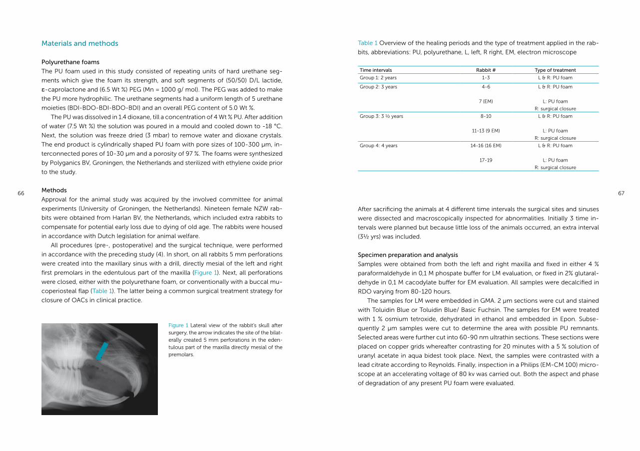
66 67
After sacrificing the animals at 4 different time intervals the surgical sites and sinuses
were dissected and macroscopically inspected for abnormalities. Initially 3 time in-
tervals were planned but because little loss of the animals occurred, an extra interval
(3½ yrs) was included.
Specimen preparation and analysis
Samples were obtained from both the left and right maxilla and fixed in either 4 %
paraformaldehyde in 0,1 M phospate buffer for LM evaluation, or fixed in 2% glutaral-
dehyde in 0,1 M cacodylate buffer for EM evaluation. All samples were decalcified in
RDO varying from 80-120 hours.
The samples for LM were embedded in GMA. 2 μm sections were cut and stained
with Toluidin Blue or Toluidin Blue/ Basic Fuchsin. The samples for EM were treated
with 1 % osmium tetroxide, dehydrated in ethanol and embedded in Epon. Subse-
quently 2 μm samples were cut to determine the area with possible PU remnants.
Selected areas were further cut into 60-90 nm ultrathin sections. These sections were
placed on copper grids whereafter contrasting for 20 minutes with a 5 % solution of
uranyl acetate in aqua bidest took place. Next, the samples were contrasted with a
lead citrate according to Reynolds. Finally, inspection in a Philips (EM-CM 100) micro-
scope at an accelerating voltage of 80 kv was carried out. Both the aspect and phase
of degradation of any present PU foam were evaluated.
Materials and methods
Polyurethane foams
The PU foam used in this study consisted of repeating units of hard urethane seg-
ments which give the foam its strength, and soft segments of (50/50) D/L lactide,
ε-caprolactone and (6.5 Wt %) PEG (Mn = 1000 g/ mol). The PEG was added to make
the PU more hydrophilic. The urethane segments had a uniform length of 5 urethane
moieties (BDI-BDO-BDI-BDO-BDI) and an overall PEG content of 5.0 Wt %.
The PU was dissolved in 1.4 dioxane, till a concentration of 4 Wt % PU. After addition
of water (7.5 Wt %) the solution was poured in a mould and cooled down to -18 °C.
Next, the solution was freeze dried (3 mbar) to remove water and dioxane crystals.
The end product is cylindrically shaped PU foam with pore sizes of 100-300 μm, in-
terconnected pores of 10-30 μm and a porosity of 97 %. The foams were synthesized
by Polyganics BV, Groningen, the Netherlands and sterilized with ethylene oxide prior
to the study.
Methods
Approval for the animal study was acquired by the involved committee for animal
experiments (University of Groningen, the Netherlands). Nineteen female NZW rab-
bits were obtained from Harlan BV, the Netherlands, which included extra rabbits to
compensate for potential early loss due to dying of old age. The rabbits were housed
in accordance with Dutch legislation for animal welfare.
All procedures (pre-, postoperative) and the surgical technique, were performed
in accordance with the preceding study (4). In short, on all rabbits 5 mm perforations
were created into the maxillary sinus with a drill, directly mesial of the left and right
first premolars in the edentulous part of the maxilla (Figure 1). Next, all perforations
were closed, either with the polyurethane foam, or conventionally with a buccal mu-
coperiosteal flap (Table 1). The latter being a common surgical treatment strategy for
closure of OACs in clinical practice.
Figure 1 Lateral view of the rabbit’s skull after surgery, the arrow indicates the site of the bilat-erally created 5 mm perforations in the eden-tulous part of the maxilla directly mesial of the premolars.
Table 1 Overview of the healing periods and the type of treatment applied in the rab-
bits, abbreviations: PU, polyurethane, L, left, R right, EM, electron microscope
Time intervals Rabbit # Type of treatment
Group 1: 2 years 1-3 L & R: PU foam
Group 2: 3 years 4-6
7 (EM)
L & R: PU foam
L: PU foam
R: surgical closure
Group 3: 3 ½ years 8-10
11-13 (9 EM)
L & R: PU foam
L: PU foam
R: surgical closure
Group 4: 4 years 14-16 (16 EM)
17-19
L & R: PU foam
L: PU foam
R: surgical closure
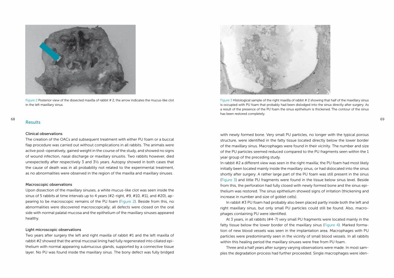
68 69Results
Clinical observations
The creation of the OACs and subsequent treatment with either PU foam or a buccal
flap procedure was carried out without complications in all rabbits. The animals were
active post-operatively, gained weight in the course of the study, and showed no signs
of wound infection, nasal discharge or maxillary sinusitis. Two rabbits however, died
unexpectedly after respectively 3 and 3½ years. Autopsy showed in both cases that
the cause of death was in all probability not related to the experimental treatment,
as no abnormalities were observed in the region of the maxilla and maxillary sinuses.
Macroscopic observations
Upon dissection of the maxillary sinuses, a white mucus-like clot was seen inside the
sinus of 5 rabbits at time intervals up to 4 years (#2-right, #9, #10, #11, and #20), ap-
pearing to be macroscopic remains of the PU foam (Figure 2). Beside from this, no
abnormalities were discovered macroscopically; all defects were closed on the oral
side with normal palatal mucosa and the epithelium of the maxillary sinuses appeared
healthy.
Light microscopic observations
Two years after surgery the left and right maxilla of rabbit #1 and the left maxilla of
rabbit #2 showed that the antral mucosal lining had fully regenerated into ciliated epi-
thelium with normal appearing submucous glands, supported by a connective tissue
layer. No PU was found inside the maxillary sinus. The bony defect was fully bridged
with newly formed bone. Very small PU particles, no longer with the typical porous
structure, were identified in the fatty tissue located directly below the lower border
of the maxillary sinus. Macrophages were found in their vicinity. The number and size
of the PU particles seemed reduced compared to the PU fragments seen within the 1
year group of the preceding study.
In rabbit #2 a different view was seen in the right maxilla; the PU foam had most likely
initially been located mainly inside the maxillary sinus, or had dislocated into the sinus
shortly after surgery. A rather large part of the PU foam was still present in the sinus
(Figure 3) and little PU fragments were found in the tissue below sinus level. Beside
from this, the perforation had fully closed with newly formed bone and the sinus epi-
thelium was restored. The sinus epithelium showed signs of irritation (thickening and
increase in number and size of goblet cells).
In rabbit #3 PU foam had probably also been placed partly inside both the left and
right maxillary sinus, but only small PU particles could still be found. Also, macro-
phages containing PU were identified.
At 3 years, in all rabbits (#4-7) very small PU fragments were located mainly in the
fatty tissue below the lower border of the maxillary sinus (Figure 4). Marked forma-
tion of new blood vessels was seen in the implantation area. Macrophages with PU
particles were predominantly seen in the vicinity of small blood vessels. In all rabbits
within this healing period the maxillary sinuses were free from PU foam.
Three and a half years after surgery varying observations were made. In most sam-
ples the degradation process had further proceeded. Single macrophages were iden-
Figure 2 Posterior view of the dissected maxilla of rabbit # 2, the arrow indicates the mucus-like clot in the left maxillary sinus.
Figure 3 Histological sample of the right maxilla of rabbit # 2 showing that half of the maxillary sinus is occupied with PU foam that probably had been dislodged into the sinus directly after surgery. As a result of the presence of the PU foam the sinus epithelium is thickened. The contour of the sinus has been restored completely.
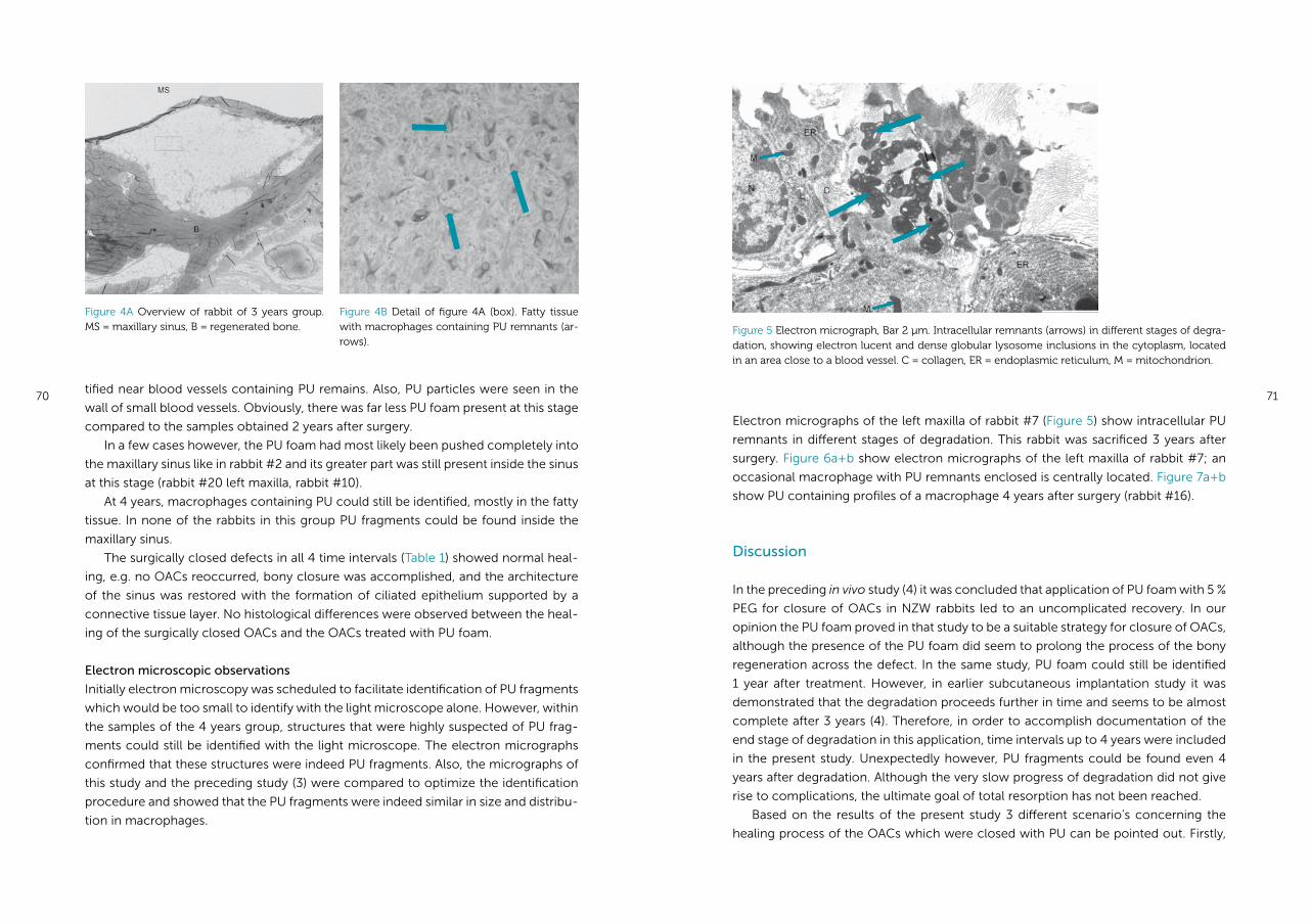
70 71tified near blood vessels containing PU remains. Also, PU particles were seen in the
wall of small blood vessels. Obviously, there was far less PU foam present at this stage
compared to the samples obtained 2 years after surgery.
In a few cases however, the PU foam had most likely been pushed completely into
the maxillary sinus like in rabbit #2 and its greater part was still present inside the sinus
at this stage (rabbit #20 left maxilla, rabbit #10).
At 4 years, macrophages containing PU could still be identified, mostly in the fatty
tissue. In none of the rabbits in this group PU fragments could be found inside the
maxillary sinus.
The surgically closed defects in all 4 time intervals (Table 1) showed normal heal-
ing, e.g. no OACs reoccurred, bony closure was accomplished, and the architecture
of the sinus was restored with the formation of ciliated epithelium supported by a
connective tissue layer. No histological differences were observed between the heal-
ing of the surgically closed OACs and the OACs treated with PU foam.
Electron microscopic observations
Initially electron microscopy was scheduled to facilitate identification of PU fragments
which would be too small to identify with the light microscope alone. However, within
the samples of the 4 years group, structures that were highly suspected of PU frag-
ments could still be identified with the light microscope. The electron micrographs
confirmed that these structures were indeed PU fragments. Also, the micrographs of
this study and the preceding study (3) were compared to optimize the identification
procedure and showed that the PU fragments were indeed similar in size and distribu-
tion in macrophages.
Figure 5 Electron micrograph, Bar 2 μm. Intracellular remnants (arrows) in different stages of degra-dation, showing electron lucent and dense globular lysosome inclusions in the cytoplasm, located in an area close to a blood vessel. C = collagen, ER = endoplasmic reticulum, M = mitochondrion.
Figure 4A Overview of rabbit of 3 years group. MS = maxillary sinus, B = regenerated bone.
Figure 4B Detail of figure 4A (box). Fatty tissue with macrophages containing PU remnants (ar-rows).
Electron micrographs of the left maxilla of rabbit #7 (Figure 5) show intracellular PU
remnants in different stages of degradation. This rabbit was sacrificed 3 years after
surgery. Figure 6a+b show electron micrographs of the left maxilla of rabbit #7; an
occasional macrophage with PU remnants enclosed is centrally located. Figure 7a+b
show PU containing profiles of a macrophage 4 years after surgery (rabbit #16).
Discussion
In the preceding in vivo study (4) it was concluded that application of PU foam with 5 %
PEG for closure of OACs in NZW rabbits led to an uncomplicated recovery. In our
opinion the PU foam proved in that study to be a suitable strategy for closure of OACs,
although the presence of the PU foam did seem to prolong the process of the bony
regeneration across the defect. In the same study, PU foam could still be identified
1 year after treatment. However, in earlier subcutaneous implantation study it was
demonstrated that the degradation proceeds further in time and seems to be almost
complete after 3 years (4). Therefore, in order to accomplish documentation of the
end stage of degradation in this application, time intervals up to 4 years were included
in the present study. Unexpectedly however, PU fragments could be found even 4
years after degradation. Although the very slow progress of degradation did not give
rise to complications, the ultimate goal of total resorption has not been reached.
Based on the results of the present study 3 different scenario’s concerning the
healing process of the OACs which were closed with PU can be pointed out. Firstly,
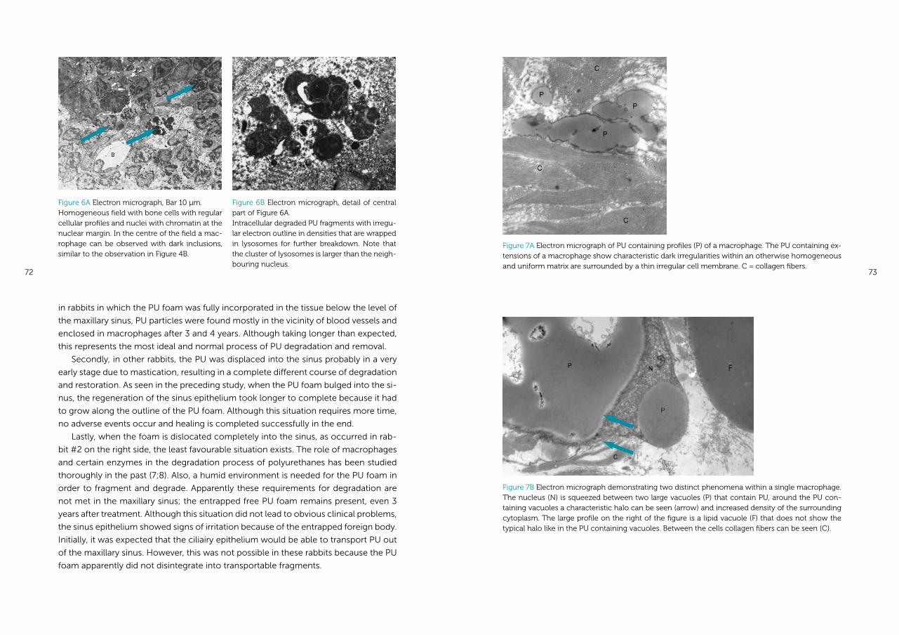
72 73
Figure 7A Electron micrograph of PU containing profiles (P) of a macrophage. The PU containing ex-tensions of a macrophage show characteristic dark irregularities within an otherwise homogeneous and uniform matrix are surrounded by a thin irregular cell membrane. C = collagen fibers.
Figure 7B Electron micrograph demonstrating two distinct phenomena within a single macrophage. The nucleus (N) is squeezed between two large vacuoles (P) that contain PU, around the PU con-taining vacuoles a characteristic halo can be seen (arrow) and increased density of the surrounding cytoplasm. The large profile on the right of the figure is a lipid vacuole (F) that does not show the typical halo like in the PU containing vacuoles. Between the cells collagen fibers can be seen (C).
Figure 6A Electron micrograph, Bar 10 μm. Homogeneous field with bone cells with regular cellular profiles and nuclei with chromatin at the nuclear margin. In the centre of the field a mac-rophage can be observed with dark inclusions, similar to the observation in Figure 4B.
Figure 6B Electron micrograph, detail of central part of Figure 6A. Intracellular degraded PU fragments with irregu-lar electron outline in densities that are wrapped in lysosomes for further breakdown. Note that the cluster of lysosomes is larger than the neigh-bouring nucleus.
in rabbits in which the PU foam was fully incorporated in the tissue below the level of
the maxillary sinus, PU particles were found mostly in the vicinity of blood vessels and
enclosed in macrophages after 3 and 4 years. Although taking longer than expected,
this represents the most ideal and normal process of PU degradation and removal.
Secondly, in other rabbits, the PU was displaced into the sinus probably in a very
early stage due to mastication, resulting in a complete different course of degradation
and restoration. As seen in the preceding study, when the PU foam bulged into the si-
nus, the regeneration of the sinus epithelium took longer to complete because it had
to grow along the outline of the PU foam. Although this situation requires more time,
no adverse events occur and healing is completed successfully in the end.
Lastly, when the foam is dislocated completely into the sinus, as occurred in rab-
bit #2 on the right side, the least favourable situation exists. The role of macrophages
and certain enzymes in the degradation process of polyurethanes has been studied
tho roughly in the past (7;8). Also, a humid environment is needed for the PU foam in
order to fragment and degrade. Apparently these requirements for degradation are
not met in the maxillary sinus; the entrapped free PU foam remains present, even 3
years after treatment. Although this situation did not lead to obvious clinical problems,
the sinus epithelium showed signs of irritation because of the entrapped foreign body.
Initially, it was expected that the ciliairy epithelium would be able to transport PU out
of the maxillary sinus. However, this was not possible in these rabbits because the PU
foam apparently did not disintegrate into transportable fragments.
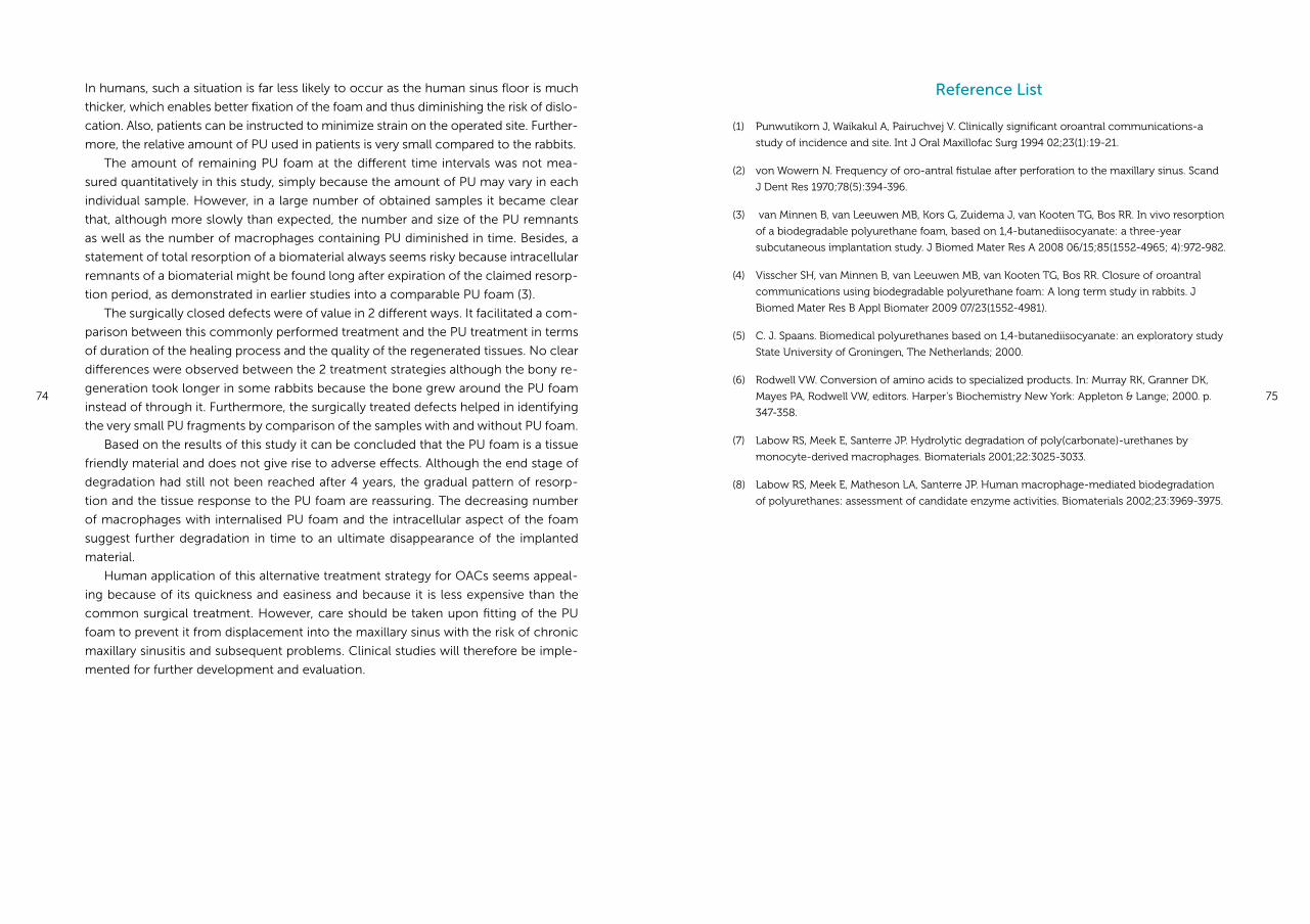
74 75
In humans, such a situation is far less likely to occur as the human sinus floor is much
thicker, which enables better fixation of the foam and thus diminishing the risk of dislo-
cation. Also, patients can be instructed to minimize strain on the operated site. Further-
more, the relative amount of PU used in patients is very small compared to the rabbits.
The amount of remaining PU foam at the different time intervals was not mea-
sured quantitatively in this study, simply because the amount of PU may vary in each
individual sample. However, in a large number of obtained samples it became clear
that, although more slowly than expected, the number and size of the PU remnants
as well as the number of macrophages containing PU diminished in time. Besides, a
statement of total resorption of a biomaterial always seems risky because intracellular
remnants of a biomaterial might be found long after expiration of the claimed resorp-
tion period, as demonstrated in earlier studies into a comparable PU foam (3).
The surgically closed defects were of value in 2 different ways. It facilitated a com-
parison between this commonly performed treatment and the PU treatment in terms
of duration of the healing process and the quality of the regenerated tissues. No clear
differences were observed between the 2 treatment strategies although the bony re-
generation took longer in some rabbits because the bone grew around the PU foam
instead of through it. Furthermore, the surgically treated defects helped in identifying
the very small PU fragments by comparison of the samples with and without PU foam.
Based on the results of this study it can be concluded that the PU foam is a tissue
friendly material and does not give rise to adverse effects. Although the end stage of
degradation had still not been reached after 4 years, the gradual pattern of resorp-
tion and the tissue response to the PU foam are reassuring. The decreasing number
of macrophages with internalised PU foam and the intracellular aspect of the foam
suggest further degradation in time to an ultimate disappearance of the implanted
material.
Human application of this alternative treatment strategy for OACs seems appeal-
ing because of its quickness and easiness and because it is less expensive than the
common surgical treatment. However, care should be taken upon fitting of the PU
foam to prevent it from displacement into the maxillary sinus with the risk of chronic
maxillary sinusitis and subsequent problems. Clinical studies will therefore be imple-
mented for further development and evaluation.
Reference List
(1) Punwutikorn J, Waikakul A, Pairuchvej V. Clinically significant oroantral communications-a
study of incidence and site. Int J Oral Maxillofac Surg 1994 02;23(1):19-21.
(2) von Wowern N. Frequency of oro-antral fistulae after perforation to the maxillary sinus. Scand
J Dent Res 1970;78(5):394-396.
(3) van Minnen B, van Leeuwen MB, Kors G, Zuidema J, van Kooten TG, Bos RR. In vivo resorption
of a biodegradable polyurethane foam, based on 1,4-butanediisocyanate: a three-year
subcutaneous implantation study. J Biomed Mater Res A 2008 06/15;85(1552-4965; 4):972-982.
(4) Visscher SH, van Minnen B, van Leeuwen MB, van Kooten TG, Bos RR. Closure of oroantral
communications using biodegradable polyurethane foam: A long term study in rabbits. J
Biomed Mater Res B Appl Biomater 2009 07/23(1552-4981).
(5) C. J. Spaans. Biomedical polyurethanes based on 1,4-butanediisocyanate: an exploratory study
State University of Groningen, The Netherlands; 2000.
(6) Rodwell VW. Conversion of amino acids to specialized products. In: Murray RK, Granner DK,
Mayes PA, Rodwell VW, editors. Harper’s Biochemistry New York: Appleton & Lange; 2000. p.
347-358.
(7) Labow RS, Meek E, Santerre JP. Hydrolytic degradation of poly(carbonate)-urethanes by
monocyte-derived macrophages. Biomaterials 2001;22:3025-3033.
(8) Labow RS, Meek E, Matheson LA, Santerre JP. Human macrophage-mediated biodegradation
of polyurethanes: assessment of candidate enzyme activities. Biomaterials 2002;23:3969-3975.
Chapter 5a
Closure of oroantral communications using biodegradable polyurethane foam:
a feasibility study
Susan H Visscher, Baucke van Minnen,
Rudolf RM Bos
Edited version of:
Journal of Oral and Maxillofacial Surgery 2010 Feb; 68(2): 281-286
78 79
Abstract
The aim of this study was to assess the feasibility of biodegradable polyurethane (PU)
foam for closure of oroantral communications (OACs).
Ten consecutive patients with fresh oroantral communications (existing < 24 hrs)
were treated with PU foam. Standardized evaluations were performed at 2 weeks and
8 weeks after closure of the OAC.
In 5 patients the OACs were closed successfully without complications. 3 patients
developed a sinusitis, which was conservatively managed with antibiotics in 2 cases.
In 1 case the sinus was reopened for irrigation, after which a buccal flap procedure
was performed. In 2 patients the OAC reoccurred and was surgically closed with a
buccal flap after thorough irrigation.
Closure of OACs with biodegradable polyurethane foam is feasible. Closure was
achieved in 7/10 patients without further surgical intervention. The complications
may be related to the fitting of the foam and the size of the defects. Taking the
complicating factors into account, further studies will be implemented to optimize
this treatment strategy.
Introduction
Oroantral communications (OACs) are usually caused by extraction of maxillary pos-
terior teeth (1;2). Although the incidence is relatively low (5%), OACs are frequently
encountered due to the high number of extractions (3;4).
OACs may close spontaneously, especially when the defect has a size below 5 mm
(5). It is, however, difficult to determine the size of the OAC clinically and therefore, it
is difficult to predict whether an OAC will heal uneventfully without intervention. To
prevent chronic sinusitis and the development of fistulas, it is therefore generally ac-
cepted that all of these defects should be surgically closed within 24 to 48 hours (5).
Surgical closure of OACs is commonly performed with a mucoperiosteal buccal
flap (5;6). Nevertheless, the use of a buccal sliding flap has several disadvantages. First-
ly, the patient often has to be referred to a maxillofacial surgeon for surgical closure
of the OAC. Secondly, the patient suffers from more postoperative pain and swelling
after surgical closure compared to an uneventful extraction (7). Thirdly, on the long
term, the depth of the buccal sulcus may permanently decrease, thereby hindering
the construction of a well-fitting dental prosthesis (8;9).
Because of the disadvantages of surgical closure, several alternative treatment
modalities have been described in literature, including third molar transplantation,
hydroxylapatite blocks, bioabsorbable root analogue and the Bio-Oss-Bio-Gide sand-
wich technique (10-13). Nevertheless, these methods all have their specific disad-
vantages and are not frequently used in clinical practice because either they are not
effective, no simplification of the standard method, or too expensive.
The goal of this feasibility study was to evaluate a new, straightforward and safe
strate gy for the closure of oroantral communications with biodegradable polyure-
thane foam. As a result of its biodegradability, the foam does not have to be removed
from the body after it has performed its function, which is considered a major ad-
vantage over non-degradable materials. In our opinion, the PU foam should make
treatment of oroantral communications easier and additionally, it will take the need
for special equipment and surgical expertise away. This should make it possible for the
general dentist to treat an OAC himself, instead of having to refer the patient to the
maxillofacial surgeon or another colleague trained in closing such defects. Besides, it
means a simple method for closure of OACs for maxillofacial surgeons as well. Lastly,
at all times the attending physician can fall back on the standard surgical procedure in
case the PU foam unexpectedly does not result in adequate closure.
80 81
Materials and methods
All procedures and materials were approved by the medical ethical committee of the
University Medical Centre Groningen (UMCG).
Biodegradable polyurethane foam (Polyganics B.V., Groningen) has been deve-
loped for the closure of OACs, made of hard urethane segments for strength and
soft segments made of D/L lactide and ε-caprolactone. The polyester soft segments
were synthesized first, and consisted of (50/50) D/L lactide/ ε-caprolactone and poly-
ethyleneglycol (PEG). The polyethyleneglycol was added to the soft segment to make
it more hydrophilic and more rapidly degradable. Chain extension was performed
resulting in polyurethane segments with a uniform length of five urethane moieties
and an overall PEG content of 5 w/ w %.
The polyurethane was dissolved in 1,4-dioxane, resulting in a concentration of 4 w/ w %
polyurethane. Water was added to obtain an interconnected pore structure, after
which the solution was poured in a mold. After cooling down the homogenous solu-
tion to -18°C, it was freeze dried to remove the water and dioxane crystals. Prior to
the study, the foams were sterilized with ethylene oxide.
The final product is a cylindrically shaped foam with a diameter of app 5 mm and
a height of approximately 7 mm (Figure 1). The porosity of the foam is app 95%. The
foam remains its strength for about 2 weeks. The highly interconnected pore struc-
ture of the product is designed for optimal tissue ingrowth.
In vivo and in vitro experiments were performed to investigate the use of PU foams as
medical devices for tissue regeneration (14-17). An in vitro degradation study showed
that the foams remained mechanically stable for two weeks (17), and animal experi-
ments proved that it enables mucosal overgrowth (18). Altogether, the results indi-
cated that the PU foam can be used safely as a biodegradable implant and showed no
different biocompatibility compared to the commercially available materials.
For this feasibility study, 10 consecutive patients with oroantral communications
were included. Inclusion took place from October 2007 to January 2008 at the De-
partment of Oral and Maxillofacial Surgery of the UMCG. The cause of the OAC was
tooth extraction in all selected patients. All OACs were closed by the same maxillofacial
surgeon and resident. Patients with a history of chronic sinusitis, patients on antibiotic
prophylaxis, or patients with acute sinusitis were excluded. Standardized evaluations
were performed at 2 weeks and 8 weeks after closure of the oroantral communication.
Recorded data included patient sex, age, smoking, medication, aetiology, reason for
extraction, location and (any) complications.
Success was considered as permanent closure of the OAC. In case of a reoccur-
rence of the OAC, the standard surgical procedure was used to achieve closure.
The OAC was confirmed by nose- and mouth blowing. In all patients, obliteration
of the antral perforation with the foam was carried out under local anaesthesia with
4% articaïn and 1:100.000 epinephrine (Aventis Pharma BV Hoevelaken). The approxi-
mate size of the perforation was estimated and a cylindrically shaped polyurethane
foam selected that resulted in a tight fit. Secondly, the PU foam was fitted in the per-
foration. Gingival margins were approximated with a 4.0 Vicryl® Rapid suture (Ethicon),
without complete mucosal closure to ensure the PU foam stayed in place (Figure
2a and 2b). All patients were advised against nose blowing. Postoperative analgesics
(Ibuprophen and/or paracetamol) and 0,2 % chlorhexidine mouth rinses 2-3 times
daily were prescribed. As in accordance with the Dutch guidelines, antibiotics or de-
congestants were not routinely prescribed. Remaining sutures were removed after 2
weeks. Intraoral photographs were taken to document the tissue healing.
Figure 1 PU Foam with electron microscope detail, showing interconnected pore structure.
82 83
Results
Table 1 Overview of clinical data of included patients
Patient
No.
Sex
(m/f)
Age
(years)
Smoker
(yes/no)
Location
OAC
Affected
roots
Indication
1 f 47 no left second molar distobuccal carious
2 m 48 no right third molar mesiobuccal carious
3 f 22 no right third molar distobuccal non-functional
4 f 44 yes right first molar palatinal
mesiobuccal
distobuccal
carious
5 m 25 no right third molar mesiobuccal preventive
6 f 72 no right first molar palatinal total tooth
extraction
7 m 46 yes left first molar distobuccal carious
8 m 42 yes left second molar mesiobuccal
distobuccal
pain
9 m 43 no left third molar mesiobuccal
distobuccal
non-functional
10 m 28 no left third molar all (fused roots) non-functional
Abbreviation: m; male, f; female, OAC; oroantral communication
Table 2 Characteristics of the treatment results
Patient
No.
Wound
infection
PU lost Sinusitis Reoccurrence
OAC
Antibiotics Surgical
intervention
1 - - - - - -
2 - - - - - -
3 - - - - - -
4 - + + + + +
5 - - - - - -
6 - - - - - -
7 - - + - + +
8 - + + + + +
9 - - + - + -
10 - - + - + -
Figure 2a Patient No 7: PU (arrow) placed in oroan-tral perforation
Figure 2b Patient No 7: PU placed in oroantral perforation, gingival margins approximated with 2 sutures
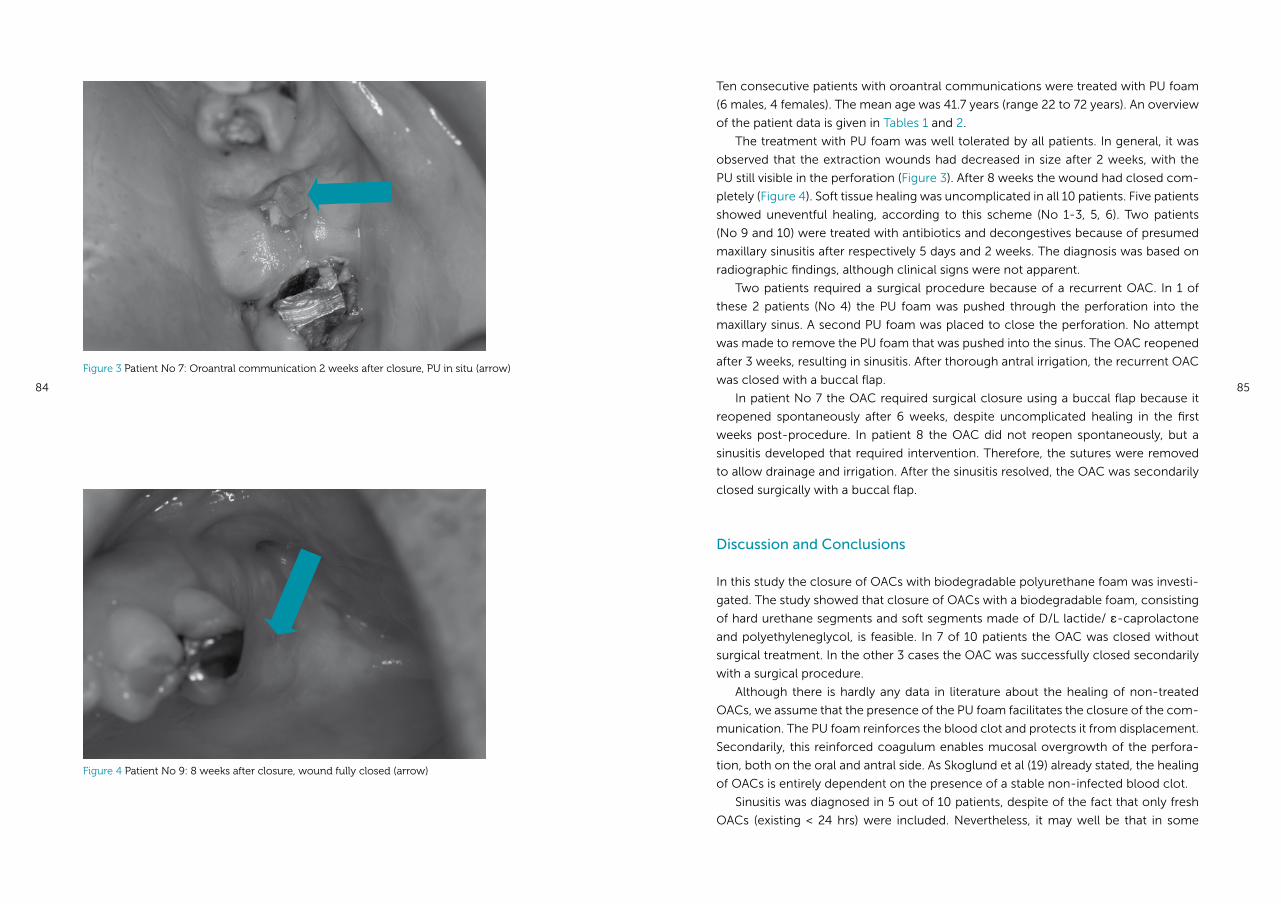
84 85
Ten consecutive patients with oroantral communications were treated with PU foam
(6 males, 4 females). The mean age was 41.7 years (range 22 to 72 years). An overview
of the patient data is given in Tables 1 and 2.
The treatment with PU foam was well tolerated by all patients. In general, it was
observed that the extraction wounds had decreased in size after 2 weeks, with the
PU still visible in the perforation (Figure 3). After 8 weeks the wound had closed com-
pletely (Figure 4). Soft tissue healing was uncomplicated in all 10 patients. Five patients
showed uneventful healing, according to this scheme (No 1-3, 5, 6). Two patients
(No 9 and 10) were treated with antibiotics and decongestives because of presumed
maxillary sinusitis after respectively 5 days and 2 weeks. The diagnosis was based on
radiographic findings, although clinical signs were not apparent.
Two patients required a surgical procedure because of a recurrent OAC. In 1 of
these 2 patients (No 4) the PU foam was pushed through the perforation into the
maxillary sinus. A second PU foam was placed to close the perforation. No attempt
was made to remove the PU foam that was pushed into the sinus. The OAC reopened
after 3 weeks, resulting in sinusitis. After thorough antral irrigation, the recurrent OAC
was closed with a buccal flap.
In patient No 7 the OAC required surgical closure using a buccal flap because it
reopened spontaneously after 6 weeks, despite uncomplicated healing in the first
weeks post-procedure. In patient 8 the OAC did not reopen spontaneously, but a
sinusitis developed that required intervention. Therefore, the sutures were removed
to allow drainage and irrigation. After the sinusitis resolved, the OAC was secondarily
closed surgically with a buccal flap.
Discussion and Conclusions
In this study the closure of OACs with biodegradable polyurethane foam was investi-
gated. The study showed that closure of OACs with a biodegradable foam, consisting
of hard urethane segments and soft segments made of D/L lactide/ ε-caprolactone
and polyethyleneglycol, is feasible. In 7 of 10 patients the OAC was closed without
surgical treatment. In the other 3 cases the OAC was successfully closed secondarily
with a surgical procedure.
Although there is hardly any data in literature about the healing of non-treated
OACs, we assume that the presence of the PU foam facilitates the closure of the com-
munication. The PU foam reinforces the blood clot and protects it from displacement.
Secondarily, this reinforced coagulum enables mucosal overgrowth of the perfora-
tion, both on the oral and antral side. As Skoglund et al (19) already stated, the healing
of OACs is entirely dependent on the presence of a stable non-infected blood clot.
Sinusitis was diagnosed in 5 out of 10 patients, despite of the fact that only fresh
OACs (existing < 24 hrs) were included. Nevertheless, it may well be that in some
Figure 4 Patient No 9: 8 weeks after closure, wound fully closed (arrow)
Figure 3 Patient No 7: Oroantral communication 2 weeks after closure, PU in situ (arrow)
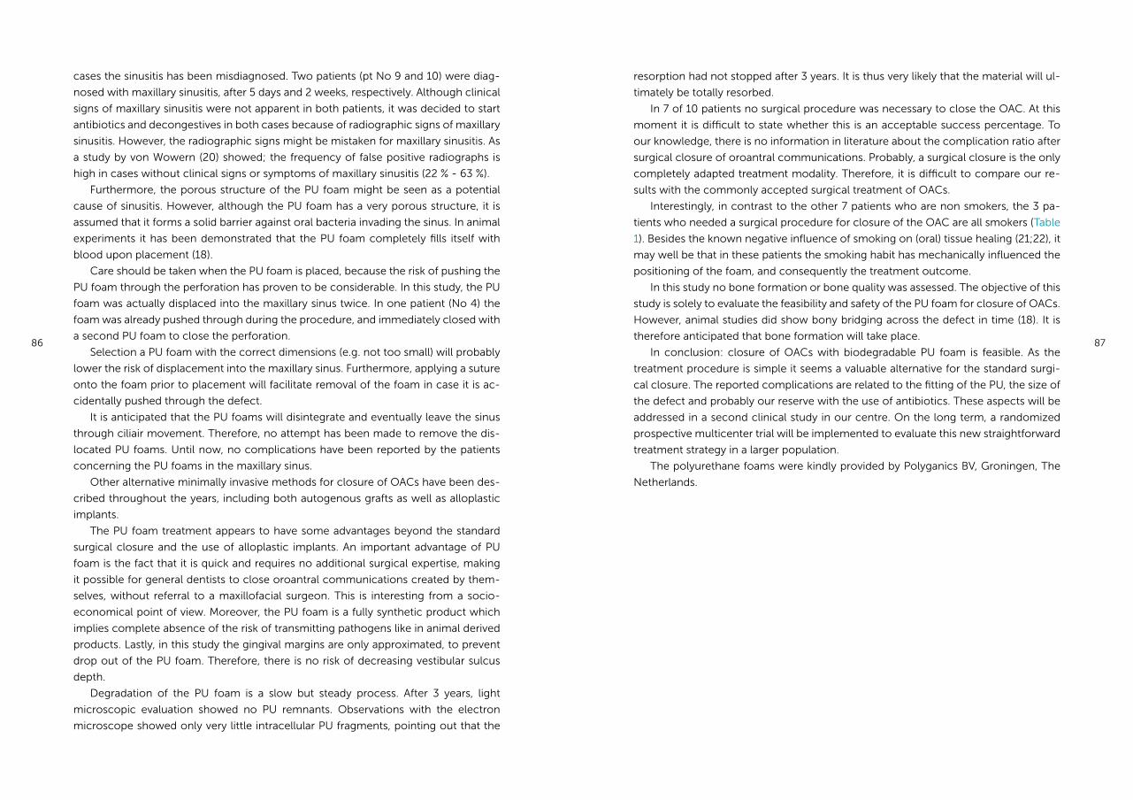
86 87
cases the sinusitis has been misdiagnosed. Two patients (pt No 9 and 10) were diag-
nosed with maxillary sinusitis, after 5 days and 2 weeks, respectively. Although clinical
signs of maxillary sinusitis were not apparent in both patients, it was decided to start
antibiotics and decongestives in both cases because of radiographic signs of maxillary
sinusitis. However, the radiographic signs might be mistaken for maxillary sinusitis. As
a study by von Wowern (20) showed; the frequency of false positive radiographs is
high in cases without clinical signs or symptoms of maxillary sinusitis (22 % - 63 %).
Furthermore, the porous structure of the PU foam might be seen as a potential
cause of sinusitis. However, although the PU foam has a very porous structure, it is
assumed that it forms a solid barrier against oral bacteria invading the sinus. In animal
experiments it has been demonstrated that the PU foam completely fills itself with
blood upon placement (18).
Care should be taken when the PU foam is placed, because the risk of pushing the
PU foam through the perforation has proven to be considerable. In this study, the PU
foam was actually displaced into the maxillary sinus twice. In one patient (No 4) the
foam was already pushed through during the procedure, and immediately closed with
a second PU foam to close the perforation.
Selection a PU foam with the correct dimensions (e.g. not too small) will probably
lower the risk of displacement into the maxillary sinus. Furthermore, applying a suture
onto the foam prior to placement will facilitate removal of the foam in case it is ac-
cidentally pushed through the defect.
It is anticipated that the PU foams will disintegrate and eventually leave the sinus
through ciliair movement. Therefore, no attempt has been made to remove the dis-
located PU foams. Until now, no complications have been reported by the patients
concerning the PU foams in the maxillary sinus.
Other alternative minimally invasive methods for closure of OACs have been des-
cribed throughout the years, including both autogenous grafts as well as alloplastic
implants.
The PU foam treatment appears to have some advantages beyond the standard
surgical closure and the use of alloplastic implants. An important advantage of PU
foam is the fact that it is quick and requires no additional surgical expertise, making
it possible for general dentists to close oroantral communications created by them-
selves, without referral to a maxillofacial surgeon. This is interesting from a socio-
economical point of view. Moreover, the PU foam is a fully synthetic product which
implies complete absence of the risk of transmitting pathogens like in animal derived
products. Lastly, in this study the gingival margins are only approximated, to prevent
drop out of the PU foam. Therefore, there is no risk of decreasing vestibular sulcus
depth.
Degradation of the PU foam is a slow but steady process. After 3 years, light
microscopic evaluation showed no PU remnants. Observations with the electron
microscope showed only very little intracellular PU fragments, pointing out that the
resorption had not stopped after 3 years. It is thus very likely that the material will ul-
timately be totally resorbed.
In 7 of 10 patients no surgical procedure was necessary to close the OAC. At this
moment it is difficult to state whether this is an acceptable success percentage. To
our knowledge, there is no information in literature about the complication ratio after
surgical closure of oroantral communications. Probably, a surgical closure is the only
completely adapted treatment modality. Therefore, it is difficult to compare our re-
sults with the commonly accepted surgical treatment of OACs.
Interestingly, in contrast to the other 7 patients who are non smokers, the 3 pa-
tients who needed a surgical procedure for closure of the OAC are all smokers (Table
1). Besides the known negative influence of smoking on (oral) tissue healing (21;22), it
may well be that in these patients the smoking habit has mechanically influenced the
positioning of the foam, and consequently the treatment outcome.
In this study no bone formation or bone quality was assessed. The objective of this
study is solely to evaluate the feasibility and safety of the PU foam for closure of OACs.
However, animal studies did show bony bridging across the defect in time (18). It is
therefore anticipated that bone formation will take place.
In conclusion: closure of OACs with biodegradable PU foam is feasible. As the
treatment procedure is simple it seems a valuable alternative for the standard surgi-
cal closure. The reported complications are related to the fitting of the PU, the size of
the defect and probably our reserve with the use of antibiotics. These aspects will be
addressed in a second clinical study in our centre. On the long term, a randomized
prospective multicenter trial will be implemented to evaluate this new straightforward
treatment strategy in a larger population.
The polyurethane foams were kindly provided by Polyganics BV, Groningen, The
Netherlands.
88 89
(15) van Minnen B, Stegenga B, van Leeuwen MB, van Kooten TG, Bos RR. A long-term in vitro
biocompatibility study of a biodegradable polyurethane and its degradation products. J
Biomed Mater Res A 2006 Feb;76(2):377-85.
(16) van Minnen B, Stegenga B, Zuidema J, Hissink CE, van Leeuwen MB, van Kooten TG, et al. An
animal model for oroantral communications: a pilot study with Gottingen minipigs. Lab Anim
2005 Jul;39(3):280-3.
(17) van Minnen B, van Leeuwen MB, Stegenga B, Zuidema J, Hissink CE, van Kooten TG, et al.
Short-term in vitro and in vivo biocompatibility of a biodegradable polyurethane foam based
on 1,4-butanediisocyanate. J Mater Sci Mater Med 2005 Mar;16(3):221-7.
(18) van Minnen B. Biodegradable polyurethane foams. Thesis. University of Groningen, the
Netherlands; 2006.
(19) Skoglund LA, Pedersen SS, Holst E. Surgical management of 85 perforations to the maxillary
sinus. Int J Oral Surg 1983 Feb;12(1):1-5.
(20) von Wowern N. Clinical and radiographic findings in maxillary sinus with oro-antral fistula. Int
J Oral Surg 1981;10(Suppl 1):138-42.
(21) Mayfield L, Soderholm G, Hallstrom H, Kullendorff B, Edwardsson S, Bratthall G, et al. Guided
tissue regeneration for the treatment of intraosseous defects using a biabsorbable membrane.
A controlled clinical study. J Clin Periodontol 1998 Jul;25(7):585-95.
(22) Balaji SM. Tobacco smoking and surgical healing of oral tissues: a review. Indian J Dent Res
2008 Oct;19(4):344-8.
Reference List
(1) Abuabara A, Cortez AL, Passeri LA, de MM, Moreira RW. Evaluation of different treatments for
oroantral/oronasal communications: experience of 112 cases. Int J Oral Maxillofac Surg 2006
Feb;35(2):155-8.
(2) Punwutikorn J, Waikakul A, Pairuchvej V. Clinically significant oroantral communications - a
study of incidence and site. Int J Oral Maxillofac Surg 1994 Feb;23(1):19-21.
(3) del Rey-Santamaria M, Valmaseda CE, Berini AL, Gay EC. Incidence of oral sinus
communications in 389 upper thirmolar extraction. Med Oral Patol Oral Cir Bucal 2006
Jul;11(4):E334-E338.
(4) Bodner L, Gatot A, Bar-Ziv J. Technical note: oroantral fistula: improved imaging with a dental
computed tomography software program. Br J Radiol 1995 Nov;68(815):1249-50.
(5) von Wowern N. Correlation between the development of an orantral fistula and the size of the
corresponding bony defect. J Oral Surg 1973;31(2):98-102.
(6) von Wowern N. Frequency of oro-antral fistulae after perforation to the maxillary sinus. Scand
J Dent Res 1970;78(5):394-6.
(7) Kinner U, Frenkel G. Alternative Methoden des Verschlusses von Mund-Antrum-Verbindungen;
Die plastische Deckung der Kieferhöhle mit lyophilisierter Dura mit alkoholischer
Prolaminlösung. ZWR 1990 Nov;99(11):890-6.
(8) Güven O. A clinical study on oroantral fistulae. J Craniomaxillofac Surg 1998 Aug;26(4):267-71.
(9) Obradovic O, Todorovic Lj, Pesic V. Investigations of the buccal sulcus depth after the use
of certain methods of oro-antral communication closure. Bull Group Int Rech Sci Stomatol
Odontol 1981;24(3):209-14.
(10) Kitagawa Y, Sano K, Nakamura M, Ogasawara T. Use of third molar transplantation for closure
of the oroantral communication after tooth extraction: a report of 2 cases. Oral Surg Oral Med
Oral Pathol Oral Radiol Endod 2003 Apr;95(4):409-15.
(11) Thoma K., Pajarola G.F., Gratz K.W., Schmidlin P.R. bioabsorbable root analogue for closure of
oroantral communications after tooth extraction:; a prospective case-cohort study. Oral Surg
Oral Med Oral Pathol Oral Radiol Endod 2006 May;101(5):558-64.
(12) Ogunsalu C. A new surgical management for oro-antral communication: the resorbable
guided tissue regeneration membrane-bone substitute sandwich technique. West Indian Med
J 2005 Sep;54(4):261-3.
(13) Zide MF, Karas ND. Hydroxylapatite block closure of oroantral fistulas: report of cases. J Oral
Maxillofac Surg 1992 Jan;50(1):71-5.
(14) van Minnen B, Stegenga B, van Leeuwen MB, van Kooten TG, Bos RR. Nonsurgical closure of
oroantral communications with a biodegradable polyurethane foam: A pilot study in rabbits. J
Oral Maxillofac Surg 2007 Feb;65(2):218-22.
Chapter 5b
Feasibility of conical biodegradable polyurethane foam for closure of oroantral
communications.
Susan H Visscher, Baucke van Minnen,
Rudolf RM Bos
Edited version of:
Journal of Oral and Maxillofacial Surgery 2011 Feb; 69(2): 390-395
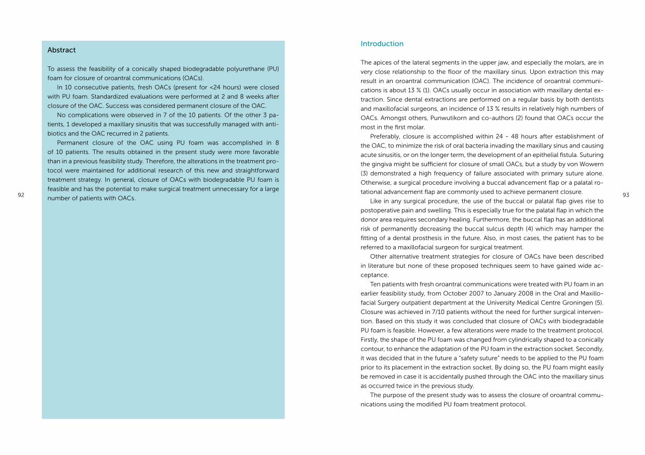
92 93
Introduction
The apices of the lateral segments in the upper jaw, and especially the molars, are in
very close relationship to the floor of the maxillary sinus. Upon extraction this may
result in an oroantral communication (OAC). The incidence of oroantral communi-
cations is about 13 % (1). OACs usually occur in association with maxillary dental ex-
traction. Since dental extractions are performed on a regular basis by both dentists
and maxillofacial surgeons, an incidence of 13 % results in relatively high numbers of
OACs. Amongst others, Punwutikorn and co-authors (2) found that OACs occur the
most in the first molar.
Preferably, closure is accomplished within 24 - 48 hours after establishment of
the OAC, to minimize the risk of oral bacteria invading the maxillary sinus and causing
acute sinusitis, or on the longer term, the development of an epithelial fistula. Suturing
the gingiva might be sufficient for closure of small OACs, but a study by von Wowern
(3) demonstrated a high frequency of failure associated with primary suture alone.
Otherwise, a surgical procedure involving a buccal advancement flap or a palatal ro-
tational advancement flap are commonly used to achieve permanent closure.
Like in any surgical procedure, the use of the buccal or palatal flap gives rise to
postoperative pain and swelling. This is especially true for the palatal flap in which the
donor area requires secondary healing. Furthermore, the buccal flap has an additional
risk of permanently decreasing the buccal sulcus depth (4) which may hamper the
fitting of a dental prosthesis in the future. Also, in most cases, the patient has to be
referred to a maxillofacial surgeon for surgical treatment.
Other alternative treatment strategies for closure of OACs have been described
in literature but none of these proposed techniques seem to have gained wide ac-
ceptance.
Ten patients with fresh oroantral communications were treated with PU foam in an
earlier feasibility study, from October 2007 to January 2008 in the Oral and Maxillo-
facial Surgery outpatient department at the University Medical Centre Groningen (5).
Closure was achieved in 7/10 patients without the need for further surgical interven-
tion. Based on this study it was concluded that closure of OACs with biodegradable
PU foam is feasible. However, a few alterations were made to the treatment protocol.
Firstly, the shape of the PU foam was changed from cylindrically shaped to a conically
contour, to enhance the adaptation of the PU foam in the extraction socket. Secondly,
it was decided that in the future a “safety suture” needs to be applied to the PU foam
prior to its placement in the extraction socket. By doing so, the PU foam might easily
be removed in case it is accidentally pushed through the OAC into the maxillary sinus
as occurred twice in the previous study.
The purpose of the present study was to assess the closure of oroantral commu-
nications using the modified PU foam treatment protocol.
Abstract
To assess the feasibility of a conically shaped biodegradable polyurethane (PU)
foam for closure of oroantral communications (OACs).
In 10 consecutive patients, fresh OACs (present for <24 hours) were closed
with PU foam. Standardized evaluations were performed at 2 and 8 weeks after
closure of the OAC. Success was considered permanent closure of the OAC.
No complications were observed in 7 of the 10 patients. Of the other 3 pa-
tients, 1 developed a maxillary sinusitis that was successfully managed with anti-
biotics and the OAC recurred in 2 patients.
Permanent closure of the OAC using PU foam was accomplished in 8
of 10 patients. The results obtained in the present study were more favorable
than in a previous feasibility study. Therefore, the alterations in the treatment pro-
tocol were maintained for additional research of this new and straightforward
treatment strategy. In general, closure of OACs with biodegradable PU foam is
feasible and has the potential to make surgical treatment unnecessary for a large
number of patients with OACs.
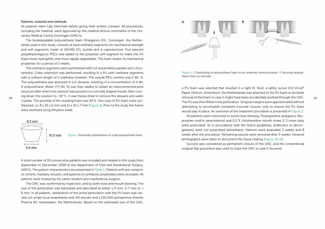
94 95
Patients, material and methods
All patients were fully informed before giving their written consent. All procedures,
including the material, were approved by the medical ethical committee of the Uni-
versity Medical Centre Groningen (UMCG).
The biodegradable polyurethane foam (Polyganics B.V., Groningen, the Nether-
lands) used in this study, consists of hard urethane segments for mechanical strength
and soft segments made of (50/50) D/L lactide and ε-caprolactone. Five percent
polyethyleneglycol (PEG) was added to the polyester soft segment to make the PU
foam more hydrophilic and more rapidly degradable. The foam retains its mechanical
properties for a period of 2 weeks.
The urethane segments were synthesized with 1,4-butanediisocyanate and 1,4 bu-
tanediol. Chain extension was performed, resulting in a PU with urethane segments
with a uniform length of 5 urethane moieties. The overall PEG content was 5 Wt. %.
The polyurethane was dissolved in 1,4-dioxane, resulting in a concentration of 4 Wt.
% polyurethane. Water (7.5 Wt. %) was then added to obtain an interconnected pore
structure after which the solution was poured in a conically shaped mould. After cool-
ing down the solution to -18 °C, it was freeze dried to remove the dioxane and water
crystals. The porosity of the resulting foam was 94 %. Two sizes of PU foam were syn-
thesized, i.e. 8 x 10 x 5 mm and 11 x 10 x 7 mm (Figure 1). Prior to the study the foams
were sterilized using ethylene oxide.
a PU foam was selected that resulted in a tight fit. Next, a safety suture (4.0 Vicryl®
Rapid, Ethicon, Amersfoort, the Netherlands) was attached to the PU foam to facilitate
removal of the foam in case it might have been accidentally pushed through the OAC.
The PU was then fitted in the perforation. Gingival margins were approximated without
attempting to accomplish complete mucosal closure, only to ensure the PU foam
would stay in place. An overview of the treatment procedure is presented in Figure 2.
All patients were instructed to avoid nose blowing. Postoperative analgesics (Ibu-
prophen and/or paracetamol) and 0,2 % chlorhexidine mouth rinses 2-3 times daily
were prescribed. As in accordance with the Dutch guidelines, antibiotics or decon-
gestants were not prescribed beforehand. Patients were evaluated 2 weeks and 8
weeks after the procedure. Remaining sutures were removed after 2 weeks. Intraoral
photographs were taken to document the tissue healing (Figure 3A-D).
Success was considered as permanent closure of the OAC, and the conventional
surgical flap procedure was used to close the OAC in case it recurred.
Figure 1 Schematic presentation of small polyurethane foam
A total number of 10 consecutive patients was included and treated in this study from
September to December 2008 at the department of Oral and Maxillofacial Surgery,
UMCG. The patient characteristics are presented in Table 1. Patients with pre-existent,
or chronic maxillary sinusitis, and patients on antibiotic prophylaxis were excluded. All
patients were treated by the same resident and maxillofacial surgeon.
The OAC was confirmed by inspection, and by both nose and mouth blowing. The
size of the perforation was estimated and described as either ≤ 5 mm, 5-7 mm or >
8 mm. In all patients, obliteration of the antral perforation with the PU foam was car-
ried out under local anaesthesia with 4% articaïn and 1:100.000 epinephrine (Aventis
Pharma BV, Hoevelaken, the Netherlands). Based on the estimated size of the OAC,
Figure 2 A Positioning of polyurethane foam in an oroantral communication. B Securing polyure-thane foam on oral side.
A B
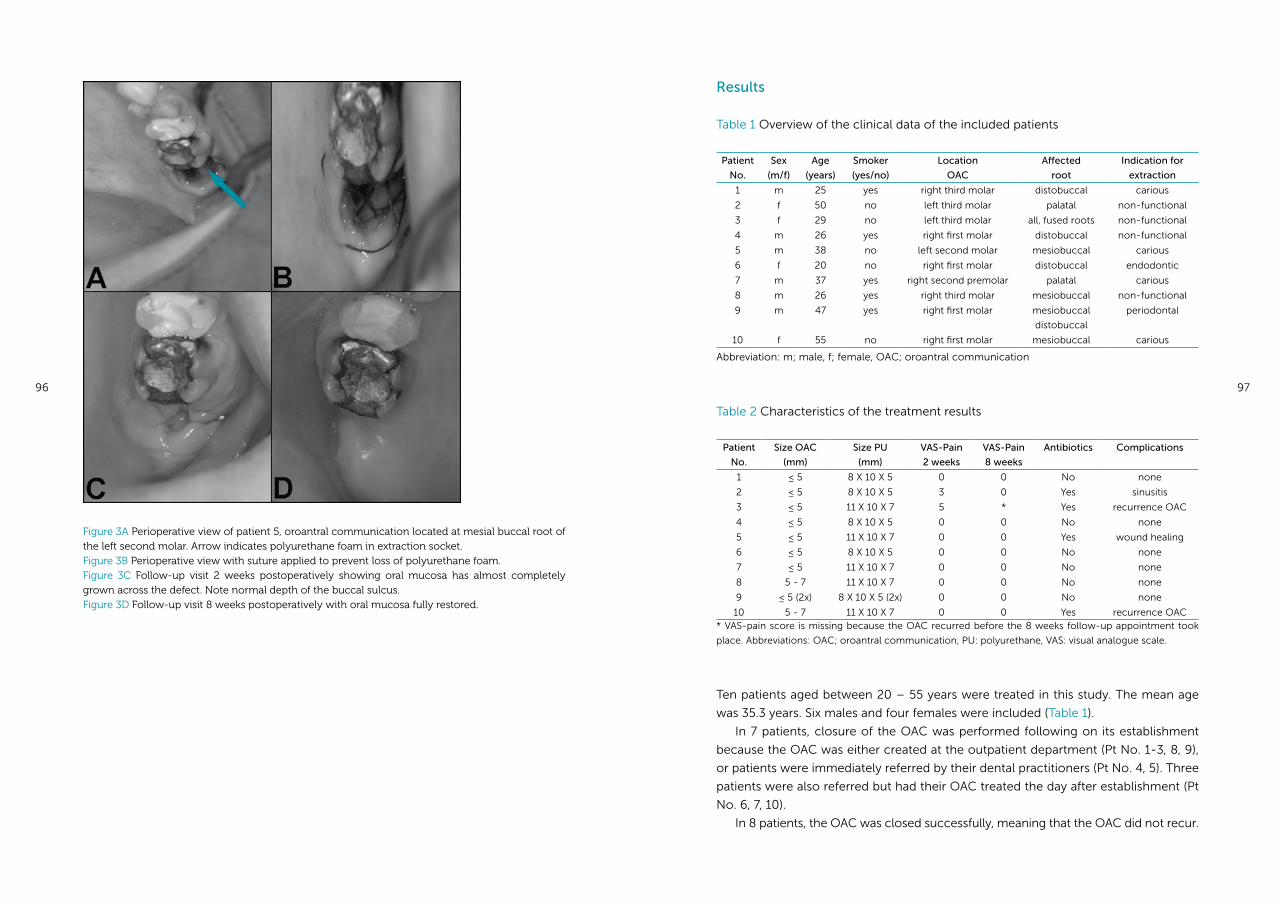
96 97
Results
Table 1 Overview of the clinical data of the included patients
Patient
No.
Sex
(m/f)
Age
(years)
Smoker
(yes/no)
Location
OAC
Affected
root
Indication for
extraction
1 m 25 yes right third molar distobuccal carious
2 f 50 no left third molar palatal non-functional
3 f 29 no left third molar all, fused roots non-functional
4 m 26 yes right first molar distobuccal non-functional
5 m 38 no left second molar mesiobuccal carious
6 f 20 no right first molar distobuccal endodontic
7 m 37 yes right second premolar palatal carious
8 m 26 yes right third molar mesiobuccal non-functional
9 m 47 yes right first molar mesiobuccal
distobuccal
periodontal
10 f 55 no right first molar mesiobuccal carious
Abbreviation: m; male, f; female, OAC; oroantral communication
Table 2 Characteristics of the treatment results
Patient
No.
Size OAC
(mm)
Size PU
(mm)
VAS-Pain
2 weeks
VAS-Pain
8 weeks
Antibiotics Complications
1 ≤ 5 8 X 10 X 5 0 0 No none
2 ≤ 5 8 X 10 X 5 3 0 Yes sinusitis
3 ≤ 5 11 X 10 X 7 5 * Yes recurrence OAC
4 ≤ 5 8 X 10 X 5 0 0 No none
5 ≤ 5 11 X 10 X 7 0 0 Yes wound healing
6 ≤ 5 8 X 10 X 5 0 0 No none
7 ≤ 5 11 X 10 X 7 0 0 No none
8 5 - 7 11 X 10 X 7 0 0 No none
9 ≤ 5 (2x) 8 X 10 X 5 (2x) 0 0 No none
10 5 - 7 11 X 10 X 7 0 0 Yes recurrence OAC* VAS-pain score is missing because the OAC recurred before the 8 weeks follow-up appointment took
place. Abbreviations: OAC; oroantral communication, PU: polyurethane, VAS: visual analogue scale.
Ten patients aged between 20 – 55 years were treated in this study. The mean age
was 35.3 years. Six males and four females were included (Table 1).
In 7 patients, closure of the OAC was performed following on its establishment
because the OAC was either created at the outpatient department (Pt No. 1-3, 8, 9),
or patients were immediately referred by their dental practitioners (Pt No. 4, 5). Three
patients were also referred but had their OAC treated the day after establishment (Pt
No. 6, 7, 10).
In 8 patients, the OAC was closed successfully, meaning that the OAC did not recur.
Figure 3A Perioperative view of patient 5, oroantral communication located at mesial buccal root of the left second molar. Arrow indicates polyurethane foam in extraction socket. Figure 3B Perioperative view with suture applied to prevent loss of polyurethane foam. Figure 3C Follow-up visit 2 weeks postoperatively showing oral mucosa has almost completely grown across the defect. Note normal depth of the buccal sulcus.Figure 3D Follow-up visit 8 weeks postoperatively with oral mucosa fully restored.
98 99
In 2 out of 10 patients the OAC recurred. Neither of these 2 patients were smokers.
This is in contrary to the previous study in which the recurrences of OACs (3 in total)
were recorded in heavy smokers. Besides the negative influence of smoking on oral
tissue healing (10;11), this difference might be explained by the difference in shape of
the polyurethane foams. It can be imagined that the cylindrically shaped PU, as used
in the first study, dislodges more easily in comparison to a conically shaped PU. This
seems especially true for smokers, who are probably more likely to put load on the PU
foam while in- and exhaling smoke.
The other alteration in the treatment protocol was the use of a safety suture. This
suture was applied to facilitate removal of the PU foam in case it was dislocated into
the sinus, which occurred twice in the previous study. In the present study none of the
foams dislodged into the sinus.
In patient No 3 extraction of the left third molar resulted in a problematic OAC
that recurred after 4 weeks. To our opinion, the only likely reason for the recurrence
of the OAC may be found in ill fitting of the PU foam due to the extended and com-
plex outline of the OAC. Specifically, this molar was positioned along the border of
the maxillary sinus and upon extraction a rather large part of the sinus floor was lost.
Besides this fact, all conditions were in favour of good healing; a cooperative young
and healthy patient, good oral hygiene and closure of the OAC immediately after ex-
traction of the molar.
In patient No. 10, closure of the OAC was accomplished the day after the extrac-
tion was performed by the dentist. The OAC unfortunately recurred after 8.5 weeks. It
is not exactly clear why the OAC recurred, but it is probably a summation of unfavour-
able circumstances: both the dental extraction and the postoperative healing course
were troublesome, the patient was referred with remaining radices of the molar in
situ, she got the flu with generalized sinusitis with persisting complaints of the right
sinus despite antibiotic treatment, and a bone sequester came loose from the extrac-
tion site. After treatment of the sinusitis the recurred OAC was surgically closed with
a buccal flap.
In our opinion, these specific failure cases do not seem to justify narrowing of the
suitability of PU foam for closure of for example large OACs or OACs without an intact
extraction socket. All the more because looking back it appeared difficult to predict
on clinical and radiographic grounds which cases were prone for complications or
failure. Cases of which we thought that could be problematic, for example because of
the absence of an intact alveole or a thin sinus floor, healed perfectly and vice versa.
VAS-scores were obtained from all patients to document the pain levels associated
with the PU treatment. In general no pain (VAS score 0) was reported at both follow-
up visits. Logically, patients with complaints such as maxillary sinusitis reported higher
pain scores. In addition, VAS scores were obtained directly after the procedure but
these did not seem to be of further value, because the treatment took place under
local anesthesia. At the first follow-up visit, patients were also asked for the pain expe-
Patient No. 5 however, returned 4 days after treatment because of mild swelling of the
buccal fold near the OAC. Antibiotics were started out of precaution and the complaints
resolved without further intervention (Figure 3A-D). Patient No. 2 developed a maxillary
sinusitis 1 week after treatment that was conservatively managed with antibiotics.
Extraction of the right first molar resulted in OACs of both buccal roots in patient
No. 9. Both OACs were subsequently closed successfully using 2 PU foams.
Extraction of the left third molar gave rise to an OAC in patient No. 3. The positioning
of this molar was rather problematic, resulting in a complicated OAC that covered a
large area of the maxillary sinus floor. Shortly after the procedure the patient developed
a sinusitis that responded well to antibiotics initially. However, the OAC recurred after
4 weeks and needed surgical closure.
In patient No. 10 the OAC recurred. The extraction of the right first molar by the
dentist had been rather difficult in this patient. Above this, she got the flu shortly after
treatment and had ongoing complaints of both maxillary sinuses since then. At first
she responded well to the antibiotics. Nevertheless, the symptoms associated with
the right maxillary sinus returned and 8.5 weeks after treatment a recurrence of the
OAC was observed. In the end, the OAC was successfully closed with a buccal flap
according to the protocol.
On average no pain or discomfort was reported in the follow up appointments
(Table 2). Higher pain scores were reported in cases with marked complications
(Pt No 2 and 3). Pt No 10 scored 0 on the pain score on both follow up visits, because
her complaints were ongoing but relatively mild at these moments.
Discussion
In this feasibility study we treated 10 patients with fresh OACs using degradable poly-
urethane foam. The goal of this study was to assess the changes made to the treat-
ment protocol; i.e. a conically shaped PU foam instead of a cylindrically shaped foam,
and application of a safety suture prior to placement. Eight of 10 patients were suc-
cessfully treated in this study, meaning that the OAC did not recur after closure with
PU foam. Based on the present study, it was concluded that the alterations to the pro-
tocol were beneficial to the treatment, and will therefore be maintained in further use.
Most OACs in this study were caused either by extraction of the third molar or first
molar. This can be explained by the high number of (impacted) third molar extractions
at the Oral and Maxillofacial Surgery outpatient department. Furthermore, the high
number of first molars inducing OACs is in line with other studies (3;6-8) as the maxil-
lary sinus has a very close relationship to the root apices of the first molars. Similar to
the retrospective study by Abuabara (9), the decade of life with the highest incidence
of OACs in the current study was the third decade, probably also because of the rather
high number of referral for (third molar) extractions in this period of life.
100 101
rienced in the days following the procedure, but obviously it was difficult to discrimi-
nate between pain caused by the extraction or by the PU treatment. It did become
clear that difficult extractions (Pt No. 3, 4, 8, 10) gave rise to more pain and discomfort.
Apart from the VAS-scores, all patients found the PU procedure quick and had a
positive overall opinion. In the future, VAS pain scores should preferably also be re-
corded after standard surgical closure, in order to compare the discomfort associated
with both treatment modalities.
In conclusion, taking the small number of treated patients in mind, the results of
this study concerning fitting of the PU foam and dislocation into the maxillary sinus
were more favourable than in the previous feasibility study. Therefore, the alterations
in the treatment protocol as used in this study will be maintained for further research
on this new strategy for closure of OACs. Nevertheless, a retrospective study will also
be performed to look into the complication rate associated with surgical treatment
of OACs. To our knowledge, this information is not yet available in literature, probably
because the surgical treatment is the golden standard and associated complications
are therefore taken for granted.
However, a success percentage of 80 % seems acceptable to proceed with further
research on this new strategy, as closure of OACs with biodegradable polyurethane
foam has the potential to make a surgical treatment unnecessary in a large number
of patients.
The polyurethane foams were kindly provided by Polyganics BV, Groningen, The
Netherlands.
Reference List
(1) Rothamel D, Wahl G, d’Hoedt B, Nentwig GH, Schwarz F, Becker J. Incidence and predictive
factors for perforation of the maxillary antrum in operations to remove upper wisdom teeth:
prospective multicentre study. Br J Oral Maxillofac Surg 2007 Jul;45(5):387-91.
(2) Punwutikorn J, Waikakul A, Pairuchvej V. Clinically significant oroantral communications-a
study of incidence and site. Int J Oral Maxillofac Surg 1994 Feb;23(1):19-21.
(3) von Wowern N. Frequency of oro-antral fistulae after perforation to the maxillary sinus. Scand
J Dent Res 1970;78(5):394-6.
(4) von Wowern N. Closure of oroantral fistula with buccal flap: Rehrmann versus Moczar. Int J
Oral Surg 1982 Jun;11(3):156-65.
(5) Visscher SH, van Minnen B, Bos RR. Closure of oroantral communications using biodegradable
polyurethane foam: a feasibility study. J Oral Maxillofac Surg 2010 Feb;68(2):281-6.
(6) Ehrl PA. Oroantral communication. Epicritical study of 175 patients, with special concern to
secondary operative closure. Int J Oral Surg 1980 Oct;9(5):351-8.
(7) Amaratunga NA. Oro-antral fistulae - a study of clinical, radiological and treatment aspects. Br
J Oral Maxillofac Surg 1986 Dec;24(6):433-7.
(8) Güven O. A clinical study on oroantral fistulae. J Craniomaxillofac Surg 1998 Aug;26(4):267-71.
(9) Abuabara A, Cortez AL, Passeri LA, de Moraes M, Moreira RW. Evaluation of different treatments
for oroantral/oronasal communications: experience of 112 cases. Int J Oral Maxillofac Surg
2006 Feb;35(2):155-8.
(10) Meechan JG, Macgregor ID, Rogers SN, Hobson RS, Bate JP, Dennison M. The effect of smoking
on immediate post-extraction socket filling with blood and on the incidence of painful socket.
Br J Oral Maxillofac Surg 1988 Oct;26(5):402-9.
(11) Balaji SM. Tobacco smoking and surgical healing of oral tissues: a review. Indian J Dent Res
2008 Oct;19(4):344-8.
Chapter 6
Is biodegradable PU foam as effective as surgery for closure of oroantral
communications? A prospective clinical trial.
Susan H Visscher, Baucke van Minnen,
Wim J Sluiter, Rudolf RM Bos
Submitted
104 105
Introduction
Surgical closure of oroantral communications (OACs), preferably within 24 hours
either with a buccal of palatal flap, still is the treatment of choice to minimize the
chance of fistula formation and chronic sinus disease (1). Application of this com-
mon surgical treatment was successful, meaning that the OAC did not recur, in about
9 out of 10 cases in a retrospective study in 308 patients (2). However, as with any
surgical procedure, the use of a buccal or palatal flap results in postoperative pain
and swelling. The use of a buccal flap also has the risk of permanently decreasing the
buccal sulcus depth which could hinder the fitting of a dental prosthesis in the future
(3). Lastly, in most cases the patient has to be referred to a maxillofacial surgeon for
closure of the OAC. A new treatment strategy for OACs should ideally overcome
these drawbacks of surgical closure. It should also be quick and easy to perform, and
result in an equal or better treatment outcome to be considered a suitable alterna-
tive.
Closure of OACs with biodegradable polyurethane (PU) foam has already been
studied extensively both in animal experiments and human studies (4-7).
However, no clinical study in a large number of patients has been conducted
until now. The purpose of the present study is therefore to analyze the effectiveness
of biodegradable PU foam for closure of OACs in a large number of patients. It was
hypothesized that closure with PU foam was at least as effective as surgical closure
in terms of the percentage of recurrences.
Patients, material and methods
Study design and sample
This study was a prospective clinical trial. All patients included in the trial were treated
with PU foam. Historical data from al large group of surgically treated patients served
as control.
All consecutive patients with fresh OACs (i.e. existing less then 24 hours) who pre-
sented to the Department of Oral and Maxillofacial Surgery at the University Medical
Center Groningen in a period of 2 years were included. Patients with pre-existent or
chronic maxillary sinusitis were excluded using a case report form containing ques-
tions regarding clinical symptoms, and additional consultation of the medical history.
When necessary, an X-Waters view was obtained. Patients on antibiotic prophylaxis
were also excluded.
All participating patients were informed and written consent was obtained. The
procedures, including the material, were approved by the medical ethical committee
of the University Medical Center Groningen (UMCG).
Before treatment age, sex, medical history, smoking habits and duration of the
Abstract
To analyze the effectiveness of biodegradable polyurethane (PU) foam for closure
of oroantral communications (OACs) in terms of recurrences, in a large number
of patients.
In this prospective trial consecutive patients with OACs existing less then 24
hours were treated with PU foam at the Oral and Maxillofacial Surgery Depart-
ment of University Medical Center Groningen (Groningen, the Netherlands). The
outcome variable was permanent closure of the OAC. The treatment outcome
was compared to results of surgical closure of OACs which were obtained in a
retrospective study, using a non inferiority design.
36 patients were included in the study (mean age 42 years). In 19 patients the
OAC was closed successfully without any complication. In 16,7 % of the patients
the OAC recurred and required additional surgical closure. A fully sequential sta-
tistical analysis proved that the results of PU treatment of OACs are significantly
inferior to conventional surgical closure (P < .05). Failure could not be related to
the size of the OAC, the location, smoking habits or the time between occurrence
and treatment.
Closure of OACs with biodegradable PU foam is not a suitable alternative for
conventional surgical closure.
106 107
Statistical analysis: non inferiority design
A non-inferiority design was chosen to objectify that the experimental treatment (clo-
sure with PU foam) is not inferior to the active control (conventional surgical closure) in
terms of recurrences of the OAC. This statistical set-up of the study was based on the
results of the retrospective study regarding conventional surgical closure of OACs (2).
A stopping rule was used in the design. The use of a stopping rule implies that the trial
is ended if the success- or failure rate of the experimental treatment exceeds a prede-
termined limit. Based on the use of this stopping rule the maximal number of patients
needed is 108. In case the failure rate was 12.5 % the probability of stopping is 50 % with-
in 49 (effective) patients and within 65 (effective) patients in case of a failure rate of 5 %.
For this group of patients inferiority of the failure rate p to H0: p ≤ p0 with the al-
ternative H1: p ≥ p1 was investigated. Based on the results of retrospective study (2),
we have chosen p0 = 0.05 and p1 = 0.125.
With a fully sequential design an upper boundary was constructed, being the maxi-
mally accepted number of events within n consecutive patients. In this way the upper
boundary serves as a fully sequential test of inferiority. When the boundary is passed,
we do not accept H0 and declare the failure rate inferior to p0. A lower boundary re-
sults in alpha (futile) = 0.05. Passage of the lower boundary would mean futile testing
and monitoring could be stopped, accepting H0. With the constant alpha (n) = 0.0155
and beta (rule) = 0.203 the cumulative probability to pass the upper boundary is 0.05
when p = p0 and 0.80 when p = p1. The maximal number of patients needed was 108.
The boundaries given are still approximations. With low values of p0 and p1 the
approximations do deviate from exact calculations. Exact calculations were therefore
performed in a spread sheet programme. The calculations were repeated with chang-
ing values of the Z-parameters, i.e. with every new inclusion and every adverse event,
until the values of alpha (rule) = 0.05, 1-beta (rule) = 0.8 and alpha (futile) = 0.05 meet
the limits set.
Results
Fourteen women and 22 men, aged between 24 and 71 years (mean 42 yrs) were in-
cluded in this study. Most of the patients were in their fourth decade of life at the time
(Figure 1). Nine of them were smokers.
The locations of the OACs were almost equally distributed; 11 times at the first
molar, 13 times at the second molar and 12 times at the third molar. Reason for ex-
traction was caries in most cases (19 x), followed by non functionality (8 x), endodon-
tic reasons (4 x), periodontal problems (3 x) and crown fracture (2 x). The size of the
OAC was estimated as being ≤ 5 mm in 19 cases and as 5- 7 mm in 18 cases. In 1
patient, extraction of the left third molar resulted in an OAC both at the palatal and
distobuccal root. In the latter patient both OACs were subsequently closed with PU
OAC were recorded. The local intraoral situation was described using the following
data: (estimated) size of the OAC, location and possible presence of radices relictae.
Experimental treatment
The biodegradable polyurethane foam (Polyganics B.V., Groningen, the Netherlands)
used in both this study and the preceding studies (4,5), consists of hard urethane
segments synthesized with 1,4-butanediisocyanate and butanediol and soft segments
made of (50/50) D/L lactide and ε-caprolactone. Five percent polyethyleneglycol
(PEG) was added to the polyester soft segment to make the PU foam more hydro-
philic and more rapidly degradable. The foam retains its mechanical properties for a
period of 2 weeks.
The urethane segments were synthesized with 1,4-butanediisocyanate and 1,4 bu-
tanediol. Chain extension was performed, resulting in a PU with urethane segments
with a uniform length of 5 urethane moieties. The overall PEG content was 5 wt%. The
polyurethane was dissolved in 1,4-dioxane, resulting in a concentration of 4 wt% poly-
urethane. Water (7.5 wt%) was then added to obtain an interconnected pore structure
after which the solution was poured in a conically shaped mould. After cooling down
the solution to -18 °C, it was freeze dried to remove the dioxane and water crystals.
The porosity of the resulting foam was 94 %. Three sizes of PU foam were synthesized,
i.e. 8 x 10 x 5 mm (size 1), 11 x 10 x 7 mm (size 2) and a cube measuring 10 x 10 x 10
mm (size 3). Prior to the study the foams were sterilized using ethylene oxide.
All patients were treated by, or under the supervision of, the same 2 maxillofacial
surgeons. The OAC was confirmed by inspection, and by both nose and mouth blowing.
The size of the perforation was estimated and documented as either ≤ 5 mm, 5-7 mm
or > 7 mm. Based on size of the OAC, a PU foam was selected that resulted in a tight
fit. Next, a safety suture (4.0 Vicryl® Rapid, Ethicon, Amersfoort, the Netherlands) was
attached to the PU foam to facilitate removal of the foam in case it had been malposi-
tioned. The PU was then fitted in the perforation. Gingival margins were approximated
with 4.0 Vicryl® Rapid without attempting to accomplish complete mucosal coverage.
The procedure was carried out under local anaesthesia with 4% articaïn and 1:100.000
epinephrine (Aventis Pharma BV, Hoevelaken, the Netherlands).
All patients were instructed to avoid putting pressure on the OAC such as
nose blowing. Postoperative analgesics (Ibuprophen and/or paracetamol) and 2 %
chlorhexidine mouth rinses 2-3 times daily were prescribed. As in accordance with
the guidelines of the Dutch Society of OMF Surgeons, antibiotics or decongestants
were not prescribed beforehand. Patients were evaluated 2 weeks and 8 weeks after
the procedure. Remaining sutures were removed after 2 weeks. Intraoral photographs
were taken to document the tissue healing.
Success was considered as permanent closure of the OAC. A conventional surgical
flap procedure (buccal or palatal flap) was used to close the OAC in case it recurred.
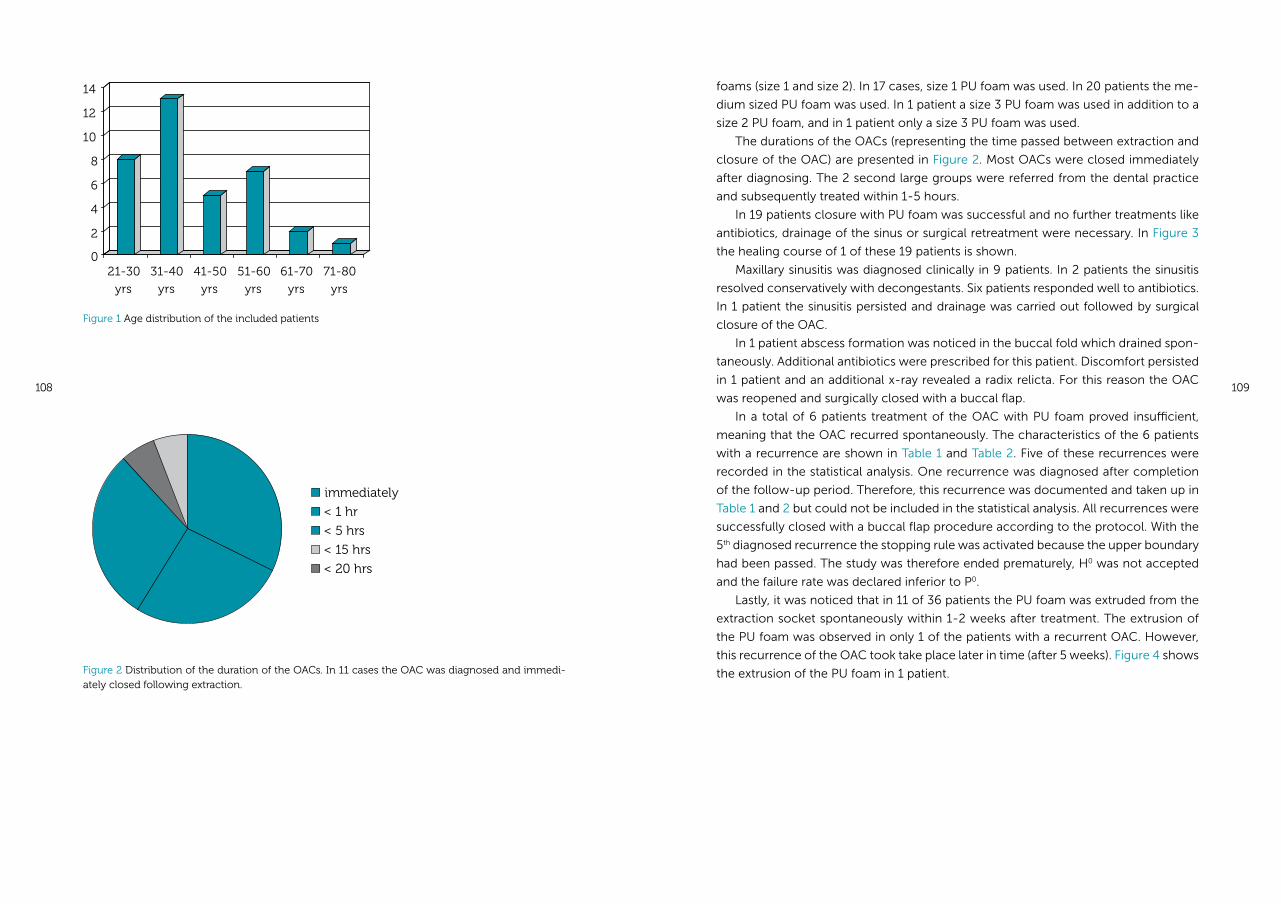
108 109
foams (size 1 and size 2). In 17 cases, size 1 PU foam was used. In 20 patients the me-
dium sized PU foam was used. In 1 patient a size 3 PU foam was used in addition to a
size 2 PU foam, and in 1 patient only a size 3 PU foam was used.
The durations of the OACs (representing the time passed between extraction and
closure of the OAC) are presented in Figure 2. Most OACs were closed immediately
after diagnosing. The 2 second large groups were referred from the dental practice
and subsequently treated within 1-5 hours.
In 19 patients closure with PU foam was successful and no further treatments like
antibiotics, drainage of the sinus or surgical retreatment were necessary. In Figure 3
the healing course of 1 of these 19 patients is shown.
Maxillary sinusitis was diagnosed clinically in 9 patients. In 2 patients the sinusitis
resolved conservatively with decongestants. Six patients responded well to antibiotics.
In 1 patient the sinusitis persisted and drainage was carried out followed by surgical
closure of the OAC.
In 1 patient abscess formation was noticed in the buccal fold which drained spon-
taneously. Additional antibiotics were prescribed for this patient. Discomfort persisted
in 1 patient and an additional x-ray revealed a radix relicta. For this reason the OAC
was reopened and surgically closed with a buccal flap.
In a total of 6 patients treatment of the OAC with PU foam proved insufficient,
meaning that the OAC recurred spontaneously. The characteristics of the 6 patients
with a recurrence are shown in Table 1 and Table 2. Five of these recurrences were
recorded in the statistical analysis. One recurrence was diagnosed after completion
of the follow-up period. Therefore, this recurrence was documented and taken up in
Table 1 and 2 but could not be included in the statistical analysis. All recurrences were
successfully closed with a buccal flap procedure according to the protocol. With the
5th diagnosed recurrence the stopping rule was activated because the upper boundary
had been passed. The study was therefore ended prematurely, H0 was not accepted
and the failure rate was declared inferior to P0.
Lastly, it was noticed that in 11 of 36 patients the PU foam was extruded from the
extraction socket spontaneously within 1-2 weeks after treatment. The extrusion of
the PU foam was observed in only 1 of the patients with a recurrent OAC. However,
this recurrence of the OAC took take place later in time (after 5 weeks). Figure 4 shows
the extrusion of the PU foam in 1 patient.
Figure 1 Age distribution of the included patients
immediately
< 1 hr
< 5 hrs
< 15 hrs
< 20 hrs
Figure 2 Distribution of the duration of the OACs. In 11 cases the OAC was diagnosed and immedi-ately closed following extraction.
0
2
4
6
8
10
12
14
21-30
yrs
31-40
yrs
41-50
yrs
51-60
yrs
61-70
yrs
71-80
yrs
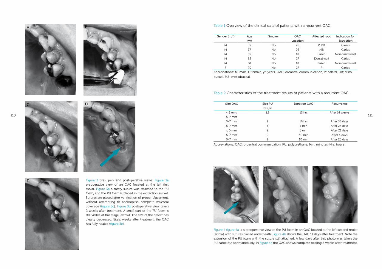
110 111
Table 1 Overview of the clinical data of patients with a recurrent OAC.
Gender (m/f) Age
(yr)
Smoker OAC
Location
Affected root Indication for
Extraction
M 39 No 28 P, DB Caries
M 37 No 26 MB Caries
M 39 No 18 Fused Non-functional
M 52 No 27 Dorsal wall Caries
M 31 No 18 Fused Non-functional
F 70 No 27 P Caries
Abbreviations: M; male, F; female, yr; years, OAC; oroantral communication, P; palatal, DB; disto-
buccal, MB; mesiobuccal.
Table 2 Characteristics of the treatment results of patients with a recurrent OAC
Size OAC Size PU
(1,2,3)
Duration OAC Recurrence
≤ 5 mm,
5-7 mm
1,2 13 hrs After 14 weeks
5-7 mm 2 16 hrs After 38 days
5-7 mm 3 5 min After 24 days
≤ 5 mm 2 5 min After 21 days
5-7 mm 2 30 min After 4 days
5-7 mm 2 10 min After 25 days
Abbreviations: OAC; oroantral communication, PU; polyurethane, Min; minutes, Hrs; hours
Figure 3 pre-, per- and postoperative views. Figure 3a preoperative view of an OAC located at the left first molar. Figure 3b a safety suture was attached to the PU foam, and the PU foam is placed in the extraction socket. Sutures are placed after verification of proper placement, without attempting to accomplish complete mucosal coverage (figure 3c). Figure 3d postoperative view taken 2 weeks after treatment. A small part of the PU foam is still visible at this stage (arrow). The size of the defect has clearly decreased. Eight weeks after treatment the OAC has fully healed (figure 3e).
Figure 4 figure 4a is a preoperative view of the PU foam in an OAC located at the left second molar (arrow) with sutures placed underneath. Figure 4b shows the OAC 11 days after treatment. Note the extrusion of the PU foam with the suture still attached. A few days after this photo was taken the PU came out spontaneously. In figure 4c the OAC shows complete healing 8 weeks after treatment.
A
C
E
B
D
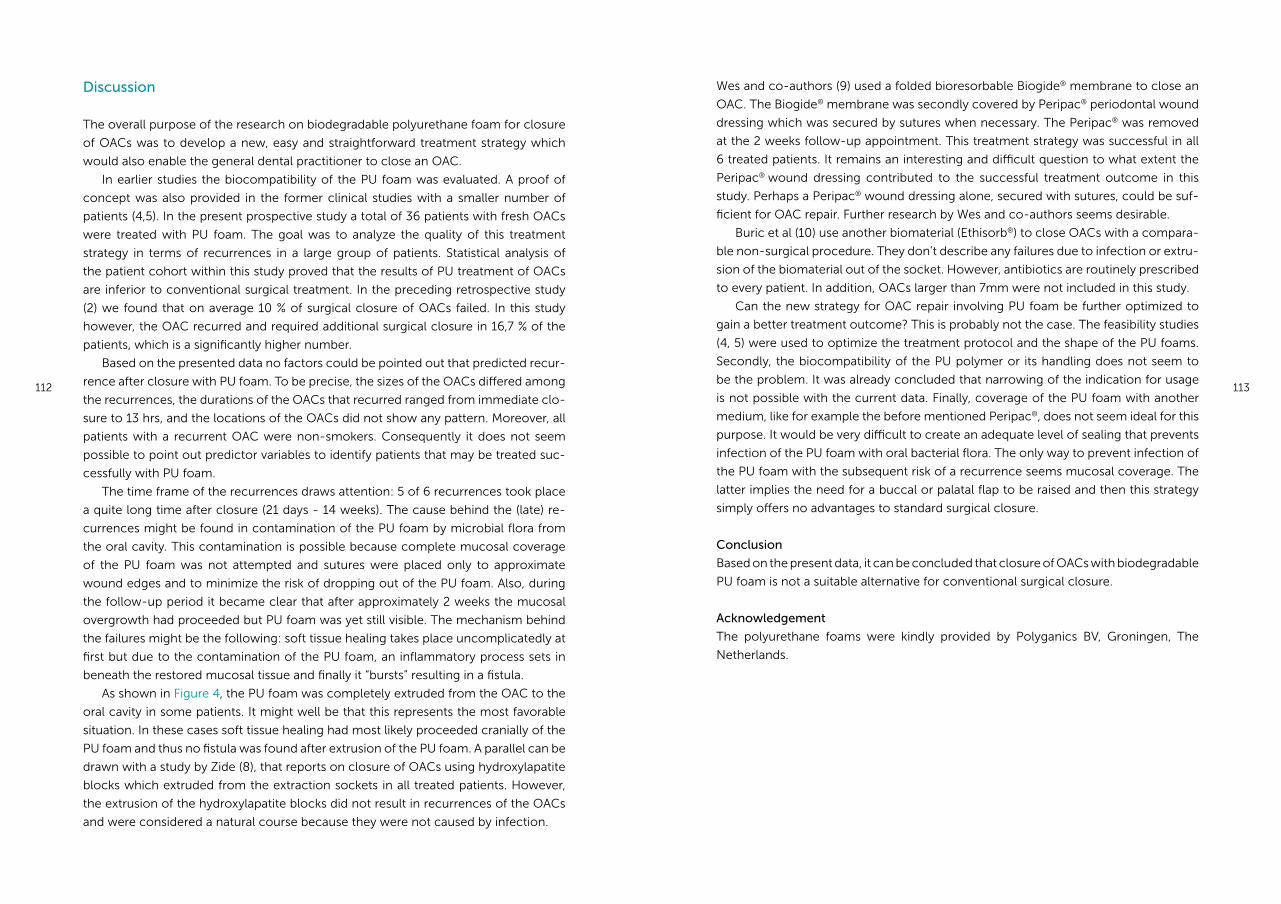
112 113
Wes and co-authors (9) used a folded bioresorbable Biogide® membrane to close an
OAC. The Biogide® membrane was secondly covered by Peripac® periodontal wound
dressing which was secured by sutures when necessary. The Peripac® was removed
at the 2 weeks follow-up appointment. This treatment strategy was successful in all
6 treated patients. It remains an interesting and difficult question to what extent the
Peripac® wound dressing contributed to the successful treatment outcome in this
study. Perhaps a Peripac® wound dressing alone, secured with sutures, could be suf-
ficient for OAC repair. Further research by Wes and co-authors seems desirable.
Buric et al (10) use another biomaterial (Ethisorb®) to close OACs with a compara-
ble non-surgical procedure. They don’t describe any failures due to infection or extru-
sion of the biomaterial out of the socket. However, antibiotics are routinely prescribed
to every patient. In addition, OACs larger than 7mm were not included in this study.
Can the new strategy for OAC repair involving PU foam be further optimized to
gain a better treatment outcome? This is probably not the case. The feasibility studies
(4, 5) were used to optimize the treatment protocol and the shape of the PU foams.
Secondly, the biocompatibility of the PU polymer or its handling does not seem to
be the problem. It was already concluded that narrowing of the indication for usage
is not possible with the current data. Finally, coverage of the PU foam with another
medium, like for example the before mentioned Peripac®, does not seem ideal for this
purpose. It would be very difficult to create an adequate level of sealing that prevents
infection of the PU foam with oral bacterial flora. The only way to prevent infection of
the PU foam with the subsequent risk of a recurrence seems mucosal coverage. The
latter implies the need for a buccal or palatal flap to be raised and then this strategy
simply offers no advantages to standard surgical closure.
Conclusion
Based on the present data, it can be concluded that closure of OACs with biodegradable
PU foam is not a suitable alternative for conventional surgical closure.
Acknowledgement
The polyurethane foams were kindly provided by Polyganics BV, Groningen, The
Netherlands.
Discussion
The overall purpose of the research on biodegradable polyurethane foam for closure
of OACs was to develop a new, easy and straightforward treatment strategy which
would also enable the general dental practitioner to close an OAC.
In earlier studies the biocompatibility of the PU foam was evaluated. A proof of
concept was also provided in the former clinical studies with a smaller number of
patients (4,5). In the present prospective study a total of 36 patients with fresh OACs
were treated with PU foam. The goal was to analyze the quality of this treatment
strategy in terms of recurrences in a large group of patients. Statistical analysis of
the patient cohort within this study proved that the results of PU treatment of OACs
are inferior to conventional surgical treatment. In the preceding retrospective study
(2) we found that on average 10 % of surgical closure of OACs failed. In this study
however, the OAC recurred and required additional surgical closure in 16,7 % of the
patients, which is a significantly higher number.
Based on the presented data no factors could be pointed out that predicted recur-
rence after closure with PU foam. To be precise, the sizes of the OACs differed among
the recurrences, the durations of the OACs that recurred ranged from immediate clo-
sure to 13 hrs, and the locations of the OACs did not show any pattern. Moreover, all
patients with a recurrent OAC were non-smokers. Consequently it does not seem
possible to point out predictor variables to identify patients that may be treated suc-
cessfully with PU foam.
The time frame of the recurrences draws attention: 5 of 6 recurrences took place
a quite long time after closure (21 days - 14 weeks). The cause behind the (late) re-
currences might be found in contamination of the PU foam by microbial flora from
the oral cavity. This contamination is possible because complete mucosal coverage
of the PU foam was not attempted and sutures were placed only to approximate
wound edges and to minimize the risk of dropping out of the PU foam. Also, during
the follow-up period it became clear that after approximately 2 weeks the mucosal
overgrowth had proceeded but PU foam was yet still visible. The mechanism behind
the failures might be the following: soft tissue healing takes place uncomplicatedly at
first but due to the contamination of the PU foam, an inflammatory process sets in
beneath the restored mucosal tissue and finally it “bursts” resulting in a fistula.
As shown in Figure 4, the PU foam was completely extruded from the OAC to the
oral cavity in some patients. It might well be that this represents the most favorable
situation. In these cases soft tissue healing had most likely proceeded cranially of the
PU foam and thus no fistula was found after extrusion of the PU foam. A parallel can be
drawn with a study by Zide (8), that reports on closure of OACs using hydroxylapatite
blocks which extruded from the extraction sockets in all treated patients. However,
the extrusion of the hydroxylapatite blocks did not result in recurrences of the OACs
and were considered a natural course because they were not caused by infection.
114 115
Reference List
(1) Visscher SH, van Minnen B, Bos RR. Closure of oroantral communications: a review of the
literature. J Oral Maxillofac Surg 2010 06;68(1531-5053; 0278-2391; 6):1384-1391.
(2) Visscher SH, van Roon MR, Sluiter WJ, van Minnen B, Bos RR. Retrospective Study on the
Treatment Outcome of Surgical Closure of Oroantral Communications. J Oral Maxillofac Surg
2011 Jul 11.
(3) von Wowern N. Closure of oroantral fistula with buccal flap: Rehrmann versus Moczar. Int J
Oral Surg 1982 06;11(3):156-165.
(4) Visscher SH, van Minnen B, Bos RR. Closure of oroantral communications using biodegradable
polyurethane foam: a feasibility study. J Oral Maxillofac Surg 2010 02;68(1531-5053; 0278-2391;
2):281-286.
(5) Visscher SH, Van Minnen B, Bos RR. Feasibility of conical biodegradable polyurethane foam for
closure of oroantral communications. J Oral Maxillofac Surg 2011 Feb;69(2):390-395.
(6) Visscher SH, van Minnen B, van Leeuwen MB, van Kooten TG, Bos RR. Closure of oroantral
communications using biodegradable polyurethane foam: A long term study in rabbits. J
Biomed Mater Res B Appl Biomater 2009 07/23(1552-4981).
(7) van Minnen B, Stegenga B, van Leeuwen MB, van Kooten TG, Bos RR. Nonsurgical closure of
oroantral communications with a biodegradable polyurethane foam: A pilot study in rabbits. J
Oral Maxillofac Surg 2007 02;65(0278-2391; 2):218-222.
(8) Zide MF, Karas ND. Hydroxylapatite block closure of oroantral fistulas: report of cases. J Oral
Maxillofac Surg 1992 01;50(1):71-75.
(9) Wes JT, Wes MF, Wes B, van Merkesteyn JP. Plastic surgery of oroantral perforations superfluous
in the future? Ned Tijdschr Tandheelkd 2010 Jul-Aug;117(7-8):369-370.
(10) Buric N. Use of N-butyl cyanoacrylate with metacryloxisulfolane (glubran 2) surgical glue for
flapless closure of oroantral communication. Implant Dent 2013 Jun;22(3):238-243.
Chapter 7
General discussion
118 119
General discussion
The research in this thesis focused on the treatment of oroantral communications
(OACs) and the development of a new, non-surgical treatment method of OACs with
a highly porous biodegradable polyurethane foam.
In Chapter 2 it was demonstrated that conventional surgery for closure of OACs
still remains the golden standard, despite the numerous alternative techniques that
have been presented throughout the years. It can be questioned whether there is
a need for a new treatment technique for closure of OACs in the first place, as the
surgical treatment methods are successful in terms of permanent closure of the per-
foration. However, surgical closure of OACs does have its drawbacks, and therefore
research into a non-surgical method of closure is worthwhile. In the current era of
implantology an alternative treatment should not interfere with buccal sulcus depth
and should leave the attached gingival in place as much as possible. In addition, a
non-surgical method should give predictable results, and should be easy and quick to
perform. These requirements would make it possible for a general dentist to treat an
OAC, which is also interesting from a socio-economical point of view.
To determine how successful a new technique should be in terms of recurrences,
literature did not provide enough information. A retrospective study (Chapter 3) was
therefore implemented and demonstrated that on average 1 out of 10 OACs recur
after surgical closure, taking in mind that surgery was performed by either residents
or maxillofacial surgeons. As with all retrospective studies, not all data that might have
been contributory could be retrieved. The method of OAC repair for example was not
explicitly documented in all cases.
A new treatment strategy involving biodegradable polyurethane foam
The research in this thesis focused on the use of fully synthetic polyurethane (PU)
foam for closure of oroantral communications.
Before this PU foam could be used in clinical trials animal experiments were imple-
mented. The OACs that were created in rabbits (Chapter 4) all healed uneventfully.
In the traditional light-microscopic evaluation of the animal experiments the PU
proved to be tissue friendly. A difficult aspect of the histopathological evaluation of
the samples was to determine the completeness of the PU degradation. It was the first
time a degradation study with this PU foam was implemented using time intervals up
to 4 years. This made it difficult to point out the PU remnants after such a long period
of degradation. Structures, that were not recognizable as cellular structures with the
electron microscope, were indicated as PU remnants. As they were repeatedly found
in the different samples and were lacking in the surgically closed defects it is very likely
that several stages of PU degradation were observed. It is, however “circumstantial ev-
idence”. The evidence that the PU is a safe biodegradable material is, however, much
stronger after the experiments presented in this thesis. The results of the long-term
degradation study support the earlier results of the long-term subcutaneous degrada-
tion study (1). Adverse effects in the clinical studies seem related to the application in
the oral environment and not to the biomaterial itself. Nevertheless, if it would have
been possible to follow the degradation and resorption process to the molecular level
the results of biological safety and resorption would have been even more convinc-
ing. It is a lot more difficult to prove whether the remnants are really degradation
products of the PU at the molecular level. Techniques like Raman Spectroscopy (2)
may be useful to detect intracellular particles. However, the fewer remnants present,
the more difficult the detection will be, as the PU is highly porous by itself and the
embedding material will also interfere with the measurements.
Ideally, all of the PU foam had degraded and disappeared such a long time after
treatment but this was clearly not the case. However, the PU remnants did not give
rise to adverse events or other harmful reactions. In addition, a decreasing number
of macrophages with internalized PU and the aspect of the PU foam suggest further
degradation in time to an ultimate end stage.
It was therefore concluded that the animal experiments provided a solid and reli-
able basis to apply the PU foam for closure of OACs in humans.
The application of PU foam for closure of OACs in humans
Two human pilot studies were conducted to determine the usefulness of PU foam for
this application. It became clear that closing OACs with PU foam is feasible although
there were some pitfalls associated with this technique. Dislocation of the PU foam
into the maxillary sinus was observed in the first pilot study. To resolve this, application
of the so called “safety suture” was introduced. This safety suture facilitated removal
of the PU foam when it was accidently pushed into the maxillary sinus. Also, the shape
of the PU foam was changed into a conical contour to improve the fitting of the PU
foam into the socket. These alterations were thought to optimize the treatment strat-
egy as the results obtained in the second pilot study were more favorable.
In the prospective clinical trial it became clear that closure of OACs with PU foam
did not result in an equal or better treatment outcome compared to standard surgical
closure. The type of PU foam tested in this thesis can therefore not be considered a
suitable alternative for conventional surgical closure.
Disappointingly, no factors could be pointed out that predicted a recurrence after
closure with PU foam. To be precise, the sizes of the OACs differed among the recur-
rences, the durations of the OACs that recurred ranged from immediate closure to 13
hours, and the locations of the OACs did not show any pattern. Moreover, all patients
with a recurrent OAC were non-smokers. Consequently it does not seem possible to
point out predictor variables to identify patients that may be treated successfully with
PU foam. It is therefore impossible to narrow the indication criteria for its use.

120 121
Can this new strategy for OAC repair involving PU foam perhaps be further optimized
to gain a better treatment outcome? This is probably not the case. The clinical appli-
cation protocol was already optimized as stated earlier. Also, the biocompatibility of
the PU or its handling does not seem to be the problem. It was already concluded that
narrowing of the indication for usage is not possible with the current data.
The mechanism behind the failures is probably the following: soft tissue healing
takes place uncomplicatedly at first but due to the contamination of the PU foam,
an inflammatory process sets in beneath the restored mucosal tissue and finally it
“bursts” resulting in a fistula. Coverage of the PU foam with another antimicrobial me-
dium, like for example Peripac® wound dressing, was considered. However, it would
be very difficult to create an adequate level of sealing that prevents infection of the
PU foam with oral bacterial flora. The only way to prevent infection of the PU foam
with the subsequent risk of a recurrence seems mucosal coverage. The latter implies
the need for a buccal or palatal flap to be raised and then this new treatment strategy
simply offers no advantages to standard surgical closure.
Future perspectives
Alternative applications of PU foam
The animal experiments and clinical trials in this thesis showed that PU foam is a tissue
friendly and safe material. Currently, fully synthetic PU foam is already being used on
a large scale as a nasal dressing after nasal interventions (NASOPORE®) and as a dres-
sing after ear surgery (OTOPORE®). In these applications the PU foam absorbs fluids,
provides pressure to prevent undesired adhesions and supports the surroun ding tis-
sue. Other surgical areas using biodegradable PU foam include peripheral nerve repair
and colorectal surgery. Also, the use of PU foam as a topical haemostatic agent is
being studied (3-5).
New and promising treatment strategies of OACs
A new method for treatment of OACs that is also usable in general dental practice
would benefit both patients and health costs. The challenge for OAC closure seems
to be the search for a material that provides a balance between an adequate seal to
prevent antimicrobial invasion of the OAC, and at the same time providing space for
bone to regenerate across the defect. The latter is important nowadays because of
the demand for implant rehabilitation. At this moment in time such a product does
not seem to be available although the research by Wes and co-authors (6) seems
the most promising. The authors used a folded bioresorbable Biogide® membrane to
close OACs. The Biogide® membrane was secondly covered by Peripac® periodontal
wound dressing which was secured by sutures when necessary. It remains an interest-
ing and difficult question to what extent the Peripac® wound dressing contributed to
the successful treatment outcome in this study. Perhaps a Peripac® wound dressing
alone, secured with sutures, could be sufficient for OAC repair. Although the Peripac®
might solve the problem of infection, the issue of the size and shape of the defect
remains. It is hardly imaginable that Biogide® can be used to close the larger and
shallower OACs. Further research by Wes and co-authors seems desirable.
Also, the initial experience of an absorbable polyglactin/polydioxanon implant
(Ethisorb®) for closure of OACs seemed encouraging for selected cases as studied by
Buric and co-authors (7). They don’t describe any failures due to infection or extru-
sion of the biomaterial out of the socket. Their treatment protocol is comparable to
the closure with PU foam. As this study is also performed with a foamy biomaterial
one might expect that adjustments to the PU foam may lead to better results in future
experiments.
In this perspective alteration of the chain length of the urethane moieties, may
be considered. The tested polymer was composed of 5 urethane moieties whereas
a length of 3 urethane moieties might lead to other mechanical properties and other
behaviour of the foam. Future research will have to show if these possible changes
in the hard segments can increase the rate of degradation while maintaining the me-
chanical properties of the foam during the healing period of the OAC. Still, the expec-
tations of the use of the three-block PU should not be too high in view of the possible
explanations for the recurrent OACs in the clinical studies. A three-block PU will also
be contaminated in the oral environment and this may cause the same clinical pro-
blems in the healing phase of the OAC.
It is important to note that Buric et al used standard antibiotic therapy in all pa-
tients. In the current era careful prescription of antibiotics is a delicate subject. It is
not likely that the current surgical treatment (not requiring antibiotic profylaxis) will be
replaced by a non-surgical method in combination with standard use of antibio tics.
In addition, it seems that sockets with rather favorable dimensions were treated in the
Buric study. That means that, even with adjustments to the PU, it is not likely that non-
surgical closure can replace surgical treatment of all types and sizes of OACs.
It can be concluded that surgical closure of OACs still is the golden standard, and
perhaps will remain in at least the near future. However it is also known that some
OACs heal spontaneously or with only a tight suture across the alveolus.
One very important and difficult aspect in the choice between surgical and non-
surgical treatment is the size of the OAC. Besides this, the ratio between the depth
and the width of the alveolus and its perforation to the sinus probably plays a role in
the healing. If the coagulum is situated in a shallow and wide alveolus spontaneous
healing will probably not take place. It is challenging to measure the size of the OAC
clinically. The size can be estimated by using metal probes with different diameters (7).
However, exact determination is desirable as it will influence the choice of treatment.
Better measurement of the size of the OAC with for example cone beam CT or the
use of an impression paste might result in a more accurate and less invasive selection
122 123
therapies. If we can predict which OACs can be left untreated or can be treated with a
single suture patients will not get a surgical treatment unnecessarily in future.Reference List
(1) van Minnen B, van Leeuwen MB, Kors G, Zuidema J, van Kooten TG, Bos RR. In vivo resorption
of a biodegradable polyurethane foam, based on 1,4-butanediisocyanate: a three-year
subcutaneous implantation study. J Biomed Mater Res A 2008 06/15;85(1552-4965; 4):972-982.
(2) van Apeldoorn AA, van Manen HJ, Bezemer JM, de Bruijn JD, van Blitterswijk CA, Otto C.
Raman imaging of PLGA microsphere degradation inside macrophages. J Am Chem Soc 2004
Oct 20;126(41):13226-13227.
(3) Broekema FI, van Oeveren W, Zuidema J, Visscher SH, Bos RR. In vitro analysis of polyurethane
foam as a topical hemostatic agent. J Mater Sci Mater Med 2011 Apr;22(4):1081-1086.
(4) Selten MH, Broekema FI, Zuidema J, van Oeveren W, Bos RR. Modified polyurethane foam
as a local hemostatic agent after dental extractions. Ned Tijdschr Tandheelkd 2013 Jul-
Aug;120(7-8):378-382.
(5) Broekema FI, van Oeveren W, Selten MH, Meijer RJ, de Wolf JT, Bos RR. In vivo hemostatic
efficacy of polyurethane foam compared to collagen and gelatin. Clin Oral Investig 2013
May;17(4):1273-1278.
(6) Wes JT, Wes MF, Wes B, van Merkesteyn JP. Plastic surgery of oroantral perforations superfluous
in the future? Ned Tijdschr Tandheelkd 2010 Jul-Aug;117(7-8):369-370.
(7) Buric N. Use of N-butyl cyanoacrylate with metacryloxisulfolane (glubran 2) surgical glue for
flapless closure of oroantral communication. Implant Dent 2013 Jun;22(3):238-243.
Chapter 8a
Summary
126 127
Introduction
Pathogenesis, incidence and treatment of oroantral communications
Oroantral communications (OACs) are open connections between the maxillary sinus
and oral cavity. OACs are usually caused by extraction of maxillary posterior teeth.
Although the incidence is relatively low, OACs are frequently encountered due to the
high number of dental extractions.
Nowadays, closure of OACs is usually performed by means of a buccal or palatal
flap procedure. In case of a small OAC, suturing the gingiva might be sufficient to
close the perforation. It is stated in literature that OACs smaller than 5 mm heal by
themselves. Evidence for this statement is lacking as the size of an OAC is difficult
to measure accurately in the clinical situation. Surgical treatment, preferably within
24 hours, is therefore recommended in many cases to minimize the risk of maxillary
sinusitis and subsequent fistula formation. The selection of the surgical technique de-
pends on the size of the communication, the time of diagnosis and the presence of an
infection. The amount and condition of the tissue available for repair and prosthetic
planning, including the possible placement of dental implants also have influence on
the surgical decisions.
Treatment strategies for oroantral communications
A wide range of surgical treatment strategies for OACs has been established through-
out the years. In Chapter 2 a literature review concerning this topic is presented. The
goal of this review was to establish whether the buccal flap still is the treatment of
choice 20 years after the last review. Secondly, it provided an overview of the com-
mon surgical treatment strategies of OACs, as well as the alternative treatment tech-
niques including their advantages and disadvantages. The treatments were divided into
techniques using autogenous soft tissue flaps or autogenous bone grafts, allogenous
grafts, xenografts and synthetic materials or metals.
It was concluded that only a few techniques to close OACs have gained wide ac-
ceptance. Some proposed new techniques proved to be too expensive. Other alter-
natives did not offer any simplification compared with the standard surgical closure.
Surgical closure of OACs by means of a buccal or palatal flap remains the treatment
of choice.
Success percentages of frequently used surgical techniques can be found in litera-
ture. However, prospective randomized comparative studies that provide evidence for
the superiority of one of the techniques have not been published. Little information
could be found about the (general) complication rate associated with surgical closure
of OACs in terms of recurrence of the OACs. Chapter 3 provides new information
on this subject. A cohort of all patients treated for an OAC in 2004 - 2008 was re-
viewed retrospectively. The recorded data included patient age and gender, location
and duration of the OAC, method of removal of the (pre)molar, presence of maxillary
sinusitis, disturbed wound healing, and the surgical treatment method. Multivariate
regression analysis showed a 15 times higher risk of a recurrence in case of a maxil-
lary sinusitis at the follow-up appointment. The presence of a maxillary sinusitis at the
follow-up appointment can therefore be considered an important determinant of the
treatment outcome of OAC repair.
The overall results of the study showed that in about 1 out of 10 patients the
OAC recurs and requires a second intervention after surgical closure. New treatment
strategies should result in an equal or better treatment outcome to be considered a
suitable alternative to standard surgical treatment.
The development of a new strategy for OAC treatment
As with any surgical procedure, the use of a buccal or palatal flap results in postop-
erative pain and swelling. The use of a buccal flap also has the risk of permanently
decreasing the buccal sulcus depth which could hinder the fitting of a dental prosthe-
sis in the future. Up to now it seems that the drawbacks of surgical closure of OACs
are accepted because useful alternatives are not available. Ideally, a new treatment
method for surgical closure of OACs gives predictable results, and overcomes the
drawbacks of conventional surgical closure. It should be easy and quick to perform,
making it possible for a dentist to treat an OAC by him/herself. The latter would also
be interesting from a socio-economical point of view.
Biodegradable polyurethane (PU) foam is a fully synthetic biodegradable product
that offers the right characteristics for a new treatment strategy for OACs. This highly
porous PU foam has excellent elastic and mechanical properties, allowing adaptation
to the extraction socket.
A new and easy to perform treatment for closure of OACs, using synthetic biode-
gradable polyurethane (PU) foam is presented in Chapter 4a. This PU foam is com-
posed of hard urethane segments, and soft segments made of D/L lactide (50/50),
ε-caprolactone and 5% polyethyleneglycol (PEG). The composition of this specific PU
foam was optimised in preceding in vivo and in vitro studies. To evaluate the use of PU
foam for this application, OACs were created in the edentulous part of the maxilla in 21
New Zealand White rabbits, and subsequently closed with PU foams. Results showed
complete healing of the oral mucosa after 4-10 weeks, healing of the antral mucosal
lining after 6 months and complete bony regeneration after 1 year. No reopening of
the defects occurred and no maxillary sinusitis was observed. Degradation of the PU
foam had not yet reached completion 1 year after implantation. It was concluded that
PU foam with 5% PEG provided adequate closure of an OAC in the rabbit model to
support healing of the oral and maxillary sinus mucosa. Also, it was found that longer
time intervals are needed to assess the complete degradation of the PU foam.
Time intervals up to 4 years (Chapter 4b) demonstrated that the OACs recovered
uncomplicatedly in all rabbits, concerning both soft tissue and bony regeneration.
128 129
The degradation process was not fully completed after 4 years but a decreasing num-
ber of macrophages with internalized PU and the aspect of the internalized PU sug-
gest further degradation in time to an ultimate end stage.
Human studies were implemented for the further development and evaluation of
this new treatment strategy of OACs (Chapter 5 and 6). A feasibility study to assess the
feasibility of biodegradable polyurethane (PU) foam for closure of OACs was carried
out in 10 consecutive patients with fresh oroantral communications (existing < 24 hrs)
(Chapter 5a).
Closure was obtained in 7/10 patients without further surgical intervention. Three
patients developed maxillary sinusitis. Based on this feasibility study it was concluded
that closure of OACs with biodegradable polyurethane foam is feasible. The compli-
cations were possibly related to the fitting of the foam and the size of the defects.
Therefore, a second human pilot study (Chapter 5b) was conducted with 2 altera-
tions made to the treatment protocol. First, the cylindrical shape of the PU foam was
changed into a conical contour. This conically shaped PU foam was thought to im-
prove adaptation to the extraction socket. Secondly, it was decided that a so called
“safety suture” had to be applied to the PU foam prior to its placement into the ex-
traction socket. The latter ensured that the PU foam could easily be removed in case
it had accidently been pushed through the OAC into the maxillary sinus. The conical
PU foam was used in 10 consecutive patients with fresh OACs and permanent closure
was obtained in 8 of 10 patients. The results obtained were more favourable than in
the previous feasibility study and therefore the alterations in the treatment protocol
were maintained for additional research in a large number of patients (Chapter 6). A
prospective clinical trial was set up to analyze the effectiveness of (conical) PU foam
for closure of OACs in terms of recurrences. As stated earlier, treatment of OACs with
a buccal or palatal flap is considered the golden standard and results in a success
percentage of 90%. In order to consider a new treatment strategy for OACs a valid op-
tion, its success percentage should be comparable. It was hypothesized that closure
with PU foam was at least as effective as surgical closure in terms of the percentage
of recurrence.
Thirty six consecutive patients with OACs existing less then 24 hours were treated
with PU foam. In 19 patients the OAC was closed successfully without any compli-
cation. In 16,7 % of the patients the OAC recurred and required additional surgical
closure. Statistical analysis proved that the results of PU treatment of OACs are signifi-
cantly inferior to conventional surgical closure (P < .05). Based on this data it can be
concluded that closure of OACs with biodegradable PU foam is not a suitable alterna-
tive for conventional surgical closure.
In conclusion, it seems that surgical closure of OACs therefore still is the golden
standard, and perhaps will remain in the near future. The reason for this is not its
superiority because there are indeed drawbacks associated with surgical closure as
mentioned above. It seems these drawbacks are taken for granted because new treat-
ment strategies, including the PU closure investigated in this thesis may be quick and
easy to perform, but also have a worse outcome in terms of recurrences and sinusitis.
As for the PU material, this thesis confirms its status of a safe biodegradable mate-
rial, which makes alternative applications of the PU in biomedical implants possible.
In Chapter 7 the results and conclusions from the previous chapters are compared
and discussed.
Chapter 8b
Nederlandse samenvatting (summary in Dutch)
132 133
ting is schaars. Hoofdstuk 3 biedt nieuwe informatie over dit onderwerp. Een retro-
spectieve studie werd geïmplementeerd waarbij het gehele patiëntencohort met
een antrumsluiting in de periode 2004-2008 werd geïncludeerd. Van deze groep
patiënten zijn de leeftijd, geslacht, plaats en bestaansduur van de antrumperforatie,
eventuele gestoorde wondgenezing en de chirurgische behandelmethode gedocu-
menteerd. Multivariate regressie analyse liet een 15 maal hoger risico op een recidief
antrumperforatie zien wanneer er sprake was van een sinusitis maxillaris ten tijde van
de controle afspraak. Een sinusitis maxillaris kan derhalve een belangrijke determinant
van het behandelresultaat beschouwd worden. In algemene zin werd geconcludeerd
dat in ongeveer 1 op de 10 patiënten een recidief antrumperforatie optreedt na chi-
rurgische antrumsluiting waarbij een nieuwe chirurgische interventie noodzakelijk
was. Bij het ontwikkelen van een nieuwe behandelmethode zou het aantal recidief
perforaties vergelijkbaar of lager moeten zijn om een geschikt alternatief genoemd te
kunnen worden.
De ontwikkeling van een nieuwe behandeloptie voor antrumperforaties
Zoals na iedere chirurgische interventie is er ook na sluiting van een antrumperforatie
middels een buccale of palatinale lap sprake van postoperatieve pijn en zwelling. Ver-
der brengt een buccale lap het risico op verstrijking van de buccale omslagplooi met
zich mee. Hierdoor kan eventuele prothetische rehabilitatie in de toekomst bemoei-
lijkt worden. Naar het schijnt worden deze nadelen simpelweg geaccepteerd omdat
er tot op heden nog geen geschiktere behandeling voorhanden is. Idealiter zou een
alternatieve behandeling voorspelbare resultaten geven en bovengenoemde nadelen
ondervangen. Het zou een simpele doeltreffende behandeling moeten zijn die een
algemeen praktiserend tandarts zelf dient te kunnen uitvoeren. Dit laatste is vanzelf-
sprekend ook interessant vanuit sociaal economisch perspectief.
Biodegradeerbaar polyurethaan (PU) schuim is een volledig synthetisch product
dat de juiste eigenschappen lijkt te hebben voor behandeling van antrumperforaties.
Het zeer poreuze schuim heeft uitstekende elastische en mechanische eigenschap-
pen waardoor adaptatie aan de extractie alveole mogelijk is.
In Hoofdstuk 4a wordt een nieuwe en eenvoudige behandeling van antrumper-
foraties middels PU schuim gepresenteerd. Dit PU schuim is opgebouwd uit harde
urethaansegmenten en zachte segmenten van D/L lactide (50/50), ε-caprolacton en 5
% polyethyleenglycol (PEG). De samenstelling van het PU schuim is in voorafgaande in
vitro en in vivo studies geoptimaliseerd. Om de geschiktheid van het materiaal in deze
toepassing te onderzoeken werd in de edentate bovenkaak van 21 New Zealand White
konijnen een antrumperforatie gecreëerd en vervolgens gesloten met PU schuim. Na
4-10 weken bleek de gingiva genezen, de antrale mucosa was hersteld na 6 maanden
en het benige genezingsproces was voltooid na 1 jaar. Er traden geen complicaties
op. Na 1 jaar bleek het PU schuim nog niet volledig te zijn gedegradeerd. Op basis van
deze studie werd vastgesteld dat met PU schuim met 5 % PEG een adequate antrum-
Inleiding
Pathogenese, incidentie en behandelopties van antrumperforaties
Antrumperforaties zijn open verbindingen tussen de neusbijholte en de mondholte.
Doorgaans is een extractie van een (pre)molaar in de bovenkaak de oorzaak van een
antrumperforatie. Doordat extracties op grote schaal worden uitgevoerd worden an-
trumperforaties vrij frequent gezien, ondanks de lage incidentie.
Tegenwoordig worden antrumperforaties in de regel middels een trapeziumvor-
mige buccale lap of palatinale rotatielap gesloten. In het geval van een kleine antrum-
perforatie kan soms worden volstaan met het strak overhechten van de gingiva. In de
literatuur wordt gesteld dat antrumperforaties kleiner dan 5mm spontaan genezen.
Er is echter geen bewijs voor deze aanname aangezien het betrouwbaar klinisch me-
ten van de afmeting van een antrumperforatie lastig is. Chirurgische behandeling, bij
voorkeur binnen 24 uur, is daarom in de meeste gevallen de aangewezen keuze om
het risico op chronische sinusitis maxillaris en fistelvorming te minimaliseren. De keu-
ze ten aanzien van de juiste chirurgische behandeling is afhankelijk van de afmeting
van de antrumperforatie, het moment van diagnosticeren en de eventuele aanwezig-
heid van een infectie. Verder spelen de hoeveelheid en conditie van de omringende
mucosa, eventuele prothetische rehabilitatie en mogelijke wens voor implantologie
nog een belangrijke rol in de behandelkeus.
Behandelopties voor antrumperforaties
In de loop der jaren is een grote verscheidenheid aan behandelopties van antrum-
perforaties in de literatuur gepresenteerd. In Hoofdstuk 2 wordt een review van deze
literatuur beschreven. Het doel van deze review was vast te stellen of behandeling van
antrumperforaties middels een buccale lap nog steeds de eerste keus is, 20 jaar na
het verschijnen van de laatste literatuur studie op dit gebied. Verder resulteerde deze
literatuur studie in een overzicht van alle gangbare chirurgische behandelopties en de
alternatieve behandelingen, inclusief de voor- en nadelen. De alternatieve behande-
lingen werden opgesplitst in technieken waarbij gebruik werd gemaakt van autogene
weke delen, autogene benige transplantaten, allogene transplantaten, xenotransplan-
taten en synthetische materialen of metalen.
Geconcludeerd werd dat er slechts enkele technieken zijn die breed gedragen
worden. Sommige technieken waren te kostbaar. Andere technieken bleken niet een-
voudiger of sneller dan conventionele chirurgische behandeling. Samengevat kan ge-
steld worden dat chirurgische sluiting van antrumperforaties middels een buccale of
palatinale lap nog steeds de gouden standaard is.
Succespercentages van chirurgische behandelingen van antrumperforaties zijn in
de literatuur beschreven. Echter, prospectief gerandomiseerde studies waarin de su-
perioriteit van een specifieke behandeling wordt aangetoond zijn niet voorhanden.
Informatie aangaande de (algemene) complicatieratio van chirurgische antrumslui-
134 135
Samengevat; chirurgische antrumsluiting is nog steeds de gouden standaard, en zal
naar het lijkt dat voorlopig nog blijven. Reden hiervoor is dat er eenvoudigweg geen
geschikter alternatief beschikbaar is.
Dit proefschrift laat zien dat PU schuim een veilig biodegradeerbaar materiaal is
dat geschikt is voor alternatieve toepassingen in biomedische implantaten.
In Hoofdstuk 7 tenslotte worden de resultaten van voorgaande hoofdstukken be-
sproken en aanbevelingen voor de toekomst beschreven.
sluiting gerealiseerd kan worden in een konijnenmodel. Het PU schuim ondersteunt
hierbij de genezing van de orale en antrale mucosa. Langere tijdsintervallen bleken
nodig om volledige degradatie van het PU schuim te kunnen beoordelen.
Tijdsintervallen tot 4 jaar (Hoofdstuk 4b) lieten ongecompliceerde genezing van
de antrumperforaties in alle konijnen zien zowel wat betreft mucosale als benige
regeneratie. Ook na 4 jaar bleek het degradatieproces van het PU schuim nog niet
het eindstadium bereikt te hebben. Echter, een afnemend aantal macrofagen met
ingesloten PU resten en het ontbreken van de kenmerkende poreuze structuur van
het geïnternaliseerde PU suggereren uiteindelijk volledige degradatie.
Humane studies werden geïmplementeerd om deze nieuwe behandeloptie verder
te ontwikkelen en evalueren (Hoofdstuk 5 en 6). Een haalbaarheidsstudie werd uitge-
voerd waarbij in 10 patiënten een verse antrumperforatie (< 24 uur bestaand) middels
PU schuim werd gesloten (Hoofdstuk 5a).
In 7/10 patiënten werd het antrum gesloten met PU schuim zonder noodzaak tot
verdere chirurgische interventie. Drie patiënten ontwikkelden een sinusitis maxillaris.
Op basis van deze haalbaarheidsstudie werd geconcludeerd dat behandeling van
antrumperforaties middels PU schuim mogelijk is. De opgetreden complicaties waren
waarschijnlijk te wijten aan de pasvorm van het PU schuim in de alveole en de afmeting
van de antrumperforaties. Een tweede humane studie werd daarom geïmplementeerd
waarbij 2 belangrijke wijzigingen in het behandelprotocol werden doorgevoerd. Ten
eerste werd de vorm van het PU schuim gewijzigd van cilindrisch in conisch. De ge-
dachte hierbij was dat een conische vorm de adaptatie aan de extractie alveole ten
goede zou komen. Ten tweede diende een “veiligheidshechting” te worden aange-
bracht in het PU schuim om het mogelijk te maken het PU schuim terug te halen in het
geval het onverhoopt in de sinus gedisloceerd werd. Het conische PU schuim werd
vervolgens in 10 patiënten met verse antrumperforaties toegepast. Permanente slui-
ting werd in 8 van de 10 gevallen gerealiseerd en besloten werd daarom de wijzigingen
in het behandelprotocol te handhaven in een prospectieve studie met een groter pa-
tiëntenaantal (Hoofdstuk 6).
Deze prospectieve studie had als doel om het conische PU schuim te beoordelen
op effectiviteit. Zoals eerder aangegeven is chirurgische antrumsluiting middels buc-
cale of palatinale lap de gouden standaard met een succespercentage van 90%. De
volgende hypothese werd daarom geformuleerd: sluiting van antrumperforaties met
PU schuim is minimaal even effectief qua succespercentage als chirurgische sluiting.
Zesendertig opeenvolgende patiënten met verse antrumperforaties werden be-
handeld met PU schuim. In 19 patiënten verliep de behandeling zonder complicaties.
In 16,7% van de patiënten trad een recidief antrumperforatie op die een nieuwe chirur-
gische interventie noodzakelijk maakte. Statistische analyse toonde aan dat antrum-
sluiting met PU schuim significant inferieur is aan conventionele chirurgische sluiting
(p <.05). Op basis van deze data kon geconcludeerd worden dat antrumsluiting middels
PU schuim geen geschikt alternatief is voor de huidige chirurgische behandelmethode.
136 137
Dankwoord
Ik heb het altijd enorm naar mijn zin gehad op de afdeling Mondziekten en Kaakchi-
rurgie en vind het nog steeds een prachtig specialisme. Het moederschap bracht mij
echter ertoe mijn bakens te verzetten. Een moeilijke beslissing maar één waar ik nog
steeds achter sta. Ik ben dankbaar voor het begrip en de positieve reacties die ik van
collega’s heb ontvangen na het kenbaar maken van mijn keuze om niet in opleiding te
gaan en voor het tandartsvak te kiezen.
En nu zit er op! Het proefschrift is klaar. Het heeft enige tijd in de koelkast gelegen
maar de intentie om het af te ronden is altijd aanwezig geweest. Dankzij de hulp van
velen is het nu toch zover. Iedereen die mij hierbij op welke manier dan ook heeft
geholpen: ontzettend bedankt!
Een aantal mensen wil ik graag in het bijzonder noemen;
Prof. dr. R.R.M. Bos, beste Ruud, vanaf het begin heb ik een goed gevoel gehad bij jou.
Je prettige manier van communiceren en het meteen aanpakken als er iets geregeld
moet worden zijn daar slechts voorbeelden van. Je hebt me verder altijd de ruimte
gegeven om op mijn manier het onderzoek te doen en hierdoor gaf je me veel ver-
trouwen. Ik ben je zeer dankbaar voor het feit dat je mijn promotor hebt willen zijn en
hoop dat je na je afscheid enorm gaat genieten met Liesbeth. Bedankt!
Dr. B. van Minnen, beste Baucke. Ik heb het stokje van jou overgenomen en het po-
lyurethaan onderzoek mogen voortzetten. Ik bof met jou als co-promotor. Want af-
gezien van je onderzoeksinhoudelijke kennis en hulp ben je ook nog eens een heel
prettige collega. Ik heb respect voor hoe jij privé en werk hebt weten te combineren
en je hebt daarmee in mijn ogen ook voor anderen de weg vrijgemaakt. Dank voor je
hulp in de breedste zin van het woord!
Prof. dr. L.G.M. de Bont, beste professor, in de tijd dat ik begon met het onderzoek was
u ons afdelingshoofd. U was dan ook diegene die me de mogelijkheid gaf de tandheel-
kunde te combineren met het promotie onderzoek. Daar ben ik u zeer erkentelijk voor.
Het wonen op het Groningse Hogeland hadden we gemeen. Ik vond het erg leuk te
hebben mogen zien hoe u woont en geniet van het buitenleven.
Prof. dr. F.K.L. Spijkervet, beste Fred, we hebben niet veel direct samengewerkt maar
desondanks heb ik ons contact altijd gewaardeerd. Mijn dank ook aan jou voor de
mogelijkheid om in mijn eigen tempo het onderzoek af te ronden.
De leden van de beoordelingscommissie, prof dr. van Merkesteyn, prof dr. Rozema en
prof. dr. G.M. Raghoebar; dank voor jullie beoordeling van mijn proefschrift.
138 139
Dr. A. van Leeuwen, beste Anne. Wij waren partners in crime tijdens onze tandheel-
kunde studie. Aan je kenmerkende humor moest ik eerst wennen maar die ben ik
enorm gaan waarderen. Het is jammer dat je niet bij mijn promotie aanwezig kan zijn
maar ik wil je via deze weg bedanken voor de leuke en gezellige jaren die we samen
als collega’s hebben gehad.
Dr. SAHJ de Visscher, beste Sebastiaan, we lijken niet alleen qua achternaam op el-
kaar. Ik heb met jou vaak het gevoel gehad dat we op een lijn zaten. Ik kijk dan ook
met een goed gevoel terug op ons contact. Ik wens je veel succes en geluk in je werk
en privé.
Dr. R. Pollard, beste Rodney, ook jij was een onderzoekskamergenoot. En ook jij hebt
na een hele turbulente periode in je leven besloten een andere weg te kiezen. Ik wens
je heel veel succes en geluk toe en bedankt voor de leuke tijd samen op de afdeling.
Dr. J.M. Meijer, beste Jiska, we hebben een hele tijd een onderzoekskamer gedeeld
wat heeft geresulteerd in een mooie vriendschap. We zien elkaar niet vaak meer, maar
als we elkaar zien pakken we de draad zo weer op! Ik denk met veel plezier terug aan
onze tijd samen op de afdeling. Bedankt daarvoor.
Lieve Riet, bedankt voor je interesse en de mogelijkheid om eens bij jou bij te tanken
en te werken aan mijn proefschrift. En lieve Jan, we missen je nog iedere dag...maar
wat ben ik blij dat ik je nog heb mogen leren kennen. We zullen je herinnering altijd
levend houden.
Lieve papa en mama, het was nogal een omweg om tandarts te worden maar door
jullie steun ben ik letterlijk en figuurlijk op een plek beland waar ik heel graag ben.
Jullie hebben me altijd gestimuleerd om ondanks het vele uitloten door te zetten en
dat heeft geresulteerd in 3 afgeronde universitaire opleidingen en een promotie. Dank
voor jullie niet aflatende steun en de mogelijkheid om mijn eigen keuzes te maken. Ik
houd van jullie!
Lieve Bas, Bart en Noortje, het proefschrift is klaar!! Het waren een paar bewogen ja-
ren voor ons viertjes met veel hoogtepunten en met als absoluut dieptepunt het veel
te jong overlijden van onze opa en (schoon-)vader Jan Langeveld.
Lieve Bas, je bent mijn absolute steun, toeverlaat en grote liefde. Als ik weer eens
doordender trek jij aan de rem. En of het nu de keuze is om niet in opleiding te gaan
of om wedstrijdrijdster te worden. Je steunt me altijd in alles. Dank voor je hulp in de
breedste zin van het woord. Ik houd ontzettend veel van jou.
Lieve Bart en Noortje, jullie komst heeft ons leven flink op de kop gezet maar in
Dr. G. Telleman, lieve Gerdien, in 2012 mocht ik jouw paranimf zijn en wat fijn dat je
nu naast mij staat. Ik ben erg blij dat je mijn collega en vriendin bent. Gelukkig blijken
de kilometers tussen Sneek en Holten niet van invloed op onze vriendschap. Samen
nascholing volgen in Zwolle is ook een hele goede manier gebleken om het nuttige
met het gezellige te combineren. Mede dankzij jouw enthousiasme en aanmoediging
is het proefschrift nu klaar. Dank je wel dat je mijn paranimf wilt zijn.
Drs. E.C. Visscher, lieve Ellen, als grote zus ken jij mij als geen ander. Ondanks dat we
enorm verschillend zijn delen we toch ook wel het een en ander. Je begrijpt vaak
precies wat ik bedoel en je bent er 24/7 voor mij. Ik heb altijd het gevoel gehad dat je
trots bent op mij en me alles gunt, of ik nou afstudeerde of een triatlon won. Ik ben erg
blij dat je mijn zus bent en vandaag mijn paranimf wil zijn. En nu niet gaan huilen hè!
Beste werknemers van het CDL en in het bijzonder Arie Nijmeijer. Jullie stonden mede
aan de basis van dit onderzoek. We hebben heel wat uren met elkaar doorgebracht op
het lab. Ik heb de samenwerking met jullie altijd als heel prettig ervaren. Jullie goede
zorg voor de proefdieren en de bereidwilligheid om te helpen heb ik enorm gewaar-
deerd. En Arie, onze gesprekken gingen over van alles en nog wat, daar denk ik met
veel plezier aan terug. Dank voor jullie hulp en tijd.
Mevr. ing. M.B.M. van Leeuwen, beste Babs. Je bent van onschatbare waarde geweest
voor de histologie van de konijnenstudies. Bedank voor je professionele bijdrage.
Dr. W. J. Sluiter, beste Wim, jij bent diegene die me heeft geholpen met de statistische
kant van de grote humane studie. Je hebt uitgebreid de tijd genomen om mij wegwijs
te maken in deze lastige materie. Dank voor je hulp.
Beste werknemers van Polyganics. Jullie zijn diegene die het PU schuim produceer-
den waar dit proefschrift om draait. Alles was altijd goed geregeld en voor elkaar. Ik
kijk positief op onze samenwerking terug. Dank daarvoor!
Ing. J. Zuidema, beste Johan. Jij bent destijds bij Polyganics mijn aanspreekpunt ge-
weest. We hebben heel wat uren samengewerkt Ik hoop dat onze wegen elkaar nog
weer eens kruisen, bv fietsend of bij een concert van Racoon. Dank voor je hulp.
Beste Lisa, Nienke, Karin en Harry, dank voor jullie interesse, gezelligheid en onder-
steuning!
Beste collega-onderzoekers, en in het bijzonder Ferdinand, Willem, Petra, Madelon,
Esther en Nico. We hebben samen heel wat tijd doorgebracht op de 3e verdieping. Ik
kijk met veel plezier terug op het bij elkaar binnenlopen voor hulp of een luisterend oor.
140
positieve zin. Ik geniet er enorm van om jullie te zien opgroeien en ben trots dat ik
jullie mama ben!