Embed Size (px)
Citation preview
Case ReportStaphylococcus aureus Myocarditis with Associated LeftVentricular Apical Thrombus
Michael McGee ,1 Emily Shiel,2 Stephen Brienesse,1,3 Stuart Murch,1 Robert Pickles,2,3
and James Leitch1,3
1Cardiovascular Department, John Hunter Hospital, Lookout Road, New Lambton Heights, NSW 2305, Australia2Infectious Diseases Department, John Hunter Hospital, Lookout Road, New Lambton Heights, NSW 2305, Australia3Department of Medicine, University of Newcastle, Newcastle, NSW, Australia
Correspondence should be addressed to Michael McGee; [email protected]
Received 7 April 2018; Accepted 16 May 2018; Published 23 May 2018
Academic Editor: Ertugrul Ercan
Copyright © 2018 Michael McGee et al. This is an open access article distributed under the Creative Commons Attribution License,which permits unrestricted use, distribution, and reproduction in any medium, provided the original work is properly cited.
Staphylococcus aureus myocarditis is a rare diagnosis with a high mortality rate, usually seen in people who areimmunocompromised. Here, we report a case of a 44-year-old man on methotrexate for rheumatoid arthritis who presented inseptic shock and was diagnosed with staphylococcus aureus myocarditis. The myocarditis was associated with a left ventricularapical thrombus, with normal systolic function. The myocarditis and associated thrombus were characterised on transthoracicechocardiogram and subsequently on cardiac magnetic resonance imaging. Cardiac magnetic resonance (CMR) imaging showedoedema in the endomyocardium, consistent with acute myocarditis, associated with an apical mural thrombus. Repeat CMR 3weeks following discharge from hospital showed marked improvement in endomyocardial oedema and complete resolution ofthe apical mural thrombus. He was treated with a 12-week course of antibiotics and anticoagulated with apixaban. The patientwas successfully managed with intravenous antibiotics and anticoagulation with complete recovery.
1. Introduction
Staphylococcus aureus is widely reported to be the mostcommon bacterial cause of myocarditis and usually occursin the setting of bacteraemia and sepsis. Rarely, it occurswithout associated infective endocarditis [1, 2]. Furthermore,difficulty in determining the diagnosis, prevalence, andaetiology of myocarditis is complicated by the infrequentuse of endomyocardial biopsy (EMB), the diagnostic goldstandard [2, 3].
Noninvasive imaging such as cardiac magnetic resonance(CMR) imaging has been shown to be reliable in the diagno-sis and monitoring of disease progression in acute myocardi-tis [4, 5]. Despite the advances of noninvasive imaging, thesensitivity and specificity of CMR for acute myocarditis arereported at 81% and 71% and 63% and 40% in chronicmyocarditis [6]. Ideally, CMR and EMB are both obtainableand are complimentary, overcoming limitations of eithertechnique alone [4].
Here, we present a case of Staphylococcus aureusmyocar-ditis with associated left ventricular apical thrombus.
2. Case History
A 44-year-old male was admitted to the intensive care unit(ICU) critically unwell with septic shock. On the day priorto admission, he had complained of lethargy, diffuse bodyaches, epigastric pain, nausea, and vomiting and had expe-rienced rigors. He had a medical background significant forrheumatoid arthritis, treated withmethotrexate 10mg weeklyfor the last five years and had recently travelled to Fiji.
On arrival of paramedics, the patient was nonresponsive,febrile, and tachycardic, with an unrecordable blood pressureand mottled appearance of the skin. He was drowsy andconfused on arrival to the emergency department and hadprolonged capillary refill of five seconds, a lactate of 6.8. Therewas no clear focus of infection on examination, and cardio-vascular examination was normal at this time. He was found
HindawiCase Reports in CardiologyVolume 2018, Article ID 7017286, 4 pageshttps://doi.org/10.1155/2018/7017286
to have an acute kidney injury, hepatic impairment, and coag-ulopathy, see Table 1. Blood cultures returned positive in 11hours for methicillin-sensitive Staphylococcus aureus, andantibiotics were changed to intravenous (IV) flucloxacillin.He was culture positive for 48 hours.
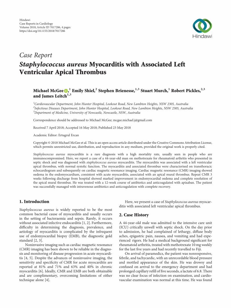
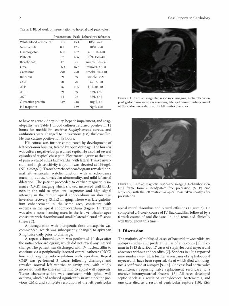
His course was further complicated by development ofleft olecranon bursitis, treated by open drainage. The bursitiswas culture negative but presumed septic. He also had severalepisodes of atypical chest pain. Electrocardiogram at the timeof pain revealed sinus tachycardia, with lateral T wave inver-sion, and high-sensitivity troponin was elevated at 139ng/L(NR< 26ng/L). Transthoracic echocardiogram revealed nor-mal left ventricular systolic function, with an echo-densemass in the apex, no valvular abnormality, and mild left atrialdilatation. The patient proceeded to cardiac magnetic reso-nance (CMR) imaging which showed increased wall thick-ness in the mid to apical wall segments and high signalintensity in the mid to apical endocardium on short tauinversion recovery (STIR) imaging. There was late gadolin-ium enhancement in the same area, consistent withoedema in the apical endomyocardium (Figure 1). Therewas also a nonenhancing mass in the left ventricular apexconsistent with thrombus and small bilateral pleural effusions(Figure 2).
Anticoagulation with therapeutic dose enoxaparin wascommenced, which was subsequently changed to apixaban5mg twice daily prior to discharge.
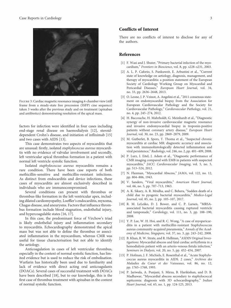
A repeat echocardiogram was performed 10 days afterthe initial echocardiogram, which did not reveal any intervalchange. The patient was discharged with IV flucloxacillin tocontinue via a peripherally inserted central catheter (PICC)line and ongoing anticoagulation with apixaban. RepeatCMR was performed 3 weeks following discharge andrevealed normal left ventricular cavity size, with mildlyincreased wall thickness in the mid to apical wall segments.Tissue characterisation was consistent with apical walloedema, which had reduced significantly in size since the pre-vious CMR, and complete resolution of the left ventricular
apical mural thrombus and pleural effusions (Figure 3). Hecompleted a 6-week course of IV flucloxacillin, followed by a6-week course of oral dicloxacillin, and remained clinicallywell throughout this time.
3. Discussion
The majority of published cases of bacterial myocarditis areautopsy studies and predate the use of antibiotics [1]. Flax-man in 1943 described 17 cases of staphylococcal myocardialabscesses without endocarditis [7]. Sanders in 1963 reportednine similar cases [8]. A further seven cases of staphylococcalmyocarditis have been reported, six of which died with diag-nosis confirmed at autopsy [9–14]. One case had aortic valveinsufficiency requiring valve replacement secondary to amassive intramyocardial abscess [15]. All cases developedseptic shock as a result of staphylococcal bacteraemia, andone case died as a result of ventricular rupture [10]. Risk
Table 1: Blood work on presentation to hospital and peak values.
Presentation Peak Laboratory reference
White blood cell count 12.5 15.4 109/L 4–11
Neutrophils 8.2 12.7 109/L 2–8
Haemoglobin 162 162 g/L 130–180
Platelets 87 466 109/L 150–400
Bicarbonate 17 25 mmol/L 22–32
Urea 16.3 16.3 mmol/L 3.5–8
Creatinine 290 290 μmol/L 60–110
Bilirubin 69 69 μmol/L< 20GGT 70 70 U/L 5–50
ALP 76 105 U/L 30–100
ALT 69 69 U/L< 50AST 74 92 U/L< 45C-reactive protein 339 348 mg/L< 5HS troponin 139 Ng/L< 26
Figure 1: Cardiac magnetic resonance imaging 4-chamber-viewpost gadolinium injection revealing late gadolinium enhancementof the endomyocardium at the left ventricular apex.
Figure 2: Cardiac magnetic resonance imaging 4-chamber view(still frame from a steady-state free precession (SSFP) cinesequence) with the left ventricular apical mass taken shortly afterpresentation.
2 Case Reports in Cardiology
factors for infection were identified in four cases includingend-stage renal disease on haemodialysis [12], steroid-dependent Crohn’s disease, and initiation of infliximab [15]and two cases with AIDS [13].
This case demonstrates two aspects of myocarditis thatare unusual: firstly, isolated staphylococcus aureus myocardi-tis with no evidence of valvular involvement and secondly,left ventricular apical thrombus formation in a patient withnormal left ventricle systolic function.
Isolated staphylococcus aureus myocarditis remains arare condition. There have been case reports of bothmethicillin-sensitive and methicillin-resistant infections.As distinct from endocarditis and device infections, thesecases of myocarditis are almost exclusively described inindividuals who are immunocompromised.
Several conditions can present with thrombus orthrombus-like formation in the left ventricular apex, includ-ing dilated cardiomyopathy, Loeffler’s endocarditis, myxoma,Chagas disease, and aneurysms. Factors that influence throm-bus formation include blood stagnation, endothelial injury,and hypercoagulable states [16, 17].
In this case, the predominant force of Virchow’s triadis likely endothelial injury and inflammation secondaryto myocarditis. Echocardiography demonstrated the apicalmass but was not able to define the thrombus or associ-ated inflammation in the myocardium, whereas CMR wasuseful for tissue characterisation but not able to identifythe aetiology.
Anticoagulation in cases of left ventricular thrombus,especially in the context of normal systolic function, has lim-ited evidence but is used to reduce the risk of embolisation.Warfarin has historically been used due to familiarity andlack of evidence with direct acting oral anticoagulants(DOACs). Several cases of successful treatment with DOACshave been described [18], but to our knowledge, this is thefirst case of thrombus treatment with apixaban in the contextof normal systolic function.
Conflicts of Interest
There are no conflicts of interest to disclose for any ofthe authors.
References
[1] F. Wasi and J. Shuter, “Primary bacterial infection of the myo-cardium,” Frontiers in Bioscience, vol. 8, pp. s228–s231, 2003.
[2] A. L. P. Caforio, S. Pankuweit, E. Arbustini et al., “Currentstate of knowledge on aetiology, diagnosis, management, andtherapy of myocarditis: a position statement of the EuropeanSociety of Cardiology Working Group on Myocardial andPericardial Diseases,” European Heart Journal, vol. 34,no. 33, pp. 2636–2648, 2013.
[3] O. Leone, J. P. Veinot, A. Angelini et al., “2011 consensus state-ment on endomyocardial biopsy from the Association forEuropean Cardiovascular Pathology and the Society forCardiovascular Pathology,” Cardiovascular Pathology, vol. 21,no. 4, pp. 245–274, 2012.
[4] H. Baccouche, H. Mahrholdt, G. Meinhardt et al., “Diagnosticsynergy of non-invasive cardiovascular magnetic resonanceand invasive endomyocardial biopsy in troponin-positivepatients without coronary artery disease,” European HeartJournal, vol. 30, no. 23, pp. 2869–2879, 2009.
[5] M. Gutberlet, B. Spors, T. Thoma et al., “Suspected chronicmyocarditis at cardiac MR: diagnostic accuracy and associa-tion with immunohistologically detected inflammation andviral persistence,” Radiology, vol. 246, no. 2, pp. 401–409, 2008.
[6] P. Lurz, I. Eitel, J. Adam et al., “Diagnostic performance ofCMR imaging compared with EMB in patients with suspectedmyocarditis,” JACC: Cardiovascular Imaging, vol. 5, no. 5,pp. 513–524, 2012.
[7] N. Flaxman, “Myocardial Abscess,” JAMA, vol. 122, no. 12,pp. 804–806, 1943.
[8] V. Sanders, “Viral myocarditis,” American Heart Journal,vol. 66, no. 5, pp. 707–713, 1963.
[9] A. K. Sikary, A. R. Mridha, and C. Behera, “Sudden death of achild due to pyogenic bacterial myocarditis,” Medico-LegalJournal, vol. 85, no. 2, pp. 105–107, 2017.
[10] R. M. LeLeiko, D. J. Bower, and C. P. Larsen, “MRSA-associated bacterial myocarditis causing ruptured ventricleand tamponade,” Cardiology, vol. 111, no. 3, pp. 188–190,2008.
[11] Y. P. Lee, W. H. Hoi, and R. C. Wong, “A case of myopericar-ditis in a patient with methicillin-resistant Staphylococcusaureus community-acquired pneumonia,” Annals of the Acad-emy of Medicine, Singapore, vol. 37, no. 3, pp. 243–242, 2008.
[12] B. Khan, R. W. Strate, and R. Hellman, “ASDIN Original Inves-tigations: Myocardial abscess and fatal cardiac arrhythmia in ahemodialysis patient with an arterio-venous fistula infection,”Seminars in Dialysis, vol. 20, no. 5, pp. 452–454, 2007.
[13] P. Hofman, J. F. Michiels, E. Rosenthal et al., “Acute Staphylo-coccus aureus myocarditis in AIDS. 2 cases,” Archives desMaladies du Coeur et des Vaisseaux, vol. 86, no. 12,pp. 1765–1768, 1993.
[14] P. Jariwala, A. Punjani, S. Mirza, B. Harikishan, and D. B.Madhawar, “Myocardial abscess secondary to staphylococcalsepticemia: diagnosis with 3D echocardiography,” IndianHeart Journal, vol. 65, no. 1, pp. 124-125, 2013.
Figure 3: Cardiacmagnetic resonance imaging 4-chamber view (stillframe from a steady-state free precession (SSFP) cine sequence)taken 3 weeks after the previous study and on treatment (apixabanand antibiotics) demonstrating resolution of the apical mass.
3Case Reports in Cardiology
[15] P. Reichardt, I. Dahnert, G. Tiller, and H. J. Hausler, “Possibleactivation of an intramyocardial inflammatory process(Staphylococcus aureus) after treatment with infliximab ina boy with Crohn disease,” European Journal of Pediatrics,vol. 161, no. 5, pp. 281–283, 2002.
[16] R. Delewi, F. Zijlstra, and J. J. Piek, “Left ventricular thrombusformation after acute myocardial infarction,” Heart, vol. 98,no. 23, pp. 1743–1749, 2012.
[17] S. Solheim, I. Seljeflot, K. Lunde et al., “Prothrombotic markersin patients with acute myocardial infarction and left ventricu-lar thrombus formation treated with pci and dual antiplatelettherapy,” Thrombosis Journal, vol. 11, no. 1, p. 1, 2013.
[18] Y. Mano, K. Koide, H. Sukegawa, M. Kodaira, and T. Ohki,“Successful resolution of a left ventricular thrombus with apix-aban treatment following acute myocardial infarction,” Heartand Vessels, vol. 31, no. 1, pp. 118–123, 2016.
4 Case Reports in Cardiology

Stem Cells International
Hindawiwww.hindawi.com Volume 2018
Hindawiwww.hindawi.com Volume 2018
MEDIATORSINFLAMMATION
of
EndocrinologyInternational Journal of
Hindawiwww.hindawi.com Volume 2018
Hindawiwww.hindawi.com Volume 2018
Disease Markers
Hindawiwww.hindawi.com Volume 2018
BioMed Research International
OncologyJournal of
Hindawiwww.hindawi.com Volume 2013
Hindawiwww.hindawi.com Volume 2018
Oxidative Medicine and Cellular Longevity
Hindawiwww.hindawi.com Volume 2018
PPAR Research
Hindawi Publishing Corporation http://www.hindawi.com Volume 2013Hindawiwww.hindawi.com
The Scientific World Journal
Volume 2018
Immunology ResearchHindawiwww.hindawi.com Volume 2018
Journal of
ObesityJournal of
Hindawiwww.hindawi.com Volume 2018
Hindawiwww.hindawi.com Volume 2018
Computational and Mathematical Methods in Medicine
Hindawiwww.hindawi.com Volume 2018
Behavioural Neurology
OphthalmologyJournal of
Hindawiwww.hindawi.com Volume 2018
Diabetes ResearchJournal of
Hindawiwww.hindawi.com Volume 2018
Hindawiwww.hindawi.com Volume 2018
Research and TreatmentAIDS
Hindawiwww.hindawi.com Volume 2018
Gastroenterology Research and Practice
Hindawiwww.hindawi.com Volume 2018
Parkinson’s Disease
Evidence-Based Complementary andAlternative Medicine
Volume 2018Hindawiwww.hindawi.com
Submit your manuscripts atwww.hindawi.com