Embed Size (px)
Citation preview
Hindawi Publishing CorporationCase Reports in OtolaryngologyVolume 2012, Article ID 814696, 8 pagesdoi:10.1155/2012/814696
Case Report
Benign Intracranial Hypertension: A Diagnostic Dilemma
Gary Y. Shaw and Stephanie K. Million
Kansas City University of Medicine and Biomedical Sciences, Kansas City, MO 64086, USA
Correspondence should be addressed to Gary Y. Shaw, [email protected]
Received 1 April 2012; Accepted 12 July 2012
Academic Editors: A. Rapoport and S. Ulualp
Copyright © 2012 G. Y. Shaw and S. K. Million. This is an open access article distributed under the Creative Commons AttributionLicense, which permits unrestricted use, distribution, and reproduction in any medium, provided the original work is properlycited.
Benign intracranial hypertension (BIH) (also known as pseudotumor cerebri and empty sella syndrome) remains a diagnosticchallenge to most physicians. The modified Dandy criteria consist of, the classic findings of headache, pulsatile tinnitus,papilledema, and elevated cerebrospinal fluid (CSF) pressure, however, these are rarely collectively present in any one patient.Furthermore, these findings can wax and wane over time. Due to the nature of this disease, both signs and symptoms may beintermittent, making definitive diagnosis difficult. Newer imaging studies, particularly the magnetic resonance venogram (MRV)along with a constellation of correlative findings and associated diseases have given new impetus in the diagnosis, treatment, andpathophysiology of this disease. This has led the authors to offer modifications to the classic Dandy criteria. This report presentsthree representative cases of BIH highlighting many of the newer advances in both diagnosis and treatment of this perplexingdisorder.
1. Introduction
In 1937, Dandy presented a report of 22 patients with ele-vated intracranial pressure not attributed to brain tumors.All of these patients presented with headaches, most alsocomplained of blurred vision, dizziness, vomiting, anddrowsiness. The author made mention of many other signsand symptoms that were experienced by these patientsincluding buzzing in the ears, funduscopic abnormalities,stumbling gate, episodic numbness, and drowsiness, to namea few. In this document Dandy explains that the elevationsin pressure seem to come and go over time, and are rarelyconstant. From this report arose the original Dandy criteriafor the diagnosis of benign intracranial hypertension. Theoriginal criteria was modified by Smith in 1985, and iscurrently known as the revised Dandy criteria (1–8), whichhas been uniformly adopted as a diagnostic paradigm forBIH. The current Dandy criteria are: (1) signs and symptomsof increased intracranial pressure; (2) no other neurologicalabnormalities or impaired level of consciousness (with theexception of CN VI palsy); (3) elevated intracranial pressure(ICP) w/normal CSF composition; (4) a computed tomog-raphy (CT) scan which shows no etiology for increased ICP(the original Dandy criteria required ventriculography); (5)
no other cause for intracranial hypertension found [1, 2].These modified criteria fail to mention that the elevation inpressure and the symptoms may wax and wane or give clearexamples of the wide array of symptoms that this disordermay present with. Furthermore, newer imaging studies,particularly the Magnetic Resonance Venogram (MRV) andupdated treatment methods have given new impetus inthe study of this disease. In light of these developmentsthe authors feel that further modifications to the Dandycriteria are in order. This paper presents three representativeexamples of BIH highlighting many of the newer advances inboth diagnosis and treatment of this perplexing disorder andoffers salient modifications to the traditional Dandy criteria.
2. Patient Number 1
A 26-year-old, mildly obese Caucasian female presentedwith a 6-month history of pulsatile tinnitus, right maxillarypressure and pain, and blurred vision in her peripherybilaterally followed by intense, debilitating, daily headacheswhich extended to her frontal areas bilaterally. The patientdenied any trauma associated with the onset of her symp-toms. She had been seen by two different physicians recentlyfor this problem; and treated with a course of antibiotics
2 Case Reports in Otolaryngology
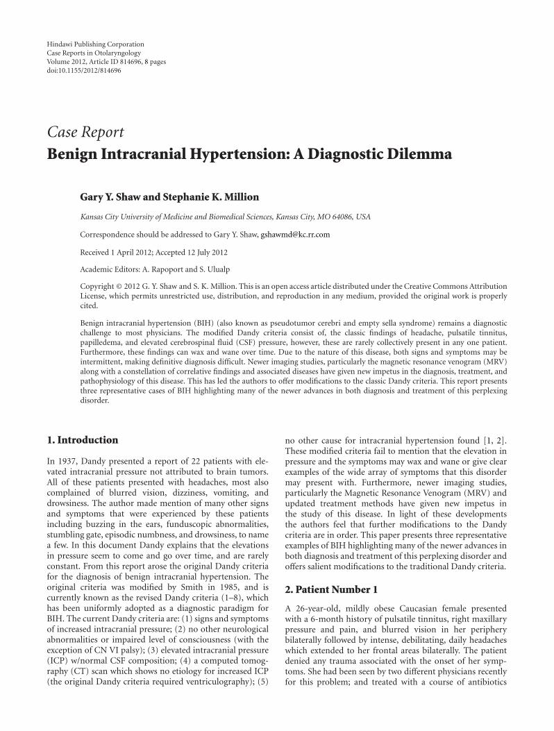
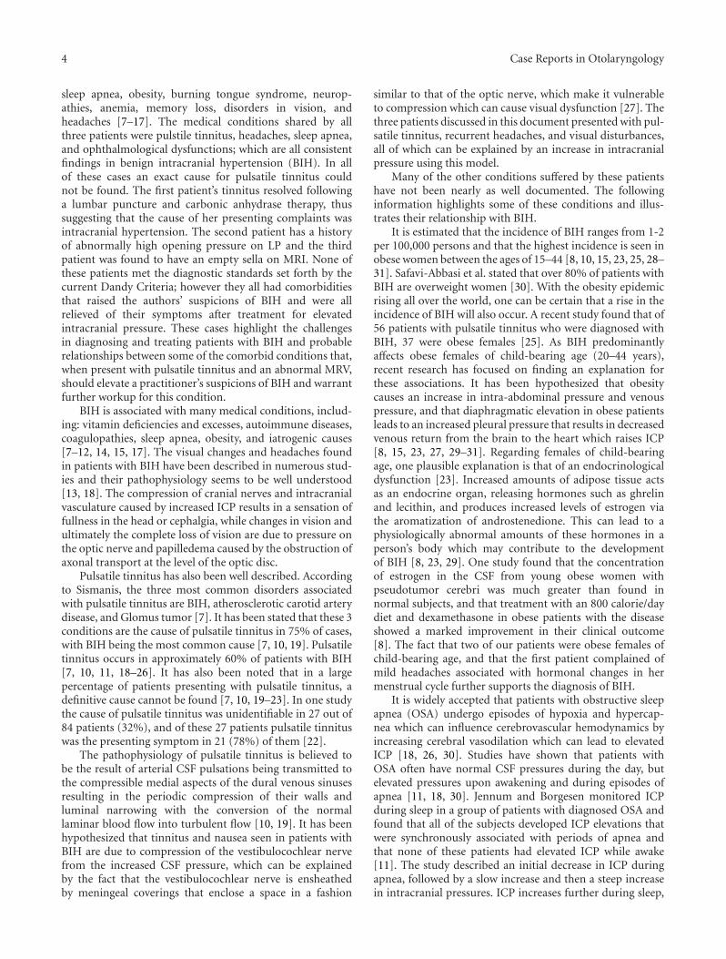
and a pressure equalization (PE) tube was placed in herright ear. Neither treatment aided in the relief of hersymptoms. The PE tube appeared to be in good positionand patent at the time of her visit. Compression of herright jugular vein led to cessation of the pulsatile tinnitus,suggesting a vascular origin. The remainder of the physicalexam was normal, with the exception of mild optic discblurring bilaterally (Figure 1(a)). Lab testing was found to benegative for rheumatoid factor, C-reactive protein, and ANA,and her erythrocyte sedimentation rate was within normallimits. Review of her head CT scan revealed no intracranialpathology, benign sinuses, and a normal appearing sellaturcica. The patient was then referred to a neurologist forfurther workup.
Over the next 5 months, the patient underwent an exten-sive evaluation in the neurology clinic and a battery of tests,including a magnetic resonance angiography (MRA) of thevenous flow of her head, and the Circle of Willis, magneticresonance imaging (MRI) of the cervical spine and brain,a lumbar puncture, two magnetic resonance venograms(MRV) of her head, coagulopathy studies, an echocar-diogram, and a sleep study. The patient was started ontopiramate 50 mg bid for treatment of her headaches. TheMRA and MRV both showed signal voids in her transverseand sagittal sinuses, with diminished signal intensity fromthe left sigmoid and transverse sinuses when compared tothe right. Neither study was able to determine whether thesefindings reflected disease, nondominance, artifact, or a com-bination of all three. Coagulation studies were positive forthe heterozygous presence of the Factor V Leiden (R506Q)mutation, and one copy of the methylenetetrahydrofolatereductase gene mutation A1298C, as well as a slight increasein antithrombin III activity of 129% (normal range 80–120%). Echocardiogram revealed the presence of a right-to- left shunt at rest, consistent with a patent foramenovale. Polysomnography resulted in the diagnosis of organicsleep apnea and restless leg syndrome. Her lumbar puncturehad an opening pressure of 150 mmH20 (normal range70–180 mmHg) and CSF analysis found no abnormalities.After the lumbar puncture, the pulsatile tinnitus completelyresolved. MRV was repeated two weeks after the lumbarpuncture and found to be normal without signal voids. Thiscorresponded with the patients clinical assessment of feelingmuch better.
At 6-months followup the patient continued to do welland her only complaint was mild headaches around the timeof menstruation; the topirimate seemed to be working toeliminate her daily headaches, the pulsatile tinnitus remainedabsent, funduscopic evaluation of her optic discs demon-strated partial resolution of the previously seen papillaryedema (Figure 1(b)).
3. Patient Number 2
Patient #2, a 43-year-old caucasian female presented with a13-year history of pulsatile tinnitus which had been gettingprogressively worse over time. She had been diagnosed ashaving atypical migraines, seizures, and chronic rhinitis.The patient had undergone numerous workups for these
(a)
(b)
Figure 1: (a) Grade I papilledema [3]. (b) Resolution of papill-edema [4].
complaints over the years, but the etiology and treatment hadyet to be determined. She claimed that the only time that shewas without these debilitating symptoms was many years agowhen a physician had given her a low dose corticosteroid.She explained that within two weeks of taking the medica-tion her symptoms had completely resolved, however theyreturned within a few months after the medication had beendiscontinued and worsened since that time. Her past medicalhistory included a microcytic anemia, hypertension, depres-sion, anxiety, fibromyalgia, sleep apnea, and hypothyroidism,visual changes with floaters, and memory loss. Fundoscopicevaluation demonstrated optic disc swelling. The results ofdiagnostic tests included a slightly elevated opening pressureon lumbar puncture, an optical CT scan revealing optic discswelling, and an MRA which was essentially normal. A recentnoncontrast brain MRI revealed her pituitary gland was inthe upper limits of normal as well as 2 small nonenhancingfoci in the left centrum semiovale region which appeared tobe chronic. Comparison to other studies was recommendeddue to these findings. Careful evaluation of the ear drum was
Case Reports in Otolaryngology 3
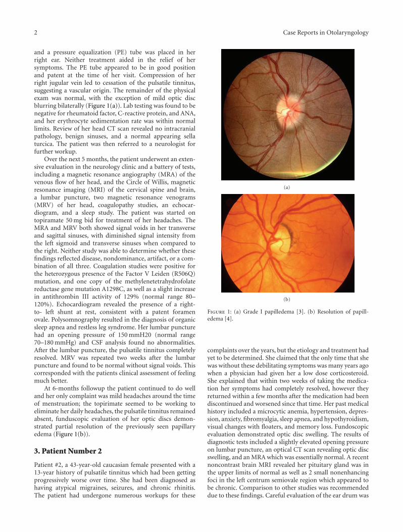
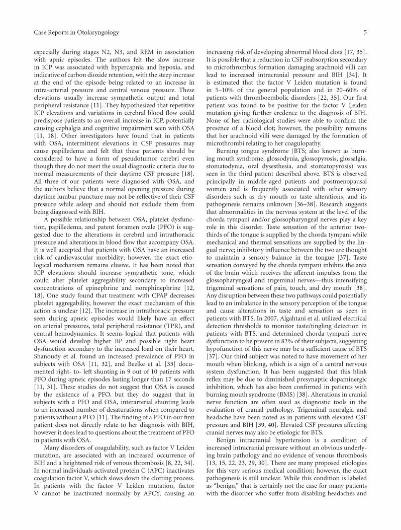
Figure 2: MRV showing narrowing of the transverse venoussinuses; most likely related to benign intracranial hypertension [5].
completed and no pulsation or bluish discoloration of thetympanic membrane was seen. The patient stated that thetinnitus in her right ear was relieved when slight pressure wasapplied just beneath her ear.
Since many of her complaints were suggestive of intracra-nial hypertension, an MRV was performed to look fordefects in dural flow, and irregularities with the jugularbulb. The MRV reported filling defects in the superiorsagittal sinus and right transverse sinus and right sigmoidsinus (Figure 2). These findings are most often observedsecondary to intraluminal thrombus and are consistent withpartial thrombosis or recanalization of a dural venous sinusthrombus. Prominence of the cortical draining veins wasalso found and noted to be most likely caused by partialobstruction due to the filling defects. The report also notedevidence of mastoiditis and suggested this as a likely triggerfor thrombus formation. A repeat MRI of the brain was alsoperformed, but revealed no evidence of abnormal enhancingmasses, acute infarction, or extra-axial fluid collection. Theparanasal sinuses were reported as normal, with some fluid inthe inferior portion of the right mastoid air cells, consistentwith mastoiditis. After antibiotic therapy, a CT was obtainedshowing resolution of the mastoid disease. The diagnosisof pseudotumor cerebri was made based on this patient’sclinical features and the MRV scans.
She was started on 500 mg of acetazolamide bid. A6-weeks followup revealed significant resolution of hersymptoms. At one year she continued to do well.
4. Patient Number 3
A 44-year-old African American female presented with com-plaints of a burning sensation in her tongue for the past6 months, temporal type headaches, and pulsatile tinnitus.Her medical history included Bell’s Palsy, chronic postnasal
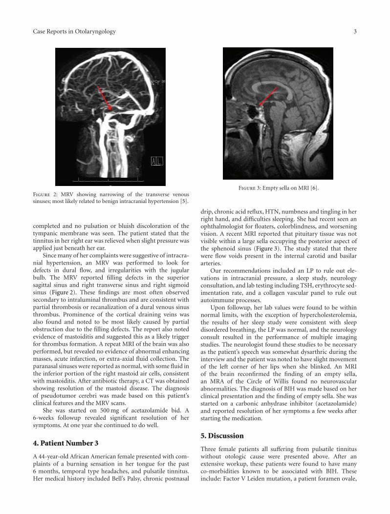
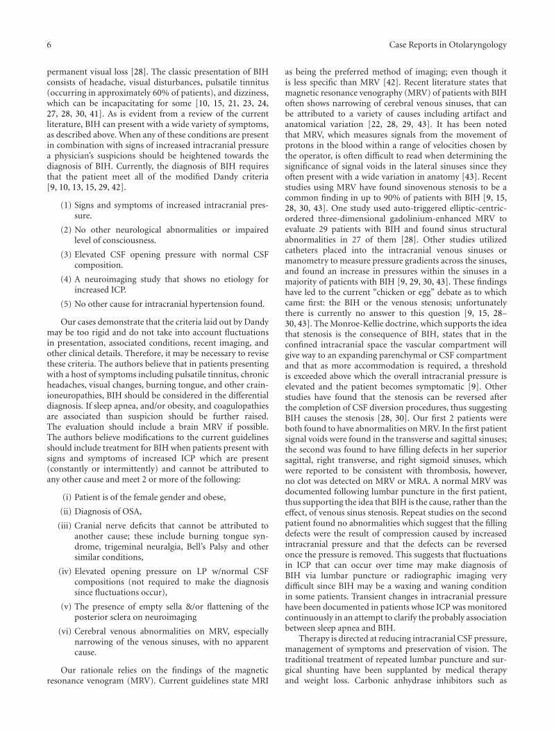
Figure 3: Empty sella on MRI [6].
drip, chronic acid reflux, HTN, numbness and tingling in herright hand, and difficulties sleeping. She had recent seen anophthalmologist for floaters, colorblindness, and worseningvision. A recent MRI reported that pituitary tissue was notvisible within a large sella occupying the posterior aspect ofthe sphenoid sinus (Figure 3). The study stated that therewere flow voids present in the internal carotid and basilararteries.
Our recommendations included an LP to rule out ele-vations in intracranial pressure, a sleep study, neurologyconsultation, and lab testing including TSH, erythrocyte sed-imentation rate, and a collagen vascular panel to rule outautoimmune processes.
Upon followup, her lab values were found to be withinnormal limits, with the exception of hypercholesterolemia,the results of her sleep study were consistent with sleepdisordered breathing, the LP was normal, and the neurologyconsult resulted in the performance of multiple imagingstudies. The neurologist found these studies to be necessaryas the patient’s speech was somewhat dysarthric during theinterview and the patient was noted to have slight movementof the left corner of her lips when she blinked. An MRIof the brain reconfirmed the finding of an empty sella,an MRA of the Circle of Willis found no neurovascularabnormalities. The diagnosis of BIH was made based on herclinical presentation and the finding of empty sella. She wasstarted on a carbonic anhydrase inhibitor (acetazolamide)and reported resolution of her symptoms a few weeks afterstarting the medication.
5. Discussion
Three female patients all suffering from pulsatile tinnituswithout otologic cause were presented above. After anextensive workup, these patients were found to have manyco-morbidities known to be associated with BIH. Theseinclude: Factor V Leiden mutation, a patient foramen ovale,
4 Case Reports in Otolaryngology
sleep apnea, obesity, burning tongue syndrome, neurop-athies, anemia, memory loss, disorders in vision, andheadaches [7–17]. The medical conditions shared by allthree patients were pulstile tinnitus, headaches, sleep apnea,and ophthalmological dysfunctions; which are all consistentfindings in benign intracranial hypertension (BIH). In allof these cases an exact cause for pulsatile tinnitus couldnot be found. The first patient’s tinnitus resolved followinga lumbar puncture and carbonic anhydrase therapy, thussuggesting that the cause of her presenting complaints wasintracranial hypertension. The second patient has a historyof abnormally high opening pressure on LP and the thirdpatient was found to have an empty sella on MRI. None ofthese patients met the diagnostic standards set forth by thecurrent Dandy Criteria; however they all had comorbiditiesthat raised the authors’ suspicions of BIH and were allrelieved of their symptoms after treatment for elevatedintracranial pressure. These cases highlight the challengesin diagnosing and treating patients with BIH and probablerelationships between some of the comorbid conditions that,when present with pulsatile tinnitus and an abnormal MRV,should elevate a practitioner’s suspicions of BIH and warrantfurther workup for this condition.
BIH is associated with many medical conditions, includ-ing: vitamin deficiencies and excesses, autoimmune diseases,coagulopathies, sleep apnea, obesity, and iatrogenic causes[7–12, 14, 15, 17]. The visual changes and headaches foundin patients with BIH have been described in numerous stud-ies and their pathophysiology seems to be well understood[13, 18]. The compression of cranial nerves and intracranialvasculature caused by increased ICP results in a sensation offullness in the head or cephalgia, while changes in vision andultimately the complete loss of vision are due to pressure onthe optic nerve and papilledema caused by the obstruction ofaxonal transport at the level of the optic disc.
Pulsatile tinnitus has also been well described. Accordingto Sismanis, the three most common disorders associatedwith pulsatile tinnitus are BIH, atherosclerotic carotid arterydisease, and Glomus tumor [7]. It has been stated that these 3conditions are the cause of pulsatile tinnitus in 75% of cases,with BIH being the most common cause [7, 10, 19]. Pulsatiletinnitus occurs in approximately 60% of patients with BIH[7, 10, 11, 18–26]. It has also been noted that in a largepercentage of patients presenting with pulsatile tinnitus, adefinitive cause cannot be found [7, 10, 19–23]. In one studythe cause of pulsatile tinnitus was unidentifiable in 27 out of84 patients (32%), and of these 27 patients pulsatile tinnituswas the presenting symptom in 21 (78%) of them [22].
The pathophysiology of pulsatile tinnitus is believed tobe the result of arterial CSF pulsations being transmitted tothe compressible medial aspects of the dural venous sinusesresulting in the periodic compression of their walls andluminal narrowing with the conversion of the normallaminar blood flow into turbulent flow [10, 19]. It has beenhypothesized that tinnitus and nausea seen in patients withBIH are due to compression of the vestibulocochlear nervefrom the increased CSF pressure, which can be explainedby the fact that the vestibulocochlear nerve is ensheathedby meningeal coverings that enclose a space in a fashion
similar to that of the optic nerve, which make it vulnerableto compression which can cause visual dysfunction [27]. Thethree patients discussed in this document presented with pul-satile tinnitus, recurrent headaches, and visual disturbances,all of which can be explained by an increase in intracranialpressure using this model.
Many of the other conditions suffered by these patientshave not been nearly as well documented. The followinginformation highlights some of these conditions and illus-trates their relationship with BIH.
It is estimated that the incidence of BIH ranges from 1-2per 100,000 persons and that the highest incidence is seen inobese women between the ages of 15–44 [8, 10, 15, 23, 25, 28–31]. Safavi-Abbasi et al. stated that over 80% of patients withBIH are overweight women [30]. With the obesity epidemicrising all over the world, one can be certain that a rise in theincidence of BIH will also occur. A recent study found that of56 patients with pulsatile tinnitus who were diagnosed withBIH, 37 were obese females [25]. As BIH predominantlyaffects obese females of child-bearing age (20–44 years),recent research has focused on finding an explanation forthese associations. It has been hypothesized that obesitycauses an increase in intra-abdominal pressure and venouspressure, and that diaphragmatic elevation in obese patientsleads to an increased pleural pressure that results in decreasedvenous return from the brain to the heart which raises ICP[8, 15, 23, 27, 29–31]. Regarding females of child-bearingage, one plausible explanation is that of an endocrinologicaldysfunction [23]. Increased amounts of adipose tissue actsas an endocrine organ, releasing hormones such as ghrelinand lecithin, and produces increased levels of estrogen viathe aromatization of androstenedione. This can lead to aphysiologically abnormal amounts of these hormones in aperson’s body which may contribute to the developmentof BIH [8, 23, 29]. One study found that the concentrationof estrogen in the CSF from young obese women withpseudotumor cerebri was much greater than found innormal subjects, and that treatment with an 800 calorie/daydiet and dexamethasone in obese patients with the diseaseshowed a marked improvement in their clinical outcome[8]. The fact that two of our patients were obese females ofchild-bearing age, and that the first patient complained ofmild headaches associated with hormonal changes in hermenstrual cycle further supports the diagnosis of BIH.
It is widely accepted that patients with obstructive sleepapnea (OSA) undergo episodes of hypoxia and hypercap-nea which can influence cerebrovascular hemodynamics byincreasing cerebral vasodilation which can lead to elevatedICP [18, 26, 30]. Studies have shown that patients withOSA often have normal CSF pressures during the day, butelevated pressures upon awakening and during episodes ofapnea [11, 18, 30]. Jennum and Borgesen monitored ICPduring sleep in a group of patients with diagnosed OSA andfound that all of the subjects developed ICP elevations thatwere synchronously associated with periods of apnea andthat none of these patients had elevated ICP while awake[11]. The study described an initial decrease in ICP duringapnea, followed by a slow increase and then a steep increasein intracranial pressures. ICP increases further during sleep,
Case Reports in Otolaryngology 5
especially during stages N2, N3, and REM in associationwith apnic episodes. The authors felt the slow increasein ICP was associated with hypercapnia and hypoxia, andindicative of carbon dioxide retention, with the steep increaseat the end of the episode being related to an increase inintra-arterial pressure and central venous pressure. Theseelevations usually increase sympathetic output and totalperipheral resistance [11]. They hypothesized that repetitiveICP elevations and variations in cerebral blood flow couldpredispose patients to an overall increase in ICP, potentiallycausing cephalgia and cognitive impairment seen with OSA[11, 18]. Other investigators have found that in patientswith OSA, intermittent elevations in CSF pressures maycause papilledema and felt that these patients should beconsidered to have a form of pseudotumor cerebri eventhough they do not meet the usual diagnostic criteria due tonormal measurements of their daytime CSF pressure [18].All three of our patients were diagnosed with OSA, andthe authors believe that a normal opening pressure duringdaytime lumbar puncture may not be reflective of their CSFpressure while asleep and should not exclude them frombeing diagnosed with BIH.
A possible relationship between OSA, platelet dysfunc-tion, papilledema, and patent foramen ovale (PFO) is sug-gested due to the alterations in cerebral and intrathoracicpressure and alterations in blood flow that accompany OSA.It is well accepted that patients with OSA have an increasedrisk of cardiovascular morbidity; however, the exact etio-logical mechanism remains elusive. It has been noted thatICP elevations should increase sympathetic tone, whichcould alter platelet aggregability secondary to increasedconcentrations of epinephrine and norephinephrine [12,18]. One study found that treatment with CPAP decreasesplatelet aggregability, however the exact mechanism of thisaction is unclear [12]. The increase in intrathoracic pressureseen during apneic episodes would likely have an effecton arterial pressures, total peripheral resistance (TPR), andcentral hemodynamics. It seems logical that patients withOSA would develop higher BP and possible right heartdysfunction secondary to the increased load on their heart.Shanoudy et al. found an increased prevalence of PFO insubjects with OSA [11, 32], and Beelke et al. [33] docu-mented right- to- left shunting in 9 out of 10 patients withPFO during apneic episodes lasting longer than 17 seconds[11, 31]. These studies do not suggest that OSA is causedby the existence of a PFO, but they do suggest that insubjects with a PFO and OSA, interarterial shunting leadsto an increased number of desaturations when compared topatients without a PFO [11]. The finding of a PFO in our firstpatient does not directly relate to her diagnosis with BIH,however it does lead to questions about the treatment of PFOin patients with OSA.
Many disorders of coagulability, such as factor V Leidenmutation, are associated with an increased occurrence ofBIH and a heightened risk of venous thrombosis [8, 22, 34].In normal individuals activated protein C (APC) inactivatescoagulation factor V, which slows down the clotting process.In patients with the factor V Leiden mutation, factorV cannot be inactivated normally by APCY, causing an
increasing risk of developing abnormal blood clots [17, 35].It is possible that a reduction in CSF reabsorption secondaryto microthrombus formation damaging arachnoid villi canlead to increased intracranial pressure and BIH [34]. Itis estimated that the factor V Leiden mutation is foundin 5–10% of the general population and in 20–60% ofpatients with thromboembolic disorders [22, 35]. Our firstpatient was found to be positive for the factor V Leidenmutation giving further credence to the diagnosis of BIH.None of her radiological studies were able to confirm thepresence of a blood clot; however, the possibility remainsthat her arachnoid villi were damaged by the formation ofmicrothrombi relating to her coagulopathy.
Burning tongue syndrome (BTS; also known as burn-ing mouth syndrome, glossodynia, glossopyrosis, glossalgia,stomatodynia, oral dysesthesia, and stomatopyrosis) wasseen in the third patient described above. BTS is observedprincipally in middle-aged patients and postmenopausalwomen and is frequently associated with other sensorydisorders such as dry mouth or taste alterations, and itspathogenesis remains unknown [36–38]. Research suggeststhat abnormalities in the nervous system at the level of thechorda tympani and/or glossopharyngeal nerves play a keyrole in this disorder. Taste sensation of the anterior two-thirds of the tongue is supplied by the chorda tympani whilemechanical and thermal sensations are supplied by the lin-gual nerve; inhibitory influence between the two are thoughtto maintain a sensory balance in the tongue [37]. Tastesensation conveyed by the chorda tympani inhibits the areaof the brain which receives the afferent impulses from theglossopharyngeal and trigeminal nerves—thus intensifyingtrigeminal sensations of pain, touch, and dry mouth [38].Any disruption between these two pathways could potentiallylead to an imbalance in the sensory perception of the tongueand cause alterations in taste and sensation as seen inpatients with BTS. In 2007, Algahtani et al. utilized electricaldetection thresholds to monitor taste/tingling detection inpatients with BTS, and determined chorda tympani nervedysfunction to be present in 82% of their subjects, suggestinghypofunction of this nerve may be a sufficient cause of BTS[37]. Our third subject was noted to have movement of hermouth when blinking, which is a sign of a central nervoussystem dysfunction. It has been suggested that this blinkreflex may be due to diminished presynaptic dopaminergicinhibition, which has also been confirmed in patients withburning mouth syndrome (BMS) [38]. Alterations in cranialnerve function are often used as diagnostic tools in theevaluation of cranial pathology. Trigeminal neuralgia andheadache have been noted as in patients with elevated CSFpressure and BIH [39, 40]. Elevated CSF pressures affectingcranial nerves may also be etiologic for BTS.
Benign intracranial hypertension is a condition ofincreased intracranial pressure without an obvious underly-ing brain pathology and no evidence of venous thrombosis[13, 15, 22, 23, 29, 30]. There are many proposed etiologiesfor this very serious medical condition; however, the exactpathogenesis is still unclear. While this condition is labeledas “benign,” that is certainly not the case for many patientswith the disorder who suffer from disabling headaches and
6 Case Reports in Otolaryngology
permanent visual loss [28]. The classic presentation of BIHconsists of headache, visual disturbances, pulsatile tinnitus(occurring in approximately 60% of patients), and dizziness,which can be incapacitating for some [10, 15, 21, 23, 24,27, 28, 30, 41]. As is evident from a review of the currentliterature, BIH can present with a wide variety of symptoms,as described above. When any of these conditions are presentin combination with signs of increased intracranial pressurea physician’s suspicions should be heightened towards thediagnosis of BIH. Currently, the diagnosis of BIH requiresthat the patient meet all of the modified Dandy criteria[9, 10, 13, 15, 29, 42].
(1) Signs and symptoms of increased intracranial pres-sure.
(2) No other neurological abnormalities or impairedlevel of consciousness.
(3) Elevated CSF opening pressure with normal CSFcomposition.
(4) A neuroimaging study that shows no etiology forincreased ICP.
(5) No other cause for intracranial hypertension found.
Our cases demonstrate that the criteria laid out by Dandymay be too rigid and do not take into account fluctuationsin presentation, associated conditions, recent imaging, andother clinical details. Therefore, it may be necessary to revisethese criteria. The authors believe that in patients presentingwith a host of symptoms including pulsatile tinnitus, chronicheadaches, visual changes, burning tongue, and other crain-ioneuropathies, BIH should be considered in the differentialdiagnosis. If sleep apnea, and/or obesity, and coagulopathiesare associated than suspicion should be further raised.The evaluation should include a brain MRV if possible.The authors believe modifications to the current guidelinesshould include treatment for BIH when patients present withsigns and symptoms of increased ICP which are present(constantly or intermittently) and cannot be attributed toany other cause and meet 2 or more of the following:
(i) Patient is of the female gender and obese,
(ii) Diagnosis of OSA,
(iii) Cranial nerve deficits that cannot be attributed toanother cause; these include burning tongue syn-drome, trigeminal neuralgia, Bell’s Palsy and othersimilar conditions,
(iv) Elevated opening pressure on LP w/normal CSFcompositions (not required to make the diagnosissince fluctuations occur),
(v) The presence of empty sella &/or flattening of theposterior sclera on neuroimaging
(vi) Cerebral venous abnormalities on MRV, especiallynarrowing of the venous sinuses, with no apparentcause.
Our rationale relies on the findings of the magneticresonance venogram (MRV). Current guidelines state MRI
as being the preferred method of imaging; even though itis less specific than MRV [42]. Recent literature states thatmagnetic resonance venography (MRV) of patients with BIHoften shows narrowing of cerebral venous sinuses, that canbe attributed to a variety of causes including artifact andanatomical variation [22, 28, 29, 43]. It has been notedthat MRV, which measures signals from the movement ofprotons in the blood within a range of velocities chosen bythe operator, is often difficult to read when determining thesignificance of signal voids in the lateral sinuses since theyoften present with a wide variation in anatomy [43]. Recentstudies using MRV have found sinovenous stenosis to be acommon finding in up to 90% of patients with BIH [9, 15,28, 30, 43]. One study used auto-triggered elliptic-centric-ordered three-dimensional gadolinium-enhanced MRV toevaluate 29 patients with BIH and found sinus structuralabnormalities in 27 of them [28]. Other studies utilizedcatheters placed into the intracranial venous sinuses ormanometry to measure pressure gradients across the sinuses,and found an increase in pressures within the sinuses in amajority of patients with BIH [9, 29, 30, 43]. These findingshave led to the current “chicken or egg” debate as to whichcame first: the BIH or the venous stenosis; unfortunatelythere is currently no answer to this question [9, 15, 28–30, 43]. The Monroe-Kellie doctrine, which supports the ideathat stenosis is the consequence of BIH, states that in theconfined intracranial space the vascular compartment willgive way to an expanding parenchymal or CSF compartmentand that as more accommodation is required, a thresholdis exceeded above which the overall intracranial pressure iselevated and the patient becomes symptomatic [9]. Otherstudies have found that the stenosis can be reversed afterthe completion of CSF diversion procedures, thus suggestingBIH causes the stenosis [28, 30]. Our first 2 patients wereboth found to have abnormalities on MRV. In the first patientsignal voids were found in the transverse and sagittal sinuses;the second was found to have filling defects in her superiorsagittal, right transverse, and right sigmoid sinuses, whichwere reported to be consistent with thrombosis, however,no clot was detected on MRV or MRA. A normal MRV wasdocumented following lumbar puncture in the first patient,thus supporting the idea that BIH is the cause, rather than theeffect, of venous sinus stenosis. Repeat studies on the secondpatient found no abnormalities which suggest that the fillingdefects were the result of compression caused by increasedintracranial pressure and that the defects can be reversedonce the pressure is removed. This suggests that fluctuationsin ICP that can occur over time may make diagnosis ofBIH via lumbar puncture or radiographic imaging verydifficult since BIH may be a waxing and waning conditionin some patients. Transient changes in intracranial pressurehave been documented in patients whose ICP was monitoredcontinuously in an attempt to clarify the probably associationbetween sleep apnea and BIH.
Therapy is directed at reducing intracranial CSF pressure,management of symptoms and preservation of vision. Thetraditional treatment of repeated lumbar puncture and sur-gical shunting have been supplanted by medical therapyand weight loss. Carbonic anhydrase inhibitors such as
Case Reports in Otolaryngology 7
acetazolamide are considered first line treatment for BIH[42]. Studies have documented acetazolamide’s success in themanagement of symptoms and stabilizing vision in 47–67%of patients [42]. Topiramate is a migraine therapy with CAHinhibition, making it another option. Furosemide and otherdiuretics cause nonspecific volume decrease throughout thebody, and therefore may be considered as adjuvant therapywith a CAH inhibitor. Mannitol is another diuretic thatcan be considered a treatment option since the primarymechanism of action is decreasing fluid within the CNS.Lumbar puncture and shunting should be reserved for severeor intractable cases. Associated morbidities such as obesityand obstructive sleep apnea should be aggressively treated.
6. Conclusion
BIH remains a diagnostic and therapeutic challenge. Recentadvancements in imaging and clinical research have neces-sitated a new approach to this fascinating disease. Withincrease in clinical impetus, better diagnostic and therapeuticmanagement will hopefully be forthcoming. Utilizing arevised, less rigid, and diagnostic criteria may aid physiciansin making the diagnosis of BIH, and permitting optimaltreatment of these patients.
References
[1] W. E. Dandy, “. Intracranial pressure without brain tumor:diagnosis and treatment,” Annals of Surgery, vol. 106, pp. 492–513, 1937.
[2] J. L. Smith, “Whence pseudotumor cerebri?” Journal of ClinicalNeuro-Ophthalmology, vol. 5, no. 1, pp. 55–56, 1985.
[3] Figure 1a: Grade I Papilledema, http://webeye.ophth.uiowa.edu/dept/IIH/pc 4.htm.
[4] Figure 1b: Resolution of Papilledema, http://webeye.ophth.uiowa.edu/dept/IIH/pc 3.htm.
[5] “Figure 2: MRV showing narrowing of the transverse venoussinuses, most likely related to pulsatile tinnitus,” http://webeye.ophth.uiowa.edu/dept/IIH/pc 3.htm.
[6] Figure 3: Empty Sella on MRI, http://www.healthcaremagic.com/articles/Empty-Sella-Turcica/2532.
[7] A. Sismanis, “Pulsatile tinnitus,” Otolaryngologic Clinics ofNorth America, vol. 36, no. 2, pp. 389–402, 2003.
[8] D. K. Binder, J. C. Horton, M. T. Lawton et al., “Ideopathicintracranial hypertension,” Neurosurgery, vol. 54, no. 3, pp.538–552, 2004.
[9] M. Jindal, L. Hiam, A. Raman, and D. Rejali, “Idiopathicintracranial hypertension in otolaryngology,” EuropeanArchives of Oto-Rhino-Laryngology, vol. 266, no. 6, pp.803–806, 2009.
[10] A. Sismanis, “Pulsatile tinnitus: a 15-year experience,” Ameri-can Journal of Otology, vol. 19, no. 4, pp. 472–477, 1998.
[11] P. Jennum and S. E. Borgesen, “Intracranial pressure andobstructive sleep apnea,” Chest, vol. 95, no. 2, pp. 279–283,1989.
[12] V. Biousse, A. Ameri, and M. G. Bousser, “Isolated intracranialhypertension as the only sign of cerebral venous thrombosis,”Neurology, vol. 53, no. 7, pp. 1537–1542, 1999.
[13] K. G. Kapoor, “Etiology of dizziness, tinnitus, and nausea inidiopathic intracranial hypertension,” Medical Hypotheses, vol.71, no. 2, pp. 310–311, 2008.
[14] J. Sussman, M. Leach, M. Greaves, R. Malia, and G. A. B.Davies-Jones, “Potentially prothrombotic abnormalities ofcoagulation in benign intracranial hypertension,” Journal ofNeurology Neurosurgery and Psychiatry, vol. 62, no. 3, pp. 229–233, 1997.
[15] O. Backhouse, T. Metcalfe, P. Goulding, M. McEvoy, and M.Menage, “Factor V Leiden mutation in association withidiopathic intracranial hypertension,” British Journal of Oph-thalmology, vol. 82, no. 7, article 844, 1998.
[16] C. A. Sila, A. J. Furlan, and J. R. Little, “Pulsatile tinnitus,”Stroke, vol. 18, no. 1, pp. 252–256, 1987.
[17] D. De Lucia, M. Napolitano, P. Di Micco, A. Niglio, A.Fontanella, and G. D. Lorio, “Benign intracranial hyperten-sion associated to blood coagulation derangements,” Throm-bosis Journal, vol. 4, article 21, 2006.
[18] V. A. Purvin, A. Kawasaki, and R. D. Yee, “Papilledema andobstructive sleep apnea syndrome,” Archives of Ophthalmology,vol. 118, no. 12, pp. 1626–1630, 2000.
[19] Y. Sogawa, R. Libman, L. Eviatar, and L. Kan, “Pulsatiletinnitus in a 16-year-old patient,” Pediatric Neurology, vol. 33,no. 3, pp. 214–216, 2005.
[20] D. Waldvogel, H. P. Mattle, M. Sturzenegger, and G. Schroth,“Pulsatile tinnitus—a review of 84 patients,” Journal ofNeurology, vol. 245, no. 3, pp. 137–142, 1998.
[21] J. A. Henry, T. L. Zaugg, P. J. Myers, C. J. Kendall, and E. M.Michaelides, “A triage guide for tinnitus,” Journal of FamilyPractice, vol. 59, no. 7, pp. 389–393, 2010.
[22] S. Randhawa and G. P. Van Stavern, “Idiopathic intracranialhypertension (pseudotumor cerebri),” Current Opinion inOphthalmology, vol. 19, no. 6, pp. 445–456, 2008.
[23] R. I. Farb, I. Vanek, J. N. Scott et al., “Idiopathic intracranialhypertension: the prevalence and morphology of sinovenousstenosis,” Neurology, vol. 60, no. 9, pp. 1418–1424, 2003.
[24] H. M. Phillippe and A. Y. Sparkman, “Venous thrombosis:preventing clots in patients at risk,” Journal of Family Practice,vol. 59, no. 6, pp. 315–321, 2010.
[25] M. C. Johansson, P. Eriksson, Y. Peker, J. Hedner, L. Rastam,and U. Lindblad, “The influence of patent foramen ovale onoxygen desaturation in obstructive sleep apnoea,” EuropeanRespiratory Journal, vol. 29, no. 1, pp. 149–155, 2007.
[26] B. M. Sanner, M. Konermann, M. Tepel, J. Groetz, C. Mum-menhoff, and W. Zidek, “Platelet function in patients withobstructive sleep apnoea syndrome,” European RespiratoryJournal, vol. 16, no. 4, pp. 648–652, 2000.
[27] J. O. Donaldson and E. Horak, “Cerebrospinal fluid oestronein pseudotumour cerebri,” Journal of Neurology Neurosurgeryand Psychiatry, vol. 45, no. 8, pp. 734–736, 1982.
[28] A. Lee and M. Wall, “Idiopathic intracranial hypertension(pseudotumor cerebri): clinical features and diagnosis,” 2010,http://www.uptodate.com/.
[29] J. N. P. Higgins, J. H. Gillard, B. K. Owler, K. Harkness,and J. D. Pickard, “MR venography in idiopathic intracranialhypertension: unappreciated and misunderstood,” Journal ofNeurology, Neurosurgery and Psychiatry, vol. 75, no. 4, pp. 621–625, 2004.
[30] S. Safavi-Abbasi, F. Di Rocco, P. Nakaji et al., “Thrombophiliadue to factor V and factor II mutations and formation of adural arteriovenous fistula: case report and review of a rareentity,” Skull Base, vol. 18, no. 2, pp. 135–142, 2008.
[31] P. Lopez-Jornet, F. Camacho-Alonso, P. Andujar-Mateos,M. Sanchez-Siles, and F. Gomez-Garcıa, “Burning mouthsyndrome: update,” Medicina Oral, Patologia Oral y CirugiaBucal, vol. 15, no. 4, pp. e562–e568, 2010.
8 Case Reports in Otolaryngology
[32] H. Shanoudy, A. Soliman, P. Raggi, J. W. Liu, D. C. Russell,and N. F. Jarmukli, “Prevalence of patent foramen ovale and itscontribution to hypoxemia in patients with obstructive sleepapnea,” Chest, vol. 113, no. 1, pp. 91–96, 1998.
[33] M. Beelke, S. Angeli, M. D. Sette et al., “Obstructive sleepapnea can be provocative for right-to-left shunting through apatent foramen ovale,” Sleep, vol. 25, no. 8, pp. 856–862, 2002.
[34] G. Friedman, N. Goldschmidt, Y. Friedlander et al., “A com-mon mutation A1298C in human methylenetetrahydrofolatereductase gene: association with plasma total homocysteineand folate concentrations,” Journal of Nutrition, vol. 129, no.9, pp. 1656–1661, 1999.
[35] T. W. Langfitt, “Clinical methods for monitoring intracranialpressure and measuring cerebral blood flow,” Clinical Neuro-surgery, vol. 22, pp. 302–320, 1975.
[36] R. S. Maurice Williams and J. Pilling, “Trigeminal sensorysymptoms associated with hydrocephalus,” Journal of Neurol-ogy Neurosurgery and Psychiatry, vol. 40, no. 7, pp. 641–644,1977.
[37] H. A. Algahtani, S. S. Baeesa, T. H. Obeid, and A. R. Abuz-inadah, “Idiopathic intracranial hypertension. Atypical pre-sentation,” Saudi Medical Journal, vol. 28, no. 5, pp. 762–765,2007.
[38] A. Lee and M. Wall, “Idiopathic intracranial hypertension(pseudotumor cerebri): epidemiology and pathogenesis,”2010, http://www.uptodate.com/.
[39] M. P. Minguez-Sanz, C. Salort-Llorca, and F. J. Silvestre-Donat, “Etiology of burning mouth syndrome: a review andupdate,” Medicina Oral, Patologia Oral y Cirugia Bucal, vol. 16,no. 2, pp. e144–e148, 2011.
[40] E. Dinces, “Etiology and diagnosis of tinnitus,” 2010, http://www.uptodate.com/.
[41] A. Sismanis, “Otologic manifestations of benign intracranialhypertension syndrome: diagnosis and management,” Laryn-goscope, vol. 97, no. 8, pp. 1–17, 1987.
[42] A. Lee and M. Wall, “Idiopathic intracranial hypertension(pseudotumor cerebri): prognosis and treatment,” 2010,http://www.uptodate.com/.
[43] S. D. Silberstein and R. C. McKinstry, “The death of idiopathicintracranial hypertension?” Neurology, vol. 60, no. 9, pp.1406–1407, 2003.

Submit your manuscripts athttp://www.hindawi.com
Stem CellsInternational
Hindawi Publishing Corporationhttp://www.hindawi.com Volume 2014
Hindawi Publishing Corporationhttp://www.hindawi.com Volume 2014
MEDIATORSINFLAMMATION
of
Hindawi Publishing Corporationhttp://www.hindawi.com Volume 2014
Behavioural Neurology
EndocrinologyInternational Journal of
Hindawi Publishing Corporationhttp://www.hindawi.com Volume 2014
Hindawi Publishing Corporationhttp://www.hindawi.com Volume 2014
Disease Markers
Hindawi Publishing Corporationhttp://www.hindawi.com Volume 2014
BioMed Research International
OncologyJournal of
Hindawi Publishing Corporationhttp://www.hindawi.com Volume 2014
Hindawi Publishing Corporationhttp://www.hindawi.com Volume 2014
Oxidative Medicine and Cellular Longevity
Hindawi Publishing Corporationhttp://www.hindawi.com Volume 2014
PPAR Research
The Scientific World JournalHindawi Publishing Corporation http://www.hindawi.com Volume 2014
Immunology ResearchHindawi Publishing Corporationhttp://www.hindawi.com Volume 2014
Journal of
ObesityJournal of
Hindawi Publishing Corporationhttp://www.hindawi.com Volume 2014
Hindawi Publishing Corporationhttp://www.hindawi.com Volume 2014
Computational and Mathematical Methods in Medicine
OphthalmologyJournal of
Hindawi Publishing Corporationhttp://www.hindawi.com Volume 2014
Diabetes ResearchJournal of
Hindawi Publishing Corporationhttp://www.hindawi.com Volume 2014
Hindawi Publishing Corporationhttp://www.hindawi.com Volume 2014
Research and TreatmentAIDS
Hindawi Publishing Corporationhttp://www.hindawi.com Volume 2014
Gastroenterology Research and Practice
Hindawi Publishing Corporationhttp://www.hindawi.com Volume 2014
Parkinson’s Disease
Evidence-Based Complementary and Alternative Medicine
Volume 2014Hindawi Publishing Corporationhttp://www.hindawi.com