Embed Size (px)
Citation preview
Pyrophosphohydrolase Activity and Inorganic PyrophosphateContent of Cultured Human Skin FibroblastsElevated Levels in SomePatients with Calcium Pyrophosphate Dihydrate Deposition Disease
Lawrence M. Ryan,* Robert L. Wortmann,* Barbara Karas,* Michael P. Lynch, and Daniel J. McCarty
*Section of Rheumatology, Department of Medicine, Medical College of Wisconsin, Milwaukee, Wisconsin 53226;and Wood Veterans Administration Medical Center, Wood, Wisconsin 53193
Abstract
In calcium pyrophosphate dihydrate (CPPD) crystal depositiondisease, metabolic abnormalities favoring extracellular inorganicpyrophosphate (PPi) accumulation have been suspected. Ele-vations of intracellular PPi in cultured skin fibroblasts from asingle French kindred with familial CPPDdeposition (19) andelevated nucleoside triphosphate pyrophosphohydrolase activity(NTPPPH), which generates PPi in extracts of CPPDcrystal-containing cartilages (14) favor this suspicion. To determinewhether NTPPPHactivity or PPi content of cells might be adisease marker expressed in extrnarticular cells, human skiu-derived fibroblasts were obtained from control donors and pa-tients affected with the sporadic and familial varieties of CPPD(CPPD-S and CPPD-F) deposition.
Intracellular PPi was elevated in both CPPD-S (P < 0.05)and CPPD-F (P < 0.01) fibroblasts compared with control fi-broblasts. Ecto-NTPPPH activity was elevated in CPPD-S (P< 0.01) but not CPPD-F. Intracellular PPi correlated with ecto-NTPPPH(P < 0.01).
Elevated PPi levels in skin fibroblasts may serve as a bio-chemical marker for patients with familial or sporadic CPPDcrystal deposition disease; ecto-NTPPPH activity further sep-arates the sporadic and familial disease types. Expression ofthese biochemical abnormalities in nonarticular cells implies ageneralized metabolic abnormality.
Introduction
In calcium pyrophosphate dihydrate (CPPD)' crystal depositiondisease, a disorder of inorganic pyrophosphate (PPi) metabolismis suspected because: (a) CPPDcrystals contain PPi; (b) synovial
This work was presented in part at the joint meeting of the CentralSociety for Clinical Research and the Midwest Section of the AmericanFederation for Clinical Research, Chicago, IL, November, 1984.
Dr. Lynch is a Copeman Traveling Fellow of the Heberden Society,Dr. McCarty is the Will and Cava Ross Professor and Chairman, De-partment of Medicine, Medical College of Wisconsin, Milwaukee, WI.Address reprint requests to Dr. Ryan, Section of Rheumatology, 8700West Wisconsin Ave., Milwaukee, WI 53226.
Receivedfor publication I April 1985 and in revisedform 23 January1986.
1. Abbreviations used in this paper: CPPD, calcium pyrophosphate dihy-drate; CPPD-F, familial CPPDcrystal deposition disease; CPPD-S, spo-radic CPPDcrystal deposition disease; DME, Dulbecco's modified Eagle'smedium; NTPPPH, nucleoside triphosphate pyrophosphohydrolase; OA,osteoarthritis; PCA, perchloric acid; Pi, inorganic phosphate; PPi, in-organic pyrophosphate; SF, synovial fluid; UDPG, uridine diphosphate
fluid (SF) PPi concentrations are elevated with respect to plasma(1 4); and (c) CPPDcrystal deposition occurs with increasedfrequency in adults with hypophosphatasia (5-7), a conditionassociated with elevated plasma and urinary PPi levels (8, 9).But systemic abnormalities in PPi metabolism have not beenidentified in most cases of CPPDcrystal deposition disease (10).Chondrocytes have been postulated to secrete PPi, since bothnormal and degenerated articular cartilage in organ culture, butnot synovium, subchondral bone, or elastic (ear) cartilage, haveelaborated extracellular PPi (11, 12). SF PPi pool and turnoverstudies in human knee joints suggested PPi production rates of0.5-1 umol/h (13), compatible with the quantities liberatedfrom human cartilage slices in organ culture (11, 12).
Tenenbaum et al. found elevated nucleoside triphosphatepyrophosphohydrolase (NTPPPH) activity in Triton X-100 ex-tracts of degenerative CPPDcrystal-containing cartilages com-pared with extracts of normal cartilages or extracts of degener-ative crystal-free cartilages (14). This chondrocyte ectoenzymegenerated PPi from a number of substrates (15, 16). NTPPPHactivity was also increased in SF from patients with CPPDde-position (17).
Lust et al. detected elevated intracellular PPi contents incultured skin fibroblasts and in Epstein-Barr virus-transformedlymphocytes from 10 affected members of a French kindredwith familial CPPI deposition, compared with cells derived fromnonaffected family members (18, 19), and cells in a single caseof sporadic CPPDcrystal deposition (20). These investigatorspostulated a generalized metabolic aberration of PPi metabolismin patients with CPPDcrystal deposition, manifested phenotyp-ically only in chondrocytes. However, interpretation of thesedata is hampered by the paucity of control values defining normalintracellular PPi and by the considerable overlap between theelevated values in fibroblasts from the French familial cases andthe normal values reported in a prior study by the same labo-ratory (20).
In view of the obvious importance of a biochemical correlatewith CPPDcrystal deposition, we assayed intracellular PPi incultured skin fibroblasts from donors in three kindreds affectedwith CPPDcrystal deposition, from patients with sporadic CPPDcrystal deposition, and, for comparison, from normal individualsand patients with primary osteoarthritis (OA). Additionally, westudied selected ectoenzyme (5' nucleotidase, inorganic pyro-phosphatase, and NTPPPH) activities in these cells to determinewhether the elevation of NTPPPHactivity observed in SF andcartilage extracts of patients with CPPDcrystal deposition alsooccurs in nonarticular tissues.
MethodsPatients. Patients with sporadic CPPDcrystal deposition (n = 13) haddefinite disease, as defined elsewhere (10). Familial CPPDdonors (n
glucose; l0-l-D, 10% fetal calf serum and 1% penicillin-streptomycin-Fungizone.
Calcium Pyrophosphate Dihydrate Deposition Disease 1689
J. Clin. Invest.C The American Society for Clinical Investigation, Inc.0021-9738/86/05/1689/05 $ 1.00Volume 77, May 1986, 1689-1693
= 11) were from three well-studied kindreds expressing CPPDdepositionas an autosomal dominant trait. Details of two of these kindreds havebeen published (21, 22). Simultaneous control biopsies were obtainedfrom spouses of affected familial members. Patients with sporadic CPPDcrystal deposition were studied for associated metabolic disease by mea-surements of serum iron, iron binding capacity, magnesium, free thyroxin,alkaline phosphatase, calcium, and phosphorus. Patients with primaryOA(n = 13) had negative joint fluid exams for CPPDcrystals and noradiographic evidence of chondrocalcinosis. As nearly half of nonagen-arians develop sporadic CPPDcrystal deposits, an overlap of values fromnormal subjects with those from patients with sporadic disease was an-ticipated. Assay of PPi content and enzyme activities in samples frompatients with OAwere thought to be a better control since these donorsdid not develop CPPDcrystal deposition, even in stressed cartilage. Lab-oratory and office personnel without joint symptoms also served as normaldonors, in addition to asymptomatic spouses of familial donors (n = 7).
Materials. Tetrasodium pyrophosphate decahydrate, ATP, uridinediphosphate glucose (UDPG), NADP, 5'AMP, trypsin, EDTA, glucose-1,6-diphosphate, phosphoglucomutase, MgCI2, ammonium molybdate,inorganic pyrophosphatase, and Tris were purchased from Sigma Chem-ical Co., St. Louis, MO; ['4C]UDPG and [32P]PPi were purchased fromNewEngland Nuclear Corp., Boston, MA. Dulbecco's modified Eagle'smedium (DME), Spinner's modified Eagle's medium, and Hank's bal-anced salt solution (HBSS) were obtained from Gibco, Grand Island,NY. Fetal bovine serum was purchased from MABioproducts, Walk-ersville, MD.
Biopsies. Biopsies were obtained from forearm skin in a manneridentical to that described by Lust et al. ( 18). Explants were transportedin DMEsupplemented with 10% fetal calf serum and 1% penicillin-streptomycin Fungizone (10-1-D).
Explants. The biopsy was washed with 10-1-D. Any hairs were re-moved. The specimen was cut into 3-4 pieces using a sterile scalpelblade. A 25-cm2 tissue culture flask which contained 4 ml of 10-1-D wasplaced upright for 10-15 min before the pieces of tissue were transferredwith a Pasteur pipette. The flask was incubated upright for 30 min at370C in 10% CO2 before it was gently placed flat. The flask was leftundisturbed for 2-3 d. If the pieces of tissue did not attach, they weretransferred to a tissue culture dish with a Pasteur pipette. A sterile mi-croscope coverslip was placed over them and 6 ml of 10- 1-D added tothe dish. The explants were fed twice weekly with 10-1-D, with care notto dislodge the attached tissues. At confluence, cells were transferred to2 tissue culture dishes by washing with HBSSand incubating for 5 minat 370 with 0.05% trypsin, 0.2% EDTA in HBSS. 7.5 ml 10-1-D wereadded to each dish. The second transfer was made to four 100-mm andeight 35-mm diameter tissue culture plates.
Biological variables. Our original attempts at measuring intracellularPPi yielded results which suggested uncontrolled variables. The effect ofindependently varying cell density, transfer number, and age of cultureon intracellular PPi levels was studied. After these studies, all determi-nations outlined below were performed on cultures of second transfercells, at 100% visual confluence, -6 wk after the initial biopsy and 48h after the most recent media change.
Assay for PPi. PPi formed during the NTPPPHreaction was deter-mined by a modified method of Cheung and Suhadolnik (23). Briefly,['4C]UDPG was quantitatively converted enzymatically to ["Ciglucose-I-PO4 and uridine triphosphate in the presence of PPi. Differential char-coal adsorption of [14C]UDPG before and after the reaction in the assaywas directly related to PPi concentration. Standard PPi concentrationsfrom 0.6 to 10.0 MMwere measured with each assay.
Intracellular levels of PPi were determined similarly, using standardPPi concentrations from 0.06 to 1.0 AM. Because these samples alsocontained [32PJPPi, the observed "4C was corrected for the overlap of 32pcounts in the 'C channel.
Standard stock PPi solutions (16 mM) were assayed for inorganicphosphate (Pi) content by the method of Fiske and SubbaRow (24) beforeand after hydrolysis by yeast inorganic pyrophosphatase as describedpreviously (25). This is necessary since stock Na.4P207- IOH20 powderreadily gains or loses waters of hydration (25), which makes calculationsof molarity by weight inaccurate.
Intracellular PPi. Cells from each of two 100-mm culture plates wereharvested and PPi content of each population was measured. The mediawas decanted and plates were washed twice with 5 ml PPi-free HBSStoremove dead cells and serum. The HBSShad been rendered PPi-free bytreating with yeast inorganic pyrophosphatase, then filtering through aCentriflo CF-25 filter (Amicon Corp., Lexington, MA) to remove py-rophosphatase. The sides of the plates were wiped dry. The plates wereplaced on ice and 0.5 ml cold pyrophosphatase-treated (PPi-free) HBSScontaining trace [32P]PPi and 1.5 M perchloric acid (PCA) was thenadded. The plates were rotated to evenly distribute the PCAand then0.025 ml cold 3M Tris was added. The cells were scraped off with arubber policeman and the suspension transferred to a cold glass test tube.The tube was centrifuged for 5 min at 650 g (Clinical Centrifuge; Inter-national Equipment Co., Damon Corp., Needham Heights, MA). Thesediment was saved for the determination of DNA. The supernatant wasneutralized with -80-95 Ml of 5 N KOH. It was again centrifuged andthe supernatant was frozen at -20'C for PPi determination. Knownamounts (0.1-5.0 AM) of PPi added to neutralized PCA supernatantsand frozen at -20'C served as internal standards. PPi recovery wasestimated by the recovery of unhydrolyzed [32P] PPi.
NTPPPHactivity. NTPPPHactivity was determined by measuringthe amount of PPi formed during incubation of cells in monolayer culturewith 1 mMATP. A 100-mm culture plate was washed 3 times with DMEto remove all of the fetal calf serum. Then 2 ml DMEcontaining I mMATP, 50 mMTris Cl, pH 7.2, was added. The plate was incubated at37°. A 200-M1 sample of media was removed at 30 min and stored at-20° for PPi determination. At 60 min, the rest of the media was removedand stored at -20° for PPi determination. The cells were released bytrypsin treatment for DNAanalysis. In preliminary studies we have de-termined that >95%of the PPi generated results from NTPPPHactivity,as we have previously shown in cartilage organ culture studies.
PPi hydrolysis. The amount of [32Plpyrophosphate hydrolyzed duringthe pyrophosphatase assay was determined by the differential precipitationof phosphorus as phosphomolybdate by triethylamine (26). 200 Ml ofthe sample was placed in a 1.5-ml microfuge tube. 10 Ml 10 mMNaPHO4,40 Ml 1.0 MPCA, 40 Ml 5%ammonium molybdate, and 10 Ml of 0.2 Mtriethylamine HCI were added sequentially with vortexing. The samplewas centrifuged for 10 s at 8,700 gin a microfuge (Brinkmann InstrumentsCo., Westbury, NY). The supernatant was transferred to a 20-ml scin-tillation vial which contained 10 ml of 0.5 MHC1. The precipitate waswashed, centrifuged twice more, and these supernatants were also addedto the scintillation vial. The precipitate was dissolved in 400 Ml acetoneand transferred to another scintillation vial containing 10 ml 0.5 MHCI.Radioactivity present in each vial was determined by Cerenkov countingas described previously (12). The percentage of hydrolysis in the blankwas subtracted from the sample value. PPi hydrolysis in the sample wasthen recorded.
Pyrophosphatase activity. Two confluent 35-mm culture plates offibroblasts in their second transfer were used for duplicate determinations.The plates were washed twice with DMEand then once with HBSS. 1.5ml of reaction mixture consisting of 640 Ml 20 mMPPi, 40 li 50 mMTris Cl buffered Spinner's modified Eagle's medium, pH 7.4, 0.2 ml 40mMEGTA, 8 ml 2mMMgCl2, and sufficient [32P]PPi to provide 10,000cpm/100 Ml was added to each plate. The plates were incubated at 370Cfor 30 min. A blank plate with no cells was also run. The media wasremoved and stored at -20° for hydrolysis determination. The cellswere released by treatment with 0.05% trypsin for DNAdetermination.
5' Nucleotidase activity. The amount of Pi released when the mono-layer fibroblasts were incubated with AMPwas measured as describedby Edwards (27). The method of Chen (28) was used to measure the Pi.Confluent 35-mm cultures were washed three times with 10 mMTrisHCI, pH 7.4, in 150 mMNaCl. 2 ml containing 50 mMTris, 4.0 mMMgCl2, 150 mMNaCl, and 0.4 mMAMPwere added to each plate andincubated at 370C. 100-Ml aliquots were removed at 5, 10, and 15 min.100 Ml of 0.150 MNaCl was added to each sample in plastic test tubes.800 Ml of a solution consisting of 2% ascorbic acid, 0.5% ammoniummolybdate, 1.2 N sulfuric acid was then added and incubated for 1-2h. The absorbance was measured at 820 nmon a DU6 spectrophotometer(Beckman Instruments, Inc., Fullerton, CA) and activity was expressed
1690 L. M. Ryan, R. L. Wortmann, B. Karas, M. P. Lynch, and D. J. McCarty
as picomoles Pi per microgram DNA. The cells were then released with0.05% trypsin for the determination of DNA.
DNAassay. DNAwas determined by a modification of the methodof Burton (29). The released cells were resuspended in 1 ml HBSSand100 Al 70% PCA was added. The macromolecular precipitate was re-moved by centrifuging at 1,700 g for 30 min and stored at -20'C. Thethawed precipitate was resuspended in 200 MA TrisCl, pH 7.4, and 200Al 0.4 N PCA, vortexed, and allowed to stand for 5 min. After centrif-ugation at 1700 g for 30 min at 4VC, 600 ml of 1.5 MPCAwas added.DNAstandards from 5 to 35 Mg were prepared in 600 Ml of 1.5 MPCA.The glass tubes containing standard or sample were incubated at 70'Cfor 30 min and allowed to cool at room temperature; 350 Ml diphenyl-amine reagent (4.0 g diphenylamine and 10 Al paraldehyde in 100 mlglacial acetic acid) was added to each tube. The tubes were incubated inthe dark for 18 h at room temperature, centrifuged for 30 min at 1700g, and the absorbance at 600 nmwas read in a spectrophotometer (model36; Beckman Instruments, Inc.).
Results
Biological variables. Our first attempts at measuring intracellularPPi content yielded widely discordant values on cells derivedfrom the same explant, suggesting uncontrolled variables. TableI shows the results of PPi measurements in cultures of cells fromsix donors at different stages of visual confluence during secondand third passage. In all samples, intracellular PPi was lowerpostconfluence than at 50% or 75% confluence. Values at com-parable stages of visual confluenc, were more often lower atthird passage than at second. The effect of age in culture wasassessed by measuring intracellular PPi in monolayer cells de-rived from a single explant on second passage at visual confluenceand 48 h after media replenishment, but plated on second passage
Table I. Intracellular PPi at Various Stagesof Visual Confluence and Passage Number
Passage Post-Patient no. 50% 75% 95% confluence
I T2 144*T3 58
2 T2 272T3 86
3t T2 93T3
4t T2 87T3 149
5 T2T3 25
6 T2 17T3 37
Mean±SD T2 123±95T3 71±47
12565
116779823
1461367733185197±4565±40
37 2468 146 87
139 5668 32344672 835 92517
846±1755±47
41
31±3317±26
Intracellular PPi was measured in duplicate on fibroblasts derivedfrom skin explants of six donors. On second (T2) or third (T3) pas-sage, cells on duplicate plates at various stages of visual confluencewere washed, lysed with 1.5 MPCA, PPi measured on the potassiumhydroxide-neutralized supernatant, and DNAmeasured in the macro-
molecular precipitate. PPi measurements were by the radiometricUDPGpyrophosphorylase method (23).* pmol PPi/Mg DNA.* Patients with CPPDdeposition disease.
Table II. PPi Content of Fibroblasts Grown from aSingle Explant Measured at Different Ages-but at Identical Stages of Confluence and Passage*
Aget PPi
wk pmol/lg DNA
4 566 17
14 716 4
Fibroblasts derived from a single explant were passaged and plated onsecond passage (T2) so as to achieve visual confluence at various inter-vals. Duplicate plates reached confluence at 4, 6, 14, and 16 wk atwhich time PPi was measured on the potassium hydroxide-neutral-ized PCAcell lysate.* Measured at visual confluence on second passage.* Interval between biopsy and PPi measurement.
at different cell densities so that confluence was reached at dif-ferent times. Table II shows the mean of duplicate determina-tions, which suggest that intracellular PPi decreases with age inculture. After these experiments, cellular PPi and ectoenzymeswere routinely measured on cells -6-7 wk after biopsy, at visualconfluence, on second passage, and precisely 48 h after mediareplenishment.
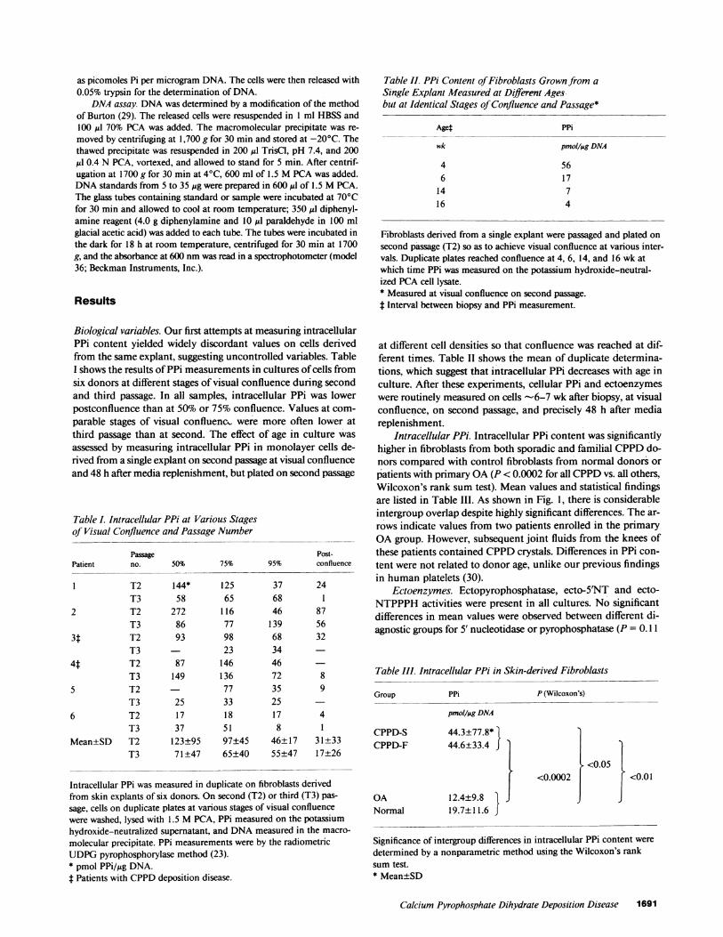
Intracellular PPi. Intracellular PPi content was significantlyhigher in fibroblasts from both sporadic and familial CPPDdo-nors compared with control fibroblasts from normal donors orpatients with primary OA(P < 0.0002 for all CPPDvs. all others,Wilcoxon's rank sum test). Mean values and statistical findingsare listed in Table III. As shown in Fig. 1, there is considerableintergroup overlap despite highly significant differences. The ar-rows indicate values from two patients enrolled in the primaryOAgroup. However, subsequent joint fluids from the knees ofthese patients contained CPPDcrystals. Differences in PPi con-tent were not related to donor age, unlike our previous findingsin human platelets (30).
Ectoenzymes. Ectopyrophosphatase, ecto-5'NT and ecto-NTPPPHactivities were present in all cultures. No significantdifferences in mean values were observed between different di-agnostic groups for 5' nucleotidase or pyrophosphatase (P = 0.11
Table III. Intracellular PPi in Skin-derived Fibroblasts
Group PPi P (Wilcoxon's)
pmoll4g DNA
CPPD-S 44.3±77.8* 1CPPD-F 44.6±33.4 J
<0.05<0.0002 <0.01I
OA 12.4±9.8 <0.01Normal 19.7±11.6 J
Significance of intergroup differences in intracellular PPi content were
determined by a nonparametric method using the Wilcoxon's ranksum test.* Mean±SD
Calcium Pyrophosphate Dihydrate Deposition Disease 1691
I
z~0
(0)*~30-
06 go020
0NORMAL OA CPPD-F CPPD-S
(7) (13) (10) (12)
Figure 1. Fibroblast PPi content in cells from normal donors and pa-tients with OA, CPPD-F, and CPPD-S. The arrows identify valuesfrom two patients originally classified as having OAin whomCPPDcrystals were subsequently found in joint fluid. Values are the mean ofduplicate cell samples.
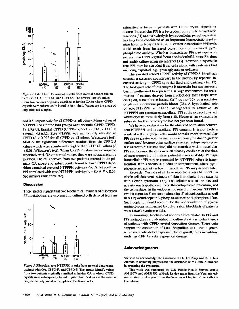
and 0.5, respectively for all CPPDvs. all other). Mean values ofNTPPPH±SDfor the four groups were: sporadic CPPD(CPPD-S), 9.9±4.8, familial CPPD(CPPD-F), 6.7±3.9; OA, 7.1± 10.1;normal, 4.6±3.2. Ecto-NTPPH was significantly elevated inCPPD(P < 0.002 for all CPPDvs. all others, Wilcoxon's test).Most of the significant differences resulted from the CPPD-Svalues which were significantly higher than CPPD-F values (P< 0.01, Wilcoxon's test). WhenCPPD-F values were comparedseparately with OAor normal values, they were not significantlyelevated. The cells derived from two patients entered in the pri-mary OAgroup and subsequently found to have CPPDdepo-sition contained elevated NTPPPHactivity (Fig. 2). IntracellularPPi correlated with ecto-NTPPPH activity (r, = 0.49, P < 0.05,Spearman's rank correlate).
Discussion
These studies suggest that two biochemical markers of disorderedPPi metabolism are expressed in cultured cells derived from an
30
zCl
420
a.a.-
. I0~W lo.Ec
0
'-.
I
NORMAL OA CPPD-F CPPD-S(7) (13) (11) (13)
Figure 2. Fibroblast ecto-NTPPPH in cells from normal donors andpatients with OA, CPPD-F, and CPPD-S. The arrows identify valuesfrom two patients originally classified as having OAin whomCPPDcrystals were subsequently found in joint fluid. Values are the mean ofenzyme activity found in two plates of cultured cells.
extraarticular tissue in patients with CPPDcrystal depositiondisease. Intracellular PPi is a by-product of multiple biosyntheticreactions (31) and its hydrolysis by intracellular pyrophosphatasehas long been considered as an important homeostatic mecha-nism favoring biosynthesis (32). Elevated intracellular PPi levelscould result from increased biosynthesis or decreased pyro-phosphatase activity. Whether intracellular PPi participates inextracellular CPPDcrystal formation is doubtful, since PPi doesnot readily diffuse across membranes (33). However, it is possiblethat PPi may be extruded from cells along with materials thatare being exported, e.g., proteoglycans or collagen.
The elevated ecto-NTPPPH activity of CPPD-S fibroblastssuggests a systemic counterpart to the previously reported in-creased activity in CPPDsynovial fluid and cartilage (16, 17).The biological role of this enzyme is uncertain but has variouslybeen hypothesized to represent a salvage mechanism for recla-mation of purines derived from nucleotides that escape fromcells (34), a membrane-bound Ca"+ pump (35), or-a regulatorof plasma membrane protein kinase (36). A hypothetical roleof ecto-NTPPPH in CPPD pathogenesis is attractive, as
NTPPPHcan generate extracellular PPi at the extracellular sitewhere crystals most likely form (10). However, an extracellularsubstrate for this ectoenzyme has not yet been found.
Wehave no explanation for the observed correlation betweenecto-NTPPPH and intracellular PPi content. It is not likely a
result of cell size (larger cells would contain more intracellularPPi due to greater volume and more ectoenzyme due to greatersurface area) because other surface enzymes (ectopyrophospha-tase and ecto-5' nucleotidase) did not correlate with intracellularPPi and because the cells were all visually confluent at the timeof measurement, diminishing potential size variability. Perhapsintracellular PPi may be generated by NTPPPHbefore its trans-location. If this occurs in a cellular compartment where pyro-phosphatase activity is low, intracellular PPi may accumulate.
Recently, Yoshida et al. have reported excess NTPPPHinwhole-cell detergent extracts of skin fibroblasts from patientswith Lowe's syndrome (37). The cellular site of the elevatedactivity was hypothesized to be the endoplasmic reticulum, notthe cell surface. In the endoplasmic reticulum, excess NTPPPH(which degrades 3'-phospho-adenosine 5'-phosphosulfate as wellas ATP) would deplete 3'-phospho-adenosine 5'-phosphosulfate.Such depletion could account for the undersulfation of glycos-aminoglycans synthesized by culture skin fibroblasts of patientswith Lowe's syndrome (38).
In summary, biochemical abnormalities related to PPi andPPi metabolism are identified in cultured extraarticular tissuesof patients with CPPDcrystal deposition disease. These datasupport the contention of Lust, Seegmiller, et al. that a gener-alized metabolic defect expressed phenotypically only in cartilageunderlies CPPDcrystal deposition disease.
Acknowledgments
Wewish to acknowledge the assistance of Dr. Ed Perry and Dr. JuliusZulman in obtaining biopsies and the assistance of Ms. Jane Alexanderin preparing the typescript.
This work was supported by U.S. Public Health Service grantsAM18074 and AM31395, a Merit Review grant from the Veterans Ad-ministration, and a grant from the Wisconsin Chapter of the ArthritisFoundation.
1692 L. M. Ryan, R. L. Wortmann, B. Karas, M. P. Lynch, and D. J. McCarty
s|||~~I
References
1. Russell, R. G. G., S. Bisaz, and H. Fleisch. 1970. Inorganic py-rophosphate in plasma, urine and synovial fluid of patients with pyro-phosphate arthropathy (chondrocalcinosis or pseudogout). Lancet. ii:899-902.
2. McCarty, D. J., S. D. Solomon, M. I. Warnock, and E. Paloyan.1971. Inorganic pyrophosphate concentrations in the synovial fluid ofarthritis patients. J. Lab. Clin. Med. 78:216-229.
3. Altman, R. D., 0. E. Muniz, J. C. Pita, and D. S. Howell. 1973.Articular chondrocalcinosis: microanalysis of pyrophosphate (PPi) insynovial fluid and plasma. Arthritis Rheum. 16:171-178.
4. Silcox, D. C., and D. J. McCarty. 1974. Elevated inorganic py-rophosphate concentrations in synovial fluid in osteoarthritis and pseu-dogout. J. Lab. Clin. Med. 83:518-531.
5. O'Duffy, D. J. 1970. Hypophosphatasia associated with calciumpyrophosphate dihydrate deposits in cartilage. Arthritis Rheum. 13:381-388.
6. Earle, A. W., A. J. Swannell, and N. W. Williamson. 1981. Py-rophosphate arthropathy in hypophosphatasia. Ann. Rheum. Dis. 40:164-170.
7. Whyte, M. P., W. A. Murphy, and M. D. Fallon. 1982. Adulthypophosphatasia with chondrocalcinosis and arthropathy: variablepenetrance of hypo-phosphatemia in a l.ge Oklahoma kindred. Am. J.Med. 72:631-641.
8. Russell, R. G. G. 1976. Excretion of inorganic pyrophosphate inhypophosphatasia. Lancet. ii:461-470.
9. Russell, R. G. G., S. Bisaz, A. Donath, D. B. Morgan, and H.Fleisch. 1971. Inorganic pyrophosphate in plasma in normal personsand in patients with hypophosphatasia, osteogenesis imperfecta and otherdisorders of bone. J. Clin. Invest. 50:961-969.
10. Ryan, L. M., and D. J. McCarty. 1985. Calcium pyrophosphatecrystal deposition disease. In Arthritis and Allied Conditions. D. J.McCarty, editor. Lea & Febiger, Philadelphia. 1515-1546.
11. Howell, D. S., 0. Muniz, J. C. Pita, and J. E. Enis. 1975. Extrusionof pyrophosphate into extracellular media by osteoarthritic cartilage in-cubates. J. Clin. Invest. 56:1473-1480.
12. Ryan, L. M., H. S. Cheung, and D. J. McCarty. 1981. Releaseof pyrophosphate by normal mammalian articular hyaline and fibro-cartilage in organ culture. Arthritis Rheum. 24:1522-1527.
13. Camerlain, M., D. J. McCarty, D. C. Silcox, and A. Jung. 1975.Inorganic pyrophosphate pool size and turnover in arthritic joints. J.Clin. Invest. 55:1373-1381.
14. Tenebaum, J., 0. Muniz, H. R. Schumacher, A. E. Good, andD. S. Howell. 1981. Comparison of phosphohydrolase activities fromarticular cartilage in calcium pyrophosphate deposition disease and pri-mary osteoarthritis. Arthritis Rheum. 24:492-500.
15. Ryan, L. M., R. L. Wortmann, B. Karas, and D. J. McCarty.1984. Cartilage nucleoside triphosphate (NTP) pyrophosphohydrolase.
I. Identification as an ectoenzyme. Arthritis Rheum. 27:913-918.16. Muniz, O., J. Pelletier, J. Martel-Pelletier, S. Morales, and D. S.
Howell. 1984. NTPpyrophosphohydrolase in human chondrocalcinoticand osteoarthritic cartilage. I. Some biochemical characteristics. ArthritisRheum. 27:186-192.
17. Rachow, J. W., and L. M. Ryan. 1983. Adenosine triphosphatepyrophosphohydrolase and pyrophosphatase activities in human synovialfluids. Clin. Res. 31:806a. (Abstr.)
18. Lust, G., G. Faure, P. Netter, A. Gaucher, and J. E. Seegmiller.1981. Evidence of a generalized metabolic defect in patients with hered-itary chondrocalcinosis. Arthritis Rheum. 24:1517-1521.
19. Lust, G., G. Faure, P. Netter, and J. E. Seegmiller. 1981. Increased
pyrophosphate in fibroblasts and lymphoblasts from patients with he-reditary diffuse articular chondrocalcinosis. Science (Wash. DC). 214:809-810.
20. Lust, G., G. Nuki, and J. E. Seegmiller. 1976. Inorganic pyro-phosphate and proteoglycan metabolism in cultured human articularchondrocytes and fibroblasts. Arthritis Rheum. 19(Suppl):479-487.
21. Perry, E., E. L. Overholt, and K. L. Newcomer. 1969. Familialoccurrence of chondrocalcinosis. Wis. Med. J. 68:321-324.
22. Richardson, B. C., N. I. Chafetz, L. D. Ferrell, J. E. Zulman, andH. K. Genant. 1983. Hereditary chondrocalcinosis (CPPD) in a Mexican-American family. Arthritis Rheum. 26:1387-1396.
23. Cheung, C. P. and R. J. Suhadolnik. 1977. Analysis of inorganicpyrophosphate at the picomole level. Anal. Biochem. 83:61-63.
24. Fiske, C. H., and Y. SubbaRow. 1925. The colorimetric deter-mination of phosphorous. J. Biol. Chem. 66:375-400.
25. Ryan, L. M., F. Kozin, and D. J. McCarty. 1979. Quantificationof human plasma inorganic pyrophosphate. I. Normal values in osteoar-thritis and calcium pyrophosphate dihydrate crystal deposition disease.Arthritis Rheum. 22:886-891.
26. Sugino, Y., and Y. Miyoshi. 1964. The specific precipitation oforthophosphate and some biochemical applications. J. Biol. Chem. 239:2360-2364.
27. Edwards, M. L., J. T. Cassidy, and I. H. Fox. 1980. Lymphocyteecto-5'-nucleotidase deficiency in hypogammaglobulinemia; clinicalcharacteristics. Clin. Immunol. Immunopathol. 17:76-88.
28. Chen, P. S., Jr., T. Y. Toribara, and H. Warner. 1956. Micro-determination of phosphorus. Anal. Biochem. 28:1756-1758.
29. Burton, K. 1956. A study of the conditions and mechanism ofthe diphenylamine reaction for the colorimetric estimation of deoxyri-bonucleic acid. Biochemistry. 62:315-323.
30. Ryan, L. M., M. P. Lynch, and D. J. McCarty. 1983. Inorganicpyrophosphate levels in blood platelets from normal donors and patientswith calcium pyrophosphate crystal deposition disease. Arthritis Rheum.26:564-566.
31. Russell, R. G. G. 1976. Metabolism of inorganic pyrophosphate(PPi). Arthritis Rheum. 19(suppl):463-478.
32. Kornberg, A. 1962. On the metabolic significance of phospho-rolytic and pyrophosphorolytic reactions. In Horizons in Biochemistry.M. Kasha and D. Pullman, editors. Academic Press, New York. 251-264.
33. Felix, R., and H. Fleisch. 1977. The effect of pyrophosphate anddiphosphonates on calcium transport in red cells. Experientia. 33:1003-1005.
34. Tran-Thi, T., J. W. Phillips, A. Schulze-Speaking, J. Rasenack,and K. Decker. 1981. Properties and biosynthetic connection of the nu-cleotide pyrophosphatases of rat liver plasma membrane and endoplasmicreticulum. Hoppe-Seyler's Z. Physiol. Chem. 362:305-316.
35. Flodgaard, H., and C. Torp-Pederson. 1978. A calcium ion-de-pendent adenosine triphosphate pyrophosphohydrolase in plasma mem-brane from rat liver. Biochem. J. 171:817-820.
36. Hutson, J. M., M. E. Fallat, S. Kamagata, P. K. Donahoe, andG. P. Budzik. 1984. Phosphorylation events during mullerian ductregression. Science (Wash. DC). 223:586-589.
37. Yoshida, H., S. Fukui, I. Yamashina, T. Tanaka, T. Sakano, T.Usui, T. Shimotsuji, H. Yaburuchi, M. Owada, and T. Kitogawa. 1982.Elevation of nucleotide pyrophosphatase activity in skin fibroblasts frompatients with Lowe's syndrome. Biochem. Biophys. Res. Commun. 107:1144-1150.
38. Fukui, S., H. Yoshida, T. Sakano, T. Tanaka, T. Usui, and I.Yamashina. 1981. Glycosaminoglycan synthesis by cultured skin fibro-blasts from patients with Lowe's syndrome. J. Biol. Chem. 256:10313-10318.
Calcium Pyrophosphate Dihydrate Deposition Disease 1693

![Review Phosphate/Pyrophosphate and MV-related Proteins in ...lation of PPi in the extracellular matrix [7], suggesting that TNSALP is required to promote calcification by removing](https://img.pdfslide.us/doc/110x75/608c128f6831c178dd1a6b7a/review-phosphatepyrophosphate-and-mv-related-proteins-in-lation-of-ppi-in-the.jpg)