Embed Size (px)
Citation preview
Case 1Case 1Case 1Case 1
TwoTwo--dayday--old girl with bilious old girl with bilious emesisemesis. .
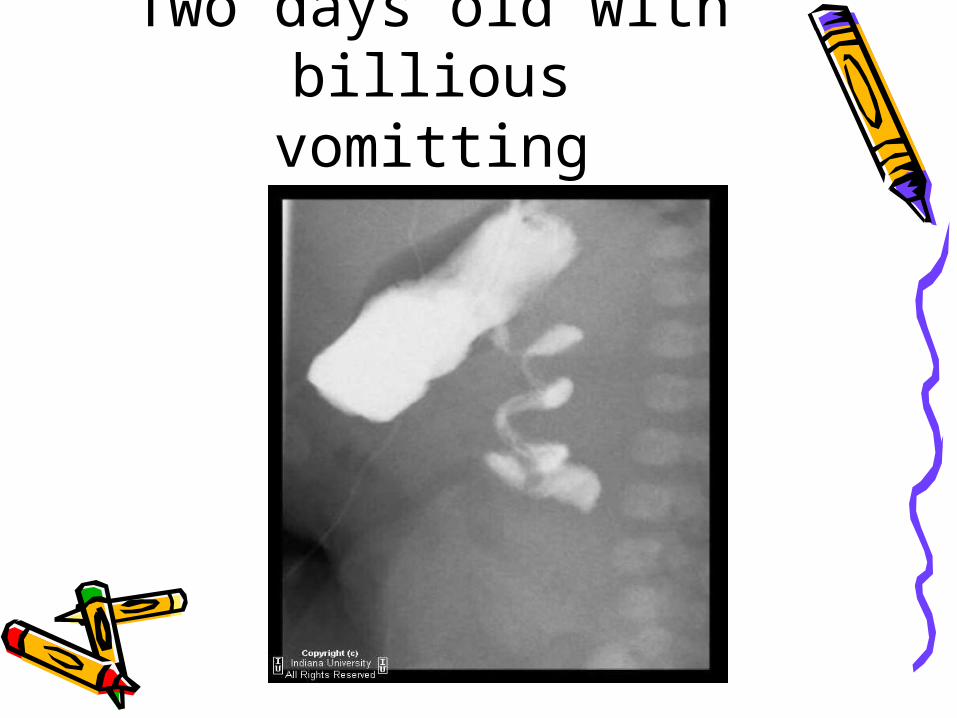
Two days old with billious
vomitting
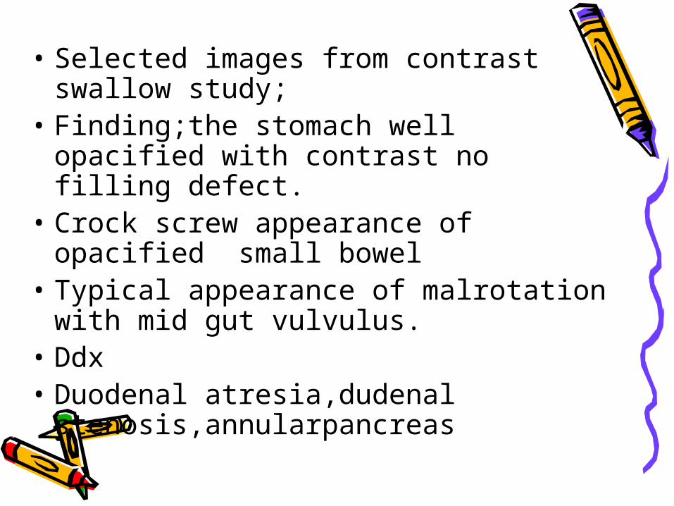
• Selected images from contrast swallow study;
• Finding;the stomach well opacified with contrast no filling defect.
• Crock screw appearance of opacified small bowel
• Typical appearance of malrotation with mid gut vulvulus.
• Ddx• Duodenal atresia,dudenal
stenosis,annularpancreas
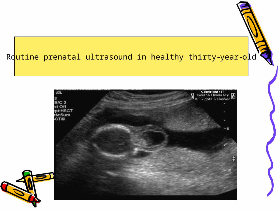
Routine prenatal ultrasound in healthy thirty-year-old

Us axial images show;• THERE IS ABNORMALITY WITH
THE FETUS HEAD.THERE IS DEFECT THROUGH THE POSTERIOR OCCIIPIT WITH HERNATED MASS WHICH CONTAIN SOLID AND CYSTIC COMPONENTS
• DDX:• CEPHALOCELE(ENCEPHALOCELE)• BRANCHIAL CLEFT CYST• CYSTIC HYGROMA• TERATOMA

Woman with breast cancer
MRI BRAIN FOR FEMALE WITH BREAST CA
• There are multiple enhancing lesion through the calvarium,diffuse enhancement of the meninges and confluent hypersignal intensity lesions on the deep whit matter in FLAIR and T2WI.
• No enhancing intraparenchymal lesion.• No mass effect• Normal ventricular system
• In keeping with hx. Ddx;METS LESION.POST RADIATION الصحيح الحلDIFFUSE INFECTIOUSDIFFUSE INFLAMMATORYSMALL VESSELS DISEASE
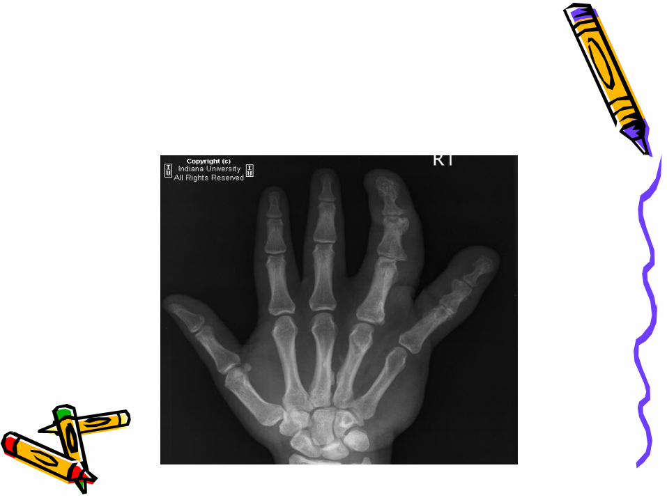
HAND XRAY
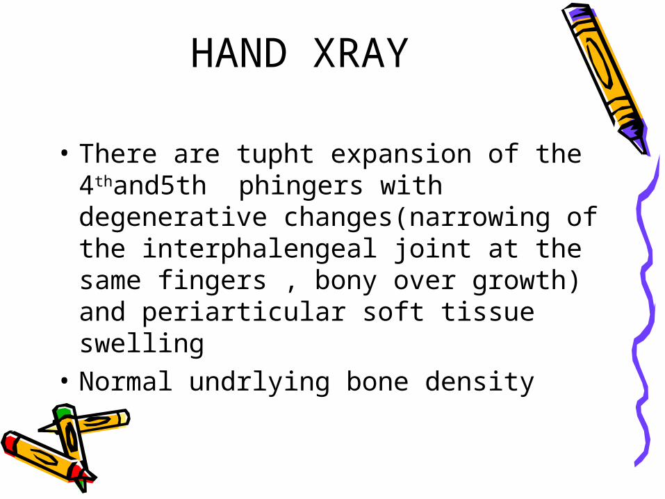
• There are tupht expansion of the 4thand5th phingers with degenerative changes(narrowing of the interphalengeal joint at the same fingers , bony over growth) and periarticular soft tissue swelling
• Normal undrlying bone density
• Ddx:• Macrodystophia lipomatosa• Hemangiomatosis• neurofibromatosis
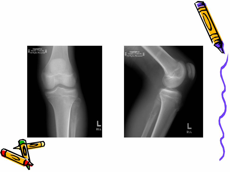
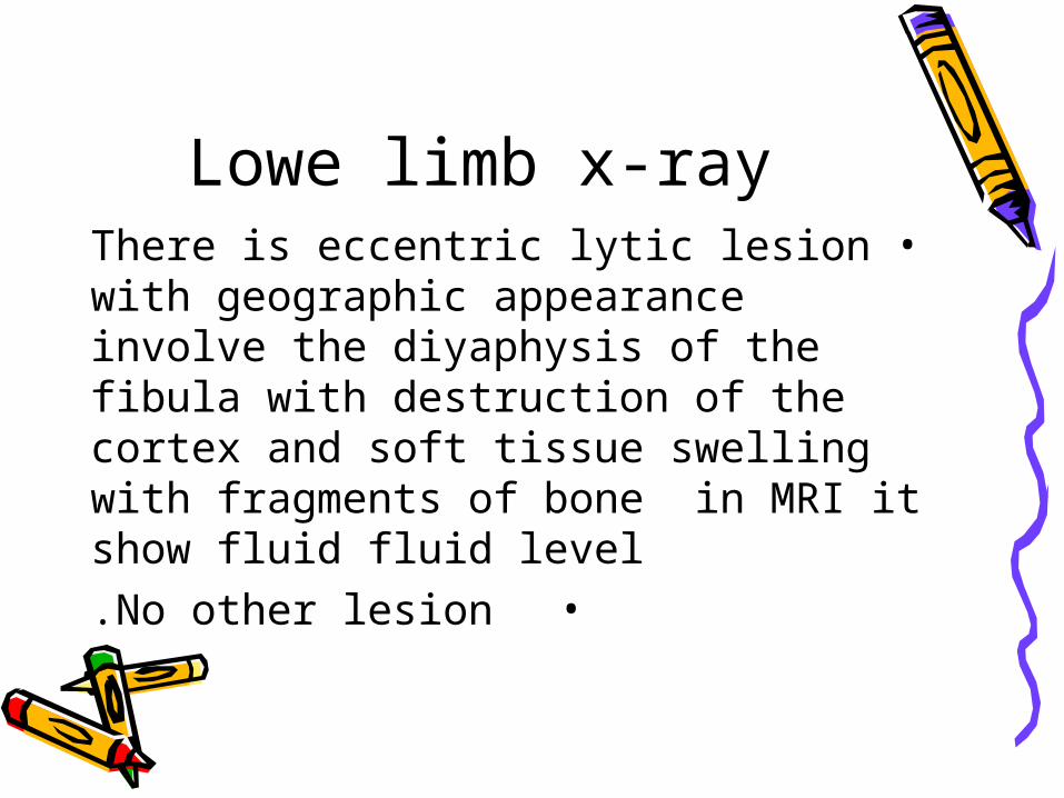
Lowe limb x-ray•There is eccentric lytic lesion with
geographic appearance involve the diyaphysis of the fibula with destruction of the cortex and soft tissue swelling with fragments of bone in MRI it show fluid fluid level
•No other lesion.
• Ddx• Telange Osteosarcoma• Ewing sarcoma• Giant cell tumor• FIBROSARCOMA

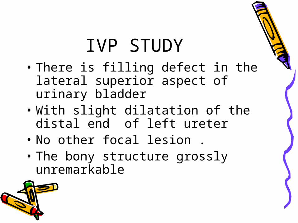
IVP STUDY• There is filling defect in the lateral
superior aspect of urinary bladder• With slight dilatation of the distal
end of left ureter• No other focal lesion .• The bony structure grossly
unremarkable
ddx• Transitional cell ca.• Squmas cell carcinoma• Lymphoma• Inflammatory process • cystitis
![DeLand News. (Deland, Florida) 1909-10-22 [p ].ufdcimages.uflib.ufl.edu/UF/00/07/58/96/00041/00347.pdfpoint They bilious being given ollicial request booklet prompt poison Notice](https://img.pdfslide.us/doc/110x75/5f990201186ce06c6f346e0c/deland-news-deland-florida-1909-10-22-p-point-they-bilious-being-given-ollicial.jpg)


![Double Incomplete Pyloromyotomy (A. Ezzat Technique): A ...incidence reported of 1 to 8 per 1000 live births [1] [2]. Projectile non bilious vomiting and its complications are common](https://img.pdfslide.us/doc/110x75/6030a50ce330ab27063bb564/double-incomplete-pyloromyotomy-a-ezzat-technique-a-incidence-reported-of.jpg)