Embed Size (px)
Citation preview

CardiacCardiacCardiacCardiac
Elisa A. Mancuso RNC, MS, Elisa A. Mancuso RNC, MS, FNSFNS
Professor of NursingProfessor of Nursing
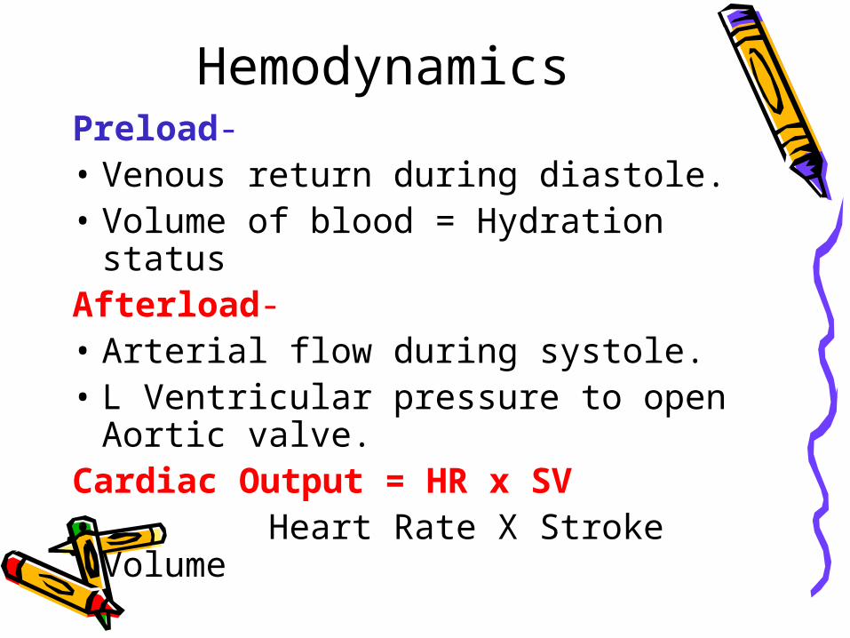
HemodynamicsPreload-• Venous return during diastole. • Volume of blood = Hydration
statusAfterload-• Arterial flow during systole.• L Ventricular pressure to open
Aortic valve.Cardiac Output = HR x SV• Heart Rate X Stroke Volume
Murmurs
Abnormal heart sounds • Improper closing or opening of valves• 80% of murmurs in kids are innocent • Close proximity of heart to chest wall • Stills Murmur –
– Blood rushing out of aorta
• Anemia • Fever

Fetal CirculationOxygenated blood → Inferior Vena Cava (IVC)
IVC → RA → FO → LA → LV → Aorta → Head & Arms– ↑↑ pressure in RA → Foramen Ovale (FO)– Bypasses the lungs and blood shunted to LA
↓ Blood returned → Superior Vena Cava (SVC)
SVC → RA → RV → PA→ DA →Aorta → Lower Body & LegsDuctus Arteriosus (DA)– Bypasses the lungs (↑↑ Pressure) – Only small portion of blood goes to pulmonary
system– Blood gets re-oxygenated via placenta
Ductus Venosis
– Bypasses the liver and shunts blood to IVC

Fetal to Neonatal Circulation
At birth lungs expand:• ↑ O2 causes pulmonary vasodilation• ↓ Pulmonary pressure (resistance)• ↑ Systemic pressure (resistance)
– LA pressure > RA pressure– Foramen Ovale closes within 1st hour of life
• ↑O2 ↓Prostaglandins (from Placenta)– Ductus Arteriosus closes within 10-
24hours.– Permanent closure by 3-4 weeks.
– PDA = Patent Ductus Arteriosus • In some cases can stay open for 3
months

Neonatal Circulation
• Blood flows from higher to lower pressure
• Systemic Pressure > Pulmonary Pressure L side > R side Blood flows from L → R side

Acyanotic Defects
• L →R side shunt of oxygenated blood
• ↑ Pulmonary blood flow • Pulmonary congestion• Heart is ineffective pump• Children prone to CHF• Prophylactic administration of
antibiotics needed

Patent Ductus Arteriosus (PDA)
Opening between the pulmonary artery (PA) and Aorta
Oxygenated blood shunted from Aorta →→PA– ↑↑ Systemic resistance
• Blood shunted to LA → LV → PA
• ↑↑ Pulmonary Congestion
• ↑↑ Back up to LA & LV
• LV Hypertrophy

PDA Clinical signs • Soft - harsh systolic newborn
murmur • Machinery type systolic and
diastolic murmur in older children• ↑ RR & moist Breath sounds• Bounding pulses • ↑ HR• Widened pulse pressure
– Large difference between the systolic – and diastolic pressure
Therapy • Indomethacin (indocin)
– Prostaglandin inhibitor promotes vasoconstriction and closure of PDA
– 3 Dose maximum q 12 hours
• Ligation of Ductus Arteriosus– Close connection to prevent return
of oxygenated blood to lungs– No open heart surgery –

Atrial Septal Defect (ASD)
Abnormal opening between the RA & LA • Blood flows from ↑↑press LA to ↓↓press RA• ↑↑ blood volume to right side of heart
– Leads to RA and RV hypertrophy • ↑↑ blood volume to lungs
– Pulmonary Congestion• DOE/ CHF symptoms• Crescendo/decrescendo systolic • ejection murmur

ASD Therapy• ASO (Amplatzer Septal
Occluder) – via cardiac cath
• Medications for CHF• Open heart surgery and
bypass, performed before school age
• Dacron patch• Low mortality rate

Ventricular Septal Defect (VSD)
Abnormal opening between RV and LV
• ↑↑O2 blood from LV to RV• ↑↑ blood to RV = RV
hypertrophy• ↑↑ pulmonary flow • ↓↓ systemic flow
• Spontaneous closure in 20%-60% within first year of life.

VSD Clinical signs • CHF = ↓↓CO, ↑HR, ↑RR, scalp
sweating, – ↑ weight gain, irritability
• Pulmonary edema• DOE, fatigue, ↓↓ PO intake• ↓↓Aorta Blood Flow
– ↓↓ femoral and brachial pulses– ↓↓ BP x 4
• Harsh holosystolic murmur with thrill
Therapy same as ASD

Pulmonic Stenosis (PS)
Narrowing of the pulmonary valve
• ↑↑ PA pressure/resistance
• ↓↓ Pulmonary Blood Flow
• Blood backs up into RV RV Hypertrophy

Clinical Signs & Therapy
• Depends on size of stenosis• Pale, lethargic, slow feeder• Systolic ejection murmur• EKG and CXR show RV Hypertrophy
Therapy• Pulmonary Valvotomy
– Angioplasty – Enlarges ↑ pulmonic valve
opening

Aortic Valvular Stenosis
Narrowing of aortic valve• ↑↑ Resistance to blood flow from LV
– causing LV Hypertrophy
• ↑↑ back-up of blood in pulmonary system – ↑↑ Pulmonary congestion
• ↓↓ blood via aorta ↓↓Systemic perfusion = ↓↓
CO

Clinical• Faint peripheral pulses RT ↓↓ CO• ↓↓ pulse pressure• Chest pain RT myocardial ischemia• Systolic ejection murmurTherapy• Commissurotomy
– Enlarge aortic valve opening via angioplasty.
• Additional surgery may be needed
• later.

Coarctation of the Aorta
• Narrowing of the aorta right after arch
• ↑↑Pressure proximal to narrowing – ↑↑ BP upper body, arms & head– Bounding pulses & warm, ruddy skin– JVD
• ↓↓Pressure distal to narrowing – ↓↓ BP lower body & legs– Weak pulses & cool, pale skin
• Difference of 20mm for systolic BP

Clinical signs• ↑↑ BP in arms ↓↓ BP legs• Weak or Absent femoral pulses• Headache, blurred vision and nose bleeds• ↑↑risk for stroke• Older kids leg pain on exertion RT ↓
bloodTherapy• Prostaglandin E – keep PDA open• Surgery
– Resect coarcted portion and reanastomosis
Cyanotic defects• Unoxygenated blood enters
systemic system • “Right to Left shunt” (R→ L)• Blood is shunted from venous to
arterial• ↑↑ CHF and hypoxic episodes
Now classified as: ↓↓ Pulmonary blood flow or Mixed blood flow defects
Transposition of Great Vessels (TGA)
• Two separate circulations!
• Aorta arises from RV– Unoxygenated blood enters aorta
→Systemic
• Pulmonary artery (PA) arises from LV– Oxygenated blood enters PA → recycled
lungs → Pulmonary veins → LA
No Oxygenated blood in systemic circulation!
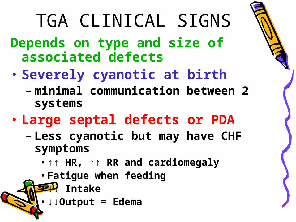
TGA CLINICAL SIGNSDepends on type and size of
associated defects• Severely cyanotic at birth
– minimal communication between 2 systems
• Large septal defects or PDA – Less cyanotic but may have CHF
symptoms•↑↑ HR, ↑↑ RR and cardiomegaly•Fatigue when feeding•↓↓ Intake •↓↓Output = Edema

THERAPYProstaglandin E1 (Prostin VR or Alprostadil) • Vasodilator • Relaxes smooth muscle of ductus arteriosus • Keeps PDA open • Provides mixing of oxygenated and
deoxygenated blood to systemic circulation.
“Rashkind procedure”• Cardiac cath to create ASD• Maintains mixing of blood• Arterial switch procedure usually
performed • in first few weeks of life

Tetralogy of FallotInvolves four cardiac defects• VSD
– Blood shunted RV→ LV• Pulmonary Stenosis
– ↓↓ blood to PA• Overriding Aorta
– Sits over VSD• RV Hypertrophy
– ↑ pressure from stenosis

Clinical signs of Tetralogy
• First cry hypoxic and cyanotic
• ↑↑ Activity = ↑↑ Hypoxia and ↑↑ Cyanosis– Pulse oximeter in low 70’s
• ↑↑ Pulmonary stenosis = ↑↑ Cyanosis• ↑↑ HR, ↑↑ RR• Tire easily can’t finish feedings = ↓↓ Intake
• Chronic O2 deficit → Polycythemia
– ↑↑ # RBC’s to supply 02 to body – ↑↑ Risk of CVA or embolism with
dehydration

Clinical Manifestations
• “Tet” Spells – ↑↑ Activity or ↑↑ Crying = ↓↓ blood flow to
brain– ↑↑ hypoxia, cyanosis and fainting
• Squatting – compensatory action– Knee chest position– ↓↓femoral blood flow ↑↑blood flow upper
body
• Clubbed fingers• Mental retardation

Therapy
• Prostaglandin E1 – Maintain PDA – ↑↑ Pulmonary perfusion
• Surgery– Patch the VSD – Open stenotic pulmonary valve– Heart Transplant with severe
defects
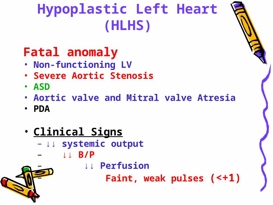
Hypoplastic Left Heart (HLHS)
Fatal anomaly• Non-functioning LV• Severe Aortic Stenosis• ASD• Aortic valve and Mitral valve Atresia • PDA
• Clinical Signs– ↓↓ systemic output– ↓↓ B/P– ↓↓ Perfusion– Faint, weak pulses (<+1)

Treatment • ExtraCorporal Membrane Oxygenation
(ECMO)– ↑↑ Risks & Costs ($250,000/day)– ↓↓ Availability @ Regional centers
• Heart Transplant– ↓↓ Donor hearts
• 3 Stage surgery if child can tolerate it.
• DNR & Letting Go – Bereavement

Tricuspid Atresia (TA)
Three major defects• No tricuspid valve• ASD & VSD• RV Hypoplasia
Lungs receive blood via– PDA– small VSD– bronchial vessels
• As long DA remains open the child
• receives adequate O2.
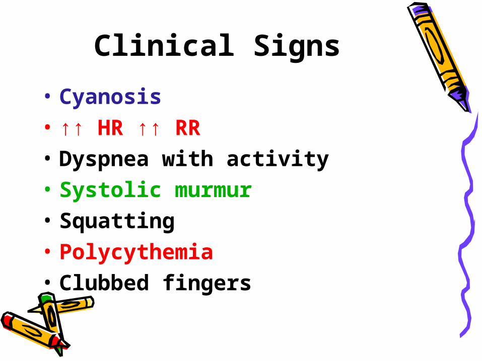
Clinical Signs
• Cyanosis• ↑↑ HR ↑↑ RR• Dyspnea with activity• Systolic murmur• Squatting• Polycythemia• Clubbed fingers

Therapy• Prostagladin E
– Maintain PDA for 2 weeks then need surgery.
• Surgery-– Anastomosis to allow blood flow to lungs. Three stages1) Blalock-Taussing @ 2 weeks of age
shunt btwn PA and Aorta2) Glenn @ 6 month to a year
shunt from SVC to PA to lungs 3) Fontan @ 2-3 years shunt from IVC to PA to lungs No more mixing of blood

Truncus Arteriosus
• One common artery arises from LV & RV.
• Overrides a large VSD• No separate PA or Aorta• Unoxygenated blood enters systemic
circulation• ↑↑ blood volume flows to lungs • ↑↑ pulmonary blood flow• ↑↑ pulmonary edema ↑↑ CHF

Treatments
• CHF and fluid overload – Lasix (1 mg/kg/dose)
•Diurectic = ↓↓ edema ↓↓ Na, ↓↓ K+
– Digoxin (Digitalization Dosing) •Cardiac glycoside = ↓↓ edema
• Surgery– VSD– R side graft

Nursing Interventions
• √ Maternal History:– Rubella, DM, ETOH or Cardiac
disease– Congenital heart disease – Chronic maternal illness– Perinatal infections (TORCH) – ertain meds maybe linked– Substance Abuse
•ETOH may be associated with FAS and Tetralogy of Fallot

Physical ExamThoracic ExamCardiac Sounds• √ Location of PMI (5th LICS MCL)• √ Rate• √ Rhythm• √ Murmurs
– location, intensity and where in cardiac & respiratory cycles
• √ visible pulsation on thorax• √ JVDBreath Sounds
– √ Rate, rhythm– √ Dyspnea and Grunting (keep alveoli
open)– √ Adventitious sounds– Moist- Pulmonary congestion or CHF

COLOR
• √ Mucous membranes– Lips, conjunctiva and nail beds.
• √ Cyanosis – @ rest or with activity
• √ clubbed fingers
• Flushed cheeks = Polycythemia – KEEP INFANTS HYDRATED! – WHY?

Pulses
• Palpate bilaterally • Compare upper and lower
extremities – Absent or ↡femoral pulses in
Coarctation
• √ Rate/Rhythm/strength (0-+4)• √ BP all four extremities
– Widened pulse pressure in PDA – ↑↑ BP upper extremities in
Coarctation.

Nutritional Status√ Intake
– Rest periods needed?– Time needed to complete feedings– ↓↓ intake, tiring due to ↓↓
available O2
√ HT, WT and HC√ Activity level-tires easily?• Developmental tasks
achieved?

Respiratory Infections
↑↑Risk– Pulmonary vascular congestion– Bacterial invasion and growth
•RT stasis of secretions (prophylaxis meds)
Therapy– Meds
•Bronchodilators•Steroids
– PD & C – O2

Compensatory Mechanisms
• Cardiomegaly– ↑ pumping action of heart = ↑ SV– ↑ use of cardiac muscle = ↑ O2 availability – ↑ size = hypertrophy
• Tachycardia >160 in infant– ↑ rate = ↑ CO– ↑ O2 to tissues and vital organs
• Polycythemia– ↑ production of RBC’s– ↑ availability of O2 to tissues – ↑ viscosity of blood – ↓↓ flow, sluggish– ↓↓ decreased peripheral circulation – High risk for CVA
• Tachypnea > 60 in infants– ↑↑ RR = ↑↑ O2

Compensatory Posturing • ↑↑ O2 to vital organs by ↓↓ workload of heart
– Less area for blood to flow = ↓↓ venous return
– “TET Spells”
• Infants– May be flaccid with extremities extended – Knee chest position (infant seat)
• Preschool– Squatting position
•occludes femoral vein = ↓↓ venous return ↓↓ workload on heart ↑↑O2 sat & ↑↑ blood to vital organs

Congestive Heart Failure CHF
Children’s CHF due to congenital heart defects
• CHF = ↓↓ Contractility of heart = ↓↓ CO • ↓↓ blood volume for systemic
circulation• ↑↑ pulmonary congestion • ↓↓ O2 and ↓↓ nutrition. Unable to meet metabolic demands

InterventionsParent teaching• Review defect and s/s when to call MD• Meds - dose, schedule, SE
– Prophylactic antibiotics – Immunizations
• Nutrition - ↑ cal formula, ↑ Fe, ↑ K+, ↑ Protein, ↓ fat, ↓ Na
• Activity- allow for rest periods for fatigue • ↓ Cardiac demands
– Position, thermoregulation• “Cardiac Cripple”
– Parents overprotect and child manipulates Set limits & discipline WNL
• Emotional support (access to NP/RN 24 hours)
• Encourage support groups (specific to defect)

MedicationsDigoxin• Action -cardiac glycoside
– ↑ Contactility of heart = ↑ efficacy = ↑ CO– Slows down SA node = ↓ HR
• DigitalizationLoading Dose = 30-40 mcg/kg/dose ÷ (½, ¼, ¼)Maintenance dose = 4-5 mcg/kg/day ÷ q 12
Nursing interventions√ Apical pulse for one full minute before giving med.
Hold med if:Infant <100 Toddler <90 Preschool <70 School age <60
Document Apical HR next to dose on MAR

Nursing Interventions
• √ I and O and √ K+ level– ↓↓ K+ = ↑↑ Dig toxicity
• √ Serum Digoxin level (0.5-2ng/dl)
• Digoxin Toxicity (>3ng/dl)– vomiting (earliest sign), nausea (↓↓ Po
intake)– lethargy and bradycardia
• Administer with 2 RN’s– Review order & √ HR parameters √ Dosage and calculation √ Actual dose in syringe a administering Document on MAR: HR & Initials @ dose

DiureticsAction- eliminates excess H2O and Na = ↑ fluid loss
↓edema and ↓work for heart and lungs
• Furosimide-Lasix (Strong acting) 1mk/kg/dose– Blocks reabsorption of Na+ H2O @ loop of Henle – ↑↑↑ loss of Na+, K+ and H2O
• Thiazides-Diuril 10-20 mg/kg/dose– Blocks reabsorption of Na+ H2O K+ distal
tubules– ↑↑↑ loss of Na+, K+ and H2O
• Aldactone (Aldosterone Inhibitor)- (K+ Sparing)– Blocks action of Aldosterone
• hormone that retains Na+ and H2O Promotes H2O and Na+ loss & Retains K+

Nursing Interventions√ Weight
– Same time, scale and amount of clothing = None!
√ I and O– weigh all diapers – √ skin tugor on sternum (tenting= dehydrated)
√ Serum electrolytes– K+, Na+, BUN and Creatine
Administer K+ supplements
KCL, Slo-K, K-Lor, K-Dur
K+ level affects Digoxin efficacy!

Prostaglandin E1 (Prostin VR)
Vasodilator (0.1 ug/kg/min)– Relaxes vascular smooth muscle – Keeps open Ductus Arteriosus (DA). – ↑ Pressure in L heart = ↑ pressure in
Aorta – Blood shunted from Aorta ➔ PDA ➔ PA– ↑ Blood to lungs =↑ perfusion = ↑
oxygenation – ↑ O2 to systemic circulation
Maintains mixing of oxygenated and deoxygenated blood in cyanotic
defects.

Prostin VR Adverse SE
• Apnea- must intubate
• Cutaneous generalized flushing
• ↓↓ BP & ↓↓ HR
• ↑↑ Seizures
• ↓↓ I & O
Hemorrhage and thrombocytopenia ✔CBC ✔ PLTS ✔ PT/PTT

Indomethacin Na (Indocin)
NSAID (0.1 -0.3 mg/kg/dose q12h
Max 3 doses)• Action
– Inhibits prostaglandin synthesis – Promotes PDA closure
• Assess presence of murmur (+) = murmer, give med(-) = closed, hold med

Indocin Adverse SE
↓↓ Renal & ↓↓ GI blood flow• ✔ I & O ✔ UA ✔ BUN/creatine• ✔ Bowel sounds • Guiac stools for necrotic bowel
– (NEC) Necrotizing enterocolitis
↓↓ Platelet function ✔ CBC ✔ PLTS ✔ PT ✔ PTT

Kawasaki Disease
• Most common acquired heart disease in children <8 years of age
• Acute febrile & multi-system disorder
• Autoimmune– Skin, mucous membranes, lymph nodes– Vasculitis ➔ cardiac complications
• ↑↑ incidence near fresh H2O• Late winter/early spring

Clinical signs
• Fever >5 days • Febrile seizures• Cervical lymphadenopathy
>1.5 cm • Bilateral non-exudative
conjunctivitis• Strawberry tongue• Dry, red, cracked lips

Clinical signs
• Truncal rash• Erythema & edema of palms and soles • “Shedding skin”
– Desquamation from fingers • ↑ WBC ↑ ESR ↑Plts
Cardiac sequella• Pericarditis• Myocarditis• Arrhythmias Coronary Artery Aneurysm
If untreated 15-25 % develop MI

Nursing Interventions
• IV Immune Globulin (IVIG)– ↓↓ the incidence of coronary aneurysm <3%
Single dose IVPB over 24 hours• ↑↑Dose ASA (100 mg/kg/day)
– √ thrombocytosis• Bed rest • ↓ O2 Demands• Petroleum jelly to lips• √ CHF: ↑HR ↑RR dyspnea crackles• Strict I & O• Tepid sponge bath
• • Complete and spontaneous recovery in 3-4
weeks!

Subacute Bacterial Endocarditis (SBE)
• Infection of valves and inner lining of heart• High risk patients = congenital heart disease • Bacteremia
– Strep Viridians- most common 70%, • Staph Aureus 20%, Candida Albicans 10%
– Enters blood stream via teeth, gums, tonsils, UTI. – Slow & insideous onset– Attaches to congenital anomalies or prosthetic valve
sites
• Vegetations– Bacteria, fibrin and plt thrombi grow on endocardium– Invade Aortic and Mitral valves ↑↑ turbulent blood flow and break off as
embolism spleen, kidney, CNS, lung and skin.

SBE Clinical Signs• Fever- low grade, intermittent or unexplained• Anorexia- malaise.
– “feel like getting the flu”• Murmur
– New or change in previous murmur • Cardiomegaly • Splenomegaly • Osler nodes-
– Red, painful nodules at finger tips• Janeway’s spots
– Painless, hemorrhagic areas on palms and soles
• Splinter hemorrhages– thin black lines under nails
• Petechiae on oral mucous membranes• HA, ↓↓motor coordination = CVA!!
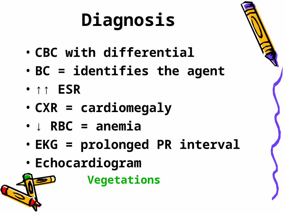
Diagnosis
• CBC with differential• BC = identifies the agent• ↑↑ ESR• CXR = cardiomegaly• ↓ RBC = anemia• EKG = prolonged PR interval• Echocardiogram
– Vegetations

Therapy• Bed rest• High dose (Meningitic) Antibiotics
– PCN, Gentamycin, Ampicillin
• IV therapy 4-6 weeks • ✔ med SEs- ✔hearing ✔renal status• Serial BC• Counsel parents regarding antibiotic prophylaxis a & p invasive procedures

Rheumatic FeverAutoimmune response to
Group A β Hemolytic Strep
• Caused by untreated/partially treated group A strep pharyngitis• Symptoms appear 2-6 weeks after infection
• Diffuse inflammatory & collagen disease– connective tissue, joints– subcutaneous tissue– Brain, heart and blood vessels

Diagnosis Jones Criteria
• Carditis– Cardiomegaly, murmur RT Mitral regurgitation– valvulitis (Endocardium ➔ Pericardium), ↑ HR
• Ashkoff bodies• Hemorrhagic lesions in heart
• Polyarthritis– Reversible and migratory – knee ➔ shoulder ➔ elbow
• Subcutaneous nodules– 1 cm non-tender swelling over bony prominences.
• Erythema marginatum– Red macular wavy rash with clear center
Chorea “ St. Vitus dance” Involuntary movements of extremities and face ↑ c anxiety ↓ c rest

Diagnosis
• ↑CPR (C-Reactive Protein)• ↑ESR• +Throat culture• ↑ ASO titer >333
– Anti-streptolysin reflects lysis of RBC
– ↑ 7 days p onset– Max. level 4-6 weeks
• +BC• EKG = prolonged P-R interval
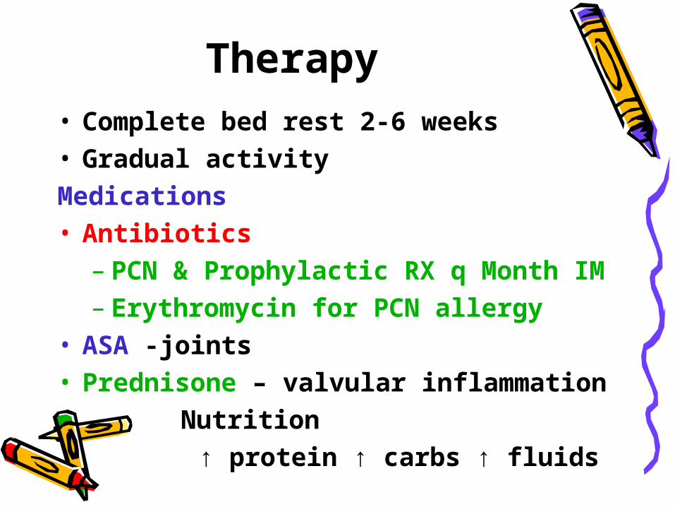
Therapy • Complete bed rest 2-6 weeks• Gradual activityMedications• Antibiotics
– PCN & Prophylactic RX q Month IM– Erythromycin for PCN allergy
• ASA -joints• Prednisone – valvular inflammation Nutrition
↑ protein ↑ carbs ↑ fluids

Iron Deficiency Anemia
• Inadequate supply of dietary iron (Fe)
• Infants – ↑ risk @ 6 months – Fetal Fe stores are depleted– ↑ milk consumption & ↓↓ protein/solid
intake
• Adolescents – ↑ growth spurt– poor nutrition – ↑ blood loss c menses
No whole milk until after 1 year.
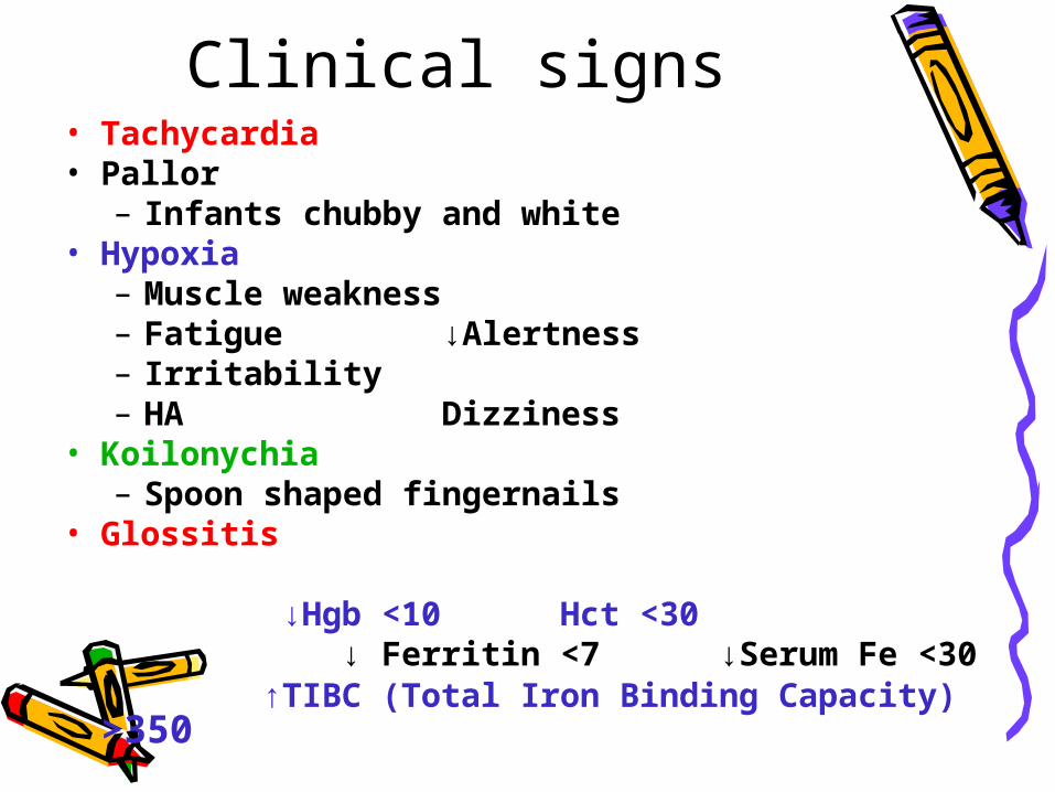
Clinical signs • Tachycardia• Pallor
– Infants chubby and white• Hypoxia
– Muscle weakness– Fatigue ↓Alertness– Irritability– HA Dizziness
• Koilonychia– Spoon shaped fingernails
• Glossitis
↓Hgb <10 Hct <30 ↓ Ferritin <7 ↓Serum Fe <30 ↑TIBC (Total Iron Binding Capacity)
>350

Therapy• Fe supplement (2-3 mg/kg/day)
– Give between meals with acidic fluids. – Takes at least 4 months to replace loss– SE: Stains teeth, black tarry stools
• Dextran (parental iron)– Z-track deep IM - buttocks only
• Nutrition Green leafy vegetables, whole wheat, beans, shellfish, egg yolk, Organ
meats,

Hemoglobinopathies
Sickle Cell Disease
• Defective HgB chain (HgS)
• RBC’s are sickle shaped – Unable to carry O2
• RBCs have a shorter life span 16-30 days

Sickle cell • Autosomal recessive
– AA = WNL– AS = trait (carrier)– SS = Disease
• Both parents have trait– 25% = AA normal– 25% = SS disease – 50% = AS (carriers)
• Only 35-45% HgB is sickled • Majority Asymptomatic
• ↑ Risk in African Americans 15-40%
A S
A AA AS
S AS SS

Pathophysiology• Vaso-occulusion
– Sickle Shaped cells stack up– Lodge in small vessels– ↓ blood flow
• Tissue Hypoxia– ↑Viscosity of blood = ↓blood flow– ↓ O2 & ↑ metabolic end products
• Tissue Ischemia– Edema & necrosis @ site
• Infarction– Brain, Kidneys & Liver

Clinical signs• First sign
– Fetal hemoglobin (HgB) is depleted – HgS hemoglobin is now dominant
• Low Hgb: 5-9 Hct: 15-30• Pallor• Jaundice
– ↑ RBC’s destroyed RT ↓life span
• Frequent URI’s• Generalized weakness• Hepatosplenomegaly

Sickle Cell Crisis = Sickling
Acutely ill RT ↓O2 and Dehydration
• ↑ Stress or ↑infection (URI GI GU)
• ↑ Temp - Dehydration• ↑ BMR ↑ O2 consumption
– leads to tissue hypoxia

Clinical Signs • Abdominal pain-
– Thrombosis to liver and spleen• Severe bone pain RT sickled joints• Hematuria and diuresis RT renal
ischemia• Seizures RT Brain thrombosis/CVA
• Acute Chest Syndrome Severe chest pain SOB, ↑ HR ↑ RR Pulmonary congestion

Therapy• Bone Marrow Transplant = ↑Prognosis • O2 humidified• Hydration
– ↑ PO intake 2.5 -3 L/day ✔ I & O• Pain control
– PCA , MSO4, Fentenyl– ASA– No Demerol ( Metabolite = ↑↑ seizures)
• Folic acid– ↑RBC
• PRBC Transfusions weekly (Hgb <10)• Splenectomy (kids <5 years)
– Prevents Splenic Sequestration – Massive entrapment of sickled cells in
spleen – HYPOVOLEMIC SHOCK!!!

Parent Teaching• Life long-frequent hospitalizations• Life Span determined by % sickled RBCs • Genetic Counseling
– Have all children tested
• Monitor fluid losses– ✔diapers ✔mucous membranes ✔Skin turgor
• ↓ Infection-Immunize on schedule– meningococcal, pneumococcal and hep B– Prophylactic PCN by 2 months of age– NO day care/malls ↓Exposure to other kids
• ↑ Coping techniques & Stress Reduction•
✔

Hemophilia A Classic Hemophilia
• Lacks Factor VIII (AHF)– AHF Anti hemolytic Factor– Severe & spontaneous bleeding – Not trauma induced
• Sex linked recessive-X chromosome – Mom transfers diseases to boys – Girls are carriers

Clinical signs• 1st indication at circumcision• Crawling = ↑ bruises on pressure areas• Hemarthrosis
– Bleeding into joint cavities (synovial space)
– Early sign = stiffness, tingling or achy– Warmth, redness, swelling– ↓ ROM & function– Alkylosis of joint
• Spontaneous bleeding– Epistaxis, loose baby teeth, Hematuria Spinal Cord Hematoma = paralysis Intracranial Hemorrhage = Death
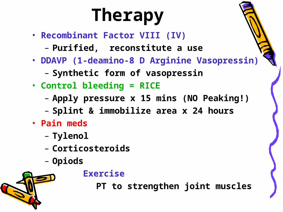
Therapy• Recombinant Factor VIII (IV)
– Purified, reconstitute a use• DDAVP (1-deamino-8 D Arginine
Vasopressin)– Synthetic form of vasopressin
• Control bleeding = RICE– Apply pressure x 15 mins (NO Peaking!)– Splint & immobilize area x 24 hours
• Pain meds– Tylenol– Corticosteroids– Opiods
Exercise PT to strengthen joint muscles
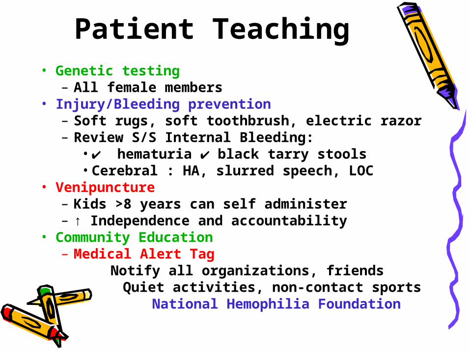
Patient Teaching• Genetic testing
– All female members• Injury/Bleeding prevention
– Soft rugs, soft toothbrush, electric razor– Review S/S Internal Bleeding:
• ✔ hematuria ✔ black tarry stools•Cerebral : HA, slurred speech, LOC
• Venipuncture– Kids >8 years can self administer– ↑ Independence and accountability
• Community Education– Medical Alert Tag Notify all organizations, friends
Quiet activities, non-contact sports National Hemophilia Foundation