Embed Size (px)
Citation preview
449 M.E.J. ANESTH 19 (2), 2007
CARDIAC ARREST IN PREGNANCY
ALIYA DABBOUS* AND FOUAD SOUKI
**
Cardiac arrest occurs only about once in every 30.000 late pregnancy, and survival from such an event is exceptional1. Not only is cardiac arrest a feared entity in this population, but it is also unexpected and catastrophic2. For this reason, it is necessary that the obstetric anesthesiologist be knowledgeable about the risk factors for cardiac arrest, the physical changes in the parturient and the management of CPR during pregnancy.
There are multiple etiologies of cardiac arrest during pregnancy. Some are pregnancy-associated, and others result form conditions that existed before pregnancy (Table 1).
Table 1
Obstetric causes Nonobstetric causes
Hemorrhage
Preeclampsia
HELLP syndrome
Amniotic fluid embolus
Anesthetic complications
Pulmonary embolus
Septic shock
Cardiovascular disease
Myocardial infarction
Trauma
Obstetric Causes
Hemorrhage
Obstetric hemorrhage can be massive and may lead to maternal
cardiac arrest. Causes of obstetric hemorrhage include abruptio placenta,
placenta previa, uterine rupture, and uterine atony3. Postpartum bleeding
From the American University of Beirut, Dept of Anesthesia, Beirut, Lebanon.
* MD, Lecturer.**MD, MS, Chief Resident
ALIYA DABBOUS & FOUAD SOUKI450
tends to be underestimated. Early diagnosis, identification, and treatment
of the source of bleeding with volume resuscitation may help avert
cardiac arrest. If hemorrhage is suspected, adequate intravenous access
should be immediately obtained. Blood is typed and cross-matched, and
the source of bleeding identified and treated. Uterine atony is the most
common etiology. It is important to restore adequate blood volume when
hemorrhage is the etiology of cardiac arrest.
Preeclampsia and HELLP
Preeclampsia is a significant contributor to maternal and fetal death
and complications. The complications of preeclampsia which precipitate
cardiac arrest include: eclampsia, pulmonary edema, cardiac dysfunction,
stroke cerebral edema and the HELLP syndrome; the latter being
associated with 40% incidence of cardiac arrest. latrogenic complications
of preeclampsia, such as magnesium toxicity and fluid overload, also
contribute to maternal cardiac arrest3,4.
If cardiac arrest complicates preeclampsia, it may also be due to
decreased intravascular volume, which is part of the pathophysiology of
preeclampsia. Hypovolemia makes cardiac compressions less efficient,
than the nonpregnant resuscitation efforts that yield 30% of the cardiac
output. Therefore volume repletion is an important part of the
resuscitation of preeclampsia3.
Amniotic Fluid Embolus
Amniotic fluid embolus is a devastating complication of pregnancy
that causes maternal death rate as high as 50%5. The cause of death in
amniotic fluid embolus is likely to be cardiac arrest5. The use of
cardiopulmonary bypass for women with life threatening amniotic fluid
embolism during labor and delivery has been reported with successful
outcome6.
CARDIAC ARREST IN PREGNANCY
M.E.J. ANESTH 19 (2), 2007
451
Anesthetic Complications
Complications of regional and general anesthesia are another
cause of cardiac arrest in labor and delivery. Even with routine dosing
and testing, epidural catheters can migrate into the intravascular or
subarachnoid space, leading to systemic toxicity or total spinal
anesthesia, respectively. Drug overdosage can lead to local anesthetic
toxicity, particularly in patients who have received multiple top-up
doses of local anesthetics during a prolonged labor. Unlike the
operating room, patients in the delivery suite receive regional
anesthesia for prolonged periods of time without the continuous
presence of anesthesiologist. It is therefore essential that the amount
of local anesthetic the patient receives be monitored from shift to
shift. Finally, it is critical to remember that induction of either
regional or general anesthesia can lead to severe hypotension in
previously hypovolemic patients3.
The effective treatment of cardiac arrest during pregnancy
requires a complete understanding of the physiologic changes seen in
the parturient. These physiologic changes can make the diagnosis and
treatment of emergent situations difficult in the labor suite. Cardiac
output is greatly influenced by the patient’s position, especially
during the third trimester. The supine hypotension syndrome can
decrease cardiac output by 30-40%3. Many women cannot tolerate the
supine position, especially after 30 weeks of gestation. For safety,
women are encouraged to maintain left uterine displacement either by
elevation of the right hip or by positioning themselves on their sides.
In the labor suite, patients are frequently positioned on their back to
facilitate cervical exams and placement of monitoring devices. If the
patients are not reminded to return to their side, especially in the
presence of epidural anesthesia, there can be a rapid decrease in
venous return, leading to hypotension and possibly cardiac arrest.
ALIYA DABBOUS & FOUAD SOUKI452
Nonobstetric Causes
Pulmonary Embolus
Pulmonary embolus (PE) is a leading cause of maternal death. A high index of suspicion should exist when a pregnant woman presents with deep vein thrombosis or PE3. Immediate institution of anticoagulation therapy with heparin and diagnostic workup will markedly decrease the risk of maternal death in patients who have thromboembolic disease. When massive pulmonary embolus leads to cardiac arrest, thrombolytic therapy or thrombectomy should be considered7.
Septic Shock
Occurs in 1 per 5000 gestations and may result in maternal death. Sepsis can result from antepartum infections, including chorioamnionitis, pneumonia, and urinary tract infections. Endomyocarditis in the postpartum period can likewise lead to sepsis. Infections must therefore be treated aggressively3.
Cardiac Disease
Cardiac disease complicating pregnancy contributes to antepartum and postpartum maternal death. The risk of death is directly related to the New York Heart Associations status. Women with stage III or IV have a high risk of death3. In these patients. pulmonary artery catheters are recommended for tight regulations intravascular volume, cardiac output and oxygen delivery3.
Myocardial Infarction
May complicate pregnancy and is more likely to occur in women
CARDIAC ARREST IN PREGNANCY
M.E.J. ANESTH 19 (2), 2007
453
with underlying conditions such as diabetes mellitus, chronic hypertension, morbid obesity and ventricular hypertrophy. Pregnancy within 6 months of myocardial infarction is also associated with maternal complications and death3.
Severe asthma and other chronic pulmonary disease may worsen during gestation and likewise lead to respiratory failure and cardiac arrest3.
Trauma or Drug Overdose
Domestic violence increases during pregnancy. Homicide and suicide are leading causes of mortality. Accidental intravenous boluses of medications have been reported7; magnesium or vancomycin can lead to severe consequences, like hypotension and cardiac arrest4,7. Case reports have linked ergot derivatives with myocardial infarction and cardiac arrest in the parturient7.
Maternal Physiologic Changes in Pregnancy
Cardiovascular Changes
After the 10th week of gestation, cardiac output is increased by 1.0 to 1.5 liters per minute8. Maternal heart rate increases throughout pregnancy, reaching a peak of 15-20 beats above nonpregnant values during the third trimester8. Maternal blood pressure is less than nonpregnant values, with systolic pressures typically about 10-15 mmHg lower than non-pregnant values. Because of these cardiac changes, patients can bleed extensively before the normally recognizable physical signs, such as tachycardia and hypotension, occurs. In addition, at about 34 weeks gestation, plasma volume is increased by 40-50%. A smaller increase in red blood cell volume occurs, resulting in a decreased hematocrit and the physiologic anemia of pregnancy8. Often, a lower hematocrit can be misinterpreted due to this phenomenon, especially in the case of hypovolemia and acute hemorrhage. Because of these compensatory mechanisms, and the
ALIYA DABBOUS & FOUAD SOUKI454
tendency for blood flow to be shunted from the uteroplacental circulation under conditions of hypovolemia, the pregnant patient can lose 35% of her blood volume before tachycardia, hypotension, and other signs of hemodynamic instability can be identified7. A parturient may appear stable while the fetus is severely deprived of blood flow and the patient is greatly compromised7.
Respiratory Changes
Tidal volume increases during pregnancy and may cause an increase in minute ventilation. Increased oxygen consumption leads to increased rates of arterial oxygen desaturation in the parturient that becomes apneic8. The expanding uterus and consequent diaphragmatic elevation decreases thoracic volume leading to a decrease in functional residual capacity (FRC)8. Due to the hormonal and physical changes of pregnancy, patients are at increased risk for difficult ventilation and failed intubation7. Increased levels of progesterone leads to delayed gastric emptying, increasing the risk for aspiration during mask ventilation and intubation1,7. Though many centers require that patients remain fasting during labor, many patients will present in spontaneous labor after consuming a large meal. Edema of the upper airway, increased breast size and generalized weight gain can delay the establishment of adequate ventilation and intubation. Therefore, it is essential that oxygenation and ventilation be restored expeditiously while maintaining cricoid pressure7.
Resuscitation of the Pregnant Woman During Cardiac Arrest
Successful resuscitation in late pregnancy is difficult because the gravid uterus acts like an abdominal binder producing an increase in intrathoracic pressure, diminished venous return, and obstruction to forward flow of blood into the abdominal aorta, especially in the supine position2,7. In addition, the gravid uterus accounts for 10% of cardiac output and this massive shunting of blood may hinder efforts at CPR. External cardiac massage can at best generate only 30% of a non-pregnant patient’s normal cardiac output and increased oxygen requirements during
CARDIAC ARREST IN PREGNANCY
M.E.J. ANESTH 19 (2), 2007
455
pregnancy make the parturient much less tolerant to hypoxia2. Tosuccessfully resuscitate a parturient in late pregnancy, a coordinated team approach is essential. Obstetricians, anesthesiologists, neonatologists and nursing staff must work efficiently and in an organized fashion to resuscitate these patients. As these events happen unexpectedly, cooperation is essential to assemble needed equipment and perform resuscitation efforts in the labor suite1.
Modifications in emergency cardiac care for the pregnant uterus consist of:
1- Left uterine displacement.
2- Aggressive airway management (see section airway under BLS and ACLS).
3- Chest thrust instead of abdominal thrust in case of foreign body airway obstruction (see BLS).
4- Aggressive restoration of circulatory volume (ACLS).
5- Perimortem Cesarean delivery – Delivery within 5 minutes if fetus is viable.
Left Uterine Displacement
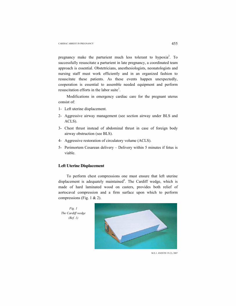
To perform chest compressions one must ensure that left uterine displacement is adequately maintained9. The Cardiff wedge, which is made of hard laminated wood on casters, provides both relief of aortocaval compression and a firm surface upon which to perform compressions (Fig. 1 & 2).
Fig. 1
The Cardiff wedge
(Ref. 1)
ALIYA DABBOUS & FOUAD SOUKI456
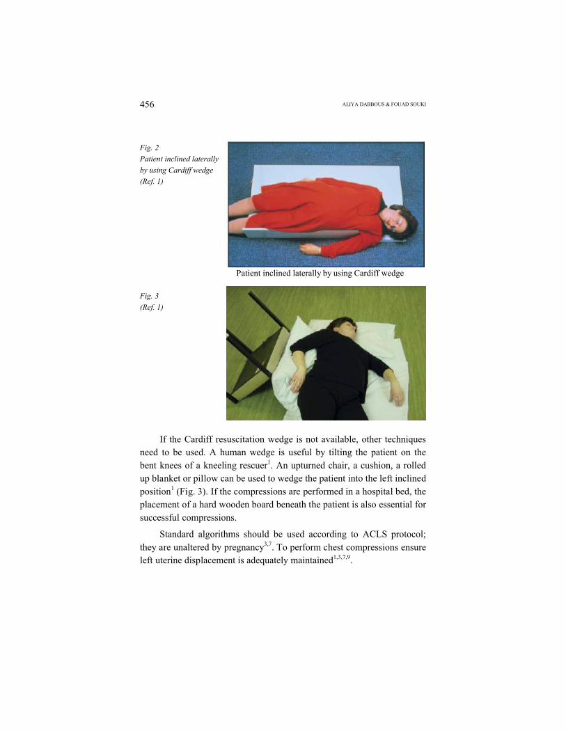
Fig. 2
Patient inclined laterally
by using Cardiff wedge
(Ref. 1)
Patient inclined laterally by using Cardiff wedge
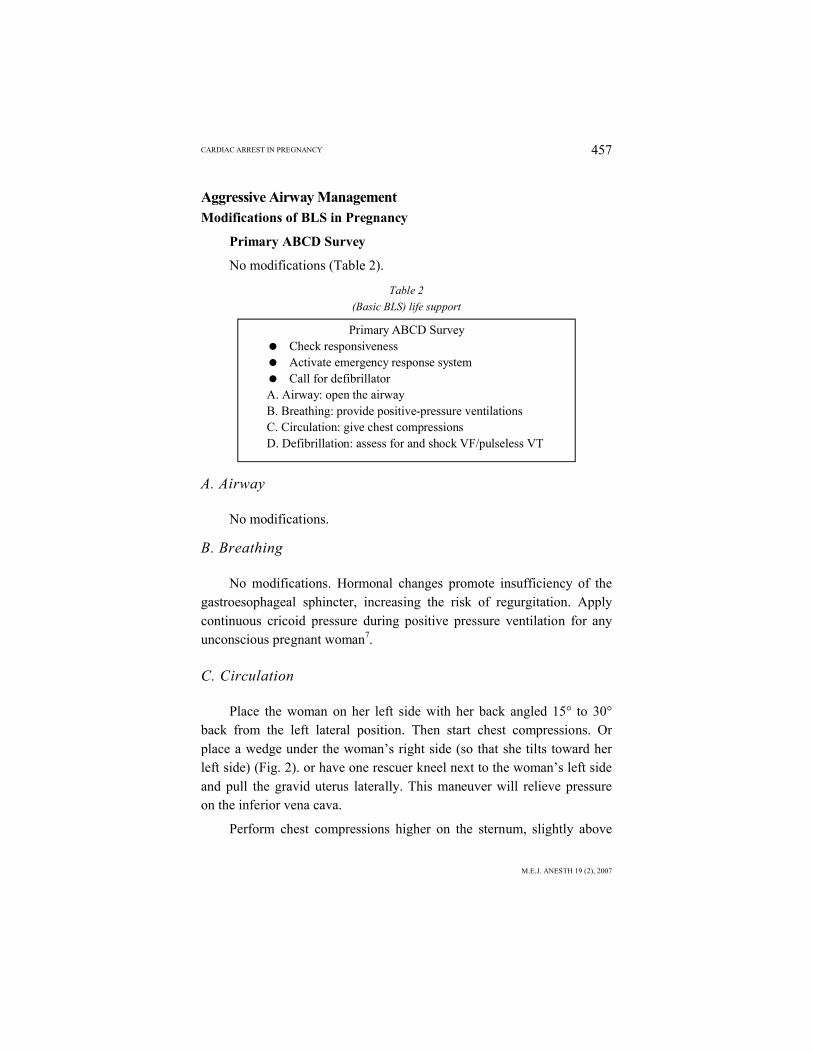
Fig. 3
(Ref. 1)
If the Cardiff resuscitation wedge is not available, other techniques need to be used. A human wedge is useful by tilting the patient on the bent knees of a kneeling rescuer1. An upturned chair, a cushion, a rolled up blanket or pillow can be used to wedge the patient into the left inclined position1 (Fig. 3). If the compressions are performed in a hospital bed, the placement of a hard wooden board beneath the patient is also essential for successful compressions.
Standard algorithms should be used according to ACLS protocol; they are unaltered by pregnancy3,7. To perform chest compressions ensure left uterine displacement is adequately maintained1,3,7,9.
CARDIAC ARREST IN PREGNANCY
M.E.J. ANESTH 19 (2), 2007
457
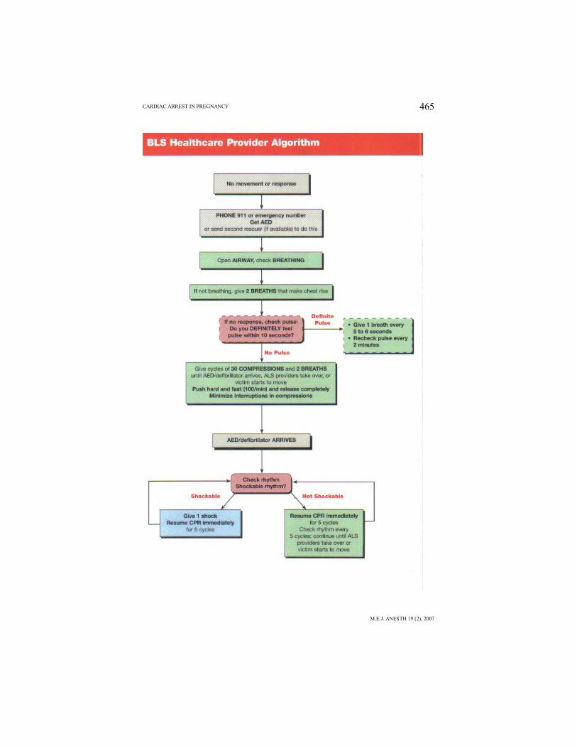
Aggressive Airway ManagementModifications of BLS in Pregnancy
Primary ABCD Survey
No modifications (Table 2).
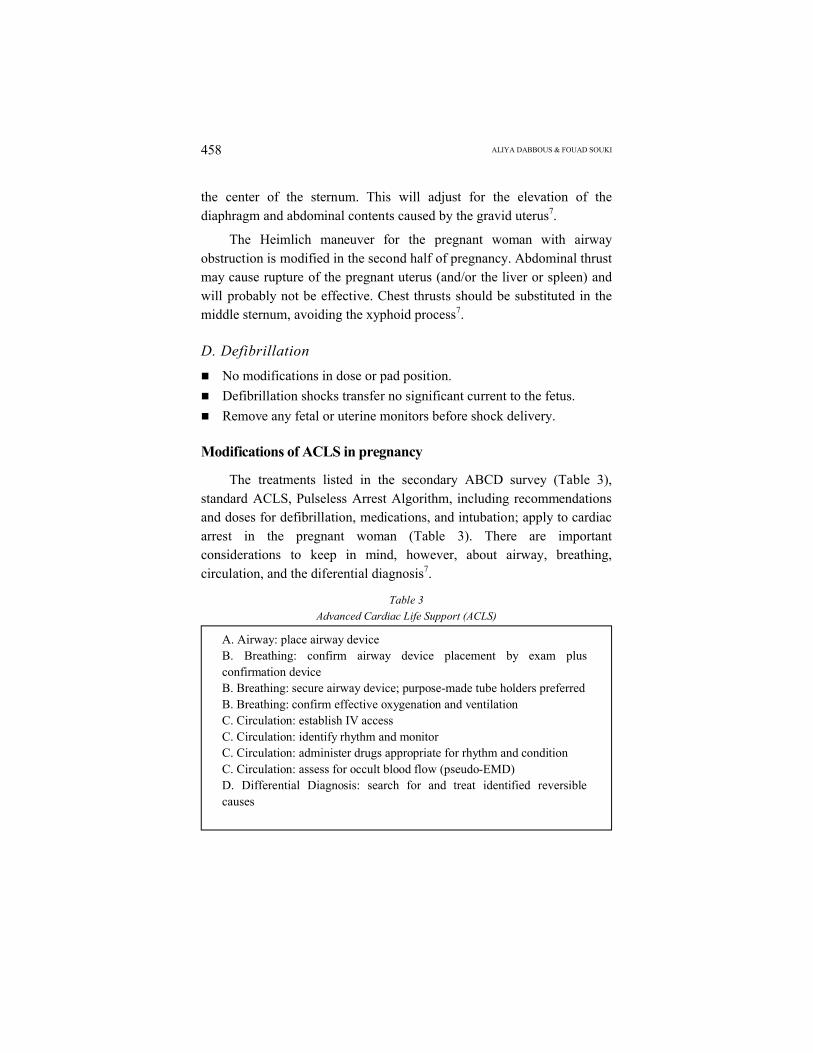
Table 2
(Basic BLS) life support
Primary ABCD Survey Check responsiveness Activate emergency response system Call for defibrillatorA. Airway: open the airwayB. Breathing: provide positive-pressure ventilationsC. Circulation: give chest compressionsD. Defibrillation: assess for and shock VF/pulseless VT
A. Airway
No modifications.
B. Breathing
No modifications. Hormonal changes promote insufficiency of the gastroesophageal sphincter, increasing the risk of regurgitation. Apply continuous cricoid pressure during positive pressure ventilation for any unconscious pregnant woman7.
C. Circulation
Place the woman on her left side with her back angled 15° to 30° back from the left lateral position. Then start chest compressions. Or place a wedge under the woman’s right side (so that she tilts toward her left side) (Fig. 2). or have one rescuer kneel next to the woman’s left side and pull the gravid uterus laterally. This maneuver will relieve pressure on the inferior vena cava.
Perform chest compressions higher on the sternum, slightly above
ALIYA DABBOUS & FOUAD SOUKI458
the center of the sternum. This will adjust for the elevation of the diaphragm and abdominal contents caused by the gravid uterus7.
The Heimlich maneuver for the pregnant woman with airway obstruction is modified in the second half of pregnancy. Abdominal thrust may cause rupture of the pregnant uterus (and/or the liver or spleen) and will probably not be effective. Chest thrusts should be substituted in the middle sternum, avoiding the xyphoid process7.
D. Defibrillation
No modifications in dose or pad position.
Defibrillation shocks transfer no significant current to the fetus.
Remove any fetal or uterine monitors before shock delivery.
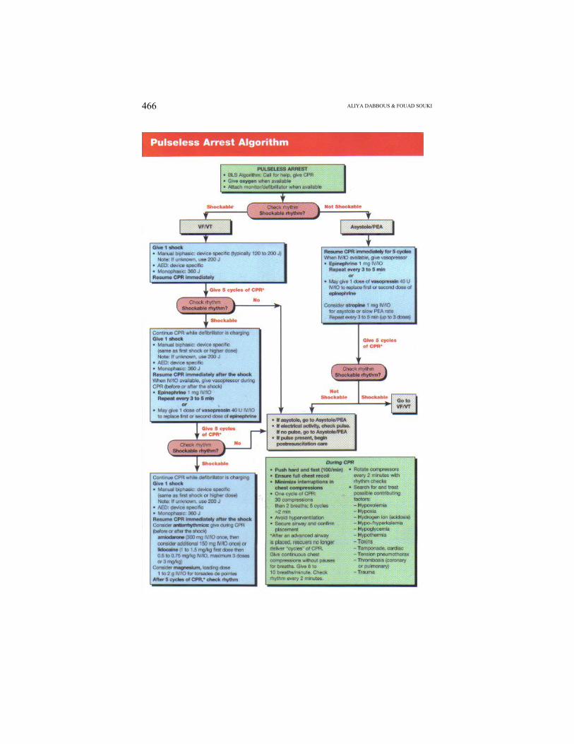
Modifications of ACLS in pregnancy
The treatments listed in the secondary ABCD survey (Table 3), standard ACLS, Pulseless Arrest Algorithm, including recommendations and doses for defibrillation, medications, and intubation; apply to cardiac arrest in the pregnant woman (Table 3). There are important considerations to keep in mind, however, about airway, breathing, circulation, and the diferential diagnosis7.
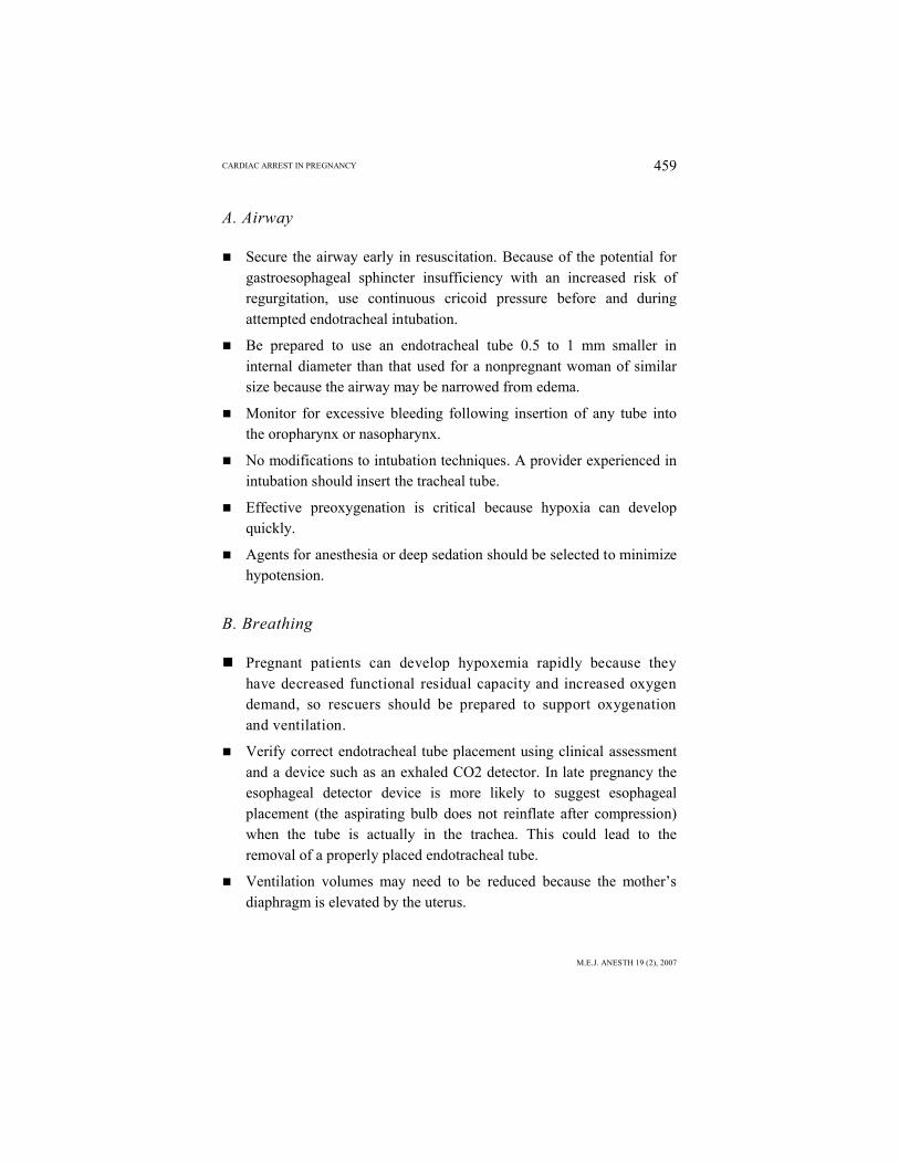
Table 3
Advanced Cardiac Life Support (ACLS)
A. Airway: place airway deviceB. Breathing: confirm airway device placement by exam plus confirmation deviceB. Breathing: secure airway device; purpose-made tube holders preferredB. Breathing: confirm effective oxygenation and ventilationC. Circulation: establish IV accessC. Circulation: identify rhythm and monitorC. Circulation: administer drugs appropriate for rhythm and conditionC. Circulation: assess for occult blood flow (pseudo-EMD)D. Differential Diagnosis: search for and treat identified reversible causes
CARDIAC ARREST IN PREGNANCY
M.E.J. ANESTH 19 (2), 2007
459
A. Airway
Secure the airway early in resuscitation. Because of the potential for gastroesophageal sphincter insufficiency with an increased risk of regurgitation, use continuous cricoid pressure before and during attempted endotracheal intubation.
Be prepared to use an endotracheal tube 0.5 to 1 mm smaller in internal diameter than that used for a nonpregnant woman of similar size because the airway may be narrowed from edema.
Monitor for excessive bleeding following insertion of any tube into the oropharynx or nasopharynx.
No modifications to intubation techniques. A provider experienced in intubation should insert the tracheal tube.
Effective preoxygenation is critical because hypoxia can develop quickly.
Agents for anesthesia or deep sedation should be selected to minimize hypotension.
B. Breathing
Pregnant patients can develop hypoxemia rapidly because they have decreased functional residual capacity and increased oxygen demand, so rescuers should be prepared to support oxygenation and ventilation.
Verify correct endotracheal tube placement using clinical assessment and a device such as an exhaled CO2 detector. In late pregnancy the esophageal detector device is more likely to suggest esophageal placement (the aspirating bulb does not reinflate after compression) when the tube is actually in the trachea. This could lead to the removal of a properly placed endotracheal tube.
Ventilation volumes may need to be reduced because the mother’s diaphragm is elevated by the uterus.

ALIYA DABBOUS & FOUAD SOUKI460
C. Circulation
Follow the ACLS guidelines for resuscitation medications.
Vasopressor agents such as epinephrine, vasopressin, and dopamine will decrease blood flow to the uterus. There are no alternatives, however, to using all indicated medications in recommended doses. The mother must be resuscitated or the chances of fetal resuscitation vanish.
Do not use the femoral vein or other lower extremity sites for venous access. Drugs administered through these sites may not reach the maternal heart unless or until the fetus is delivered.
D. Differential Diagnosis
The same reversible causes of cardiac arrest that occur in nonpregnant women can occur during pregnancy. But providers should be familiar with pregnancy-specific diseases and procedural complications. Providers should try to identify these common and reversible causes of cardiac arrest in pregnancy during resuscitation attempts and decide whether to perform emergency hysterotomy. The use of abdominal ultrasound by a skilled operator should be considered in detecting pregnancy and possible causes of the cardiac arrest, but this should not delay other treatments8. Excess magnesium sulfate as a treatment in women with eclampsia can lead to cardiac arrest, particularly if the woman becomes oliguric. Empiric calcium administration may be life saving. A case was reported by Swartjes et al. (1992) of successful resuscitation with calcium gluconate of an eclamptic who received magnesium overdose4.
ACLS Drugs
Current recommendations are that ACLS protocols be followed in pregnancy as they are in nonpregnant individual. Vasopressor agents such as epinephrine, dopamine, and vasopressin will decrease uterine blood flow. There are, however, no alterations to using all indicated medications
CARDIAC ARREST IN PREGNANCY
M.E.J. ANESTH 19 (2), 2007
461
in recommended doses. The mother must be resuscitated even if the chances of fetal resuscitation vanish7.
Epinephrine
1 mg IV q3-5 min.
High dose epinephrine is no longer recommended.
Vasopressin
40 U IV.
One time dose (wait 5-10 minutes before starting epinephrine).
Can replace first or second dose of epinephrine in all ACLS algorithms.
Vasopressin can only be given once.
Amiodarone (class 2b)
300 mg IV push.
May repeat once at 150 mg in 3-5 min.
Max cumulative dose = 2.2g IV/24 hours.
Lidocaine
1-1.5 mg/kg IV q3-5 min.
Max 3 mg/kg.
Magnesium Sulfate
1-2g IV (over 2 min) for suspected hypomagnesemia or torsades de pointes (polymorphic VT).
Bicarbonate
1 meq/kg IV for reasons below.
Class 1: hyperkalemia.
Class 2a: bicarbonate responsive acidosis, tricyclic overdose, to alkalize urine for aspirin overdose.
Class 2b: prolonged arrest.
ALIYA DABBOUS & FOUAD SOUKI462
Not for hypercarbia related acidosis, not for routine use in cardiac arrest.
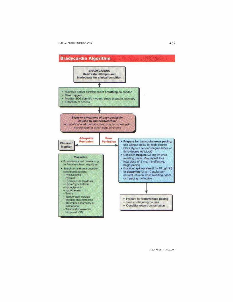
Transcutaneous pacing (TCP) has not been shown to improve survival in asystole and is therefore not recommended to be used. TCP isonly used in symptomatic bradycardia, second and third degree AV block when signs and symptoms of poor perfusion are present. Also in newly acquired left, right or alternating bundle branch block or bifasicular block in the setting of acute myocardial infarction. Signs of poor perfusion include: orthostatic hypotension, diaphoresis, pulmonary congestion on physical examination or chest x-ray, frank congestive heart failure or pulmonary edema; and bradycardia related escape rhythm, frequent premature ventricular complexes.
Terminating In-hospital Resuscitation Efforts
If a reversible cause is not rapidly identified and the patient fails to respond to the BLS primary survey and ACLS secondary survey management, termination of resuscitation efforts may be appropriate. The decision to terminate resuscitation efforts rests with the treating physician in the hospital and is based on consideration of many factors:
Time to CPR.
Time to defibrillation.
Comorbid disease.
Prearrest state.
Initial arrest rhythm.
Response to resuscitation measures.
None of these factors alone or in combination is clearly predictive of outcome. The duration of resuscitative efforts, however, is an important factor associated with poor outcome.
Duration of Resuscitative Efforts
Available scientific studies have shown that in the absence of mitigating factors, prolonged resuscitative efforts are unlikely to be
CARDIAC ARREST IN PREGNANCY
M.E.J. ANESTH 19 (2), 2007
463
successful and they can be discontinued if there no return of spontaneous circulation (ROSC) at anytime during or following 20 minutes of cumulative BLS and ACLS. If ROSC of any duration occurs, it may be appropriate to consider extending the resuscitative efforts. It may also be appropriate to consider other causes such as, drug overdose, and severe prearrest hypothermia (e.g. submersion in icy water) when deciding whether to extend resuscitative efforts. Think of potentially reversible causes.
Perimortem Cesarean Delivery and Delivery Within 5 Minutes
Attempts at resuscitation in the pregnant patients are unfortunately met with failure secondary to negative impact of the anatomic and physiologic changes of pregnancy11-15. If cardiac arrest occurs in the first half of gestation, the purpose of CPR is to resuscitate the mother. If she is resuscitated, it is likely that the pregnancy will proceed and the fetal viability will not be compromised. In this setting, emergency delivery of the fetus is not likely to improve the mother’s chances of survival and certainly the fetus will not survive. However, beyond the threshold of viability (24 weeks of gestation or greater) there are data to suggest that delivery may actually improve maternal survival. Delivery of the fetus will decrease aortocaval compressions and therefore improve venous return and cardiac output. In addition, the cardiac output will increase secondary to the 25-56% increase in intravascular volume that occurs when the uterus is emptied and autotransfusion occurs11-14. An additional benefit is that chest compressions will be more effective once the gravid uterus is evacuated. The functional residual capacity will likewise increase, improving oxygenation during resuscitation efforts11-14.
Several case reports document the successful resuscitation of pregnant women in cardiac arrest after perimortem cesarean delivery11,15.
The time interval from cardiac arrest to delivery is probably the single most important prognostic factor for fetal survival. If the fetus is delivered within 5 minutes of maternal cardiac arrest, intact neurological survival is markedly increased. Therefore, during CPR, to maximize
ALIYA DABBOUS & FOUAD SOUKI464
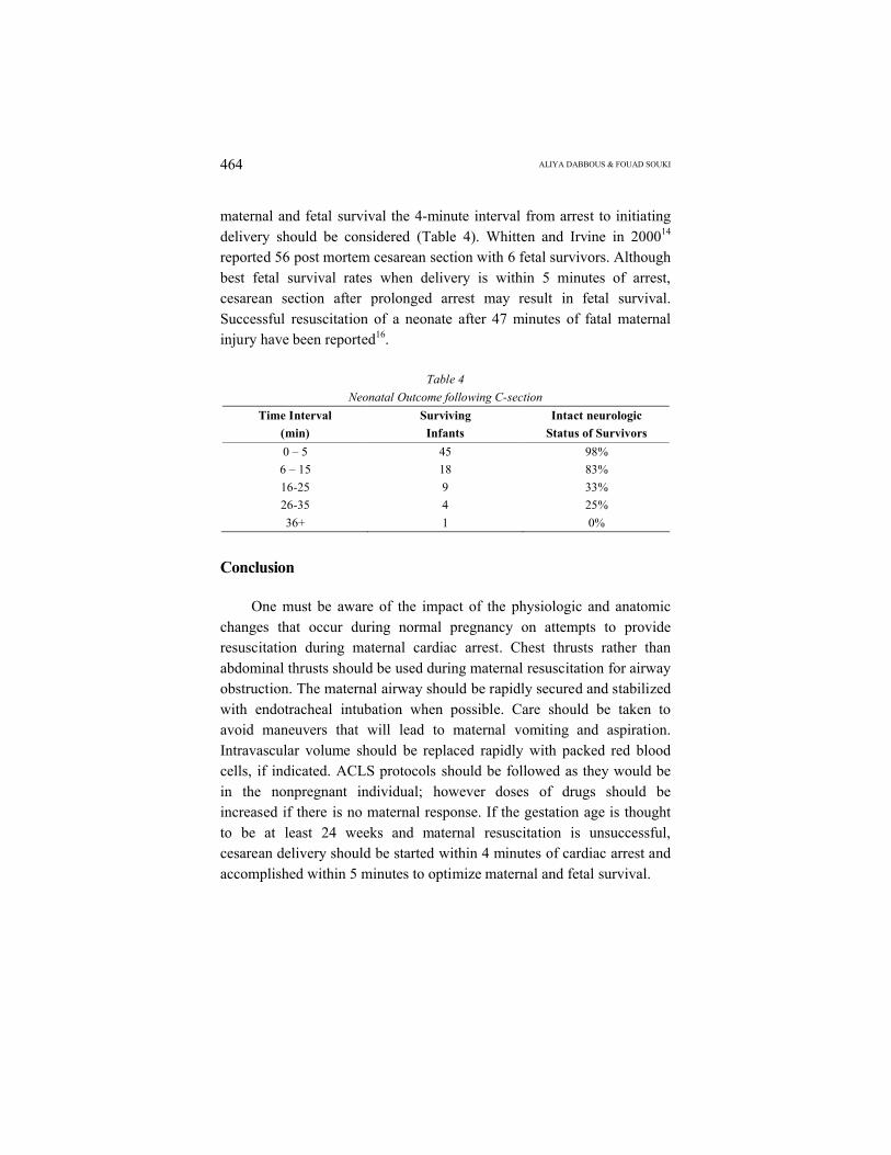
maternal and fetal survival the 4-minute interval from arrest to initiating delivery should be considered (Table 4). Whitten and Irvine in 200014
reported 56 post mortem cesarean section with 6 fetal survivors. Although best fetal survival rates when delivery is within 5 minutes of arrest, cesarean section after prolonged arrest may result in fetal survival. Successful resuscitation of a neonate after 47 minutes of fatal maternal injury have been reported16.
Table 4
Neonatal Outcome following C-section
Time Interval
(min)
Surviving
Infants
Intact neurologic
Status of Survivors
0 – 5 45 98%
6 – 15 18 83%
16-25 9 33%
26-35 4 25%
36+ 1 0%
Conclusion
One must be aware of the impact of the physiologic and anatomic changes that occur during normal pregnancy on attempts to provide resuscitation during maternal cardiac arrest. Chest thrusts rather than abdominal thrusts should be used during maternal resuscitation for airway obstruction. The maternal airway should be rapidly secured and stabilized with endotracheal intubation when possible. Care should be taken to avoid maneuvers that will lead to maternal vomiting and aspiration. Intravascular volume should be replaced rapidly with packed red blood cells, if indicated. ACLS protocols should be followed as they would be in the nonpregnant individual; however doses of drugs should be increased if there is no maternal response. If the gestation age is thought to be at least 24 weeks and maternal resuscitation is unsuccessful, cesarean delivery should be started within 4 minutes of cardiac arrest and accomplished within 5 minutes to optimize maternal and fetal survival.
CARDIAC ARREST IN PREGNANCY
M.E.J. ANESTH 19 (2), 2007
465
ALIYA DABBOUS & FOUAD SOUKI466
CARDIAC ARREST IN PREGNANCY
M.E.J. ANESTH 19 (2), 2007
467
ALIYA DABBOUS & FOUAD SOUKI468
References
1. STEPHEN MORRIS, MARK STACEY: ABC of Resuscitation in pregnancy. BMJ; 327, 1277-79, 2003.2. Cardiac Arrest in Labor and Delivery: A current Review SOAP Newsletter winter, 2003.3. WHITTY JE: Maternal cardiac arrest during pregnancy. Clinical J Obstet Gynec; 45(2), 377-92,
2003.4. SWARTJES JM, SCHUTTLE MF, ET AL: Management of eclampsia: cardiopulmonary arrest resulting
from magnesium sulfate overdose. Eur Obstet Gynecol Reprod Biol; 47(1):73-75, 1992.5. CLARK S, HANKIN G, DUDLEY D: Amniotic fluid embolism: Analysis of the national registry. Am J
Obstet Gynecol; 172:1158-69, 1995.6. Amniotic fluid embolism causing catastrophic pulmonary vasoconstriction: Diagnosis by
transesophageal echocardiogram and treatment by cardiopulmonary bypass. Obst and Gynecol; 102:496-498, 2003.
7. Cardiac Arrest Associated with Pregnancy: Circulation; 112:150-163, 2005.8. CHESTNUT, DAVID H: Obstetric Anesthesia Principles and Practice 2nd Ed, p. 17-42, 1999.9. KINSELLA SM: Lateral tilt for pregnant women: why 15 degrees? Anesthesia; 58(9):835-6, 2003.10. American Heart Association Guidelines for Cardiopulmonary Resuscitation and Emergency
Cardiovascular Care: Circulation; 2005, 112, Issue 24 Supplement; December 13, 2005.11. KATZ VL, DOTTERS DJ, ET AL: Perimortem cesarean delivery. Obstet Gynecol; 68:571, 1986.12. LANOIX R, AKKAPED V, ET AL: Perimortem cesarean section: case reports and recommendations.
Acad Emerg Med; 2(12):1063-7, 1995.13. FINEGOLD H, DARWICH, ET AL: Successful resuscitation after maternal cardiac arrest by immediate
cesarean section in the labor room. Anesthesiology; 96(5):1278, 2002.14. WHITTEN M AND IRVINE LM: Postmortem and perimortem cesarean section: what are the
indications? J R Soc Med; 93(1):6-9, 2000.15. POOLE JH, LONG J: Maternal mortality-a review of current trends. Crit Care Nurs Clin North Am;
16:227-230, 2004.16. LOPEZ-ZENO JA, CARLO WA, ET AL: Infant survival following delayed postmortem cesarean
delivery. Obstet Gynecol; 76(5):991-992, 1990.