Embed Size (px)
Citation preview

Carbapenemases in Klebsiella pneumoniae and OtherEnterobacteriaceae: an Evolving Crisis of Global Dimensions
L. S. Tzouvelekis,a A. Markogiannakis,b M. Psichogiou,c P. T. Tassios,a and G. L. Daikosc
Department of Microbiologya and First Department of Propaedeutic Medicine,c School of Medicine, University of Athens, and Department of Pharmacy, Laiko GeneralHospital,b Athens, Greece
INTRODUCTION . . . . . . . . . . . . . . . . . . . . . . . . . . . . . . . . . . . . . . . . . . . . . . . . . . . . . . . . . . . . . . . . . . . . . . . . . . . . . . . . . . . . . . . . . . . . . . . . . . . . . . . . . . . . . . . . . . . . . . . . . . . . . . . . . . . . . . . . . . . .682GENETIC CONTEXT, SUBSTRATE SPECTRA, AND �-LACTAM RESISTANCE PHENOTYPES . . . . . . . . . . . . . . . . . . . . . . . . . . . . . . . . . . . . . . . . . . . . . . . . . . . . . . . . . . . . . . . .683
KPC Carbapenemases . . . . . . . . . . . . . . . . . . . . . . . . . . . . . . . . . . . . . . . . . . . . . . . . . . . . . . . . . . . . . . . . . . . . . . . . . . . . . . . . . . . . . . . . . . . . . . . . . . . . . . . . . . . . . . . . . . . . . . . . . . . . . . . . . . . .683M�Ls . . . . . . . . . . . . . . . . . . . . . . . . . . . . . . . . . . . . . . . . . . . . . . . . . . . . . . . . . . . . . . . . . . . . . . . . . . . . . . . . . . . . . . . . . . . . . . . . . . . . . . . . . . . . . . . . . . . . . . . . . . . . . . . . . . . . . . . . . . . . . . . . . . . . .685OXA-48 . . . . . . . . . . . . . . . . . . . . . . . . . . . . . . . . . . . . . . . . . . . . . . . . . . . . . . . . . . . . . . . . . . . . . . . . . . . . . . . . . . . . . . . . . . . . . . . . . . . . . . . . . . . . . . . . . . . . . . . . . . . . . . . . . . . . . . . . . . . . . . . . . . .685
GLOBAL SPREAD OF CPE . . . . . . . . . . . . . . . . . . . . . . . . . . . . . . . . . . . . . . . . . . . . . . . . . . . . . . . . . . . . . . . . . . . . . . . . . . . . . . . . . . . . . . . . . . . . . . . . . . . . . . . . . . . . . . . . . . . . . . . . . . . . . . . . . .685Producers of KPC Types . . . . . . . . . . . . . . . . . . . . . . . . . . . . . . . . . . . . . . . . . . . . . . . . . . . . . . . . . . . . . . . . . . . . . . . . . . . . . . . . . . . . . . . . . . . . . . . . . . . . . . . . . . . . . . . . . . . . . . . . . . . . . . . . . .685Producers of M�Ls . . . . . . . . . . . . . . . . . . . . . . . . . . . . . . . . . . . . . . . . . . . . . . . . . . . . . . . . . . . . . . . . . . . . . . . . . . . . . . . . . . . . . . . . . . . . . . . . . . . . . . . . . . . . . . . . . . . . . . . . . . . . . . . . . . . . . . .686Producers of OXA-48 . . . . . . . . . . . . . . . . . . . . . . . . . . . . . . . . . . . . . . . . . . . . . . . . . . . . . . . . . . . . . . . . . . . . . . . . . . . . . . . . . . . . . . . . . . . . . . . . . . . . . . . . . . . . . . . . . . . . . . . . . . . . . . . . . . . . .686
DETECTION OF CPE. . . . . . . . . . . . . . . . . . . . . . . . . . . . . . . . . . . . . . . . . . . . . . . . . . . . . . . . . . . . . . . . . . . . . . . . . . . . . . . . . . . . . . . . . . . . . . . . . . . . . . . . . . . . . . . . . . . . . . . . . . . . . . . . . . . . . . . . .687MHT . . . . . . . . . . . . . . . . . . . . . . . . . . . . . . . . . . . . . . . . . . . . . . . . . . . . . . . . . . . . . . . . . . . . . . . . . . . . . . . . . . . . . . . . . . . . . . . . . . . . . . . . . . . . . . . . . . . . . . . . . . . . . . . . . . . . . . . . . . . . . . . . . . . . . .687Detection of M�Ls Based on Chelating Agents . . . . . . . . . . . . . . . . . . . . . . . . . . . . . . . . . . . . . . . . . . . . . . . . . . . . . . . . . . . . . . . . . . . . . . . . . . . . . . . . . . . . . . . . . . . . . . . . . . . . . . . . . .687Detection of KPCs Based on Boronates. . . . . . . . . . . . . . . . . . . . . . . . . . . . . . . . . . . . . . . . . . . . . . . . . . . . . . . . . . . . . . . . . . . . . . . . . . . . . . . . . . . . . . . . . . . . . . . . . . . . . . . . . . . . . . . . . . .688Detection by Use of Chromogenic Media . . . . . . . . . . . . . . . . . . . . . . . . . . . . . . . . . . . . . . . . . . . . . . . . . . . . . . . . . . . . . . . . . . . . . . . . . . . . . . . . . . . . . . . . . . . . . . . . . . . . . . . . . . . . . . . .688Molecular Detection of Carbapenemase Genes . . . . . . . . . . . . . . . . . . . . . . . . . . . . . . . . . . . . . . . . . . . . . . . . . . . . . . . . . . . . . . . . . . . . . . . . . . . . . . . . . . . . . . . . . . . . . . . . . . . . . . . . . .688Detection of Carbapenemase Activity by Spectrophotometry . . . . . . . . . . . . . . . . . . . . . . . . . . . . . . . . . . . . . . . . . . . . . . . . . . . . . . . . . . . . . . . . . . . . . . . . . . . . . . . . . . . . . . . . . . .688Detection of Carbapenemase Activity by Mass Spectrometry. . . . . . . . . . . . . . . . . . . . . . . . . . . . . . . . . . . . . . . . . . . . . . . . . . . . . . . . . . . . . . . . . . . . . . . . . . . . . . . . . . . . . . . . . . . .688
ANTIMICROBIAL AGENTS AGAINST CPE . . . . . . . . . . . . . . . . . . . . . . . . . . . . . . . . . . . . . . . . . . . . . . . . . . . . . . . . . . . . . . . . . . . . . . . . . . . . . . . . . . . . . . . . . . . . . . . . . . . . . . . . . . . . . . . . . .688In Vitro Activity. . . . . . . . . . . . . . . . . . . . . . . . . . . . . . . . . . . . . . . . . . . . . . . . . . . . . . . . . . . . . . . . . . . . . . . . . . . . . . . . . . . . . . . . . . . . . . . . . . . . . . . . . . . . . . . . . . . . . . . . . . . . . . . . . . . . . . . . . . . .688In Vitro Synergy . . . . . . . . . . . . . . . . . . . . . . . . . . . . . . . . . . . . . . . . . . . . . . . . . . . . . . . . . . . . . . . . . . . . . . . . . . . . . . . . . . . . . . . . . . . . . . . . . . . . . . . . . . . . . . . . . . . . . . . . . . . . . . . . . . . . . . . . . . .689In Vitro Pharmacodynamic Models . . . . . . . . . . . . . . . . . . . . . . . . . . . . . . . . . . . . . . . . . . . . . . . . . . . . . . . . . . . . . . . . . . . . . . . . . . . . . . . . . . . . . . . . . . . . . . . . . . . . . . . . . . . . . . . . . . . . . . .689Experimental Infection Models . . . . . . . . . . . . . . . . . . . . . . . . . . . . . . . . . . . . . . . . . . . . . . . . . . . . . . . . . . . . . . . . . . . . . . . . . . . . . . . . . . . . . . . . . . . . . . . . . . . . . . . . . . . . . . . . . . . . . . . . . . .690Comments on Experimental Studies . . . . . . . . . . . . . . . . . . . . . . . . . . . . . . . . . . . . . . . . . . . . . . . . . . . . . . . . . . . . . . . . . . . . . . . . . . . . . . . . . . . . . . . . . . . . . . . . . . . . . . . . . . . . . . . . . . . . .690
ANTIMICROBIAL THERAPY . . . . . . . . . . . . . . . . . . . . . . . . . . . . . . . . . . . . . . . . . . . . . . . . . . . . . . . . . . . . . . . . . . . . . . . . . . . . . . . . . . . . . . . . . . . . . . . . . . . . . . . . . . . . . . . . . . . . . . . . . . . . . . . . .690Review of Clinical Studies . . . . . . . . . . . . . . . . . . . . . . . . . . . . . . . . . . . . . . . . . . . . . . . . . . . . . . . . . . . . . . . . . . . . . . . . . . . . . . . . . . . . . . . . . . . . . . . . . . . . . . . . . . . . . . . . . . . . . . . . . . . . . . . .690
CPE IN HEALTH CARE SETTINGS . . . . . . . . . . . . . . . . . . . . . . . . . . . . . . . . . . . . . . . . . . . . . . . . . . . . . . . . . . . . . . . . . . . . . . . . . . . . . . . . . . . . . . . . . . . . . . . . . . . . . . . . . . . . . . . . . . . . . . . . . . .694Epidemiology. . . . . . . . . . . . . . . . . . . . . . . . . . . . . . . . . . . . . . . . . . . . . . . . . . . . . . . . . . . . . . . . . . . . . . . . . . . . . . . . . . . . . . . . . . . . . . . . . . . . . . . . . . . . . . . . . . . . . . . . . . . . . . . . . . . . . . . . . . . . .694Infection Control Strategies . . . . . . . . . . . . . . . . . . . . . . . . . . . . . . . . . . . . . . . . . . . . . . . . . . . . . . . . . . . . . . . . . . . . . . . . . . . . . . . . . . . . . . . . . . . . . . . . . . . . . . . . . . . . . . . . . . . . . . . . . . . . . .695Tracing of Carriers . . . . . . . . . . . . . . . . . . . . . . . . . . . . . . . . . . . . . . . . . . . . . . . . . . . . . . . . . . . . . . . . . . . . . . . . . . . . . . . . . . . . . . . . . . . . . . . . . . . . . . . . . . . . . . . . . . . . . . . . . . . . . . . . . . . . . . . .695Intervention . . . . . . . . . . . . . . . . . . . . . . . . . . . . . . . . . . . . . . . . . . . . . . . . . . . . . . . . . . . . . . . . . . . . . . . . . . . . . . . . . . . . . . . . . . . . . . . . . . . . . . . . . . . . . . . . . . . . . . . . . . . . . . . . . . . . . . . . . . . . . .698
Environmental cleaning and decolonization of patients . . . . . . . . . . . . . . . . . . . . . . . . . . . . . . . . . . . . . . . . . . . . . . . . . . . . . . . . . . . . . . . . . . . . . . . . . . . . . . . . . . . . . . . . . . . . . .698Judicious antimicrobial use . . . . . . . . . . . . . . . . . . . . . . . . . . . . . . . . . . . . . . . . . . . . . . . . . . . . . . . . . . . . . . . . . . . . . . . . . . . . . . . . . . . . . . . . . . . . . . . . . . . . . . . . . . . . . . . . . . . . . . . . . . . .698
Success Stories . . . . . . . . . . . . . . . . . . . . . . . . . . . . . . . . . . . . . . . . . . . . . . . . . . . . . . . . . . . . . . . . . . . . . . . . . . . . . . . . . . . . . . . . . . . . . . . . . . . . . . . . . . . . . . . . . . . . . . . . . . . . . . . . . . . . . . . . . . .698NOVEL AGENTS AGAINST CPE . . . . . . . . . . . . . . . . . . . . . . . . . . . . . . . . . . . . . . . . . . . . . . . . . . . . . . . . . . . . . . . . . . . . . . . . . . . . . . . . . . . . . . . . . . . . . . . . . . . . . . . . . . . . . . . . . . . . . . . . . . . . .698
Antibiotics . . . . . . . . . . . . . . . . . . . . . . . . . . . . . . . . . . . . . . . . . . . . . . . . . . . . . . . . . . . . . . . . . . . . . . . . . . . . . . . . . . . . . . . . . . . . . . . . . . . . . . . . . . . . . . . . . . . . . . . . . . . . . . . . . . . . . . . . . . . . . . . .698Sulfactams . . . . . . . . . . . . . . . . . . . . . . . . . . . . . . . . . . . . . . . . . . . . . . . . . . . . . . . . . . . . . . . . . . . . . . . . . . . . . . . . . . . . . . . . . . . . . . . . . . . . . . . . . . . . . . . . . . . . . . . . . . . . . . . . . . . . . . . . . . . . .698Plazomicin . . . . . . . . . . . . . . . . . . . . . . . . . . . . . . . . . . . . . . . . . . . . . . . . . . . . . . . . . . . . . . . . . . . . . . . . . . . . . . . . . . . . . . . . . . . . . . . . . . . . . . . . . . . . . . . . . . . . . . . . . . . . . . . . . . . . . . . . . . . . .699Aminoacyl-tRNA synthetase inhibitors . . . . . . . . . . . . . . . . . . . . . . . . . . . . . . . . . . . . . . . . . . . . . . . . . . . . . . . . . . . . . . . . . . . . . . . . . . . . . . . . . . . . . . . . . . . . . . . . . . . . . . . . . . . . . . . .699
Carbapenemase Inhibitors . . . . . . . . . . . . . . . . . . . . . . . . . . . . . . . . . . . . . . . . . . . . . . . . . . . . . . . . . . . . . . . . . . . . . . . . . . . . . . . . . . . . . . . . . . . . . . . . . . . . . . . . . . . . . . . . . . . . . . . . . . . . . . .699Penem derivatives . . . . . . . . . . . . . . . . . . . . . . . . . . . . . . . . . . . . . . . . . . . . . . . . . . . . . . . . . . . . . . . . . . . . . . . . . . . . . . . . . . . . . . . . . . . . . . . . . . . . . . . . . . . . . . . . . . . . . . . . . . . . . . . . . . . . .6991-�-Methylcarbapenems . . . . . . . . . . . . . . . . . . . . . . . . . . . . . . . . . . . . . . . . . . . . . . . . . . . . . . . . . . . . . . . . . . . . . . . . . . . . . . . . . . . . . . . . . . . . . . . . . . . . . . . . . . . . . . . . . . . . . . . . . . . . . .699Sulfones. . . . . . . . . . . . . . . . . . . . . . . . . . . . . . . . . . . . . . . . . . . . . . . . . . . . . . . . . . . . . . . . . . . . . . . . . . . . . . . . . . . . . . . . . . . . . . . . . . . . . . . . . . . . . . . . . . . . . . . . . . . . . . . . . . . . . . . . . . . . . . . .699Succinic acids (non-�-lactams) . . . . . . . . . . . . . . . . . . . . . . . . . . . . . . . . . . . . . . . . . . . . . . . . . . . . . . . . . . . . . . . . . . . . . . . . . . . . . . . . . . . . . . . . . . . . . . . . . . . . . . . . . . . . . . . . . . . . . . . .699Thiols (non-�-lactams) . . . . . . . . . . . . . . . . . . . . . . . . . . . . . . . . . . . . . . . . . . . . . . . . . . . . . . . . . . . . . . . . . . . . . . . . . . . . . . . . . . . . . . . . . . . . . . . . . . . . . . . . . . . . . . . . . . . . . . . . . . . . . . . . .699Avibactam . . . . . . . . . . . . . . . . . . . . . . . . . . . . . . . . . . . . . . . . . . . . . . . . . . . . . . . . . . . . . . . . . . . . . . . . . . . . . . . . . . . . . . . . . . . . . . . . . . . . . . . . . . . . . . . . . . . . . . . . . . . . . . . . . . . . . . . . . . . . .699
CONCLUDING REMARKS AND PERSPECTIVES . . . . . . . . . . . . . . . . . . . . . . . . . . . . . . . . . . . . . . . . . . . . . . . . . . . . . . . . . . . . . . . . . . . . . . . . . . . . . . . . . . . . . . . . . . . . . . . . . . . . . . . . . . . . .699REFERENCES . . . . . . . . . . . . . . . . . . . . . . . . . . . . . . . . . . . . . . . . . . . . . . . . . . . . . . . . . . . . . . . . . . . . . . . . . . . . . . . . . . . . . . . . . . . . . . . . . . . . . . . . . . . . . . . . . . . . . . . . . . . . . . . . . . . . . . . . . . . . . . . .700
INTRODUCTION
Klebsiella pneumoniae is encountered as a saprophyte in hu-mans and other mammals, colonizing the gastrointestinal
tract, skin, and nasopharynx; it is also found in various environ-mental niches (soil, water, etc.) (11). In the past, it was consideredan important causative agent of community-acquired (CA) infec-tions, including a severe form of pneumonia. Recently, while CA
Address correspondence to G. L. Daikos, [email protected].
Copyright © 2012, American Society for Microbiology. All Rights Reserved.
doi:10.1128/CMR.05035-11
682 cmr.asm.org Clinical Microbiology Reviews p. 682–707 October 2012 Volume 25 Number 4
on July 24, 2020 by guesthttp://cm
r.asm.org/
Dow
nloaded from

pneumonia due to K. pneumoniae has become rare, novel mani-festations of CA infections, such as liver abscess complicated byendophthalmitis and other metastatic infections, have been de-scribed (140).
In the early 1970s, both the epidemiology and spectrum ofinfections caused by K. pneumoniae changed dramatically whenthis bacterium was established in the hospital environment andbecame a (still) leading cause of nosocomial infections. Not only isit found in the gastrointestinal tracts of patients, at frequencies ashigh as 80%, but high carriage rates have also been recorded forpatient nasopharynges and hands (212). This considerable effi-ciency of colonization, enhanced by acquired resistance to antibi-otics, enables K. pneumoniae to persist and spread rapidly inhealth care settings (119). Although not inherently resistant toantibiotics, since it produces only moderate amounts of chromo-somal penicillinases, K. pneumoniae is a notorious “collector” ofmultidrug resistance plasmids. During the 1970s to 1980s, thesewere commonly plasmids encoding resistance to aminoglyco-sides. Later, however, K. pneumoniae became the index species forplasmids encoding extended-spectrum �-lactamases (ESBLs)—mostly TEMs and SHVs active against newer cephalosporins—along with a variety of genes conferring resistance to drugs otherthan �-lactams (212). The successive addition of genetic elementsencoding resistance to aminoglycosides and extended-spectrum�-lactams, coupled with the rapid accumulation of chromosomalmutations conferring resistance to fluoroquinolones, left carbap-enems as the first-choice drugs for the treatment of health care-associated infections caused by K. pneumoniae.
This was true until approximately 2000, when we began wit-nessing a global crisis of unprecedented dimensions due to therapid dissemination of multidrug-resistant (MDR) K. pneu-moniae strains producing “carbapenemases” encoded by trans-missible plasmids. Later, other clinically important enterobacte-rial species, including Escherichia coli, acquired carbapenemasegenes (202). Thus, it appears probable that as in the ESBL “era,” K.pneumoniae again functions as a pool of potent �-lactamases. Theclinically most important carbapenemases in Enterobacteriaceaeare the class A enzymes of the KPC type and the zinc-dependentclass B metallo-�-lactamases (M�Ls), represented mainly by the
VIM, IMP, and NDM types. The plasmid-expressed class D car-bapenemases of the OXA-48 type complete the picture (Table 1)(107, 166, 202).
Carbapenemase-producing enterobacteria (CPE) cause seri-ous infections in debilitated and immunocompromised patients,in association with prolonged hospital stays and increased mor-tality rates, ranging from 24% to as high as 70%, depending on thestudy population (14, 24, 28, 67, 175, 187, 203, 233, 274). Giventhe critical condition of these patients, treatment should be timely,aggressive, and rapidly efficacious. However, therapeutic optionsare obviously limited, and unfortunately, the introduction of newantimicrobials such as tigecycline or the “reinvention” of colistinhas far from entirely resolved this problem, as discussed in a latersection.
In this review, we attempt to (i) describe the microbiologicaland epidemiological characteristics of carbapenemase-producingEnterobacteriaceae and (ii) present in a critical manner the avail-able data regarding the antimicrobial treatment and infectioncontrol practices used to combat infections caused by these bac-teria.
GENETIC CONTEXT, SUBSTRATE SPECTRA, AND �-LACTAMRESISTANCE PHENOTYPES
KPC Carbapenemases
KPC �-lactamases (KPC-2 to KPC-13; molecular class A) (www.lahey.org/studies) exhibit activity against a wide spectrum of�-lactams, including penicillins, older and newer cephalosporins,aztreonam, and carbapenems (Table 2) (191). Structural studiesand comparisons with the TEM-1 and SHV-1 penicillinases indi-cated that positioning of the catalytic residues in KPCs may allowaccommodation of the bulky �-substituents of carbapenems in amanner facilitating the subsequent acylation and deacylationsteps (123).
blaKPC genes detected so far in K. pneumoniae are all carried onplasmids. Sequences adjacent to blaKPC genes display rather lim-ited diversity, suggesting a single or at least a limited number oforiginal sources. Segments of the Tn3-related Tn4401 transposon,occurring in four isoforms, are invariably present upstream of
TABLE 1 Types, classification, variants, and species distribution of plasmid-mediated carbapenemases encountered in Enterobacteriaceae
TypeMolecular class(subclass)a
Functionalgroupb Variants Species
KPC A 2f KPC-2 to -13 K. pneumoniae, E. coli, Klebsiella oxytoca, S.marcescens, Enterobacter spp., C. freundii,Salmonella enterica, Raultella spp.
VIM B (B1) 3a VIM-1, -2, -4, -5, -6 K. pneumoniae, E. coli, K. oxytoca, S. marcescensVIM-11, -12, -13, -19, -23 Serratia liquefaciens, Enterobacter spp., C. freundiiVIM-24, -25, -26, -27, -32 Morganella morganii, Proteus stuartii, P. mirabilis
IMP B (B1) 3a IMP-1, -3, -4, -6, -8 K. pneumoniae, E. coli, K. oxytoca, S. marcescensIMP-11, -24, -27 Enterobacter spp., Citrobacter spp., P. mirabilis,
Proteus rettgeri, Shigella flexneri, M. morganii
NDM B (B1) 3a NDM-1, -4, -5, -6 K. pneumoniae, E. coli, Enterobacter spp., K.oxytoca, C. freundii, M. morganii, Providenciaspp.
OXA D 2df OXA-48, -163, -181 K. pneumoniae, E. coli, C. freundii, P. mirabilisa See reference 109.b See reference 37.
Carbapenemases in Enterobacteriaceae
October 2012 Volume 25 Number 4 cmr.asm.org 683
on July 24, 2020 by guesthttp://cm
r.asm.org/
Dow
nloaded from

blaKPC (63, 180). Tn4401 is bracketed by 39-bp imperfect invertedrepeats and bounded by different 5-bp target site duplications(Fig. 1, structure I) (180). These structures indicate the operationof a replicative transposition mechanism (typical of Tn3-liketransposons) that allows spread of KPC-encoding sequencesamong different genetic units and has resulted in the emergence ofdistinct KPC-encoding plasmids belonging to various Inc groups,
such as FII (probably derivatives of the characteristic FII virulenceplasmid of K. pneumoniae), L/M, and N (63). The same geneticstructures have been identified in KPC-positive isolates of otherenterobacterial species (Table 1).
In line with their substrate spectra, KPC enzymes confer onenterobacteria decreased susceptibility or resistance to virtually all�-lactam antibiotics. Moreover, there have been studies reporting
TABLE 2 Hydrolytic efficiencies of representative carbapenemase variants against various �-lactam substrates
�-Lactamase
Hydrolytic efficiency (kcat/Km) (s�1 �M�1)a against:
ReferenceImipenem Meropenem Ceftazidime Cefotaxime Aztreonam Cefoxitin Cephalothin Penicillin G
KPC-2 0.29 0.27 ND 0.10 0.08 0.002 0.84 1.90 271KPC-3 1.90 1.40 0.03 0.50 ND 0.50 3.50 ND 4VIM-1 0.13 0.26 0.08 0.68 — 0.20 5.10 0.04 93VIM-2 3.80 2.50 0.05 5.80 — 1.20 11.8 4.0 74VIM-4 23.0 0.90 ND ND — ND 36.0 3.10 137VIM-5 0.29 0.05 0.001 0.09 — ND ND 0.26 95VIM-19 6.0 2.0 0.02 30.0 — 0.50 ND 5.0 227VIM-27 0.26 ND ND 0.82 — 0.03 8.30 ND 198IMP-1 1.20 0.12 0.18 0.35 — 2.0 2.40 0.62 136IMP-4 0.35 0.18 0.07 0.14 — ND 0.43 0.08 51NDM-1 0.21 0.25 0.03 0.58 — 0.02 0.40 0.68 273NDM-4 0.46 0.31 0.06 1.20 — — 0.50 ND 189OXA-48 0.14 �0.001 0.001 0.05 — ND 0.15 6.10 217a ND, not determined; —, no hydrolysis detected.
FIG 1 Schematic depiction of representative sequences from enterobacterial plasmids, showing the association of carbapenemase-encoding genes with variousmobile elements. (I) The blaKPC-2-containing Tn4401 transposon from plasmid pNYC (GenBank accession no. EU176011) (180). (II and III) RepresentativeVIM-encoding sequences from plasmids pNL194 (GenBank accession no. GU585907) (167) and pCC416 (GenBank accession no. AJ704863) (59), respectively.(IV) A blaIMP-carrying sequence from plasmid pFP10-2 (GenBank accession no. HQ651093) (146). (V and VI) Sequences containing blaNDM-1 carried by aplasmid from K. pneumoniae 05-506 (GenBank accession no. FN396876) (273) and by plasmid p271A (GenBank accession no. HQ162469) (218), respectively.(VII and VIII) The OXA-48-encoding transposon Tn1999 from plasmid pA-1 (GenBank accession no. AY236073) (217) and the blaOXA-163-containing segmentfrom plasmid p6299 (GenBank accession no. HQ700343) (216), respectively.
Tzouvelekis et al.
684 cmr.asm.org Clinical Microbiology Reviews
on July 24, 2020 by guesthttp://cm
r.asm.org/
Dow
nloaded from

on the emergence of KPC-positive K. pneumoniae exhibiting adecreased outer membrane permeability that enhances �-lactamresistance levels (134). Although the MICs of carbapenems vary,the latest Clinical and Laboratory Standards Institute (CLSI) andEuropean Committee for Antimicrobial Susceptibility Testing(EUCAST) breakpoints classify most KPC-producing isolates asresistant to these drugs (55; www.eucast.org). It should also benoted that carbapenem and oxyimino-�-lactam MICs are com-monly higher for species with derepressed production of theirchromosomally encoded AmpCs, such as enterobacters, than forK. pneumoniae (166, 191).
M�Ls
M�Ls constitute a class of enzymes (molecular class B) that, de-spite their significant amino acid sequence diversity, share threedistinct functional properties: (i) capability of hydrolyzing car-bapenems, (ii) resistance to mechanism-based inhibitors, and (iii)susceptibility to chelating agents such as EDTA. The latter prop-erty results from their unique mechanism of hydrolysis, in whichdivalent cations, most commonly Zn2�, are essential for the nu-cleophilic attack of the �-lactam ring. Phylogenetic analysis sug-gests the existence of three M�L lineages: B1, B2, and B3 (13). Inaddition to chromosomally encoded M�Ls, subgroup B1 includesthe acquired enzymes of the VIM, IMP, GIM, SPM, SIM, AIM,DIM, and NDM types, all of which, surprisingly, are still of un-known origin (61). Of these, several variants of the VIM, IMP, andNDM types have been encountered in K. pneumoniae and otherEnterobacteriaceae (Table 1). These �-lactamases are active againstpenicillins, older and newer cephalosporins, and carbapenems,although significant variations in hydrolytic efficiency exist evenbetween enzymes of the same type (Table 2). Also, they are inca-pable of inactivating aztreonam. This is due mainly to the fact thatB1 M�Ls bind monobactams with a very low affinity. Moreover,docking experiments have indicated that positioning of the drugwithin the active site does not favor hydrolysis (213).
The blaVIM and blaIMP variants identified in K. pneumoniae sofar occur as gene cassettes incorporated into the variable regions ofclass 1 integrons (Fig. 1, structures II, III, and IV) (254), with theexception of a class 2 and a class 3 IMP-encoding integron (174,235). In contrast, blaNDM genes are not associated with integrons(Fig. 1, structures V and VI) (114, 218, 273). The wide variety of K.pneumoniae plasmids encoding M�Ls implies the operation ofmobilization mechanisms. Two non-mutually exclusive possibil-ities are likely to account for this dissemination of M�L genes indistinct plasmids: (i) reshuffling of M�L cassettes among plasmid-borne integrons and (ii) en bloc mobilization of M�L gene-con-taining structures through transposition and/or recombinationevents. Indeed, insertion elements (ISs) such as IS26, ISEc33,ISSen4, and ISAba125, either alone or as parts of transposons (e.g.,Tn3 and Tn1696), commonly flank M�L-encoding regions (Fig.1, structures II to VI) (42, 59, 114, 165, 167, 215, 218, 273). At leastsome of the latter elements apparently participate in the spread ofM�L-encoding sequences among different genetic units.
The three M�L types encountered in K. pneumoniae have alsobeen identified on numerous occasions in other enterobacterialspecies, including E. coli (VIM, IMP, and NDM), Enterobactercloacae (mainly VIM and IMP), Serratia marcescens (mainly IMP),and Proteus mirabilis (mainly VIM) (Table 1) (61, 166). In thesespecies, most genetic platforms of M�L genes are similar to thosefound in K. pneumoniae.
The baseline phenotype expected from M�L-producing entero-bacteria includes (i) resistance to amino-, carboxy-, and ureido-penicillins, penicillin-clavulanate combinations, and cefoxitin;(ii) decreased susceptibility to piperacillin-tazobactam and oxy-imino cephalosporins; and (iii) elevated MICs of carbapenemscompared to the epidemiological cutoff values. In actual fact,however, such a minimal resistance phenotype is observed rarely,if at all, among clinical isolates, since M�L producers most oftenpossess additional mechanisms that increase carbapenem resis-tance levels, such as elevated expression of the M�L itself and,most importantly, impaired outer membrane permeability (40,152). The latter mechanism apparently plays a significant role indetermining carbapenem resistance levels, as also indicated by thewide range of carbapenem MICs among M�L producers belong-ing to the same species and even to the same lineage (40, 152, 166).Also, ESBLs such as SHVs, often encountered among VIM pro-ducers (152, 224), expand the resistance phenotype to includeaztreonam resistance.
OXA-48
OXA-type �-lactamases (molecular class D), such as OXA-23,OXA-24/40, and OXA-58, encountered frequently in acinetobac-ters, exhibit relatively weak carbapenemase activity (256). In 2001,OXA-48, a distinct OXA enzyme (�50% amino acid sequenceidentity with the other OXA enzymes) with significant carbapen-emase activity, was identified in K. pneumoniae (217). Its hydro-lytic efficiency against imipenem is approximately 10-fold higherthan those of the acinetobacter OXAs (256). Data from the crystalstructure of OXA-48 and molecular dynamics studies suggest thatthe process of carbapenem hydrolysis is different from that for theother OXA carbapenemases. For OXA-48, hydrolysis relies onthe rotation of the carbapenem’s �-hydroxyethyl group within theactive site in a manner that allows movement of the deacylatingwater toward the acylated serine residue (73). Consequently,OXA-48-producing K. pneumoniae isolates exhibit elevated MICsof carbapenems that are still frequently lower than the respectivebreakpoints. OXA-48 also hydrolyzes penicillins and early cepha-losporins, but its activity against oxyimino cephalosporins is weak(217). Other carbapenem-hydrolyzing variants of OXA-48 (OXA-163 and -181) have also emerged in K. pneumoniae (216, 220,221). blaOXA-48 is invariably carried by transmissible plasmids re-sponsible for its spread among K. pneumoniae and other entero-bacterial species, such as E. coli and Citrobacter freundii (Table 1)(46, 217). Moreover, the plasmid-borne blaOXA-48-containing se-quences are associated with IS1999, an IS4 family element in-volved in mobilization and expression of �-lactam resistancegenes (Fig. 1, structures VII and VIII) (8). This association pro-vides further possibilities for blaOXA-48 to be transferred to othergenetic units.
GLOBAL SPREAD OF CPE
Producers of KPC Types
A rapid and extensive dissemination of KPC-producing K. pneu-moniae was first noticed in the northeastern parts of the UnitedStates during the first decade of the 21st century. Surveillancestudies suggested that the epicenter of this epidemic was the stateof New York (25, 27, 29). Later, isolates producing KPC-2 (270)and KPC-3 (a point mutant of KPC-2) (4) became established inhospitals in neighboring states, apparently due to transfer of col-
Carbapenemases in Enterobacteriaceae
October 2012 Volume 25 Number 4 cmr.asm.org 685
on July 24, 2020 by guesthttp://cm
r.asm.org/
Dow
nloaded from

onized patients (83, 84, 126). During the same period, KPC-pro-ducing K. pneumoniae also emerged in Latin America (171, 201,252) and Israel (139). Other countries, such as China (257) andGreece (102), soon followed. The Chinese data originate from alimited number of hospitals; thus, the actual extent of spread ofKPC producers in China remains unknown. In Greece, KPC-pos-itive K. pneumoniae became dominant in tertiary care hospitals,reaching epidemic proportions in a matter of approximately 2years (99). In Northern and Western European countries, KPCprevalence remains low. In these countries (e.g., Switzerland, Ire-land, United Kingdom, France, Sweden, Norway, the Nether-lands, and Denmark), most reports concern sporadic isolates in-troduced by patients from high-prevalence areas (181, 230, 266).Nevertheless, a multihospital outbreak has already occurred inFrance (43). Higher prevalences have been reported from Polandand Italy, where KPC producers appear to be established in vari-ous regions (12, 103). The rapid global dissemination of KPC-producing K. pneumoniae implies multiple transmission routes.According to a widely held scenario, an important event was theintroduction of KPC-positive K. pneumoniae from the UnitedStates to Israel, followed by spread to neighboring countries andvia Greece to other European countries (260). However, indexcases to confirm this scenario were not identified with certainty(154). KPC enzymes have been detected in a large number of K.pneumoniae sequence types (STs) (99). Nevertheless, the vast ma-jority of isolates with these enzymes worldwide belong to ST258.This ST is strongly associated with KPC production and with iso-lates exhibiting multidrug resistance, but one could also speculateon additional—though yet unknown—inherent traits responsiblefor its high rate of transmissibility. Whatever these eventually turnout to be, KPC-producing ST258 K. pneumoniae can undeniablybe regarded as one of the most successful multidrug-resistant nos-ocomial pathogens known to date.
Not unexpectedly, KPC-producing isolates of various other en-terobacterial species, including E. coli and E. cloacae, have beenreported in settings where the prevalence of KPC-positive K.pneumoniae is high. Outbreaks of KPC-producing E. coli haveoccurred in health care facilities in various countries, includingthe United States, Israel, and Greece (29, 105, 160, 249). Also,sporadic KPC-positive isolates of a wide variety of other entero-bacterial species have been described worldwide (Table 1) (166,191).
Producers of M�Ls
K. pneumoniae strains producing enzymes belonging to any of thethree M�L families (VIM, IMP, and NDM) have already achievedinternational spread, though significant local differences do exist.VIM-positive K. pneumoniae was first observed around 2001 to2003 in Southern Europe and was introduced later to NorthernEurope (e.g., Germany, France, and the Scandinavian countries)and the United States, mostly through colonized patients trans-ferred from high-prevalence areas (61). Isolation rates of VIM-positive K. pneumoniae in Northern Europe and the United Statesremain low, though some infection clusters limited to single hos-pitals have been reported (107). In addition, sporadic cases havebeen recorded in Tunisia (129), South Korea (272), and Venezuela(155). Until recently, VIM-producing K. pneumoniae and otherenterobacteria were frequently isolated in Mediterranean coun-tries, reaching epidemic proportions only in Greece (107, 251).However, up-to-date surveillance data from this country indicate
that these organisms have been in decline since 2009 (G. L. Daikos,unpublished data).
Acquisition of IMP M�Ls by K. pneumoniae was described dur-ing the 1990s, primarily in Japan, as well as in Taiwan and Singa-pore (225). IMP-positive K. pneumoniae clinical isolates remainfrequent in Japan (94). IMP-4-producing K. pneumoniae strainshave also caused hospital outbreaks in China (162) and Australia(206). In addition, IMP-positive clinical enterobacteria, such as S.marcescens and E. cloacae, have been reported in the same area, i.e.,Japan, South Korea, and Taiwan (225). Dissemination of IMP-producing Enterobacteriaceae in the rest of the world appears to belimited, with single cases identified in Turkey, Lebanon, Brazil,and the United States (3, 72, 148, 174). As usual, limitations anddifferences in surveillance systems in different countries inevita-bly affect the reliability and comparability of international epide-miological data on IMP (or indeed VIM)-positive K. pneumoniae.
In stark contrast, the results of the internationally concertedeffort and resources allocated for the elucidation of the transmis-sion routes and public health impact of enterobacteria, mainly E.coli and K. pneumoniae strains producing NDM, the most recentlyidentified M�L type, were spectacular. These efforts produced awealth of data regarding the epidemiology of NDM producers.The epicenter of their epidemic is the Indian subcontinent, wherethe high isolation frequency of these microorganisms in healthcare facilities, as well as their extensive spread in various environ-mental niches, has been documented repeatedly (130, 192). Fur-thermore, the blaNDM genes have spread to various enterobacterialspecies other than K. pneumoniae and E. coli (Table 1) (255). Also,a second reservoir of NDM-producing K. pneumoniae strainsseems to exist in the central Balkans, but its link with the Indianepidemic remains uncertain (108, 150). In contrast, the recentspread of NDM producers in Western Europe, North America,Australia, and the Far East has clearly been attributed to patientswho originated mainly from India, Pakistan, and Bangladesh(192). A characteristic of NDM-producing K. pneumoniae isolateshas so far been their rapid dissemination; indeed, infected or col-onized humans without obvious connection to the Indian epi-demic are increasingly being reported in several countries (125,190, 214).
Producers of OXA-48
OXA-48-producing K. pneumoniae was first detected sporadicallyin Turkey, in 2001 (217). Hospital outbreaks in the main cities ofthis country soon followed (45). About the same time, OXA-48-positive K. pneumoniae isolates were also identified in other Mid-dle Eastern and North African countries (46, 62) as well as inWestern European countries, including the United Kingdom, Bel-gium, France, Germany, and the Netherlands. Emergence ofOXA-48 producers in the latter countries has been attributedmainly to colonized patients transferred from North Africa (107).Recently, an important outbreak due to an OXA-48-producing K.pneumoniae strain was reported in a Dutch hospital (220). How-ever, there are no indications of an overall significant spread ofthese microorganisms across Europe. Although the Middle Eastand North Africa remain the main foci of infection, the recentisolation of K. pneumoniae isolates producing OXA-48-type en-zymes in India (47), Senegal (173), and Argentina (216) suggestsan expansion that can safely be considered global. Additionally,the recent isolation of OXA-48 producers belonging to species
Tzouvelekis et al.
686 cmr.asm.org Clinical Microbiology Reviews
on July 24, 2020 by guesthttp://cm
r.asm.org/
Dow
nloaded from

other than K. pneumoniae underlines the spreading potential ofblaOXA-48 (Table 1) (46).
DETECTION OF CPE
Counterintuitive as it may sound, carbapenemase production byenteric bacilli does not necessarily confer significant resistance tocarbapenems. Before the introduction of the new carbapenembreakpoints by CLSI and EUCAST in 2010 (55; www.eucast.org),carbapenemase-positive isolates (as determined by phenotypictests) with relatively low carbapenem MICs were reported withoutinterpretation of their susceptibility status. This directly impliedthe possibility of therapeutic failure for carbapenem regimens,passing to clinicians a mixed message of dubious value. Today,after the introduction of the new, lower breakpoints, the situationhas been simplified: laboratories report the MICs of carbapenemsirrespective of carbapenemase production. On the other hand,various enterobacterial isolates lacking enzymes with appreciablecarbapenemase activity may exhibit elevated MICs of carbapen-ems. Consequently, this may exclude from use a viable group ofantibiotics. Application of simple and reliable carbapenemase-de-tecting tests nevertheless remains useful for monitoring of carbap-enemase-producing microorganisms in order to inform appro-priate infection control policies in health care settings.
A large-scale and cost-effective approach for deciding whichisolates are carbapenemase producers based solely on phenotypictests should rely on the epidemiological cutoff (ECOFF) values fornonsusceptibility. These values depend on carbapenem MIC dis-tributions of carbapenemase producers as opposed to wild-typestrains. Considering the respective distributions for K. pneu-moniae and E. coli compiled by EUCAST, MICs of �1 �g/ml forimipenem and �0.5 �g/ml for meropenem and ertapenem havebeen proposed (57). According to the CLSI, which has not definedECOFFs, selection of isolates for testing can rely on clinical break-points: isolates that test intermediate or resistant to at least onecarbapenem as well as resistant to a “subclass III cephalosporin”(cefotaxime, ceftazidime, ceftriaxone, cefoperazone, or cefti-zoxime) should be tested further. Ertapenem is considered themost sensitive indicator (29). It should be noted, however, thatuse of this drug may cause specificity problems: decreased perme-ability, combined with either production of CTX-M or overpro-duction of AmpC �-lactamases, can significantly affect the MIC ofertapenem and therefore lower the detection specificity (57, 264).
Screening criteria, however, may and should be adapted de-pending on the epidemiological situation in a given ecologicalsetting. Application of the CLSI criteria is expected to be adequatein settings where carbapenemase producers have already been es-tablished. On the other hand, occasional adoption of less stringentcriteria (i.e., the use of lower cutoffs or reducing the concentra-tions of the selective agents used for screening) in low-prevalencesettings may facilitate the timely detection of CPE emergence orearly dissemination and therefore allow the swift implementationof measures preventing their further spread. The potential pre-vention benefits of such an approach are likely to counterbalancethe burden of increased false-positive results.
There have been numerous studies that deal with technicalissues of carbapenemase detection methods, comparing their per-formances mainly for K. pneumoniae and E. coli. We thereforebriefly review these methods and their principles.
MHT
The cloverleaf or modified Hodge test (MHT) is based on theinactivation of meropenem or ertapenem by whole cells of car-bapenemase-producing organisms. MHT has been used exten-sively as a phenotypic method for the detection of carbapenemaseactivity (55), and it is the only carbapenemase detection methodrecommended by the CLSI for screening purposes. There are,however, various shortcomings with MHT. The assay cannot dis-tinguish the type of carbapenemase involved. Most importantly,false-positive results have been observed with isolates producingCTX-M-type ESBLs or increased amounts of AmpC �-lactamases(cephalosporinases) (166, 200). Moreover, sensitivity problems(false-negative results) may occur, mainly with M�L-producingenterobacterial isolates exhibiting weak carbapenemase activity(166). Also, MHT is probably unreliable in detecting NDM-1-producing K. pneumoniae, though the relevant observations re-gard a limited number of isolates (49, 169). Replacement of Mu-eller-Hinton agar by MacConkey agar has been proposed as ameans to increase the sensitivity of MHT for detection of isolatesproducing M�Ls or OXA carbapenemases. Improved perfor-mance was attributed to the enhanced release of periplasmic en-zymes caused by the bile salts included in MacConkey medium(141). This modification, however, has not been evaluated sys-tematically. Overall, MHT, although remaining a convenient as-say, cannot be used as the sole method for the detection of carbap-enemase-positive Enterobacteriaceae in the clinical laboratory.
Detection of M�Ls Based on Chelating Agents
Phenotypic detection of M�L producers in the clinical laboratoryis based mainly on the specific inhibition of M�Ls by EDTA (193).Additionally, various techniques utilizing other chelating agents,such as dipicolinic acid and 1,10-phenanthroline, as well as thiolcompounds such as 2-mercaptopropionic and mercaptoaceticacid, have been developed (166). Use of a combination of chela-tors, e.g., EDTA plus 2-mercaptopropionic acid, has also beenproposed (124). These compounds, by depriving the M�L of hy-drolytically essential Zn divalent cations, render it inactive against�-lactams. The most common M�L detection tests employ a diskof a hydrolyzable �-lactam (typically a carbapenem, though cef-tazidime has also been used extensively) placed close to a disk witha given amount of an M�L inhibitor (most commonly EDTA),hence the term “double-disk synergy test” (DDST). Formation ofa synergy pattern is indicative of M�L production. A drawback ofthis approach is that interpretation is subjective and cannot bequantified. Alternatively, the �-lactam disk is potentiated with aninhibitor, and the diameter of its inhibition zone is then comparedwith that of the �-lactam disk alone, hence the term “combineddisk test” (CDT). An increase of the inhibition zone diameterabove a predefined cutoff value denotes M�L activity. Variousgradient diffusion methods (e.g., Etest [bioMérieux, Solna, Swe-den]), utilizing strips containing imipenem and EDTA, are basedon the same principle. In general, M�L detection methods basedon �-lactam– chelator combinations perform well for K. pneu-moniae and E. coli, while they have not been tested systematicallyfor other enterobacterial species. Also, the user should always con-sider the potentially detrimental effects of chelating agents on bac-terial growth. Additionally, interpretation difficulties are to beexpected with M�L producers exhibiting low carbapenem MICs.
Carbapenemases in Enterobacteriaceae
October 2012 Volume 25 Number 4 cmr.asm.org 687
on July 24, 2020 by guesthttp://cm
r.asm.org/
Dow
nloaded from

Detection of KPCs Based on Boronates
Phenotypic detection of KPC production is based on the suscep-tibility of KPCs to boronic acid and its derivatives, i.e., phenyl-boronic and 3-aminophenylboronic acid. Boronate derivatives,which structurally resemble �-lactams, have long been used inprobing the function of �-lactamases, especially class C enzymes.In 2008, Pasteran et al. (201) observed that boronates preferen-tially inhibit KPC-type �-lactamases. This report was soon fol-lowed by studies proposing detection techniques using boronicacids combined with a carbapenem, mostly in the CDT format(75, 104, 248). As with the above-described M�L detection tests,experience with the boronate-based detection of KPC producers islimited mainly to K. pneumoniae. Specificity problems may arisewith isolates producing AmpC-type �-lactamases (cephalospori-nases), since boronic acid derivatives are potent inhibitors of theseenzymes. The problem can be alleviated partly by the simultane-ous use of cloxacillin, which preferentially inhibits cephalospori-nases (104). It should also be noted that boronate-based assays areineffective in detecting KPC-positive K. pneumoniae in the case ofcoproduction of VIM �-lactamase (100).
Detection by Use of Chromogenic Media
At least two selective agar media allowing different carbapen-emase-producing microorganisms to be recognized are commer-cially available: CHROMagar-KPC (CHROMagar; BBL) and Bril-liance CRE agar (Thermo Fisher Scientific). Species aredistinguished by colony color. The reliability of these media hasnot yet been evaluated rigorously. Nevertheless, they have beenused successfully for surveillance cultures on various occasions(196, 208, 229).
Molecular Detection of Carbapenemase Genes
Many clinical laboratories employ “in-house” PCR-based meth-ods for the detection of carbapenemase genes to get around theproblems of phenotypic detection methods and to reduce report-ing times. In addition, PCR-based methods allow detection ofOXA-type carbapenemases for which specific phenotypic testshave not been developed. Simplex PCR assays targeting a singlecarbapenemase type have been used successfully in numerousstudies, although there is no consensus regarding the oligonucle-otide primers that should be used for each bla gene group. Multi-plex and real-time PCR methods that allow the identification ofmultiple carbapenemase gene types and that further shorten thedetection time, in the case of real-time PCR, have also been uti-lized (20, 70, 172, 219, 253). Also, real-time PCR assays can befollowed by a melting curve step to allow the accurate identifica-tion of carbapenemase gene variants (163). PCR- and hybridiza-tion-based kits for detection of the main carbapenemase genetypes, for example, Hyplex MBL ID and Hyplex CarbOxa ID kits(BAG Health Care, Lich, Germany), have also been developed bythe industry. Although manufacturers claim that these methodshave the potential to be used directly on clinical samples (9), theirdiagnostic usefulness remains to be evaluated systematically indifferent settings. Microarray technology was recently added tothe list of molecular methods aiming at the rapid and reliableidentification of multiple resistance determinants. The Check-KPC ESBL microarray and its expanded version, Check-MDRCT102 (Check-Points Health BV, Wageningen, Netherlands),have been used successfully for detection within a single reactiontube of a wide variety of bla genes, including most clinically rele-
vant carbapenemase genes (58, 179). Nevertheless, the term “mac-roarray” may be more suitable given the relatively small numberof genes tested.
An issue with all molecular methods is that the range of resis-tance genes to be detected is predefined, so these methods maymiss novel gene types.
Detection of Carbapenemase Activity bySpectrophotometry
Assessment of carbapenemase activity by spectrophotometry iscarried out using crude or partially purified enzyme extracts and acarbapenem, commonly imipenem. It is considered the referencemethod for the verification of carbapenemase activity. This labo-rious and technically demanding approach, however, is limited toreference laboratories.
Detection of Carbapenemase Activity by MassSpectrometry
Matrix-assisted laser desorption ionization–time of flight massspectrometry (MALDI-TOF MS) is the latest advancement in therecognition of carbapenemase activity. The method is based onthe ionization in high vacuum of the material under examinationand its subsequent acceleration in an electrical field. The sizes offragments can be inferred from the time of flight within the elec-trical field. MALDI-TOF MS has been introduced in the clinicallaboratory mainly as a means for species identification. However,the method is highly versatile and can be used for the recognitionof various compounds, including antibiotic degradation prod-ucts. Recently, MALDI-TOF MS was used successfully to identifycarbapenem hydrolysis products, thus confirming carbapenemaseactivity in Gram-negative isolates (35, 115). However, experiencewith this methodology is still limited.
ANTIMICROBIAL AGENTS AGAINST CPE
In Vitro Activity
Susceptibility of the infecting isolate is one of the key factors indeciding on a suitable antimicrobial chemotherapy. In vitro sus-ceptibility data from numerous studies throughout the world in-dicate that colistin, tigecycline, and fosfomycin are the most effec-tive antibacterials against clinical enterobacteria producing eitherKPCs or M�Ls. Drug effectiveness, however, differs depending onthe extent of spread of resistant isolates in each setting. Indeed, agrowing number of studies indicate that the activity of these drugsis decreasing rapidly (7, 21, 80, 85, 88, 96, 128, 142, 151, 156, 185,186, 226, 243, 247, 275, 277). In addition, rates of susceptibility tofluorinated quinolones are generally low, reflecting the multi-drug-resistant nature of CPE. Low susceptibility rates are alsofound for other, clinically less important antimicrobials, such asnitrofurantoin (breakpoints are available only for E. coli) andchloramphenicol, both of which are drugs that are tested less fre-quently. Among the currently used aminoglycosides, only genta-micin has so far retained good activity against producers of KPCsand acquired M�Ls of the VIM type. The majority of NDM pro-ducers, however, are resistant to all clinically available aminogly-cosides due to coproduction of 16S rRNA methylases (18). Of the�-lactams, the most effective compounds seem to be the carbap-enems, which at first sight appears to be paradoxical. However,applications of different carbapenem breakpoints by the CLSI andEUCAST must be taken into account. It has been estimated that in
Tzouvelekis et al.
688 cmr.asm.org Clinical Microbiology Reviews
on July 24, 2020 by guesthttp://cm
r.asm.org/
Dow
nloaded from

Greek hospitals, some 10 to 15% of CPE (a bacterial populationconsisting mainly of KPC-2-producing K. pneumoniae strains)appear resistant by the CLSI interpretive criteria but susceptibleaccording to EUCAST (Daikos, unpublished data). Aztreonam,though withstanding hydrolysis by acquired M�Ls, exhibits lim-ited activity against the respective CPE due to the frequent copro-duction of ESBLs, mainly of the SHV and CTX-M types (60).Finally, temocillin, a 6-�-methoxy derivative of ticarcillin, seemsto exhibit moderate activity against KPC-producing K. pneu-moniae and E. coli, but the relevant data are limited (1).
The data summarized above, obtained from a wide variety ofsettings, should nevertheless be treated with caution given thedifferent methodologies used. Additionally, there have been re-ports that automated systems have “inherent” problems in reli-ably determining carbapenem MICs for CPE (33, 101, 199, 244,246). Also, there are difficulties in interpreting carbapenem sus-ceptibility data for KPC producers due to heterogeneous resis-tance-like phenomena (191).
In Vitro Synergy
Given the limited therapeutic options for the management of in-fections caused by carbapenemase-producing K. pneumoniaestrains, several investigators have evaluated combinations of dif-ferent antimicrobial agents for potential synergistic effects againstthese organisms, relying mostly on time-kill methods. It should benoted, however, that in most synergy studies the most frequentcompounds used were the polymyxins (polymyxin B and poly-myxin E).
In 2005, Bratu and colleagues found that polymyxin B, at 0.5times its MIC, exhibited synergistic activity with rifampin against15 of 16 KPC-positive K. pneumoniae isolates. A synergistic effectwas also seen in 10 of these isolates when polymyxin B was com-bined with imipenem (30). In a later study, it was found that atriple-drug combination of polymyxin B with rifampin and dorip-enem at 1/4 their MICs exhibited high bactericidal activity, de-fined as a decrease of �3 log CFU/ml in 24 h, for five E. coli andtwo K. pneumoniae clinical isolates, all producing KPC-type en-zymes (250). Similarly, the combination of colistin (polymyxin E)and tigecycline has been reported as synergistic against KPC-pos-itive K. pneumoniae isolates in time-kill experiments (222). Inter-actions of colistin and imipenem were also examined in time-killexperiments with 42 VIM-producing K. pneumoniae isolates froma Greek hospital (241). In general, the combination of imipenemwith colistin exhibited improved bactericidal activity against iso-lates that were susceptible either to both agents or to colistin alone.More specifically, the combination was synergistic against 50% ofthe colistin-susceptible isolates and indifferent against the re-maining 50%, irrespective of the imipenem MIC. In contrast, forisolates that were nonsusceptible to colistin, the combination wasantagonistic for 55.6% of the isolates and synergistic for only 11%of the isolates. These data are partly reminiscent of those reportedby Elemam et al. (79), who examined the interactions of poly-myxin B with several antimicrobials against 12 KPC-producing K.pneumoniae isolates that had elevated polymyxin B MIC values,using a broth microdilution assay in a checkerboard pattern. Syn-ergy was observed with the combinations of polymyxin B plusrifampin and polymyxin B plus doxycycline, at achievable serumdrug concentrations for the antimicrobial agents tested. Less pro-nounced synergy was noted with polymyxin B and tigecycline,whereas no synergy was evident between polymyxin B and the
other antimicrobial agents tested, including imipenem and genta-micin.
Given that fosfomycin retains its activity against the majority ofCPE, it is reasonable to consider administering this compoundagainst CPE infections, but always in combination with anotherantimicrobial agent, as the rate of mutation to fosfomycin resistanceis worringly high (188). Recently, the interactions of fosfomycin withmeropenem, colistin, and gentamicin against KPC-positive K. pneu-moniae isolates were studied using time-kill experiments (238).Combinations of fosfomycin with meropenem and colistin weresynergistic in 64.7 and 11.8% of KPC-producing K. pneumoniaeisolates, respectively, whereas the combination with gentamicinwas indifferent. In addition, combinations of fosfomycin withmeropenem, colistin, and gentamicin prevented the developmentof resistance to fosfomycin in 69.2, 53.8, and 81.8% of examinedisolates, respectively. Similar results were obtained by anotherstudy that demonstrated synergy of fosfomycin with imipenem,meropenem, doripenem, colistin, netilmicin, and tigecycline for74, 70, 74, 36, 42, and 30% of 50 KPC-producing K. pneumoniaeisolates, respectively (228).
Time-kill assays have also been used to comparatively assess theactivities of aztreonam and carbapenems. As mentioned previ-ously, aztreonam is not hydrolyzed by M�Ls and therefore is apotentially useful agent against M�L producers. A time-kill studyassessed the in vitro activity of aztreonam in comparison to car-bapenems against VIM-1-producing ESBL-negative K. pneu-moniae isolates (197). Aztreonam exhibited slow bactericidal ac-tivity that was sustained for 24 h, whereas carbapenems resulted inmore rapid bacterial killing for the first 6 h but regrowth to thelevel of antibiotic-free controls at 24 h.
In Vitro Pharmacodynamic Models
In a chemostat model simulating human pharmacokinetics, it wasshown that optimized doses of meropenem (simulation of 2 gevery 8 h, infused over 3 h, in humans) can achieve bactericidalactivity against KPC-producing K. pneumoniae isolates with lowmeropenem MICs, despite the presence of an active carbapen-emase. In this model, actual meropenem concentrations were sig-nificantly lower than intended, presumably due to rapid in vitrohydrolysis of meropenem by the released KPC enzyme. Despitethis situation, meropenem achieved a rapid, �3-log CFU reduc-tion of all KPC isolates within 6 h, but this effect was maintainedfor only two of the three KPC-producing isolates (with mero-penem MICs of 2 and 8 �g/ml) for which adequate drug exposurehad been attained (32).
The effect of tigecycline alone or in combination with mero-penem was assessed in an in vitro pharmacodynamic model sim-ulating human epithelial lining fluid drug concentrations againstfive KPC-producing K. pneumoniae isolates displaying mero-penem MICs between 8 and 64 �g/ml and tigecycline MICs be-tween 1 and 2 �g/ml. Tigecycline alone did not produce a reduc-tion in bacterial density in any of the isolates studied, except forone with a tigecycline MIC of 1 �g/ml, in which an initial reduc-tion was nevertheless followed by rapid regrowth. Meropenemalone, on the other hand, produced a rapid bactericidal effect forisolates with meropenem MICs of 8 and 16 �g/ml, but this effectwas not maintained and was also followed by regrowth. Unlikemonotherapy with tigecycline or meropenem, their combinationcaused a significant reduction in CFU/ml at 24 and 48 h for iso-lates with tigecycline and meropenem MICs of �2 and �16 �g/
Carbapenemases in Enterobacteriaceae
October 2012 Volume 25 Number 4 cmr.asm.org 689
on July 24, 2020 by guesthttp://cm
r.asm.org/
Dow
nloaded from

ml, respectively, compared to the case with either agent alone.None of the studied regimens, however, was able to maintain asignificant bactericidal effect for periods over 48 h (262).
Experimental Infection Models
Several investigators have examined the efficacy of differentagents, alone or in combination, against CPE isolates by usingdifferent experimental infection models. Daikos et al. (66) as-sessed the activity of two dosing regimens of imipenem (30 and 60mg/kg of body weight every 2 h [q2h]) against VIM-1-producingK. pneumoniae isolates in the neutropenic murine thigh infectionmodel. Animals were infected with three VIM-1-positive isolates(with imipenem MICs of 2, 4, and 32 �g/ml) and a susceptibleclinical isolate (with an imipenem MIC of 0.125 �g/ml) not pro-ducing any �-lactamase with broad-spectrum activity. The bacte-ricidal effect was greatest against the susceptible non-VIM-1-pro-ducing isolate, intermediate against the “susceptible” VIM-1producers (imipenem MICs of 2 and 4 �g/ml), and minimalagainst the resistant VIM-1 isolate (imipenem MIC of 32 �g/ml).However, with administration of a higher dose of imipenem (60mg/kg q2h) and attainment of a drug exposure (cumulative per-centage of a 24-h period that the drug concentration exceeds theMIC under steady-state pharmacokinetic conditions [%TMIC]) ofapproximately 40%, a more pronounced antibacterial effectagainst all VIM-1-producing isolates, including the highly resis-tant one, was achieved.
The use of carbapenems in the treatment of CPE infections waspursued further by Bulik and Nicolau (34), who evaluated theefficacy of doripenem against KPC-producing K. pneumoniae iso-lates with MICs ranging from 4 to 32 �g/ml in both immunocom-petent and neutropenic mice. In these experiments, the authorsused doripenem doses simulating human pharmacokinetics ob-served after administration of 1 or 2 g every 8 h as a 4-h infusion.The 1-g dose simulation was able to produce only a bacteriostaticresponse for the isolates with MICs of 4 and 8 �g/ml, whereas the2-g dose simulation achieved a similar effect for isolates withMICs of up to 16 �g/ml. Relative to neutropenic mice, a reductionin bacterial density was observed in the immunocompetent ani-mals, with overall decreases of up to 1 log, with either the 1- or 2-gdoripenem dose simulation. A critical interpretation of the animalinfection model data just summarized suggests that optimizedregimens of carbapenems are able to achieve at least a static effectin severely compromised hosts and a modest bactericidal effect inimmunocompetent animals infected with KPC-positive isolateswith MICs of up to 8 �g/ml.
The efficacy of carbapenems and aztreonam was also evaluatedin a rabbit model of peritoneal abscess caused by an ESBL-negativeVIM-producing E. coli isolate. MICs of imipenem, meropenem,ertapenem, and aztreonam were 1, �0.25, 1.5, and �0.25 �g/ml,respectively. Carbapenems and aztreonam were shown to be ef-fective in the treatment of this infection with regard to reductionsin bacterial densities and mortality of the animals compared withthose of untreated controls. Aztreonam, however, resulted in amore favorable outcome overall than that seen with carbapenems(239).
Comments on Experimental Studies
The number of studies assessing the interaction of antimicrobialswith CPE in the laboratory, whether using time-kill assays or ex-perimental animal infections, is remarkably small. In addition,
clinically important CPE, such as those producing NDM-typeM�Ls or OXA-48, have not yet been studied in this manner. It istherefore obvious that additional studies of this kind are required,given the extent and severity of the problem posed by CPE. Thesynergy data from time-kill studies present some discrepancies.These data should therefore be interpreted with caution, sinceslight differences in experimental conditions (e.g., a relativelysmall change in the MIC fraction for one or more drugs) couldresult in significant changes in the apparent effect of a given com-bination. Despite these limitations, however, the data from time-kill studies indicate a variety of antibiotic combinations with po-tential synergistic effects against CPE.
In pharmacodynamic models and, most importantly, experi-mental infections in animals, the antibiotics preferably evaluatedso far have been the carbapenems. This might appear unexpected,since therapy with carbapenems in the majority of human infec-tions caused by CPE would be considered “inappropriate” basedon MICs. Yet the relevant data, though limited, may be taken asindicating that approaches such as modification of dosingschemes warrant further attention.
ANTIMICROBIAL THERAPY
In studies examining the outcomes of CPE infections, older age,severity of underlying illness, comorbid conditions of the host,intensive care unit (ICU) stay, resistance to carbapenems, andadministration of inappropriate antimicrobial treatment (partlydue to CPE multidrug resistance compromising empirical ther-apy) are the most important independent predictors of treatmentfailure (14, 67, 175, 205, 233, 274). In the absence of controlledcomparative trials, however, an overall critical appraisal of antibi-otic treatment schemes inevitably has to be based on a variety ofcase reports, case series, retrospective studies, and observationalstudies. Moreover, these studies are focused on K. pneumoniae, asclinical experience with other CPE is quite limited. Therefore, theassessment we attempt here lacks many of the characteristics of arigorous meta-analysis but may provide some guidance on thetreatment of CPE-infected patients.
Review of Clinical Studies
We performed a systematic search of MEDLINE and compiled 34studies containing the necessary information to estimate the effi-cacies of different antimicrobials in relation to their MICs for theinfecting organisms (Tables 3 and 4). A total of 301 patients wereidentified, including 161 infected with KPC-producing K. pneu-moniae and 140 infected with M�L-producing K. pneumoniae.The vast majority of these patients had serious infections: 244 hadbloodstream infections (BSIs), 32 had pneumonia, 8 had urinarytract infections, 4 had tracheobronchitis, 3 had wound infections,and 7 had other infections. Three patients reported as having uri-nary colonization were excluded. Of the remaining 298 patients,242 (81.1%) received appropriate therapy (with at least one drugto which the infecting organism was classified as susceptible invitro), while 56 (18.9%) received inappropriate therapy (no drugto which the infecting organism was classified as susceptible invitro). To facilitate comparisons, patients were classified intoseven groups according to treatment regimen, as follows: regimenA, combination therapy with �2 active drugs, one of which was acarbapenem; regimen B, combination therapy with �2 activedrugs, not including a carbapenem; regimen C, monotherapy withan aminoglycoside; regimen D, monotherapy with a carbapenem;
Tzouvelekis et al.
690 cmr.asm.org Clinical Microbiology Reviews
on July 24, 2020 by guesthttp://cm
r.asm.org/
Dow
nloaded from

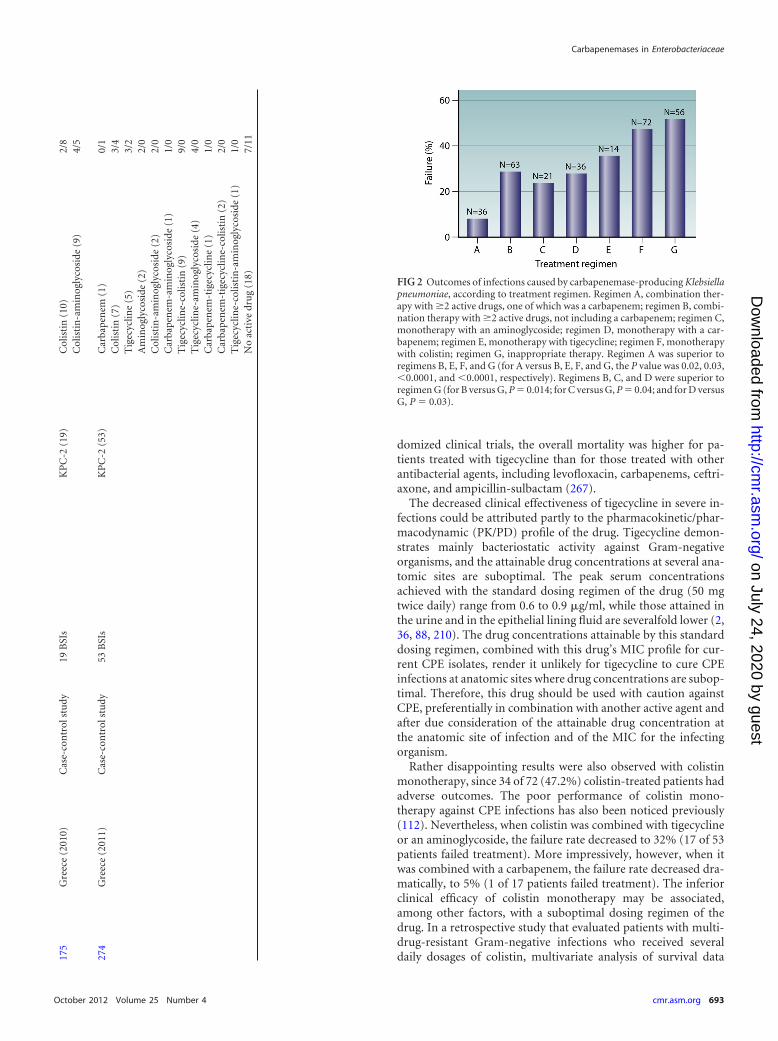
regimen E, monotherapy with tigecycline; regimen F, mono-therapy with colistin; and regimen G, inappropriate therapy (Fig.2). It should be noted that the carbapenem susceptibility statuswas taken as reported in relevant studies in which the previousCLSI interpretive criteria were applied (54).
The lowest failure rate (8.3%) was observed for patients whoreceived combination therapies including a carbapenem (regimenA). In addition, the therapeutic efficacy of this regimen was supe-rior to those of regimens B, E, F, and G (for A versus B, the P valueis 0.02, the odds ratio [OR] is 4.4, and the 95% confidence interval[95% CI] is 1.19 to 16.19; for A versus E, the P value is 0.03, the ORis 6.11, and the 95% CI is 1.22 to 30.58; for A versus F, the P valueis �0.0001, the OR is 9.84, and the 95% CI is 2.76 to 35.03; and forA versus G, the P value is �0.0001, the OR is 11.81, and the 95% CIis 3.24 to 43.06). Combination therapy not including a carbap-enem (regimen B), as well as monotherapy with either an amino-glycoside (regimen C) or a carbapenem (regimen D), was never-theless effective compared to inappropriate therapy (for B versusG, the P value is 0.014, the OR is 2.68, and the 95% CI is 1.26 to5.73; for C versus G, the P value is 0.04, the OR is 3.44, and the 95%CI is 1.11 to 10.67; and for D versus G, the P value is 0.03, the OR
is 2.79, and the 95% CI is 1.14 to 6.86). On the other hand, treat-ment with tigecycline and colistin as single active agents resultedin failure rates comparable to that observed for patients who re-ceived inappropriate therapy (Fig. 2). These observations raiseconcerns about the use of tigecycline or colistin as a single agent inthe treatment of serious carbapenemase-producing K. pneu-moniae infections and support the notion of administering drugcombinations preferentially including a carbapenem when sus-ceptibility data allow.
The limited efficacy of tigecycline revealed by the present anal-ysis is in line with the recent warning issued by the U.S. Food andDrug Administration (FDA) against the use of this agent for seri-ous infections (91a). The FDA, in a pooled analysis of 13 clinicaltrials, found an increased mortality risk associated with the use oftigecycline compared to other drugs to treat a variety of seriousinfections. A higher mortality rate was seen most clearly for pa-tients treated for ventilator-associated pneumonia and bacter-emia (9/18 [50.0%] tigecycline-treated patients versus 1/13[7.7%] comparator drug-treated patients). The cause of excessdeath in these trials most likely was related to progression of theinfection. Similarly, in a recent meta-analysis including 15 ran-
TABLE 3 Clinical studies, antimicrobial therapies, and outcomes for patients infected with M�L-producing K. pneumoniae
ReferenceCountry (yr ofpublication) Study design
No. of patients withindicated type of infection
Type of MBL (no. ofisolates)
Treatment(no. of patients)
Outcome (no.of successes/no. of failures)
121 Greece (2004) Case reports 4 (2 BSIs, 1 case ofmediastinitis, 1 boneinfection)
VIM-1 (4) Colistin (1) 1/0
86 Greece (2008) Tigecycline (1) 1/056 Spain (2008) Tigecycline-colistin (2) 1/1223 Ireland (2010)
269 Taiwan (2001) Case series 3 BSIs IMP-8 (3) Carbapenem (3) 1/2143 Taiwan (2004) Case series 3 (2 pneumonias, 1 BSI) IMP-type enzyme (3) Carbapenem (1) 1/0
Carbapenem-aminoglycoside (2) 2/0
240 Greece (2008) Case series 17 (14 BSIs, 3 pneumonias) VIM-1 (17) Colistin (6) 6/0Tigecycline (1) 0/1Colistin-aminoglycoside (2) 2/0Colistin doxycycline (1) 0/1Carbapenem-colistin (6) 5/1Carbapenem-aminoglycoside-
doxycycline (1)1/0
175 Greece (2010) Case-control study 18 BSIs VIM-1 (17) Colistin (10) 6/4VIM-type enzyme (1) Colistin-aminoglycoside (8) 4/4
64 Greece (2007) Retrospective study 28 BSIs VIM-1 (28) Carbapenem (8) 7/1Colistin (4) 0/4Aminoglycoside (3) 2/1Carbapenem-aminoglycoside (6) 6/0Carbapenem-colistin (1) 1/0Aztreonam-aminoglycoside (2) 1/1No active drug (4) 2/2
67 Greece (2009) Prospectiveobservationalstudy
67 BSIs VIM-1 (67) Carbapenem (14) 11/3Carbapenem-colistin (8) 8/0Carbapenem-aminoglycoside (4) 3/1Colistin (15) 11/4Aminoglycoside (8) 5/3No active drug (18) 13/5
Carbapenemases in Enterobacteriaceae
October 2012 Volume 25 Number 4 cmr.asm.org 691
on July 24, 2020 by guesthttp://cm
r.asm.org/
Dow
nloaded from

TA
BLE
4C
linic
alst
udi
es,a
nti
mic
robi
alth
erap
ies,
and
outc
omes
for
pati
ents
infe
cted
wit
hK
PC
-pro
duci
ng
K.p
neum
onia
e
Ref
eren
ceC
oun
try
(yr
ofpu
blic
atio
n)
Stu
dyde
sign
No.
ofpa
tien
tsw
ith
indi
cate
din
fect
ion
Typ
eof
�-l
acta
mas
e(n
o.of
isol
ates
)T
reat
men
tw
ith
acti
vedr
ug
(no.
ofpa
tien
ts)
Ou
tcom
e(n
o.of
succ
esse
s/n
o.of
failu
res)
252
Col
ombi
a(2
006)
Cas
ere
port
s23
(10
BSI
s,10
pneu
mon
ias,
1en
doca
rdit
is,1
liver
absc
ess,
1em
pyem
a)
KP
C-2
(19)
Car
bape
nem
(4)
3/1
153
USA
(200
6)K
PC
-3(2
)C
olis
tin
(3)
2/1
257
Ch
ina
(200
7)K
PC
-typ
een
zym
e(2
)T
igec
yclin
e(1
)0/
171
USA
(200
7)A
min
ogly
cosi
de(2
)1/
16
USA
(200
8)T
igec
yclin
e-co
listi
n(2
)1/
116
2C
hin
a(2
008)
Tig
ecyc
line-
amin
ogly
cosi
de(1
)1/
081
USA
(200
8)C
olis
tin
-am
inog
lyco
side
(1)
1/0
159
USA
(200
9)A
min
ogly
cosi
de-fl
uor
oqu
inol
one
(1)
1/0
17Is
rael
(200
9)C
arba
pen
em-a
min
ogly
cosi
de(1
)1/
015
8U
SA(2
009)
No
acti
vedr
ug
(7)
1/6
80U
SA(2
009)
116
USA
(201
0)13
8B
razi
l(20
11)
52T
aiw
an(2
011)
10Sw
itze
rlan
d(2
011)
113
USA
(201
1)
25U
SA(2
004)
Cas
ese
ries
4(1
BSI
,2u
rin
ary
trac
tin
fect
ion
s[U
TIs
],1
pneu
mon
ia)
KP
C-2
(4)
Car
bape
nem
(1)
1/0
Car
bape
nem
-col
isti
n(1
)1/
0C
arba
pen
em-a
min
ogly
cosi
de(1
)1/
0C
olis
tin
(1)
0/1
258
USA
(200
9)C
ase
seri
es21
(5pn
eum
onia
s,5
BSI
s,4
case
sof
trac
heo
bron
chit
is,5
UT
Is,1
case
ofm
enin
giti
s,1
surg
ical
site
infe
ctio
n[S
SI])
KP
C-3
(21)
Car
bape
nem
(4)
2/2
Tig
ecyc
line
(5)
4/1
Am
inog
lyco
side
(3)
3/0
Car
bape
nem
-tig
ecyc
line
(1)
0/1
Tig
ecyc
line-
amin
ogly
cosi
de(1
)1/
0N
oac
tive
dru
g(7
)3/
4
154
Gre
ece
(200
9)C
ase
seri
es13
(9pn
eum
onia
s,4
BSI
s)K
PC
-2(1
3)A
min
ogly
cosi
de(2
)2/
0T
igec
yclin
e-co
listi
n(8
)6/
2C
olis
tin
-am
inog
lyco
side
(3)
3/0
83U
SA(2
009)
Cas
ese
ries
7(3
BSI
s,1
UT
I,3
uri
nar
yco
lon
izat
ion
s)K
PC
-2(1
)C
olis
tin
(1)
0/1
KP
C-3
(6)
Col
isti
n-a
min
ogly
cosi
de(2
)0/
2N
oac
tive
dru
g(4
)0/
4
182
USA
(200
9)C
ase
seri
es3
BSI
sK
PC
-2(3
)T
etra
cycl
ine-
amin
ogly
cosi
de(1
)1/
0C
olis
tin
(3)
1/2
237
Gre
ece
(201
0)C
ase
seri
es17
(11
BSI
s,2
SSIs
,1U
TI,
2pn
eum
onia
s,1
case
ofch
olan
giti
s)
KP
C-2
(17)
Col
isti
n(1
1)6/
5T
igec
yclin
e(1
)1/
0A
min
ogly
cosi
de(1
)1/
0C
olis
tin
-am
inog
lyco
side
(2)
1/1
Tig
ecyc
line-
colis
tin
-am
inog
lyco
side
(1)
1/0
No
acti
vedr
ug
(1)
1/0
Tzouvelekis et al.
692 cmr.asm.org Clinical Microbiology Reviews
on July 24, 2020 by guesthttp://cm
r.asm.org/
Dow
nloaded from
domized clinical trials, the overall mortality was higher for pa-tients treated with tigecycline than for those treated with otherantibacterial agents, including levofloxacin, carbapenems, ceftri-axone, and ampicillin-sulbactam (267).
The decreased clinical effectiveness of tigecycline in severe in-fections could be attributed partly to the pharmacokinetic/phar-macodynamic (PK/PD) profile of the drug. Tigecycline demon-strates mainly bacteriostatic activity against Gram-negativeorganisms, and the attainable drug concentrations at several ana-tomic sites are suboptimal. The peak serum concentrationsachieved with the standard dosing regimen of the drug (50 mgtwice daily) range from 0.6 to 0.9 �g/ml, while those attained inthe urine and in the epithelial lining fluid are severalfold lower (2,36, 88, 210). The drug concentrations attainable by this standarddosing regimen, combined with this drug’s MIC profile for cur-rent CPE isolates, render it unlikely for tigecycline to cure CPEinfections at anatomic sites where drug concentrations are subop-timal. Therefore, this drug should be used with caution againstCPE, preferentially in combination with another active agent andafter due consideration of the attainable drug concentration atthe anatomic site of infection and of the MIC for the infectingorganism.
Rather disappointing results were also observed with colistinmonotherapy, since 34 of 72 (47.2%) colistin-treated patients hadadverse outcomes. The poor performance of colistin mono-therapy against CPE infections has also been noticed previously(112). Nevertheless, when colistin was combined with tigecyclineor an aminoglycoside, the failure rate decreased to 32% (17 of 53patients failed treatment). More impressively, however, when itwas combined with a carbapenem, the failure rate decreased dra-matically, to 5% (1 of 17 patients failed treatment). The inferiorclinical efficacy of colistin monotherapy may be associated,among other factors, with a suboptimal dosing regimen of thedrug. In a retrospective study that evaluated patients with multi-drug-resistant Gram-negative infections who received severaldaily dosages of colistin, multivariate analysis of survival data17
5G
reec
e(2
010)
Cas
e-co
ntr
olst
udy
19B
SIs
KP
C-2
(19)
Col
isti
n(1
0)2/
8C
olis
tin
-am
inog
lyco
side
(9)
4/5
274
Gre
ece
(201
1)C
ase-
con
trol
stu
dy53
BSI
sK
PC
-2(5
3)C
arba
pen
em(1
)0/
1C
olis
tin
(7)
3/4
Tig
ecyc
line
(5)
3/2
Am
inog
lyco
side
(2)
2/0
Col
isti
n-a
min
ogly
cosi
de(2
)2/
0C
arba
pen
em-a
min
ogly
cosi
de(1
)1/
0T
igec
yclin
e-co
listi
n(9
)9/
0T
igec
yclin
e-am
inog
lyco
side
(4)
4/0
Car
bape
nem
-tig
ecyc
line
(1)
1/0
Car
bape
nem
-tig
ecyc
line-
colis
tin
(2)
2/0
Tig
ecyc
line-
colis
tin
-am
inog
lyco
side
(1)
1/0
No
acti
vedr
ug
(18)
7/11
FIG 2 Outcomes of infections caused by carbapenemase-producing Klebsiellapneumoniae, according to treatment regimen. Regimen A, combination ther-apy with �2 active drugs, one of which was a carbapenem; regimen B, combi-nation therapy with �2 active drugs, not including a carbapenem; regimen C,monotherapy with an aminoglycoside; regimen D, monotherapy with a car-bapenem; regimen E, monotherapy with tigecycline; regimen F, monotherapywith colistin; regimen G, inappropriate therapy. Regimen A was superior toregimens B, E, F, and G (for A versus B, E, F, and G, the P value was 0.02, 0.03,�0.0001, and �0.0001, respectively). Regimens B, C, and D were superior toregimen G (for B versus G, P � 0.014; for C versus G, P � 0.04; and for D versusG, P � 0.03).
Carbapenemases in Enterobacteriaceae
October 2012 Volume 25 Number 4 cmr.asm.org 693
on July 24, 2020 by guesthttp://cm
r.asm.org/
Dow
nloaded from

showed that a lower total daily dosage of intravenous colistin wasassociated with increased mortality (90). It is therefore critical toadminister an adequate total daily dosage of colistin to critically illpatients, particularly to those who are on renal replacement ther-apy, in order to accomplish efficacious levels according to currentrecommendations (97). An additional factor that could be detri-mental to a patient’s outcome is the delay in attaining an effica-cious drug concentration with the standard treatment regimen ofcolistin. This could be overcome by administering a loading doseof the drug (211).
Although colistin has been used extensively in critically ill pa-tients infected with multidrug-resistant Gram-negative organ-isms, its optimum dosing regimen remains to be defined. Animalinfection models have shown that the ratio of the area under theconcentration-time curve for the free, unbound fraction of thedrug (fAUC) to the MIC is the PK/PD index that is linked moststrongly to an antibacterial effect, indicating the importance ofachieving adequate time exposure to colistin across the day byadministering the drug twice or three times a day (77, 78). Incontrast, however, several features of this drug, such as its pro-longed half-life, its concentration-dependent killing, and a phe-nomenon known as “adaptive resistance” that has not been appre-ciated adequately (50, 68, 92, 236), favor a once-daily dosingregimen, provided that such a scheme is not proven to be morenephrotoxic. Thus, a better understanding of the complex PK/PDfeatures of colistin will be essential in devising dosing regimenswith improved efficacy against CPE infections.
Among the publications available in MEDLINE, we were able toidentify 15 studies reporting on 50 patients infected with carbap-enemase-positive K. pneumoniae, all of whom had received car-bapenem monotherapy (meropenem or imipenem). Twenty-nineof the respective isolates exhibited carbapenem MICs of �2 �g/ml. In seven and six isolates, the MICs were equal to 4 and 8 �g/ml,respectively. The remaining eight isolates were inhibited in vitroby carbapenem concentrations of �8 �g/ml. Note that, as indi-cated by the reported outcomes of these patients, the therapeuticefficacy of carbapenems increased from 25% for a MIC of �8�g/ml to 66.7% for a MIC of 8 �g/ml, 71.4% for a MIC of 4 �g/ml,and 72.4% for a MIC of 2 �g/ml or less (Table 5). Clinical expe-rience with carbapenem monotherapy is indeed limited. Yet wemay consider the above data to indicate that carbapenems couldprovide some therapeutic benefit in infections caused by carbap-enemase-producing K. pneumoniae, even for strains with interme-diate susceptibility to carbapenems. It should be pointed out here
that these observations do not contradict the findings of the ex-perimental infection models discussed herein or those of humanPK/PD studies (34, 118, 131, 145). Carbapenems display time-dependent bactericidal killing when free drug concentrations re-main above the MIC for 40 to 50% of the time between dosingintervals. The probability of attaining a 50% TMIC target for anisolate with a MIC of 4 �g/ml is 69% for the traditional dosingregimen (e.g., 30-min infusion of 1 g every 8 h for meropenem)and increases to 100% for the high-dose/prolonged-infusion reg-imen (e.g., 3-h infusion of 2 g every 8 h for meropenem). For aMIC of 8 �g/ml, only the high-dose/prolonged-infusion regimendisplays a relatively high probability (85%) of bactericidal targetattainment (131).
Whether we can use carbapenems in the presence of a carbap-enemase is an issue that remains to be answered (65, 245). How-ever, faced with the daily challenge of managing critically ill pa-tients and the dearth of alternative therapeutic options, some ofwhich have not been investigated satisfactorily and/or whose effi-cacy in certain situations remains questionable, use of a carbap-enem against an organism with a MIC of �4 or even �8 �g/ml,using a high-dose/prolonged-infusion regimen and in combina-tion with another active agent, preferentially gentamicin or colis-tin, seems reasonable.
The number of CPE isolates exhibiting resistance to almost allavailable agents is worryingly high in various settings (85). Giventhat fosfomycin displays good in vitro activity against most CPE,this agent could be selected as salvage therapy in situations wheretherapeutic options are very limited (89). Although the main in-dication of fosfomycin remains the treatment of lower urinarytract infection, some investigators have included this drug in var-ious combination schemes to treat systemic infections caused byCPE (87, 164). Available data, however, are too limited to allow asound hypothesis as to its efficacy. Also, the potential of fosfomy-cin to rapidly select resistant mutants during therapy is a matter ofconsideration (188).
The clinical data reviewed here allow for some reasonable no-tions but not for solid conclusions, since it was not possible tomeasure and adjust for certain important variables (e.g., host-related factors, severity of infections, and dosing and timing ofinitiation of treatment). Thus, we cannot exclude the possibilitythat our analysis, in some cases, might have resulted in biasedassociations between antimicrobial treatment and outcome. Nev-ertheless, given that the majority of patients infected with CPE aredebilitated, with various underlying diseases, and that more than90% of them have severe infections (BSIs or pneumonias), it isunlikely that residual confounding could account to an apprecia-ble extent for the significantly different failure rates between treat-ment groups.
CPE IN HEALTH CARE SETTINGS
Epidemiology
The prevalence of CPE, primarily K. pneumoniae, in several insti-tutions in areas of endemicity may vary between 20 and 40% (28,99, 133, 233). Initially, CPE appeared to cause hospital-acquiredinfections, mainly in ICU patients (28, 29, 67, 233, 265). Morerecently, however, they have spread in different health care set-tings, including long-term care facilities (LTCF) (16, 53, 83, 161,249).
Several investigators have evaluated the factors associated with
TABLE 5 Results of carbapenem monotherapy in 50 CPE-infectedpatients from 15 studiesa
MIC of carbapenem(�g/ml)
No. ofpatients
No. ofsuccesses
No. offailures % Failure
�1 17 12 5 29.42 12 9 3 25.04 7 5 2 28.68 6 4 2 33.3
Subtotal 42 30 12 28.6b
�8 8 2 6 75.0b
Total 50 32 18 36a See references 25, 64, 67, 81, 113, 143, 153, 159, 162, 240, 252, 257, 258, 269, and 275.b P � 0.02, odds ratio � 7.5, and 95% confidence interval � 1.32 to 42.52.
Tzouvelekis et al.
694 cmr.asm.org Clinical Microbiology Reviews
on July 24, 2020 by guesthttp://cm
r.asm.org/
Dow
nloaded from

increased risk for acquisition of CPE in the hospital setting (69, 91,98, 106, 117, 132, 205, 233, 261). Investigators from Israel haveshown that poor functional status of the host, prior antibiotictherapy, and stay in the ICU are independent risk factors for col-onization or infection with carbapenem-resistant K. pneumoniae(233). Other factors that have been associated with CPE includesolid organ or stem cell transplantation (122, 204), presence of abiliary catheter (120), multiple invasive devices (157), prior sur-gery, and the presence of wounds (106). Antibiotic selection pres-sure may be an additional factor that influences colonization withthese organisms. Case-control studies have shown that almost ev-ery class of antibiotics can select for CPE (69, 91, 98, 117, 120, 132,203, 261). What appears to be more important, however, is thecumulative number of prior antibiotic exposures rather than theuse of a specific class of antibiotics (69, 205).
Since members of the Enterobacteriaceae constitute part of thehuman enteric flora, once CPE colonize the intestinal tract, car-riage may persist for a long time (232). Based on limited experi-ence (90, 116, 207), it appears that colonization with CPE is pro-longed and lasts at least several months. A prolonged duration ofcolonization means a larger reservoir of colonized patients, exert-ing more colonization pressure, which in turn will result in higherrates of patient-to-patient transmission. CPE cross-transmissionoccurs more efficiently in health care settings where infection con-trol practices are poor. Indeed, in a surgical unit where hand hy-giene compliance was 21%, the probability of a patient becomingcolonized with CPE was 7.1% per week of hospitalization, and theincidence of new acquisitions was 9.1/1,000 patient-days (39). Itcan be supported that once the first case of CPE infection is rec-ognized in a health care facility, these organisms may have alreadyspread widely and colonized a substantial number of patients.Colonization may be extensive and pass largely unnoticed in in-stitutions located in regions of endemicity, as evident in severalstudies. Calfee et al. (41) reported that 37% of patients with car-bapenem-resistant K. pneumoniae colonization were first identi-fied by surveillance cultures. During an outbreak of carbapen-emase-producing K. pneumoniae in Israel, a point prevalencesurvey demonstrated that 16 (5.4%) of 298 patients screened werecolonized with carbapenemase producers, and notably, 11 (69%)of these carriers would have remained undetected without theperformance of active surveillance cultures (261).
More importantly, CPE colonization may evolve to infection,with detrimental effects for the host (23, 41, 261). Although dataregarding the infection/colonization ratio are very limited, it isestimated that a proportion of colonized patients (10 to 30%) willdevelop CPE infection. This proportion is probably related to theseverity of the underlying disease of the host and appears to behigher for severely immunocompromised patients (e.g., patientsin induction chemotherapy for acute myelogenous leukemia orpost-allogeneic stem cell transplantation).
Infection Control Strategies
The rapid and worldwide dissemination of a variety of CPE re-flects, to various extents, increased antibiotic selection pressure,carriage of the acquired carbapenemase genes by mobile geneticunits, and probably the enhanced spreading potential of specificclones, such as K. pneumoniae ST258. Notwithstanding these fac-tors, the fact that CPE outbreaks occur principally in settingswhere infection control practices are inadequate shows that thereremains plenty of room for curbing CPE spread.
CPE seem to have a high potential for spread not only frompatient to patient within a health care facility but also through“cycling” of patients between institutions in the same region (263)and/or across borders from high- to low-prevalence countries,which, for instance, has happened repeatedly in Europe (107),threatening every health care system. To address this public healththreat, it is imperative to formulate a preparedness plan beforeCPE have the opportunity to become endemic. In areas where thishas already happened, on the other hand, control measures mustinclude a multifaceted approach coordinated by the nationalhealth authorities, as indicated in a recent study from Israel (234).
In response to this need, both the Centers for Disease Controland Prevention (CDC) and a group of experts from the EuropeanSociety of Clinical Microbiology and Infectious Diseases (ESC-MID) have published guidelines for interventions to control CPEtransmission in acute health care facilities (44, 48). These recom-mendations are based primarily on experience with other MDRorganisms (www.cdc.gov/hicpac/mdro/mdro_0.html) and typi-cally include detection, isolation or cohorting, and other en-hanced infection control measures. It is expected that the increas-ing number of studies dealing with CPE epidemiology will soonlead to guidelines specifically targeting CPE.
Tracing of Carriers
Critical to the success of interrupting cross-transmission of CPEin a health care facility is the timely identification of colonizedand/or infected patients. Thus, every clinical microbiology labo-ratory should establish a reliable detection methodology. Addi-tionally, resources and trained personnel should be readily avail-able to carry out point prevalence surveys as well as activesurveillance cultures, a demanding yet highly effective approachfor detecting carriers (15, 53, 177). The most suitable anatomicsites for surveillance cultures appear to be the perianal area and therectum (31, 261). In patients with surgical wounds, decubitus ul-cers, a urinary catheter, or bronchial secretions, the respective sitescould also be screened. Several versions of culture-based tech-niques have been described. In some of them, differences are lim-ited to the concentration of carbapenem used for the initialscreening, with the lowest being that proposed by EUCAST (0.25�g/ml of meropenem), based on an epidemiologic cutoff. Addi-tionally, several PCR-based techniques for active surveillance havebeen described (111, 135, 231). PCR assays are rapid and usuallymore sensitive than culture-based methods. However, their maindisadvantages are that they do not provide information on thecarbapenemase-producing species and can assess the presence ofonly already known resistance genes.
In settings with low CPE prevalence, laboratories should mon-itor clinical culture results to determine whether CPE have beenisolated in the facility (48). If a CPE is identified by clinical culture,a point prevalence survey should be performed in selected wards(e.g., ICUs and units where CPE have been identified). Detectionof additional CPE carriers should then be followed by active sur-veillance covering a wider range of patients with potential epide-miological links to persons from whom CPE have been isolated[e.g., patients from the same unit and patients cared for by thesame health care worker(s)]. Active surveillance should be contin-ued until no new CPE cases are identified.
In areas where CPE are endemic, an increased likelihood existsfor importation of CPE into a previously CPE-free health carefacility. Upon admission of patients at increased risk of CPE car-
Carbapenemases in Enterobacteriaceae
October 2012 Volume 25 Number 4 cmr.asm.org 695
on July 24, 2020 by guesthttp://cm
r.asm.org/
Dow
nloaded from
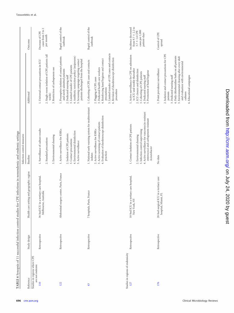
TA
BLE
6Sy
nop
sis
of11
succ
essf
uli
nfe
ctio
nco
ntr
olst
udi
esfo
rC
PE
infe
ctio
ns
inn
onen
dem
ican
den
dem
icse
ttin
gs
Ref
eren
ceSt
udy
desi
gnH
ealt
hca
rese
ttin
gan
dge
ogra
phic
regi
on
Infe
ctio
nco
ntr
olm
easu
res
Ou
tcom
eB
asel
ine
Add
itio
nal
Stu
dies
inre
gion
sw
her
eC
PE
are
not
ende
mic
110
Ret
rosp
ecti
ve36
-bed
ICU
ina
tert
iary
care
hos
pita
l,M
elbo
urn
e,A
ust
ralia
1.Su
rvei
llan
ceof
cult
ure
resu
lts
1.U
niv
ersa
lcon
tact
prec
auti
ons
inIC
UD
ecre
ase
ofC
PE
case
sfr
om3
to1
per
mon
th2.
Stan
dard
prec
auti
ons
2.Si
ngl
e-ro
omis
olat
ion
ofC
PE
pati
ents
(all
war
ds)
3.E
nvi
ron
men
talc
lean
ing
3.R
estr
icti
onof
carb
apen
emu
se
122
Ret
rosp
ecti
veA
bdom
inal
surg
ery
cen
ter,
Par
is,F
ran
ce1.
Act
ive
surv
eilla
nce
for
ESB
Ls1.
Pre
empt
ive
isol
atio
nof
con
tact
pati
ents
and
new
lyad
mit
ted
pati
ents
Rap
idco
ntr
olof
the
outb
reak
2.Is
olat
ion
ofC
PE
pati
ents
2.D
edic
ated
nu
rsin
gst
aff
3.C
onta
ctpr
ecau
tion
s3.
Lim
ited
tran
sfer
ofC
PE
pati
ents
4.E
nvi
ron
men
tald
isin
fect
ion
4.A
nti
biot
icre
stri
ctio
npo
licy
(im
ipen
em)
5.A
ctiv
esu
rvei
llan
ce5.
Scre
enin
gca
mpa
ign
targ
etin
gco
nta
ctpa
tien
tsdi
sch
arge
dfr
omth
eh
ospi
tal
43R
etro
spec
tive
7h
ospi
tals
,Par
is,F
ran
ce1.
Nat
ion
alea
rly
war
nin
gsy
stem
for
mu
ltir
esis
tan
tis
olat
es1.
Coh
orti
ng
ofC
PE
case
san
dco
nta
cts
Rap
idco
ntr
olof
the
outb
reak
2.A
ctiv
esu
rvei
llan
cefo
rE
SBLs
2.Fl
aggi
ng
ofC
PE
case
s3.
Act
ive
scre
enin
gof
con
tact
pati
ents
3.D
edic
ated
hea
lth
care
wor
kers
4.E
valu
atio
nof
duod
enos
cope
disi
nfe
ctio
npr
acti
ces
4.R
ein
forc
ing
han
dh
ygie
ne
and
con
tact
prec
auti
ons
5.Li
mit
edtr
ansf
erof
CP
Eca
ses
and
con
tact
s6.
Rev
isio
nof
duod
enos
cope
disi
nfe
ctio
npr
oced
ure
Stu
dies
inre
gion
sof
ende
mic
ity
127
Ret
rosp
ecti
ve10
-bed
ICU
ina
tert
iary
care
hos
pita
l,N
ewY
ork,
NY
1.C
onta
ctis
olat
ion
ofC
PE
pati
ents
1.A
ctiv
esu
rvei
llan
cefo
rC
PE
onad
mis
sion
toIC
Uan
dw
eekl
yth
erea
fter
Inci
den
cede
crea
sed
from
9.7
2.
2to
3.7
1.
6C
PE
case
spe
r1,
000
pati
ent-
days
2.E
nvi
ron
men
talc
lean
ing
2.IC
Ucl
osu
rean
ddi
sin
fect
ion
3.In
fect
ion
con
trol
supe
rvis
ing
3.C
ohor
tin
gof
CP
Epa
tien
ts4.
Act
ive
surv
eilla
nce
for
van
com
ycin
-res
ista
nt
ente
roco
ccia
nd
carb
apen
em-r
esis
tan
tA
cine
toba
cter
4.D
edic
ated
nu
rsin
gst
aff
5.P
rom
otio
nof
han
dh
ygie
ne
176
Ret
rosp
ecti
ve20
-bed
surg
ical
ICU
ina
tert
iary
care
hos
pita
l,M
iam
i,FL
No
data
1.P
oin
tpr
eval
ence
surv
eilla
nce
Con
trol
ofC
PE
spre
ad2.
Isol
atio
nan
dco
nta
ctpr
ecau
tion
sfo
rC
PE
pati
ents
3.D
edic
ated
nu
rsin
gst
aff
4.D
aily
chlo
rhex
idin
eba
ths
onal
lpat
ien
ts5.
En
viro
nm
enta
lcle
anin
gaf
ter
ever
ysh
ift
and
eval
uat
ion
wit
hen
viro
nm
enta
lcu
ltu
res
6.E
duca
tion
alca
mpa
ign
s
Tzouvelekis et al.
696 cmr.asm.org Clinical Microbiology Reviews
on July 24, 2020 by guesthttp://cm
r.asm.org/
Dow
nloaded from

177
Ret
rosp
ecti
ve70
-bed
lon
g-te
rmac
ute
care
hos
pita
l,C
hic
ago,
IL1.
Act
ive
surv
eilla
nce
onad
mis
sion
1.A
ctiv
esu
rvei
llan
cecu
ltu
res
for
CP
Ean
dpo
int
prev
alen
cesu
rvey
sdu
rin
gth
ein
terv
enti
on
Col
oniz
atio
npr
eval
ence
ofC
PE
decr
ease
dpr
ogre
ssiv
ely,
from
21%
to12
,6,
3,an
d0%
2.B
asel
ine
poin
tpr
eval
ence
surv
eilla
nce
2.Is
olat
ion
and
con
tact
prec
auti
ons
for
CP
Epa
tien
ts3.
Pre
empt
ive
isol
atio
nof
hig
h-r
isk
pati
ents
4.E
nvi
ron
men
talc
ult
ure
san
den
han
ced
envi
ron
men
talc
lean
ing
5.D
aily
chlo
rhex
idin
eba
ths
for
allp
atie
nts
6.E
duca
tion
alca
mpa
ign
83R
etro
spec
tive
Lon
g-te
rmac
ute
care
hos
pita
l,So
uth
Flor
ida
No
data
1.A
ctiv
esu
rvei
llan
cecu
ltu
reT
erm
inat
ion
ofth
eou
tbre
ak2.
Poi
nt
prev
alen
cesu
rvey
3.Is
olat
ion
and
con
tact
prec
auti
ons
for
CP
Epa
tien
ts4.
Ded
icat
edn
urs
ing
staf
fan
deq
uip
men
t
106
Ret
rosp
ecti
veT
erti
ary
care
hos
pita
l,P
uer
toR
ico
No
data
1.C
onta
ctpr
ecau
tion
sfo
rC
PE
pati
ents
Con
trol
ofth
eou
tbre
ak2.
Coh
orti
ng
ofC
PE
pati
ents
3.D
edic
ated
nu
rsin
gst
aff
4.H
and
hyg
ien
eau
dits
5.IC
Ucl
osu
re6.
Res
tric
tion
ofbr
oad-
spec
tru
man
tibi
otic
s7.
Act
ive
surv
eilla
nce
onad
mis
sion
toh
igh
-ri
sku
nit
s(I
CU
,dia
bete
sw
ard)
and
wee
kly
ther
eaft
er
15R
etro
spec
tive
Ter
tiar
yca
reh
ospi
tal,
Tel
Has
hom
er,I
srae
l1.
Con
tact
prec
auti
ons
for
CP
Eca
ses
1.A
ctiv
esu
rvei
llan
ceon
adm
issi
onto
ICU
and
inst
ep-d
own
un
its
and
wee
kly
ther
eaft
er
Inci
den
cede
crea
sed
from
6.93
to1.
8C
PE
case
spe
r10
,000
pati
ent-
days
2.In
oth
erde
part
men
ts,a
ctiv
esu
rvei
llan
ceof
pati
ents
wit
hep
idem
iolo
gic
links
toC
PE
carr
iers
3.D
aily
repo
rtin
gof
CP
Eca
ses
toh
ospi
tal
man
ager
and
the
nat
ion
alco
ordi
nat
or
53P
rosp
ecti
vein
terv
enti
onst
udy
Ter
tiar
yca
reh
ospi
tal,
Reh
ovot
,Isr
ael
No
data
1.A
ctiv
esu
rvei
llan
ceon
adm
issi
onto
ICU
,in
room
mat
esof
new
CP
Eca
ses
orca
rrie
rs,a
nd
inpa
tien
tsat
hig
hri
skfo
rca
rria
ge
Inci
den
cede
crea
sed
from
8.2
to0.
5C
PE
case
per
10,0
00pa
tien
t-da
ys2.
Isol
atio
n-c
ohor
tin
gan
dco
nta
ctpr
ecau
tion
sfo
rC
PE
case
san
dca
rrie
rs3.
Ded
icat
edn
urs
ing
staf
f4.
En
viro
nm
enta
lcle
anin
gan
ddi
sin
fect
ing
duri
ng
hos
pita
lsta
yan
daf
ter
disc
har
ge5.
Edu
cati
onan
dtr
ain
ing
toal
lmed
ical
staf
fm
embe
rs,p
atie
nts
,an
dca
regi
vers
6.A
uto
mat
icw
arn
ing
syst
em
234
Pro
spec
tive
inte
rven
tion
stu
dy27
acu
teca
reh
ospi
tals
,Isr
ael
No
data
1.Is
olat
ion
-coh
orti
ng
and
con
tact
prec
auti
ons
for
CP
Epa
tien
tsan
dca
rrie
rsM
onth
lyin
cide
nce
decr
ease
dfr
om55
.5to
11.7
CP
Eca
ses
per
100,
000
pati
ent-
days
2.D
edic
ated
nu
rsin
gst
affa
nd
equ
ipm
ent
3.M
anda
tory
repo
rtin
gto
publ
ich
ealt
hau
thor
itie
sof
ever
yC
PE
case
4.E
stab
lish
men
tof
Tas
kFo
rce
onA
nti
mic
robi
alR
esis
tan
cean
dIn
fect
ion
Con
trol
Carbapenemases in Enterobacteriaceae
October 2012 Volume 25 Number 4 cmr.asm.org 697
on July 24, 2020 by guesthttp://cm
r.asm.org/
Dow
nloaded from

riage (such as residency in an LTCF, previous stay in an ICU,prolonged hospitalization in the previous 6 months, or presenceof indwelling devices), preemptive isolation while awaiting sur-veillance culture results can prevent early transmission events (19,43, 53, 122). Performing a surveillance culture before the dis-charge of a patient into the community or an LTCF may also beuseful to avoid transfer of CPE to additional niches (83, 122). It isalso important to communicate the results of screening and toprovide alerts for previously identified CPE carriers for every re-admitted patient. The surveillance strategy should be definedclearly for each setting and evaluated periodically according to thecurrent situation and available resources (19, 48).
Intervention
When a CPE carrier is identified, the infection control personnelshould be notified immediately. Isolation or cohorting of CPEcarriers seems to be the main prevention measure (43, 83, 106,110, 122, 127, 176, 177, 234). Assignment of dedicated health careworkers and use of separate equipment for carriers are additionalinterventions that have been employed successfully in several out-breaks (19, 43, 122, 176, 177, 234). In the Israeli experience, phys-ical separation of carriers from noncarriers and assignment ofdedicated nursing staff to care for carriers on all shifts were themost important components of the intervention measures in halt-ing transmission (234).
In addition to isolation, personnel in acute health care facilitiesshould use contact precautions (wearing a gown and gloves) whencaring for patients colonized or infected with CPE in order tominimize indirect transmission of the organism (44, 48; www.cdc.gov/hicpac/mdro/mdro_0.html). In LTCFs, it is only practical toapply contact precautions for those patients who are severely illand have conditions that may facilitate transmission (e.g., diar-rhea or decubitus ulcers) (83). The success of the interventionshould be monitored constantly, and when failure is observed, aroot cause analysis should be performed (19).
Environmental cleaning and decolonization of patients.Cleaning of the inanimate environment and equipment in prox-imity to a CPE carrier, along with daily antiseptic (chlorhexidine)baths to cleanse patients’ skin, were included in the bundles ofintervention measures that successfully controlled two recent out-breaks in the United States (176, 177). Available data do not allowfor an assessment of the usefulness of the systematic application ofthese practices. Indeed, persisting environmental contaminationwith Enterobacteriaceae is limited compared to that of other or-ganisms (144). In addition, culturing of surfaces and equipmentto investigate their role in the transmission chain is not usuallyrequired, unless the inanimate environment or shared equipmentis potentially linked to an outbreak (43). Such an event, however,does not seem to occur frequently with CPE. On the other hand,daily antiseptic baths have been proven efficacious in preventing
central vascular catheter-associated bloodstream infections (178).This practice therefore appears to warrant further evaluation withrespect to CPE-positive settings.
Selective decolonization of the gut by use of oral gentamicin inpatients undergoing chemotherapy and allogeneic stem cell trans-plantation has achieved a 66% eradication rate of the CPE carrier state(278). This approach should be adopted cautiously, however, as gen-tamicin is commonly used for the treatment of CPE infections, and itsexcessive use may select CPE that are resistant to this drug as well.
Judicious antimicrobial use. The role of restriction of antibioticsin controlling CPE needs further evaluation. As mentioned previ-ously, almost every antimicrobial class can select for CPE. In thisregard, cumulative exposure to antibiotics is likely to be more impor-tant than selection exerted by a specific agent. Therefore, formularyinterventions should focus on reduction of overall antibiotic usage(19).
Success Stories
There have been various successful attempts to control CPE out-breaks in both endemic and nonendemic settings. The relevantstudies (summarized in Table 6) concern single-center outbreaks,except for one describing a countrywide epidemic in Israel (234).Although some differences in approach did exist, the interven-tions implemented were largely based on the rationale of infectioncontrol strategies mentioned previously, with their main compo-nents being surveillance cultures, isolation and cohorting, contactprecautions, and assignment of dedicated staff.
The design of these studies does not allow us to accurately classifymeasures according to their effectiveness. Critical interpretation ofthe published data, however, suggests that application of a bundle ofinfection control measures may be required for maximum contain-ment of CPE. Controlled studies and mathematical modeling of CPEtransmission and prevention are needed to specify the most appro-priate procedures for containment or even eradication of CPE.
NOVEL AGENTS AGAINST CPE
Antibiotics
Not unexpectedly, many new antibiotics under development tar-get multidrug-resistant bacteria. The relatively small number ofnew antibacterials active against MDRs does not necessarily meana lack of industry interest but rather reflects the countless difficul-ties posed from the early stages of designing to the introductioninto clinical practice. Although not specifically focused on CPE,some of these experimental antibacterials, belonging to diverseantimicrobial classes, exhibit high activity against these microor-ganisms. We present below a few indicative compounds, prefer-entially those that are under advanced clinical testing (Table 7).
Sulfactams. The sulfactams comprise a distinct series of mono-cyclic �-lactams (with tigemonam being the first member) exhib-
TABLE 7 Experimental antimicrobial agents active against carbapenemase-producing Enterobacteriaceae
Drug Compound type Relevant target Reference
BAL30072 Siderophore-containing sulfactam Enterobacteriaceae, including M�L producers 194Plazomicin (ACHN-490) Sisomicin derivative Gram-negative organisms, including carbapenemase
producers149
GSK2251052 Leucyl-tRNA synthetase inhibitor Gram-negative organisms, including carbapenemaseproducers
5
Tzouvelekis et al.
698 cmr.asm.org Clinical Microbiology Reviews
on July 24, 2020 by guesthttp://cm
r.asm.org/
Dow
nloaded from

iting potent activity against the Enterobacteriaceae. Additionally,other structurally related monocyclic compounds, such as aztreo-nam, are virtually unaffected by the hydrolytic activity of M�Ls.Of special interest is a novel group of siderophore-coupled sulfac-tams, represented by BAL30072 (194); the coupled siderophoresenhance activity against M�L producers by facilitating the en-trance rate through the outer membrane, even in strains withdefective permeability (195). Animal experiments have furthersupported the therapeutic potential of BAL30072 against M�L-positive enterobacteria (168).
Plazomicin. Formerly known as ACHN-490, plazomicin is asisomicin derivative with substitutions at positions 1 and 6 (ahydroxyaminobutyric and a hydroxyethyl group, respectively).This antibiotic resists most aminoglycoside-modifying enzymes(but not 16S rRNA methylases) and exhibits potent bactericidalactivity against many MDRs, including CPE (149). It is currentlyundergoing a phase 2 study for use against complicated urinarytract infections (276).
Aminoacyl-tRNA synthetase inhibitors. Until recently, theaminoacyl-tRNA synthetase inhibitors included only mupirocin,an isoleucyl-tRNA synthetase inhibitor of limited clinical use. De-velopment of novel boron-containing aminoacyl-tRNA synthe-tase inhibitors added compounds with kinetics suitable for sys-tematic use that are also active against Gram-negative organisms.A promising compound, GSK2251052, which inhibits the leucyl-tRNA synthetase and is effective against CPE and other MDRs, iscurrently under clinical evaluation (5).
Carbapenemase Inhibitors
Despite almost 3 decades of efforts to develop �-lactamase inhib-itors, only three, clavulanic acid, tazobactam, and sulbactam, havebeen made available as therapeutics. Fortunately, a number ofpotential �-lactamase inhibitors are under evaluation (for a com-prehensive review, see reference 76). The majority of currentlytested compounds are �-lactam derivatives, though a number ofdiverse non-�-lactam substances also exhibit significant inhibi-tory activity. Only compounds that also happen to inhibit ac-quired carbapenemases (mainly KPCs and M�Ls) at clinically rel-evant concentrations are mentioned briefly here (Table 8).
Penem derivatives. The penem derivatives include the proto-type, BRL 42715, and a series of heterocyclic methylidene penemswith significant inhibitory activity against serine �-lactamases(molecular classes A, C, and D). BLI-489, a bicyclic derivative, hasbeen shown to be capable of reducing the MICs of piperacillinagainst enterobacteria producing KPC enzymes (209).
1-�-Methylcarbapenems. 1-�-Methylcarbapenems have beensynthesized by various substitutions in the carbapenem nucleus,
which contains a methyl group at position C-1 (the same nucleusas that in doripenem). Interesting members of the group are com-pounds J-110,411 and J-111,225, which inhibit class A and C�-lactamases, as well as IMP-type M�Ls, at low concentrations(183, 184).
Sulfones. C-6-substituted penicillin sulfones are primarily in-hibitors of class A �-lactamases. However, compounds with amercaptomethyl substituent at C-6 exhibit strong inhibitory ac-tivity against M�Ls (38).
Succinic acids (non-�-lactams). Various succinic acid deriva-tives [2,3-(S,S)-disubstituted succinic acids] exhibit potent inhib-itory activity against the IMP-type M�Ls, restoring carbapenemsusceptibility in members of the Enterobacteriaceae producingthese enzymes (170).
Thiols (non-�-lactams). Thiol compounds, such as thioman-delic acids, are considered effective inhibitors of M�Ls, especiallythose of the VIM and IMP types. They act by bridging the zinc ionsof the active site, thus displacing the catalytic water molecule (147,268).
Avibactam. Formerly known as NXL104, avibactam, a non-�-lactam compound, is likely to be the most promising experimentalinhibitor and is expected to be introduced soon into antimicrobialchemotherapy. It is a bridged diazabicyclo (EC 3.2.1) octanonewith excellent activity against virtually all serine �-lactamases butno activity against molecular class B enzymes (22). In the currentera of carbapenemases, the ability of avibactam to inhibit KPC�-lactamases at very low concentrations (50% inhibitory concen-tration [IC50] of 38 nM) is important (22, 242). MICs of variousnewer �-lactams tested against KPC producers in the presence ofavibactam have clearly shown the in vitro efficacy of these combi-nations (82). Most importantly, the therapeutic efficacy of theceftazidime-avibactam combination has been documented formurine infection models (259).
CONCLUDING REMARKS AND PERSPECTIVES
The current public health crisis due to the international spread ofcarbapenemase-producing multidrug-resistant enterobacteriahas caught us unprepared, despite clear signs of this problem aris-ing years ago. Most of the recent papers describing yet anotheremergence of a CPE or a CPE outbreak conclude, almost invari-ably, with the urgent need for measures to contain these microor-ganisms. This, however, begs the question: precisely what mea-sures are to be taken? First of all, a clearer and more accuratepicture of the situation at the global level, based on data that arevalid for comparisons, is necessary. For instance, in developedcountries, it appears that many studies have been conducted insingle institutions or a small number of tertiary care hospitals and
TABLE 8 Experimental �-lactamase inhibitors active against carbapenemases from Enterobacteriaceae
Inhibitor Compound typeInhibition spectrum(�-lactamase classes)
Susceptiblecarbapenemasesa Reference(s)
BLI-489 Penem A, C, D KPC type 209J-110,411 and J-111,225 1-�-Methyl carbapenem A, C, B IMP type 183, 184Mercaptomethyl sulfones C-6-substituted penicillin sulfone B VIM and IMP types 382,3-(S,S)-Disubstituted succinic
acidsSuccinic acid B IMP type 170
Thiomandelic acids Thiol B VIM and IMP types 147, 268Avibactam (NLX104) Diazabicyclo-octanone A, C, D KPC type 22a Only carbapenemase types with documented susceptibility to the respective inhibitor are included.
Carbapenemases in Enterobacteriaceae
October 2012 Volume 25 Number 4 cmr.asm.org 699
on July 24, 2020 by guesthttp://cm
r.asm.org/
Dow
nloaded from

cover limited times. Therefore, biased sampling is probable, atleast in some cases. Moreover, systematic reports from many Af-rican countries, the Balkans, the Middle East, and vast areas inAsia are scarce, if they exist at all. The collection of epidemiolog-ical data that are as complete as possible is imperative for theimplementation of effective yet affordable and sustainable mea-sures against CPE, especially in the most affected countries. In thisrespect, if the expressed international concern is indeed genuine,resources from international public health organizations shouldbe mobilized and allocated appropriately.
Some new drugs active against CPE are indeed in an advancedstage of development, but very few of them are expected to beclinically available soon. Thus, in the foreseeable future, we shallcontinue to rely on the available antibiotics. Nevertheless, thestudies summarized above show that there is yet room to improveour therapeutic approaches. Colistin and tigecycline are amongthe most frequently used agents in the treatment of CPE infec-tions. However, as discussed here, laboratory and clinical datasupporting this practice are insufficient. Although it has to beadmitted that alternative options have not yet been documentedsolidly, well-designed clinical trials aiming to (i) determine theoptimum dosing regimen of colistin, (ii) define CPE infectionsthat could be controlled effectively by tigecycline, (iii) exploit thePK/PD features of carbapenems, and (iv) unravel the most effec-tive drug combinations may prove valuable.
REFERENCES1. Adams-Haduch JM, Potoski BA, Sidjabat HE, Paterson DL, Doi Y.
2009. Activity of temocillin against KPC-producing Klebsiella pneu-moniae and Escherichia coli. Antimicrob. Agents Chemother. 53:2700 –2701.
2. Agwuh KN, MacGowan A. 2006. Pharmacokinetics and pharmacody-namics of the tetracyclines including glycylcyclines. J. Antimicrob. Che-mother. 58:256 –265.
3. Aktas Z, Bal C, Midilli K, Poirel L, Nordmann P. 2006. First IMP-1-producing Klebsiella pneumoniae isolate in Turkey. Clin. Microbiol. In-fect. 12:695– 696.
4. Alba J, Ishii Y, Thomson K, Moland ES, Yamaguchi K. 2005. Kineticsstudy of KPC-3, a plasmid-encoded class A carbapenem-hydrolyzing�-lactamase. Antimicrob. Agents Chemother. 49:4760 – 4762.
5. Alley D. 2011. Novel boron-containing aminoacyl-tRNA synthetase in-hibitors with Gram-negative bacterial activity, abstr 1071. Abstr. 51stIntersci. Conf. Antimicrob. Agents Chemother., Chicago, IL.
6. Anthony KB, et al. 2008. Clinical and microbiological outcomes ofserious infections with multidrug-resistant Gram-negative organismstreated with tigecycline. Clin. Infect. Dis. 46:567–570.
7. Antoniadou A, et al. 2007. Colistin-resistant isolates of Klebsiella pneu-moniae emerging in intensive care unit patients: first report of a multi-clonal cluster. J. Antimicrob. Chemother. 59:786 –790.
8. Aubert D, Naas T, Héritier C, Poirel L, Nordmann P. 2006. Functionalcharacterization of IS1999, an IS4 family element involved in mobiliza-tion and expression of �-lactam resistance genes. J. Bacteriol. 188:6506 –6514.
9. Avlami A, et al. 2010. Detection of metallo-�-lactamase genes in clinicalspecimens by a commercial multiplex PCR system. J. Microbiol. Meth-ods 83:185–187.
10. Babouee B, et al. 2011. Emergence of four cases of KPC-2 and KPC-3-carrying Klebsiella pneumoniae introduced to Switzerland, 2009 –10.Euro Surveill. 16:19817. http://www.eurosurveillance.org/ViewArticle.aspx?ArticleId�19817.
11. Bagley ST. 1985. Habitat association of Klebsiella species. Infect. Control6:52–58.
12. Baraniak A, et al. 2009. The emergence of Klebsiella pneumoniae ST258with KPC-2 in Poland. Antimicrob. Agents Chemother. 53:4565– 4567.
13. Bebrone C. 2007. Metallo-�-lactamases (classification, activity, geneticorganization, structure, zinc coordination) and their superfamily.Biochem. Pharmacol. 74:1686 –1701.
14. Ben-David D, et al. 2012. Outcome of carbapenem resistant Klebsiellapneumoniae bloodstream infections. Clin. Microbiol. Infect. 18:54 – 60.
15. Ben-David D, et al. 2010. Potential role of active surveillance in thecontrol of a hospital-wide outbreak of carbapenem-resistant Klebsiellapneumoniae infection. Infect. Control Hosp. Epidemiol. 31:620 – 626.
16. Ben-David D, et al. 2011. Carbapenem-resistant Klebsiella pneumoniaein post-acute-care facilities in Israel. Infect. Control Hosp. Epidemiol.32:845– 853.
17. Benenson S, et al. 2009. Carbapenem-resistant Klebsiella pneumoniaeendocarditis in a young adult. Successful treatment with gentamicin andcolistin. Int. J. Infect. Dis. 13:e295– e298.
18. Berçot B, Poirel L, Nordmann P. 2011. Updated multiplex polymerasechain reaction for detection of 16S rRNA methylases: high prevalenceamong NDM-1 producers. Diagn. Microbiol. Infect. Dis. 71:442– 445.
19. Bilavsky E, Schwaber MJ, Carmeli Y. 2010. How to stem the tide ofcarbapenemase-producing Enterobacteriaceae?: proactive versus reactivestrategies. Curr. Opin. Infect. Dis. 23:327–331.
20. Bisiklis A, Papageorgiou F, Frantzidou F, Alexiou-Daniel S. 2007.Specific detection of blaVIM and blaIMP metallo-�-lactamase genes in asingle real-time PCR. Clin. Microbiol. Infect. 13:1201–1203.
21. Bogdanovich T, et al. 2011. Colistin-resistant, Klebsiella pneumoniaecarbapenemase (KPC)-producing Klebsiella pneumoniae belonging tothe international epidemic clone ST258. Clin. Infect. Dis. 53:373–376.
22. Bonnefoy A, et al. 2004. In vitro activity of AVE1330A, an innovativebroad-spectrum non-�-lactam �-lactamase inhibitor. J. Antimicrob.Chemother. 54:410 – 417.
23. Borer A, et al. 8 September 2011. Risk factors for developing clinicalinfection with carbapenem-resistant Klebsiella pneumoniae in hospitalpatients initially only colonized with carbapenem-resistant K. pneu-moniae. Am. J. Infect. Control [Epub ahead of print.] doi:10.1016/j.ajic.2011.05.022.
24. Borer A, et al. 2009. Attributable mortality rate for carbapenem-resistant Klebsiella pneumoniae bacteremia. Infect. Control Hosp. Epide-miol. 30:972–976.
25. Bradford PA, et al. 2004. Emergence of carbapenem-resistant Klebsiellaspecies possessing the class A carbapenem-hydrolyzing KPC-2 and in-hibitor-resistant TEM-30 �-lactamases in New York City. Clin. Infect.Dis. 39:55– 60.
26. Bratu S, et al. 2007. Detection and spread of Escherichia coli possessingthe plasmid-borne carbapenemase KPC-2 in Brooklyn, New York. Clin.Infect. Dis. 44:972–975.
27. Bratu S, Landman D, Alam M, Tolentino E, Quale J. 2005. Detectionof KPC carbapenem-hydrolyzing enzymes in Enterobacter spp. fromBrooklyn, New York. Antimicrob. Agents Chemother. 49:776 –778.
28. Bratu S, et al. 2005. Rapid spread of carbapenem-resistant Klebsiellapneumoniae in New York City: a new threat to our antibiotic armamen-tarium. Arch. Intern. Med. 165:1430 –1435.
29. Bratu S, et al. 2005. Emergence of KPC-possessing Klebsiella pneu-moniae in Brooklyn, New York: epidemiology and recommendations fordetection. Antimicrob. Agents Chemother. 49:3018 –3020.
30. Bratu S, et al. 2005. Carbapenemase-producing Klebsiella pneumoniaein Brooklyn, NY: molecular epidemiology and in vitro activity of poly-myxin B and other agents. J. Antimicrob. Chemother. 56:128 –132.
31. Buehlmann M, Fankhauser H, Laffer R, Bregenzer T, Widmer AF.2010. The inguinal skin: an important site of colonization with extended-spectrum �-lactamase-producing Enterobacteriaceae. Infect. ControlHosp. Epidemiol. 31:427– 428.
32. Bulik CC, et al. 2010. Comparison of the activity of a human simulated,high-dose, prolonged infusion of meropenem against Klebsiella pneu-moniae producing the KPC carbapenemase versus that against Pseu-domonas aeruginosa in an in vitro pharmacodynamic model. Antimicrob.Agents Chemother. 54:804 – 810.
33. Bulik CC, et al. 2010. Comparison of meropenem MICs and suscepti-bilities for carbapenemase-producing Klebsiella pneumoniae isolates byvarious testing methods. J. Clin. Microbiol. 48:2402–2406.
34. Bulik CC, Nicolau DP. 2010. In vivo efficacy of simulated human dosingregimens of prolonged-infusion doripenem against carbapenemase-producing Klebsiella pneumoniae. Antimicrob. Agents Chemother. 54:4112– 4115.
35. Burckhardt I, Zimmermann S. 2011. Using matrix-assisted laser de-sorption ionization–time of flight mass spectrometry to detect carbap-enem resistance within 1 to 2.5 hours. J. Clin. Microbiol. 49:3321–3324.
36. Burkhardt O, et al. 2009. Tigecycline possibly underdosed for the treat-
Tzouvelekis et al.
700 cmr.asm.org Clinical Microbiology Reviews
on July 24, 2020 by guesthttp://cm
r.asm.org/
Dow
nloaded from

ment of pneumonia: a pharmacokinetic viewpoint. Int. J. Antimicrob.Agents 34:101–102.
37. Bush K, Jacoby GA. 2010. Updated functional classification of �-lacta-mases. Antimicrob. Agents Chemother. 54:969 –976.
38. Buynak JD, et al. 2004. Penicillin-derived inhibitors that simultaneouslytarget both metallo- and serine-�-lactamases. Bioorg. Med. Chem. Lett.14:1299 –1304.
39. Buzala G, et al. 2011. Risk factors for carbapenem resistant Enterobac-teriaceae carriage on admission to a surgical unit and acquisition rateduring hospitalization, abstr P687. Abstr. 21st Eur. Congr. Clin. Micro-biol. Infect. Dis., Milan, Italy.
40. Cagnacci S, et al. 2008. Bloodstream infections caused by multidrug-resistant Klebsiella pneumoniae producing the carbapenem-hydrolysingVIM-1 metallo-�-lactamase: first Italian outbreak. J. Antimicrob. Che-mother. 61:296 –300.
41. Calfee D, Jenkins SG. 2008. Use of active surveillance cultures to detectasymptomatic colonization with carbapenem-resistant Klebsiella pneu-moniae in intensive care unit patients. Infect. Control Hosp. Epidemiol.29:966 –968.
42. Carattoli A, et al. 2010. Complete nucleotide sequence of the IncNplasmid pKOX105 encoding VIM-1, QnrS1 and SHV-12 proteins in En-terobacteriaceae from Bolzano, Italy compared with IncN plasmids en-coding KPC enzymes in the U. S. A. J. Antimicrob. Chemother. 65:2070 –2075.
43. Carbonne A, et al. 2010. Control of a multi-hospital outbreak of KPC-producing Klebsiella pneumoniae type 2 in France, September to October2009. Euro Surveill. 15:19734. http://www.eurosurveillance.org/ViewArticle.aspx?ArticleId�19734.
44. Carmeli Y, et al. 2010. Controlling the spread of carbapenemase-producing gram-negatives: therapeutic approach and infection control.Clin. Microbiol. Infect. 16:102–111.
45. Carrër A, et al. 2008. Spread of OXA-48-positive-carbapenem-resistantKlebsiella pneumoniae isolates in Istanbul, Turkey. Antimicrob. AgentsChemother. 52:2950 –2954.
46. Carrër A, et al. 2010. Spread of OXA-48-encoding plasmid in Turkeyand beyond. Antimicrob. Agents Chemother. 54:1369 –1373.
47. Castanheira M, et al. 2011. Early dissemination of NDM-1- and OXA-181-producing Enterobacteriaceae in Indian hospitals: report from theSENTRY Antimicrobial Surveillance Program, 2006 –2007. Antimicrob.Agents Chemother. 55:1274 –1278.
48. Centers for Disease Control and Prevention (CDC). 2009. Guidancefor control of infections with carbapenem-resistant or carbapenemase-producing Enterobacteriaceae in acute care facilities. MMWR Morb.Mortal. Wkly. Rep. 58:256 –260.
49. Centers for Disease Control and Prevention (CDC). 2010. Detection ofEnterobacteriaceae isolates carrying metallo-�-lactamase—UnitedStates, 2010. MMWR Morb. Mortal. Wkly. Rep. 59:750.
50. Cheng HY, Chen YF, Peng HL. 2010. Molecular characterization of thePhoPQ-PmrD-PmrAB mediated pathway regulating polymyxin B resis-tance in Klebsiella pneumoniae CG43. J. Biomed. Sci. 17:60 –76.
51. Chu YW, et al. 2001. IMP-4, a novel metallo-�-lactamase from noso-comial Acinetobacter spp. collected in Hong Kong between 1994 and1998. Antimicrob. Agents Chemother. 45:710 –714.
52. Chung KP, et al. 2011. Arrival of Klebsiella pneumoniae carbapenemase(KPC)-2 in Taiwan. J. Antimicrob. Chemother. 66:1182–1184.
53. Ciobotaro P, Oved M, Nadir E, Bardenstein R, Zimhony O. 2011. Aneffective intervention to limit the spread of an epidemic carbapenem-resistant Klebsiella pneumoniae strain in an acute care setting: from the-ory to practice. Am. J. Infect. Control 39:671– 677.
54. Clinical and Laboratory Standards Institute. 2010. Performance stan-dards for antimicrobial susceptibility testing: 19th informational supple-ment. CLSI document M100-S20. Clinical and Laboratory StandardsInstitute, Wayne, PA.
55. Clinical and Laboratory Standards Institute. 2010. Performance stan-dards for antimicrobial susceptibility testing; 20th informational supple-ment (June 2010 update). CLSI document M100-S20-U. Clinical andLaboratory Standards Institute, Wayne, PA.
56. Cobo J, et al. 2008. Use of tigecycline for the treatment of prolongedbacteremia due to a multiresistant VIM-1 and SHV-12 �-lactamase-producing Klebsiella pneumoniae epidemic clone. Diagn. Microbiol. In-fect. Dis. 60:319 –322.
57. Cohen Stuart J, et al. 2010. Guideline for phenotypic screening and
confirmation of carbapenemases in Enterobacteriaceae. Int. J. Antimi-crob. Agents 36:205–210.
58. Cohen Stuart J, Voets G, Scharringa J, Fluit AC, Leverstein-Van HallMA. 1 March 2012. Detection of carbapenemase producing Enterobac-teriaceae with a commercial DNA Microarray. J. Med. Microbiol. [Epubahead of print.] doi:10.1099/jmm.0.041673-0.
59. Colinon C, Miriagou V, Carattoli A, Luzzaro F, Rossolini GM. 2007.Characterization of the IncA/C plasmid pCC416 encoding VIM-4 andCMY-4 �-lactamases. J. Antimicrob. Chemother. 60:258 –262.
60. Cornaglia G, et al. 2007. Metallo-�-lactamases as emerging resistancedeterminants in Gram-negative pathogens: open issues. Int. J. Antimi-crob. Agents 29:380 –388.
61. Cornaglia G, Giamarellou H, Rossolini GM. 2011. Metallo-�-lactamases: a last frontier for �-lactams? Lancet Infect. Dis. 11:381–393.
62. Cuzon G, Naas T, Lesenne A, Benhamou M, Nordmann P. 2010.Plasmid-mediated carbapenem-hydrolysing OXA-48 �-lactamase inKlebsiella pneumoniae from Tunisia. Int. J. Antimicrob. Agents 36:91–93.
63. Cuzon G, et al. 2010. Worldwide diversity of Klebsiella pneumoniae thatproduce �-lactamase blaKPC-2 gene. Emerg. Infect. Dis. 16:1349 –1356.
64. Daikos GL, et al. 2007. VIM-1-producing Klebsiella pneumoniae blood-stream infections: analysis of 28 cases. Int. J. Antimicrob. Agents 29:471–473.
65. Daikos GL, Markogiannakis A. 2011. Carbapenemase-producing Kleb-siella pneumoniae: (when) might we still consider treating with carbap-enems? Clin. Microbiol. Infect. 17:1135–1141.
66. Daikos GL, et al. 2007. Activity of imipenem against VIM-1 metallo-�-lactamase-producing Klebsiella pneumoniae in the murine thigh infec-tion model. Clin. Microbiol. Infect. 13:202–205.
67. Daikos GL, et al. 2009. Prospective observational study of the impact ofVIM-1 metallo-�-lactamase on the outcome of patients with Klebsiellapneumoniae bloodstream infections. Antimicrob. Agents Chemother.53:1868 –1873.
68. Daikos GL, et al. 2010. Serum bactericidal activity of three differentdosing regimens of colistin with implications for optimum clinical use. J.Chemother. 22:175–178.
69. Daikos GL, et al. 2010. Risk factors for bloodstream infection withKlebsiella pneumoniae producing VIM-1 metallo-�-lactamase. J. Anti-microb. Chemother. 65:784 –788.
70. Dallenne C, Da Costa A, Decré D, Favier C, Arlet G. 2010. Develop-ment of a set of multiplex PCR assays for the detection of genes encodingimportant �-lactamases in Enterobacteriaceae. J. Antimicrob. Che-mother. 65:490 – 495.
71. Daly MW, Riddle DJ, Ledeboer NA, Dunne WM, Ritchie DJ. 2007.Tigecycline for treatment of pneumonia and empyema caused by car-bapenemase-producing Klebsiella pneumoniae. Pharmacotherapy 27:1052–1057.
72. Daoud Z, Hobeika E, Choucair A, Rohban R. 2008. Isolation of the firstmetallo-�-lactamase producing Klebsiella pneumoniae in Lebanon. Rev.Esp. Quimioter. 21:123–126.
73. Docquier JD, et al. 2009. Crystal structure of the OXA-48 �-lactamasereveals mechanistic diversity among class D carbapenemases. Chem.Biol. 16:540 –547.
74. Docquier JD, et al. 2003. On functional and structural heterogeneity ofVIM-type metallo-�-lactamases. J. Antimicrob. Chemother. 51:257–266.
75. Doi Y, et al. 2008. Simple disk-based method for detection of Klebsiellapneumoniae carbapenemase-type �-lactamase by use of a boronic acidcompound. J. Clin. Microbiol. 46:4083– 4086.
76. Drawz SM, Bonomo RA. 2010. Three decades of beta-lactamase inhib-itors. Clin. Microbiol. Rev. 23:160 –201.
77. Dudhani RV, et al. 2010. Elucidation of the pharmacokinetic/pharmacodynamic determinant of colistin activity against Pseudomonasaeruginosa in murine thigh and lung infection models. Antimicrob.Agents Chemother. 54:1117–1124.
78. Dudhani RV, Turnidge JD, Nation RL, Li JJ. 2010. fAUC/MIC is themost predictive pharmacokinetic/pharmacodynamic index of colistinagainst Acinetobacter baumannii in murine thigh and lung infectionmodels. J. Antimicrob. Chemother. 65:1984 –1990.
79. Elemam A, Rahimian J, Doymaz M. 2010. In vitro evaluation of anti-biotic synergy for polymyxin B-resistant carbapenemase-producingKlebsiella pneumoniae. J. Clin. Microbiol. 48:3558 –3562.
80. Elemam A, Rahimian J, Mandell W. 2009. Infection with panresistant
Carbapenemases in Enterobacteriaceae
October 2012 Volume 25 Number 4 cmr.asm.org 701
on July 24, 2020 by guesthttp://cm
r.asm.org/
Dow
nloaded from

Klebsiella pneumoniae: a report of 2 cases and a brief review of the liter-ature. Clin. Infect. Dis. 49:271–274.
81. Endimiani A, et al. 2008. Presence of plasmid-mediated quinoloneresistance in Klebsiella pneumoniae isolates possessing blaKPC in theUnited States. Antimicrob. Agents Chemother. 52:2680 –2682.
82. Endimiani A, Choudhary Y, Bonomo RA. 2009. In vitro activity ofNXL104 in combination with �-lactams against Klebsiella pneumoniaeisolates producing KPC carbapenemases. Antimicrob. Agents Che-mother. 53:3599 –3601.
83. Endimiani A, et al. 2009. Emergence of blaKPC-containing Klebsiellapneumoniae in a long-term acute care hospital: a new challenge to ourhealthcare system. J. Antimicrob. Chemother. 64:1102–1110.
84. Endimiani A, et al. 2009. Characterization of blaKPC-containing Kleb-siella pneumoniae isolates detected in different institutions in the easternU. S. A. J. Antimicrob. Chemother. 63:427– 437.
85. Endimiani A, et al. 2010. In vitro activity of fosfomycin against blaKPC-containing Klebsiella pneumoniae isolates, including those nonsuscep-tible to tigecycline and/or colistin. Antimicrob. Agents Chemother. 54:526 –529.
86. Evagelopoulou P, Myrianthefs P, Markogiannakis A, Baltopoulos G,Tsakris A. 2008. Multidrug-resistant Klebsiella pneumoniae mediastini-tis safely and effectively treated with prolonged administration of tigecy-cline. Clin. Infect. Dis. 46:1932–1933.
87. Falagas ME, Giannopoulou KP, Kokolakis GN, Rafailidis PI. 2008.Fosfomycin: use beyond urinary tract and gastrointestinal infections.Clin. Infect. Dis. 46:1069 –1077.
88. Falagas ME, Karageorgopoulos DE, Nordmann P. 2011. Therapeuticoptions for infections with Enterobacteriaceae producing carbapenem-hydrolyzing enzymes. Future Microbiol. 6:653– 666.
89. Falagas ME, et al. 2010. Antimicrobial susceptibility of multidrug-resistant (MDR) and extensively drug-resistant (XDR) Enterobacteria-ceae isolates to fosfomycin. Int. J. Antimicrob. Agents 35:240 –243.
90. Falagas ME, et al. 2010. Colistin therapy for microbiologically docu-mented multidrug-resistant Gram-negative bacterial infections: a retro-spective cohort study of 258 patients. Int. J. Antimicrob. Agents 35:194 –199.
91. Falagas ME, et al. 2007. Risk factors of carbapenem-resistant Klebsiellapneumoniae infections: a matched case control study. J. Antimicrob.Chemother. 60:1124 –1130.
91a.(a)FDA. 1 September 2010, posting date. FDA drug safety communica-tion: increased risk of death with Tygacil (tigecycline) compared to otherantibiotics used to treat similar infections. http://www.fda.gov/Drugs/DrugSafety/ucm224370.htm.
92. Fernández L, Breidenstein EB, Hancock RE. 2011. Creeping baselinesand adaptive resistance to antibiotics. Drug Resist. Updat. 14:1–21.
93. Franceschini N, et al. 2000. Purification and biochemical characteriza-tion of the VIM-1 metallo-�-lactamase. Antimicrob. Agents Chemother.44:3003–3007.
94. Fukigai S, et al. 2007. Nosocomial outbreak of genetically related IMP-1�-lactamase-producing Klebsiella pneumoniae in a general hospital inJapan. Int. J. Antimicrob. Agents 29:306 –310.
95. Gacar GG, et al. 2005. Genetic and enzymatic properties of metallo-�-lactamase VIM-5 from a clinical isolate of Enterobacter cloacae. Antimi-crob. Agents Chemother. 49:4400 – 4403.
96. Gales AC, Jones RN, Sader HS. 2011. Contemporary activity of colistinand polymyxin B against a worldwide collection of Gram-negativepathogens: results from the SENTRY Antimicrobial Surveillance Pro-gram (2006 – 09). J. Antimicrob. Chemother. 66:2070 –2074.
97. Garonzik SM, et al. 2011. Population pharmacokinetics of colistinmethanesulfonate and formed colistin in critically ill patients from amulticenter study provide dosing suggestions for various categories ofpatients. Antimicrob. Agents Chemother. 55:3284 –3294.
98. Gasink LB, Edelstein PH, Lautenbach E, Synnestvedt M, Fishman NO.2009. Risk factors and clinical impact of Klebsiella pneumoniae carbap-enemase-producing K. pneumoniae. Infect. Control Hosp. Epidemiol.30:1180 –1185.
99. Giakkoupi P, et al. 2011. An update of the evolving epidemic of blaKPC-
2-carrying Klebsiella pneumoniae in Greece (2009 –10). J. Antimicrob.Chemother. 66:1510 –1513.
100. Giakkoupi P, et al. 2009. Emerging Klebsiella pneumoniae isolates co-producing KPC-2 and VIM-1 carbapenemases. Antimicrob. Agents Che-mother. 53:4048 – 4050.
101. Giakkoupi P, et al. 2005. Discrepancies and interpretation problems in
susceptibility testing of VIM-1-producing Klebsiella pneumoniae isolates.J. Clin. Microbiol. 43:494 – 496.
102. Giakoupi P, et al. 2009. Greek system for the surveillance of antimicro-bial resistance. KPC-2-producing Klebsiella pneumoniae infections inGreek hospitals are mainly due to a hyperepidemic clone. Euro Surveill.14:19218. http://www.eurosurveillance.org/ViewArticle.aspx?ArticleId�19218.
103. Giani T, et al. 2009. Emergence in Italy of Klebsiella pneumoniae se-quence type 258 producing KPC-3 carbapenemase. J. Clin. Microbiol.47:3793–3794.
104. Giske CG, et al. 2011. A sensitive and specific phenotypic assay fordetection of metallo-�-lactamases and KPC in Klebsiella pneumoniaewith the use of meropenem disks supplemented with aminophenylbo-ronic acid, dipicolinic acid and cloxacillin. Clin. Microbiol. Infect. 17:552–556.
105. Goren MG, Navon-Venezia S, Chmelnitsky I, Carmeli Y. 2010. Car-bapenem-resistant KPC-2-producing Escherichia coli in a Tel Aviv med-ical center, 2005 to 2008. Antimicrob. Agents Chemother. 54:2687–2691.
106. Gregory CJ, et al. 2010. Outbreak of carbapenem-resistant Klebsiellapneumoniae in Puerto Rico associated with a novel carbapenemase vari-ant. Infect. Control Hosp. Epidemiol. 31:476 – 484.
107. Grundmann H, et al. 2010. Carbapenem-non-susceptible Enterobacte-riaceae in Europe: conclusions from a meeting of national experts. EuroSurveill. 15:19711. http://www.eurosurveillance.org/ViewArticle.aspx?ArticleId�19711.
108. Halaby T. 13 February 2012. A case of New Delhi metallo-�-lactamase 1(NDM-1)-producing Klebsiella pneumoniae from the Balkan region inthe Netherlands with putative secondary transmission. Antimicrob.Agents Chemother. [Epub ahead of print.] doi:10.1128/AAC.00111-12.
109. Hall BG, Barlow M. 2005. Revised Ambler classification of �-lactamases.J. Antimicrob. Chemother. 55:1050 –1051.
110. Herbert S, et al. 2007. Large outbreak of infetion and colonization withgram-negative pathogens carrying the metallo-�-lactamase gene blaIMP-4
at a 320-bed tertiary hospital in Australia. Infect. Control Hosp. Epide-miol. 28:98 –101.
111. Hindiyeh M, et al. 2008. Rapid detection of blaKPC carbapenemase genesby real-time PCR. J. Clin. Microbiol. 46:2879 –2883.
112. Hirsch EB, Tam VH. 2010. Detection and treatment options for Kleb-siella pneumoniae carbapenemases (KPCs): an emerging cause of multi-drug-resistant infection. J. Antimicrob. Chemother. 65:1119 –1125.
113. Ho VP, et al. 2011. Use of meropenem by continuous infusion to treat apatient with a Bla(kpc-2)-positive Klebsiella pneumoniae blood streaminfection. Surg. Infect. (Larchmt.) 12:325–327.
114. Ho PL, et al. 2011. Complete sequencing of pNDM-HK encodingNDM-1 carbapenemase from a multidrug-resistant Escherichia colistrain isolated in Hong Kong. PLoS One 21:e17989. doi:10.1371/journal.pone.0017989.
115. Hrabák J, Walková R, Studentová V, Chudácková E, Bergerová T.2011. Carbapenemase activity detection by matrix-assisted laser desorp-tion ionization–time of flight mass spectrometry. J. Clin. Microbiol. 49:3222–3227.
116. Humphries RM, et al. 2010. Successful treatment of pan-resistant Kleb-siella pneumoniae pneumonia and bacteraemia with a combination ofhigh-dose tigecycline and colistin. J. Med. Microbiol. 59:1383–1386.
117. Hussein K, et al. 2009. Carbapenem resistance among Klebsiella pneu-moniae isolates: risk factors, molecular characteristics, and susceptibilitypatterns. Infect. Control Hosp. Epidemiol. 30:666 – 671.
118. Jaruratanasirikul S, Sriwiriyajan S, Punyo J. 2005. Comparison of thepharmacodynamics of meropenem in patients with ventilator-associatedpneumonia following administration by 3-hour infusion or bolus injec-tion. Antimicrob. Agents Chemother. 49:1337–1339.
119. Jarvis WR, Munn VP, Highsmith AK, Culver DH, Hughes JM. 1985.The epidemiology of nosocomial infections caused by Klebsiella pneu-moniae. Infect. Control 6:68 –74.
120. Jeon MH, et al. 2008. Risk factors for the acquisition of carbapenem-resistant Escherichia coli among hospitalized patients. Diagn. Microbiol.Infect. Dis. 62:402– 406.
121. Karabinis A, et al. 2004. Colistin for Klebsiella pneumoniae-associatedsepsis. Clin. Infect. Dis. 38:e7– e9.
122. Kassis-Chikhani N, et al. 2010. Extended measures for controlling anoutbreak of VIM-1 producing imipenem-resistant Klebsiella pneumoniaein a liver transplant centre in France, 2003–2004. Euro Surveill. 15:19713.http://www.eurosurveillance.org/ViewArticle.aspx?ArticleId�19713.
Tzouvelekis et al.
702 cmr.asm.org Clinical Microbiology Reviews
on July 24, 2020 by guesthttp://cm
r.asm.org/
Dow
nloaded from

123. Ke W, Bethel CR, Thomson JM, Bonomo RA, van den Akker F. 2007.Crystal structure of KPC-2: insights into carbapenemase activity in classA �-lactamases. Biochemistry 46:5732–5740.
124. Kim S-Y, Hong SG, Moland ES, Thomson KS. 2007. Convenient testusing a combination of chelating agents for detection of metallo-�-lactamases in the clinical laboratory. J. Clin. Microbiol. 45:2798 –2801.
125. Kim MN, et al. 2012. Nosocomial clustering of NDM-1-producingKlebsiella pneumoniae sequence type 340 strains in four patients at aSouth Korean tertiary care hospital. J. Clin. Microbiol. 50:1433–1436.
126. Kitchel B, et al. 2009. Molecular epidemiology of KPC-producing Kleb-siella pneumoniae in the United States: clonal expansion of MLST se-quence type 258. Antimicrob. Agents Chemother. 53:3365–3370.
127. Kochar S, et al. 2009. Success of an infection control program to reducethe spread of carbapenem-resistant Klebsiella penumoniae. Infect. Con-trol Hosp. Epidemiol. 30:447– 452.
128. Kontopoulou K, et al. 2010. Hospital outbreak caused by Klebsiellapneumoniae producing KPC-2 �-lactamase resistant to colistin. J. Hosp.Infect. 76:70 –73.
129. Ktari S, et al. 2006. Emergence of multidrug-resistant Klebsiella pneu-moniae isolates producing VIM-4 metallo-�-lactamase, CTX-M-15extended-spectrum �-lactamase, and CMY-4 AmpC �-lactamase in aTunisian university hospital. Antimicrob. Agents Chemother. 50:4198 –4201.
130. Kumarasamy KK, et al. 2010. Emergence of a new antibiotic resistancemechanism in India, Pakistan, and the UK: a molecular, biological, andepidemiological study. Lancet Infect. Dis. 10:597– 602.
131. Kuti JL, Dandekar PK, Nightingale CH, Nicolau DP. 2003. Use ofMonte Carlo simulation to design an optimized pharmacodynamic dos-ing strategy for meropenem. J. Clin. Pharmacol. 43:1116 –1123.
132. Kwak YG, et al. 2005. Risk factors for the acquisition of carbapenem-resistant Klebsiella pneumoniae among hospitalized patients. Microb.Drug Resist. 11:165–169.
133. Landman D, et al. 2007. Evolution of antimicrobial resistance amongPseudomonas aeruginosa, Acinetobacter baumannii and Klebsiella pneu-moniae in Brooklyn, NY. J. Antimicrob. Chemother. 60:78 – 82.
134. Landman D, Bratu S, Quale J. 2009. Contribution of OmpK36 tocarbapenem susceptibility in KPC-producing Klebsiella pneumoniae. J.Med. Microbiol. 58:1303–1308.
135. Landman D, Salvani JK, Bratu S, Quale J. 2005. Evaluation of tech-niques for detection of carbapenem-resistant Klebsiella pneumoniae instool surveillance cultures. J. Clin. Microbiol. 43:5639 –5641.
136. Laraki N, et al. 1999. Biochemical characterization of the Pseudomonasaeruginosa 101/1477 metallo-�-lactamase IMP-1 produced by Esche-richia coli. Antimicrob. Agents Chemother. 43:902–906.
137. Lassaux P, et al. 2011. Biochemical and structural characterization of thesubclass B1 metallo-�-lactamase VIM-4. Antimicrob. Agents Che-mother. 55:1248 –1255.
138. Leão RS, et al. 2011. KPC-2 carbapenemase-producing Klebsiella pneu-moniae isolates from patients with cystic fibrosis. J. Cyst. Fibros. 10:140 –142.
139. Leavitt A, Navon-Venezia S, Chmelnitsky I, Schwaber MJ, Carmeli Y.2007. Emergence of KPC-2 and KPC-3 in carbapenem-resistant Kleb-siella pneumoniae strains in an Israeli hospital. Antimicrob. Agents Che-mother. 51:3026 –3029.
140. Lederman ER, Crum NF. 2005. Pyogenic liver abscess with a focus onKlebsiella pneumoniae as a primary pathogen: an emerging disease withunique clinical characteristics. Am. J. Gastroenterol. 100:322–331.
141. Lee K, et al. 2010. Improved performance of the modified Hodge testwith MacConkey agar for screening carbapenemase-producing Gram-negative bacilli. J. Microbiol. Methods 83:149 –152.
142. Lee J, Patel G, Huprikar S, Calfee DP, Jenkins SG. 2009. Decreasedsusceptibility to polymyxin B during treatment for carbapenem-resistantKlebsiella pneumoniae infection. J. Clin. Microbiol. 47:1611–1612.
143. Lee NY, et al. 2004. Clinical experience of bacteremia caused by metallo-�-lactamase-producing gram-negative organisms. J. Microbiol. Immu-nol. Infect. 37:343–349.
144. Lemmen SW, Häfner H, Zolldann D, Stanzel S, Lütticken R. 2004.Distribution of multi-resistant Gram-negative versus Gram-positivebacteria in the hospital inanimate environment. J. Hosp. Infect. 56:191–197.
145. Li C, Kuti JL, Nightingale CH, Nicolau DP. 2006. Population pharma-cokinetic analysis and dosing regimen optimization of meropenem inadult patients. J. Clin. Pharmacol. 46:1171–1178.
146. Li B, et al. 2011. First report of Klebsiella oxytoca strain coproducingKPC-2 and IMP-8 carbapenemases. Antimicrob. Agents Chemother. 55:2937–2941.
147. Lienard BM, et al. 2008. Structural basis for the broad-spectrum inhi-bition of metallo-�-lactamases by thiols. Org. Biomol. Chem. 6:2282–2294.
148. Lincopan N, et al. 2005. First isolation of metallo-�-lactamase-producing multiresistant Klebsiella pneumoniae from a patient in Brazil.J. Clin. Microbiol. 43:516 –519.
149. Livermore DM, et al. 2011. Activity of aminoglycosides, includingACHN-490, against carbapenem-resistant Enterobacteriaceae isolates. J.Antimicrob. Chemother. 66:48 –53.
150. Livermore DM, Walsh TR, Toleman M, Woodford N. 2011. BalkanNDM-1: escape or transplant? Lancet Infect. Dis. 11:164.
151. Livermore DM, et al. 2011. What remains against carbapenem-resistantEnterobacteriaceae? Evaluation of chloramphenicol, ciprofloxacin, colis-tin, fosfomycin, minocycline, nitrofurantoin, temocillin and tigecycline.Int. J. Antimicrob. Agents 37:415– 419.
152. Loli A, et al. 2006. Sources of diversity of carbapenem resistance levels inKlebsiella pneunoniae carrying blaVIM-1. J. Antimicrob. Chemother. 58:669 – 672.
153. Lomaestro BM, Tobin EH, Shang W, Gootz T. 2006. The spread ofKlebsiella pneumoniae carbapenemase-producing K. pneumoniae to up-state New York. Clin. Infect. Dis. 43:e26 – e28.
154. Maltezou HC, et al. 2009. Outbreak of infections due to KPC-2-producing Klebsiella pneumoniae in a hospital in Crete (Greece). J. Infect.58:213–219.
155. Marcano D, et al. 2008. First isolation of a VIM-producing Klebsiellapneumoniae from a seven-year-old child in Venezuela. J. Infect. Dev.Ctries. 2:241–244.
156. Marchaim D, et al. 2011. Outbreak of colistin-resistant, carbapenem-resistant Klebsiella pneumoniae in metropolitan Detroit, Michigan. An-timicrob. Agents Chemother. 55:593–599.
157. Marchaim D, Navon-Venezia S, Schwaber MJ, Carmeli Y. 2008. Iso-lation of imipenem-resistant Enterobacter species: emergence of KPC-2carbapenemase, molecular characterization, epidemiology, and out-comes. Antimicrob. Agents Chemother. 52:1413–1418.
158. Marschall J, et al. 2009. Presence of the KPC carbapenemase gene inEnterobacteriaceae causing bacteremia and its correlation with in vitrocarbapenem susceptibility. J. Clin. Microbiol. 47:239 –241.
159. Mathers AJ, et al. 2009. Fatal cross infection by carbapenem-resistantKlebsiella in two liver transplant recipients. Transpl. Infect. Dis. 11:257–265.
160. Mavroidi A, et al. 2012. Emergence of Escherichia coli sequence type 410(ST410) with KPC-2 �-lactamase. Int. J. Antimicrob. Agents 39:247–250.
161. McGuinn M, Hershow RC, Janda WM. 2009. Escherichia coli andKlebsiella pneumoniae carbapenemase in long-term care facility, Illinois,USA. Emerg. Infect. Dis. 15:988 –989.
162. Mendes RE, et al. 2008. Carbapenem-resistant isolates of Klebsiellapneumoniae in China and detection of a conjugative plasmid (blaKPC-2
plus qnrB4) and a blaIMP-4 gene. Antimicrob. Agents Chemother. 52:798 –799.
163. Mendes RE, et al. 2007. Rapid detection and identification of metallo-�-lactamase-encoding genes by multiplex real-time PCR assay and meltcurve analysis. J. Clin. Microbiol. 45:544 –547.
164. Michalopoulos A, et al. 2010. Intravenous fosfomycin for the treatmentof nosocomial infections caused by carbapenem-resistant Klebsiellapneumoniae in critically ill patients: a prospective evaluation. Clin. Mi-crobiol. Infect. 16:184 –186.
165. Miriagou V, Carattoli A, Tzelepi E, Villa L, Tzouvelekis LS. 2005.IS26-associatedIn4-typeintegronsformingmultiresistanceloci inentero-bacterial plasmids. Antimicrob. Agents Chemother. 49:3541–3543.
166. Miriagou V, et al. 2010. Acquired carbapenemases in Gram-negativebacterial pathogens: detection and surveillance issues. Clin. Microbiol.Infect. 16:112–122.
167. Miriagou V, et al. 2010. Sequence of pNL194, a 79.3-kilobase IncNplasmid carrying the blaVIM-1 metallo-�-lactamase gene in Klebsiellapneumoniae. Antimicrob. Agents Chemother. 54:4497– 4502.
168. Miriagou V, et al. 2010. Efficacy of BAL30072, alone and combined withmeropenem, against VIM-producing enterobacteria in a murine thighinfection model, abstr P1240. Abstr. 20th Eur. Congr. Clin. Microbiol.Infect. Dis., Vienna, Austria.
169. Mochon AB, et al. 2011. New Delhi metallo-�-lactamase (NDM-1)-
Carbapenemases in Enterobacteriaceae
October 2012 Volume 25 Number 4 cmr.asm.org 703
on July 24, 2020 by guesthttp://cm
r.asm.org/
Dow
nloaded from

producing Klebsiella pneumoniae: case report and laboratory detectionstrategies. J. Clin. Microbiol. 49:1667–1670.
170. Moloughney JG, Thomas JD, Toney JH. 2005. Novel IMP-1 metallo-�-lactamase inhibitors can reverse meropenem resistance in Escherichiacoli expressing IMP-1. FEMS Microbiol. Lett. 243:65–71.
171. Monteiro J, Fernandes Santos A, Asensi MD, Peirano G, Gales AC.2009. First report of KPC-2-producing Klebsiella pneumoniae strains inBrazil. Antimicrob. Agents Chemother. 53:333–334.
172. Monteiro J, Widen RH, Pignatari AC, Kubasek C, Silbert S. 2012.Rapid detection of carbapenemase genes by multiplex real-time PCR. J.Antimicrob. Chemother. 67:906 –909.
173. Moquet O, et al. 2011. Class D OXA-48 carbapenemase in multidrug-resistant enterobacteria, Senegal. Emerg. Infect. Dis. 17:143–144.
174. Moulds NM, Thomson KS, Hanson ND. 2011. IMP-27, a novel metal-lo-�-lactamase (MBL) associated with a class II integron identified in anisolate of Proteus mirabilis, abstr C1-1212. Abstr. 51st Intersci. Conf.Antimicrob. Agents Chemother., Chicago, IL.
175. Mouloudi E, et al. 2010. Bloodstream infections caused by metallo-�-lactamase/Klebsiella pneumoniae carbapenemase-producing K. pneu-moniae among intensive care unit patients in Greece: risk factors forinfection and impact of type of resistance on outcomes. Infect. ControlHosp. Epidemiol. 31:1250 –1256.
176. Munoz-Price LS, et al. 2010. Successful eradication of a monoclonalstrain of Klebsiella pneumoniae during a K. pneumoniae carbapenemase-producing K. pneumoniae outbreak in a surgical intensive care unit inMiami, Florida. Infect. Control Hosp. Epidemiol. 31:1074 –1077.
177. Munoz-Price LS, et al. 2010. Successful control of an outbreak of Kleb-siella pneumoniae carbapenemase-producing K. pneumoniae at a long-term acute care hospital. Infect. Control Hosp. Epidemiol. 31:341–347.
178. Munoz-Price LS, Hota B, Stemer A, Weinstein RA. 2009. Prevention ofbloodstream infections by use of daily chlorhexidine baths for patients ata long-term acute care hospital. Infect. Control Hosp. Epidemiol. 30:1031–1035.
179. Naas T, Cuzon G, Bogaerts P, Glupczynski Y, Nordmann P. 2011.Evaluation of a DNA microarray (Check-MDR CT102) for rapid detec-tion of TEM, SHV, and CTX-M extended-spectrum �-lactamases and ofKPC, OXA-48, VIM, IMP, and NDM-1 carbapenemases. J. Clin. Micro-biol. 49:1608 –1613.
180. Naas T, et al. 2008. Genetic structures at the origin of acquisition of the�-lactamase blaKPC gene. Antimicrob. Agents Chemother. 52:1257–1263.
181. Naas T, Nordmann P, Vedel G, Poyart C. 2005. Plasmid-mediatedcarbapenem-hydrolyzing �-lactamase KPC in a Klebsiella pneumoniaeisolate from France. Antimicrob. Agents Chemother. 49:4423– 4424.
182. Nadkarni AS, Schliep T, Khan L, Zeana CB. 2009. Cluster of blood-stream infections caused by KPC-2 carbapenemase producing Klebsiellapneumoniae in Manhattan. Am. J. Infect. Control 37:121–126.
183. Nagano R, Adachi Y, Hashizume T, Morishima H. 2000. In vitroantibacterial activity and mechanism of action of J-111,225, a novel 1�-methylcarbapenem, against transferable IMP-1 metallo-�-lactamaseproducers. J. Antimicrob. Chemother. 45:271–276.
184. Nagano R, et al. 1999. Carbapenem derivatives as potential inhibitors ofvarious �-lactamases, including class B metallo-�-lactamases. Antimi-crob. Agents Chemother. 43:2497–2503.
185. Neonakis IK, et al. 2010. Resistance status and evolution trends ofKlebsiella pneumoniae isolates in a university hospital in Greece: ineffec-tiveness of carbapenems and increasing resistance to colistin. Chemo-therapy 56:448 – 452.
186. Neonakis IK, Stylianou K, Daphnis E, Maraki S. 2011. First case ofresistance to tigecycline by Klebsiella pneumoniae in a European univer-sity hospital. Indian J. Med. Microbiol. 29:78 –79.
187. Neuner EA, et al. 2011. Treatment and outcomes in carbapenem-resistant Klebsiella pneumoniae bloodstream infections. Diagn. Micro-biol. Infect. Dis. 69:357–362.
188. Nilsson A, Berg OG, Aspevall O, Kahlmeter G, Andersson DI. 2003.Biological costs and mechanisms of fosfomycin resistance in Escherichiacoli. Antimicrob. Agents Chemother. 47:2850 –2858.
189. Nordmann P, Boulanger AE, Poirel L. 2012. NDM-4 metallo-�-lactamase with increased carbapenemase activity from Escherichia coli.Antimicrob. Agents Chemother. 56:2184 –2186.
190. Nordmann P, Couard JP, Sansot D, Poirel L. 2012. Emergence of anautochthonous and community-acquired NDM-1-producing Klebsiellapneumoniae in Europe. Clin. Infect. Dis. 54:150 –151.
191. Nordmann P, Cuzon G, Naas T. 2009. The real threat of Klebsiellapneumoniae carbapenemase-producing bacteria. Lancet Infect. Dis.9:228 –236.
192. Nordmann P, Poirel L, Toleman MA, Walsh TR. 2011. Does broad-spectrum �-lactam resistance due to NDM-1 herald the end of the anti-biotic era for treatment of infections caused by Gram-negative bacteria?J. Antimicrob. Chemother. 66:689 – 692.
193. Osano E, et al. 1994. Molecular characterization of an enterobacterialmetallo-�-lactamase found in a clinical isolate of Serratia marcescens thatshows imipenem resistance. Antimicrob. Agents Chemother. 38:71–78.
194. Page MG, Dantier C, Desarbre E. 2010. In vitro properties ofBAL30072, a novel siderophore sulfactam with activity against multire-sistant gram-negative bacilli. Antimicrob. Agents Chemother. 54:2291–2302.
195. Page MG, Heim J. 2009. New molecules from old classes: revisiting thedevelopment of beta-lactams. IDrugs 12:561–565.
196. Panagea T, et al. 2011. Evaluation of CHROMagar KPC for the detectionof carbapenemase-producing Enterobacteriaceae in rectal surveillancecultures. Int. J. Antimicrob. Agents 37:124 –128.
197. Panagiotakopoulou A, et al. 2007. Comparative in vitro killing of car-bapenems and aztreonam against Klebsiella pneumoniae producingVIM-1 metallo-�-lactamase. Int. J. Antimicrob. Agents 29:360 –362.
198. Papagiannitsis CC, et al. 2011. Characterization of metallo-�-lactamaseVIM-27, an A57S mutant of VIM-1 associated with Klebsiella pneu-moniae ST147. Antimicrob. Agents Chemother. 55:3570 –3572.
199. Pasteran F, Lucero C, Soloaga R, Rapoport M, Corso A. 2011. Can weuse imipenem and meropenem Vitek 2 MICs for detection of suspectedKPC and other carbapenemase producers among species of Enterobacte-riaceae? J. Clin. Microbiol. 49:697–701.
200. Pasteran F, Mendez T, Guerriero L, Rapoport M, Corso A. 2009.Sensitive screening tests for suspected class A carbapenemase productionin species of Enterobacteriaceae. J. Clin. Microbiol. 47:1631–1639.
201. Pasteran FG, et al. 2008. Klebsiella pneumoniae carbapenemase-2, Bue-nos Aires, Argentina. Emerg. Infect. Dis. 14:1178 –1180.
202. Patel G, Bonomo RA. 2011. Status report on carbapenemases: chal-lenges and prospects. Expert Rev. Anti Infect. Ther. 9:555–570.
203. Patel G, Huprikar S, Factor SH, Jenkins SG, Calfee DP. 2008. Out-comes of carbapenem-resistant Klebsiella pneumoniae infection and theimpact of antimicrobial and adjunctive therapies. Infect. Control Hosp.Epidemiol. 29:1099 –1106.
204. Patel G, Perez F, Bonomo RA. 7 October 2010. Carbapenem-resistantEnterobacteriaceae and Acinetobacter baumannii: assessing their impacton organ transplantation. Curr. Opin. Organ Transplant. [Epub ahead ofprint.] doi:10.1097/MOT.0b013e3283404373.
205. Patel N, et al. 2011. Clinical epidemiology of carbapenem-intermediateor -resistant Enterobacteriaceae. J. Antimicrob. Chemother. 66:1600 –1608.
206. Peleg AY, Franklin C, Bell JM, Spelman DW. 2005. Dissemination ofthe metallo-�-lactamase gene blaIMP-4 among gram-negative pathogensin a clinical setting in Australia. Clin. Infect. Dis. 41:1549 –1556.
207. Perez F, et al. 2010. Carbapenem-resistant Acinetobacter baumannii andKlebsiella pneumoniae across a hospital system: impact of post-acute carefacilities on dissemination. J. Antimicrob. Chemother. 65:1807–1818.
208. Perry JD, et al. 2011. Prevalence of faecal carriage of Enterobacteriaceaewith NDM-1 carbapenemase at military hospitals in Pakistan, and eval-uation of two chromogenic media. J. Antimicrob. Chemother. 66:2288 –2294.
209. Petersen PJ, et al. 2009. Establishment of in vitro susceptibility testingmethodologies and comparative activities of piperacillin in combinationwith the �-lactamase inhibitor BLI-489. Antimicrob. Agents Chemother.53:370 –384.
210. Peterson LR. 2008. A review of tigecycline—the first glycylcycline. Int. J.Antimicrob. Agents 32(Suppl 4):S215–S222.
211. Plachouras D, et al. 2009. Population pharmacokinetic analysis of colis-tin methanesulfonate and colistin after intravenous administration incritically ill patients with infections caused by gram-negative bacteria.Antimicrob. Agents Chemother. 53:3430 –3436.
212. Podschun R, Ullmann U. 1998. Klebsiella spp. as nosocomial pathogens:epidemiology, taxonomy, typing methods, and pathogenicity factors.Clin. Microbiol. Rev. 11:589 – 603.
213. Poeylaut-Palena AA, et al. 2007. A minimalistic approach to identifysubstrate binding features in B1 metallo-�-lactamases. Bioorg. Med.Chem. Lett. 17:5171–5174.
Tzouvelekis et al.
704 cmr.asm.org Clinical Microbiology Reviews
on July 24, 2020 by guesthttp://cm
r.asm.org/
Dow
nloaded from

214. Poirel L, Benouda A, Hays C, Nordmann P. 2011. Emergence ofNDM-1-producing Klebsiella pneumoniae in Morocco. J. Antimicrob.Chemother. 66:2781–2783.
215. Poirel L, Bonnin RA, Nordmann P. 2011. Analysis of the resistome of amultidrug-resistant NDM-1-producing Escherichia coli strain by high-throughput genome sequencing. Antimicrob. Agents Chemother. 55:4224 – 4229.
216. Poirel L, et al. 2011. OXA-163, an OXA-48-related class D �-lactamasewith extended activity toward expanded-spectrum cephalosporins. An-timicrob. Agents Chemother. 55:2546 –2551.
217. Poirel L, Héritier C, Tolün V, Nordmann P. 2004. Emergence ofoxacillinase-mediated resistance to imipenem in Klebsiella pneumoniae.Antimicrob. Agents Chemother. 48:15–22.
218. Poirel L, Lagrutta E, Taylor P, Pham J, Nordmann P. 2010. Emergenceof metallo-�-lactamase NDM-1-producing multidrug-resistant Esche-richia coli in Australia. Antimicrob. Agents Chemother. 54:4914 – 4916.
219. Poirel L, Walsh TR, Cuvillier V, Nordmann P. 2011. Multiplex PCR fordetection of acquired carbapenemase genes. Diagn. Microbiol. Infect.Dis. 70:119 –123.
220. Potron A, Kalpoe J, Poirel L, Nordmann P. 2011. European dissemi-nation of a single OXA-48-producing Klebsiella pneumoniae clone. Clin.Microbiol. Infect. 17:E24 –E26.
221. Potron A, et al. 2011. Characterization of OXA-181, a carbapenem-hydrolyzing class D �-lactamase from Klebsiella pneumoniae. Antimi-crob. Agents Chemother. 55:4896 – 4899.
222. Pournaras S, et al. 2011. Activity of tigecycline alone and in combinationwith colistin and meropenem against Klebsiella pneumoniae carbapen-emase (KPC)-producing Enterobacteriaceae strains by time-kill assay.Int. J. Antimicrob. Agents 37:244 –247.
223. Prior AR, et al. 2010. First identified case of VIM-producing carbap-enem-resistant Klebsiella pneumoniae in the Republic of Ireland associ-ated with fatal outcome. Euro Surveill. 15:19752. http://www.eurosurveillance.org/ViewArticle.aspx?ArticleId�19752.
224. Psichogiou M, et al. 2008. Ongoing epidemic of blaVIM-1-positive Kleb-siella pneumoniae in Athens, Greece: a prospective survey. J. Antimicrob.Chemother. 61:59 – 63.
225. Queenan AM, Bush K. 2007. Carbapenemases: the versatile �-lacta-mases. Clin. Microbiol. Rev. 20:440 – 458.
226. Rodríguez-Avial C, Rodríguez-Avial I, Merino P, Picazo JJ. 2012.Klebsiella pneumoniae: development of a mixed population of carbap-enem and tigecycline resistance during antimicrobial therapy in a kidneytransplant patient. Clin. Microbiol. Infect. 18:61– 66.
227. Rodriguez-Martinez JM, Nordmann P, Fortineau N, Poirel L. 2010.VIM-19, a metallo-�-lactamase with increased carbapenemase activityfrom Escherichia coli and Klebsiella pneumoniae. Antimicrob. AgentsChemother. 54:471– 476.
228. Samonis G, Maraki S, Karageorgopoulos DE, Vouloumanou EK, Fala-gas ME. 31 July 2011. Synergy of fosfomycin with carbapenems, colistin,netilmicin, and tigecycline against multidrug-resistant Klebsiella pneu-moniae, Escherichia coli, and Pseudomonas aeruginosa clinical isolates.Eur. J. Clin. Microbiol. Infect. Dis. [Epub ahead of print.] doi:10.1007/s10096-011-1360-5.
229. Samra Z, et al. 2008. Evaluation of CHROMagar KPC for rapid detec-tion of carbapenem-resistant Enterobacteriaceae. J. Clin. Microbiol. 46:3110 –3111.
230. Samuelsen O, et al. 2009. Emergence of clonally related Klebsiella pneu-moniae isolates of sequence type 258 producing plasmid-mediated KPCcarbapenemase in Norway and Sweden. J. Antimicrob. Chemother. 63:654 – 658.
231. Schechner V, et al. 2009. Evaluation of PCR-based testing for surveil-lance of KPC-producing carbapenem-resistant members of the Entero-bacteriaceae family. J. Clin. Microbiol. 47:3261–3265.
232. Schwaber MJ, Carmeli Y. 2008. Carbapenem-resistant Enterobacteria-ceae: a potential threat. JAMA 300:2911–2913.
233. Schwaber MJ, et al. 2008. Predictors of carbapenem-resistant Klebsiellapneumoniae acquisition among hospitalized adults and effect of acquisi-tion on mortality. Antimicrob. Agents Chemother. 52:1028 –1033.
234. Schwaber MJ, et al. 2011. Containment of a country-wide outbreak ofcarbapenem-resistant Klebsiella pneumoniae in Israeli hospitals via a na-tionally implemented intervention. Clin. Infect. Dis. 52:848 – 855.
235. Senda K, et al. 1996. PCR detection of metallo-�-lactamase gene(blaIMP) in gram-negative rods resistant to broad-spectrum �-lactams. J.Clin. Microbiol. 34:2909 –2913.
236. Skiada A, Markogiannakis A, Plachouras D, Daikos GL. 2011. Adaptiveresistance to cationic compounds in Pseudomonas aeruginosa. Int. J. An-timicrob. Agents 37:187–193.
237. Souli M, et al. 2010. An outbreak of infection due to �-lactamase Kleb-siella pneumoniae carbapenemase 2-producing K. pneumoniae in a Greekuniversity hospital: molecular characterization, epidemiology and out-comes. Clin. Infect. Dis. 50:364 –373.
238. Souli M, et al. 2011. In vitro interactions of antimicrobial combinationswith fosfomycin against KPC-2-producing Klebsiella pneumoniae andprotection of resistance development. Antimicrob. Agents Chemother.55:2395–2397.
239. Souli M, et al. 2011. Efficacy of carbapenems against a metallo-�-lactamase-producing Escherichia coli clinical isolate in a rabbit intra-abdominal abscess model. J. Antimicrob. Chemother. 66:611– 617.
240. Souli M, et al. 2008. Clinical experience of serious infections caused byEnterobacteriaceae producing VIM-1 metallo-�-lactamase in a Greekuniversity hospital. Clin. Infect. Dis. 46:847– 854.
241. Souli M, et al. 2009. Does the activity of the combination of imipenemand colistin in vitro exceed the problem of resistance in metallo-�-lactamase-producing Klebsiella pneumoniae isolates? Antimicrob. AgentsChemother. 53:2133–2135.
242. Stachyra T, et al. 2009. In vitro activity of the �-lactamase inhibitorNXL104 against KPC-2 carbapenemase and Enterobacteriaceae express-ing KPC carbapenemases. J. Antimicrob. Chemother. 64:326 –329.
243. Suh JY, et al. 2010. Nonclonal emergence of colistin-resistant Klebsiellapneumoniae isolates from blood samples in South Korea. Antimicrob.Agents Chemother. 54:560 –562.
244. Tenover FC, et al. 2006. Carbapenem resistance in Klebsiella pneu-moniae not detected by automated susceptibility testing. Emerg. Infect.Dis. 12:1209 –1213.
245. Thomson KS. 2010. Extended-spectrum-�-lactamase, AmpC, and car-bapenemase issues. J. Clin. Microbiol. 48:1019 –1025.
246. Thomson KS, Robledo IE, Vázquez GJ, Moland ES. 2011. KPC screen-ing by updated BD Phoenix and Vitek 2 automated systems. J. Clin.Microbiol. 49:3386 –3387.
247. Tóth A, et al. 2010. Emergence of a colistin-resistant KPC-2-producingKlebsiella pneumoniae ST258 clone in Hungary. Eur. J. Clin. Microbiol.Infect. Dis. 29:765–769.
248. Tsakris A, et al. 2009. Use of boronic acid disk tests to detect extended-spectrum �-lactamases in clinical isolates of KPC carbapenemase-possessing Enterobacteriaceae. J. Clin. Microbiol. 47:3420 –3426.
249. Urban C, et al. 2008. Carbapenem-resistant Escherichia coli harboringKlebsiella pneumoniae carbapenemase �-lactamases associated withlong-term care facilities. Clin. Infect. Dis. 46:e127– e130.
250. Urban C, Mariano N, Rahal JJ. 2010. In vitro double and triple bacte-ricidal activities of doripenem, polymyxin B, and rifampin against mul-tidrug-resistant Acinetobacter baumannii, Pseudomonas aeruginosa, Kleb-siella pneumoniae, and Escherichia coli. Antimicrob. Agents Chemother.54:2732–2734.
251. Vatopoulos A. 2008. High rates of metallo-�-lactamase-producingKlebsiella pneumoniae in Greece—a review of the current evidence.Euro Surveill. 13:8023. http://www.eurosurveillance.org/ViewArticle.aspx?ArticleId�8023.
252. Villegas MV, et al. 2006. First detection of the plasmid-mediated class Acarbapenemase KPC-2 in clinical isolates of Klebsiella pneumoniae fromSouth America. Antimicrob. Agents Chemother. 50:2880 –2882.
253. Voets GM, et al. 2011. A set of multiplex PCRs for genotypic detectionof extended-spectrum �-lactamases, carbapenemases, plasmid-mediated AmpC �-lactamases and OXA �-lactamases. Int. J. Antimi-crob. Agents 37:356 –359.
254. Walsh TR, Toleman MA, Poirel L, Nordmann P. 2005. Metallo-�-lactamases: the quiet before the storm? Clin. Microbiol. Rev. 18:306 –325.
255. Walsh TR, Weeks J, Livermore DM, Toleman MA. 2011. Dissemina-tion of NDM-1 positive bacteria in the New Delhi environment and itsimplications for human health: an environmental point prevalencestudy. Lancet Infect. Dis. 11:355–362.
256. Walther-Rasmussen J, Høiby N. 2006. OXA-type carbapenemases. J.Antimicrob. Chemother. 57:373–383.
257. Wei ZQ, et al. 2007. Plasmid-mediated KPC-2 in a Klebsiella pneu-moniae isolate from China. Antimicrob. Agents Chemother. 51:763–765.
258. Weisenberg SA, Morgan DJ, Espinal-Witter R, Larone HD. 2009.Clinical outcomes of patients with KPC-producing Klebsiella pneu-
Carbapenemases in Enterobacteriaceae
October 2012 Volume 25 Number 4 cmr.asm.org 705
on July 24, 2020 by guesthttp://cm
r.asm.org/
Dow
nloaded from
moniae following treatment with imipenem or meropenem. Diagn. Mi-crobiol. Infect. Dis. 64:233–235.
259. Weiss WJ, et al. 2009. Efficacy of NXL104 in combination with ceftazi-dime in murine infection models, abstr B-1339. Abstr. 49th Intersci.Conf. Antimicrob. Agents Chemother., San Francisco, CA.
260. Wernli D, et al. 2011. A call for action: the application of The Interna-tional Health Regulations to the global threat of antimicrobial resistance.PLoS Med. 8:e1001022. doi:10.1371/journal.pmed.1001022.
261. Wiener-Well Y, et al. 2010. Carriage rate of carbapenem-resistant Kleb-siella pneumoniae in hospitalised patients during a national outbreak. J.Hosp. Infect. 74:344 –349.
262. Wiskirchen DE, Koomanachai P, Nicasio AM, Nicolau DP, Kuti JL.2011. In vitro pharmacodynamics of simulated pulmonary exposures oftigecycline alone and in combination against Klebsiella pneumoniae iso-lates producing a KPC carbapenemase. Antimicrob. Agents Chemother.55:1420 –1427.
263. Won SY, et al. 2011. Emergence and rapid regional spread of Klebsiellapneumoniae carbapenemase-producing Enterobacteriaceae. Clin. Infect.Dis. 53:532–540.
264. Woodford N, et al. 2007. Ertapenem resistance among Klebsiella andEnterobacter submitted in the UK to a reference laboratory. Int. J. Anti-microb. Agents 29:456 – 459.
265. Woodford N, et al. 2004. Outbreak of Klebsiella pneumoniae producinga new carbapenem-hydrolyzing class A �-lactamase, KPC-3, in a NewYork medical center. Antimicrob. Agents Chemother. 48:4793– 4799.
266. Woodford N, et al. 2008. Arrival of Klebsiella pneumoniae producingKPC carbapenemase in the United Kingdom. J. Antimicrob. Chemother.62:1261–1264.
267. Yahav D, Lador A, Paul M, Leibovici L. 2011. Efficacy and safety oftigecycline: a systematic review and meta-analysis. J. Antimicrob. Che-mother. 66:1963–1971.
268. Yamaguchi Y, et al. 2007. Crystallographic investigation of the inhibi-tion mode of a VIM-2 metallo-�-lactamase from Pseudomonas aerugi-nosa by a mercaptocarboxylate inhibitor. J. Med. Chem. 50:6647– 6653.
269. Yan JJ, Ko WC, Tsai SH, Wu HM, Wu JJ. 2001. Outbreak of infectionwith multidrug-resistant Klebsiella pneumoniae carrying blaIMP-8 in auniversity medical center in Taiwan. J. Clin. Microbiol. 39:4433– 4439.
270. Yigit H, et al. 2001. Novel carbapenem-hydrolyzing �-lactamase,KPC-1, from a carbapenem-resistant strain of Klebsiella pneumoniae.Antimicrob. Agents Chemother. 45:1151–1161.
271. Yigit H, et al. 2003. Carbapenem-resistant strain of Klebsiella oxytocaharboring carbapenem-hydrolyzing �-lactamase KPC-2. Antimicrob.Agents Chemother. 47:3881–3889.
272. Yong D, et al. 2006. Increasing prevalence and diversity of metallo-�-lactamases in Pseudomonas spp., Acinetobacter spp., and Enterobacteria-ceae from Korea.Antimicrob. Agents Chemother. 50:1884 –1886.
273. Yong D, et al. 2009. Characterization of a new metallo-�-lactamasegene, blaNDM-1, and a novel erythromycin esterase gene carried on aunique genetic structure in Klebsiella pneumoniae sequence type 14 fromIndia. Antimicrob. Agents Chemother. 53:5046 –5054.
274. Zarkotou O, et al. 2011. Predictors of mortality in patients with blood-stream infections caused by KPC-producing Klebsiella pneumoniae andimpact of appropriate antimicrobial treatment. Clin. Microbiol. Infect.17:1798 –1803.
275. Zarkotou O, et al. 2010. Risk factors and outcomes associated withacquisition of colistin-resistant KPC-producing Klebsiella pneumoniae: amatched case-control study. J. Clin. Microbiol. 48:2271–2274.
276. Zhanel GG. 2011. New aminoglycosides for Gram-negative infections,abstr 1070. Abstr. 51st Intersci. Conf. Antimicrob. Agents Chemother.,Chicago, IL.
277. Zhang R, Cai JC, Zhou HW, Nasu M, Chen GX. 2011. Genotypiccharacterization and in vitro activities of tigecycline and polymyxin B forEnterobacteriaceae with decreased susceptibility to carbapenems. J. Med.Microbiol. 60:1813–1819.
278. Zuckerman T, et al. 2011. SCT in patients with carbapenem resistantKlebsiella pneumoniae: a single center experience with oral gentamicinfor the eradication of carrier state. Bone Marrow Transplant. 46:1226 –1230.
Leonidas S. Tzouvelekis, Pharm.D., Ph.D., isAssociate Professor of Microbiology at theSchool of Medicine, National and KapodistrianUniversity of Athens, Athens, Greece, and a re-search collaborator at the Laboratory of Bacte-riology, Hellenic Pasteur Institute, Athens,Greece. His main research interests include var-ious aspects of beta-lactamase-mediated resis-tance in enterobacterial species. He is also in-volved in projects regarding the epidemiologyof hospital- and community-acquired anti-biotic-resistant pathogens.
Antonios Markogiannakis, Pharm.D., Ph.D.,obtained his bachelor’s degree in pharmacy atthe University of Messina, Italy, before obtain-ing a master’s degree in public health at the Na-tional School of Public Health, Athens, Greece,and subsequently a Ph.D. in the Department ofMicrobiology, Medical School, National andKapodistrian University of Athens, Athens,Greece. He works as a clinical pharmacist atLaiko General Hospital of Athens. His main re-search interests focused on the mechanisms ofbacterial resistance to antibiotics. He also participates in antibiotic auditprojects.
Mina Psichogiou, M.D., Ph.D., is a Lecturer ofInternal Medicine and Infectious Diseases at theNational and Kapodistrian University of Ath-ens, Athens, Greece. After her qualification inmedicine, she worked in the Department of Hy-giene and Epidemiology, where she obtainedher Ph.D. Dr. Psichogiou had her clinical train-ing in internal medicine and infectious diseasesat the First Department of Propaedeutic Medi-cine, Laiko General Hospital, Athens, Greece,and then worked at the Hellenic Center for Dis-ease Control and Prevention. During the past 6 years, she has gained broadexperience in the clinical aspects, epidemiology, and treatment of multi-drug-resistant infections. Her research interests include health care-associ-ated infections and the epidemiology of infectious diseases.
Tzouvelekis et al.
706 cmr.asm.org Clinical Microbiology Reviews
on July 24, 2020 by guesthttp://cm
r.asm.org/
Dow
nloaded from

Panayiotis T. Tassios, B.Sc., Ph.D., is AssistantProfessor in Molecular Microbiology at the De-partment of Microbiology, Medical School, Na-tional and Kapodistrian University of Athens,Athens, Greece. Since 1994, he has been work-ing on the epidemiology and molecular typingof bacteria causing both health care-associatedand community-acquired infections. He hasbeen both Secretary (2003 to 2005) and Chair-person (2005 to 2007) of the ESCMID StudyGroup on Epidemiological Markers. Since2003, he has collaborated as an expert with the European Center for DiseasePrevention and Control (ECDC), the French National Health Institute (In-stitut de Veille Sanitaire), the French Ministry of Research and New Tech-nologies, and the Italian Ministry of Education, Universities and Research.
George L. Daikos, M.D., Ph.D., is AssociateProfessor of Medicine and Infectious Diseasesat the School of Medicine, National and Kapo-distrian University of Athens, Athens, Greece.He is Director of the Antimicrobial Chemo-therapy Research Laboratory at the First De-partment of Propaedeutic Medicine at this in-stitution. Dr. Daikos performed his residency ininternal medicine at Wayne State University,Detroit, MI, and a fellowship in infectious dis-eases at University of Illinois, Chicago, IL. He isboard certified in both internal medicine and infectious diseases. His re-search focuses on health care-associated infections, antimicrobial resistance,and the molecular epidemiology of infectious diseases.
Carbapenemases in Enterobacteriaceae
October 2012 Volume 25 Number 4 cmr.asm.org 707
on July 24, 2020 by guesthttp://cm
r.asm.org/
Dow
nloaded from