Embed Size (px)
Citation preview
RESEARCH Open Access
Camel milk peptide improves woundhealing in diabetic rats by orchestrating theredox status and immune responseHossam Ebaid1,2*, Bahaa Abdel-salam2,3, Iftekhar Hassan1, Jameel Al-Tamimi1, Ali Metwalli4,5 and Ibrahim Alhazza1
Abstract
Background: Diabetes mellitus alters oxidative stability and immune response. Here, we investigated the impact ofa peptide extracted from camel milk (CMP) on the oxidative status, transcription factor kappa-B (NF-kB) andinflammatory cytokine in diabetic wounds.
Methods: Rats were assigned into three groups: control, diabetic induced (DM) and diabetic induced with multipledoses of CMP for a week (DM-CMP).
Results: DM showed a sharp decline in the activity of major antioxidant enzymes such as superoxide dismutase(SOD), catalase (CAT) and glutathione (GSH) compared to the control. The DM-CMP group, however, showed anoticeable replenishment in the activity of these enzymes compared to the DM group. The CMP-treated group alsoshowed a normal level of lipid peroxidation marker (MDA) compared to the DM rats. Furthermore, ELISA analysis ofserum TNF-α protein showed an elevated level in diabetic rats in comparison to control serum. However, RT-PCRanalysis of locally wounded skin tissues revealed that diabetes down-regulates the RNA expression of both TNF-αand MIF genes in comparison to the control samples but that CMP was found to restore RNA expressionsignificantly. Although it was elevated in CMP-treated rats after one day of wound incision, the NF-kB protein levelwas significantly decreased seven days after the incision in comparison to the animals in the diabetic group.
Conclusion: CMP, therefore, can be seen an effective antioxidant and immune stimulant that induces oxidativestability and speeds up wound healing in diabetic model animals, making it a potential adjuvant in improvingwound healing in those with diabetic conditions.
Keywords: Diabetic model, Camel milk peptide, Oxidative stress, TNF-α, IL-1β, Wound healing
BackgroundOxygen free radicals are normally balanced by the pres-ence of adequate endogenous antioxidant defences [1].An imbalance between endogenous/exogenous oxidantsand the antioxidant system in a living organism is calledoxidative stress. This type of stress has been implicated inthe pathogenesis of various diseases including diabetesmellitus [2–4]. One of the major concerns in respect todiabetic patients is their prolonged inflammatory period
that delays, or exerts a derogatory effect, on the naturalwound healing process.Normally, wound healing is initiated by an inflamma-
tory phase that is followed by a proliferation of fibro-blasts and endothelial cells, and then by the productionand reorganization of the extracellular matrix, leading torepair or regeneration. The inflammatory phase provokesthe recruitment of leukocytes that produce growth factorsand remove debris from the wound [5–7]. The healingprocess requires an interaction between inflammatorycells and biochemical mediators, which is stimulated by anumber of mitogens and chemotactic factors [8].IL-1β and TNF-αare critical to the normal inflamma-
tory phase of wound healing, while NF-kB is requiredfor the induction of pro-inflammatory cytokines, such as
* Correspondence: [email protected] of Zoology, College of Science, King Saud University, P.O. Box2455, Riyadh 11451, Saudi Arabia2Department of Zoology, Faculty of Science, El-Minia University, El-Minia,EgyptFull list of author information is available at the end of the article
© 2015 Ebaid et al. Open Access This article is distributed under the terms of the Creative Commons Attribution 4.0International License (http://creativecommons.org/licenses/by/4.0/), which permits unrestricted use, distribution, andreproduction in any medium, provided you give appropriate credit to the original author(s) and the source, provide a link tothe Creative Commons license, and indicate if changes were made. The Creative Commons Public Domain Dedication waiver(http://creativecommons.org/publicdomain/zero/1.0/) applies to the data made available in this article, unless otherwise stated.
Ebaid et al. Lipids in Health and Disease (2015) 14:132 DOI 10.1186/s12944-015-0136-9
IL-1β, TNF-α and IL-6 [9]. IL-1β and TNF-α modulatethe expression of the chemokines and adhesion moleculesnecessary for the recruitment of inflammatory cells to thesite of injury [10, 11]. The inflammatory phase recruitsleukocytes that produce growth factors and remove debrisfrom the wound [5]. Impairment of leukocyte recruitmentis associated with delayed wound healing [7]. Neutrophilsrelease highly active antimicrobial substances, proteinases[12] and inflammatory cytokines which also have crucialroles in the healing of wounds.Our previous work based on the proteins derived from
camel milk have demonstrated their enhanced wound heal-ing capacity in diabetic as well as older animals [13–15].Additionally, our recent study [16] revealed the ability ofthis protein to direct immune processes and its ability totrigger the proliferation of peripheral blood mononuclearcells (PBMCs) [17]. In the present piece of work, wehypothesize that wound healing in diabetic animals couldbe improved by supplementing camel milk derived proteinfractions. Thus, the camel milk proteins were enzymaticallydigested into different peptide fractions in order to find theactive one to improve the diabetic wound healing.
Materials and methodsEnzymatic and degree of hydrolysis of camel milkwhey proteinsCamel milk was obtained from a camel breed (Majaheem)from the Najd region in Saudi Arabia. Camel milk wasused to isolate the whey proteins as previously described[13–15]. Briefly, Skim milk was acidified to pH 4.3 using1 M HCl. The precipitated casein was removed by centri-fugation, and the supernatant containing the whey proteinwas saturated with ammonium sulfate (70 % saturation)and incubated overnight at 4 °C. The precipitated wheyprotein was collected by centrifugation and dialyzedagainst distilled water for 48 h at 4 °C using a Spectra/Pro®Membrane, MWCO 6000–8000 Da. The obtaineddialyzate was lyophilized using a Unitop 600SL, [VirtisCompany, Gardiner, New York 12525 USA] and were keptat −20 °C until use. The dialyzate containing non-denatured whey protein was freeze-dried and refrigerateduntil use.A 2.5 % whey proteins solution was suspended in
water at pH 7.0 using 1 mol NaOH. After completesolublization, the temperature was adjusted at 37 °C andthe trypsin enzyme was added to proteins in ratio 1/100and incubated. The enzyme inactivation was achieved byheating the samples in boiling water bath for 5 min.Samples were cooled under running tap water followed bytheir storage in the refrigerator until usage. Degree ofprotein hydrolysis was determined using the ortho-phthaldialdehyde based method. Then protein fractionswere independently tested to their bioactivities (woundclosure rate, histological features of cutaneous epidermal
and dermal events, and oxidative stability). Among them,the peptide fraction 1 (camel milk peptide: CMP) waschosen for wound healing experiments in vivo.
Ethical approval and preparation of un-denatured camelmilk whey proteinsCamel milk was obtained from a camel breed (Majaheem)from the Najd region (Alazeria farm; GPS: 300 02 47/ 30002 27) in Saudi Arabia. Specific permissions were notrequired for activities in this private farm as this study didnot involve endangered or protected species. Regardingexperimental animals, all procedures were conducted inaccordance with the standards set forth under the guide-lines for the care and use of experimental animals by theCommittee for the Purpose of Control and Supervision ofExperiments on Animals and the National Institutes ofHealth. The study protocol (care and handling of experi-mental animals) was approved by the Animal EthicsCommittee of the Zoology Department in the College ofScience at King Saud University, Riyadh (KSA).
Diabetes induction and wound incisionDiabetes was induced by a single injection of freshly dis-solved streptozocin (STZ) at the standardised dose of60 mg/kg of body weight in a 0.1 Mcitrate buffer (pH 4.5)into the peritoneum region of the rats. Control ratsreceived only equal volume of citrate buffer. After sevendays, the rats were screened for serum glucose levels inwhich a serum glucose level ≥ 200 mg/dl after 2 h ofglucose intake were considered as diabetic and selectedfor further studies.Rats were anaesthetized with isoflurane. Their backs
were gently shaved with sterilization using an alcoholswab. The wound biopsy model used in this experimenthas been described previously [18] with little modification.The shaved skin was pinched and folded, and the woundwas punched through the full thickness of the folded skinto form a 2 × 5 mm rectangle below the shoulder blades ineach rat.
Experimental designTwenty four healthy male Sprague–Dawley rats, 8 weeksold and weighting 150 g to 200 g, were purchased fromthe Animal Breeding Center in college of pharmacy,King Saud University (Riyadh, KSA). This murine modelwas used to mimic the pathophysiological features seenin human diabetic wounds as previously described [19].The animals were assigned to three groups: 1) the firstgroup was remained as a wounded non-diabetic controlgroup (CN) and was given phosphate buffered saline, 2)the second group was a wounded diabetic group (DM)that was treated with phosphate buffered saline, 3) thethird group was a wounded diabetic group that was or-ally supplemented with camel milk peptide (CMP) at a
Ebaid et al. Lipids in Health and Disease (2015) 14:132 Page 2 of 10
dose of 25 mg/kg of body weight for 7 days (was dailyadministered as a single dose by gavage for 7 days) duringwounding healing. Additional supplementary groups suchas diabetic rats treated with whey protein fraction 2 andthe non-injuried diabetic groups were studied for confirm-ing the results of the three main groups. However, datafrom supplementary groups are not included in this study.Seven animals from each group were sacrificed under
mild diethyl ether anesthesia after 1st and 7th day afterthe wounding.
Evaluation of the intensity of histochemical stainingAll expression patterns were analyzed by two independ-ent investigators with histology expertise on skin andpancreas. The expression ranged from 1 = low (<10 %),2 =medium (10–25 %), 3 = high (26–70 %), 4 = very high(71 %) were followed under the present study [20].
Measurement of wound closureThe procedure for measuring wound closure was con-ducted according to previously described by Lim et al.[9]. Wounds from individual rats were digitally photo-graphed every day. A standard rectangle equivalent in sizeto the initial wound area was drawn beside the wound andused as a reference. Wound size was calculated by deter-mining the area of the wound each day in comparison tothe area of the standard rectangle. Wound closure wasexpressed as the ratio of the initial wound size to thewound area (each day after wounding). A higher ratioindicates faster wound closure.
Collection of blood and tissue samplesTwo blood samples were immediately collected. The firstsample was used for serum analysis. Plasma was isolatedfrom the second sample using EDTA (ethylenediamine-tetra acetic acid) as an anticoagulant. Samples of plasmaand serum were separated for analysis by centrifugingthe blood for 15 min at 2000 rpm.
Assay of Antioxidant Enzymes (SOD and CAT)The activity of different antioxidant enzymes in the liverhomogenates was assayed with standard protocols. CuZn superoxide dismutase (CuZnSOD) was assayed byautoxidation of pyrogallol [21] while catalase (CAT) wasassayed by decomposition of hydrogen peroxide [22].
Estimation of GSH and MDA levelsThe level of reduced glutathione (GSH) was estimated inthe liver homogenates by method of Jollow et al. [23].The extent of lipid peroxidation was estimated by themethod of Buege and Aust [24] involving the measure-ment of total malondialdehyde (MDA).
ELISA assay for the inflammatory cytokines TNF-αand NF-kBSera and liver homogenates were tested for TNF-α andNF-kB, respectively by ELISA according to the manufac-turer’s instructions for the corresponding rat immunoassaykits (Abcam, USA). The optical densities were measured at405 nm. The detection limits were set according to thelog-log correlative coefficient of the standard curve.
RT-PCR analysis of RNA expression of MIF and TNF- α inwounded skinQuantification of mRNA expression by real-time poly-merase chain reaction cDNA from the above preparationwas subjected to PCR amplification using 96-well opticalreaction plates in the ABI Prism 7500 System (AppliedBiosystems®). The 25-μl reaction mixture contained0.1 μl of 10 μM forward primer and 0.1 μl of 10 μM re-verse primer (40 μM final concentration of each primer),12.5 μl of SYBR Green Universal astermix, 11.05 μl ofnucleasefree water, and 1.25 μl of cDNA sample. Theprimers used in the current study were chosen frompubmed com. The RT-PCR data was analyzed using therelative gene expression method, as described in AppliedBiosystems ® User Bulletin No. 2. The data are presentedas the fold change in gene expression normalized to theendogenous reference gene and relative to a calibrator.
Statistical analysisThe statistical analysis was performed using MINITAB,State College, PA, Version 13.1, 2002. The data werenormally distributed with homogeneous variances. One-way ANOVA statistical measure was used to determinethe overall effect of each treatment. This measure wassupplemented by individual comparison between the dif-ferent treatments using Tukey’s method for pairwisecomparisons. The results were expressed as mean (M) ±standard deviation (SD). Only statistically significantdifferences with P < 0.05 were found between the treat-ment group and the control, and between the treatmentgroup and the diabetic group considered.
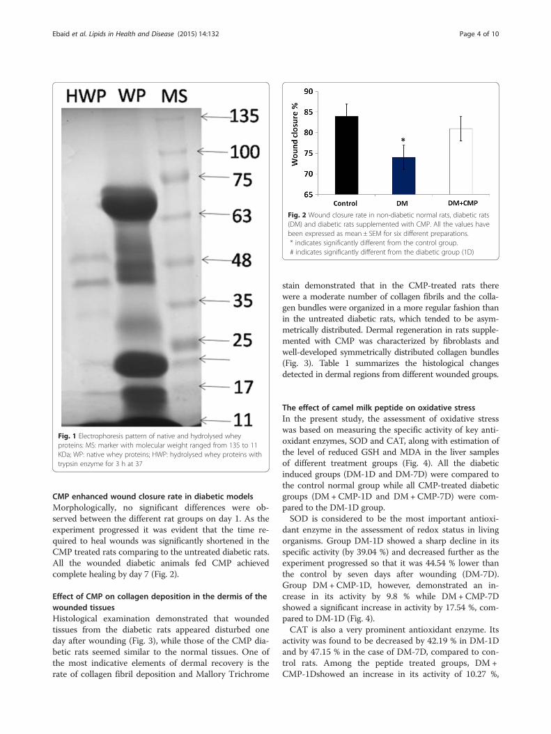
ResultsElectrophoresis pattern of the hydrolysed whey proteinsElectrophoresis-pattern clearly revealed that the camelwhey proteins extensively hydrolysed during incubationtime for 3 h with trypsin enzyme (Fig. 1). After hydroly-sis whey protein hydrolysates were separated by centrifu-gation into several fractions; the total unhydrolysed wheyprotein, the hyrolysed whey protein, long chain peptideand the camel milk peptide1 (CMP). Different fractionswere independently tested. Among them, the CMP(3 kDa) was chosen for wound healing experiments invivo.
Ebaid et al. Lipids in Health and Disease (2015) 14:132 Page 3 of 10
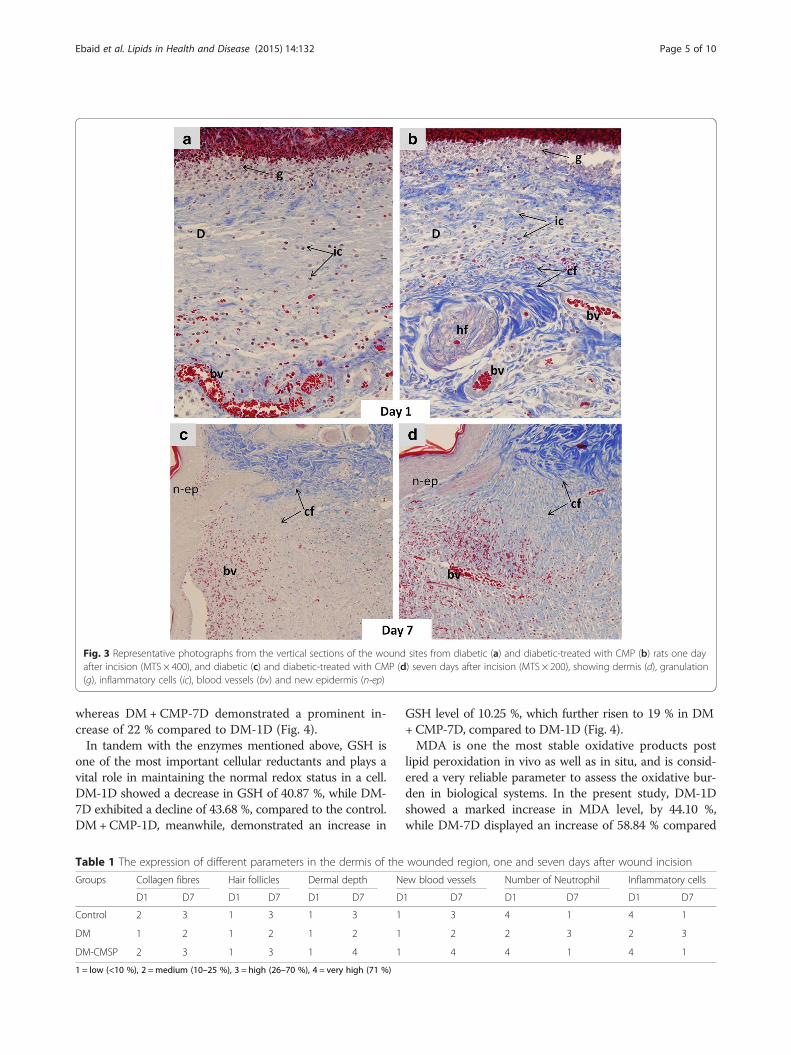
CMP enhanced wound closure rate in diabetic modelsMorphologically, no significant differences were ob-served between the different rat groups on day 1. As theexperiment progressed it was evident that the time re-quired to heal wounds was significantly shortened in theCMP treated rats comparing to the untreated diabetic rats.All the wounded diabetic animals fed CMP achievedcomplete healing by day 7 (Fig. 2).
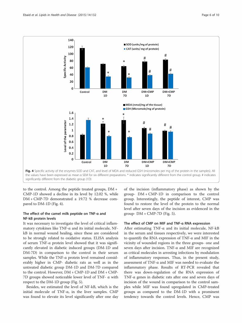
Effect of CMP on collagen deposition in the dermis of thewounded tissuesHistological examination demonstrated that woundedtissues from the diabetic rats appeared disturbed oneday after wounding (Fig. 3), while those of the CMP dia-betic rats seemed similar to the normal tissues. One ofthe most indicative elements of dermal recovery is therate of collagen fibril deposition and Mallory Trichrome
stain demonstrated that in the CMP-treated rats therewere a moderate number of collagen fibrils and the colla-gen bundles were organized in a more regular fashion thanin the untreated diabetic rats, which tended to be asym-metrically distributed. Dermal regeneration in rats supple-mented with CMP was characterized by fibroblasts andwell-developed symmetrically distributed collagen bundles(Fig. 3). Table 1 summarizes the histological changesdetected in dermal regions from different wounded groups.
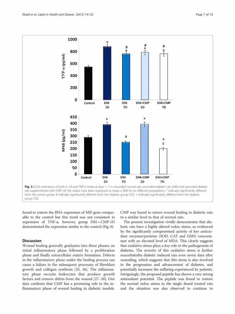
The effect of camel milk peptide on oxidative stressIn the present study, the assessment of oxidative stresswas based on measuring the specific activity of key anti-oxidant enzymes, SOD and CAT, along with estimation ofthe level of reduced GSH and MDA in the liver samplesof different treatment groups (Fig. 4). All the diabeticinduced groups (DM-1D and DM-7D) were compared tothe control normal group while all CMP-treated diabeticgroups (DM+CMP-1D and DM+CMP-7D) were com-pared to the DM-1D group.SOD is considered to be the most important antioxi-
dant enzyme in the assessment of redox status in livingorganisms. Group DM-1D showed a sharp decline in itsspecific activity (by 39.04 %) and decreased further as theexperiment progressed so that it was 44.54 % lower thanthe control by seven days after wounding (DM-7D).Group DM+CMP-1D, however, demonstrated an in-crease in its activity by 9.8 % while DM+CMP-7Dshowed a significant increase in activity by 17.54 %, com-pared to DM-1D (Fig. 4).CAT is also a very prominent antioxidant enzyme. Its
activity was found to be decreased by 42.19 % in DM-1Dand by 47.15 % in the case of DM-7D, compared to con-trol rats. Among the peptide treated groups, DM +CMP-1Dshowed an increase in its activity of 10.27 %,
Fig. 1 Electrophoresis pattern of native and hydrolysed wheyproteins: MS: marker with molecular weight ranged from 135 to 11KDa; WP: native whey proteins; HWP: hydrolysed whey proteins withtrypsin enzyme for 3 h at 37
Fig. 2 Wound closure rate in non-diabetic normal rats, diabetic rats(DM) and diabetic rats supplemented with CMP. All the values havebeen expressed as mean ± SEM for six different preparations.* indicates significantly different from the control group.# indicates significantly different from the diabetic group (1D)
Ebaid et al. Lipids in Health and Disease (2015) 14:132 Page 4 of 10
whereas DM +CMP-7D demonstrated a prominent in-crease of 22 % compared to DM-1D (Fig. 4).In tandem with the enzymes mentioned above, GSH is
one of the most important cellular reductants and plays avital role in maintaining the normal redox status in a cell.DM-1D showed a decrease in GSH of 40.87 %, while DM-7D exhibited a decline of 43.68 %, compared to the control.DM+CMP-1D, meanwhile, demonstrated an increase in
GSH level of 10.25 %, which further risen to 19 % in DM+CMP-7D, compared to DM-1D (Fig. 4).MDA is one the most stable oxidative products post
lipid peroxidation in vivo as well as in situ, and is consid-ered a very reliable parameter to assess the oxidative bur-den in biological systems. In the present study, DM-1Dshowed a marked increase in MDA level, by 44.10 %,while DM-7D displayed an increase of 58.84 % compared
Fig. 3 Representative photographs from the vertical sections of the wound sites from diabetic (a) and diabetic-treated with CMP (b) rats one dayafter incision (MTS × 400), and diabetic (c) and diabetic-treated with CMP (d) seven days after incision (MTS × 200), showing dermis (d), granulation(g), inflammatory cells (ic), blood vessels (bv) and new epidermis (n-ep)
Table 1 The expression of different parameters in the dermis of the wounded region, one and seven days after wound incision
Groups Collagen fibres Hair follicles Dermal depth New blood vessels Number of Neutrophil Inflammatory cells
D1 D7 D1 D7 D1 D7 D1 D7 D1 D7 D1 D7
Control 2 3 1 3 1 3 1 3 4 1 4 1
DM 1 2 1 2 1 2 1 2 2 3 2 3
DM-CMSP 2 3 1 3 1 4 1 4 4 1 4 1
1 = low (<10 %), 2 =medium (10–25 %), 3 = high (26–70 %), 4 = very high (71 %)
Ebaid et al. Lipids in Health and Disease (2015) 14:132 Page 5 of 10
to the control. Among the peptide treated groups, DM+CMP-1D showed a decline in its level by 12.02 %, whileDM+CMP-7D demonstrated a 19.72 % decrease com-pared to DM-1D (Fig. 4).
The effect of the camel milk peptide on TNF-α andNF-kB protein levelsIt was necessary to investigate the level of critical inflam-matory cytokines like TNF-α and its initial molecule, NF-kB in normal wound healing, since these are consideredto be strongly related to oxidative status. ELISA analysisof serum TNF-α protein level showed that it was signifi-cantly elevated in diabetic induced groups (DM-1D andDM-7D) in comparison to the control in their serumsamples. While the TNF-α protein level remained consid-erably higher in CMP- diabetic rats as well as in theuntreated diabetic group DM-1D and DM-7D comparedto the control. However, DM+CMP-1D and DM+CMP-7D groups showed noticeable lower level of TNF- α withrespect to the DM-1D group (Fig. 5).Besides, we estimated the level of NF-kB, which is the
initial molecule of TNF-α, in the liver samples. CMPwas found to elevate its level significantly after one day
of the incision (inflammatory phase) as shown by thegroup- DM +CMP-1D in comparison to the controlgroup. Interestingly, the peptide of interest, CMP wasfound to restore the level of the protein to the normallevel after seven days of the incision as evidenced in thegroup- DM +CMP-7D (Fig. 5).
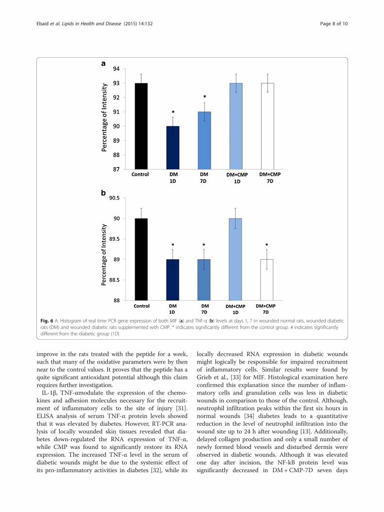
The effect of CMP on MIF and TNF-α RNA expressionAfter estimating TNF-α and its initial molecule, NF-kBin the serum and tissues respectively, we were interestedto quantify the RNA expression of TNF-α and MIF in thevicinity of wounded regions in the three groups- one andseven days after incision. TNF-α and MIF are recognizedas critical molecules in arresting infections by modulationof inflammatory responses. Thus, in the present study,assessment of TNF-α and MIF was needed to evaluate theinflammatory phase. Results of RT-PCR revealed thatthere was down-regulation of the RNA expression ofTNF-α genes in diabetic rats after one and seven days ofincision of the wound in comparison to the control sam-ples while MIF was found upregulated in CMP-treatedgroups as compared to the DM-1D with a prominenttendency towards the control levels. Hence, CMP was
Fig. 4 Specific activity of the enzymes-SOD and CAT, and level of MDA and reduced GSH (micromoles per mg of the protein in the samples). Allthe values have been expressed as mean ± SEM for six different preparations. * indicates significantly different from the control group. # indicatessignificantly different from the diabetic group (1D)
Ebaid et al. Lipids in Health and Disease (2015) 14:132 Page 6 of 10
found to restore the RNA expression of MIF gene compar-able to the control but this trend was not consistent inexpression of TNF-α, however, group DM+CMP-1Ddemonstrated the expression similar to the control (Fig. 6).
DiscussionWound healing generally graduates into three phases: aninitial inflammatory phase followed by a proliferationphase and finally extracellular matrix formation. Defectsin the inflammatory phase under the healing process cancause a failure in the subsequent processes of fibroblastgrowth and collagen synthesis [25, 26]. The inflamma-tory phase recruits leukocytes that produce growthfactors and remove debris from the wound [27–30]. Ourdata confirms that CMP has a promising role in the in-flammatory phase of wound healing in diabetic models.
CMP was found to return wound healing in diabetic ratsto a similar level to that of normal rats.The present investigation vividly demonstrates that dia-
betic rats have a highly altered redox status, as evidencedby the significantly compromised activity of key antioxi-dant enzymes/proteins (SOD, CAT and GSH) concomi-tant with an elevated level of MDA. This clearly suggeststhat oxidative stress plays a key role in the pathogenesis ofdiabetes. The severity of this oxidative stress is furtherexacerbatedin diabetic induced rats even seven days afterwounding, which suggests that this stress is also involvedin the progression and advancement of diabetes, andpotentially increases the suffering experienced by patients.Intriguingly, the proposed peptide has shown a very strongantioxidant potential. The peptide was found to restorethe normal redox status in the single dosed treated ratsand the situation was also observed to continue to
Fig. 5 ELISA estimation of both IL-1β and TNF-α levels at days 1, 7 in wounded normal rats, wounded diabetic rats (DM) and wounded diabeticrats supplemented with CMP. All the values have been expressed as mean ± SEM for six different preparations. * indicates significantly differentfrom the control group. # indicates significantly different from the diabetic group (1D). + indicates significantly different from the diabeticgroup (7D)
Ebaid et al. Lipids in Health and Disease (2015) 14:132 Page 7 of 10
improve in the rats treated with the peptide for a week,such that many of the oxidative parameters were by thennear to the control values. It proves that the peptide has aquite significant antioxidant potential although this claimrequires further investigation.IL-1β, TNF-αmodulate the expression of the chemo-
kines and adhesion molecules necessary for the recruit-ment of inflammatory cells to the site of injury [31].ELISA analysis of serum TNF-α protein levels showedthat it was elevated by diabetes. However, RT-PCR ana-lysis of locally wounded skin tissues revealed that dia-betes down-regulated the RNA expression of TNF-α,while CMP was found to significantly restore its RNAexpression. The increased TNF-α level in the serum ofdiabetic wounds might be due to the systemic effect ofits pro-inflammatory activities in diabetes [32], while its
locally decreased RNA expression in diabetic woundsmight logically be responsible for impaired recruitmentof inflammatory cells. Similar results were found byGrieb et al., [33] for MIF. Histological examination hereconfirmed this explanation since the number of inflam-matory cells and granulation cells was less in diabeticwounds in comparison to those of the control. Although,neutrophil infiltration peaks within the first six hours innormal wounds [34] diabetes leads to a quantitativereduction in the level of neutrophil infiltration into thewound site up to 24 h after wounding [13]. Additionally,delayed collagen production and only a small number ofnewly formed blood vessels and disturbed dermis wereobserved in diabetic wounds. Although it was elevatedone day after incision, the NF-kB protein level wassignificantly decreased in DM+ CMP-7D seven days
Fig. 6 A: Histogram of real time PCR gene expression of both MIF (a) and TNF-α (b) levels at days 1, 7 in wounded normal rats, wounded diabeticrats (DM) and wounded diabetic rats supplemented with CMP. * indicates significantly different from the control group. # indicates significantlydifferent from the diabetic group (1D)
Ebaid et al. Lipids in Health and Disease (2015) 14:132 Page 8 of 10
after incision, in comparison to the diabetic one. NF-kBis required for the induction of pro-inflammatory cyto-kines, such as IL-1β, TNF-α and IL-6 [9]. The oxidativestability induced by CMP may, therefore, mediate the acti-vation of NF-kB, leading to the activation of the inflamma-tory cascade and the stimulation of wound healing,resulting in faster wound closure rate in CMP rats.In addition, we have found that diabetes down-regulated
the RNA expression of MIF genes both one and sevendays after incision in comparison to the control samplesunder the present investigation. Grieb et al. [32] foundthat locally decreased levels of MIF in chronic woundexudates might be responsible for impaired recruitment ofendothelial progenitor cells. Accordingly, diabetic woundsin this study were delayed in their closure rate in compari-son to control wounds. This may be due to the fact thatMIF is a critical molecule in pro-inflammatory innate im-mune responses, being involved in arresting infections[35–38]. From another point of view, these two factorscan be considered as a diagnostic biomarker for auto-immune and inflammatory diseases including diabetes[39–41]. Therefore, prolonged elevation of MIF and in-flammatory cytokines has been found to be responsiblefor impaired healing [14].
ConclusionCMP has a strong antioxidant potential that reduces theeffects of oxygen free radicals and lipid peroxidation byorchestrating the overall antioxidant system to theoptimum in vivo. Moreover, CMP is a potential stimu-lant in normalizing the inflammatory cytokines andrestoring high levels of TNF-α mediated by NF-kB in dia-betic rats. An increase in neutrophil infiltration at woundsites post CMP administration in diabetic rats speeded upthe normal inflammatory events of the healing process.However, investigation of the deep mechanisms in woundhealing phases, as well as structural analysis, are necessaryto identify the mode of action of this peptide.
AbbreviationsALP: Alkaline phosphatase; CAT: Catalase; GSH: Glutathione;MDA: Malondialdehyde; MTS: Mallory Trichrome stain; SOD: Superoxidedismutase; NF-kB: Transcription factor kappa-B; TNF-α: Tumor necrosis factoralpha; IL-1β: Interleukin 1β.
Competing interestsThe authors declare that there to be no competing interests.
Authors’ contributionsHE designed this study, analysed the data, prepared the figures, andprepared and finalized the manuscript. JA was responsible of the animalmodel and histological investigations. BA was responsible for PCR analysis. IHperformed oxidative stress analysis and write its description and discussion.AM and IA were responsible for the extraction and preparation of the CMP.All authors have read and approved the final version of the manuscript.
AcknowledgementThe authors would like to acknowledge the support of National Plan forScience, Technology, & Innovation (NPST) at King Saud University under
project number (12-BIO-2483-02). This support is highly appreciated andacknowledged.
Author details1Department of Zoology, College of Science, King Saud University, P.O. Box2455, Riyadh 11451, Saudi Arabia. 2Department of Zoology, Faculty ofScience, El-Minia University, El-Minia, Egypt. 3Department of Biology, Collegeof Science and Humanities in Quwiaya, Riyadh 11961, Saudi Arabia.4Department of Food Science, College of Agriculture and Food Science, KingSaud University, Riyadh, Saudi Arabia. 5Department of Dairy, Faculty ofAgriculture, El-Minia University, El-Minia, Egypt.
Received: 30 March 2015 Accepted: 15 October 2015
References1. Hall ED, Yonkers PA, Horan KL. Correlation between attenuation of
posttraumatic spinal cord ischemia and preservation of tissue vitamin E bythe 21-aminosteroid U-Z4006F: Evidence for an in vivo antioxidantmechanism. J Neurotrauma. 1989;6:169–76.
2. Ashour M, Salem S, Hassaneen H, EL-Gadban H, Elwan N, Awad A, et al.Antioxidant status and insulin dependent diabetes mellitus (IDDM). JClinBiochemNutr. 1999;26:99–107.
3. Hsu WT, Tsai LY, Lin SK, Hsiao K, Chen BH. Effects of diabetes duration andglycemic control on free radicals in children with type 1 diabetes mellitus.Ann Clin Lab Sci. 2006;36:174–8.
4. Hassan I, Chibber S, Khan AA, Naseem I. Cisplatin-induced neurotoxicity invivo can be alleviated by riboflavin under photoillumination. Cancer BiotherRadiopharm. 2013;28:160–8.
5. Clark R. Cutaneous tissue repair. I. Basic biologic consideration. J AmAcadDermatol. 1985;13:701–25.
6. Clark R. Molecular and cellular biology of wound repair. New York: PlenumPress. 1996;2:553–0.
7. Graves DT, Nooh N, Gillen T, Davey M, Patel S, Cottrell D, et al. IL-1 plays acritical role in oral, but not dermal, wound healing. J Immunol.2001;167:5316–20.
8. Altavilla D, Saitta A, Cucinotta D, Galeano M, Deodato B, Colonna M, et al.Inhibition of lipid peroxidation restores impaired vascular endothelialgrowth factor expression and stimulates wound healing and angiogenesisin the genetically diabetic mouse. J Diabetes. 2001;50:667–74.
9. Lim Y, Levy M, Bray TM. Dietary zinc alters early inflammatory responsesduring cutaneous wound healing in weanling CD-1 mice. J Nutr.2004;134:811–6.
10. Khurram SA, Bingle L, McCabe BM, Farthing PM, Whawell SA.The chemokinereceptors CXCR1 and CXCR2 regulate oral cancer cell behaviour. J OralPathol Med. 2014;43(9):667–74.
11. Zhang X, Kohli M, Zhou Q, Graves DT, Amar S. Short- and longterm effectsof IL-1 and TNF antagonists on periodontal wound healing. J Immunol.2004;173:3514–23.
12. Eming SA, Krieg T, Davidson JM. Inflammation in wound repair: molecularand cellular mechanisms. J Invest Dermatol. 2007;127:514–25.
13. Ebaid H, Salem A, Sayed A, Metwalli A. Whey protein enhances normalinflammatory responses during cutaneous wound healing in diabeticrats.Lipids Health Dis. 2011;10:235.
14. Ebaid H, Ahmed O, Mahmoud A, Ahmed R. Limiting prolongedinflammation during proliferation and remodeling phases of wound healingin streptozotocin-induced diabetic rats supplemented with camelundenatured whey protein. BMC Immunol. 2013;14:31.
15. Ebaid H. Neutrophil depletion in the early inflammatory phase delayedcutaneous wound healing in older rats: improvements due to the use ofun-denatured camel whey protein. J DiagnPathol. 2014;9:46.
16. Ebaid H, Al-Khalifa M, Isa A, Gadoa S. Bioactivity of Samsum ant(Pachycondylasennaarensis) venom against lipopolysaccharides throughantioxidant and upregulation of Akt1signaling in rats. Lipids Health Dis.2012;11:93.
17. Badr G, Ebaid H, Mohany M, Abuelsaad AS. Modulation of immune cellproliferation and chemotaxis towards CC chemokine ligands (CCL)-21 andCXC chemokine ligand (CXCL)-12 in un-denatured whey protein-treatedmice. J nutritBiochem. 2012;23:1640–6.
18. Schwentker A, Vodovotz Y, Weller R, Billiar TR. Nitricoxide and wound repair:role of cytokines? NitricOxide. 2002;7:1–10.
Ebaid et al. Lipids in Health and Disease (2015) 14:132 Page 9 of 10
19. Wang T, Gu Q, Zhao J, Mei J, Shao M, Pan Y, et al. Calcium alginateenhances wound healing by up-regulating the ratio of collagen types I/III indiabetic rats. Int J Clin Exp Pathol. 2015;8:6636–45.
20. Souil E, Capon A, Mordon S, Dinh-Xuan AT, Polla BS, Bachelet M. Treatmentwith 815-nm diode laser induces long-lasting expression of 72-kDa heatshock protein in normal rat skin. Br J Dermatol. 2001;144:260–6.
21. Marklund S, Marklund G. Involvement of the superoxide anion radical in theautoxidation of pyrogallol and a convenient assay for superoxide dismutase.Eur J Biochem. 1974;47:469–74.
22. Aebi H. Catalase in vitro. Methods Enzymol. 1984;105:121–6.23. Jollow DJ, Mitchell JR, Zampaglione N, Gillette JR. Bromobenzene induced
liver necrosis. Protective role of glutathione and evidence for3,4bromobenzene oxide as the hepatotoxic metabolite. Pharmacol.1974;11:151–69.
24. Buege JA, Aust SD. Microsomal lipid peroxidation. Methods Enzymol.1978;52:302–10.
25. Loots MA, Lamme EN, Zeegelaar J, Mekkes JR, Bos JD, Middelkoop E.Differences in cellular infiltrate and extracellular matrix of chronic diabeticand venous ulcers versus acute wounds. J Invest Dermatol. 1998;111:850–7.
26. Moore K, Ruge F, Harding KG. T lymphocytes and the lack of activatedmacrophages in wound margin biopsies from chronic leg ulcers. Br JDermatol. 1997;137:188–94.
27. Devalaraja RM, Nanney LB, Du J, Qian Q, Yu Y, Devalaraja MN, et al. Delayedwound healing in CXCR2 knockout mice. J Invest Dermatol. 2000;115:234–44.
28. Mori R, Kondo T, Nishie T, Ohshima T, Asano M. Impairment of skin woundhealing in beta-1,4-galactosyltransferase-deficient mice with reducedleukocyte recruitment. Am J Pathol. 2004;164:1303–14.
29. Miller LS, O’Connell RM, Gutierrez MA, Pietras EM, Shahangian A, Gross CE,et al. MyD88 mediatesneutrophil recruitment initiated by IL-1R but not TLR2activation in immunity against Staphylococcus aureus. Immunity.2006;24:79–91.
30. Abdel-Salam BKA, Ebaid H. Expression of CD11b and CD18 onpolymorphonuclear neutrophils stimulated with interleukin-2. CentralEuropean J Immunol. 2014;39:209.
31. Bickel M, Nothen SM, Freiburghaus K, Shire D. Chemokine expression inhuman oral keratinocyte cell lines and keratinized mucosa. J Dent Res.1996;75:1827–34.
32. Ebaid H, Al-Tamimi J, Hassan I, Alhazza I, Al-Khalifa M. Antioxidant Bioactivity ofSamsum Ant (Pachycondylasennaarensis) Venom Protects against CCL4-Induced Nephrotoxicity in Mice. J OxidMedic and CelluLongev. 2014;76:30–61.
33. Grieb G, Simons D, Eckert L, Hemmrich M, Steffens G, Bernhagen J, et al.Levels of macrophage migration inhibitory factor and glucocorticoids inchronic wound patients and their potential interactions with impairedwound endothelial progenitor cell migration. Wound Repair Regen.2012;20:707–14.
34. Kindt T, Goldsby R, Osborne B. Kuby Immunology. 6th ed. New York: WHFreeman and company; 2007.
35. Sanchez-Zamora Y, Rodriguez-Sosa M. The Role of MIF in Type 1 and Type 2Diabetes Mellitus. J Diabetes Res 2014;2014:804519. doi: 10.1155/2014/804519. Epub 2014 Jan 2.
36. Terrazas CA, Juarez I, Terrazas L, Saavedra R, Calleja EA, Rodriguez-Sosa M.Toxoplasma gondii: impairedmaturation and pro-inflammatory response ofdendritic cells in MIF deficient mice favors susceptibility to infection.ExperParasitology. 2010;126:348–58.
37. Flores M1, Saavedra R, Bautista R, Viedma R, Tenorio EP, Leng L. et al.Macrophage migration inhibitory factor (MIF) is critical for the hostresistance against Toxoplasma gondii. FASEB J. 2008;22(10):3661–71.
38. Reyes JL, Terrazas LI, Espinoza B, Cruz-Robles D, Soto V, Rivera-Montoya I .Macrophage migration inhibitory factor contributes to host defense againstacute TrypanosomacruziInfection. J Infection Immunity. 2006;74(6):3170–9.
39. Finucane OM, Reynolds CM, McGillicuddy FC, Roche HM. Insights into therole of macrophage migration inhibitory factor in obesity and insulinresistance. J Proc Nutr Soc. 2012;71:622–33.
40. Stojanovic I, Saksida T, Stosic-Grujicic S. Beta cell function: the role ofmacrophage migration inhibitory factor. J Immunol Res. 2012;52:81–8.
41. Grieb G, Merk M, Bernhagen J, Bucala R. Macrophage migration inhibitoryfactor (MIF): a promising biomarker. J Drug News Perspect. 2010;23:257–64.
Submit your next manuscript to BioMed Centraland take full advantage of:
• Convenient online submission
• Thorough peer review
• No space constraints or color figure charges
• Immediate publication on acceptance
• Inclusion in PubMed, CAS, Scopus and Google Scholar
• Research which is freely available for redistribution
Submit your manuscript at www.biomedcentral.com/submit
Ebaid et al. Lipids in Health and Disease (2015) 14:132 Page 10 of 10