Embed Size (px)
Citation preview
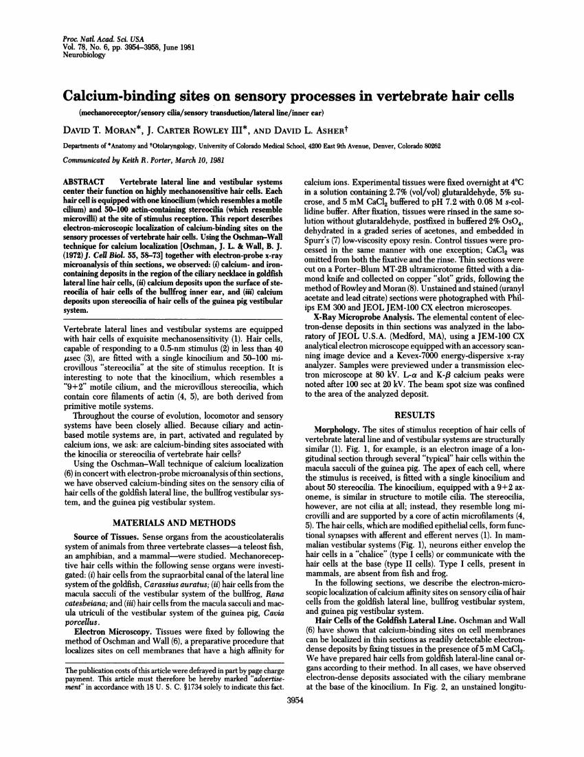
Proc. NatL Acad. Sci. USAVol. 78, No. 6, pp. 3954-3958, June 1981Neurobiology
Calcium-binding sites on sensory processes in vertebrate hair cells(mechanoreceptor/sensory cilia/sensory transduction/lateral line/inner ear)
DAVID T. MORAN*, J. CARTER ROWLEY III*, AND DAVID L. ASHERtDepartments of *Anatomy and tOtolaryngology, University of Colorado Medical School, 4200 East 9th Avenue, Denver, Colorado 80262
Communicated by Keith R. Porter, March 10, 1981
ABSTRACT Vertebrate lateral line and vestibular systemscenter their function on highly mechanosensitive hair cells. Eachhair cell is equipped with one kinocilium (which resembles a motilecilium) and 50-100 actin-containing stereocilia (which resemblemicrovilli) at the site of stimulus reception. This report describeselectron-microscopic localization of calcium-binding sites on thesensory processes of vertebrate hair cells. Using the Oschman-Walltechnique for calcium localization [Oschman, J. L. & Wall, B. J.(1972) J. Cell Biol. 55, 58-73] together with electron-probe x-raymicroanalysis of thin sections, we observed: (i) calcium- and iron-containing deposits in the region of the ciliary necldace in goldfishlateral line hair cells, (ii) calcium deposits upon the surface of ste-reocilia of hair cells of the bullfrog inner ear, and (iii) calciumdeposits upon stereocilia of hair cells of the guinea pig vestibularsystem.
Vertebrate lateral lines and vestibular systems are equippedwith hair cells of exquisite mechanosensitivity (1). Hair cells,capable of responding to a 0.5-nm stimulus (2) in less than 40,usec (3), are fitted with a single kinocilium and 50-100 mi-crovillous "stereocilia" at the site of stimulus reception. It isinteresting to note that the kinocilium, which resembles a"9+2" motile cilium, and the microvillous stereocilia, whichcontain core filaments of actin (4, 5), are both derived fromprimitive motile systems.
Throughout the course of evolution, locomotor and sensorysystems have been closely allied. Because ciliary and actin-based motile systems are, in part, activated and regulated bycalcium ions, we ask: are calcium-binding sites associated withthe kinocilia or stereocilia of vertebrate hair cells?
Using the Oschman-Wall technique of calcium localization(6) in concert with electron-probe microanalysis ofthin sections,we have observed calcium-binding sites on the sensory cilia ofhair cells of the goldfish lateral line, the bullfrog vestibular sys-tem, and the guinea pig vestibular system.
MATERIALS AND METHODSSource of Tissues. Sense organs from the acousticolateralis
system of animals from three vertebrate classes-a teleost fish,an amphibian, and a mammal-were studied. Mechanorecep-tive hair cells within the following sense organs were investi-gated: (i) hair cells from the supraorbital canal of the lateral linesystem ofthe goldfish, Carassius auratus; (ii) hair cells from themacula sacculi of the vestibular system of the bullfrog, Ranacatesbeiana; and (iii) hair cells from the macula sacculi and mac-ula utriculi of the vestibular system of the guinea pig, Caviaporcellus.
Electron Microscopy. Tissues were fixed by following themethod ofOschman and Wall (6), a preparative procedure thatlocalizes sites on cell membranes that have a high affinity for
calcium ions. Experimental tissues were fixed overnight at 40Cin a solution containing 2.7% (vol/vol) glutaraldehyde, 5% su-crose, and 5 mM CaCl2 buffered to pH 7.2 with 0.08 M s-col-lidine buffer. After fixation, tissues were rinsed in the same so-lution without glutaraldehyde, postfixed in buffered 2% OS04,dehydrated in a graded series of acetones, and embedded inSpurr's (7) low-viscosity epoxy resin. Control tissues were pro-cessed in the same manner with one exception; CaCl2 wasomitted from both the fixative and the rinse. Thin sections werecut on a Porter-Blum MT-2B ultramicrotome fitted with a dia-mond knife and collected on copper "slot" grids, following themethod ofRowley and Moran (8). Unstained and stained (uranylacetate and lead citrate) sections were photographed with Phil-ips EM 300 and JEOL JEM-100 CX electron microscopes.X-Ray Microprobe Analysis. The elemental content of elec-
tron-dense deposits in thin sections was analyzed in the labo-ratory of JEOL U.S.A. (Medford, MA), using a JEM-100 CXanalytical electron microscope equipped with an accessory scan-ning image device and a Kevex-7000 energy-dispersive x-rayanalyzer. Samples were previewed under a transmission elec-tron microscope at 80 kV. L-a and K-,8 calcium peaks werenoted after 100 sec at 20 kV. The beam spot size was confinedto the area of the analyzed deposit.
RESULTSMorphology. The sites of stimulus reception of hair cells of
vertebrate lateral line and of vestibular systems are structurallysimilar (1). Fig. 1, for example, is an electron image of a lon-gitudinal section through several "typical" hair cells within themacula sacculi of the guinea pig. The apex of each cell, wherethe stimulus is received, is fitted with a single kinocilium andabout 50 stereocilia. The kinocilium, equipped with a 9+2 ax-oneme, is similar in structure to motile cilia. The stereocilia,however, are not cilia at all; instead, they resemble long mi-crovilli and are supported by a core of actin microfilaments (4,5). The hair cells, which are modified epithelial cells, form func-tional synapses with afferent and efferent nerves (1). In mam-malian vestibular systems (Fig. 1), neurons either envelop thehair cells in a "chalice" (type I cells) or communicate with thehair cells at the base (type II cells). Type I cells, present inmammals, are absent from fish and frog.
In the following sections, we describe the electron-micro-scopic localization ofcalcium affinity sites on sensory cilia ofhaircells from the goldfish lateral line, bullfrog vestibular system,and guinea pig vestibular system.
Hair Cells of the Goldfish Lateral Line. Oschman and Wall(6) have shown that calcium-binding sites on cell membranescan be localized in thin sections as readily detectable electron-dense deposits by fixing tissues in the presence of5 mM CaCl2.We have prepared hair cells from goldfish lateral-line canal or-gans according to their method. In all cases, we have observedelectron-dense deposits associated with the ciliary membraneat the base of the kinocilium. In Fig. 2, an unstained longitu-
3954
The publication costs ofthis article were defrayed in part by page chargepayment. This article must therefore be hereby marked "advertise-ment" in accordance with 18 U. S. C. §1734 solely to indicate this fact.
Proc. Natl. Acad. Sci. USA 78 (1981) 3955
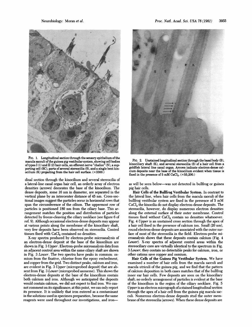
Ar~-FIG. 1. Longitudinal section through the sensory epithelium ofthe
macula sacculi ofthe guinea pig vestibular system, showing cell bodiesoftypes I (1) and 1 (2) hair cells, an efferent nerve "chalice" (N), a sup-porting cell (SC), parts of several stereocilia (S), and a single bent kin-ocilium (K) projecting from the hair cell surface. (x3300.)
dinal section through the kinocilium and several stereocilia ofa lateral-line canal organ hair cell, an orderly array of electrondensities (arrows) decorates the base of the kinocilium. Thedense deposits, some 16 nm in diameter, are separated in thevertical plane by an intercenter distance of 45 nm. Cross-sec-tional images suggest the particles occur in horizontal rows thatspan the circumference of the cilium. The uppermost row ofparticles is positioned 180 nm from the ciliary base. This ar-
rangement matches the position and distribution of particlesdetected by freeze-cleaving the ciliary necklace (see figure 6 ofref. 9). Although occasional electron-dense deposits may appearat various points along the membrane of the kinociliary shaft,very few deposits have been observed on stereocilia. Controltissues fixed with CaCl2 contained no densities.
X-ray spectra produced by electron-probe microanalysis ofan electron-dense deposit at the base of the kinocilium areshown in Fig. 3 Upper. Electron-probe microanalysis data froman adjacent control area within the same ciliary shaft are shownin Fig. 3 Lower. The two spectra have peaks in common: os-
mium from the fixative, chlorine from the epoxy embedment,and copper from the grid. Two distinct peaks, calcium and iron,are evident in Fig. 3 Upper (microprobed deposit) that are ab-sent from Fig. 3 Lower (microprobed axoneme). This shows theelectron-dense deposits at the base of the kinocilium containboth calcium and iron. Although we anticipated the depositswould contain calcium, we did not expect to find iron. We can-
not comment on its significance; at this point, we can only reportits presence. It is unlikely that iron entered as a contaminantin the solutions used in specimen preparation, because the samereagents were used throughout our investigations, and iron-
FIG. 2. Unstained longitudinal section through the basal body (B),kinociliary shaft (K), and several stereocilia (S) of a hair cell from agoldfish lateral line canal organ. Arrows indicate electron-dense cal-cium deposits near the base of the kinocilium evident when tissue isfixed in the presence of 5 mM CaCl2. (x55,200.)
as will be seen below-was not detected in bullfrog or guineapig hair cells.
Hair Cells of the Bullfrog Vestibular System. In contrast tothe lateral line, when hair cells from the macula sacculi of thebullfrog vestibular system are fixed in the presence of 5 mMCaC12 the kinocilia do not display electron-dense deposits. Thestereocilia, however, do display numerous electron densitiesalong the external surface of their outer membrane. Controltissues fixed without CaC12 contain no densities whatsoever.Fig. 4 Upper is an unstained cross section through the apex ofa hair cell fixed in the presence of calcium ion. Small (20 nm),round electron-dense deposits are associated with the outer sur-face of most of the stereocilia in the field. Electron-probe mi-croanalysis shows that these deposits contain calcium (Fig. 4Lower). X-ray spectra of adjacent control areas within thestereociliary core are virtually identical to the spectrum in Fig.3 Lower; they contain no detectable peaks for calcium, iron, orother cations save copper and osmium.
Hair Cells of the Guinea Pig Vestibular System. We haveexamined a number of hair cells from the macula sacculi andmacula utriculi of the guinea pig, and we find that the patternof calcium deposition in both cases matches that of the bullfroginner ear hair cells. Few deposits are seen on the kinociliaryshaft; no orderly arrangement of particles is evident at the baseof the kinocilium in the region of the ciliary necklace. Fig. 5Upper is an electron micrograph ofa stained longitudinal sectionthrough the apex of a hair cell from the guinea pig macula sac-culi. Numerous electron-dense deposits stud the outer mem-brane ofthe stereocilia (arrows). When these dense deposits are
Neurobiology: Moran et al -
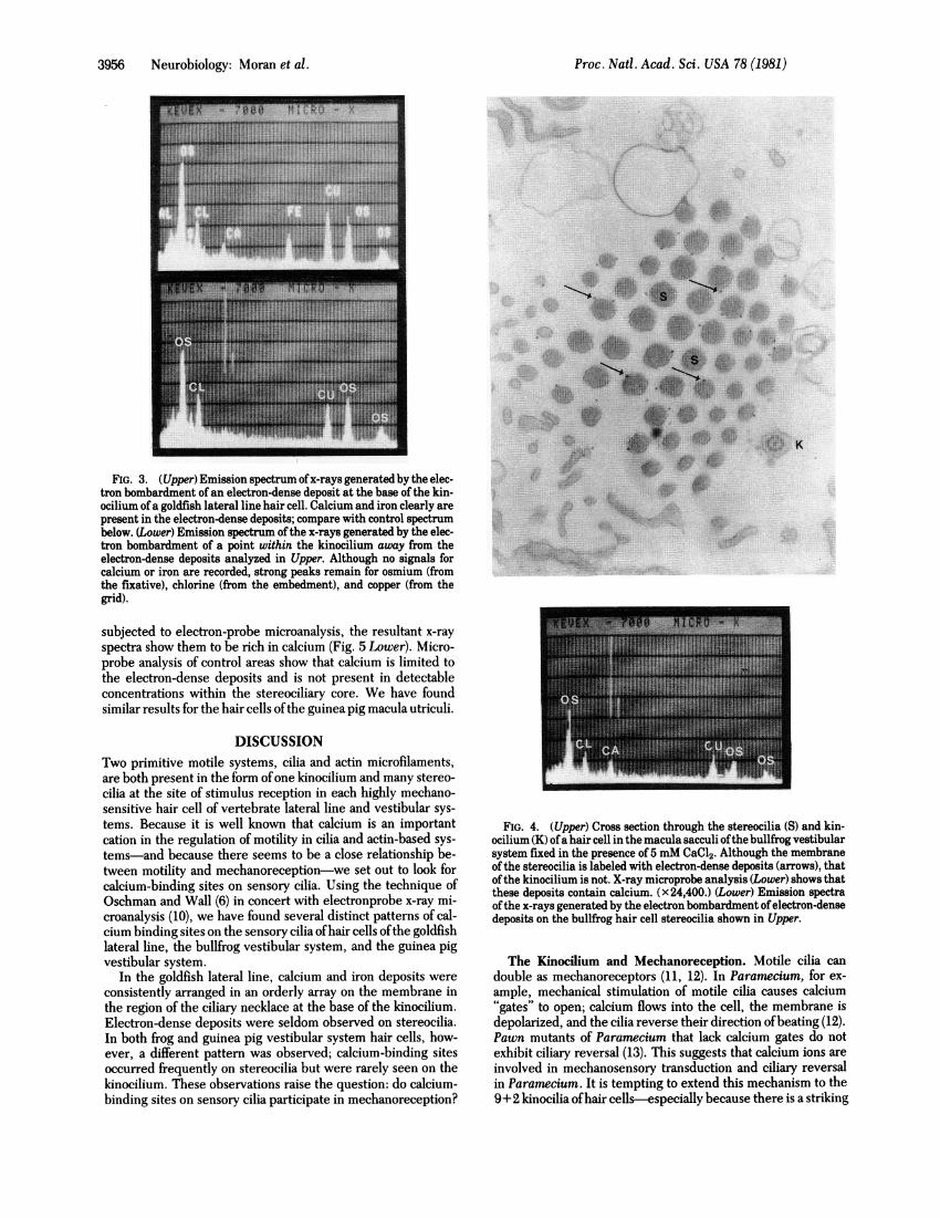
Proc. Natl. Acad. Sci. USA 78 (1981)~~~~~~~~~~~~~~~~~~~~~~~......_FIG. 3. (Upper) Emission spectrum ofx-rays generated by the elec-
tron bombardment of an electron-dense deposit at the base ofthe kin-ocilium ofa goldfish lateral line hair cell. Calcium and iron clearly arepresent in the electron-dense deposits; compare with control spectrumbelow. (Lower) Emission spectrum ofthe x-rays generated by the elec-tron bombardment of a point within the kinocilium away from theelectron-dense deposits analyzed in Upper. Although no signals forcalcium or iron are recorded, strong peaks remain for osmium (fromthe fixative), chlorine (from the embedment), and copper (from thegrid).
subjected to electron-probe microanalysis, the resultant x-rayspectra show them to be rich in calcium (Fig. 5 Lower). Micro-probe analysis of control areas show that calcium is limited tothe electron-dense deposits and is not present in detectableconcentrations within the stereociliary core. We have foundsimilar results for the hair cells ofthe guinea pig macula utriculi.
DISCUSSIONTwo primitive motile systems, cilia and actin microfilaments,are both present in the form ofone kinocilium and many stereo-cilia at the site of stimulus reception in each highly mechano-sensitive hair cell of vertebrate lateral line and vestibular sys-tems. Because it is well known that calcium is an importantcation in the regulation of motility in cilia and actin-based sys-tems-and because there seems to be a close relationship be-tween motility and mechanoreception-we set out to look forcalcium-binding sites on sensory cilia. Using the technique ofOschman and Wall (6) in concert with electronprobe x-ray mi-croanalysis (10), we have found several distinct patterns of cal-cium binding sites on the sensory cilia ofhair cells ofthe goldfishlateral line, the bullfrog vestibular system, and the guinea pigvestibular system.
In the goldfish lateral line, calcium and iron deposits wereconsistently arranged in an orderly array on the membrane inthe region of the ciliary necklace at the base of the kinocilium.Electron-dense deposits were seldom observed on stereocilia.In both frog and guinea pig vestibular system hair cells, how-ever, a different pattern was observed; calcium-binding sitesoccurred frequently on stereocilia but were rarely seen on thekinocilium. These observations raise the question: do calcium-binding sites on sensory cilia participate in mechanoreception?
FIG. 4. (Upper) Cross section through the stereocilia (S) and kin-ocilium (K) ofa hair cell in the macula sacculi ofthe bullfrog vestibularsystem fixed in the presence of 5 mM CaCl2. Although the membraneofthe stereocilia is labeled with electron-dense deposits (arrows), thatofthe kinocilium is not. X-ray microprobe analysis (Lower) shows thatthese deposits contain calcium. (X24,400.) (Lower) Emission spectraofthe x-rays generated by the electron bombardment ofelectron-densedeposits on the bullfrog hair cell stereocilia shown in Upper.
The Kinocilium and Mechanoreception. Motile cilia candouble as mechanoreceptors (11, 12). In Paramecium, for ex-ample, mechanical stimulation of motile cilia causes calcium"gates" to open; calcium flows into the cell, the membrane isdepolarized, and the cilia reverse their direction ofbeating (12).Pawn mutants of Paramecium that lack calcium gates do notexhibit ciliary reversal (13). This suggests that calcium ions areinvolved in mechanosensory transduction and ciliary reversalin Paramecium. It is tempting to extend this mechanism to the9+2 kinocilia ofhair cells-especially because there is a striking
3956 Neurobiology: Moran et al.
7: P.

Proc. Natl. Acad. Sci. USA 78 (1981) 3957
FIG. 5. (Upper) Longitudinal section through the stereocilia (S) ofhair cells from a macula sacculi of the guinea pig vestibular systemfixed in the presence of 5 mM CaCl2. Arrows indicate electron-densedeposits that are shown (Lower) to contain calcium by x-ray microprobeanalysis. (x27,000.) (Lower) Emission spectra of the x-rays generatedby the electron bombardment of electron-dense deposits on the guineapig hair cell stereocilia shown in Upper.
correspondence between the axonemal plane of the active (andrecovery) stroke of motile cilia and the axonemal plane of thestimulatory (and inhibitory) displacements of the kinocilium inthe hair bundle of hair cells (1). Two pieces of evidence, how-ever, indicate hair cell kinocilia are not necessary elements formechanoreceptive function in hair cells. First, patients withKartagener syndrome-the so-called "immotile cilia syn-
drome"-have not been reported to show evidence ofvestibulardysfunctions. Because many of these patients present a con-
genital lack of one or both dynein arms on the outer doublets
of somatic cilia (14) (they have chronic bronchitis due to im-motile airway cilia) and germ cell cilia (they are sterile due toimmotile spermatozoa), we can conclude that kinociliary mo-tility is not necessary for proper function of human vestibularsystem hair cells. Second-and more conclusive-the elegantexperiments ofHudspeth and Jacobs (15) clearly show that bull-frog vestibular hair cells can function quite well in vitro afterphysical removal of the kinocilium. Removal of the kinociliumdoes not diminish their mechanosensitivity or their directionalsensitivity. Although these experiments have not yet been doneon lateral line receptors, it is probably safe to extend these find-ings to fish. Why, then, is the kinocilium there? And why isthere such a precise arrangement ofcalcium-binding sites at theciliary base? Although the answers to these questions are un-known, we agree with Hudspeth and Jacobs (15) that the roleof the kinocilium in vertebrate hair cells is most probably de-velopmental. The calcium-binding sites on goldfish kinociliacorrespond precisely to the position of the membrane particlesin the ciliary necklace described by Gilula and Satir (9). Estab-lishment of the ciliary necklace particles seems to be the pri-mary event in the development of the cilium (9, 16). Becauselateral line and vestibular hair cells are directionally sensitive(1, 2, 15)-they function as vector analyzers-it is likely thatsome sort ofstructural polarity is established in the hair cell apexduring development. The elaboration of the ciliary necklace ata specific site on the hair cell surface could determine the po-sition of the kinocilium, and the development ofthe kinocilium,along with its basal body and attached basal foot, could influencethe establishment of a structural polarity within the apical poleof the cell.
Stereocilia and Mechanoreception. Hair cells fitted withstereocilia alone can function as highly sensitive mechanore-ceptors. Human cochlear hair cells, for example, have stereo-cilia but no kinocilium. The kinocilium, present during devel-opment, is lost at birth, leaving its basal body behind. Theexperiments of Hudspeth and Jacobs (15) show that hair cellscan function not only after removal of the kinocilium but alsoafter removal of half the stereocilia.
Stereocilia are not cilia at all, but rather resemble microvilli.Microvilli have core filaments of actin and can actively shortenin length (17). Stereocilia, too, have a core of 6-nm actin mi-crofilaments (4, 5). Because calcium ions are important to theregulation of actin-based motile systems, because calcium ap-pears to be a necessary cofactor for the mechanoreceptive re-sponse in hair cells (18), and because stereocilia have actin coresand have calcium-binding sites in their membrane, it is tempt-ing to speculate that calcium-mediated motile mechanisms par-ticipate in mechanoreception. Recent findings by Tilney et al.(5), however, indicate stereocilia of lizard ear hair cells, unlikemicrovilli, have an arrangement of actin microfilaments that isnot designed to shorten. They conclude, "the stereocilium isa large, rigid structure designed to move as a lever." Thus therelationship between calcium, motility, and mechanosensitivityin stereocilia, though intriguing, remains obscure.
We thank Mr. David Harling ofJEOL (Medford, MA) for performingthe electron-probe microanalysis and Betty Aguilar and Judy Paden fortyping the manuscript. This work was supported by National ScienceFoundation Research Grant BNS 77-03317 to D.T. M.
1. Flock, A. (1971) in Handbook of Sensory Physiology, Principlesof Receptor Physiology, ed. Loewenstein, W. R. (Springer, Ber-lin), Vol. 1, pp. 396441.
2. Hudspeth, A. J. & Corey, D. P. (1977) Proc. Natl. Acad. Sci.USA 74, 2407-2411.
3. Corey, D. P.. & Hudspeth, A. J. (1979) Biophys. J. 26, 499-506.4. Flock, A. & Cheung, H. C. (1977)J. Cell Biol. 75, 339-343.
Neurobiology: Moran et al.

3958 Neurobiology: Moran et al.
5. Tilney, L. G., de Rosier, D. & Murray, M. (1979) 1. Cell Biol.83, 310A (abstr.).
6. Oschman, J. L. & Wall, B. J. (1972) J. Cell Biol. 55, 58-73.7. Spurr, A. R. (1969) J. Ultrastruct. Res. 26, 31-43.8. Rowley, J. C., III & Moran, D. T. (1975) Ultramicroscopy 1,
151-155.9. Gilula, N. B. & Satir, P. (1972)J. Cell Biol. 53, 494509.
10. Oschman, J. L., Hall, T. A., Peters, P. D. & Wall, B. J. (1974)J. Cell Biol. 61, 156-165.
i1. Horridge, G. A. (1968) Interneurons (Freeman, San Francisco).12. Naitoh, Y. & Eckert, R. (1974) in Cilia and Flagella, ed. Sleigh,
M. A. (Academic, New York), pp. 305-352.
Proc. Nati. Acad. Sci. USA 78 (1981)
13. Kung, C. (1976) in Cell Motility, Cold Spring Harbor Confer-ences on Cell Proliferation, eds. Goldman, R. D., Pollard, T. D.& Rosenbaum, J. L. (Cold Spring Harbor Laboratory, ColdSpring Harbor, NY), Vol. 3, Book C, pp. 941-947.
14. Afzelius, B. A. & Eliasson, R. (1979) J. Ultrastruct. Res. 69,43-52.
15. Hudspeth, A. J. & Jacobs, R. (1979) Proc. Natl. Acad. Sci. USA76, 1506-1509.
16. Cordier, A. C. & Haumont, S. (1979) Am. J. Anat. 156, 91-97.17. Rodewald, R., Newman, S. B. & Karnovsky, M. J. (1976)J. Cell
Biol. 70, 541-554.18. Corey, D. P. & Hudspeth, A. J. (1979) Nature (London) 281,
675-676.


![The Binding Nucleotides andBivalentCations Calcium-and ...molecular-weight polymers (>200000mol.wt.) were sometimesseen. GelsrunatpH2.4afterincubationwithMg[y_32P]-ATPbythemethodsofJordon](https://img.pdfslide.us/doc/110x75/60744c82ac519f757526bfa1/the-binding-nucleotides-andbivalentcations-calcium-and-molecular-weight-polymers.jpg)