Embed Size (px)
Citation preview
Louisiana State UniversityLSU Digital Commons
LSU Master's Theses Graduate School
2010
Caged Morpholino Oligonucleotide for Control ofGene Expression in ZebrafishChad Michael JarreauLouisiana State University and Agricultural and Mechanical College, [email protected]
Follow this and additional works at: https://digitalcommons.lsu.edu/gradschool_theses
Part of the Engineering Commons
This Thesis is brought to you for free and open access by the Graduate School at LSU Digital Commons. It has been accepted for inclusion in LSUMaster's Theses by an authorized graduate school editor of LSU Digital Commons. For more information, please contact [email protected].
Recommended CitationJarreau, Chad Michael, "Caged Morpholino Oligonucleotide for Control of Gene Expression in Zebrafish" (2010). LSU Master'sTheses. 3969.https://digitalcommons.lsu.edu/gradschool_theses/3969
CAGED MORPHOLINO OLIGONUCLEOTIDE FOR CONTROL OF GENE EXPRESSION
IN ZEBRAFISH
A Thesis
Submitted to the Graduate Faculty of the
Louisiana State University and
Agricultural and Mechanical College
In partial fulfillment of the
Requirements for the degree of
Masters of Science in Biological and Agricultural Engineering
In
The Department of Biological and Agricultural Engineering
by
Chad M. Jarreau
B.S., Louisiana State University, 2007
May, 2010
ii
TABLE OF CONTENTS
ABSTRACT ............................................................................................................. iv
CHAPTER 1: INTRODUCTION .............................................................................. 1
1.1 Caged Compounds – Unidirectional Mechanism for Control of Genetic Expression ......... 1 1.1.1 Desired Characteristics of Caged Molecules ............................................................. 2
1.1.1.1 Quantum Yield ............................................................................................. 3
1.1.1.2 Extinction Coefficient .................................................................................. 3
1.1.1.3 Photorelease Properties ................................................................................ 4
1.1.1.4 Rate of Uncaging .......................................................................................... 5
1.1.1.5 Off-Target Effect .......................................................................................... 5
1.2 Reaction Mechanisms and Release Rates ............................................................................ 5 1.2.1 Ortho-Alkylated Nitrobenzyl Compounds ................................................................. 6
1.3 Examples of Caged Compounds .......................................................................................... 9
1.3.1Small Molecules........................................................................................................ 10 1.3.1.1 Neurotransmitters - Caged Glutamate ........................................................ 10
1.3.1.2 Nucleotides and Nucleosides ...................................................................... 10 1.3.2 Macromolecules ....................................................................................................... 11
1.3.2.1 Peptides and Proteins .................................................................................. 12
1.3.2.2 Oligonucleotides and Nucleic Acids .......................................................... 13 1.4 General Oligonucleotide Caging Methods ......................................................................... 14
1.4.1 Statistical Backbone Caging .................................................................................... 14
1.4.2 Site Specific Attachment.......................................................................................... 15
1.4.3 Alternative Novel Approaches ................................................................................. 16 1.5 Light Sources for Photo-Activation of Caged Compounds ............................................... 20 1.6 Gene-Silencing Oligonucleotides – Tools for Gene Knockdown ...................................... 21
1.6.1 The Opportunity for Antisense Oligonucleotides .................................................... 21 1.6.2 The Challenges Faced by the Field .......................................................................... 22
1.7 Basic Principles – RNA Intermediary Metabolism ............................................................ 23 1.7.1 Antisense Action ...................................................................................................... 24 1.7.2 Potential Sites of Disruption .................................................................................... 26
1.7.2.1 Splicing Transitioning ................................................................................ 26 1.7.2.2 Transational Arrest ..................................................................................... 26
1.7.2.3 5’ Caping Alterations ................................................................................. 27 1.7.2.4 Inhition of 3’ Polyadenylation .................................................................... 27
1.7.2.5 RNase H Enzyme ....................................................................................... 28 1.7.3 Factors Influencing Antisense Drug Selectivity ...................................................... 28
1.7.3.1 Affinity ....................................................................................................... 29 1.7.3.2 Specificity for Nucleic Acid Sequence ...................................................... 30 1.7.3.3 Protein Binding .......................................................................................... 31
1.7.3.4 Levels of Taget mRNA .............................................................................. 32 1.8 Morpholino Antisense Oligonucleotides – Robust Nucleic Acid Analogs for Gene
Silencing ................................................................................................................................... 32
1.9 Bioconjugate Techniques – Chemical Coupling to Nucleic Acids .................................... 35
iii
1.9.1 Zero Length Cross Linkers ...................................................................................... 36
1.9.2 Carbodiimides .......................................................................................................... 36
1.9.2.1 EDC ............................................................................................................ 36 1.9.2.2 Sulfo-NHS .................................................................................................. 38
1.9.2.3 DCC ........................................................................................................... 38
1.10 In vivo Zebrafish Model ................................................................................................... 35 1.11 References ........................................................................................................................ 40
CHAPTER 2. SYNTHESIS AND ANALYSIS OF BASE-CAGED
MORPHOLINO OLIGONUCLEOTIDES ..............................................................49 2.1 Introduction ....................................................................................................................... 49
2.2 Experimental Procedures .................................................................................................. 54
2.2.1 General Synthetic Procedure .................................................................................... 54
2.2.1.1 DNA/ Morpholino Oligonucleotide Conjugation Reactions ....................... 56
2.2.2 Caged Oligonucleotide Purification Scheme .......................................................... 56
2.2.3 Spectroscopic Characterization of Native, Caged, and Flashed Oligonucleotides . 56
2.2.4 UVA Irradiation ...................................................................................................... 57
2.2.5 Oligonucleotide In Vitro Hybridization Assay ....................................................... 57
2.2.6 Oligonucleotide Gel Electrophoresis ...................................................................... 58
2.2.7 Morpholino Microinjections ................................................................................... 58
2.2.8 Behavior .................................................................................................................. 59
2.2.9 Zebrafish Husbandry, IACUC ................................................................................. 59
2.3 Results and Discussion ...................................................................................................... 60
2.3.1 Absorbance Spectroscopic Characterization of Native, Caged, and Flashed
Oligonucleotides ............................................................................................................... 60
2.3.2 Oligonucleotide In Vitro Hybridization Characterization ........................................ 62
2.3.3 Electrophoretic Analysis of Hybridization Properties ............................................. 65
2.3.4 Zebrafish Behavior Response to Nicotine ............................................................... 67
2.3.5 Light Dosing Effects on Behavior Response ........................................................... 69
2.3.6 In vivo Experimentation - Injecting Caged Morpholino Oligonucleotides into
Zebrafish ........................................................................................................................... 71
2.4 Conclusions ........................................................................................................................ 75
2.5 References .......................................................................................................................... 76
CHAPTER 3. CONCLUSIONS AND FUTURE DIRECTIONS ...........................79 3.1 Conclusions ...................................................................................................................... 79
3.2 Future Directions ............................................................................................................... 80
VITA .......................................................................................................................82
iv
ABSTRACT
To control gene expression in vivo with spatial and temporal precision remains a
significant hurdle in laboratory studies of development as well as clinical genetic therapies. Here
we demonstrate such control over gene expression by use of photochemistry to reversibly
inactivate the hybridization of a nucleic acid analog used for specific protein knockdown. A
morpholino oligonucleotide, commonly used for knockdown of protein expression in
developmental studies, was “caged” using carbodiimide conjugate chemistry which yielded
photocleavable adducts that can be removed with light exposure. Photochemical inactivation
approaches to produce caged molecules have been used to control the spatiotemporal activity of
biomolecules such as nucleotides, neurotransmitters, proteins and nucleic acids. In this case, the
morpholino oligonucleotide was caged through direct alkylation of exocylic amines with a
carboxylic acid-based nitrobenzyl cage compound to demonstrate blockade of hybridization. Due
to the site of attachment on nucleobases, results indicate that presumably, any nucleic acid
antisense molecule could be used in this reaction scheme and thus, effectively caged. The degree
of cage alkylation was determined using absorbance spectrophotometry, and the light-induced
control over hybridization was characterized with gel-shift and fluorescence-based melting
temperature assays. Using a behavioral assay in the zebrafish embryonic model as an endpoint
for synthetic molecule assessment, in vivo demonstration of light-induced protein knockdown
was shown where caged morpholino oligonucleotides do not possess protein knockdown activity
until exposed to near-UV light. Perfect binary on/off behavioral responses with light exposure
were not observed in the in vivo studies, presumably due to the statistical, or random-style of
cage attachment to the many suitable bases on the oligonucleotide. This investigation should act
to expand caged morpholino oligonucleotide technologies, and more generally antisense

v
technologies as a whole due to the ease of synthesis required in caging these compounds, as well
as further the understanding of molecular mechanisms governing embryonic development.
1
CHAPTER 1: INTRODUCTION
1.1 Caged Compounds – Unidirectional Mechanism for Control of Genetic Expression
A remaining challenge to developmental biology is the spatiotemporal study of genes
with regard to cell proliferation, migration, and differentiation [Ouyang et al., 2009].
Shestopalov and Chen have explored several genetic approaches for conditional gene regulation
that provided key insights to the molecular mechanisms of cell patterning and function
[Shestopalov et al, 2008]. Aside from the biological approaches commonly employed, a class of
chemical compounds coined “caged” molecules are also used as tools to aid in unveiling the
mysteries of cellular activity. These compounds functionally encapsulate biomolecules and
render them inactive. The inactivated, or silenced, compounds can later be liberated by
photomanipulation via a specific wavelength, characteristic to the chromophore, which restores
activity and allows perturbation of the targeted bioprocess [Ellis-Davies, 2007]. This chemical
approach to studying biological function quickly gained popularity due to its prolific application
and invaluableness in concern to the aforementioned classes of biological study.
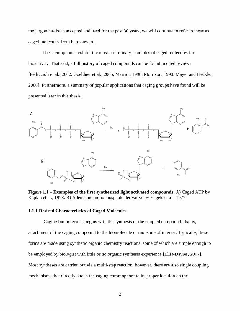
“Caged compounds,” or this class of light activatable molecules, originated in 1978 with
Kaplan et al.’s synthesis of phosphate caged adenosine triphosphate (Fig1A) [Kaplan et al.,
1978]. One year earlier, Engels et al. synthesized a cyclic adenosine monophosphate derivative
(Fig1B) containing a photolabile leaving group. The light activatable phenomenon of this
molecule was not the focus of the paper, nor was the “caged/caging” terminology ever inducted
into scientific vocabulary, so inception was postponed [Engels et al., 1977]. As Figure 1.1
depicts, “caging” is a rather ambiguous or liberally used term to describe the protection. More
accurately, these can simply be described as light removable protecting groups. However, since
2
the jargon has been accepted and used for the past 30 years, we will continue to refer to these as
caged molecules from here onward.
These compounds exhibit the most preliminary examples of caged molecules for
bioactivity. That said, a full history of caged compounds can be found in cited reviews
[Pelliccioli et al., 2002, Goeldner et al., 2005, Marriot, 1998, Morrison, 1993, Mayer and Heckle,
2006]. Furthermore, a summary of popular applications that caging groups have found will be
presented later in this thesis.
1.1.1 Desired Characteristics of Caged Molecules
Caging biomolecules begins with the synthesis of the coupled compound, that is,
attachment of the caging compound to the biomolecule or molecule of interest. Typically, these
forms are made using synthetic organic chemistry reactions, some of which are simple enough to
be employed by biologist with little or no organic synthesis experience [Ellis-Davies, 2007].
Most syntheses are carried out via a multi-step reaction; however, there are also single coupling
mechanisms that directly attach the caging chromophore to its proper location on the
Figure 1.1 – Examples of the first synthesized light activated compounds. A) Caged ATP by
Kaplan et al., 1978. B) Adenosine monophosphate derivative by Engels et al., 1977
3
biomolecule. There exist a number of general guidelines (listed in bold below), for design and/or
use of caging molecules and caged compounds, which will greatly optimize the photochemical
reaction. This convention should be verified and followed when introducing any caging
compound to a cellular environment:
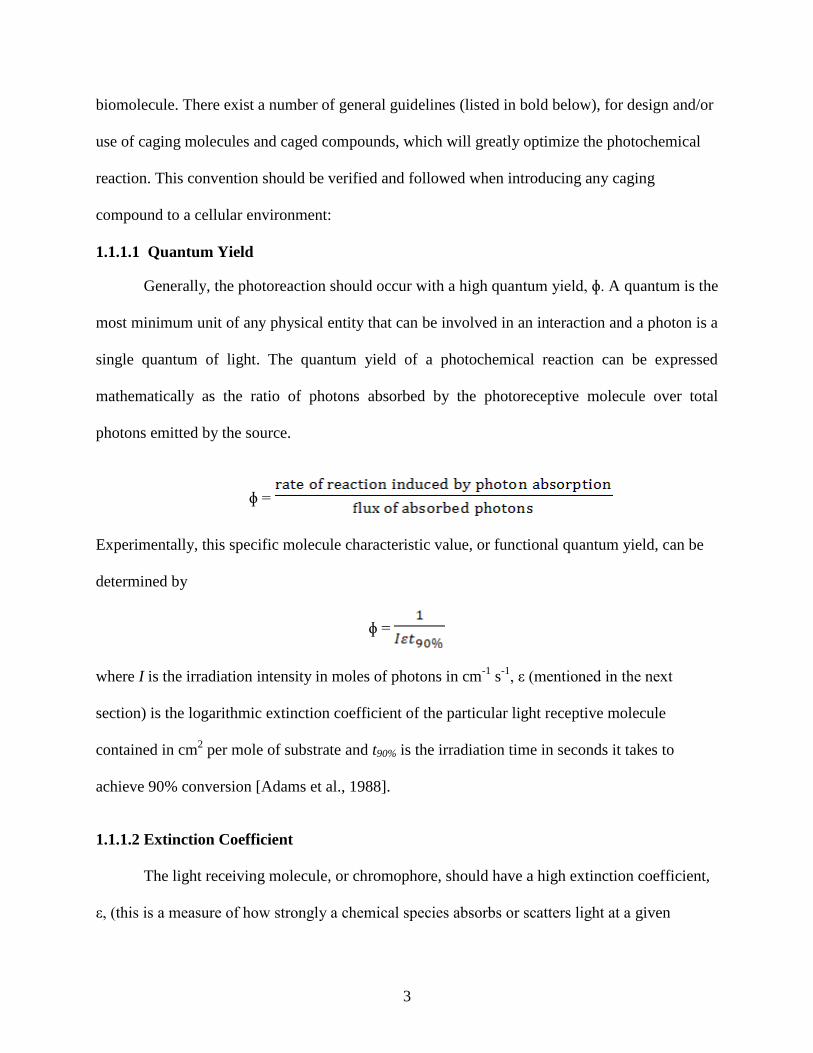
1.1.1.1 Quantum Yield
Generally, the photoreaction should occur with a high quantum yield, ɸ. A quantum is the
most minimum unit of any physical entity that can be involved in an interaction and a photon is a
single quantum of light. The quantum yield of a photochemical reaction can be expressed
mathematically as the ratio of photons absorbed by the photoreceptive molecule over total
photons emitted by the source.
ɸ =
Experimentally, this specific molecule characteristic value, or functional quantum yield, can be
determined by
ɸ =
where I is the irradiation intensity in moles of photons in cm-1
s-1
, ε (mentioned in the next
section) is the logarithmic extinction coefficient of the particular light receptive molecule
contained in cm2 per mole of substrate and t90% is the irradiation time in seconds it takes to
achieve 90% conversion [Adams et al., 1988].
1.1.1.2 Extinction Coefficient
The light receiving molecule, or chromophore, should have a high extinction coefficient,
ε, (this is a measure of how strongly a chemical species absorbs or scatters light at a given
4
wavelength) at wavelengths above 300nm. Light at wavelengths below 300nm are particularly
avoided because at these wavelengths, many biological samples themselves have high extinction
coefficients, thus they can readily absorb high energy light that has a wide range of deleterious
effects. Lower uncaging efficiencies have proven to be damaging to the cellular environment.
The organism itself cannot tolerate the number or density of photons needed to uncage the
compound without also receiving the deleterious effects of this bombardment.
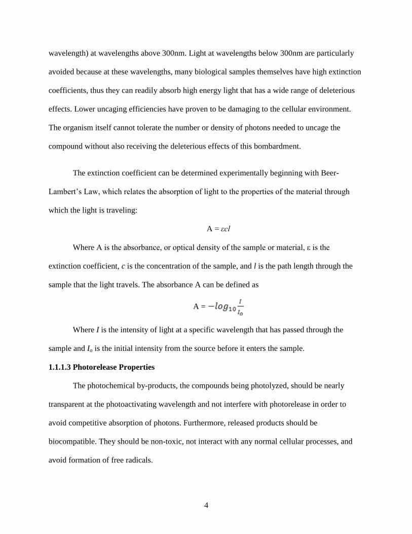
The extinction coefficient can be determined experimentally beginning with Beer-
Lambert’s Law, which relates the absorption of light to the properties of the material through
which the light is traveling:
A = εcl
Where A is the absorbance, or optical density of the sample or material, ε is the
extinction coefficient, c is the concentration of the sample, and l is the path length through the
sample that the light travels. The absorbance A can be defined as
A =
Where I is the intensity of light at a specific wavelength that has passed through the
sample and Io is the initial intensity from the source before it enters the sample.
1.1.1.3 Photorelease Properties
The photochemical by-products, the compounds being photolyzed, should be nearly
transparent at the photoactivating wavelength and not interfere with photorelease in order to
avoid competitive absorption of photons. Furthermore, released products should be
biocompatible. They should be non-toxic, not interact with any normal cellular processes, and
avoid formation of free radicals.
5
1.1.1.4 Rate of Uncaging
In kinetic studies, the rate of uncaging essentially must be more rapid than the process
being studied. The speed requirements will be fully dependent on the phenomenon being
analyzed.
1.1.1.5 Off-Target Effects
The caged agent must be biologically inert before photolysis. It must not elicit any
cellular response, nor act as an agonist or antagonist when applied at a useful/working
concentration in the biological preparation. It should be soluble in the target (aqueous) media,
and may be required to bypass biological membranes [reviewed in Pelliccioli et al., 2002].
Upon actualization of these design requirements, the compounds themselves can be
induced to in vivo analysis. However, because of the time dependency of biological processes, a
distinct understanding of the reaction mechanism, and specifically the release rate of the
compound, is strongly recommended.
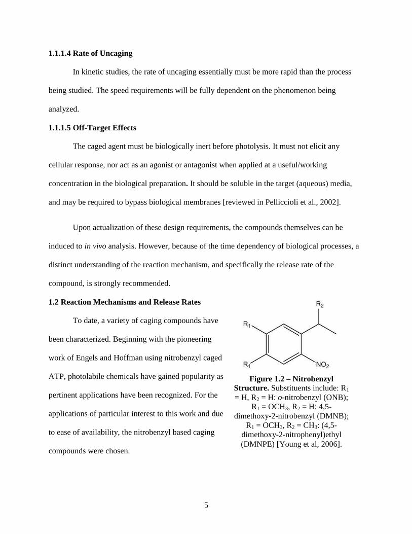
1.2 Reaction Mechanisms and Release Rates
To date, a variety of caging compounds have
been characterized. Beginning with the pioneering
work of Engels and Hoffman using nitrobenzyl caged
ATP, photolabile chemicals have gained popularity as
pertinent applications have been recognized. For the
applications of particular interest to this work and due
to ease of availability, the nitrobenzyl based caging
compounds were chosen.
Figure 1.2 – Nitrobenzyl
Structure. Substituents include: R1
= H, R2 = H: o-nitrobenzyl (ONB);
R1 = OCH3, R2 = H: 4,5-
dimethoxy-2-nitrobenzyl (DMNB);
R1 = OCH3, R2 = CH3: (4,5-
dimethoxy-2-nitrophenyl)ethyl
(DMNPE) [Young et al, 2006].
6
1.2.1 Ortho-Alkylated Nitrobenzyl Compounds
Of several very effective caging groups the 2–nitrobenzyl group was chosen due to the
availability of this compound. In general, nitrobenzyl groups are by far the most commonly
utilized caging agent due to their practical synthesis and, in most cases, relative ease of
attachment to the intended active site. Furthermore, these compounds are readily and easily
decorated with electron donating functional groups (CH3O) that “red–shift” the absorption
maximum. Particularly, the attachment of multiple groups produces a hyperchromatic shift in the
absorbance, which enables the chromophore to absorb a longer, lower energy wavelength of light
that is less photodamaging. The adverse consequence of these attachments are typically found in
a reduction of photolysis efficiency [Aujard et al., 2006].
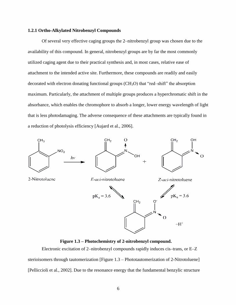
Electronic excitation of 2–nitrobenzyl compounds rapidly induces cis–trans, or E–Z
sterioisomers through tautomerization [Figure 1.3 – Phototautomerization of 2-Nitrotoluene]
[Pelliccioli et al., 2002]. Due to the resonance energy that the fundamental benzylic structure
Figure 1.3 – Photochemistry of 2-nitrobenzyl compound.
7
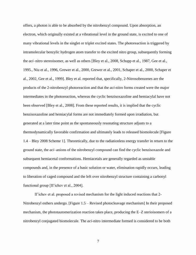
offers, a photon is able to be absorbed by the nitrobenzyl compound. Upon absorption, an
electron, which originally existed at a vibrational level in the ground state, is excited to one of
many vibrational levels in the singlet or triplet excited states. The photoreaction is triggered by
intramolecular benzylic hydrogen atom transfer to the excited nitro group, subsequently forming
the aci–nitro stereoisomer, as well as others [Bley et al., 2008, Schupp et al., 1987, Gee et al.,
1995., Niu et al., 1996, Grewer et al., 2000, Grewer et al., 2001, Schaper et al., 2000, Schaper et
al., 2002, Gee et al., 1999]. Bley et al. reported that, specifically, 2-Nitrosobenzenes are the
products of the 2-nitrobenzyl photoreaction and that the aci-nitro forms created were the major
intermediates in the photoreaction, whereas the cyclic benzisoxazoline and hemiacylal have not
been observed [Bley et al., 2008]. From these reported results, it is implied that the cyclic
benzisoxazoline and hemiacylal forms are not immediately formed upon irradiation, but
generated at a later time point as the spontaneously resonating structure adjusts to a
thermodynamically favorable confirmation and ultimately leads to released biomolecule [Figure
1.4 – Bley 2008 Scheme 1]. Theoretically, due to the radiationless energy transfer in return to the
ground state, the aci–anions of the nitrobenzyl compound can find the cyclic benzisoxazole and
subsequent hemiacetal conformations. Hemiacetals are generally regarded as unstable
compounds and, in the presence of a basic solution or water, elimination rapidly occurs, leading
to liberation of caged compound and the left over nitrobenzyl structure containing a carbonyl
functional group [II’ichev et al., 2004].
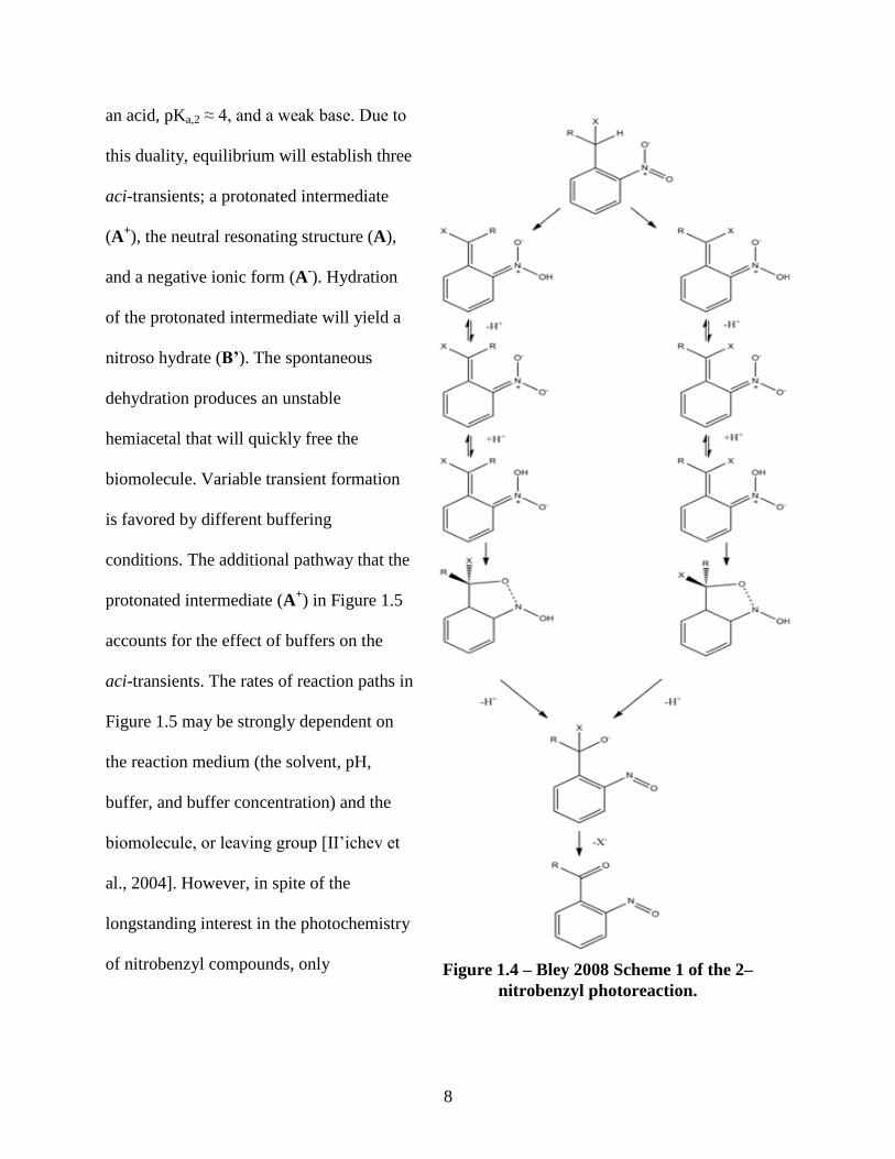
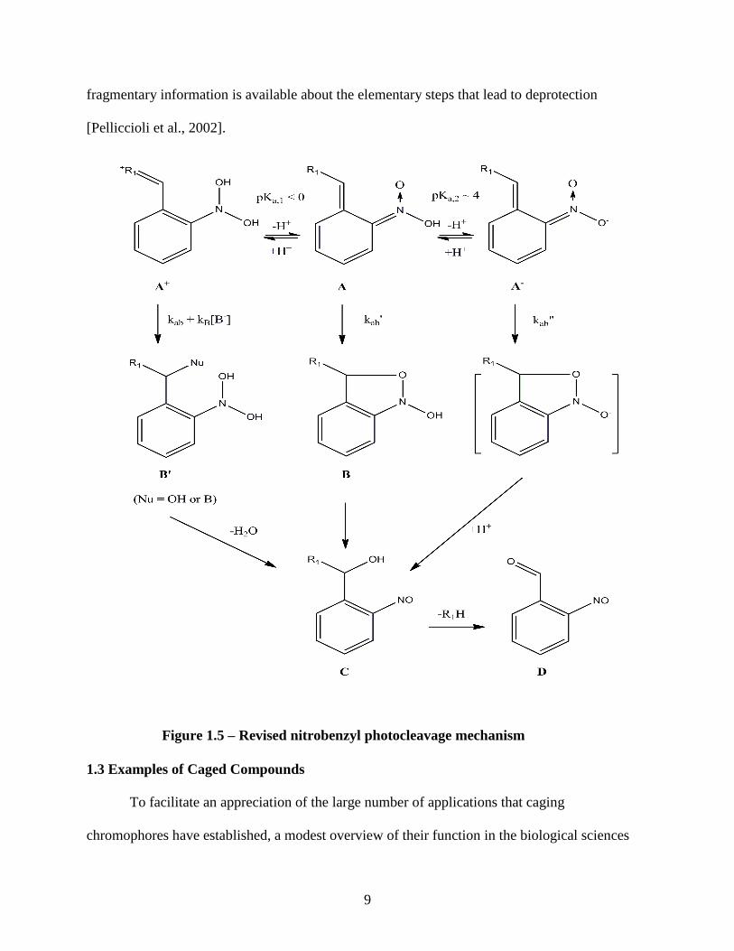
II’ichev et al. proposed a revised mechanism for the light induced reactions that 2-
Nitrobenzyl esthers undergo. [Figure 1.5 – Revised photocleavage mechanism] In their proposed
mechanism, the phototautomerization reaction takes place, producing the E–Z sterioisomers of a
nitrobenzyl conjugated biomolecule. The aci-nitro intermediate formed is considered to be both
8
an acid, pKa,2 ≈ 4, and a weak base. Due to
this duality, equilibrium will establish three
aci-transients; a protonated intermediate
(A+), the neutral resonating structure (A),
and a negative ionic form (A-). Hydration
of the protonated intermediate will yield a
nitroso hydrate (B’). The spontaneous
dehydration produces an unstable
hemiacetal that will quickly free the
biomolecule. Variable transient formation
is favored by different buffering
conditions. The additional pathway that the
protonated intermediate (A+) in Figure 1.5
accounts for the effect of buffers on the
aci-transients. The rates of reaction paths in
Figure 1.5 may be strongly dependent on
the reaction medium (the solvent, pH,
buffer, and buffer concentration) and the
biomolecule, or leaving group [II’ichev et
al., 2004]. However, in spite of the
longstanding interest in the photochemistry
of nitrobenzyl compounds, only Figure 1.4 – Bley 2008 Scheme 1 of the 2–
nitrobenzyl photoreaction.
9
fragmentary information is available about the elementary steps that lead to deprotection
[Pelliccioli et al., 2002].
1.3 Examples of Caged Compounds
To facilitate an appreciation of the large number of applications that caging
chromophores have established, a modest overview of their function in the biological sciences
Figure 1.5 – Revised nitrobenzyl photocleavage mechanism
10
will be presented beginning with related studies of small molecules. Macromolecular caged
compounds will then be briefly presented and, finally, transitioning to our applications,
attachment to oligonucleotides (or short nucleic acid/nucleic acid analog polymer, typically 20 or
fewer base pairs) and nucleic acids.
1.3.1 Small Molecules
The following sections are designed to give the reader an appreciation of applications that
biomolecules find when they can be rendered inert. This is by no means an exhaustive list.
Excellent reviews containing more examples and applications can be readily explored [Ellis-
Davies, 2007, Pelliccioli et al., 2002, Mayer and Heckel, 2006].
1.3.1.1 Neurotransmitters - Caged Glutamate
Perturbation of sensory processing and manipulation of neural activity with single cell
resolution is a long standing desire of neuroscientists for applications in disease treatment. Using
caged glutamate, an action potential can be induced in only a small number of neurons which can
disclose information on the neuronal circuitry that will, expectantly, lead biologist to their goal
[Matsuzaki et al., 2008]. Caged glutamate is the most widely used caged neurotransmitter by
biologist, and many approaches to caging this molecule have been effected using different
chromophores and caging strategies [Ellis-Davies, in press)].
1.3.1.2 Nucleotides and Nucleosides
As previously mentioned, the Kaplan, Forbush, Hoffman group were the pioneers of the
“caging” frontier. In their 1978 work, they successfully caged ATP using a five step synthesis
reaction. Beginning with the nitrobenzyl caging compound, they created caged phosphate which
was coupled to the terminal phosphate of adenosine diphosphate, ADP. Formation of the caged
adenosine triphosphate followed. Demonstration of photolytic features of caged ATP lead to the
11
applicable objectives, which were the effects of ATP/caged ATP on sodium/potassium pump
governing ATP-ase and its effects on sodium/potassium transport associated with its enzymatic
activity in human red blood cells [Kaplan et al., 1978].
One year prior to the Kaplan 1978 publication, Engels et al., “caged” cAMP. Although
their original objectives of adding a chemical moiety to the adenosine structure were to enhance
lipophilicity, they were concerned with restoring the original activity of this important second
messenger. While some of 6 different triesters of cAMP were synthesized with the intention of
direct hydrolysis to free the biomolecule, 2 involved the nitrobenzyl moiety for release via
photolysis. As a delivery vehicle for enhanced lipophilicity, the results were validated in rat
glioma cells [Engels et al., 1977]. The caged ATP and cAMP were the first reported caged
compounds to be synthesized, used, and uncaged in living cells. Since that time many
nucleotides and their analogs have been caged for biological applications [reviewed in Pelliccioli
et al., 2002].
1.3.2 Macromolecules
Similar to that of small molecules, blocking the function of macromolecules has been met
with much success. It is generally accepted that significant differences lie in targeting the active
site of the molecule as opposed to strategies of attachment to small molecules where the mere
presence of the blocking compound can impose a steric hindrance, affecting action. Similar to
the overview of small molecules, this section on macromolecules will generally describe the use
of caged large molecules.
A considerable variety of caged peptides (short polymers formed from linking amino
acids in a defined order) have been made, specifically in the form of enzymes and polypeptides.
Caged enzymes usually serve as inhibitory peptides designed to disrupt protein–protein
12
interactions and caged polypeptides act as the larger counterparts to standard small molecule
caged compounds. In this case, where direct attachment to the enzyme or protein is advanced, its
catalytic function is blocked by the caging chromophore [Ellis-Davies, 2007]. The progress of
macromolecular caging is often more difficult than that of small molecules due to the fact that
caged peptides appear to be inherently unstable. However, development of these complex
conjugates is a fundamental necessity because they provide an inactivation approach that is
conceptually distinct from that offered by the small molecules. This idea is highlighted by the
fact that many cellular processes are not regulated by cofactors, but by direct protein–protein
interaction [Ellis-Davies, 2007].
1.3.2.1 Peptides and Proteins
Proteins and peptides have been caged by many commercially available reagents that
modify specific amino acid residues. The ability to target particular locations on the biomolecule
is especially important because blockage of the active site, usually through steric hindrance, will
render it inert. Identifying and targeting certain locations, a crucial facet to this method that
ultimately optimizes monetary and temporal resources, is achieved by predetermined
bioconjugate techniques that are fully dependent on well known and characterized chemical
reactions [Ellis-Davies, 2007]. There are several points to consider when planning caged protein
synthesis: (i) due to its size, use of caged proteins will inherently introduce a percentage of
residual activity effectively complicating outcomes in the biological system (in most situations,
an equilibrium will establish, greatly reducing the probability of caging 100%, leaving the small
percent of residual activity to potentially introduce ambiguous results), (ii) full recovery of
function is problematic, especially when “shotgun” or “statistical” caging is the method of
inactivation, and (iii) there is no especially efficient way to introduce these silenced compounds
13
to an in vivo environment [Ellis-Davies, 2007]. Additionally, proteins typically contain a large
number of nucleophilic sites (sites that preferably attach to an electrophilic counterpart through
the donation of electrons) which can be difficult to interact in a site-specific manner with
exogenous caging agents, when the endogenous, competitive interactions with other proteins in
the cellular environment is a much more favorable reaction [Curley et al., 1999]. Also, there may
not be an appropriate nucleophilic residue necessary for interaction, at or near the desired site of
modification. Fortunately, many of these problems have been circumvented in recent studies.
The two primary mechanisms of passage are beyond the scope of this review [refer to Ellis-
Davies paper 18, 49, 50, and 51] [Mendel et al., 1991, Petersson et al., 2003, Muralidharan et al.,
2006, and Hahn et al., 2004]. In summary there is an exponential growth of complexity for
caging reactions introduced by increasing molecular weight compounds.
1.3.2.2 Oligonucleotides and Nucleic Acids
Effective alteration of gene activity at precise times and locations is especially attractive
for delineation of protein function in whole organisms, and where uncaging technology is being
increasingly applied to accelerate this work. Genetic function can be efficiently controlled by
caging deoxyribonucleic acids (DNA) and (messenger) ribonucleic acids (mRNA) fragments, as
well as gene-regulating oligonucleotides. Thus far, the most efficient method of caging mRNAs
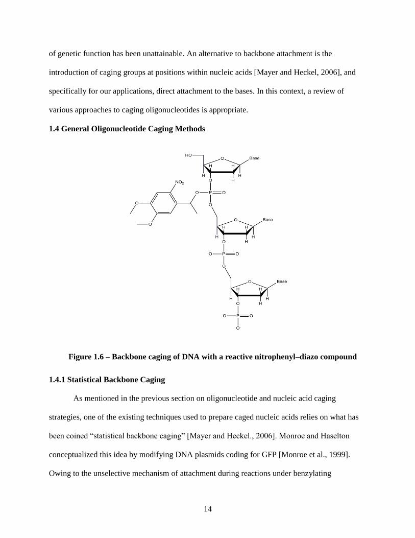
or DNAs has proven to be the most useful biologically [Ellis-Davis, 2007]. “Direct, multi-site,”
“shotgun,” or “statistical” caging, terms coined to describe a specific phenomena, of DNA with
reactive nitrophenyl–diazo compounds, thoroughly binds and inactivates the molecule [Monroe
et al., 1999] [Figure 1.6]. The elegance of the approach of statistical backbone caging clearly lies
in its ease and simplicity of preparation [Mayer and Heckel, 2006]. However, at least with the
modifying reactions and caging chromophores used to date, systematic binary (on/off) response
14
of genetic function has been unattainable. An alternative to backbone attachment is the
introduction of caging groups at positions within nucleic acids [Mayer and Heckel, 2006], and
specifically for our applications, direct attachment to the bases. In this context, a review of
various approaches to caging oligonucleotides is appropriate.
1.4 General Oligonucleotide Caging Methods
1.4.1 Statistical Backbone Caging
As mentioned in the previous section on oligonucleotide and nucleic acid caging
strategies, one of the existing techniques used to prepare caged nucleic acids relies on what has
been coined “statistical backbone caging” [Mayer and Heckel., 2006]. Monroe and Haselton
conceptualized this idea by modifying DNA plasmids coding for GFP [Monroe et al., 1999].
Owing to the unselective mechanism of attachment during reactions under benzylating
Figure 1.6 – Backbone caging of DNA with a reactive nitrophenyl–diazo compound
15
conditions, the plasmids were modified with a statistical distribution of the altering agent. In this
approach, probable locations of attachment are estimated and selected as likely sites that are
dependent on the chemical species being exploited. Although this approach brings one as far as a
hypothetical structure of the chemical species complex, the statistical analysis begins with
calculations based on the extinction coefficient of the caging chromophore. Based on the
extinction coefficient, the number of caging groups per oligonucleotide sequence can be
quantified [Friedman 2006].
1.4.2 Site Specific Attachment
The appeal of the statistical backbone method stems from its simplicity of preparation.
However its primary downfall, which calls for implementation of alternative strategies, is the
crux that full restoration, or complete removal of attachment compounds, is not yet attainable.
The most obvious alternative would be a direct, site-specific attachment scheme. The alternative
to circumvent the disadvantageous properties introduced by statistical attachment is to accurately
target locations at defined positions on nucleic acids.
This is exactly the approach that Chaulk and MacMillan took one year prior to Monroe
and Haselton’s work. In their conception, a complementary approach was developed that allowed
for the isolation of specific RNA structures or complex formations through transient blockings or
caging of particular RNA functional groups involved in the transition between two different
states, particularly the 2’ hydroxyl functionality [Chaulk et al., 1998]. The 2’ hydroxyl group was
chosen as a blocking site due to its association with general RNA functionality, since specific 2’
hydroxyls act as nucleophiles in a number of biologically relevant transesterifications
[Uhlenbeck, 1987, Branch et al., 1991, Buzayan et al., 1986, Guo et al., 1995, Peebles et al.,
1986]. The 2’ modified RNA was used to control hammerhead ribozyme (ribonucleic acid
16
enzyme) activity with light. Ribozymes are RNA molecules that are able to catalyze a chemical
reaction. Modification of the 2’ group with a nitrobenzyl blocking compound stopped catalytic
activity of the enzyme whereas upon irradiation, full restoration of activity was restored. This is
a hallmark example of site–specific, finely controlled, binary behavior prior to and post
irradiation albeit at the demand of a markedly higher synthetic effort. Although different
synthetic methods are employed and target areas are varied, the aforementioned techniques have
commonality, namely, backbone caging which assumes the existence of the very nature of
operation nucleic acids are still intact. This nature is the Watson–Crick interaction capability. A
complete explanation of how backbone caging affects hybridization has not yet been offered.
Alternatively, assuming labeling on the bases, one can clearly understand that the presence of the
caging groups offers a steric hindrance that disables the hydrogen bonding ability of an
oligonucleotide sequence.
1.4.3 Alternative Novel Approaches
Before proceeding, notable approaches have been employed that are acutely different
from the somewhat expectable approaches mentioned until now. Met with success, the pioneers
who crafted these techniques engaged diverse tools of molecular biology with a single specific
design and function that fortunately operated in the hypothesized manner. Several of these tools
that exercise nucleobase caging techniques, or direct Watson–Crick disruption, will now be
discussed.
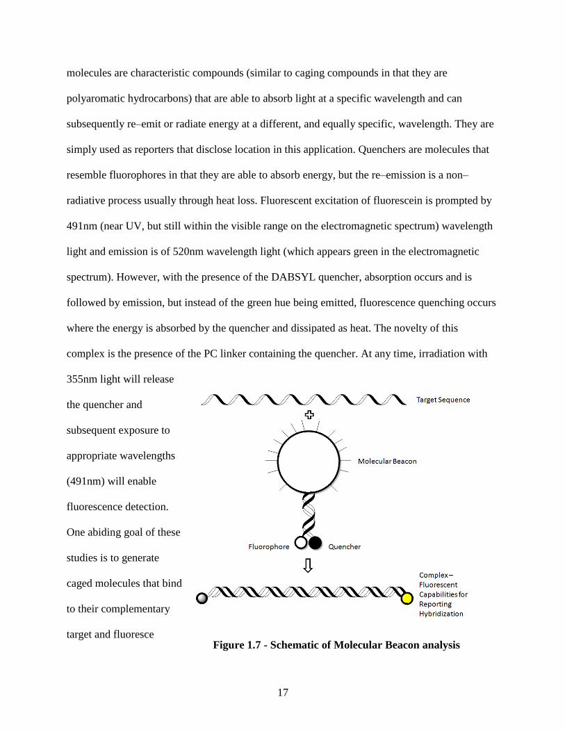
Dmochowski et al. employed a synthetic route for incorporating a photocleavable (PC)
linker containing a DABSYL (fluorescence quencher) moiety and fluorescein at adjacent
cytidines (cytosine nucleoside, formed when cytosine is attached to the ribose ring, or in the case
of DNA deoxycytidine) in the middle of a 25mer oligonucleotide [Tang et al., 2004]. Fluorescent
17
molecules are characteristic compounds (similar to caging compounds in that they are
polyaromatic hydrocarbons) that are able to absorb light at a specific wavelength and can
subsequently re–emit or radiate energy at a different, and equally specific, wavelength. They are
simply used as reporters that disclose location in this application. Quenchers are molecules that
resemble fluorophores in that they are able to absorb energy, but the re–emission is a non–
radiative process usually through heat loss. Fluorescent excitation of fluorescein is prompted by
491nm (near UV, but still within the visible range on the electromagnetic spectrum) wavelength
light and emission is of 520nm wavelength light (which appears green in the electromagnetic
spectrum). However, with the presence of the DABSYL quencher, absorption occurs and is
followed by emission, but instead of the green hue being emitted, fluorescence quenching occurs
where the energy is absorbed by the quencher and dissipated as heat. The novelty of this
complex is the presence of the PC linker containing the quencher. At any time, irradiation with
355nm light will release
the quencher and
subsequent exposure to
appropriate wavelengths
(491nm) will enable
fluorescence detection.
One abiding goal of these
studies is to generate
caged molecules that bind
to their complementary
target and fluoresce Figure 1.7 - Schematic of Molecular Beacon analysis
18
when triggered with light [Tang et al., 2004]. In this way, one can determine exactly when the
ligand/receptor complex formed and is present.
Analogous to the quenched fluorophore, a novel idea arising from this technology
introduced and combined another set of molecular diagnostics, providing an additional degree of
precise control. To begin, molecular beacons, first synthesized in 1996, are nucleic acid probes
that recognize and report the presence of specific nucleic acids in a homogeneous solution or
environment [Tyagi et al., 1996]. The beacons undergo a spontaneous structural change when a
target nucleic acid sequence is identified that ultimately brings about a fluorogenic state [Figure
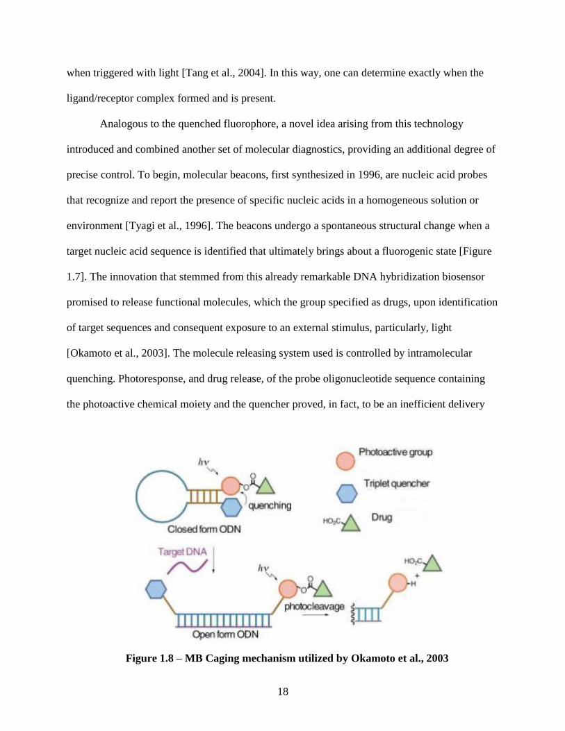
1.7]. The innovation that stemmed from this already remarkable DNA hybridization biosensor
promised to release functional molecules, which the group specified as drugs, upon identification
of target sequences and consequent exposure to an external stimulus, particularly, light
[Okamoto et al., 2003]. The molecule releasing system used is controlled by intramolecular
quenching. Photoresponse, and drug release, of the probe oligonucleotide sequence containing
the photoactive chemical moiety and the quencher proved, in fact, to be an inefficient delivery
Figure 1.8 – MB Caging mechanism utilized by Okamoto et al., 2003
19
mechanism when the existing state was in the “closed loop” form, whereas, upon Watson–Crick
coupling with its target and consecutive irradiation, rapid release of the functional molecule was
observed [Okamoto et al., 2003] [Figure 1.8].
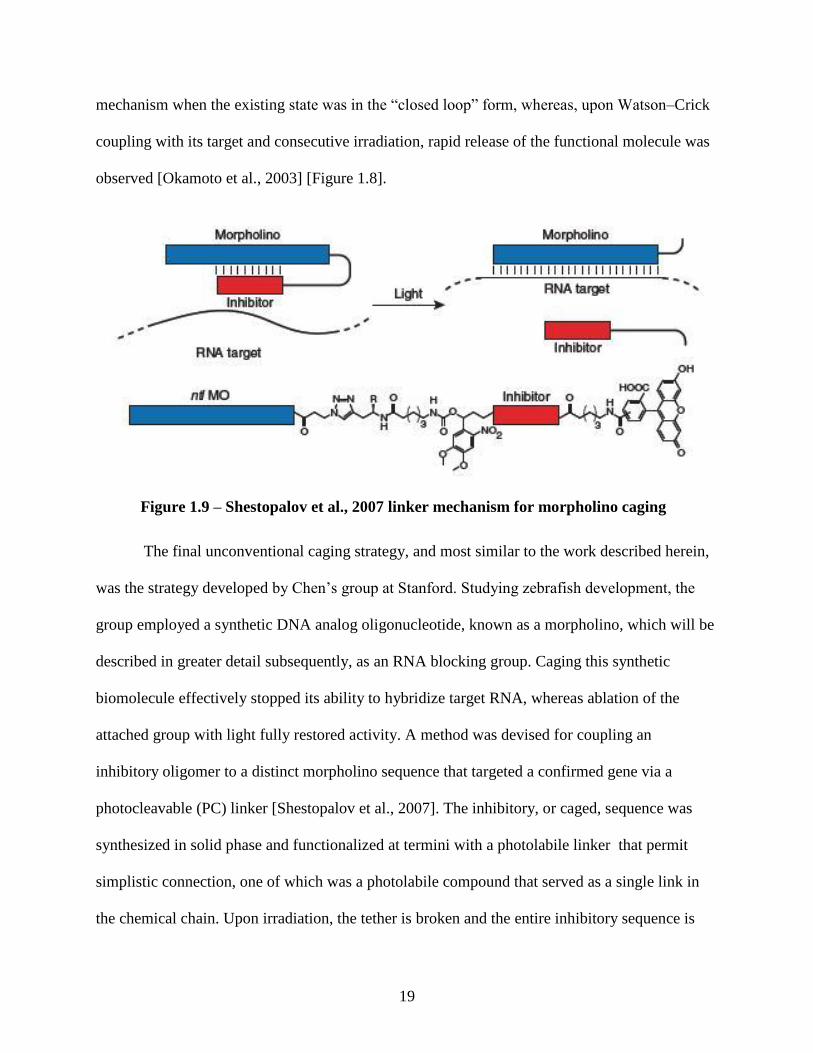
The final unconventional caging strategy, and most similar to the work described herein,
was the strategy developed by Chen’s group at Stanford. Studying zebrafish development, the
group employed a synthetic DNA analog oligonucleotide, known as a morpholino, which will be
described in greater detail subsequently, as an RNA blocking group. Caging this synthetic
biomolecule effectively stopped its ability to hybridize target RNA, whereas ablation of the
attached group with light fully restored activity. A method was devised for coupling an
inhibitory oligomer to a distinct morpholino sequence that targeted a confirmed gene via a
photocleavable (PC) linker [Shestopalov et al., 2007]. The inhibitory, or caged, sequence was
synthesized in solid phase and functionalized at termini with a photolabile linker that permit
simplistic connection, one of which was a photolabile compound that served as a single link in
the chemical chain. Upon irradiation, the tether is broken and the entire inhibitory sequence is
Figure 1.9 – Shestopalov et al., 2007 linker mechanism for morpholino caging
20
released [Figure 1.9]. Functional studies in whole organisms exploit a seemingly infinite number
of drugs. The constitutive activity of these reagents limit their experimental utility, ultimately
creating a daunting enigma. Owing to this puzzle, caging compounds have been identified by
their aptitude for elucidation of this fundamental dilemma.
1.5 Light Sources for Photo-Activation of Caged Compounds
A large number of photon sources are available for photo-activation. These variable
devices offer advantageous and disadvantageous elements that are especially dependent on the
applications involving the caged compound that require their use. Characteristics one should
consider when choosing the appropriate light source for their application include: heat generation
by the source, the spectra that is generated, and density of radiation incident. Without performing
a full review of available light sources, which can be found at [Ellis-Davies, 2007, Casey et al.,
2009], it is sufficient to mention that, currently, the majority of uncaging experiments exploit
flash lamps and lasers [Rapp et al., 1989, Blidner et al., 2008, Mikat et al., 2007, Ando et al.,
2004, Shah et al., 2005, Young et al., 2008, Shestopalov et al., 2007, Kaplan et al., 1978]. For
our application, we apply a specific UV-light source for our studies. Distinguishing from others,
we operate a device, commercially tagged, “Green–Spot” that uses industry standard pressurized
100 watt mercury lamp, mounted vertically, in a dichroic–coated elliptical reflector to generate
an intense, 5mm spot of light in the UVA, UVB, UVC, and visible range [American Ultraviolet
Co]. The device is marketed by its simplistic design, interface and reliability of operation. The
optics system contains a removable quartz IR filter that allows UV light to pass from the 300-480
nm range [Forman, 2007]. The Green–Spot device uses a standard fiber optic cable, or light
guide, designed to operate in the 320 – 500nm range. Confining spectral output to 365nm is
21
preferable, due to the fact that the methoxy-nitrobenzyl absorbs maximally at 355nm, as well as
reducing the potential for undesirable heat accretion [Forman, 2007].
1.6 Gene-Silencing Oligonucleotides – Tools for Gene Knockdown
1.6.1 The Opportunity for Antisense Oligonucleotides
An ever increasing opportunity for antisense drugs is becoming more evident as the
windings of molecular biology and disease diagnosis are being unraveled. This understanding at
the molecular level validates the approach as a whole since biological process dissection enables
and promotes rational drug design which almost entirely abolishing conventional drug synthesis
methods. The conception of this technology derives from a sound understanding of nucleic acid
structure and function that is ultimately dependent on Watson–Crick hybridization [Watson,
1953], and fundamentally promises gene-selective reagents and drugs [Crooke, 2007]. Thus,
arguably, clear demonstration that nucleic acid hybridization is feasible and moderately
controllable, and the advances of in situ hybridization and probe technology, establishes
evidence for the most basic elements of the foundation supporting the antisense theory [Gillespie
et al., 1965, Thompson et al., 1990].
The first use of antisense oligonucleotides as therapeutic agents was performed in the
work of Zamecnik and Stevenson in 1978 [Zamecnik et al., 1978]. Here, a 13 base pair
tridecamer was synthesized that was complementary to an equal length segment of the 3’ and 5’
reiterated terminal sequences of Rous sarcoma virus (RSV) 35SRNA. The RSV 35S RNA
contains a 21 nucleotide sequence just internal to the 5’ cap that is exactly identical to the same
length segment adjacent to the poly A terminus at the 3’ end of the molecule, approximately
10,000 base pairs away. The reiterating terminal sequences critically impact in circularization of
viral DNA, prior to its integration into the host genome. Induction of the tridecamer, targeting
22
the 3’ and 5’ locations, into cell culture infected with RSV resulted in inhibition of virus
production [Zamecnik et al., 1978]. This implied that the oligonucleotide displayed evidence of
antiviral activity. However, the most impactful result that the authors reported was the possible
sites for attachment via hybridization on the viral RNA and potential mechanism of action of the
oligonucleotide.
1.6.2 The Challenges Faced by the Field
Essentially, the development of antisense technology is the creation of a new
pharmacology, or more specifically, an entire new study of how these exogenous chemicals alter
the normal biological function of the organisms with which they interact [Crooke, 2007]. The
receptors for this drug are defined nucleic acid sequences in a target strand. Accordingly,
perfectly analogous with enzymatic kinetic analysis, from a pharmacological perspective, a
comprehension of the structure, function, and intermediary metabolism of RNA is essential.
Conceptually, the mechanisms of action that antisense molecules can assume is divided
into three classes: pre– or non–hybridization, hybridization, and posthybridization. These
hybridization properties must be considered owing to the basis that typical cellular levels of
target mRNAs is less than 100 copies per cell, and at such a low concentration, the interaction
that occurs usually has a negligible effect on the total antisense drug concentration.
Consequentially, these unaccounted for off–target effects, primarily with cellular and
extracellular proteins, attribute to the pharmacokinetics and non–pharmacologically–based
toxicological properties of antisense drugs. Finally, while impressive progress has been reported
about understanding posthybridization processes, curiously, very little has been said about how
hybridization to a specific target RNA occurs, in particular, the kinetics of intracellular
hybridization events [Crooke, 2007].
23
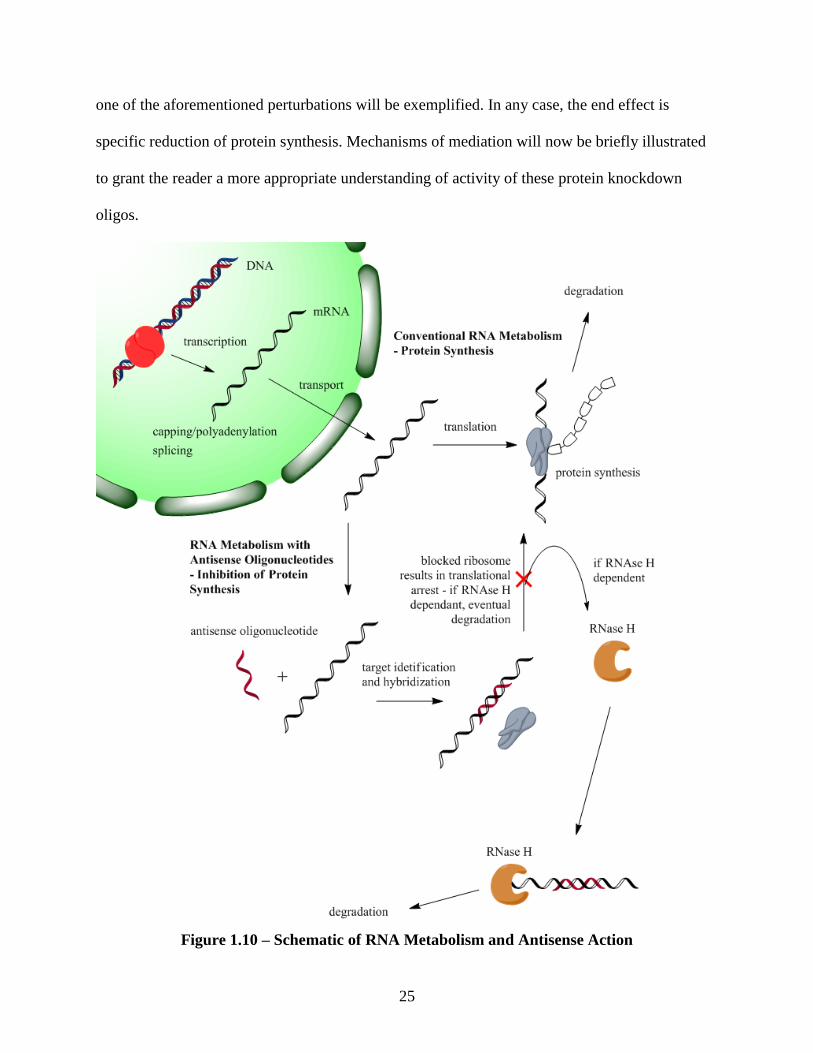
1.7 Basic Principles – RNA Intermediary Metabolism
Oligonucleotides are molecules designed to alter RNA intermediary metabolism. They
are designed to modulate the information transfer from gene to protein. Figure 1.10 depicts this
mechanism. RNA metabolism is initiated with transcription. Specific sequences of DNA are
recognized by transcription initiation complexes that act to locally denature DNA by separating
the double stranded sequence into two independent identifiable strands. A member of the RNA
polymerase family, a protein family that can assemble RNA polymers from genetic information
in the form of DNA, can then complex with the single stranded DNA and serve its function to
transcribe one strand of the DNA (the antisense strand) to its sense pre–mRNA polymer.
Typically during transcription, the 5’ end of the pre–mRNA is “capped” by adding a methyl–
guanosine, and also by methylation of one or two adjacent sugar residues. This process is vital to
creating mature mRNA, which is able to undergo translation. Capping also ensures the mRNA
polymer’s stability as it undergoes transcription in protein synthesis and may affect a number of
valuable RNA processing events [Mizumoto et al., 1987]. Between the 5’ cap and the sight
where translation occurs is a stretch of nucleotides that are untranslated, known as the 5’
untranslated region (5’ UTR), that also serve to affect mRNA half life and transitional efficiency
[Ross, 1988]. Similar to the 5’end of the mRNA, the 3’ end also undergoes modification. On the
3’ side exists several hundred nucleotides beyond the translation termination signal. Much like
the 5’ bit, this untranslated region too plays a role in determining the half life of the mRNA as a
whole. Moreover, post transcriptional modifications in the form of a polyadenylated tail stabilize
mRNA. This modifier is crucial for transporting mature mRNA out of the nucleus, and
additionally, it may serve significant roles in the cytoplasm [Friedman et al., 1987, Manley,
1988].
24
Besides modifying the 3’ and 5’ ends of pre–mRNA, proper splicing is important in
conventional mRNA metabolism. Because eukaryotic genes usually contain ancillary sequences
(introns) embedded between active protein coding regions (exons), proper function entails
excision of these superfluous regions with the remaining segments spliced together. Splicing
reactions are complex, highly regulated, involve specific sequences, small molecular weight
RNA species, and numerous proteins [Crooke, 2007]. Since RNA splicing involves withdrawal
of delineated intronic sequences and coupling of exonic fragments, alternative splicing reactions,
or removal of alternative sequences, will generate different mature mRNAs and therefore,
different proteins. Although introns have been observed as waste, important sequences are
conserved and have been recognized to play roles in coding for proteins, antisense transcripts,
and noncoding RNAs [Black, 2003]. Once capping, polyadenylation, and splicing occurs, mature
mRNAs are shuttled out of the nucleus and into the cytoplasm where they engage in translation.
As previously maintained, the half life of these compounds range from minutes to hours, and are
highly regulated.
1.7.1 Antisense Action
Simply stated, an antisense oligonucleotide is a short strand of deoxyribonucleotide or
deoxyribonucleotide analogue that is complementary to a sense strand of mRNA. As a
consequence of the complementation, the antisense strand can hybridize to the sense sequence by
way of Watson–Crick base pairing. Formation of this heteroduplex induces a number of potential
circumstances including: induction of RNase H activity which leads to mRNA destruction, steric
hindrance and sequential translational arrest by blockage of the ribosomal units, and/or
interference with mRNA maturation by inhibiting splicing or destabilization of pre–mRNA in the
nucleus[Chan et al., 2006]. Depending on the mode of action of the particular oligonucleotide,
25
one of the aforementioned perturbations will be exemplified. In any case, the end effect is
specific reduction of protein synthesis. Mechanisms of mediation will now be briefly illustrated
to grant the reader a more appropriate understanding of activity of these protein knockdown
oligos.
Figure 1.10 – Schematic of RNA Metabolism and Antisense Action
26
1.7.2 Potential Sites of Disruption
Traditionally, competitive antagonists are assumed to alter biological processes through
binding to receptors, effectively preventing agonists from accessing and inducing normal
biological process. Binding of oligonucleotides to endemic complementary sequences may
inhibit RNA interaction with proteins, nucleic acids, or other intracellular machinery necessary
for proper intermediary metabolism [Crook, 2007].
1.7.2.1 Splicing Transitioning
Splicing reactions, the excision of introns necessary in the intermediary metabolism of
mRNA, are sequence specific and demand the concerted action of spliceosomes, therefore,
oligonucleotides that append to mRNA that naturally undergo the excision process may prevent
amalgamation of necessary factors (proteins) and prevent fragmentation [Crooke, 2007]. This
would result in prevention of mature mRNA synthesis. A number of chemically modified
antisense oligonucleotides have been shown to alter spicing in vitro and in vivo [Sazani et al.,
2003]. Considering the necessity of oligonucleotide hybrids that are more robust and resistant to
degradation, certain chemical alterations to structure are required. Specifically, chemically
modified antisense products such as:s fully modified 2’–methoxy, 2’–MOE, peptide nucleic acid
(PNA), morpholino (which will be described in more detail later), and fully modified locked
nucleic acid (LNA) analogs [Sazani et al., 2003].
Alternative splicing, a subset of splicing reactions that allows for the assembly of
necessary proteins but alters the output of the original intended reaction, is also a domain of
focus because theoretical conception of alternative proteins can be envisioned.
1.7.2.2 Translational Arrest
Translational arrest, as hinted by the name, is a supposition that translation is inhibited. In
this stage of RNA metabolism, some process denies the messenger RNA from being read and
27
construed to protein. Because polysomes are capable of “melting” or disassociating structures in
RNA, translational inhibitors classically interact in the 5’ untranslated region (UTR), the
translation initiation codon area, or an internal ribosomal entry (IRE) sequence [Crooke, 2001,
Crooke, 1999]. However, inhibitors designed to bind to sites in coding sequences have also been
shown to be active [Crooke, 2007].
1.7.2.3 5’ Capping Alterations
An imperative elementary step in RNA processing and metabolism is 5’ capping. The
pervasiveness of this action is a testament to its importance and it serves a number of roles in the
cell which are exemplified by stabilizing the pre–mRNA as well as the mature mRNA, essential
binding to the nuclear matrix, and its presence plays a role in transport of mRNA out of the
nucleus. Several oligonucleotides that bind near the cap site have shown favorable silencing
activity, presumably by inhibiting standard protein binding required for attachment of the
capping molecule [Saxena et al., 1990, Westermann et al., 1989]. Also, oligonucleotides have
been designed to bind to the 5’ cap structure that selectively identify reagents that cleave the 5’
cap itself off of the mRNA. This demonstrated that addition and subsequent abstraction of the 5’
cap still serves as an effective means to inhibit the binding of translation initiation factors [Baker
et al., 1992].
1.7.2.4 Inhibition of 3’ Polyadenylation
Similar to 5’ capping, in the 3’ UTR of pre-mRNA, there are sequences that necessitate
posttranscriptional addition of hundreds of nucleotide long tracts of polyadenylate. Analogously,
polyadenylation stabilizes mRNA and plays a multitude of other roles in RNA metabolism.
Logically, using the same approach as disruption of 5’ capping protein binding, interactions in
the 3’–terminal region could inhibit polyadenylation and destabilize the RNA species. Although
28
many oligonucleotides target the 3’ UTR and display antisense activity, only one study has
typified evidence for alterations in polyadenylation [Vickers et al., 2001]. Here, fully modified
2’–MOE antisense agents prompted polyadenylation to be redirected, effectively leading to RNA
stability increases and enhanced protein synthesis.
1.7.2.5 RNase H Enzymes
The RNase H knockdown mechanism has proven to be the most widely studied system to
date. In this pathway, RNase H enzymes hydrolyze RNA in RNA–DNA hybrids [Stein et al.,
1969]. In addition, there are RNases that recognize double stranded RNA, or RNA/RNA
duplexes, and denature these complexes. Although it has been accepted that ASOs (antisense
oligonucleotides) similar in structure to DNA cause target RNA reduction by binding RNA and
inducing indigenous RNase H enzymes to degrade the product (by creating duplexes that serve
as a substrate for the protein), definitive proof that this mechanism is responsible for the
observed knockdown effects in vivo has been lacking [Crooke, 1999, Crooke 2001]. However,
the addition of members of this protein family to DNA/ RNA heteroduplexes in cell free systems
results in degradation of target RNA [Wu et al., 1999, Denisov et al., 2001, Crooke et al., 1995].
The same has been displayed with DNA–like antisense oligonucleotides [Crooke et al., 2001].
Thanks to the substantial progress reported thus far in understanding RNase H, their roles served
with ASO complexes, and the factors that influence function, many groups feel that the
intellectual framework for designing optimized RNase H dependent ASOs exists, and they are
issuing vast resources to the cause as they see a potential for success [Crooke, 2007].
1.7.3 Factors Influencing Antisense Drug Selectivity
The following are examples of factors that influence antisense drug selectivity (a full list
can be found and reviewed by Crooke, 2007).
29
1.7.3.1 Affinity
The contingency for oligonucleotide binding is dependent on hybridization interactions
with its receptor sequence. The two prominent determinants for augmentation of the free energy
of coupling, also known as oligonucleotide affinity, are hydrogen bonding and base stacking
within the double helix [Crooke, 2007]. These two characteristics bear such a substantial
contribution that they can be fully accredited with governing the affinity; in other words, affinity
increases as the length, or number of interacting bases, increases. Affiliation between two
sequences also varies by the sequential composition. The nearest–neighbor model supports the
prediction of the free energy of binding for DNA–DNA and RNA–RNA hybrids with relatively
high fidelity [Breslauer et al., 1986, Freier et al., 1986].
As with other drug–receptor interactions, activity requires a threshold level of affinity to
be exceeded. For many antisense oligonucleotides, the minimum length required to display any
function is 12–14 nucleotides [Crooke, 2007].
Albeit theoretical bonding for single stranded oligonucleotide interactions are relatively
large, pragmatically, association constants are substantially lower owing to a number of
constituents. Undoubtedly, the most important facet contributing to this anomaly is that RNA can
adopt a variety of secondary and tertiary structures [Chastain et al., 1993, Ecker 1993]. A
concomitant circumstance that can potentially impact binding affinity, in a negative way, is the
potential for oligonucleotides to form secondary and tertiary structures within and among
themselves. As a general guideline, avoiding duplex formation entails choosing oligonucleotides
that do not contain self–complementary regions. However, unforeseen structures such as
tetrameric complexes that are not well understood have been reported. These complexes,
consisting of guanosine quartets (found in oligonucleotides containing multiple guanosines) and
30
other base sequences can be highly stable, and can prevent antisense interaction as well as have a
number of biological effects, but have confounded interpretation of experiments [Tuerk et al.,
1990, Wyatt et al., 1994, Wang et al., 1993, Leroy et al,. 1993, Gehring et al., 1993].
Additional forthcoming considerations for antisense lie in in vivo testing. Since RNA and
oligonucleotide structures are dependent on the ionic milieu and interactions with proteins and
polycations, the in vivo environment is considerably more complicated to anticipate.
Proportionally, little is known about the interplay between these complexities and the effects on
true affinities between oligonucleotides and target sequences [Crooke, 2007], so there is still
work to be done before the full potential of clinical antisense therapies are realized.
1.7.3.2 Specificity for Nucleic Acid Sequence
Gene targeting using antisense oligonucleotides, appears to be orders of magnitude more
specific compared to traditional drug design which targets a particular class of receptors, where
nonspecific interactions with other receptors and proteins containing similar active site
geometries introduce side effects. This specificity derives from the selectivity of Watson–Crick
or other types of base pairings. Also of note is the fact that DNA/RNA molecules are not
synthetic therapeutics, whose cellular targets may be numerous and unknown. The decrease in
affinity associated with a mismatched base pair is astounding, and varies as a function of the
mismatch itself, the positioning of the mismatch along the oligonucleotide hybridizing, and the
sequences surrounding the mismatch [Crooke, 2007]. For example, in a conventional interaction
between complementary 18–mers, the change in the Gibbs free energy of binding induced by a
single mismatch varies from +0.2 to +0.4 kcal/mol/modification at 100mM NaCl, or relatively,
impacts this length oligonucleotide by decreasing the affinity 500–fold [Freier et al., 1992].
31
At the genomic level, any given sequence of 17 residues is expected to occur only once
[Thein et al., 1986]. Stepping down to RNA which is not as copious, and assuming a random
distribution of sequences, any arrangement of 13 bases is expected to occur only once in the
cellular RNA population; contrasting mammalian cells, an 11–mer or possibly smaller
oligonucleotide could identify and bind to a unique sequence [Helene, 1993].
Additional determinants that conceivably alter specificity are RNA secondary and tertiary
structures, whereby these formations practically assure that not all sequences are readily
accessible. Particularized design of oligonucleotides to interact with portions of RNA involved in
the maintenance of these structures can theoretically enhance specificity and, if the structure
enhances stability or function of the polynucleotide, potency [Crooke, 2007]. Ensuing, in many
cases, both RNA and DNA interact extensively with proteins. Due to this synergy, it is
conceivable that far more diversity will be met in response to an antisense oligonucleotide
addressing these protein interacting sequences than might be predicted solely on the basis of
differences in nucleic acid sequence [Crooke, 2007].
1.7.3.3 Protein Binding
As previously mentioned, influencing the selectivity and potency of antisense drugs
stems from RNA binding interactions with proteins. Although understanding RNA structure
provides crucial information that enhances identification of optimal binding sites for antisense
drugs, insufficiencies stem from the fact that RNA binds a multitude of proteins at various
positions [Saunders et al., 2003]. Any protein participation at or near a target site may adversely
affect the efficacy of the antisense drug. Despite the progress made in understanding antisense
drug activity, relatively little is known about antisense/protein agonistic behavior [Crooke,
2007].
32
1.7.3.4 Levels of Target mRNA
While one may conceive that the concentration, transcription rate, and/or stability of
RNA may influence the effectiveness of antisense drugs, experimental results prove that these
factors do not have such a strong impact on antisense performance [Miraglia et al., 2000].
Miraglia et al.’s study varied concentrations of either exogenous or induced endogenous RNA
from 1 to 400 copies per cell and showed that the deviated collection of nucleic acid had no
effect on the potency or efficacy of the antisense drugs. They went on to show that transcription
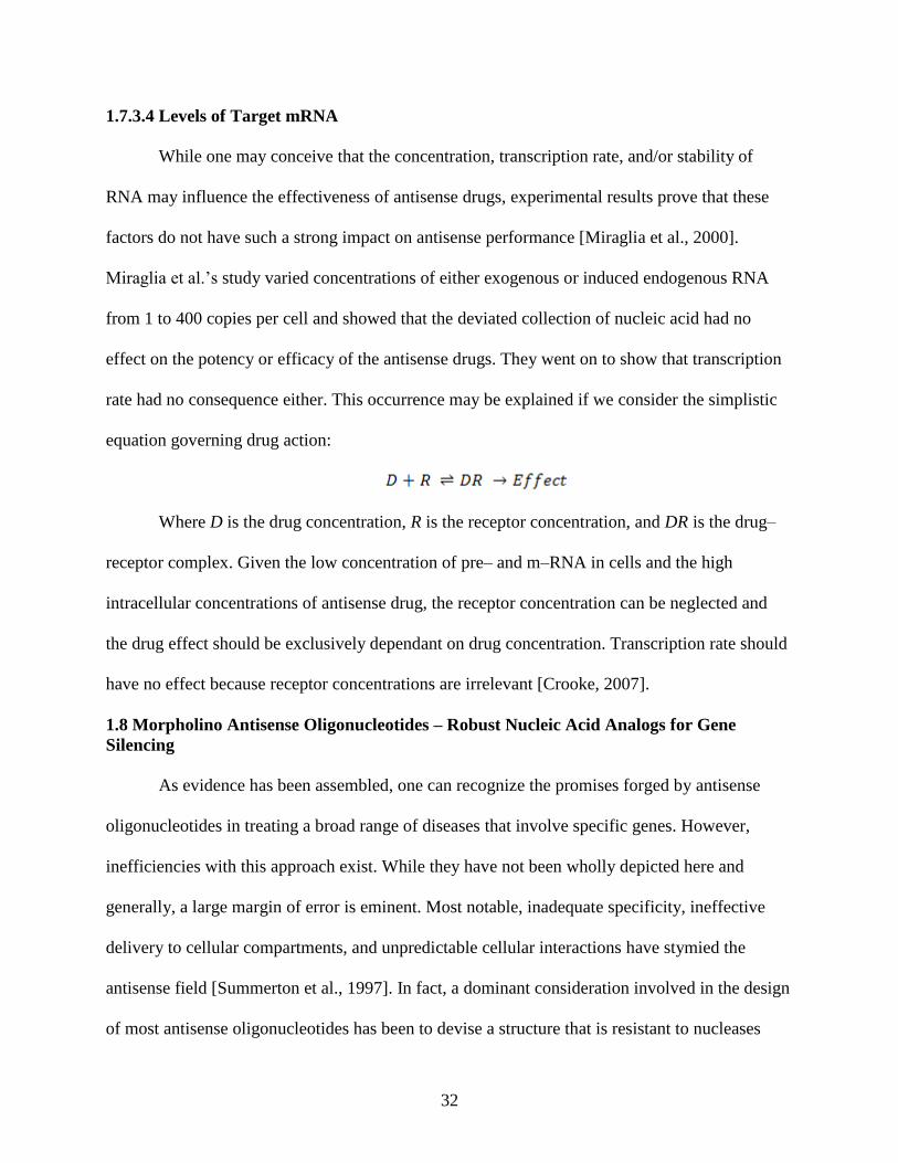
rate had no consequence either. This occurrence may be explained if we consider the simplistic
equation governing drug action:
Where D is the drug concentration, R is the receptor concentration, and DR is the drug–
receptor complex. Given the low concentration of pre– and m–RNA in cells and the high
intracellular concentrations of antisense drug, the receptor concentration can be neglected and
the drug effect should be exclusively dependant on drug concentration. Transcription rate should
have no effect because receptor concentrations are irrelevant [Crooke, 2007].
1.8 Morpholino Antisense Oligonucleotides – Robust Nucleic Acid Analogs for Gene
Silencing
As evidence has been assembled, one can recognize the promises forged by antisense
oligonucleotides in treating a broad range of diseases that involve specific genes. However,
inefficiencies with this approach exist. While they have not been wholly depicted here and
generally, a large margin of error is eminent. Most notable, inadequate specificity, ineffective
delivery to cellular compartments, and unpredictable cellular interactions have stymied the
antisense field [Summerton et al., 1997]. In fact, a dominant consideration involved in the design
of most antisense oligonucleotides has been to devise a structure that is resistant to nucleases
33
while still resembling nucleic acids. In an effort to circumvent these dilemmas, multiple
generations of antisense oligonucleotides have been generated, many of which aid, but cannot
fully elude the problematic actions including physiological degradation, off-target effects, and
other functional insufficiencies. Thus far, one of the seemingly most promising members of the
second generation family of synthetic oligonucleotides that avoid these negative effects is the
phosphorodiamidate morpholino oligomers.
The phosphorodiamidate morpholino oligomers (PMO) are comprised of
(dimethylamino) phosphinylideneoxy–linked morpholino backbone moieties, which is where
their informal designation, morpholino, is derived. [Figure 1.11] The morpholino moieties
contain heterocyclic base recognizing subdivisions of DNA (A, C, G, and T) attached to a
substituted morpholine ring system. When linked to each other via the (dimethylamino)
phosphinylideneoxy function, the employable group formed by intersubunit linkage is commonly
referred to as a phosphorodiamidate [Crooke, 2007].
As previously mentioned, morpholinos offer beneficial features more explicitly stated as;
the characteristic that PMOs are highly resistant to degradation [Hudziak et al., 1996], PMOs
mechanism of action eschews oligomers serving as cofactors for enzymatic cleavage of RNA,
thus they appropriate an RNase H free pathway [Giles et al., 1993, Stein et al., 1997],
morpholinos refrain from forming G–quartet structures (tetrameric complexes mentioned earlier)
capable of off target gene regulation [Burgess et al., 1993, Hudziak et al., 2000], and PMOs do
not bind to cellular and extracellular proteins [Stein et al., 1993, Stein, 1994, Shoeman et al.,
1997].
The lack of iterated charge appears to eliminate non–targeted binding to cellular
components other than RNA. The limited (negligible) coupling with proteins and weak

34
interactions with cell membranes imply that PMOs available for hybridization with RNA will be
minimally competed for by nonspecific binding [Crooke, 2007]. This phenomenon forms the
basis for the hypothesis that the sequence–dependent pharmacokinetics for PMOs is unique
comparatively to the ionic oligomer chemistries [Arora et al., 2004].
Figure 1.11 – Morpholino Oligonucleotide Structure
35
Thus far, we have established a platform that has combined the subject material of caged
compounds and antisense oligonucleotides with particular emphasis on the nitrobenzyl labeling
compound and morpholino oligonucleotides. This data presented has served to embody the
current progression in the fields of both photoactivatable compounds and antisense nucleic acids.
With this information, we are now set to explain the thesis endeavor which is to manipulate the
morpholino oligonucleotide, an entity that is distinctively designed to resist any sort of reaction
other than with its target, to foster attachment or reaction of a caging “silencing” compound, and
acquire unidirectional control over the morpholino oligonucleotide’s cellular activity.
Minor data is available about successfully caging PMOs. As previously introduced, Chen
and colleagues used the tethering of a complementary strand which acted to impede activity and
was subsequently removed with precisely controlled wavelength light. This approach, while very
effective, is impractical for interlab usage by cause of the complex synthesis demanded.
Herein, we describe a statistical base-caging approach that effectively inhibits
morpholino hybridization when the photosensitive labeling compound is coupled. Exposure to
near-UV wavelengths of energy abolishes the coupling and restores hybridization activity. The
encouragement that this approach offers is ease of synthesis, in that, commercially available
PMOs can be inserted into the protocol and successfully altered in a batch style process, as
opposed to the complexities offered in synthesis by the Chen group where solid phase synthesis
is required for construction of the inhibitory sequence.
1.9 Bioconjugate Techniques – Chemical Coupling to Nucleic Acids
Bioconjugate techniques integrating nucleic acids have become one of the most eminent
areas of crosslinking and modification chemistry. Without delving exhaustively into the field of
chemistry and structure of nucleic acids and oligonucleotides, highlighted are the critical
36
functionalities necessary for transition to the appropriated chemical synthesis method employed
in this study, namely, the lack of the existing phosphate backbone (found on DNA and absent on
the PMO) which lead to alternative propositions.
1.9.1 Zero Length Cross Linkers
The smallest available reagent sets for bioconjugation are termed zero length cross
linkers, which mediate the combination of two molecules by forming a covalent bond containing
no additional atoms.
1.9.2 Carbodiimides
Carbodiimides are used to mediate the formation of amide linkages between carboxylates
and amines or phosphoramidate linkages between phosphates and amines [Hoare and Koshland,
1966, Chu et al., 1986, Ghosh et al., 1990]. Carbodiimides are seemingly, the most accepted type
of zero length cross linking agent in use being efficient in forming conjugates between two
proteins, peptides and proteins, oligonucleotides and proteins, biomolecules and functionalized
surfaces or particles, or any combination of these with small molecules [Bioconjugate
Techniques 2nd
Edition – find actual reference]. These compounds can be divided into two
subsets: water soluble and water insoluble. For most bioconjugating applications, the water
soluble carbodiimides find precedence, because most biological macromolecules are soluble in
aqueous buffer solutions. Water insoluble carbodiimides, in contrast, are frequently exploited in
peptide synthesis and other affixing reactions involving molecules soluble in organic solvents.
1.9.2.1 EDC
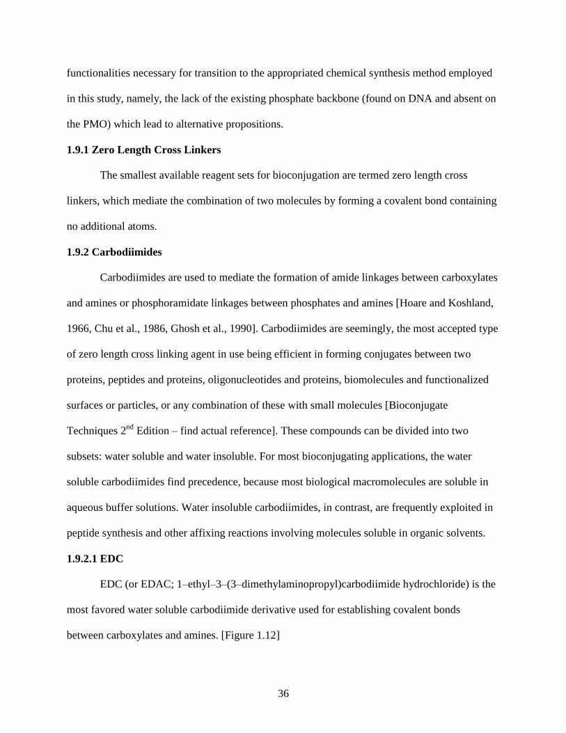
EDC (or EDAC; 1–ethyl–3–(3–dimethylaminopropyl)carbodiimide hydrochloride) is the
most favored water soluble carbodiimide derivative used for establishing covalent bonds
between carboxylates and amines. [Figure 1.12]
37
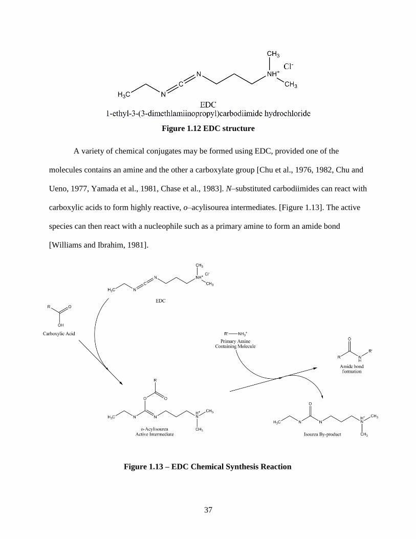
A variety of chemical conjugates may be formed using EDC, provided one of the
molecules contains an amine and the other a carboxylate group [Chu et al., 1976, 1982, Chu and
Ueno, 1977, Yamada et al., 1981, Chase et al., 1983]. N–substituted carbodiimides can react with
carboxylic acids to form highly reactive, o–acylisourea intermediates. [Figure 1.13]. The active
species can then react with a nucleophile such as a primary amine to form an amide bond
[Williams and Ibrahim, 1981].
1.1.1.1
1.1.1.2
1.1.1.3
1.1.1.4
Figure 1.12 EDC structure
Figure 1.13 – EDC Chemical Synthesis Reaction
38
1.9.2.2 Sulfo–NHS
Sulfo–NHS may be used in combination with carbodiimides to form active ester
functionalities with carboxylate groups. Sulfo–NHS esters are hydrophilic reactive groups that
couple rapidly with amines on target molecules [Staros, 1982; Denney and Blobel, 1984; Kotite
et al., 1984]. The advantage of adding sulfo–NHS to carbodiimide reactions is to increase the
solubility and stability of the active intermediate, which reacts with the attacking amine
[Bioconjugate Techniques 2nd
Edition]. Sulfo coupled reactions are highly efficient and usually
increase the yield of conjugation significantly over that obtained solely with the carbodiimide
[Staros et al., 1986].
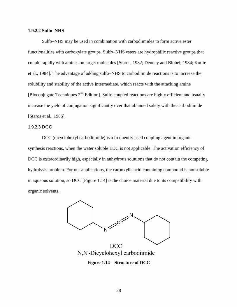
1.9.2.3 DCC
DCC (dicyclohexyl carbodiimide) is a frequently used coupling agent in organic
synthesis reactions, when the water soluble EDC is not applicable. The activation efficiency of
DCC is extraordinarily high, especially in anhydrous solutions that do not contain the competing
hydrolysis problem. For our applications, the carboxylic acid containing compound is nonsoluble
in aqueous solution, so DCC [Figure 1.14] is the choice material due to its compatibility with
organic solvents.
Figure 1.14 – Structure of DCC
39
1.10 In vivo Zebrafish Model
In vitro analyses are critical for any drug characterization scheme, but where the
technology really finds applicability and true test of efficacy is in the in vivo system. We selected
the organism Danio rerio, commonly called zebrafish, as our in vivo system. The zebrafish was
chosen for reasons including ease of maintaining whole populations, its genome is completely
sequenced and available, and that it is typically regarded as the model system to investigate
fundamental principles of developmental biology and genetics [Lieschke et al., 2007]. However,
of particular interest is the optical clarity of this organism throughout its lifetime from embryo to
adult. Serving as a model organism, we chose an easily verifiable biological endpoint displayed
by this organism, particularly, the nicotine induced behavior response. This behavior response
stems from the binding of nicotine to nicotinic acetylcholine receptors (nAChRs) in the
developing embryo. Nicotine and acetylcholine are both agonists for this family of receptors and
both of these chemicals modulate embryonic motor output in acute exposure paradigms [Thomas
et al., 2009]. The nAChRs are ligand–gated pentameric ion channels which have been well–
characterized [Purves et al., 2008]. Particular interest is vested in the α2 subunit, as it plays a
crucial role in functionality pertaining to nicotine response [Zirger et al., 2003]. The presence of
α2 containing receptors allows nicotine to trigger channel opening, allowing sodium, calcium,
and potassium flux, leading to rapid cell depolarization, and resulting in the firing of an action
potential. The gross effect of acute exposure to nicotine results in increased muscular bend rates
in zebrafish embryos which can be readily quantified. Incorporation of α2 splice blocking
morpholinos in zebrafish embryos inhibits proper protein production and thus receptor function,
resulting in no behavior response to nicotine exposure. Using our caging chemistry, we
40
synthesize caged α2 splice blocking morpholino oligonucleotides and utilize this biological
endpoint for in vivo testing.
Caged morpholinos could provide the means to stop functional protein production in
various stages of zebrafish embryonic development, a goal currently unattainable due to the
requirement of delivering these antisense agents via microinjection at the 1–2 cell stage. In the
case of studying genes such as the nAChRs that are directly involved in developmental
processes, knockdown at these early stages could disrupt the biology to the point that endpoint
analyses become impossible. A caged morpholino injected at the 1–2 cell stage could remain
inactive and be carried through cell divisions such that when photoactivated at later times (and
even specific locations, the biological response to gene suppression could be more accurately
studied. In this study, we seek to demonstrate such control over commonly used morpholinos by
caging them and subsequently photoactivating them to control hybridization and thus gene
silencing in developing zebrafish embryos.
1.11 References
1. Adams, S. R., J. P. Y. Kao, et al. (1988). "BIOLOGICALLY USEFUL CHELATORS
THAT RELEASE CA-2+ UPON ILLUMINATION." Journal of the American Chemical
Society 110(10): 3212-3220.
2. Ando, H., T. Furuta, et al. (2004). Photo-mediated gene activation by using caged mRNA
in zebrafish embryos. Zebrafish:2nd Edition Genetics Genomics and Informatics. 77:
159-+.
3. Aujard, I., C. Benbrahim, et al. (2006). "o-Nitrobenzyl photolabile protecting groups with
red-shifted absorption: Syntheses and uncaging cross-sections for one- and two-photon
excitation." Chemistry-a European Journal 12(26): 6865-6879.
4. Baker, B. F., L. Miraglia, et al. (1992). "MODULATION OF EUKARYOTIC
INITIATION FACTOR-4E BINDING TO 5'-CAPPED OLIGORIBONUCLEOTIDES
BY MODIFIED ANTISENSE OLIGONUCLEOTIDES." Journal of Biological
Chemistry 267(16): 11495-11499.
41
5. Black, D. L. (2003). "Mechanisms of alternative pre-messenger RNA splicing." Annual
Review of Biochemistry 72: 291-336.
6. Bley, F., K. Schaper, et al. (2008). "Photoprocesses of molecules with 2-nitrobenzyl
protecting groups and caged organic acids." Photochemistry and Photobiology 84(1):
162-171.
7. Blidner, R. A., K. R. Svoboda, et al. (2008). "Photoinduced RNA interference using
DMNPE-caged 2 '-deoxy-2 '-fluoro substituted nucleic acids in vitro and in vivo."
Molecular Biosystems 4(5): 431-440.
8. Branch, A. D. and H. D. Robertson (1991). "EFFICIENT TRANS-CLEAVAGE AND A
COMMON STRUCTURAL MOTIF FOR THE RIBOZYMES OF THE HUMAN
HEPATITIS-DELTA AGENT." Proceedings of the National Academy of Sciences of the
United States of America 88(22): 10163-10167.
9. Breslauer, K. J., R. Frank, et al. (1986). "PREDICTING DNA DUPLEX STABILITY
FROM THE BASE SEQUENCE." Proceedings of the National Academy of Sciences of
the United States of America 83(11): 3746-3750.
10. Burgess, T. L., E. F. Fisher, et al. (1995). "The antiproliferative activity of c-myb and c-
myc antisense oligonucleotides in smooth muscle cells is caused by a nonantisense
mechanism." Proceedings of the National Academy of Sciences of the United States of
America 92(9): 4051-4055.
11. Buzayan, J. M., W. L. Gerlach, et al. (1986). "NONENZYMATIC CLEAVAGE AND
LIGATION OF RNAS COMPLEMENTARY TO A PLANT-VIRUS SATELLITE
RNA." Nature 323(6086): 349-353.
12. Casey, J. P., R. A. Blidner, et al. (2009). "Caged siRNAs for Spatiotemporal Control of
Gene Silencing." Molecular Pharmaceutics 6(3): 669-685.
13. Chan, J. H. P., S. H. Lim, et al. (2006). "Antisense oligonucleotides: From design to
therapeutic application." Clinical and Experimental Pharmacology and Physiology 33(5-
6): 533-540.
14. Chaulk, S. G. and A. M. MacMillan (1998). "Caged RNA: photo-control of a ribozyme
reaction." Nucleic Acids Research 26(13): 3173-3178.
15. Chu, F. S. and I. Ueno (1977). "PRODUCTION OF ANTIBODY AGAINST
AFLATOXIN-B1." Applied and Environmental Microbiology 33(5): 1125-1128.
16. Crooke, S. T. (1999). "Molecular mechanisms of action of antisense drugs." Biochimica
Et Biophysica Acta-Gene Structure and Expression 1489(1): 31-44.
42
17. Crooke, S. T. (2008). Antisense Drug Technology: Principles, Strategies, and
Applications. Boca Ranton, CRC Press Taylor & Francis Group.
18. Crooke, S. T., K. M. Lemonidis, et al. (1995). "KINETIC CHARACTERISTICS OF
ESCHERICHIA-COLI RNASE H1 - CLEAVAGE OF VARIOUS ANTISENSE
OLIGONUCLEOTIDE-RNA DUPLEXES." Biochemical Journal 312: 599-608.
19. Curley, K. and D. S. Lawrence (1999). "Light-activated proteins." Current Opinion in
Chemical Biology 3(1): 84-88.
20. Denisov, A. Y., A. M. Noronha, et al. (2001). "Solution structure of an arabinonucleic
acid (ANA)/RNA duplex in a chimeric hairpin: comparison with 2 '-fluoro-ANA/RNA
and DNA/RNA hybrids." Nucleic Acids Research 29(21): 4284-4293.
21. Denny, J. B. and G. Blobel (1984). "I-125-LABELED CROSSLINKING REAGENT
THAT IS HYDROPHILIC, PHOTOACTIVATABLE, AND CLEAVABLE THROUGH
AN AZO LINKAGE." Proceedings of the National Academy of Sciences of the United
States of America-Biological Sciences 81(17): 5286-5290.
22. Ellis-Davies, G. C. R. (2007). "Caged compounds: photorelease technology for control of
cellular chemistry and physiology." Nature Methods 4(8): 619-628.
23. Engels, J. and E. J. Schlaeger (1977). "SYNTHESIS, STRUCTURE, AND
REACTIVITY OF ADENOSINE CYCLIC 3',5'-PHOSPHATE BENZYL TRIESTERS."
Journal of Medicinal Chemistry 20(7): 907-911.
24. Freier, S. M., R. Kierzek, et al. (1986). "IMPROVED FREE-ENERGY PARAMETERS
FOR PREDICTIONS OF RNA DUPLEX STABILITY." Proceedings of the National
Academy of Sciences of the United States of America 83(24): 9373-9377.
25. Friedman, D. I., M. J. Imperiale, et al. (1987). "RNA 3' END FORMATION IN THE
CONTROL OF GENE-EXPRESSION." Annual Review of Genetics 21: 453-488.
26. Friedman Adam & Norbert Perrimon. (2006). “A functional RNAi screen for regulators
of receptor tyrosine kinase and ERK signalling." Nature 444, 230-234
27. J Forman, M Dietrich, WT Monroe. (2007) Photobiological and thermal effects of
photoactivating UVA light doses on cell culture. Photochemical & Photobiological
Sciences. 6: 649-658
28. Gee, K. R., L. Niu, et al. (1995). "CAGED BIOACTIVE CARBOXYLATES -
SYNTHESIS, PHOTOLYSIS STUDIES, AND BIOLOGICAL CHARACTERIZATION
OF A NEW CAGED N-METHYL-D-ASPARTIC ACID." Journal of Organic Chemistry
60(13): 4260-4263.
43
29. Gee, K. R., L. Niu, et al. (1999). "Synthesis and photochemistry of a photolabile
precursor of N-methyl-D-aspartate (NMDA) that is photolyzed in the microsecond time
region and is suitable for chemical kinetic investigations of the NMDA receptor."
Biochemistry 38(10): 3140-3147.
30. Gehring, K., J. L. Leroy, et al. (1993). "A TETRAMERIC DNA-STRUCTURE WITH
PROTONATED CYTOSINE.CYTOSINE BASE-PAIRS." Nature 363(6429): 561-565.
31. Giles, R. V., D. G. Spiller, et al. (1993). "CHIMERIC OLIGODEOXYNUCLEOTIDE
ANALOGS - ENHANCED CELL UPTAKE OF STRUCTURES WHICH DIRECT
RIBONUCLEASE-H WITH HIGH SPECIFICITY." Anti-Cancer Drug Design 8(1): 33-
51.
32. Gillespi.D and Spiegelm.S (1965). "A QUANTITATIVE ASSAY FOR DNA-RNA
HYBRIDS WITH DNA IMMOBILIZED ON A MEMBRANE." Journal of Molecular
Biology 12(3): 829-&.
33. Grewer, C., S. Abdollah, et al. (2001). "Substrate translocation kinetics of excitatory
amino acid carrier 1 probed with laser-pulse photolysis of a new photolabile precursor of
D-aspartic acid." Biochemistry 40(1): 232-240.
34. Grewer, C., J. Jager, et al. (2000). "A new photolabile precursor of glycine with improved
properties: A tool for chemical kinetic investigations of the glycine receptor."
Biochemistry 39(8): 2063-2070.
35. Guo, H. C. T. and R. A. Collins (1995). "EFFICIENT TRANS-CLEAVAGE OF A
STEM-LOOP RNA SUBSTRATE BY A RIBOZYME DERIVED FROM
NEUROSPORA VS RNA." Embo Journal 14(2): 368-376.
36. Hahn, M. E. and T. W. Muir (2004). "Photocontrol of Smad2, a multiphosphorylated
cell-signaling protein, through caging of activating phosphoserines." Angewandte
Chemie-International Edition 43(43): 5800-5803.
37. Hoare, D. G. and D. E. Koshland (1966). "A PROCEDURE FOR SELECTIVE
MODIFICATION OF CARBOXYL GROUPS IN PROTEINS." Journal of the American
Chemical Society 88(9): 2057-&.
38. Hudziak, R. M., E. Barofsky, et al. (1996). "Resistance of morpholino
phosphorodiamidate oligomers to enzymatic degradation." Antisense & Nucleic Acid
Drug Development 6(4): 267-272.
39. Hudziak, R. M., J. Summerton, et al. (2000). "Antiproliferative effects of steric blocking
phosphorodiamidate morpholino antisense agents directed against c-myc." Antisense &
Nucleic Acid Drug Development 10(3): 163-176.
44
40. Il'ichev, Y. V., M. A. Schworer, et al. (2004). "Photochemical reaction mechanisms of 2-
nitrobenzyl compounds: Methyl ethers and caged ATP." Journal of the American
Chemical Society 126(14): 4581-4595.
41. Kaplan, J. H., B. Forbush, et al. (1978). "RAPID PHOTOLYTIC RELEASE OF
ADENOSINE 5'-TRIPHOSPHATE FROM A PROTECTED ANALOG -
UTILIZATION BY NA-K PUMP OF HUMAN RED BLOOD-CELL GHOSTS."
Biochemistry 17(10): 1929-1935.
42. Kotite, N. J., J. V. Staros, et al. (1984). "INTERACTION OF SPECIFIC PLATELET
MEMBRANE-PROTEINS WITH COLLAGEN - EVIDENCE FROM CHEMICAL
CROSS-LINKING." Biochemistry 23(13): 3099-3104.
43. Leroy, J. L., K. Gehring, et al. (1993). "ACID MULTIMERS OF
OLIGODEOXYCYTIDINE STRANDS - STOICHIOMETRY, BASE-PAIR
CHARACTERIZATION, AND PROTON-EXCHANGE PROPERTIES." Biochemistry
32(23): 6019-6031.
44. Manley, J. L. (1988). "POLYADENYLATION OF MESSENGER-RNA
PRECURSORS." Biochimica Et Biophysica Acta 950(1): 1-12.
45. Matsuzaki, M., G. C. R. Ellis-Davies, et al. (2008). "Three-Dimensional Mapping of
Unitary Synaptic Connections by Two-Photon Macro Photolysis of Caged Glutamate." J
Neurophysiol 99(3): 1535-1544.
46. Mayer, G. and A. Heckel (2006). "Biologically active molecules with a "light switch"."
Angewandte Chemie-International Edition 45(30): 4900-4921.
47. Mendel, D., J. A. Ellman, et al. (1991). "CONSTRUCTION OF A LIGHT-ACTIVATED
PROTEIN BY UNNATURAL AMINO-ACID MUTAGENESIS." Journal of the
American Chemical Society 113(7): 2758-2760.
48. Mikat, V. and A. Heckel (2007). "Light-dependent RNA interference with nucleobase-
caged siRNAs." Rna-a Publication of the Rna Society 13(12): 2341-2347.
49. Miraglia, L., A. T. Watt, et al. (2000). "Variations in mRNA content have no effect on
the potency of antisense oligonucleotides." Antisense & Nucleic Acid Drug Development
10(6): 453-461.
50. Mizumoto, K. and Y. Kaziro (1987). "MESSENGER-RNA CAPPING ENZYMES
FROM EUKARYOTIC CELLS." Progress in Nucleic Acid Research and Molecular
Biology 34: 1-28.
51. Monroe, W. T., M. M. McQuain, et al. (1999). "Targeting expression with light using
caged DNA." Journal of Biological Chemistry 274(30): 20895-20900.
45
52. Muralidharan, V. and T. W. Muir (2006). "Protein ligation: an enabling technology for
the biophysical analysis of proteins." Nature Methods 3(6): 429-438.
53. Niu, L., K. R. Gee, et al. (1996). "Synthesis and photochemical properties of a kainate
precursor and activation of kainate and AMPA receptor channels on a microsecond time
scale." Biochemistry 35(6): 2030-2036.
54. Okamoto, A., K. Tanabe, et al. (2003). "Phototriggered drug release from functionalized
oligonucleotides by a molecular beacon strategy." Angewandte Chemie-International
Edition 42(22): 2502-2504.
55. Ouyang, X. H., I. A. Shestopalov, et al. (2009). "Versatile Synthesis and Rational Design
of Caged Morpholinos." Journal of the American Chemical Society 131(37): 13255-
13269.
56. Peebles, C. L., P. S. Perlman, et al. (1986). "A SELF-SPLICING RNA EXCISES AN
INTRON LARIAT." Cell 44(2): 213-223.
57. Pelliccioli, A. P. and J. Wirz (2002). "Photoremovable protecting groups: reaction
mechanisms and applications." Photochemical & Photobiological Sciences 1(7): 441-458.
58. Petersson, E. J., G. S. Brandt, et al. (2003). Caging proteins through unnatural amino acid
mutagenesis. Biophotonics, Pt A. San Diego, Academic Press Inc. 360: 258-273.
59. Rapp, G. and K. Guth (1988). "A LOW-COST HIGH-INTENSITY FLASH DEVICE
FOR PHOTOLYSIS EXPERIMENTS." Pflugers Archiv-European Journal of Physiology
411(2): 200-203.
60. Ross, J. (1988). "MESSENGER-RNA TURNOVER IN EUKARYOTIC CELLS."
Molecular Biology & Medicine 5(1): 1-14.
61. Saunders, L. R. and G. N. Barber (2003). "The dsRNA binding protein family: critical
roles, diverse cellular functions." Faseb Journal 17(9): 961-983.
62. Saxena, S. K. and E. J. Ackerman (1990). "MICROINJECTED OLIGONUCLEOTIDES
COMPLEMENTARY TO THE ALPHA-SARCIN LOOP OF 28-S RNA ABOLISH
PROTEIN-SYNTHESIS IN XENOPUS OOCYTES." Journal of Biological Chemistry
265(6): 3263-3269.
63. Sazani, P. and R. Kole (2003). "Therapeutic potential of antisense oligonucleotides as
modulators of alternative splicing." Journal of Clinical Investigation 112(4): 481-486.
64. Schaper, K., D. Dommaschke, et al. (2000). "AM1 calculations on the mechanism of the
o-nitrobenzyl photochemistry." Journal of Information Recording 25(3-4): 339-354.
46
65. Schaper, K., S. A. M. Mobarekeh, et al. (2002). "Synthesis and photophysical
characterization of a new, highly hydrophilic caging group." European Journal of Organic
Chemistry(6): 1037-1046.
66. Schupp, H., W. K. Wong, et al. (1987). "MECHANISTIC STUDIES OF THE
PHOTOREARRANGEMENT OF ORTHO-NITROBENZYL ESTERS." Journal of
Photochemistry 36(1): 85-97.
67. Shah, S., S. Rangarajan, et al. (2005). "Light activated RNA interference." Angewandte
Chemie-International Edition 44(9): 1328-1332.
68. Shestopalov, I. A. and J. K. Chen (2008). "Chemical technologies for probing embryonic
development." Chemical Society Reviews 37(7): 1294-1307.
69. Shestopalov, I. A., S. Sinha, et al. (2007). "Light-controlled gene silencing in zebrafish
embryos." Nature Chemical Biology 3(10): 650-651.
70. Shoeman, R. L., R. Hartig, et al. (1997). "Fluorescence microscopic comparison of the
binding of phosphodiester and phosphorothioate (antisense) oligodeoxyribonucleotides to
subcellular structures, including intermediate filaments, the endoplasmic reticulum, and
the nuclear interior." Antisense & Nucleic Acid Drug Development 7(4): 291-308.
71. Staros, J. V. (1982). "N-HYDROXYSULFOSUCCINIMIDE ACTIVE ESTERS -
BIS(N-HYDROXYSULFOSUCCINIMIDE) ESTERS OF 2 DICARBOXYLIC-ACIDS
ARE HYDROPHILIC, MEMBRANE-IMPERMEANT, PROTEIN CROSS-LINKERS."
Biochemistry 21(17): 3950-3955.
72. Staros, J. V., R. W. Wright, et al. (1986). "ENHANCEMENT BY N-
HYDROXYSULFOSUCCINIMIDE OF WATER-SOLUBLE CARBODIIMIDE-
MEDIATED COUPLING REACTIONS." Analytical Biochemistry 156(1): 220-222.
73. Stein, C. A. (1995). "DOES ANTISENSE EXIST." Nature Medicine 1(11): 1119-1121.
74. Stein, C. A. and Y. C. Cheng (1993). "ANTISENSE OLIGONUCLEOTIDES AS
THERAPEUTIC AGENTS - IS THE BULLET REALLY MAGICAL." Science
261(5124): 1004-1012.
75. Stein, D., E. Foster, et al. (1997). "A specificity comparison of four antisense types:
Morpholino, 2'-O-methyl RNA, DNA, and phosphorothioate DNA." Antisense & Nucleic
Acid Drug Development 7(3): 151-157.
76. Stein, H. and P. Hausen (1969). "ENZYME FROM CALF THYMUS DEGRADING
RNA MOIETY OF DNA-RNA HYBRIDS - EFFECT ON DNA-DEPENDENT RNA
POLYMERASE." Science 166(3903): 393-&.
47
77. Summerton, J., D. Stein, et al. (1997). "Morpholino and phosphorothioate antisense
oligomers compared in cell-free and in-cell systems." Antisense & Nucleic Acid Drug
Development 7(2): 63-70.
78. Summerton, J. and D. Weller (1997). "Morpholino antisense oligomers: Design,
preparation, and properties." Antisense & Nucleic Acid Drug Development 7(3): 187-
195.
79. Tang, X. J. and I. J. Dmochowski (2004). "Synthesis of photoactivatable antisense
oligonucleotides." Abstracts of Papers of the American Chemical Society 228: 040-
BIOL.
80. Thompson, J. D. and D. Gillespie (1990). "CURRENT CONCEPTS IN
QUANTITATIVE MOLECULAR HYBRIDIZATION." Clinical Biochemistry 23(4):
261-266.
81. Tuerk, C. and L. Gold (1990). "SYSTEMATIC EVOLUTION OF LIGANDS BY
EXPONENTIAL ENRICHMENT - RNA LIGANDS TO BACTERIOPHAGE-T4 DNA-
POLYMERASE." Science 249(4968): 505-510.
82. Tyagi, S. and F. R. Kramer (1996). "Molecular beacons: Probes that fluoresce upon
hybridization." Nature Biotechnology 14(3): 303-308.
83. Uhlenbeck, O. C. (1987). "A SMALL CATALYTIC OLIGORIBONUCLEOTIDE."
Nature 328(6131): 596-600.
84. Vickers, T. A., J. R. Wyatt, et al. (2001). "Fully modified 2 ' MOE oligonucleotides
redirect polyadenylation." Nucleic Acids Research 29(6): 1293-1299.
85. Vikram, A., R. D. Gayathri, et al. (2004). "Neutrally Charged Phosphorodiamidate
Morpholino Antisense Oligomers: Uptake, Efficacy and Pharmacokinetics." Current
Pharmaceutical Biotechnology 5: 431-439.
86. Wang, K. Y., S. McCurdy, et al. (1993). "A DNA APTAMER WHICH BINDS TO AND
INHIBITS THROMBIN EXHIBITS A NEW STRUCTURAL MOTIF FOR DNA."
Biochemistry 32(8): 1899-1904.
87. Watson, J. D. and F. H. C. Crick (1953). "GENETICAL IMPLICATIONS OF THE
STRUCTURE OF DEOXYRIBONUCLEIC ACID." Nature 171(4361): 964-967.
88. Westermann, P., B. Gross, et al. (1989). "INHIBITION OF EXPRESSION OF SV40
VIRUS LARGE T-ANTIGEN BY ANTISENSE
OLIGODEOXYRIBONUCLEOTIDES." Biomedica Biochimica Acta 48(1): 85-93.
89. Williams, A. and I. T. Ibrahim (1981). "CARBODIIMIDE CHEMISTRY - RECENT
ADVANCES." Chemical Reviews 81(6): 589-636.
48
90. Wu, H. J., W. F. Lima, et al. (2004). "Determination of the role of the human RNase H1
in the pharmacology of DNA-like antisense drugs." Journal of Biological Chemistry
279(17): 17181-17189.
91. Wyatt, J. R., T. A. Vickers, et al. (1994). "COMBINATORIALLY SELECTED
GUANOSINE-QUARTET STRUCTURE IS A POTENT INHIBITOR OF HUMAN-
IMMUNODEFICIENCY-VIRUS ENVELOPE-MEDIATED CELL-FUSION."
Proceedings of the National Academy of Sciences of the United States of America 91(4):
1356-1360.
92. Yamada, H., T. Imoto, et al. (1981). "SELECTIVE MODIFICATION OF ASPARTIC
ACID-101 IN LYSOZYME BY CARBODIIMIDE REACTION." Biochemistry 20(17):
4836-4842.
93. Young, D. D., H. Lusic, et al. (2008). "Gene Silencing in Mammalian Cells with Light-
Activated Antisense Agents." Chembiochem 9(18): 2937-2940.
94. Zamecnik, P. C. and M. L. Stephenson (1978). "INHIBITION OF ROUS-SARCOMA
VIRUS-REPLICATION AND CELL TRANSFORMATION BY A SPECIFIC
OLIGODEOXYNUCLEOTIDE." Proceedings of the National Academy of Sciences of
the United States of America 75(1): 280-284.
95. Zhang, G. S., S. W. Li, et al. (1986). "PRODUCTION AND CHARACTERIZATION OF
ANTIBODY AGAINST DEOXYNIVALENOL TRIACETATE." Journal of Food
Protection 49(5): 336-339.
49
CHAPTER 2: SYNTHESIS AND ANALYSIS OF BASE-CAGED
MORPHOLINO OLIGONUCLEOTIDES
2.1 Introduction
A remaining challenge to developmental biology is the spatiotemporal study of genes with
regard to cell proliferation, migration, and differentiation [Ouyang et al., 2009]. Aside from
commonly employed biological approaches, a class of chemical compounds coined “caged”
molecules are also used as tools to aid in unveiling the mysteries of cellular activity. These
compounds functionally encapsulate biomolecules and render them inactive. The inactivated, or
silenced, biomolecules can later be liberated by photomanipulation of a specific wavelength,
characteristic to the chromophore, which restores activity and allows perturbation of the targeted
bioprocess [Ellis-Davies, 2007]. This externally triggered chemical approach to studying and
regulating biological function has quickly garnered research interest due to its invaluable
application in both laboratory and potentially, clinical settings. Effective alteration of gene
activity at precise times and locations is especially attractive for delineation of gene function in
whole organisms, and where caging technology is being increasingly applied. Antisense
technology is the study of how these exogenous nucleic acid oligonucleotides alter normal
biological function of the organisms with which they interact [Crooke, 2007]. The receptors for
these drugs are defined nucleic acid sequences in an intracellular DNA or RNA target. Although
antisense molecules are the subject of expansive studies in developmental biology and medicine,
inadequate stability and specificity, off-target effects, susceptibility to enzymatic degradation,
ineffective delivery to cellular compartments, and unpredictable cellular interactions have
previously stymied the antisense field [Summerton et al., 1997]. One of the seemingly most
promising members of the second generation family of synthetic oligonucleotides that surmount
50
these inadequacies is the phosphorodiamidate morpholino oligomers (PMOs) (Figure 2.1),
comprised of (dimethylamino) phosphinylideneoxy–linked morpholino backbone moieties. The
morpholino moieties contain heterocyclic base recognizing subdivisions of DNA (A, C, G, and
T) attached to a substituted morpholine ring system. PMOs offer many beneficial features,
including: a high resistance to enzymatic degradation[Hudziak et al., 1996], an RNAse H free
pathway [Giles et al., 1993, Stein et al., 1997], reduced off-target gene regulation [Burgess et al.,
1993, Hudziak et al., 2000], and an absence of binding affinity to cellular and extracellular
proteins [Stein et al., 1993, Stein, 1994, Shoeman et al., 1997].
We demonstrate a unique manipulation of the morpholino oligonucleotide, an entity that
is distinctively designed to resist any sort of reaction other than with its targeted complementary
nucleic acid, to foster attachment or reaction of a caging “silencing” compound, resulting in
unidirectional control over its gene-regulating activity. Caging biomolecules begins with
synthesis of the coupling compound, followed by attachment of that caging compound to the
biomolecule or molecule of interest. Typically, these bioconjugates are made using synthetic
organic chemistry reactions, ranging from single coupling mechanisms to complex multi-step
reactions [Ellis-Davies, 2007].
Through widespread use, antisense oligonucleotides have evolved through multiple
generations of structural change in an attempt to evade previously listed off target effects
encountered in the cellular environment. As the development of these compounds have removed
many of the previously utilized constructs, notably the phosphate backbone, new tailored
chemistries for targeted attachment of blocking groups to these new constructs have had to be
incorporated. Thus far, the most common method of caging mRNAs or DNAs has proven to be
the most useful biologically [Ellis-Davis, 2007]. “Direct, multi-site,” “shotgun,” or “statistical”
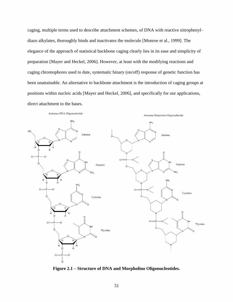
51
caging, multiple terms used to describe attachment schemes, of DNA with reactive nitrophenyl–
diazo alkylates, thoroughly binds and inactivates the molecule [Monroe et al., 1999]. The
elegance of the approach of statistical backbone caging clearly lies in its ease and simplicity of
preparation [Mayer and Heckel, 2006]. However, at least with the modifying reactions and
caging chromophores used to date, systematic binary (on/off) response of genetic function has
been unattainable. An alternative to backbone attachment is the introduction of caging groups at
positions within nucleic acids [Mayer and Heckel, 2006], and specifically for our applications,
direct attachment to the bases.
Figure 2.1 – Structure of DNA and Morpholino Oligonucleotides.
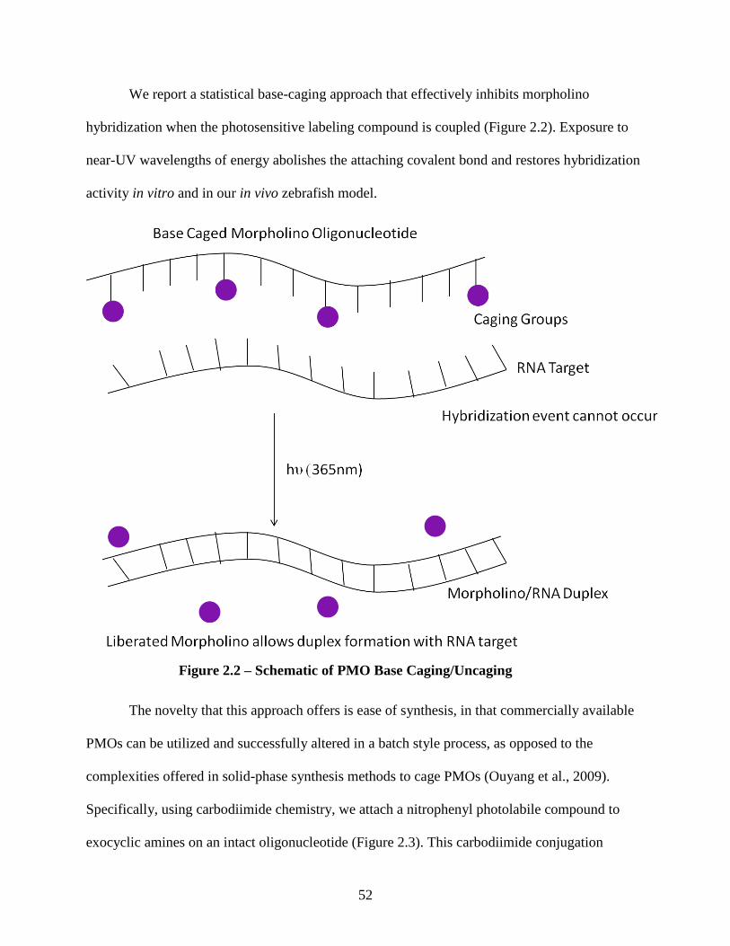
52
We report a statistical base-caging approach that effectively inhibits morpholino
hybridization when the photosensitive labeling compound is coupled (Figure 2.2). Exposure to
near-UV wavelengths of energy abolishes the attaching covalent bond and restores hybridization
activity in vitro and in our in vivo zebrafish model.
The novelty that this approach offers is ease of synthesis, in that commercially available
PMOs can be utilized and successfully altered in a batch style process, as opposed to the
complexities offered in solid-phase synthesis methods to cage PMOs (Ouyang et al., 2009).
Specifically, using carbodiimide chemistry, we attach a nitrophenyl photolabile compound to
exocyclic amines on an intact oligonucleotide (Figure 2.3). This carbodiimide conjugation
Figure 2.2 – Schematic of PMO Base Caging/Uncaging
53
scheme capitalizes on reacting a carboxylic acid caged compound with exocyclic amines in an
intact oligonucleotide, and thus could be amenable to many different forms of nucleic acids
(Figure 2.1). Sampling two structurally distinct oligonucleotides, we demonstrate successful
caging of both DNA and PMOs with our novel chemical caging approach. Our synthetic scheme
for caged PMOs fulfills the design requirements for caged biomolecules as described above. The
use of carbodiimide chemistry for the successful caging of antisense oligonucleotides has
relevance to the fields of caged compounds, antisense technology, gene therapy, and
developmental biology.
In vitro analyses are critical for any drug characterization scheme, but where the
technology really finds applicability and true test of efficacy is in the in vivo system. We selected
the organism Danio rerio, commonly called zebrafish, as our in vivo system. The zebrafish was
chosen for reasons including ease of maintaining whole populations, its genome is completely
sequenced and available, and that it is typically regarded as the model system to investigate
fundamental principles of developmental biology and genetics [Lieschke et al., 2007]. However,
of particular interest is the optical clarity of this organism throughout its lifetime from embryo to
adult. Serving as a model organism, we chose an easily verifiable biological endpoint displayed
by this organism, particularly, the nicotine induced behavior response. This behavior response
stems from the binding of nicotine to nicotinic acetylcholine receptors (nAChRs) in the
developing embryo. Nicotine and acetylcholine are both agonists for this family of receptors and
both of these chemicals modulate embryonic motor output in acute exposure paradigms [Thomas
et al., 2009]. The nAChRs are ligand–gated pentameric ion channels which have been well–
characterized [Purves et al., 2008]. Particular interest is vested in the α2 subunit, as it plays a
crucial role in functionality pertaining to nicotine response [Zirger et al., 2003]. The presence of
54
α2 containing receptors allows nicotine to trigger channel opening, allowing sodium, calcium,
and potassium flux, leading to rapid cell depolarization, and resulting in the firing of an action
potential. The gross effect of acute exposure to nicotine results in increased muscular bend rates
in zebrafish embryos which can be readily quantified. Incorporation of α2 splice blocking
morpholinos in zebrafish embryos inhibits proper protein production and thus receptor function,
resulting in no behavior response to nicotine exposure. Using our caging chemistry, we
synthesize caged α2 splice blocking morpholino oligonucleotides and utilize this biological
endpoint for in vivo testing.
Caged morpholinos could provide the means to stop functional protein production in
various stages of zebrafish embryonic development, a goal currently unattainable due to the
requirement of delivering these antisense agents via microinjection at the 1–2 cell stage. In the
case of studying genes such as the nAChRs that are directly involved in developmental
processes, knockdown at these early stages could disrupt the biology to the point that endpoint
analyses become impossible. A caged morpholino injected at the 1–2 cell stage could remain
inactive and be carried through cell divisions such that when photoactivated at later times (and
even specific locations, the biological response to gene suppression could be more accurately
studied. In this study, we seek to demonstrate such control over commonly used morpholinos by
caging them and subsequently photoactivating them to control hybridization and thus gene
silencing in developing zebrafish embryos.
2.2 Experimental Procedures
2.2.1 General Synthetic Procedures
All reactions were carried out in Seal–Rite® 1.5mL microcentrifuge tubes (USA
Scientific, Ocala, FL) at RTP using commercially available reagents without further purification,
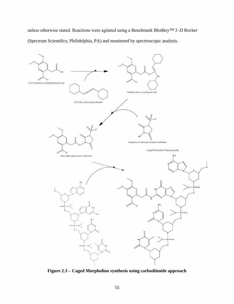
55
unless otherwise stated. Reactions were agitated using a Benchmark BlotBoy™ 3–D Rocker
(Spectrum Scientifics, Philidelphia, PA) and monitored by spectroscopic analysis.
Figure 2.3 – Caged Morpholino synthesis using carbodiimide approach
56
2.2.1.1 DNA/Morpholino Oligonucleotide Conjugation Reaction
50mg of N,N’–Dicyclohexylcarbodiimide (DCC) (Thermo Scientific, Waltham, MA) and
5mg of (4,5–dimethoxy–2–nitrophenyl) acetic acid (DMNPAA) (Alfa Aesar, Ward Hill, MA)
were dissolved into 500µL of dimethylformamide (DMF) (Sigma–Aldrich, St. Louis MO) and
allowed to react at RTP for 15 minutes. After 15 minutes of reaction, 15mg of N–
hydroxysulfosuccinimide (Sulfo–NHS) (Thermo Scientific, Waltham, MA) dissolved in 50µL
distilled water was added to the reaction. The mixture was vortexed for a few seconds to ensure
dispersion. Following vortexing, it was allowed to react for 15 minutes at RTP. After the second
reaction sequence, 30µg of Morpholino oligonucleotide (sequence: 5’– ATG CAA AGT ATC
AAC TTA CCA CAT C –3’, Gene–Tools LLC., Philomath, OR) was added to the vessel and
allowed to react at RTP for 18 hours in dark conditions.
2.2.2 Caged Oligonucleotide Purification Scheme
Sephadex LH–20 was functionalized using water as the functionalizing agent as
described by GE Healthcare, and prepared as a media slurry. The sephadex slurry (4ml) was
added to a PD–10 desalting column (GE Healthcare, Waukesha, WI). For a given reaction, 6
1mL fractions were collected from the Sephadex column. Following fractionation, recovered
product (verified through UV–Vis spectroscopy) was spin filtered using Amicon centrifugal
filters (Amicon Ultra 3K – 0.5mL 3000MWCO Centrifugal Filters) until the eluent showed no
absorbance at 355nm.
2.2.3 Spectroscopic Characterization of Native, Caged, and Flashed Oligonucleotides
DNA oligonucleotides, 4,5–dimethoxy–2–nitrophenyl acetic acid (DMNPAA)
conjugated “caged” DNA oligonucleotides, morpholino oligonucleotides, and DMNPAA
conjugated “caged” morpholino oligonucleotides were dissolved (70ng/µL) in 0.2µm filter
57
purified distilled water (Synergy – Millipore, Billerica, MA) and scanned from 200nm to 500nm
for maximum absorption (ThermoSpectronic Genesys 6 UV–Vis Spectrophotometer). Relative
absorbances at 260nm and 355nm were used to determine the average amount of cage moieties
per oligonucleotide, similar to the approach used by Ghosn et al. (Ghosn et al., 2005)
2.2.4 UVA Irradiation
DMNPAA caged oligonucleotides were exposed to UVA light for 4 minutes in 0.5mL
TempAssure PCR tubes (USA Scientific), where the light source output was located 3cm away
from the sample. The Greenspot, a 100–watt, mercury lamp with a 5mm x 1000mm light guide,
produces a peak spectral output at 365±8 nm (American Ultraviolet, Lebanon, IN) (Forman et
al., 2007). The lamp has a fluence of 206 mW/cm2 with short bandpass (1.5mm thick, 2.4mm
diameter SWP–2502U–400, Lambda Research Optics, CA) and IR filters (818–ST–UV detector,
Newport Corporation, Irvine, CA) in place.
2.2.5 Oligonucleotide In Vitro Hybridization Assay
Hybridization melting curve assays and gel electrophoresis techniques were employed to
demonstrate functional attributes of native, caged, and flashed DNA and morpholino
oligonucleotides. The oligonucleotide samples were designated as native sample (unreacted
DNA or morpholino oligonucleotide, which was never introduced to the reaction conditions and
served as a positive control), caged samples (oligonucleotides containing photolabile blocking
group), and flashed sample (oligonucleotides exposed to 365nm light causing photolysis of the
blocking group), 1µg of each oligonucleotide sample was hybridized with 1µg of its
complementary DNA, along with 15µL of intercalating 100X SYBR Green I (Invitrogen –
SYBR Green 1 nucleic acid gel stain; 10000X concentrate in DMSO), 20uL of 10X
hybridization buffer (1M NaCl, 100mM NaP, 1mM EDTA, pH 7.0 – 7.5), and distilled water
58
was also added to the reaction tubes (Bio–Rad CLR PCR tube, Flat Cap Strips) to bring the final
volume to 100µL. Triplicate samples were first held at 95˚C for 5min in a thermal cycler (Bio–
Rad MJ Mini 48–Well Personal Thermal Cycler)and followed by cooling to 5˚C over 5min.
Oligonucleotide melting curves were generated by measuring the fluorescence of SYBR Green I
during a linear temperature transition from 5˚C to 95˚C at 0.1˚C/s.
2.2.6 Oligonucleotide Gel Electrophoresis
For gel electrophoretic analysis, 500ng of morpholino oligonucleotide hybridized to its
DNA complement was added to each well of a 15% TBE polyacrylamide gel (Bio–Rad) and run
for 90 minutes at 85V. Gels were stained after electrophoresis with 1X SYBR–Gold (Invitrogen
– SYBR Gold nucleic acid gel stain; 10000X concentrate in DMSO) in 1% TBE buffer for 15
minutes. Gel images were captured using Quantity One (Bio–Rad – 1–D Analysis Software).
2.2.7 Morpholino Microinjections
Morpholino solutions were prepared immediately prior to the microinjection procedure. A
control morpholino (Sequence: 5’– CCT CTT ACC TCA GTT ACA ATT TAT A –3’) was
injected at 0.25 mM and α2 splice blocking morpholinos (native, caged, and uncaged in vitro)
were injected at 2mM (~17µg/µL). All morpholino solutions were prepared in 0.1% phenol red
(Sigma) and 125ng/µL rhodamine-conjugated dextran (Sigma, St. Louis, Missouri, USA) to
facilitate confirmation of successful injections. Each solution was loaded into a micropipette
needle pulled on a Flaming/Brown P-97 micropipette puller (Sutter Instruments, Novato, CA)
using fire-polished borosilicate glass capillaries with an internal filament (outer diameter of 1.2
mm, Sutter Instruments). All solutions were then injected in the yolk stream of 1-2 cell stage
zebrafish embryos [Nasevicius et al., 2000]. At ~6 hours post fertilization (hpf), all injected
zebrafish embryos were individually screened for fluorescence on a Zeiss Axiovert inverted
59
microscope equipped with epifluorescence using a 10x objective. Embryos with no fluorescence
were removed and were not used for any subsequent experiments. At 12 hpf, half of embryos
injected with the caged α2 morpholinos were placed in 3 mL of embryo media (15-20 embryos
per petri dish) and were irradiated in vivo with the Green Spot UVA light source, at 24 J/cm2
[Dong et al., 2007]. All zebrafish experiments were performed in the presence of minimal light
to minimize photolysis of caged compounds. Embryos were raised at 28oC and protected from
light until behavioral analysis.
2.2.8 Behavior
Behavioral analysis based on nicotine binding to nAChRs provided a biological endpoint
for assessing functionality of the α2 splice block morpholino and caged α2 morpholino
oligonucleotides. Embryos were placed in embryo media in 10mm Petri dishes and their baseline
spontaneous activity in their chorions in the form of spinal musculature bends was videotaped for
3 minutes using a Kohu video camera mounted to a Zeiss SV6 dissecting microscope. The
embryos were quickly transferred into a nicotine solution (60μM) ((-)-nicotine, catalog # N3876-
5ml, Sigma, St. Louis, Missouri, USA) and the musculature bends were recorded for 1-3
minutes. The motor output was quantified for each individual embryo as the number of bends in
a one-minute epoch. Following behavioral examination embryos were placed in fresh embryo
media and raised at 28oC. All behavior experiments were carried out at ~25-26
oC with minimal
light.
2.2.9 Zebrafish Husbandry, IACUC
Wild-type zebrafish purchased from EkkWill Waterlife resources were kept at standard
laboratory conditions (28°C on a 14 hr light: 10 hr dark photo-period in a recirculating system).
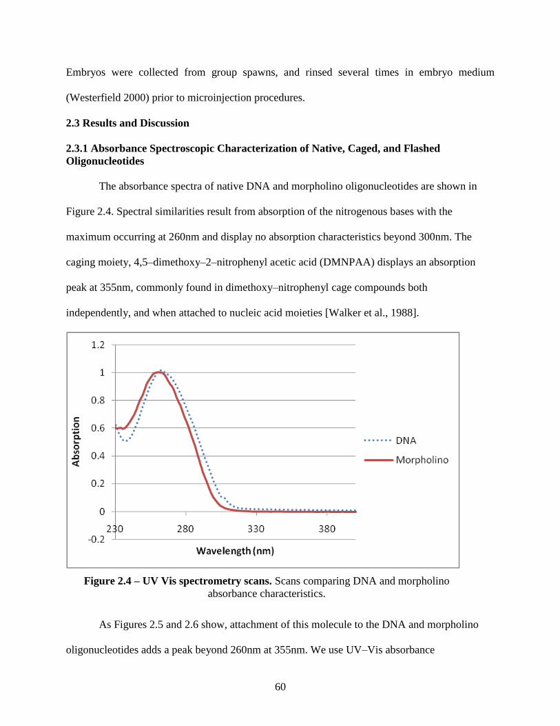
60
Embryos were collected from group spawns, and rinsed several times in embryo medium
(Westerfield 2000) prior to microinjection procedures.
2.3 Results and Discussion
2.3.1 Absorbance Spectroscopic Characterization of Native, Caged, and Flashed
Oligonucleotides
The absorbance spectra of native DNA and morpholino oligonucleotides are shown in
Figure 2.4. Spectral similarities result from absorption of the nitrogenous bases with the
maximum occurring at 260nm and display no absorption characteristics beyond 300nm. The
caging moiety, 4,5–dimethoxy–2–nitrophenyl acetic acid (DMNPAA) displays an absorption
peak at 355nm, commonly found in dimethoxy–nitrophenyl cage compounds both
independently, and when attached to nucleic acid moieties [Walker et al., 1988].
As Figures 2.5 and 2.6 show, attachment of this molecule to the DNA and morpholino
oligonucleotides adds a peak beyond 260nm at 355nm. We use UV–Vis absorbance
Figure 2.4 – UV Vis spectrometry scans. Scans comparing DNA and morpholino
absorbance characteristics.

61
spectroscopy as a quantitative assay to approximate the amount of attachment that can be
achieved through the carbodiimide reaction. By recording the relative peak heights at 260nm,
355nm, and 500nm, percent of attachment can be calculated as previously described [Ghosn et
al., 2005]. Interestingly, when comparing this novel base attachment scheme to backbone caging
methods, knockdown of hybridization can be achieved with as little as 12 - 16% attachment
(corresponding to 3 – 4 caged groups per 25mer oligo) to the required 70 – 80% [Ghosn et al.,
2005] (corresponding to 14 – 16 caged groups per 20mer oligo), indicating a more efficient
disruption of hybridization by base caging as compared to phosphate caging. Importantly, this
assay is used as a quantitative means to determine reaction success.
Figure 2.5 – Spectroscopic Scans comparing Native, Caged, and Flashed DNA. 355nm peak
(indicative of the caging compound) comparing native, caged, and flashed DNA samples. Inset
shows an enlarged scale to better quantify 355nm absorbances associated with cage attachment
and release.
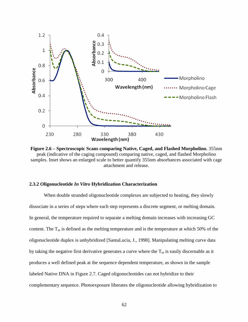
62
2.3.2 Oligonucleotide In Vitro Hybridization Characterization
When double stranded oligonucleotide complexes are subjected to heating, they slowly
dissociate in a series of steps where each step represents a discrete segment, or melting domain.
In general, the temperature required to separate a melting domain increases with increasing GC
content. The Tm is defined as the melting temperature and is the temperature at which 50% of the
oligonucleotide duplex is unhybridized [SantaLucia, J., 1998]. Manipulating melting curve data
by taking the negative first derivative generates a curve where the Tm is easily discernable as it
produces a well defined peak at the sequence dependent temperature, as shown in the sample
labeled Native DNA in Figure 2.7. Caged oligonucleotides can not hybridize to their
complementary sequence. Photoexposure liberates the oligonucleotide allowing hybridization to
Figure 2.6 – Spectroscopic Scans comparing Native, Caged, and Flashed Morpholino. 355nm
peak (indicative of the caging compound) comparing native, caged, and flashed Morpholino
samples. Inset shows an enlarged scale to better quantify 355nm absorbances associated with cage
attachment and release.
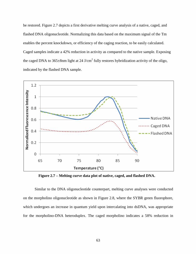
63
be restored. Figure 2.7 depicts a first derivative melting curve analysis of a native, caged, and
flashed DNA oligonucleotide. Normalizing this data based on the maximum signal of the Tm
enables the percent knockdown, or efficiency of the caging reaction, to be easily calculated.
Caged samples indicate a 42% reduction in activity as compared to the native sample. Exposing
the caged DNA to 365±8nm light at 24 J/cm2 fully restores hybridization activity of the oligo,
indicated by the flashed DNA sample.
Similar to the DNA oligonucleotide counterpart, melting curve analyses were conducted
on the morpholino oligonucleotide as shown in Figure 2.8, where the SYBR green fluorophore,
which undergoes an increase in quantum yield upon intercalating into dsDNA, was appropriate
for the morpholino-DNA heteroduplex. The caged morpholino indicates a 58% reduction in
Figure 2.7 – Melting curve data plot of native, caged, and flashed DNA.
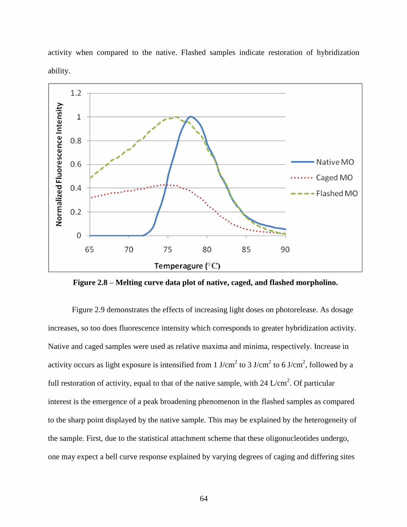
64
activity when compared to the native. Flashed samples indicate restoration of hybridization
ability.
Figure 2.9 demonstrates the effects of increasing light doses on photorelease. As dosage
increases, so too does fluorescence intensity which corresponds to greater hybridization activity.
Native and caged samples were used as relative maxima and minima, respectively. Increase in
activity occurs as light exposure is intensified from 1 J/cm2 to 3 J/cm
2 to 6 J/cm
2, followed by a
full restoration of activity, equal to that of the native sample, with 24 L/cm2. Of particular
interest is the emergence of a peak broadening phenomenon in the flashed samples as compared
to the sharp point displayed by the native sample. This may be explained by the heterogeneity of
the sample. First, due to the statistical attachment scheme that these oligonucleotides undergo,
one may expect a bell curve response explained by varying degrees of caging and differing sites
Figure 2.8 – Melting curve data plot of native, caged, and flashed morpholino.
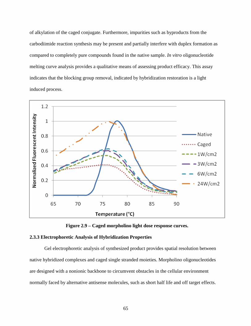
65
of alkylation of the caged conjugate. Furthermore, impurities such as byproducts from the
carbodiimide reaction synthesis may be present and partially interfere with duplex formation as
compared to completely pure compounds found in the native sample. In vitro oligonucleotide
melting curve analysis provides a qualitative means of assessing product efficacy. This assay
indicates that the blocking group removal, indicated by hybridization restoration is a light
induced process.
2.3.3 Electrophoretic Analysis of Hybridization Properties
Gel electrophoretic analysis of synthesized product provides spatial resolution between
native hybridized complexes and caged single stranded moieties. Morpholino oligonucleotides
are designed with a nonionic backbone to circumvent obstacles in the cellular environment
normally faced by alternative antisense molecules, such as short half life and off target effects.
Figure 2.9 – Caged morpholino light dose response curves.
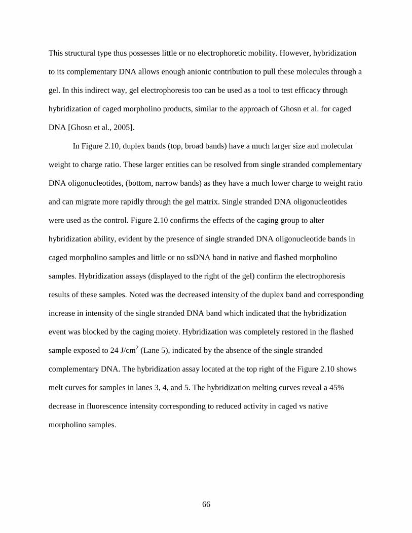
66
This structural type thus possesses little or no electrophoretic mobility. However, hybridization
to its complementary DNA allows enough anionic contribution to pull these molecules through a
gel. In this indirect way, gel electrophoresis too can be used as a tool to test efficacy through
hybridization of caged morpholino products, similar to the approach of Ghosn et al. for caged
DNA [Ghosn et al., 2005].
In Figure 2.10, duplex bands (top, broad bands) have a much larger size and molecular
weight to charge ratio. These larger entities can be resolved from single stranded complementary
DNA oligonucleotides, (bottom, narrow bands) as they have a much lower charge to weight ratio
and can migrate more rapidly through the gel matrix. Single stranded DNA oligonucleotides
were used as the control. Figure 2.10 confirms the effects of the caging group to alter
hybridization ability, evident by the presence of single stranded DNA oligonucleotide bands in
caged morpholino samples and little or no ssDNA band in native and flashed morpholino
samples. Hybridization assays (displayed to the right of the gel) confirm the electrophoresis
results of these samples. Noted was the decreased intensity of the duplex band and corresponding
increase in intensity of the single stranded DNA band which indicated that the hybridization
event was blocked by the caging moiety. Hybridization was completely restored in the flashed
sample exposed to 24 J/cm2 (Lane 5), indicated by the absence of the single stranded
complementary DNA. The hybridization assay located at the top right of the Figure 2.10 shows
melt curves for samples in lanes 3, 4, and 5. The hybridization melting curves reveal a 45%
decrease in fluorescence intensity corresponding to reduced activity in caged vs native
morpholino samples.
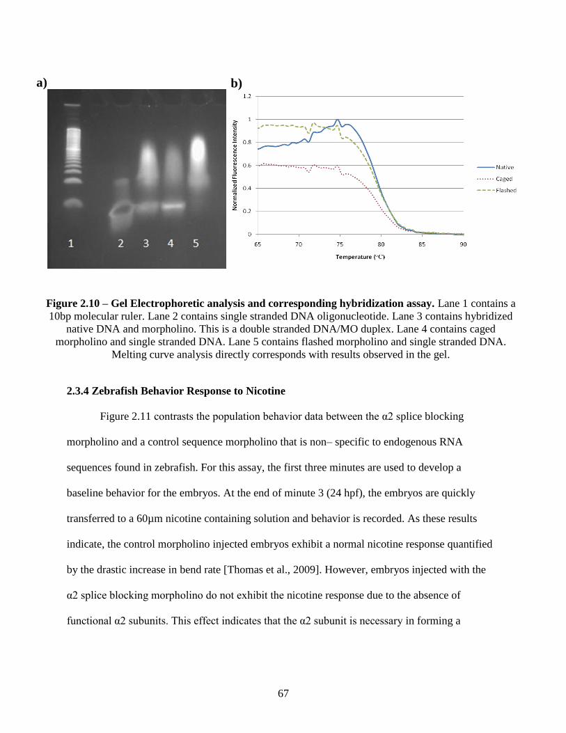
67
2.3.4 Zebrafish Behavior Response to Nicotine
Figure 2.11 contrasts the population behavior data between the α2 splice blocking
morpholino and a control sequence morpholino that is non– specific to endogenous RNA
sequences found in zebrafish. For this assay, the first three minutes are used to develop a
baseline behavior for the embryos. At the end of minute 3 (24 hpf), the embryos are quickly
transferred to a 60µm nicotine containing solution and behavior is recorded. As these results
indicate, the control morpholino injected embryos exhibit a normal nicotine response quantified
by the drastic increase in bend rate [Thomas et al., 2009]. However, embryos injected with the
α2 splice blocking morpholino do not exhibit the nicotine response due to the absence of
functional α2 subunits. This effect indicates that the α2 subunit is necessary in forming a
Figure 2.10 – Gel Electrophoretic analysis and corresponding hybridization assay. Lane 1 contains a
10bp molecular ruler. Lane 2 contains single stranded DNA oligonucleotide. Lane 3 contains hybridized
native DNA and morpholino. This is a double stranded DNA/MO duplex. Lane 4 contains caged
morpholino and single stranded DNA. Lane 5 contains flashed morpholino and single stranded DNA.
Melting curve analysis directly corresponds with results observed in the gel.
a) b)
68
functional receptor. This also establishes biological endpoints between where caged and flashed
samples are expected to operate.
Figure 2.11 – Nicotine induced behavior response. At 3minutes, nicotine was introduced and
bend rate was calculated for the control morpholino and α2 morpholino injected population.
Ultimately, UVA light will be used to liberate caged molecules. Thus, we had to
determine if UVA doses affected the behavior response. Knowing behavioral endpoints set by
the control morpholino and α2 splice blocking morpholino, effects of light on embryonic
development and specifically nicotine induced behavior had to be assessed. Embryos were raised
to 12 hpf and irradiated for 4min and 10min. Based on these times, the doses that the embryos
were exposed to 24 J/cm2 and 60 J/cm
2, respectively, from the Greenspot UVA light source.
Figure 2.12 displays population data for the nicotine response. At min 5 (26 hpf), control and
flashed embryos were exposed to 60µM nicotine and behavior was recorded. Consistent with the
control population, photoexposed embryos display the same behavior pattern of increased bend
Nicotine Introduced
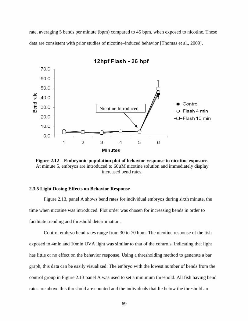
69
rate, averaging 5 bends per minute (bpm) compared to 45 bpm, when exposed to nicotine. These
data are consistent with prior studies of nicotine–induced behavior [Thomas et al., 2009].
2.3.5 Light Dosing Effects on Behavior Response
Figure 2.13, panel A shows bend rates for individual embryos during sixth minute, the
time when nicotine was introduced. Plot order was chosen for increasing bends in order to
facilitate trending and threshold determination.
Control embryo bend rates range from 30 to 70 bpm. The nicotine response of the fish
exposed to 4min and 10min UVA light was similar to that of the controls, indicating that light
has little or no effect on the behavior response. Using a thresholding method to generate a bar
graph, this data can be easily visualized. The embryo with the lowest number of bends from the
control group in Figure 2.13 panel A was used to set a minimum threshold. All fish having bend
rates are above this threshold are counted and the individuals that lie below the threshold are
Figure 2.12 – Embryonic population plot of behavior response to nicotine exposure.
At minute 5, embryos are introduced to 60µM nicotine solution and immediately display
increased bend rates.
Nicotine Introduced
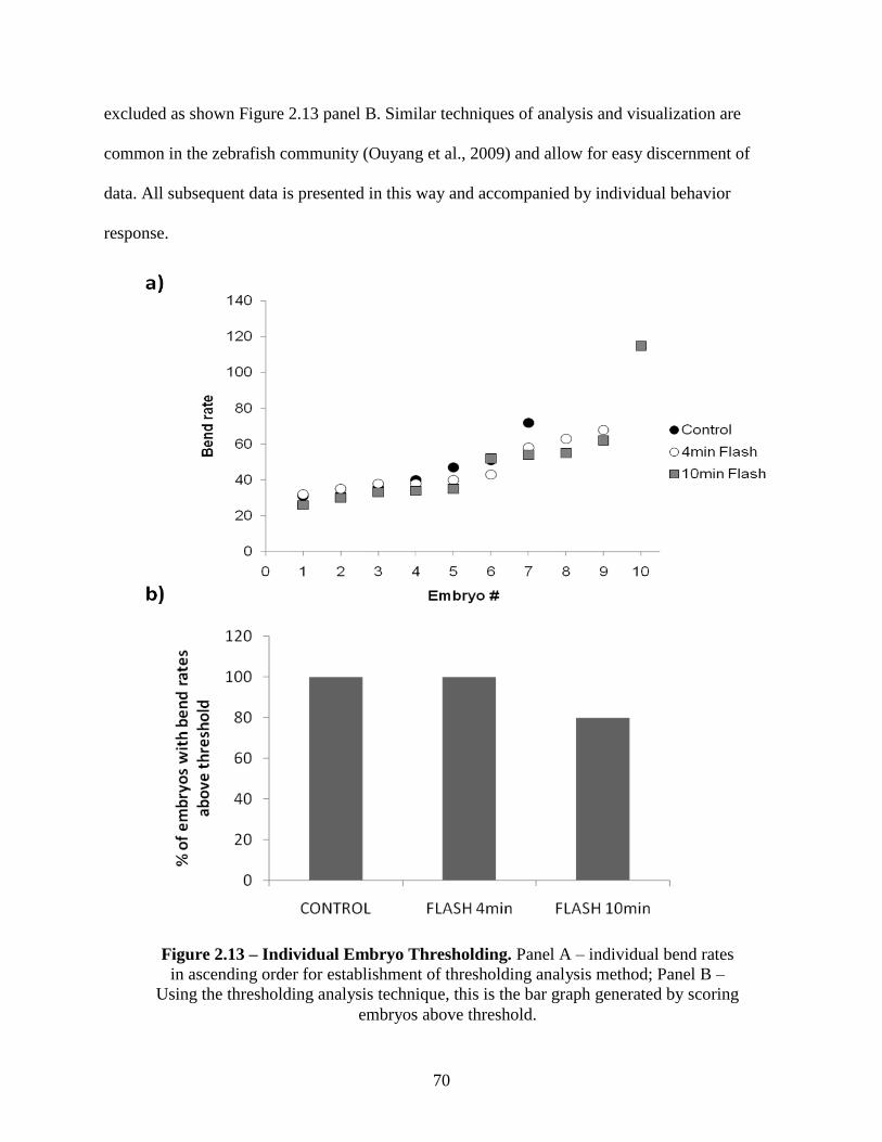
70
excluded as shown Figure 2.13 panel B. Similar techniques of analysis and visualization are
common in the zebrafish community (Ouyang et al., 2009) and allow for easy discernment of
data. All subsequent data is presented in this way and accompanied by individual behavior
response.
Figure 2.13 – Individual Embryo Thresholding. Panel A – individual bend rates
in ascending order for establishment of thresholding analysis method; Panel B –
Using the thresholding analysis technique, this is the bar graph generated by scoring
embryos above threshold.
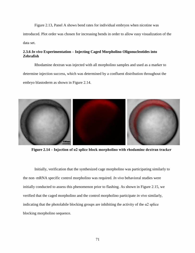
71
Figure 2.13, Panel A shows bend rates for individual embryos when nicotine was
introduced. Plot order was chosen for increasing bends in order to allow easy visualization of the
data set.
2.3.6 In vivo Experimentation – Injecting Caged Morpholino Oligonucleotides into
Zebrafish
Rhodamine dextran was injected with all morpholino samples and used as a marker to
determine injection success, which was determined by a confluent distribution throughout the
embryo blastoderm as shown in Figure 2.14.
Initially, verification that the synthesized cage morpholino was participating similarly to
the non–mRNA specific control morpholino was required. In vivo behavioral studies were
initially conducted to assess this phenomenon prior to flashing. As shown in Figure 2.15, we
verified that the caged morpholino and the control morpholino participate in vivo similarly,
indicating that the photolabile blocking groups are inhibiting the activity of the α2 splice
blocking morpholino sequence.
Figure 2.14 – Injection of α2 splice block morpholino with rhodamine dextran tracker
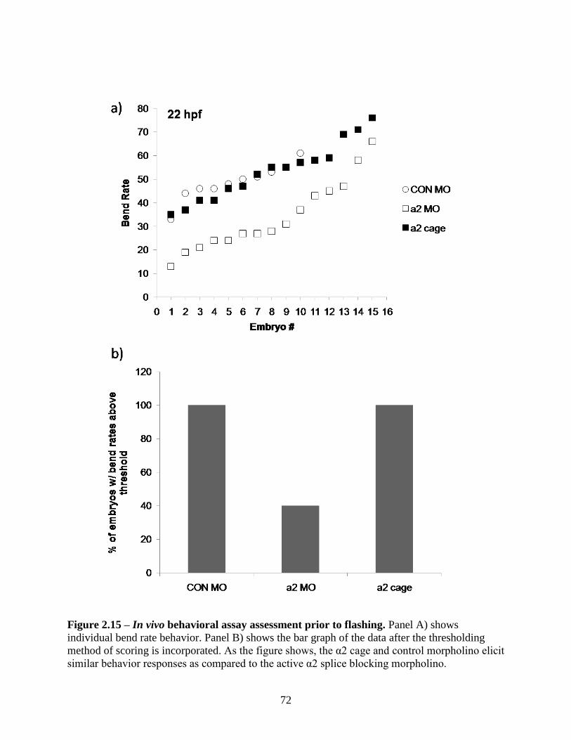
72
Figure 2.15 – In vivo behavioral assay assessment prior to flashing. Panel A) shows
individual bend rate behavior. Panel B) shows the bar graph of the data after the thresholding
method of scoring is incorporated. As the figure shows, the α2 cage and control morpholino elicit
similar behavior responses as compared to the active α2 splice blocking morpholino.
73
For in vivo experiments, control morpholino sequence, native α2 splice blocking
sequence provided by Gene Tools, synthesized base caged morpholino α2, and in vitro flashed
(24J/cm2 365±8nm UVA light source) base caged α2 sequence were injected into the embryo
yolk at the 1–2 cell stage, the common time point for morpholino introduction [Nasevicius et al.,
2000] and imaged 6–7 hpf to determine injection success (Figure 2.14) using rhodamine dextran
to track the injected material’s location. The sample number of embryos injected with the base
caged morpholino oligonucleotides was doubled so that half of that subpopulation could be
flashed in vivo. At 24 hpf, injected embryos were analyzed using the techniques previously
detailed for UVA effects on bend rate. Figure 2.16, Panel A shows individual bend rate data for
each morpholino injected group. Using the embryo with the lowest bend rate of the control
group, a threshold is set at 38 bpm. All fish above this threshold are counted for the sample set
where all below the set threshold are excluded from the data set depicted in panel B. Panel B
shows that α2 injected embryos have a marked decrease in individuals having bend rates above
the set threshold, again indicating the absence of functional nicotine binding receptors in those
embryos. Embryos injected with caged α2 MO display similar characteristics to that of the
control sample population, indicating that the action of the α2 splice blocking morpholino was
stopped or reduced. Half of the embryos designated as “α2 cage” were removed from that set and
exposed to 24J/cm2 UVA light (365±8nm). These embryos were designated as “uncaged in vivo”
and as the graph shows, have a reduced number of bend rates when compared to the caged
subpopulation. This indicates a restoration of activity of the splice block morpholino due to the
removal of photolabile blocking groups. The in vitro flashed sample set includes embryos
injected with caged morpholino that was exposed to 24J/cm2 light prior to injection.
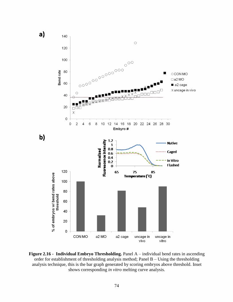
74
Figure 2.16 - Individual Embryo Thresholding. Panel A – individual bend rates in ascending
order for establishment of thresholding analysis method; Panel B – Using the thresholding
analysis technique, this is the bar graph generated by scoring embryos above threshold. Inset
shows corresponding in vitro melting curve analysis.
75
The phenomenon of not being able to achieve efficient in vitro uncaging as compared to
the in vivo uncaging was often observed throughout experimentation. This was thought to be
contributed to by the concentration of the caged sample prior to and post injection, which has
been shown to affect uncaging efficiencies [Ellis – Davies, 2007]. The inset on panel B is
corresponding hybridization data. Interestingly, as the melting curve assay indicated, the in vitro
flashed sample never regained hybridization activity. This chemical assay predicted biological
function which translated to observable behavior.
2.4 Conclusions
We have shown a novel reaction scheme to cage exocyclic primary amines on intact DNA
and morpholino oligonucleotides using a statistical attachment strategy that could be applied to
any antisense oligonucleotide. Both in vitro and in vivo assays confirm the disruption of
hybridization in the caged form, which can be recovered with light exposure. Using zebrafish
nicotine induced behavior response for in vivo validation of the presence of α2 subunits located
on nAChR pentameters, rendering a diminished response to nicotine treatment. Caged splice
blocking morpholinos could not hybridize to α2 mRNA because of the steric inhibition
introduced by the presence of photolabile blocking groups. This enabled fish injected with the
caged morpholino to properly synthesize the α2 protein and undergo normal nicotine response.
Having these two endpoints to operate with, we tested the functionality of our caged
oligonucleotide. Utilizing the full potential of the caging technology, spatial and temporal
activation of the morpholino’s splice blocking ability was within our control. At 12 hpf, we have
shown the ability to effectively liberate the morpholino and stop α2 production, confirmed by the
behavior assay. Normal efficacy of morpholino oligonucleotides last 3 days post injection
[Sumanas et al., 2002]. Using delayed time points for photoactivation, it may be possible to
76
extend the half–life of the morpholino beyond what has been observed. Furthermore, we were
able to accurately predict in vivo efficacy based on the results obtained from in vitro
experimentation.
This is the first time that statistical caging has been actualized on the morpholino
oligonucleotide through direct attachment to the bases. Initial attempts using DMNPE caging
strategies developed for phosphodiester backbone nucleic acids proved unsuccessful due to the
lack of available binding sites on the morpholino [Monroe et al., 1999, Blidner et al., 2008].
Additionally, pertaining to the morpholino, this effort was initially designed to make the moiety
more electrophilic, but being incapable of this, ultimately was trying to force
nucleophilic/nucleophilic interaction.
This interdisciplinary study combined applications from biological chemistry, biological
engineering, rational drug design, and developmental biology. Based on results obtained through
chemical analysis, we can accurately and reliably predict biological function in whole organisms
through the use of a reliable and reproducible behavior assay. This investigation should act to
expand caged morpholino oligonucleotide technologies, and more generally antisense
technologies as a whole due to the ease of synthesis required in caging these compounds, as well
as to facilitate further understanding of the molecular mechanisms governing embryonic
development.
2.5 References
1. Burgess, T. L., E. F. Fisher, et al. (1995). "The antiproliferative activity of c-myb and c-
myc antisense oligonucleotides in smooth muscle cells is caused by a nonantisense
mechanism." Proceedings of the National Academy of Sciences of the United States of
America 92(9): 4051-4055.
2. Crooke, S. T. (2008). Antisense Drug Technology: Principles, Strategies, and
Applications. Boca Ranton, CRC Press Taylor & Francis Group.
77
3. CW Luetje and J Patrick. (1991) “Both alpha- and beta-subunits contribute to the agonist
sensitivity of neuronal nicotinic acetylcholine receptors.” Journal of Neuroscience. 11,
837-845.
4. Ellis-Davies, G. C. R. (2007). "Caged compounds: photorelease technology for control of
cellular chemistry and physiology." Nature Methods 4(8): 619-628.
5. Ghosn, B., F. R. Haselton, et al. (2005). "Control of DNA hybridization with
photocleavable adducts." Photochemistry and Photobiology 81(4): 953-959.
6. Giles, R. V., D. G. Spiller, et al. (1993). "CHIMERIC OLIGODEOXYNUCLEOTIDE
ANALOGS - ENHANCED CELL UPTAKE OF STRUCTURES WHICH DIRECT
RIBONUCLEASE-H WITH HIGH SPECIFICITY." Anti-Cancer Drug Design 8(1): 33-
51.
7. Graham J. Lieschke & Peter D. Currie (2007). “Animal models of human disease:
zebrafish swim into view.” Nature Reviews Genetics 8, 353-367
8. Hudziak, R. M., E. Barofsky, et al. (1996). "Resistance of morpholino
phosphorodiamidate oligomers to enzymatic degradation." Antisense & Nucleic Acid
Drug Development 6(4): 267-272.
9. Jeffrey M Zirger, Christine E Beattie, Dennis B McKay and R Thomas Boyd (2003)
“Cloning and expression of zebrafish neuronal nicotinic acetylcholine receptors.” Gene
Expression Patterns. 3(6): 747-754
10. Mayer, G. and A. Heckel (2006). "Biologically active molecules with a "light switch"."
Angewandte Chemie-International Edition 45(30): 4900-4921.
11. Monroe, W. T., M. M. McQuain, et al. (1999). "Targeting expression with light using
caged DNA." Journal of Biological Chemistry 274(30): 20895-20900.
12. Nasevicius, A. and S. C. Ekker (2000). "Effective targeted gene 'knockdown' in
zebrafish." Nature Genetics 26(2): 216-220.
13. Ouyang, X. H., I. A. Shestopalov, et al. (2009). "Versatile Synthesis and Rational Design
of Caged Morpholinos." Journal of the American Chemical Society 131(37): 13255-
13269.
14. Purves, Dale, George J. Augustine, David Fitzpatrick, William C. Hall, Anthony-Samuel
LaMantia, James O. McNamara, and Leonard E. White (2008). Neuroscience. 4th ed.
Sinauer Associates. pp. 122–6
15. SantaLucia, J. (1998). "A unified view of polymer, dumbbell, and oligonucleotide DNA
nearest-neighbor thermodynamics." Proceedings of the National Academy of Sciences of
the United States of America 95(4): 1460-1465.
78
16. Shoeman, R. L., R. Hartig, et al. (1997). "Fluorescence microscopic comparison of the
binding of phosphodiester and phosphorothioate (antisense) oligodeoxyribonucleotides to
subcellular structures, including intermediate filaments, the endoplasmic reticulum, and
the nuclear interior." Antisense & Nucleic Acid Drug Development 7(4): 291-308.
17. Stein, C. A. and Y. C. Cheng (1993). "ANTISENSE OLIGONUCLEOTIDES AS
THERAPEUTIC AGENTS - IS THE BULLET REALLY MAGICAL." Science
261(5124): 1004-1012.
18. Stein, D., E. Foster, et al. (1997). "A specificity comparison of four antisense types:
Morpholino, 2'-O-methyl RNA, DNA, and phosphorothioate DNA." Antisense & Nucleic
Acid Drug Development 7(3): 151-157.
19. Sumanas, S. and J. D. Larson (2002). "Morpholino phosphorodiamidate oligonucleotides
in zebrafish: A recipe for functional genomics?" Briefings in Functional Genomics and
Proteomics 1(3): 239-256.
20. Summerton, J. and D. Weller (1997). "Morpholino antisense oligomers: Design,
preparation, and properties." Antisense & Nucleic Acid Drug Development 7(3): 187-
195.
21. Thomas, L. T., L. Welsh, et al. (2009). "Acute nicotine exposure and modulation of a
spinal motor circuit in embryonic zebrafish." Toxicology and Applied Pharmacology
239(1): 1-12.
22. Walker, J. W., G. P. Reid, et al. (1988). "PHOTOLABILE 1-(2-
NITROPHENYL)ETHYL PHOSPHATE-ESTERS OF ADENINE-NUCLEOTIDE
ANALOGS - SYNTHESIS AND MECHANISM OF PHOTOLYSIS." Journal of the
American Chemical Society 110(21): 7170-7177.
23. Wasterfield M. (2000). “The Zebrafish Book, A guide for laboratory use of Zebrafish
(Danio Rerio)”. Eugene, OR, University of Oregon Press
79
CHAPTER 3: CONCLUSIONS AND FUTURE DIRECTIONS
3.1 Conclusions
In this study, through direct alkylation of exocyclic amines on intact oligonucleotides, we
have successfully devised a method for synthesis of antisense oligonucleotides. Specifically
DNA antisense oligonucleotides and morpholino antisense oligonucleotides but, presumably any
available structure could be inserted into the reaction scheme and successfully caged. The caged
compounds that we synthesized were characterized by both in vitro and in vivo methods.
Absorbance spectroscopy served as a first quantitative check for reaction success. Comparing
relative absorbance values from 260nm, 355nm, and 500nm, caging efficiency was calculated.
Once this was done, qualitative means of analysis was performed in the form of hybridization
assays including melting curve analysis and electrophoretic gel mobility. Upon completion of
these methods, we were confident in the product which allowed a transition to in vivo validation.
Performed in the zebrafish model, a well characterized behavior response was used to assess the
efficacy of the caged product. Behavior provides an exceptional means of genetic manipulation
due to the fact that whole organisms are preserved and can be used to further confirm results in
other assays. This interdisciplinary study combined applications from biological chemistry,
biological engineering, rational drug design, and developmental biology. Based on results
obtained through chemical analysis, we can accurately and reliably predict biological function in
whole organisms through the use of a reliable and reproducible behavior assay. This
investigation should act to expand caged morpholino oligonucleotide technologies, and more
generally antisense technologies as a whole due to the ease of synthesis required in caging these
compounds, as well as gain an understanding of the molecular mechanisms governing embryonic
development.
80
3.2 Future Directions
Future directions of this work include the following:
HPLC purification and analysis of caged products is a critical method of cleanup for
synthesis reactions. The current purification scheme is rather intense. Incorporation of HPLC to
not only purify, but separate caged products from unreacted species would aid in reducing
variation in both in vitro and in vivo results.
We only touched the surface of mass spectrometry analysis on the caged morpholino
products. Considering MALDI and ESI as possible routes of validation, we tested both methods.
MALDI proved to be unable to process samples in their current state. We hypothesized that this
was due to the inability of the caged morpholino to crystallize on the matrix and that the caging
reaction was somehow changing the chemistry of the oligonucleotide. ESI proved feasible but
the signal was noisy, reconfirming the need to efficiently purify these products through HPLC
analysis.
A 9–ethylguanine conjugation reaction is essential to confirm that the caging
compound is attaching to the exocyclic amine on this compound. However, due to the fact that
the current purification scheme requires species with MW greater than 3000, it was not plausible
at the time.
NMR analysis of purified products would also confirm location of the caging group
along the oligonucleotide.
Also important is quantification of the lifetime efficacy of the morpholinos in vivo. It
is currently known that morpholinos are active up to 3 days post-injection in zebrafish. It will be
important to assess if the caging moieties extend the lifetime of morpholinos in in vivo
environments.
81
Because the α2 morpholino acts through splice blocking, RT-PCR would compliment
behavioral analysis by confirming the mRNA splicing changes. This would be a final endpoint
following nicotine-induced behavioral response assay.
Similar to RT-PCR, immunohistochemistry would compliment results obtained via
behavioral analysis, via antibody-antigen interactions on α2 receptors. Using fluorescent
microscopy imaging techniques, we would confirm observations from in vivo studies. Both RT-
PCR and immunohistochemical staining could analyze single fish to further confirm results.
82
VITA
Chad Michael Jarreau, son of Tim and Peggy Miller, was born in February, 1984, in Baton
Rouge, Louisiana. He graduated from False River Academy in 2002. In May, 2007, he earned his
Bachelor of Science in Biological and Agricultural Engineering degree from Louisiana State
University from Baton Rouge, Louisiana. He plans to obtain his Master of Science in Biological
and Agricultural Engineering degree in May 2010 under the direction of his primary advisor Dr.
Todd Monroe, and committee members Dr. Kurt Svoboda and Dr. Marybeth Lima.


![[ ] Caged Drug Delivery P](https://img.pdfslide.us/doc/110x75/58edc5381a28abf2328b46d1/-caged-drug-delivery-p.jpg)