Embed Size (px)
Citation preview
Physiologic Behavioural Changes of Precancerous Leukoplakia Cell linesExerted to Extremely Low Frequency Electric FieldAhmed Korraah1, Margarete Odenthal2, Marion Kopp3, Nadarajah Vigneswaran4, Peter G Sacks5, Hans Peter Dienes2, Hartmut Stützer6 and WilhelmNiedermeier3*
1Department of Oral Pathology, Faculty of Dentistry, Suez Canal University, Egypt2Institute for Pathology, University Hospital, Cologne, Germany3Department of Prosthetic Dentistry, Dental School, University of Cologne, Germany4Department of Diagnostic Sciences, School of Dentistry, University of Texas, USA5Department of Basic Science and Craniofacial Biology, College of Dentistry, New York University, USA6Institute for Medical Statistics, Informatics and Epidemiology, University of Cologne, Germany*Corresponding author: Wilhelm Niedermeier, Department of Prosthetic Dentistry, Dental School, University of Cologne, Kerpenerstr. 32, 50931 Cologne, Germany,Tel: 0049-478-6337; Fax: 0049-478-5982; E-mail: [email protected]
Received date: November 18, 2014, Accepted date: December 26, 2014, Published date: December 31, 2014Copyright: © 2015 Korraah A, et al. This is an open-access article distributed under the terms of the Creative Commons Attribution License, which permits unrestricted use, distribution, and reproduction in any medium, provided the original author and source are credited.
Abstract
Objectives: Oral galvanism arising from the presence of two different metallic fillings in the mouth that may reachup to 16V/m, induces several changes in oral environment such as gingival swelling and erythema, mucosal pain,lichenoid reactions and leukoplakia.
Study design: To investigate in vitro the physiologic reactions of oral leukoplakia caused by electrostaticcorrosion potentials, human leukoplakia cell lines (MSK-LEUK1), cultivated in KGM-2 supplemented with bullet kit in5% CO2 humidified air at 37°C, were exposed to electric field strength of 1-20 V/m for 24 hours in a pseudo realisticapparatus called “Pulse chamber”. Following this, the cells were analysed for proliferation using BrdU assay, and forapoptosis using TUNEL assay. Findings were assessed utilizing fluorescent microscopy. Ultra structural changeswere studied by TEM. Data was evaluated statistically using non parametric Chi-square test.
Results: Electric field strength of 1-10V/m led to upregulation of cell proliferation rate between 10.64% and 44.06% (p=0.0001). The apoptotic index increased significantly (p=0.0001) from 20.03% at 1V/m to 46.56% at 10V/m. Leukoplakia cells treated with 16V/m show individual cell keratinization.
Conclusion: Oral galvanism induces subcellular changes in oral precancerous leukoplakia cells in vitro thatresemble some of the histopathologic features of oral squamous cell carcinoma cells in vivo.
Keywords: Oral galvanism (OG); Apoptosis; Pulse chamber; MSK-LEUK1 cell lines
Abbrevations: A: Ampere; Ac/Me: Acetone/Methanol fixative; AI: Apoptotic index; ANCC: Apoptotic negative control cells; ATP: Adenosine triphosphate; BrdU: 5-Bromo-2´-deoxyuridine; BSA: Bovine Serum Antigen; C: Celsius; Ca2+: Calcium ions; CO2: Carbon dioxide, dm: decimetre; DAPI: 4',6-diamidino-2-phenylindole; DC: Direct current; DCV: Direct Current Voltage; DNA: Desoxyribonucleic acid; EDTA: Ethylene diamine tetraacetic acid; EF: Electric Field; EFS: Electric field strength; EFSC: Electric field stimulated cells; ELSC: Electrode stimulated cells; EMF: Electromagnetic Field; FBS: Foetal bovine serum; H2O2: Hydrogen peroxide; HUH-7: Hepatoma cell lines; Ig: Immunoglobulin; KGM-2=Keratinocytes growth medium-2; ml=millilitre; mS/m=millisiemens/meter; mV=millivolt; MSK-LEUK1=Leukoplakia cell lines; n=number of carried out experiments; nm: nanometer; NADPH: Nicotinamide adenine dinucleotide hydrogen; OG: Oral galvanism; OPM: Oral premalignant; OSCC: Oral squamous cell carcinoma; pH: Power of hydrogen; PBS: Phosphate buffered saline; PCC: Positive Control Cells; PFA: Paraformaldehyde; PI: Proliferation index; PNCC: Proliferating negative control cells; Redox=reduction- Oxidation; Rpm=revolution per minute;
ROS=Reactive oxygen species; S=seconds; V/m: Volt per meter; T1N0M0=Tumor stage 1, no lymph node involvement and no Metastasis; TMR=: Tetramethylrhodamine; TUNEL: Terminal uridine deoxy nucleotidyl transferase dUTP nick end labelling
IntroductionIn dental fields, gold, cobalt, palladium, silver, chromium, copper,
zinc, tin and nickel are used. When there are two or more differentdental metallic restorations in the presence of saliva, an electrical fieldis induced that is called oral galvanism (OG), in which saliva plays therole of the conducting solution (the electrolyte) as it containsinorganic ions as a major component [1]. The generated electric fieldmay reach 950 mV between an aluminium splint and a gold crown [2].
Oral Leukoplakia is defined clinically in 1968 as a white patch, notless than 5 mm in diameter, which cannot be rubbed off with a bluntinstrument and which cannot be classified as any other diagnosabledisease [3]. It appears clinically white but histopathologically variesbetween simple hyperkeratosis to severe epithelial dysplasia orcarcinoma in situ. Recently, Warnakulasuriya defined oral leukoplakiaas white plaques of questionable risk having excluded (other) known
JBR Journal of InterdisciplinaryMedicine and Dental Science
Korraah A, et al., J Interdiscipl Med Dent Sci 2015, 3:1 DOI: 10.4172/2376-032X.1000165
Research Article Open Access
J Interdiscipl Med Dent SciISSN: 2376-032X JIMDS, an open access journal
Volume 3 • Issue 1 • 1000165
JBR Jour
nal o
f Int
erdis
ciplinary Medicine and Dental Science
ISSN: 2376-032X
diseases or disorders that carry no increased risk for cancer [4]. It isone of the most frequent types of oral precancerous lesions and showsa variable rate of malignant transformation to oral squamous cellcarcinoma (OSCC) [5]. Pindborg mentioned that oral squamous cellcarcinoma (OSCC) is frequently preceded by oral leukoplakia, whichhas a malignant transformation rate between 0.13-6% [6,7]. OSCCgenesis passes through many genetic and non-genetic steps that havetwo clinical forms: (1) oral premalignant (OPM) lesion, which willtransform into (2) OSCC [8,9]. OPM lesions usually appear eitherwhite (leukoplakia) or red (erythroplakia). Leukoplakia represents 85%of such lesions and has a prevalence rate of 2–8% in people over theage of 70 [10]. The basis of our approach was that oral squamouscarcinoma is very frequently preceded by a precancerous and thatestablished squamous carcinomas of the mouth are invariablysurrounded by leukoplakia, erythroplakia or both lesions [6].
There has been much controversy regarding whether oral galvanism(OG) really plays a role in the development of oral cancer, lichenplanus-like lesions, and various other oral diseases. Previous studiesindicate that oral galvanism could lead to the development of oralleukoplakia and the removal of the metallic restorations resulted in theresolution of these lesions [11-13]. In addition, a remarkable recoveryof diseases of the oral mucosa was noted when harmful potentials wereeliminated [14-16].
In the wake of the pioneering work of Kerr, the importance of cellloss through apoptosis (active cell death) and accidental cell death(passive cell death) and the role of apoptosis in tumorigenesis has beenincreasingly recognized [17]. Although apoptosis is a naturallyoccurring cellular event that helps maintain organism homeostasis, itsrole in the field of oncology, especially in tumorigenesis eruptsrecently and current researches are now focused on the role of theunbalance between cell proliferation and cell apoptosis intumorigenesis [17,18].
In the present study, we examined the physiologic behaviour of oralprecancerous leukoplakia cell lines regarding its proliferative andapoptotic activities when exerted to extremely low frequency electricfield treatment.
Material and Methods
Cell linesLeukoplakia cell lines (MSK-LEUK1) derived from dysplastic
leukoplakia adjacent to an early invasive OSCC (T1N0M0) involvingthe tongue of a 47-year-old female was used in our research. Thesecells showed loss of response to calcium and have no terminaldifferentiation (characteristics of premalignant keratinocytes) [7,19].
A serum-free keratinocyte growth medium (KGM-2) supplementedwith growth factors (Bullet kit; Cat#CC-3103; Lot#01119272; LONZAWalkersville, Inc, Chicago, IL, USA) was used for the growth of MSK-LEUK1 cells.It was changed every 2 days and the cells were trypsinizedevery week with a sequential method. Trypsin 2.5%/ EDTA 0.03% wasused for trypsinization. The cells were plated onto 6cm cell culturePetri dishes. Experiments were performed with sub-confluent cellcultures. All cell lines were incubated under standard conditions with5% CO2 at 37°C in a humidified atmosphere.
Enzymatic dissociation of cell lines with trypsin/EDTA (Sequentialtrypsinization)
The adherent growing cells were enzymatically dissociated using37°C warm trypsin/EDTA.
First, the trypsin/EDTA mixture was heated in water bath at 37°C.After the cell culture medium was removed from the monolayer, thecells were briefly washed with 5 ml PBS to remove any static mediumradicals, which can affect the action of trypsin. The cells thenincubated in a 5 ml trypsin/EDTA mixture for 5 minutes in anincubator. After this period, the majority of the cells resolved from thesurface of the monolayer. A stopping medium was prepared in a 25 mltube. It consists of 4 ml KGM-2 and 1 ml of foetal bovine serum. Thedetached cell suspension was transported into the stopping medium.The still attached cells were then incubated for another 5 minutes withTrypsin/EDTA. This step was repeated until all the monolayers weredetached. The Trypsin/EDTA cell suspension was centrifuged with700 rpm for 5 minutes in 15°C. The supernatant was discarded and 2ml of KGM-2 was added. Lastly, the cell suspension was distributedevenly in Petri dishes and incubated.
In order to have several vials of MSK-LEUK1 cells, vials of cells arefrozen under -70°C in liquid nitrogen. Thawing of cells should be inwater bath in 37°C till the water crystals disappear.
Cell cultures treatment with electrical field (pulse chamber)Petri dishes with sub-confluent cell cultures were transferred to a
custom-made electropulse apparatus (Figures 1 and 2), which allowedrotating electrical fields to avoid decomposition and pH gradients inthe cell culture medium and polarization of cells [20]. The pulsechamber setup consisted of 14 electrodes formed with an alloy called“BiOcclus 4” (DeguDent GmbH, P.O.Box 1364, D-63403 Hanau,Germany) which consists of gold and platinum. The distance betweeneach two opposite electrodes was 5 cm and they were immersed at theborder of the Petri dish into the cell culture medium, which has aspecific conductivity of 15.75 mS/m. The electrodes were connected toa direct current supply, which generated direct current (DC) voltagepulses with duration of 0.9s. After an interval of 0.1s, the voltagesupply automatically coupled to the next electrode pair to initiate thesubsequent voltage pulse. During the experiments voltage pulsesranging from 50 to 1000 mV were applied, which resulted in electricalfield strength (EFS) of 1 to 20V/m respectively. Before and during eachexperiment, it was assured that the EFS was in agreement with theinput DC voltage in the centre of the Petri dish.The exposure devicewas placed in an incubator with 5% CO2 at 37oC in a humidifiedatmosphere. The temperature, osmolarity and pH of the cell culturestreated with electrical fields were controlled before, during and afterthe experiments. For controls, MSK-LEUK1 cells were treated in acomparable manner in a second pulse chamber that containedelectrodes immersed into the culture medium but were not connectedto a voltage supply. Both the experimental and the control pulsechambers were placed in the same incubator during the experiments.The MSK-LEUK1 cells were prepared before the experiments byallowing them to grow on 1 cm in diameter cover slips that wereplaced in the centre of the cell culture dish (the area of optimum fieldhomogeneity). The confluence of cells on the cover slip must be 50%.
Citation: Ahmed Korraah, Margarete Odenthal, Marion Kopp, Nadarajah Vigneswaran, Peter G Sacks, et al. (2015) Physiologic BehaviouralChanges of Precancerous Leukoplakia Cell lines Exerted to Extremely Low Frequency Electric Field. J Interdiscipl Med Dent Sci 3: 165. doi:10.4172/2376-032X.1000165
Page 2 of 9
J Interdiscipl Med Dent SciISSN: 2376-032X JIMDS, an open access journal
Volume 3 • Issue 1 • 1000165
Figure 1: Incubation chamber for electrical field treatment of MSK-LEUK1 cells. 2 points in the centre of the Petri dish were identifiedfor the assessment of the potential difference.
Figure 2:Scheme of the pulse chamber showing the distribution ofthe 14 electrodes.Electric field strength (EFS) was calculated usingthe following equation:EFS = Voltage / Distance between 2electrodes (V/m)
Experimental conditions were prepared in such a way as to avoidundesirable effects that might interfere with the specificity and thesensitivity of results. For example, effects that might arise fromcorrosion of electrodes, decomposition of cell culture medium andchange of temperature and pH were all controlled in preliminaryexperiments (data not shown).
The voltage generated from electrodes without anyconnection with DC supply
In order to get accurate results through the avoidance of externalvoltages, a preliminary experiment was conducted to measure thevoltage generated from the 14 electrodes when they were notconnected to the DC supply. Electrodes were placed into a 6 cm Petridish containing 4 ml KGM-2. To assess field homogeneity from the
site where cells were taken for analysis, the potential differencebetween two points in the middle of the Petri dish with a distance of 5mm (Figure 1) was measured every hour for 24 hours using a sensitivevoltmeter (KEITHLEY, 2000 Multimeter, ID-NR:20009867, Cleveland,Ohio, USA).
Determination of the best fixative for 5-Bromo-2´-deoxyuridine (BrdU) assay:
Many fixatives (ethanol, paraformaldehyde and acetone/methanol)were used in BrdU assay. In order to determine the best fixative whichgives the best results, BrdU assay was done on hepatoma cell lines(HUH-7) exerted to 200 mV for 24 hours. The experiment was done 3times and each time the fixation of the cells was done using a differentfixative. Each experiment was repeated 2 times (n=2). A negativecontrol and cells exerted to electrodes without EMF were included inthe experiment.
5-Bromo-2´-deoxyuridine (BrdU) labeling and detection kit I forthe proliferation assessment
In order to study the proliferation rate of MSK-LEUK1 cells, BrdUlabeling and detection kit I (cat#11296736001; Lot#13410600; ROCHE,Basel, Switzerland) was chosen. BrdU detection kit can detect theentrance of cells in the cell cycle through the detection of DNAreplication, and this is by the detection of the uptake of a syntheticlabelled nucleotide called 5-bromo-2´-deoxy-uridine by the cellularDNA.
EFS of 1 to 20V/m was applied on MSK-LEUK1 cells, each for 24hours. Afterwards, cells were incubated with BrdU labeling mediumfor 1 hour at 37°C in a humidified atmosphere and fixed with ethanolfor 20 minutes at -20°C. Cells were covered with anti-BrdU workingsolution for 30 minutes at 37°C, then with anti-mouse-Ig-fluoresceinworking solution for 30 minutes (37°C). The counter stain was 4',6-diamidino-2-phenylindole (DAPI) for 1 minute (DAPI enters readilyin living cells, binds to DNA and appears blue with fluorescencemicroscopy). Plates were rinsed three times between each step withwashing buffer. Cover slips were mounted on glass slides andexamined with a fluorescence microscope with an excitationwavelength in the range of 450-500 nm; the detection was in the rangeof 515-565 nm (green). Each experiment was performed using thesame generation of MSK-LEUK1 cells, i.e. three Petri dishes wereprepared from the same passage of MSK-LEUK1 cells:
a) EF- stimulated MSK-LEUK1 cells (EFSC), b) cells that wereexposed to electrodes without EFS (electrode-stimulated cells) (ELSC)and c) proliferating negative control cells (PNCC) (cells that weresubjected neither to EFS nor to electrodes).
Results were evaluated by calculating the proliferation index (PI) ofcells. PI was calculated using the equation:
PI = (Number of proliferating cells / Total number of cells) X 100(%)
Proliferating MSK-LEUK1 cells were counted by certifying eachgreen (proliferating) cell with the light aperture of the fluorescencemicroscope to identify normal cells. The experiment was done 2 times(n=2).
Although all reserves were taken to standardize the conditions ofexperiments, the problem of different generations of MSK-LEUK1cells still evolved. Every generation had its own proliferation activitywhich differs from a cell passage to the other i.e. the proliferation
Citation: Ahmed Korraah, Margarete Odenthal, Marion Kopp, Nadarajah Vigneswaran, Peter G Sacks, et al. (2015) Physiologic BehaviouralChanges of Precancerous Leukoplakia Cell lines Exerted to Extremely Low Frequency Electric Field. J Interdiscipl Med Dent Sci 3: 165. doi:10.4172/2376-032X.1000165
Page 3 of 9
J Interdiscipl Med Dent SciISSN: 2376-032X JIMDS, an open access journal
Volume 3 • Issue 1 • 1000165
activity of the 6th passage differs from that of 5th passage. Therefore,to overcome this, we calculated the corrected mean of PI as follows:
Corrected mean= value of experiment of EFSC X factor (PNCC).
Factor (PNCC) = value of experiment of PNCC /mean of allexperiments of PNCC.
Apoptosis detection using Terminal deoxynucleotidyltransferase-mediated dUTP nick end labeling (TUNEL) immunohistochemistry
The apoptotic rate of MSK-LEUK1 cells was studied by using the insitu cell death detection kit, TMR red (cat#12156792910;Lot#13754100; Roche, Basel, Switzerland). This technique relies on theuse of exogenous enzyme called terminal deoxy-nucleotidyltransferase(TdT) to incorporate labelled nucleotides into the 3’-hydroxyl (3’OH)recessed termini of DNA breaks that distinguish apoptosis fromaccidental cell death.
EFS of 1 to 20V/m were applied on MSK-LEUK1 cells, each for 24hours. Afterwards cells were fixed with paraformaldehyde 4% in PBS(pH 7.4) for 1 hour at 15 to 25°C and incubated on ice withpermeabilization solution (0.1% triton X-100 in 0.1% sodium citrate)for 2 minutes. Cells were then covered with 50µl TUNEL reactionmixture (mixture between enzyme solution and labeling solution in aratio of 1:9) for 1 hour at 37°C in the dark. The counter stain wasDAPI for 1 minute. Phosphate buffer solution (PBS) was used to washthe cells 3 times between each step. Cover slips were mounted on glassslides. TMR red labeled nucleotides, incorporated in nucleotidepolymers, were detected and quantified by fluorescence microscopywith an excitation wavelength in the range of 520-560 nm; thedetection was in the range of 570-620nm (maximum 580nm, red).Each experiment was performed using the same generation of MSK-LEUK1 cells, i.e. four Petri dishes were prepared from the samepassage of MSK-LEUK1 cells:
a) EFSC, b) ELSC, c) positive control (PCC) (treatment of fixed andpermeabilized MSK-LEUK1 cells with DNase I recombinant (3000U/ml– 3 U/ml in 50 mM Tris-HCl, pH 7.5, 1 mg/ml BSA) for 10 minat 15 to 25°C to induce DNA strand breaks, prior to labelingprocedures, and d) apoptotic negative control cells (ANCC)(treatment of fixed and permeabilized MSK-LEUK1 cells in 50 µl /cover slip in labeling solution without terminal transferase instead ofTUNEL reaction mixture).
Results were evaluated by calculating the apoptotic index (AI) ofcells. AI was calculated using the equation:
AI = Number of apoptotic cells / Total number of cells) X 100 (%)
Apoptotic MSK-LEUK1 cells were counted by certifying each red(apoptotic) cell with the light aperture of the fluorescence microscopeto identify normal cells. The experiment was done 2 times (n=2).
Although all reserves were taken to standardize conditions ofexperiments, the problem of different generations of MSK-LEUK1cells still evolved. Every generation had its own apoptotic activity.Therefore, to overcome this, we calculated the corrected mean of PI asfollows:
Corrected mean= value of experiment of EFSC X factor (ANCC).
Factor (ANCC) = value of experiment of ANCC /mean of allexperiments of ANCC.
Transmission electron microscopy (TEM)
For the determination of ultrastructural changes that occur toMSK-LEUK1 cells during apoptosis, three experiments wereconducted. The exposure of cells to 12V/m, 16V/m and 20V/m, eachfor 24 hours. A negative control was also performed through the non-exposure of MSK-LEUK1 cells to any DCV. MSK-LEUK1 cells weretrypsinized, centrifuged for 5 minutes, fixed using gluteraldehyde 2.5%overnight, and then washed. Next, osmium acid was applied for 2hours and cells were dehydrated using ascending concentrations ofethanol. The soft araldite was poured in gelatin capsules and capsuleswere turned upon cells until the araldite hardened. After 48 hours,blocks were ultrasectioned and sections were placed on cupper grids tobe examined with TEM.
Statistical MethodsLaboratory protocols were registered and analyzed in the data
module of the Statistical Package for Social Science (SPSS-16) forWindows. The chi-square test was used to ascertain differencesbetween results obtained from the effect of applied EFS on MSK-LEUK1 cells and those obtained without EFS. A value of P<0.05 wasconsidered significant.
ResultsMeasurement of the voltage evolving from electrodes without any
connection with DC supply. The voltage generated by the electrodes inculture medium, but without any connection to a DC supply was 41.47mV ± 11.31 (n=2) (Table 1).
Time1st exp.Voltage(mV)
2nd exp.Voltage(mV)
Mean(mV) Mean (V/m)
1h 37 53 45 0.9
2h 37 46 41.5 0.83
3h 37 29 33 0.66
4h 29 53 41 0.82
5h 32 60 46 0.92
6h 37 59 48 0.96
7h 39 23 31 0.62
8h 39 19 29 0.58
9h 40 13 26.5 0.53
19h 50 39 44.5 0.89
20h 45 23 34 0.68
21h 35 28 31.5 0.63
22h 52 88 70 1.4
23h 45 61 53 1.1
24h 53 43 48 0.96
Mean 40.47 42.47 41.47 0.83
Table 1: Potential difference between 2 points in the KGM-2medium.These are the potential differences between 2 identifiedpoints in the middle of the petri dish. The measurements were takenevery hour for 24 hours (n=2).
Citation: Ahmed Korraah, Margarete Odenthal, Marion Kopp, Nadarajah Vigneswaran, Peter G Sacks, et al. (2015) Physiologic BehaviouralChanges of Precancerous Leukoplakia Cell lines Exerted to Extremely Low Frequency Electric Field. J Interdiscipl Med Dent Sci 3: 165. doi:10.4172/2376-032X.1000165
Page 4 of 9
J Interdiscipl Med Dent SciISSN: 2376-032X JIMDS, an open access journal
Volume 3 • Issue 1 • 1000165
Determination of the best fixative for BrDU assayWith the usage of ethanol fixative, the mean of PI of HUH-7 cells
was approximately 27%. In case of paraformaldehyde (PFA), the meanof PI of HUH-7 cells was approximately 33%, while it was 34% withthe usage of acetone/methanol fixative (Ac/Me) (Table 2).
Ethanol PFA Ac/Me
PI (1) 26.73% 32.87% 32.54%
PI (2) 28.48% 34.40% 35%
Table 2:BrdU assay for HUH-7 cells with different fixatives (PI: thepercentage of the proliferation rate of HUH-7 cells; PFA:paraformaldehyde fixative; Ac/Me: acetone/methanol fixative).
From the above results, it was found that paraformaldehyde andAc/Me were the best fixatives for BrDU assay. But ethanol was used asa fixative in the experiments because it gave best results with negativecontrol and with cells exerted to electrodes without EMF.
Oral mucosa leukoplakia cells proliferation after electricalfield treatment
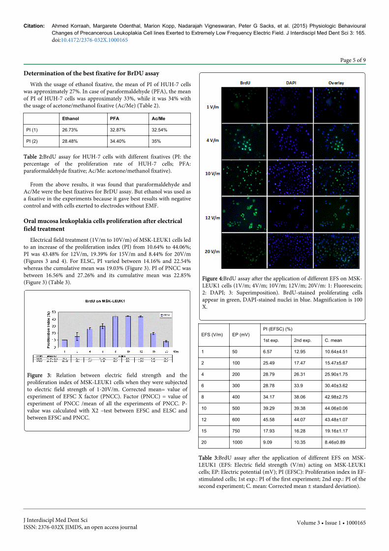
Electrical field treatment (1V/m to 10V/m) of MSK-LEUK1 cells ledto an increase of the proliferation index (PI) from 10.64% to 44.06%;PI was 43.48% for 12V/m, 19.39% for 15V/m and 8.44% for 20V/m(Figures 3 and 4). For ELSC, PI varied between 14.16% and 22.54%whereas the cumulative mean was 19.03% (Figure 3). PI of PNCC wasbetween 16.56% and 27.26% and its cumulative mean was 22.85%(Figure 3) (Table 3).
Figure 3: Relation between electric field strength and theproliferation index of MSK-LEUK1 cells when they were subjectedto electric field strength of 1-20V/m. Corrected mean= value ofexperiment of EFSC X factor (PNCC). Factor (PNCC) = value ofexperiment of PNCC /mean of all the experiments of PNCC. P-value was calculated with X2 –test between EFSC and ELSC andbetween EFSC and PNCC.
Figure 4:BrdU assay after the application of different EFS on MSK-LEUK1 cells (1V/m; 4V/m; 10V/m; 12V/m; 20V/m: 1: Fluorescein;2: DAPI; 3: Superimposition). BrdU-stained proliferating cellsappear in green, DAPI-stained nuclei in blue. Magnification is 100X.
EFS (V/m) EP (mV)PI (EFSC) (%)
1st exp. 2nd exp. C. mean
1 50 6.57 12.95 10.64±4.51
2 100 25.49 17.47 15.47±5.67
4 200 28.79 26.31 25.90±1.75
6 300 28.78 33.9 30.40±3.62
8 400 34.17 38.06 42.98±2.75
10 500 39.29 39.38 44.06±0.06
12 600 45.58 44.07 43.48±1.07
15 750 17.93 16.28 19.16±1.17
20 1000 9.09 10.35 8.46±0.89
Table 3:BrdU assay after the application of different EFS on MSK-LEUK1 (EFS: Electric field strength (V/m) acting on MSK-LEUK1cells; EP: Electric potential (mV); PI (EFSC): Proliferation index in EF-stimulated cells; 1st exp.: PI of the first experiment; 2nd exp.: PI of thesecond experiment; C. mean: Corrected mean ± standard deviation).
Citation: Ahmed Korraah, Margarete Odenthal, Marion Kopp, Nadarajah Vigneswaran, Peter G Sacks, et al. (2015) Physiologic BehaviouralChanges of Precancerous Leukoplakia Cell lines Exerted to Extremely Low Frequency Electric Field. J Interdiscipl Med Dent Sci 3: 165. doi:10.4172/2376-032X.1000165
Page 5 of 9
J Interdiscipl Med Dent SciISSN: 2376-032X JIMDS, an open access journal
Volume 3 • Issue 1 • 1000165
Upregulation of apoptosis of oral mucosa leukoplakia cellsby electrical field treatment
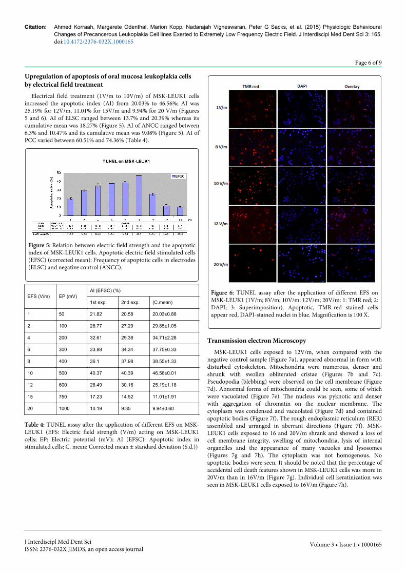
Electrical field treatment (1V/m to 10V/m) of MSK-LEUK1 cellsincreased the apoptotic index (AI) from 20.03% to 46.56%; AI was25.19% for 12V/m, 11.01% for 15V/m and 9.94% for 20 V/m (Figures5 and 6). AI of ELSC ranged between 13.7% and 20.39% whereas itscumulative mean was 18.27% (Figure 5). AI of ANCC ranged between6.3% and 10.47% and its cumulative mean was 9.08% (Figure 5). AI ofPCC varied between 60.51% and 74.36% (Table 4).
Figure 5: Relation between electric field strength and the apoptoticindex of MSK-LEUK1 cells. Apoptotic electric field stimulated cells(EFSC) (corrected mean): Frequency of apoptotic cells in electrodes(ELSC) and negative control (ANCC).
EFS (V/m) EP (mV)AI (EFSC) (%)
1st exp. 2nd exp. (C.mean)
1 50 21.82 20.58 20.03±0.88
2 100 28.77 27.29 29.85±1.05
4 200 32.61 29.38 34.71±2.28
6 300 33.88 34.34 37.75±0.33
8 400 36.1 37.98 38.55±1.33
10 500 40.37 40.39 46.56±0.01
12 600 28.49 30.16 25.19±1.18
15 750 17.23 14.52 11.01±1.91
20 1000 10.19 9.35 9.94±0.60
Table 4: TUNEL assay after the application of different EFS on MSK-LEUK1 (EFS: Electric field strength (V/m) acting on MSK-LEUK1cells; EP: Electric potential (mV); AI (EFSC): Apoptotic index instimulated cells; C. mean: Corrected mean ± standard deviation (S.d.))
Figure 6: TUNEL assay after the application of different EFS onMSK-LEUK1 (1V/m; 8V/m; 10V/m; 12V/m; 20V/m: 1: TMR red; 2:DAPI; 3: Superimposition). Apoptotic, TMR-red stained cellsappear red, DAPI-stained nuclei in blue. Magnification is 100 X.
Transmission electron MicroscopyMSK-LEUK1 cells exposed to 12V/m, when compared with the
negative control sample (Figure 7a), appeared abnormal in form withdisturbed cytoskeleton. Mitochondria were numerous, denser andshrunk with swollen obliterated cristae (Figures 7b and 7c).Pseudopodia (blebbing) were observed on the cell membrane (Figure7d). Abnormal forms of mitochondria could be seen, some of whichwere vacuolated (Figure 7e). The nucleus was pyknotic and denserwith aggregation of chromatin on the nuclear membrane. Thecytoplasm was condensed and vacuolated (Figure 7d) and containedapoptotic bodies (Figure 7f). The rough endoplasmic reticulum (RER)assembled and arranged in aberrant directions (Figure 7f). MSK-LEUK1 cells exposed to 16 and 20V/m shrank and showed a loss ofcell membrane integrity, swelling of mitochondria, lysis of internalorganelles and the appearance of many vacuoles and lysosomes(Figures 7g and 7h). The cytoplasm was not homogenous. Noapoptotic bodies were seen. It should be noted that the percentage ofaccidental cell death features shown in MSK-LEUK1 cells was more in20V/m than in 16V/m (Figure 7g). Individual cell keratinization wasseen in MSK-LEUK1 cells exposed to 16V/m (Figure 7h).
Citation: Ahmed Korraah, Margarete Odenthal, Marion Kopp, Nadarajah Vigneswaran, Peter G Sacks, et al. (2015) Physiologic BehaviouralChanges of Precancerous Leukoplakia Cell lines Exerted to Extremely Low Frequency Electric Field. J Interdiscipl Med Dent Sci 3: 165. doi:10.4172/2376-032X.1000165
Page 6 of 9
J Interdiscipl Med Dent SciISSN: 2376-032X JIMDS, an open access journal
Volume 3 • Issue 1 • 1000165

Figure 7: MSK-LEUK1 cells: (a. Negative control; b. Swollenmitochondria with obliterated cristae (12V/m); c. Different formsof apoptotic mitochondria (12V/m); d. Appearance of vacuoles andblebbing (12V/m); e. Vacuolated mitochondria (12V/m); f. RER areassembled in bundles and the appearance of apoptotic bodies(12V/m); g. Accidental cell death (20V/m); h. Individual cellkeratinization (16V/m))
Statistical analysisFor EFS of 1, 4, 6, 8, 10, 12 and 20V/m, the ratio between EFSC and
ELSC was significantly increased (P=0.0001).
Chi-square test in apoptotic assay showed the following results:
• EF-stimulation of 1 to 12V/m increased significantly (P<0.05) theapoptotic EFSC/non-apoptotic ANCC ratio.
• The ratio between EFSC and ELSC significantly increased (P<0.05)for EFS 2 to 20V/m.
• The ratio between ELSC and ANCC significantly increased(P<0.05) for EFS 1 to 20V/m.
DiscussionThe current study investigated effects of extremely low frequency
electric field treatment on the physiologic behaviour of oral
premalignant cells from the point of view of its proliferative andapoptotic activity.
It has been previously discussed that OG could lead to a change intaste, burning sensation in the oral mucosa and tongue, xerostomiaand erythema, headache and pain in eyes, arms, legs, back, neck, earsand joints [21]. Also itching, nausea, tinnitus and associated diseaseslike multiple sclerosis, myasthenia, rheumatoid arthritis and asthmacould be symptoms of OG [22]. Corrosion currents coming from OGcause teeth destruction [23,24], bone resorption [25] and several oralinflammatory disorders through the high acidity and highconcentration of dissolved metal ions formed [26]. Oral leukoplakiamay also develop in the oral mucosa due to the chronic irritationcaused by OG [11-13]. Inovay (1961) mentioned that OG could causeoral leukoplakia and lichen planus and he noted a remarkableresolution of leukoplakia when harmful potentials were eliminated[14,16]. Considering the need for well-controlled scientificexperiments to provide a definitive proof of cause and effect for OG onvarious human diseases including oral precancers and cancers, weundertook this study to evaluate the effect of OG on precancer cells invitro.
Normally, electric currents arise by unidirectional ion transport inepithelial cell layers and generate an essentially direct current (DC)voltage across the layer [27].Bergman (1986) said the basis forgalvaniccell formation is always a redox reaction:oxidationat anode (loss ofelectron) and reduction at cathode (gain of electron) [28]. In the oralcavity, there are two reasons for galvanic cell formation: differentmetals and different concentrations of the same metal [28]. Sutow(2004) found that galvanic currents for couples such as amalgam-amalgam and amalgam-gold are generally below 15 µA. He mentionedthat due to mutual polarization, the anodic corrosion rate of the morenegative metal (anode) will be accelerated, while the corrosion rate ofthe more positive metal (cathode) will be reduced [29].200µA/dm2was the maximum current density obtained between the conventionalamalgam and type III gold alloy, while no current densities wereobtained between gold and cobalt-chromium alloys [30].In ourresearch, the intensity of current was measured in all experiments andthe mean electrical current was between 5 and 20µA (data not shown).
Nowadays electromagnetic fields (EMF) are the concern of manyscientific investigators as they could potentially cause cancer [31].They suggest that if EMF have any effect on the process ofcarcinogenesis, they are more likely to act as promoters that influencethe progression of cancer than initiators of the disease. In this role,fields would enhance the proliferation of genetically altered cells ratherthan causing the initial lesion in DNA or chromatin [31]. Valberg in1997 supposed that for electric or magnetic fields to initiate orpromote inconvenient health effects in biological systems, they musthave changed the size, shape, charge, chemical state, or energy ofbiological molecules or structures. The biological system would havean actual effect when it felt the amplified change [32].
The utilization of BrdU labeling and detection assay in the study ofcellular proliferation allowed differentiation between viable andproliferating cells. BrdU can be incorporated into the newlysynthesized DNA of replicating cells during the S-phase of the cellcycle, substituting for thymidine. Antibodies specific for BrdU can beused to detect the incorporated chemical, thus indicating DNAsynthesizing and proliferating cells. DNA replication, which isindicated by the renewed BrdU assay, is the indicator of the entranceof cells in cell cycle and their proliferation. In our study, it was provedthat EFS from 2 to 10V/m could upregulate the proliferation index
Citation: Ahmed Korraah, Margarete Odenthal, Marion Kopp, Nadarajah Vigneswaran, Peter G Sacks, et al. (2015) Physiologic BehaviouralChanges of Precancerous Leukoplakia Cell lines Exerted to Extremely Low Frequency Electric Field. J Interdiscipl Med Dent Sci 3: 165. doi:10.4172/2376-032X.1000165
Page 7 of 9
J Interdiscipl Med Dent SciISSN: 2376-032X JIMDS, an open access journal
Volume 3 • Issue 1 • 1000165
(PI) of oral leukoplakia cell lines from 15% to approximately 44%(Figures 3 and 4).
In the present research, the voltage generated by electrodes inculture medium but without any connection to a DC supply wasmeasured at approximately 41 ± 11mV (n=2). This indicates thepresence of individual potentials for every metal restoration, even ifthe amount of material is small.
The effect of electrodes with no connection to a DC supply onMSK-LEUK1 cells, which is similar to amalgam-amalgam coupling inthe oral cavity, was measured to identify the effect of the presence ofmetal in an electrolyte. For EFS of 12V/m in the proliferation assay, PIwas 43.48% in EF-stimulated MSK-LEUK1 cells (EFSC), 22.54% inelectrode-stimulated cells (ELSC) and 22.17% in negative control cells(PNCC). From the above results, we concluded that electrodes alonehad little proliferative effect on OPM cell lines, while DC voltagestimulation nearly doubled the proliferation of these cell lines. At 1V/m, PI was 10.64% in EFSC while it was 18.89% in ELSC. This maybe due to inherent variability in MSK-LEUK1. The effect of the currenton MSK-LEUK1 cells is, similar to that occurring in the oral cavity, incontrast to the effect of electrodes alone. It could be attributed to thefact that the latter produced steady current on one side of the cellswhereas the former applied rotating currents that hit the cells fromdifferent sides and thus had a greater impact.
Apoptosis, or programmed cell death, is a normal physiologicprocess for maintaining tissue homeostasis [17]. There are two types ofapoptosis: extracellular (extrinsic inducers) or intracellular (intrinsicinducers). Extracellular signals may include hormones, growth factors,nitric oxideor cytokines. Intracellular apoptotic signalling is a reactioninitiated by a cell in response to stress [33,34]. The apoptotic processin our experiments could be of intracellular type where DC voltage isconsidered as chronic stress to OPM cells. An increasing number ofinvestigators favourin situ labeling of apoptotic cells through theTUNEL assay, which allows both high sensitivity and preciseidentification of the cell population involved [35]. TUNEL reactionpreferentially labels DNA strand breaks generated during apoptosis.This allows discrimination of apoptosis from accidental cell death andfrom primary DNA strand breaks induced by cytostatic drugs orirradiation. In the present study, it was proved that EFS from 1V/m to10V/m could up-regulate the apoptosis of OPM lesions in vitro from21% to 47% (Figures 5 and 6). For EFS of 8 V/m, the apoptotic index(AI) was 38.55% in EFSC, 20.39% in ELSC and 9.45% in negativecontrol cells (ANCC). From the above results, we concluded thatelectrodes alone had an apoptotic effect on leukoplakia cell lines andthis effect was accelerated, nearly double, when these electrodes wereconnected to a DC supply.
In order to explain the increase of both the proliferation and theapoptosis of MSK-LEUK1, findings of Sauer H (2002) and WartenbergM (2008) were used. OG expresses chronic irritation on leukoplakiacells; this will lead to the increase of ROS concentration in cells.Intracellular ATP will go through anion channels to the extracellularcompartment leading to the activation of purinergic receptors, whichin turn lead to the opening of calcium channels and the influx of Ca2+inside cells. Intracellular increase of Ca2+ concentration will activatemembrane receptors, which will activate Phospholipase C andgenerate inositol triphosphate that in turn leads to entrance ofquiescent leukoplakia cells into cell cycle and tumour cell proliferationand growth. In the meantime, ROS increases the expression ofNADPH oxidase, considered a pro-apoptotic enzyme. Moreover, theincrease of ROS lead to intracellular release of apoptotic signals and
the formation of apoptotic proteins which target mitochondria andlead to the release of cytochrome C into cytoplasm; that in turn lead tothe activation of caspase 3, a protein who plays an important role inthe execution of apoptosis and cancer incidence [20,36,37]. Someauthors proved that DC voltage gives rise to the generation of ROS in avariety of preparations including multicellular tumor spheroids [38],embryonic stem cells [39], primary human monocytes andlymphocytes [40] as well as human oral mucosa cancer cells [20]. It isalso possible that within stimulated MSK-LEUK1 cells, a few cellsproliferate more than others do, while other cells by the strongstimulus become apoptotic.
The data of the present study was, in part, inconsistent withfindings of Wartenberg in 2008 that cited a decrease in theproliferation rate of oral squamous carcinoma cell lines exposed to adirect current electrical field of field strength between 2 and 16V/m[20]. As the field strength was increased from 2 to 10V/m, there was anincrease in the percentage of proliferating MSK-LEUK1 cells. On theother hand, at levels of 12, 15 and 20V/m, the proliferation ratedecreased. The latter may be due to death of MSK-LEUK1 cellsthrough either accidental cell death (which are not marked in ourstudy) or apoptosis, considering that the number of cells is constant.The behavioural difference between both cell lines to DC voltage couldbe due to their differentiation status. The more differentiated MSK-LEUK1 cells proliferate while the less differentiated OSCC cellsnecrose under the effect of DC.
Other data from the present study was consistent with findings ofWartenberg in 2008 [20]. As field strength was increased from 1 to10V/m, there was an increase in the percentage of apoptotic MSK-LEUK1 cells. However, it was proved that at fields of 12, 15 and20V/m, the apoptotic rate decreased. The latter may be due to death ofMSK-LEUK1 cells through accidental cell death (which are notmarked in our experiments) other than apoptosis, considering that thenumber of cells is constant.
This study supports the observation that OG arising in vivo in theoral mucosa may disrupt the dynamic balance between proliferationand cell death leading to the development of oral mucosal diseasessuch as epithelial dysplasia and cancer.
An interesting and important finding in the electron microscopyexamination of MSK-LEUK1 cells exposed to EMF was the presence ofindividual cell keratinization (intraepithelial keratinization), amorphologic feature commonly seen in malignant oral epithelial cells[41,42]. This finding suggested that that oral galvanism-inducedsubcellular changes in oral precancer cells in vitro closely simulates themorphologic features of oral squamous cell carcinoma cells in vivo.
In conclusion, OG increases the proliferation of leukoplakia cellsand induces cell apoptosis. So, the usage of different metallicrestorations in the same patient should be avoided, especially when thetwo restorations are near to each other. Lastly, studies upon electricfields should be directed to test changes in cell signaling, particularlyeffects on ornithine decarboxylase (ODC) (due to its role inproliferation of cancer as it is upregulated in many types of cancer)and Na+K+-ATPase activity (due to its role in ion transport).
References1. Momoi Y, Asanuma A, Kohno A, Yanagisawa K (1986) A measurement
of galvanic current and electrical potential in extracted human teeth. JDent Res 65: 1441-1444.
Citation: Ahmed Korraah, Margarete Odenthal, Marion Kopp, Nadarajah Vigneswaran, Peter G Sacks, et al. (2015) Physiologic BehaviouralChanges of Precancerous Leukoplakia Cell lines Exerted to Extremely Low Frequency Electric Field. J Interdiscipl Med Dent Sci 3: 165. doi:10.4172/2376-032X.1000165
Page 8 of 9
J Interdiscipl Med Dent SciISSN: 2376-032X JIMDS, an open access journal
Volume 3 • Issue 1 • 1000165

2. SCHRIEVER W, DIAMOND LE (1952) Electromotive forces and electriccurrents caused by metallic dental fillings. J Dent Res 31: 205-229.
3. Pindborg JJ, Jolst O, Renstrup G, Roed-Petersen B (1968) Studies in oralleukoplakia: a preliminary report on the period pervalence of malignanttransformation in leukoplakia based on a follow-up study of 248 patients.J Am Dent Assoc 76: 767-771.
4. Warnakulasuriya S, Johnson NW, van der Waal I (2007) Nomenclatureand classification of potentially malignant disorders of the oral mucosa. JOral Pathol Med 36: 575-580.
5. López M, Aguirre JM, Cuevas N, Anzola M, Videgain J, et al. (2003) Genepromoter hypermethylation in oral rinses of leukoplakia patients--adiagnostic and/or prognostic tool? Eur J Cancer 39: 2306-2309.
6. Pindborg JJ, Daftary DK, Mehta FS (1977) A follow-up study of sixty-oneoral dysplastic precancerous lesions in Indian villagers. Oral Surg OralMed Oral Pathol 43: 383-390.
7. Pindborg JJ. Oral Cancer and Precancer 1st ed. Bristol: John Wright:1980.
8. Lippman SM, Sudbø J, Hong WK (2005) Oral cancer prevention and theevolution of molecular-targeted drug development. J ClinOncol 23:346-356.
9. Vigneswaran N, Beckers S, Waigel S, Mensah J, Wu J, et al. (2006)Increased EMMPRIN (CD 147) expression during oral carcinogenesis.ExpMolPathol 80: 147-159.
10. Neville B, Damm D, Allen C, Bouquot J (2002) Oral and MaxillofacialPathology: Epithelial Pathology. WB Saunders, Philadelphia, USA.p315-387.
11. Ullmann K (1932) Leukoplakia caused by electrogalvanic currentgenerated in the oral cavity. Wien KlinWochenschr 45: 840-844.
12. Lain ES (1933) Electrogalvanic lesions of the oral cavity produced bymetallic dentures. The Journal of American Medical Association 100:717-720.
13. Muller AW1, Van Loon LA, Davidson CL (1990) Electrical potentials ofrestorations in subjects without oral complaints. J Oral Rehabil 17:419-424.
14. Inovay J, Banoczy J (1961) The role of electrical potential differences inthe etiology of chronic diseases of the oral mucosa. J Dent Res 40:884-890.
15. Bánóczy J, Roed-Petersen B, Pindborg JJ, Inovay J (1979) Clinical andhistologic studies on electrogalvanically induced oral white lesions. OralSurg Oral Med Oral Pathol 48: 319-323.
16. Psarras V, Derand T, Nilner K (1994) Effect of selenium on mercuryvapour released from dental amalgams: an in vitro study. Swed Dent J 18:15-23.
17. Kerr JF, Wyllie AH, Currie AR (1972) Apoptosis: a basic biologicalphenomenon with wide-ranging implications in tissue kinetics. Br JCancer 26: 239-257.
18. Thompson CB1 (1995) Apoptosis in the pathogenesis and treatment ofdisease. Science 267: 1456-1462.
19. Sacks PG (1996) Cell, tissue and organ culture as in vitro models to studythe biology of squamous cell carcinomas of the head and neck. CancerMetastasis Rev 15: 27-51.
20. Wartenberg M, Wirtz N, Grob A, Niedermeier W, Hescheler J et al.(2008) Direct current electrical fields induce apoptosis in oral mucosacancer cells by NADPH oxidase-derived reactive oxygen species.Bioelectromagnetics 29: 47-54.
21. Johannson BI, Stenman E, Bergman M (1986) Clinical registration ofcharge transfer between dental metallic materials in patients withdisorders and/or discomfort allegedly caused by corrosion. Scand J DentRes 94: 357-63.
22. Schmalz G, Garhammer P (2002) Biological interactions of dental castalloys with oral tissues. Dent Mater 18: 396-406.
23. Chase HS (1879) Oral Electricity. Dent Cosmos 21:205-207.24. Meyer RD, Meyer J, Taloumis LJ (1993) Intraoral galvanic corrosion:
literature review and case report. J Prosthet Dent 69: 141-143.25. Olmedo D, Fernández MM, Guglielmotti MB, Cabrini RL (2003)
Macrophages related to dental implant failure. Implant Dent 12: 75-80.26. Marek M (1992) Interactions between dental amalgams and the oral
environment. Adv Dent Res 6: 100-109.27. Jaffe LF, Nuccitelli R (1977) Electrical controls of development. Annu
Rev BiophysBioeng 6: 445-476.28. Bergman M (1986) Corrosion in the oral cavity--potential local and
systemic effects. Int Dent J 36: 41-44.29. Sutow EJ, Maillet WA, Taylor JC, Hall GC (2004) In vivo galvanic
currents of intermittently contacting dental amalgam and other metallicrestorations. Dent Mater 20: 823-831.
30. Arvidson K, Johansson EG (1985) Galvanic currents between dentalalloys in vitro. Scand J Dent Res 93: 467-473.
31. Repacholi MH, Greenebaum B (1999) Interaction of static and extremelylow frequency electric and magnetic fields with living systems: healtheffects and research needs. Bioelectromagnetics 20: 133-160.
32. Valberg PA, Kavet R, Rafferty CN (1997) Can low-level 50/60 Hz electricand magnetic fields cause biological effects? Radiat Res 148: 2-21.
33. Cotran RS, Kumar V, Collins T, editors (1999) Robbins pathologic basisof disease. WB Saunders Co, Philadelphia, USA. p1-29.
34. Latenser BA (2008) Apoptotic death in deep partial thickness burnsversus normal skin of burned patients. J Surg Res 146: 161-163.
35. Gold R, Schmied M, Giegerich G, Breitschopf H, Hartung HP, et al.(1994) Differentiation between cellular apoptosis and necrosis by thecombined use of in situ tailing and nick translation techniques. LabInvest 71: 219-225.
36. Sauer H, Stanelle R, Hescheler J, Wartenberg M (2002) The DCelectrical-field-induced Ca2+ response and growth stimulation ofmulticellular tumor spheroids are mediated by ATP release andpurinergic receptor stimulation. J Cell Sci 115: 3265-73.
37. Valko M, Leibfritz D, Moncol J, Cronin MT, Mazur M et al. (2007) Freeradicals and antioxidants in normal physiological functions and humandisease. Int J Biochem Cell Biol 39: 44-84.
38. Wartenberg M, Hescheler J, Sauer H (1997) Electrical fields enhancegrowth of cancer spheroids by reactive oxygen species and intracellularCa2+. Am J Physiol 272: R1677-1683.
39. Sauer H, Bekhite MM, Hescheler J, Wartenberg M (2005) Redox controlof angiogenic factors and CD31-positive vessel-like structures in mouseembryonic stem cells after direct current electrical field stimulation. ExpCell Res 304: 380-390.
40. Lantow M, Lupke M, Frahm J, Mattsson MO, Kuster N, et al. (2006) ROSrelease and Hsp70 expression after exposure to 1,800 MHzradiofrequency electromagnetic fields in primary human monocytes andlymphocytes. Radiat Environ Biophys 45: 55-62.
41. Kramer IR, el-Labban NG, Sonkodi S (1974) Further studies on lesions ofthe oral mucosa using computer-aided analyses of histological features.Br J Cancer 29: 223-231.
42. Reibel J (2003) Prognosis of oral pre-malignant lesions: significance ofclinical, histopathological, and molecular biological characteristics. CritRev Oral Biol Med 14: 47-62.
Citation: Ahmed Korraah, Margarete Odenthal, Marion Kopp, Nadarajah Vigneswaran, Peter G Sacks, et al. (2015) Physiologic BehaviouralChanges of Precancerous Leukoplakia Cell lines Exerted to Extremely Low Frequency Electric Field. J Interdiscipl Med Dent Sci 3: 165. doi:10.4172/2376-032X.1000165
Page 9 of 9
J Interdiscipl Med Dent SciISSN: 2376-032X JIMDS, an open access journal
Volume 3 • Issue 1 • 1000165


![BP520 JBR 517 JBR - Universal Airlinesuvairlines.com/admin/resources/LHBP.pdfABONY 1L [ABO1L], JBR 1L Licensed to BRITISH AIRWAYS PLC, . Printed from JeppView disc 23-06. Notice: After](https://img.pdfslide.us/doc/110x75/603a9ab8acfc0749f75c4eb7/bp520-jbr-517-jbr-universal-abony-1l-abo1l-jbr-1l-licensed-to-british-airways.jpg)