Embed Size (px)
Citation preview

C h a p t e r
22
The Cardiovascular
System —Vessels and Circulation
PowerPoint® Lecture Slides
prepared by Jason LaPres
North Harris College
Houston, Texas
Copyright © 2009 Pearson Education, Inc.,
publishing as Pearson Benjamin Cummings

Introduction
The cardiovascular system is a closed system
that circulates blood.
There are two groups of blood vessels:
One supplies the lungs (the pulmonary circuit).
The other supplies the rest of the body (the
systemic circuit).
Blood is pumped from the heart into both the
pulmonary and systemic (aortic) trunks
simultaneously.
Copyright © 2009 Pearson Education, Inc., publishing as Pearson Benjamin Cummings

Histological Organization of Blood Vessels
Blood vessel walls
Tunica intima
Inner layer
Endothelium and CT
Tunica media
Middle layer
Smooth muscle and CT
Adventitia
Outer layer
Mostly CT
Copyright © 2009 Pearson Education, Inc., publishing as Pearson Benjamin Cummings

Histological Organization of Blood Vessels
Figure 22.1 Histological Comparison of Typical Arteries and Veins
Copyright © 2009 Pearson Education, Inc., publishing as Pearson Benjamin Cummings

Histological Organization of Blood Vessels
Figure 22.2 Histological Structure of Blood Vessels
Copyright © 2009 Pearson Education, Inc., publishing as Pearson Benjamin Cummings

Histological Organization of Blood Vessels
Types of arteries:
Elastic arteries, or conducting arteries, are large
vessels with diameters of up to 2.5 cm (1 in.).
Muscular arteries, or distribution arteries (also
known as medium-sized arteries), transport blood
to the body’s skeletal muscle and internal organs.
Arterioles are considerably smaller than muscular
arteries.
Arterioles have an average diameter of about 30 μm.
Copyright © 2009 Pearson Education, Inc., publishing as Pearson Benjamin Cummings

Histological Organization of Blood Vessels
Figure 22.3 A Plaque Blocking a Peripheral Artery
Copyright © 2009 Pearson Education, Inc., publishing as Pearson Benjamin Cummings

Histological Organization of Blood Vessels
Capillaries:
Smallest and most delicate blood vessels
Their walls permit exchange between the blood and the surrounding interstitial fluids
Types of capillaries:
Continuous— has a complete endothelial lining
Fenestrated— contains “windows” in their walls due to perforated endothelial lining
Sinusoids— specialized fenestrated capillaries found in selected tissues (such as the liver) that allow very slow blood flow
Copyright © 2009 Pearson Education, Inc., publishing as Pearson Benjamin Cummings

Histological Organization of Blood Vessels
Figure 22.4 Structure of Capillaries
Copyright © 2009 Pearson Education, Inc., publishing as Pearson Benjamin Cummings

Histological Organization of Blood Vessels
Figure 22.5 Organization of a Capillary Bed
Copyright © 2009 Pearson Education, Inc., publishing as Pearson Benjamin Cummings
Histological Organization of Blood Vessels
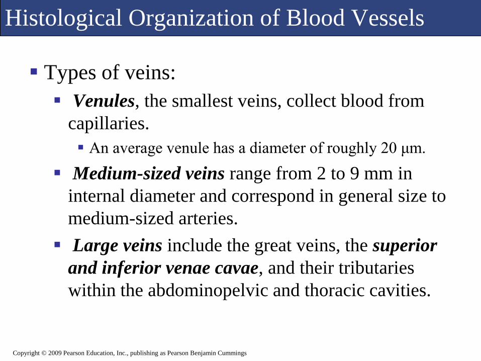
Types of veins:
Venules, the smallest veins, collect blood from
capillaries.
An average venule has a diameter of roughly 20 μm.
Medium-sized veins range from 2 to 9 mm in
internal diameter and correspond in general size to
medium-sized arteries.
Large veins include the great veins, the superior
and inferior venae cavae, and their tributaries
within the abdominopelvic and thoracic cavities.
Copyright © 2009 Pearson Education, Inc., publishing as Pearson Benjamin Cummings

Histological Organization of Blood Vessels
Figure 22.6 Function of Valves in the Venous System
Copyright © 2009 Pearson Education, Inc., publishing as Pearson Benjamin Cummings

Histological Organization of Blood Vessels
Figure 22.7 The Distribution of Blood in the Cardiovascular System
Copyright © 2009 Pearson Education, Inc., publishing as Pearson Benjamin Cummings

Blood Vessel Distribution
Distribution of arteries and veins on the left and right sides is usually identical except near the heart.
A single vessel may have several different names as it crosses specific anatomical boundaries, making accurate anatomical descriptions possible.
Arteries and veins often make anastomoticconnections that reduce the impact of blockage of a single vessel.
Copyright © 2009 Pearson Education, Inc., publishing as Pearson Benjamin Cummings

Blood Vessel Distribution
Figure 22.8 An Overview of the General Pattern of Circulation
Copyright © 2009 Pearson Education, Inc., publishing as Pearson Benjamin Cummings

Blood Vessel Distribution
Pulmonary circuit
Carries deoxygenated blood to the lungs then back to the heart
Includes the pulmonary trunk, the left and right pulmonary arteries, and the pulmonary veins
Systemic circuit
Carries oxygenated blood to the body tissues
Includes the coronary circuit, which supplies the myocardium
Copyright © 2009 Pearson Education, Inc., publishing as Pearson Benjamin Cummings

Blood Vessel Distribution
Figure 22.9a The Pulmonary Circuit: (a) Anatomy of the Pulmonary Circuit
Copyright © 2009 Pearson Education, Inc., publishing as Pearson Benjamin Cummings

Blood Vessel Distribution
Figure 22.9b The Pulmonary Circuit: (b) Coronary Angiogram
Copyright © 2009 Pearson Education, Inc., publishing as Pearson Benjamin Cummings

Blood Vessel Distribution
Figure 22.10 An Overview of the Systemic Arterial System
Copyright © 2009 Pearson Education, Inc., publishing as Pearson Benjamin Cummings

Blood Vessel Distribution
Figure 22.11 Aortic Angiogram
Copyright © 2009 Pearson Education, Inc., publishing as Pearson Benjamin Cummings

Blood Vessel Distribution
Figure 22.12a Arteries of the Chest and Upper Limb: (a) Arteries of the Chest and Upper Limb
Copyright © 2009 Pearson Education, Inc., publishing as Pearson Benjamin Cummings

Blood Vessel Distribution
Figure 22.12b Arteries of the Chest and Upper Limb: (b) Right Forearm, Anterior View
Copyright © 2009 Pearson Education, Inc., publishing as Pearson Benjamin Cummings
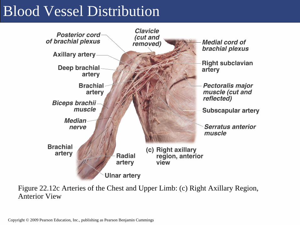
Blood Vessel Distribution
Figure 22.12c Arteries of the Chest and Upper Limb: (c) Right Axillary Region, Anterior View
Copyright © 2009 Pearson Education, Inc., publishing as Pearson Benjamin Cummings

Blood Vessel Distribution
Figure 22.12d Arteries of the Chest and Upper Limb: (d) Summary of Arterial Distribution from Aortic Arch
Copyright © 2009 Pearson Education, Inc., publishing as Pearson Benjamin Cummings

Blood Vessel Distribution
Figure 22.13a Arteries of the Neck and Head: (a) Arteries of Neck and Head, an Oblique Lateral View from the Right Side
Copyright © 2009 Pearson Education, Inc., publishing as Pearson Benjamin Cummings

Blood Vessel Distribution
Figure 22.13b Arteries of the Neck and Head: (b) Angiogram, Lateral Projection
Copyright © 2009 Pearson Education, Inc., publishing as Pearson Benjamin Cummings

Blood Vessel Distribution
Figure 22.14 Major Arteries of the Neck
Copyright © 2009 Pearson Education, Inc., publishing as Pearson Benjamin Cummings

Blood Vessel Distribution
Figure 22.15a The Arterial Supply to the Brain: (a) Arteries of the Brain, Inferior Vew
Copyright © 2009 Pearson Education, Inc., publishing as Pearson Benjamin Cummings

Blood Vessel Distribution
Figure 22.15b The Arterial Supply to the Brain: (b) Arteries Injected to Show Cerebral Arterial Circle
Copyright © 2009 Pearson Education, Inc., publishing as Pearson Benjamin Cummings

Blood Vessel Distribution
Figure 22.15c The Arterial Supply to the Brain: (c) Corrosion Cast of Cerebral Arteries, Left Cerebral Hemisphere
Copyright © 2009 Pearson Education, Inc., publishing as Pearson Benjamin Cummings

Blood Vessel Distribution
Figure 22.16 Major Arteries of the Trunk
Copyright © 2009 Pearson Education, Inc., publishing as Pearson Benjamin Cummings
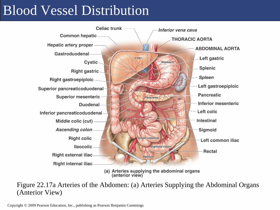
Blood Vessel Distribution
Figure 22.17a Arteries of the Abdomen: (a) Arteries Supplying the Abdominal Organs (Anterior View)
Copyright © 2009 Pearson Education, Inc., publishing as Pearson Benjamin Cummings

Blood Vessel Distribution
Figure 22.17b Arteries of the Abdomen: (b) Angiogram of the Abdominal Aorta
Copyright © 2009 Pearson Education, Inc., publishing as Pearson Benjamin Cummings

Blood Vessel Distribution
Figure 22.18a Major Arteries of the Lower Limb, Part I: (a) Anterior View
Copyright © 2009 Pearson Education, Inc., publishing as Pearson Benjamin Cummings
Blood Vessel Distribution
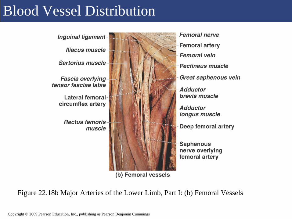
Figure 22.18b Major Arteries of the Lower Limb, Part I: (b) Femoral Vessels
Copyright © 2009 Pearson Education, Inc., publishing as Pearson Benjamin Cummings

Blood Vessel Distribution
Figure 22.19a Major Arteries of the Lower Limb, Part II: (a) Posterior View
Copyright © 2009 Pearson Education, Inc., publishing as Pearson Benjamin Cummings

Blood Vessel Distribution
Figure 22.19b Major Arteries of the Lower Limb, Part II: (b) Major Arteries of the Lower Limb
Copyright © 2009 Pearson Education, Inc., publishing as Pearson Benjamin Cummings

Blood Vessel Distribution
Figure 22.20 A Summary of the Arterial System
Copyright © 2009 Pearson Education, Inc., publishing as Pearson Benjamin Cummings

Blood Vessel Distribution
Figure 22.29 Radiograph of Tony’s Left Subclavian Artery Showing Decreased Diameter
Copyright © 2009 Pearson Education, Inc., publishing as Pearson Benjamin Cummings

Blood Vessel Distribution
Figure 22.21 An Overview of the Systemic Venous System
Copyright © 2009 Pearson Education, Inc., publishing as Pearson Benjamin Cummings

Blood Vessel Distribution
Figure 22.22a Major Veins of the Head and Neck: (a) Veins of the Head and Neck, Lateral View
Copyright © 2009 Pearson Education, Inc., publishing as Pearson Benjamin Cummings

Blood Vessel Distribution
Figure 22.22b Major Veins of the Head and Neck: (b) Venous Drainage of the Brain, Inferior View
Copyright © 2009 Pearson Education, Inc., publishing as Pearson Benjamin Cummings

Blood Vessel Distribution
Figure 22.23 The Venous Drainage of the Trunk and Upper Limb
Copyright © 2009 Pearson Education, Inc., publishing as Pearson Benjamin Cummings

Blood Vessel Distribution
Figure 22.24a A Summary Flowchart of the Venous System: (a) Tributaries of the Superior Vena Cava
Copyright © 2009 Pearson Education, Inc., publishing as Pearson Benjamin Cummings

Blood Vessel Distribution
Figure 22.24b A Summary Flowchart of the Venous System: (b) Tributaries of the Inferior Vena Cava
Copyright © 2009 Pearson Education, Inc., publishing as Pearson Benjamin Cummings

Blood Vessel Distribution
Figure 22.24c A Summary Flowchart of the Venous System: (c) Summary of the Veins of the Lower Limb
Copyright © 2009 Pearson Education, Inc., publishing as Pearson Benjamin Cummings

Blood Vessel Distribution
Figure 22.25a The Venous Drainage of the Lower Limb: (a) Anterior View
Copyright © 2009 Pearson Education, Inc., publishing as Pearson Benjamin Cummings

Blood Vessel Distribution
Figure 22.25b The Venous Drainage of the Lower Limb: (b) Posterior View
Copyright © 2009 Pearson Education, Inc., publishing as Pearson Benjamin Cummings

Blood Vessel Distribution
Figure 22.26 The Hepatic Portal System
Copyright © 2009 Pearson Education, Inc., publishing as Pearson Benjamin Cummings

Cardiovascular Changes at Birth
Changes are made to adapt to extra-uterine life
Blood shunts close
Blood begins to be pumped to the lungs
Newborn oxygenates blood for the first time
Copyright © 2009 Pearson Education, Inc., publishing as Pearson Benjamin Cummings

Cardiovascular Changes at Birth
Figure 22.27a Changes in Fetal Circulation at Birth: (a) Full-term fetus (Before Birth)
Copyright © 2009 Pearson Education, Inc., publishing as Pearson Benjamin Cummings

Cardiovascular Changes at Birth
Figure 22.27b Changes in Fetal Circulation at Birth: (b) After Delivery
Copyright © 2009 Pearson Education, Inc., publishing as Pearson Benjamin Cummings

Cardiovascular Changes at Birth
Figure 22.27c Changes in Fetal Circulation at Birth: (c) Fetal Circulatory Pattern
Copyright © 2009 Pearson Education, Inc., publishing as Pearson Benjamin Cummings

Cardiovascular Changes at Birth
Figure 22.28 Congenital Cardiovascular Problems
Copyright © 2009 Pearson Education, Inc., publishing as Pearson Benjamin Cummings

Aging and the Cardiovascular System
Changes occur in the blood, heart, and blood vessels.
Blood changes
Decreased HCT
Thrombi and emboli form more easily
Blood pools in legs
Heart changes
Reduced efficiency and elasticity
Atherosclerosis of coronary vessels
Scar tissue forms
Blood vessel changes
Loss of elasticity
Calcium deposits damage vessel walls
Copyright © 2009 Pearson Education, Inc., publishing as Pearson Benjamin Cummings