Embed Size (px)
Citation preview

Recognizing and managing the fetus with channelopathy
Bettina Cuneo MD
Professor of Pediatrics and Obstetrics University of Colorado and Children’s
Hospital Colorado
Disclosure: I am a consultant for Philips Ultrasound

(Fetal) Long QT Syndrome: Background
• An inherited channelopathy and the leading cause of sudden arrhythmic death in children and young adults
• Causative in ~10% • “normal” IUFDs (Crotti et. al. JAMA 2013)
• SIDS (Schwartz et. al. Circulation 2007)
• neonatal epilepsy deaths (Tu E. et al. Brain Pathol 2011)
• Fetal ascertainment is poor even with a + family history • Live-born population: 1/2-2500 individuals • Fetal population: 1/8658 (Flock U J Mat Fetal Neo Med 2015) • Most common presentation: sinus bradycardia, a subtle rhythm
disturbance often unappreciated to be abnormal • Even fetuses with signature rhythms of VT and/or 2°AVB
• Unsuspected, undiagnosed or misdiagnosed
• Delivered prematurely and/or by C-section
• Failure to recognize fetal LQTS is a missed opportunity for primary prevention of life threatening ventricular arrhythmias

“…This report (of the first confirmed case of Romano Ward syndrome diagnosed prenatally) confirms that moderate fetal bradycardia (110-120 bpm) does not indicate fetal distress, but indicates that fetuses should be studied for fetal cardiac conduction defects in the newborn period”
1989: First Case of Fetal Bradycardia Recognized as LQTS
Vigliani M. J Reprod Med 1995
Mother, maternal grandmother and infant had prolonged QTc on ECG
Sinus Rates of Fetal LQTS Subjects
Mitc
hell
J e
t al C
ircul
atio
n 20
12
OB definition of bradycardia
50th
3rd
97th
Mitchell J Circulation 2012

5
• Retrospective study 3rd trimester (29-41 weeks)
• FHR from 184 fetuses with parental LQT1
• 110 mutation carriers • FHR varied with number
of mutations and disease severity
• Some double mutation carriers had FHR>110 bpm
“..the current OB standard for fetal bradycardia is not useful with regards to LQTS…but what FHR should signal the need for what type of follow-up is not yet known.”
143 ± 5
131 ± 10
111 ± 6
More on FHR and LQTS Winbo A et al. Circ Arrhythm Electrophysiol 2015; 8:805-814
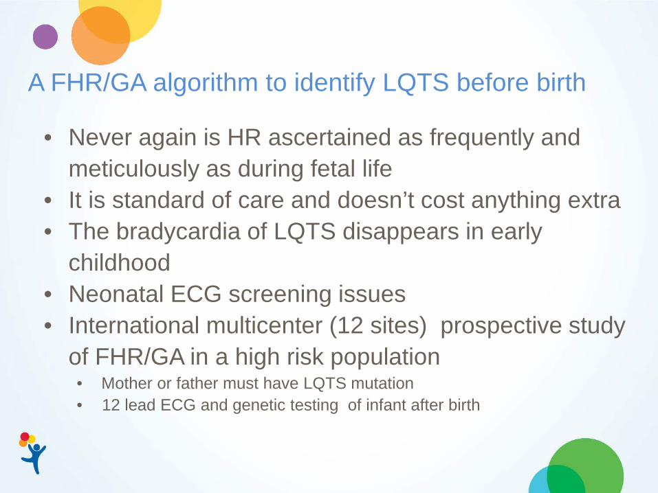
A FHR/GA algorithm to identify LQTS before birth
• Never again is HR ascertained as frequently and meticulously as during fetal life
• It is standard of care and doesn’t cost anything extra • The bradycardia of LQTS disappears in early
childhood • Neonatal ECG screening issues • International multicenter (12 sites) prospective study
of FHR/GA in a high risk population • Mother or father must have LQTS mutation • 12 lead ECG and genetic testing of infant after birth
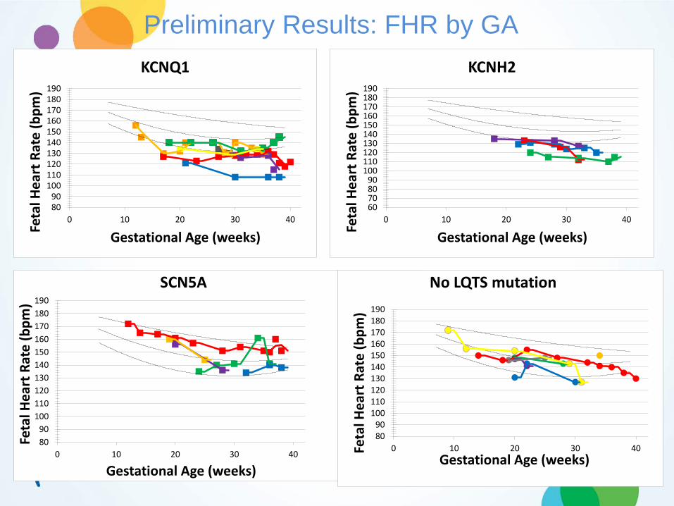
Preliminary Results: FHR by GA
8090
100110120130140150160170180190
0 10 20 30 40
Feta
l Hea
rt R
ate
(bpm
)
Gestational Age (weeks)
SCN5A
8090
100110120130140150160170180190
0 10 20 30 40
Feta
l Hea
rt R
ate
(bpm
)
Gestational Age (weeks)
KCNQ1
8090
100110120130140150160170180190
0 10 20 30 40Feta
l Hea
rt R
ate
(bpm
)
Gestational Age (weeks)
No LQTS mutation
60708090
100110120130140150160170180190
0 10 20 30 40
Feta
l Hea
rt R
ate
(bpm
)
Gestational Age (weeks)
KCNH2
Maybe its more than the FHR/Rhythm? Other features of fetal LQTS
IRT ICT IVCT IVRT

IRT differentiates immune-mediated 2° from LQTS “2° AVB”
IRT ICT
Anti-SSA mediated 2° AVB LQTS
Sonesson S-E et al. Ultrasound Obstet Gynecol 2014; 44: 171–175
LQTS: IRT longer (105 v. 47.5 ±13.8 ms) ICT shorter (7 v. 60.9 ± 22.6 ms)
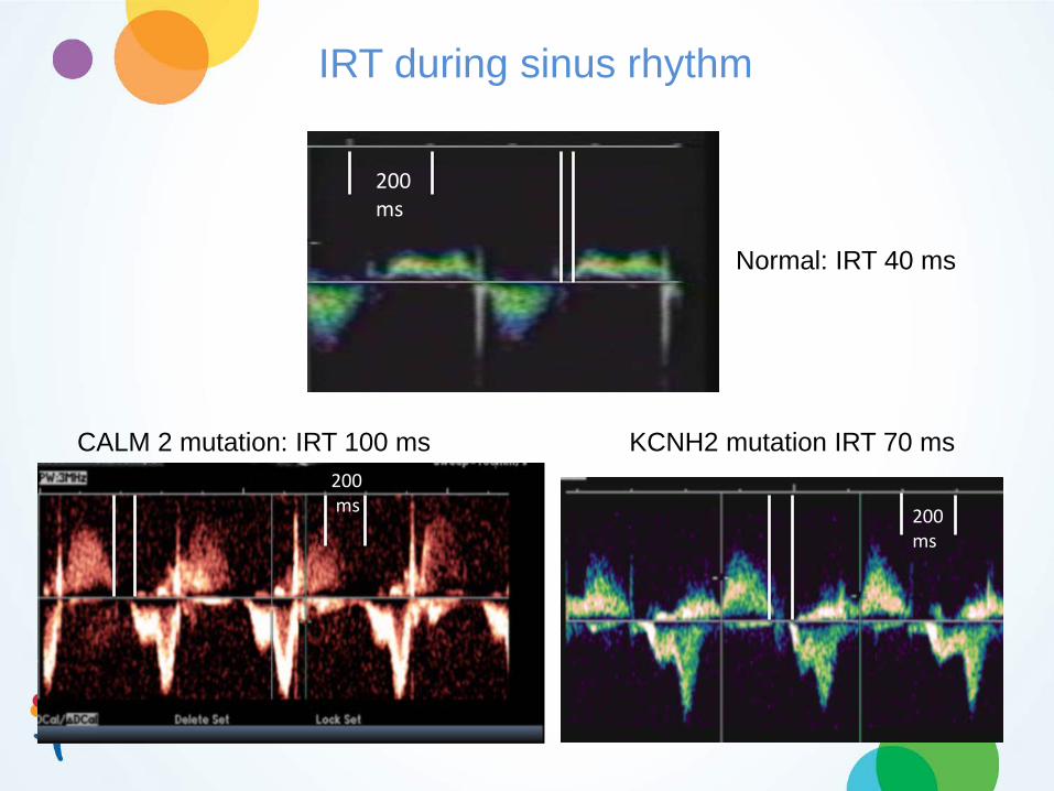
IRT during sinus rhythm
Normal: IRT 40 ms
CALM 2 mutation: IRT 100 ms KCNH2 mutation IRT 70 ms
200 ms
200 ms 200
ms

CALM 2 mutation
.Acherman RJ et al. Right ventricular noncompaction associated with long QT in a fetus with right ventricular hypertrophy and cardiac arrhythmias Prenat Diagn 2008; 28: 551–553
Cardiac Dysfunction in LQTS

Non-invasive ‘gold standard’ for LQTS diagnosis: Fetal Magnetocardiography (fMCG):
Recorded without direct contact with source (mother) Superconducting quantum Interference device (SQUID) Unaffected by amniotic fluid or vernix Excellent signal to noise ratio
Limited maternal (signal) interference Can be recorded from 18-40 weeks

Results: Genetics of LQTS Rhythms
5 Fetal TdP + 2° AVB
SCN5A CALM 2
KCNH2
3 Fetal 2° AVB
R1623Q (n=2) L409P
G628S T613K
Not tested
8
CACNA1C G406A
39 referred for fMCG
31 (26) Sinus Bradycardia
KCNQ1 SCN5A E1784K (1)
Red = +family history
Circulation. 2013;128:2183-2191
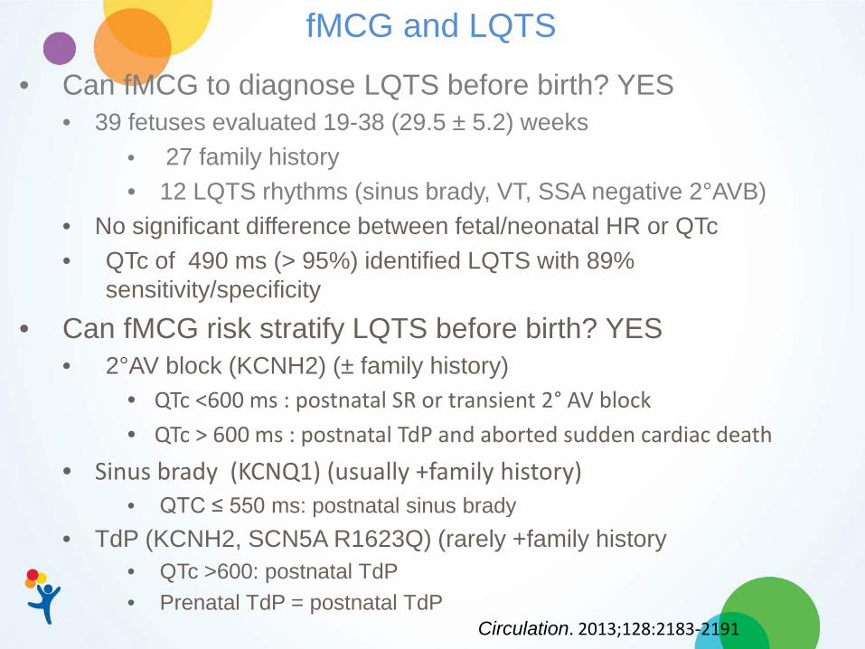
fMCG and LQTS
• Can fMCG to diagnose LQTS before birth? YES • 39 fetuses evaluated 19-38 (29.5 ± 5.2) weeks
• 27 family history • 12 LQTS rhythms (sinus brady, VT, SSA negative 2°AVB)
• No significant difference between fetal/neonatal HR or QTc • QTc of 490 ms (> 95%) identified LQTS with 89%
sensitivity/specificity
• Can fMCG risk stratify LQTS before birth? YES • 2°AV block (KCNH2) (± family history)
• QTc <600 ms : postnatal SR or transient 2° AV block • QTc > 600 ms : postnatal TdP and aborted sudden cardiac death
• Sinus brady (KCNQ1) (usually +family history) • QTC ≤ 550 ms: postnatal sinus brady
• TdP (KCNH2, SCN5A R1623Q) (rarely +family history • QTc >600: postnatal TdP • Prenatal TdP = postnatal TdP
Circulation. 2013;128:2183-2191
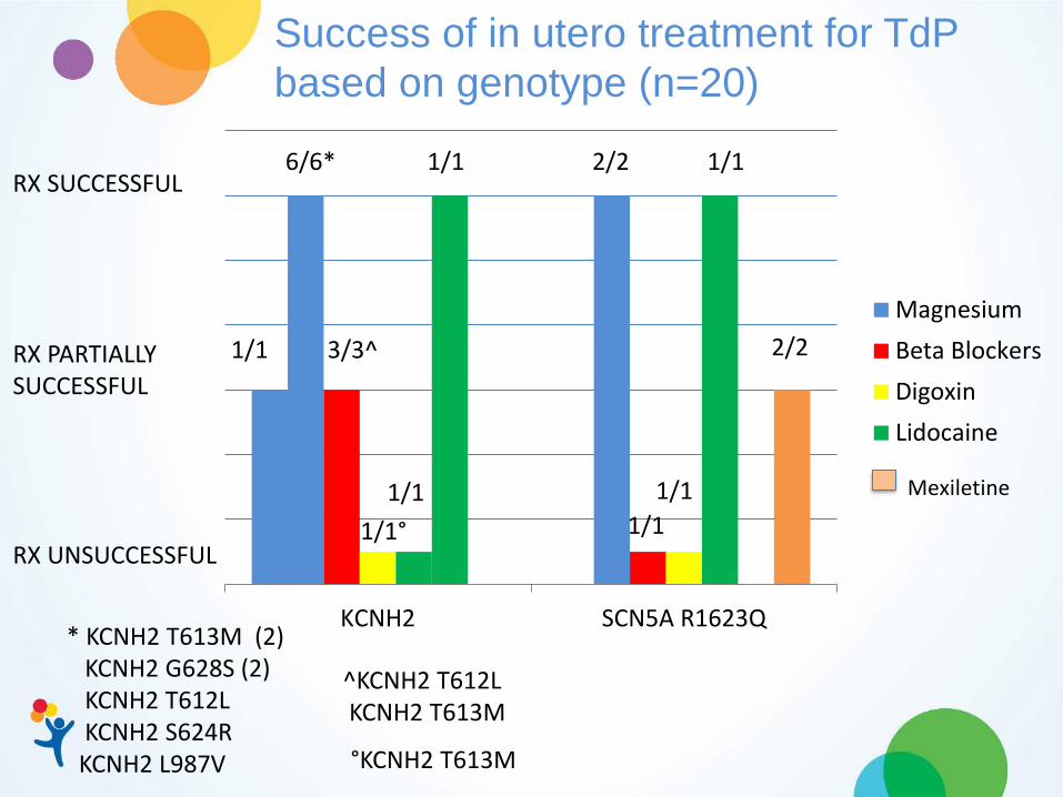
KCNH2 SCN5A R1623Q
MagnesiumBeta BlockersDigoxinLidocaine
1/1 Mexiletine
RX SUCCESSFUL 6/6*
3/3^ 1/1 RX PARTIALLY SUCCESSFUL
1/1° RX UNSUCCESSFUL
* KCNH2 T613M (2) KCNH2 G628S (2) KCNH2 T612L KCNH2 S624R KCNH2 L987V
^KCNH2 T612L KCNH2 T613M
°KCNH2 T613M
2/2
1/1
1/1
2/2
1/1
1/1
Success of in utero treatment for TdP based on genotype (n=20)

Fetal Surveillance w. LQTS Family History
• Monthly FHR • After 32 weeks qo week FHR
• Monthly FHR • Between 20-24 weeks:
• Fetal echo • After 30 weeks
• Follow-up fMCG • q week non-stress testing • qo week fetal echo
If LQT2 + If LQT3 +
• Postnatal ECG and Genetic testing
If LQT1 +
• Treat maternal Mg and/or 25,OH Vit D deficiency • No QT prolonging meds • Continue maternal BB if mother LQTS + • fMCG at 24-28 wks
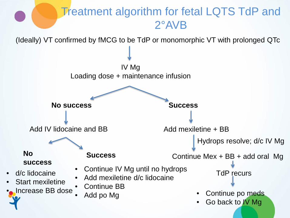
Treatment algorithm for fetal LQTS TdP and 2°AVB
(Ideally) VT confirmed by fMCG to be TdP or monomorphic VT with prolonged QTc
IV Mg Loading dose + maintenance infusion
Success
Add IV lidocaine and BB
No success
Add mexiletine + BB
Continue Mex + BB + add oral Mg
Hydrops resolve; d/c IV Mg
TdP recurs
• Continue po meds • Go back to IV Mg
• Continue IV Mg until no hydrops • Add mexiletine d/c lidocaine • Continue BB • Add po Mg
• d/c lidocaine • Start mexiletine • Increase BB dose
Success No success

Future Directions
• Embrace a paradigm shift from post-event recognition of LQTS and secondary prevention to prenatal recognition and primary prevention of ventricular arrhythmia • Adopt a population based strategy before birth to most
effectively identify individuals at risk for sudden death. • Develop an ascertainment technique with high
sensitivity/specificity • Educate OB colleagues about presentation and in utero
LQTS management • Improve communication between “pediatric” “adult” and
“fetal” cardiologists
• Prevent sudden death in the young by identifying risk of sudden death in the youngest
References 1. Hofbeck U, Ulmer H, Beinder E et al. Prenatal findings in patients with prolonge QT interval in the neonatal
period. Heart, 1997: 77: 198-204. 2. Lin MT, Hsich FJ, Shyu MK, et al. Postnatal outcome of fetal bradycardia without significant cardiac
abnormalities. Am Heart J 2004; 147: 540-544. 3. Anuwutnavin S, Wanitpongpan P, Chungsomprasong P, et al. Fetal long QT syndrome manifested as
Atrioventricular block and ventricular tachycardia: A case report and review of the literature. Pediatr Cardiol 2013 34: 1995-1962.
4. Simpson JM, Maxwell D, Rosenthal E, Gill H. Fetal ventricular tachycardia secondary to long QT sydrome treated with intravenous magnesium: a case report and review of the literature. Ultrasound Obstet Gynecol 2009; 34:475-480.
5. Ohkuchi A, Shiraishi H, Minakami H, et. al. Fetus with long QT syndrome manifested by tachyarrhythmia: A case report. Prenat Diagn 1999;19: 990-992.
6. Chang IK, Shyu MK, Lee CN. Prenatal diagnosis and treatment of fetal long QT syndrome: a case report. Prenat Diagn 2002; 22: 1209-1212.
7. Acherman RJ, Evans WN, Schwartz JK, et al. Right ventricular noncompaction associated with long QT a fetus with right ventricular hypertrophy and cardiac arrhythmias. Prenat Diagn 2008;28:551-553.
8. Flock A, HerbergU, Gembruch U, Mertz WM. Clinical spectrum of fetal long QT syndrome: A single center experience. Journal of Maternal-Fetal and Neonatal Medicine 2015; 28:1731-1735.
9. Sonesson SE, Eliasson H, Conner P, Wahren-Herlenius M. Doppler echocardiographic isovolumetric timre interals in the dignosis of fetal blocked atrial bigeminy and 2:1 AV block. Ultrasound Obstet Gynceol 2014; 44:171-175.
10. Cuneo BF, Strasburger JF, Yu S, Horigome H, Hosono T, Kandori A, Wakai RT. In utero diagnosis of Long QT syndrome by magnetocardiography. Circulation. 2013;128(20):2183-91. 11. Cuneo BF, Etheridge SP, Horigome H, Sallee D, Moon-Grady A, Weng HY, Ackerman MJ, Benson DW.
Arrhythmia phenotype during fetal life suggests long QT syndrome genotype. Circ Arrhythm Electrophysiol. 2013;6(5):946-51
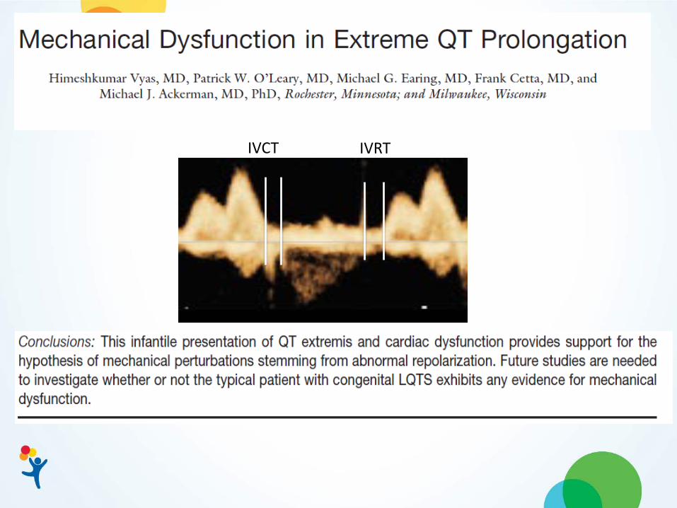
12. Vyas H, O’Leary PW, Earing MG, Cetta F, Ackerman MJ. Mechanical dysfunction in extreme QT prolongation. J Am Soc Echocardiogr 2008; 21 (5) : 511.e15–17.
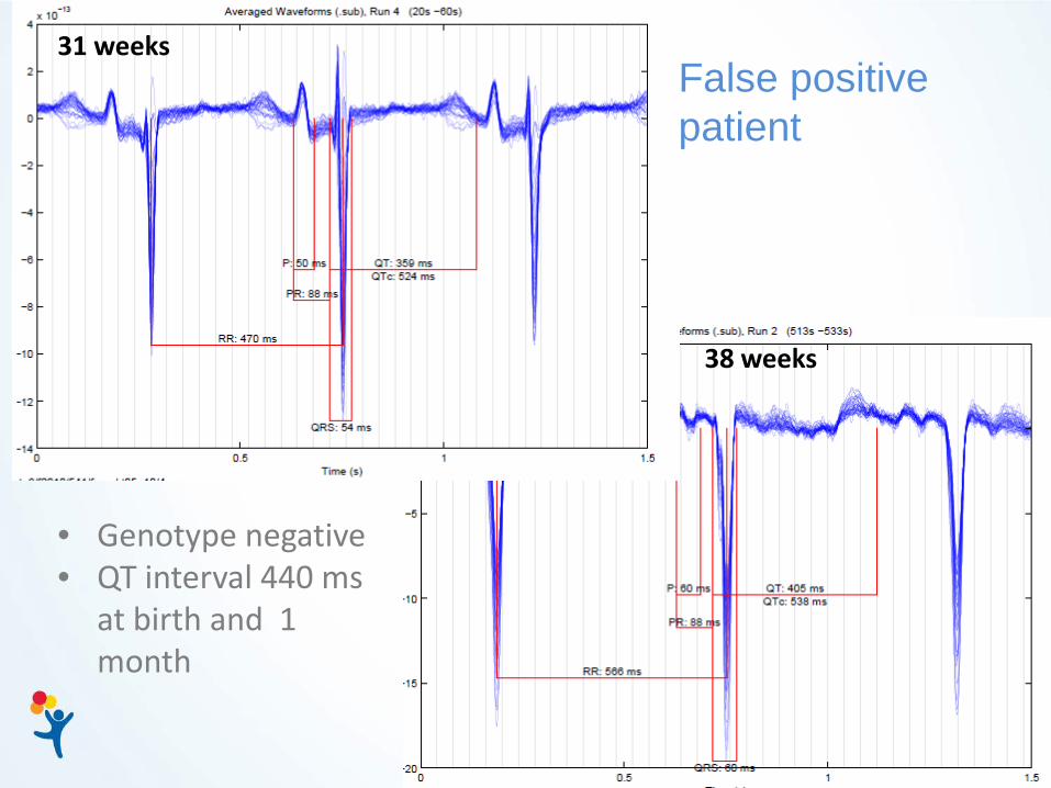
False positive patient
31 weeks
38 weeks
• Genotype negative • QT interval 440 ms
at birth and 1 month
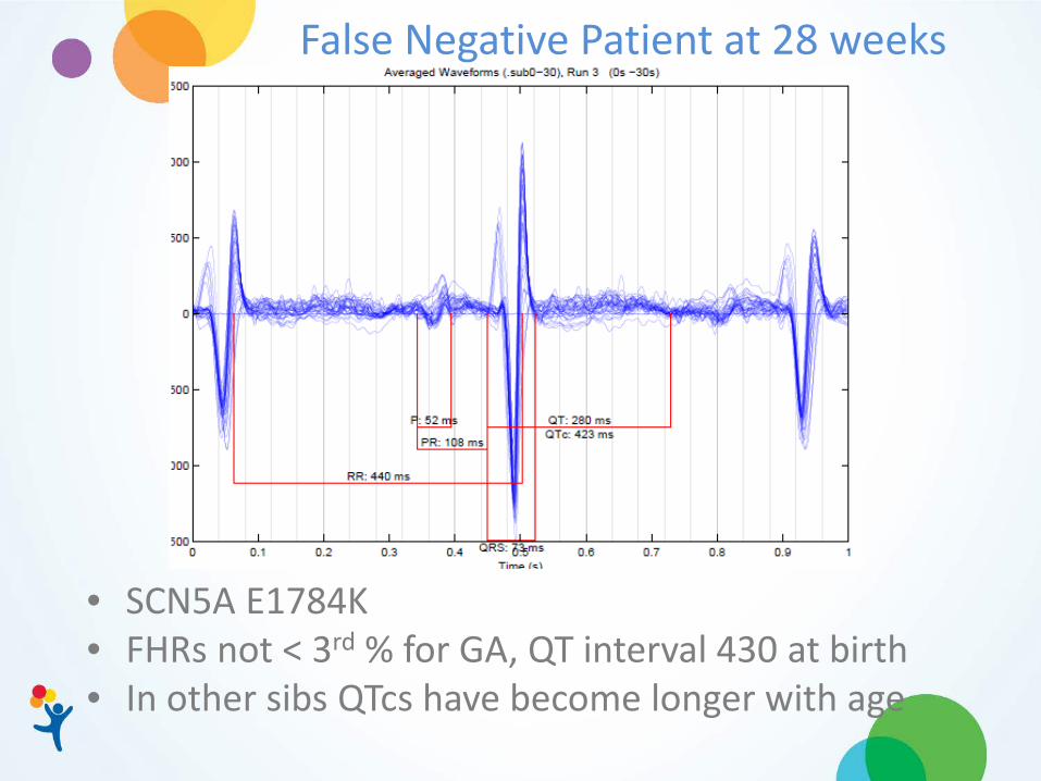
False Negative Patient at 28 weeks
• SCN5A E1784K • FHRs not < 3rd % for GA, QT interval 430 at birth • In other sibs QTcs have become longer with age

Which Beta Blocker?
Beta Blocker Approximate % transfer
Protein Binding
Nadolol Unknown 20-30%
Propranolol Fetal ~20% maternal concentration 93%
Atenolol Fetal and maternal concentrations~ equal <5%
Metoprolol Fetal and maternal concentrations ~ equal 10%
Conclusions The clinical profile of LQTS patients with complex fetal arrhythmias is suggestive of mutation type Fetal TdP and QTc > 580 ms are harbingers of postnatal adverse cardiac events Fetal treatment can abolish or reduce the incidence of TdP and prolong gestation
Author GA (wks)
Hydrops/CHD
In utero Rx
Control? Mutation Outcome
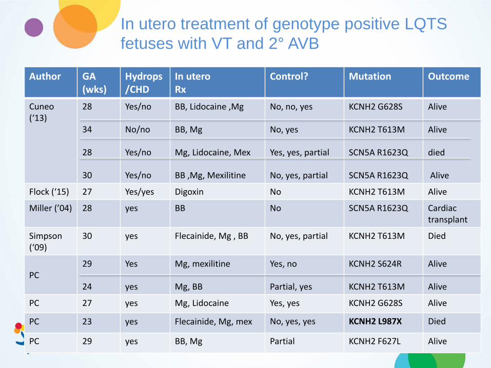
Cuneo (‘13)
28 34 28 30
Yes/no No/no Yes/no Yes/no
BB, Lidocaine ,Mg BB, Mg Mg, Lidocaine, Mex BB ,Mg, Mexilitine
No, no, yes No, yes Yes, yes, partial No, yes, partial
KCNH2 G628S KCNH2 T613M SCN5A R1623Q SCN5A R1623Q
Alive Alive died Alive
Flock (‘15) 27 Yes/yes Digoxin No KCNH2 T613M Alive
Miller (’04) 28 yes BB No SCN5A R1623Q Cardiac transplant
Simpson (‘09)
30 yes Flecainide, Mg , BB No, yes, partial KCNH2 T613M Died
PC
29 24
Yes yes
Mg, mexilitine Mg, BB
Yes, no Partial, yes
KCNH2 S624R KCNH2 T613M
Alive Alive
PC 27 yes Mg, Lidocaine Yes, yes KCNH2 G628S Alive
PC 23 yes Flecainide, Mg, mex No, yes, yes KCNH2 L987X Died
PC 29 yes BB, Mg Partial KCNH2 F627L Alive
In utero treatment of genotype positive LQTS fetuses with VT and 2° AVB

Author GA (wks)
Hydrops/CHD
Medication Control? Genotype Outcome
Hofbeck (‘97) 32 yes Flecainide BB
No Partial
ND Died
Ohkuchi (‘99) 34 yes Digoxin No ND Alive
Chang (‘02) 26 yes Lidocaine BB Digoxin
No Partial Less hydrops
ND Alive
Lin (‘04) 35 31 26
No No No
BB BB BB
No No No
ND ND ND
Died Died Alive
Anuwutnavin (’12)
32 No BB Partial ND Alive
Fukushima (‘10)
24 No Mg yes ND Alive
In utero treatment of phenotype positive LQTS fetuses with VT and 2° AVB

Sinus bradycardia 2° AVB Ventricular tachycardia
Fetal Surveillance with LQTS rhythm: + or - LQTS family history
fMCG + for LQTS
• Recheck family history • ECGs of 1° Relatives • Genetic testing if QTc prolonged • Treat maternal Mg and/or 25, OH Vit D deficiency • No QT prolonging meds
• 1X weekly FHR • Every 2 week fetal
echo
• 2x weekly FHR • 1x week fetal echo
SB 2° AVB VT
• Postnatal ECG • Genetic testing
Resolution Rare/Intermittent No heart failure Progression
Observe or Treat?
Recurrent with heart failure
Treat Continuation

23 19 18
= Misscarriages and unknown gender
= dead
or = mutation / pacemaker
1
1
2
2
3
3
3
4
4
4
5
5
6
6
7
7
8
8
9
9
10
10
11
11
12
1. 2. 3. 4.
1 2
13
12 13
14
14
15
15
16
16
17
17
18 19 20
20
21
21
23
22
1 2 3 4
= unborn
22
= consanguineous
Generation 2:
2: ICD, not tested
10: PM and + mutation
Generation 3:
1, 2, 3: ICD, not tested
5: + mutation
8. ICD and + mutation
And what about that family history?
KCNH2 L987X Slide courtesy of Professor Frank Pillekamp MD, Dusseldorf

Magnesium Beta Blocker Lidocaine Mexilitine
Sucessful RxUnsuccessful Rx
N=10 N= 6 N=3 N=4
Results of in utero treatment for TdP
100%
0%
Partial

Fetal LQTS Consortium
North America
Children's Hospital Colorado Denver CO USA
Vanderbilt University Nashville TN USA
University of Utah Salt Lake City UT,
USA
University of Rochester, Rochester NY, USA
Mayo Clinic, Rochester MN, USA
University of Toronto, Ontario, CA
Europe Center Cardiac Arrhythmias of Genetic Origin Milan, Italy
The University of Amsterdam, the Netherlands
Bonn University. Bonn Germany
University of Munster, Germany
German Heart Center Munich Germany Hospital Bichat-Claude Bernard, Paris, FR
University of Oslo, Oslo, Norway
Royal Brompton Hospital, London, UK
University of Helsinki, Helsinki, Finland
Umea University, Umea, Sweden
Total Enrollment=52 from 8 active sites Complete data from 24 subjects

Presentation of Fetal LQTS
Rhythm N Geno-typed
Mutation De Novo Family
Sinus brady 32 26 KCNQ1 0 100%
Fetal TdP 4 4 KCNH2 25% 75%
Fetal TdP + 2° AVB
22 13 (6) KCNH2 (7) SCN5A
100% 0
Neonatal TdP 7 4 (3) SCN5A (1) CALM 2
100% 0
Neonatal TdP + 2° AVB
8 3 (1) KCNH2 (2) SCN5A
100% 0

A FHR/GA algorithm to identify LQTS before birth: Preliminary results
• International multicenter (15 sites) study pregnancies with maternal or paternal KCNQ1, KCNH2 or SCN5A (fetallqts.com) • Review FHR/GA throughout pregnancy • Postnatal ECG and genetic testing
• Total Enrollment = 52 from 8 active sites • Complete data from 24 subjects
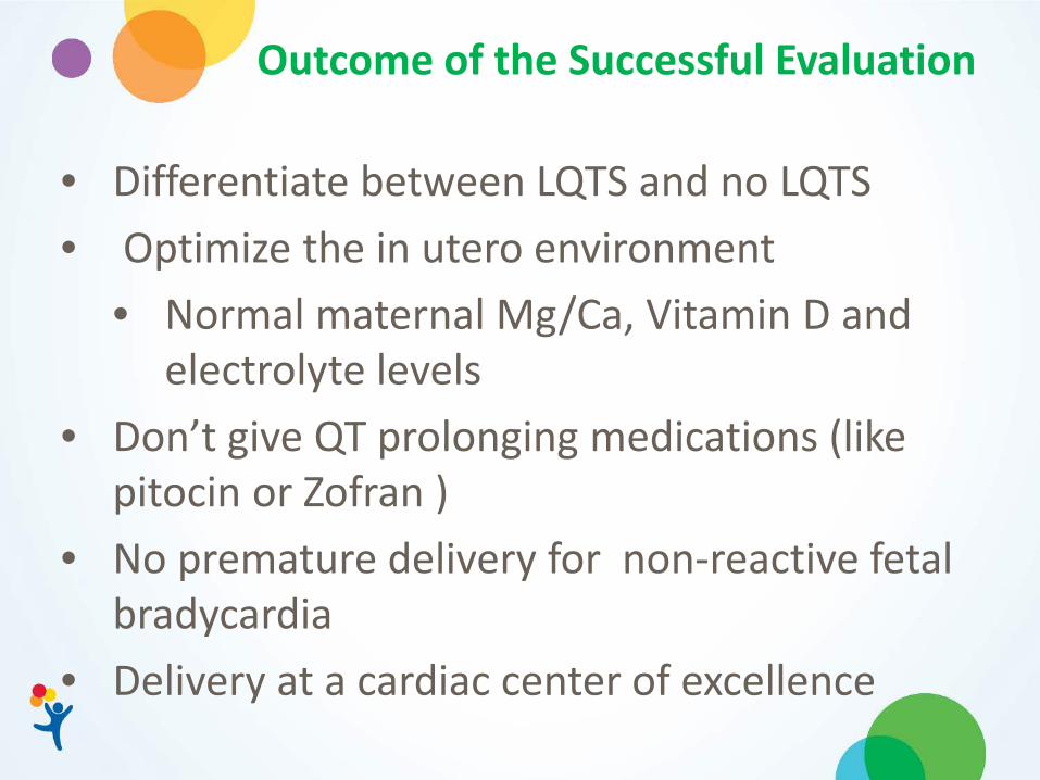
Outcome of the Successful Evaluation
• Differentiate between LQTS and no LQTS • Optimize the in utero environment
• Normal maternal Mg/Ca, Vitamin D and electrolyte levels
• Don’t give QT prolonging medications (like pitocin or Zofran )
• No premature delivery for non-reactive fetal bradycardia
• Delivery at a cardiac center of excellence

Evaluation of Suspected Channelopathies
Improve ascertainment The fetus at risk
• FH LQTS • LQTS rhythm (including bradycardia)
Elucidate the electrophysiology of TdP
Risk stratify pre/postnatal care In utero management and delivery of the LQTS fetus
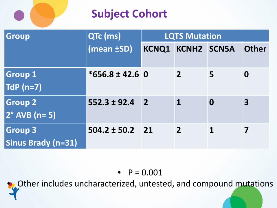
Group QTc (ms) (mean ±SD)
LQTS Mutation KCNQ1
KCNH2
SCN5A
Other
Group 1 TdP (n=7)
*656.8 ± 42.6 0 2 5 0
Group 2 2° AVB (n= 5)
552.3 ± 92.4 2 1 0 3
Group 3 Sinus Brady (n=31)
504.2 ± 50.2 21 2 1 7
• P = 0.001 • Other includes uncharacterized, untested, and compound mutations
Subject Cohort
Summary of Results TdP or TdP + 2° AV block
All w. SCN5A had aborted or sudden cardiac death in utero or within the first year of life despite medical +/- pacemaker Rx All w. KCNH2 survived w. medical +/ - pacemaker Rx