Embed Size (px)
Citation preview

I
BACTERIAL CONTAMINATION OF POWDER MILK
By: Ragia Hussein Elhussein Mohammed
B.Sc. Faculty of Science Al Neelain University
Supervisor: Dr. Khalid Mohammed Suleiman
A thesis submitted to the University of Khartoum in
Partial Fulfillment of the Requirements for Master of Science in Microbiology
Department of Microbiology Faculty of Veterinary Medicine
University of Khartoum August 2007

II
DEDICATION
To Soul of my Father
To my dear Mother
To my dear Husband

III

IV
Abstract
The present study was carried out to isolate, identify and
to determine the extent of contamination of milk powder. Sixty
powered milk samples were collected from retail shops in
Alkalakla, Alsagana, Alhag Yousif, Ombeda, Dar Alsalama and
Shambat.
Samples were cultured on blood and MacConkeys agar
and isolated bacteria were identified by primary and
secondary methods. Bacterial growth was demonstrated in
(56.67%) powdered milk samples.
Results of present investigation showed that powdered
milk is contaminated with numerous species of spore- forming
aerobic bacilli which include Bacillus cereus (7.5%) and
B.amyloiquefacients (50%), B.mycoides. B.firmest,
B.pantothenicus, B.pumilus, B.megaterium (2.5%) B.licheniformis
(12.5%) and B.thuringiensis (10%).
The highest rate of bacterial isolation was documented in
powdered milk samples collected from Dar Alsalam (35%) and
the least was detected in samples obtained from Ombada
(2.5%).
The viable bacterial counts of powdered milk sampled
from three area ranged between 1.2x10² – 3.5 x 10¹⁰ CFU/gm.

V
ةـــــــــــــــالخلاص
. المجففنأجريت الدراسة لعزل وتعريف الأنواع البكتيرية التي تلوث اللب
لات التجارية الصغيرة في عينة من اللبن المجفف من المح) 60(تم جمع
.مناطق الكلاكلة، السجانة، الحاج يوسف، أم بده، دار السلام و شمبات
م تزريع هذه العينات في وسط اجار الدم واجار الماكونكي و عرفت البكتريا ت
عينات اللبن . المعزولة باستخدام الصفات المزرعية والاختبارات الكيموحيوية
).%56.67(المجفف التي نمت بها البكتريا
نتائج البحث أوضحت أن اللبن المجفف ملوث بعدد من السلالات البكتيرية
-:يات والتي شملت الأنواعلجنس الصو
B.cereus (7.5%), B.amyloiquefacients(50%), B.mycoides. B.firmest, B.pantothenicus, B.megaterium (2.5%) B.licheniformis (12.5%) and B.thuringiensis (10%).
عينات التي اعلي معدل لعزل البكتريا من عينات اللبن تم توضيحها في ال
بة سجلت في العينات من منطقة واقل نس%) 35(جمعت من منطقة دار السلام
%).2.5(بده ام
نتائج العد البكتيري الحي أوضحت أن عينات اللبن المجفف تحتوي على أعداد
وحدة مكونة لمستعمرة 1.2x10² – 3.5 x 10¹⁰مقدرة من البكتيريا تتراوح بين
.في الجرام

VI

VII

VIII

IX

X

XI

1
Introduction
The world is faced with a problem of food shortage and milk products
are considered as a partial solution for this problem in developing countries.
But these products are vulnerable to spoilage by certain microorganisms,
some of which are beneficial and others are harmful to human beings.
The number and type of microorganisms present in foods are influenced
by many factors, that include the general environment for which food was
obtained, the microbiological quality of the food in its processed state, the
sanitary conditions under which the product was handled and processed and
finally the adequacy of subsequent packing, handling and storage conditions
in maintaining the flora at low level (Jay, 1986).
Food spoilage and food poisoning by microbes have been known since
1880 ( Jay,1970) , and there were many food borne diseases that have been
referred to as food poisoning , such as brucellosis , scarlet fever , typhoid
fever , diphtheria and others were associated with consumption of milk .
Banwart (1981) reported that for contamination control of the food, the
microbial load must kept as low as possible and the main sources of
contamination must be importantly known.
Numerous studies have documented that powder milk could
contaminated by bacteria. In national institute of public health it was
reported that on spray drying of milk artificially contaminated with Bacillus
cereus spores (In't veld, 1993).
Study performed in New Zealand for sample of milk powder revealed
that it's contaminated by Bacillus licheniformis and Bacillus subtilis
(Ronimus et al., 2006).

2
Anthoer study of sample from different countries were examined, the
dominated isolates was Anoxybacillus flevithermus followed by Bacillus
licheniformis (Rucket et al., 2004). A previous study also demonstrated that
Bacillus cereus isolated from powdered milk produce diarrhoeal enterotoxin
(Reves et al., 2007).
The objectives of this study to isolate and identify the bacterial species
contaminating powder milk and to determine the extent of this
contamination by estimating the viable bacterial count.

3
CHAPTER ONE
LITRATURE REVIEW
1.1 Milk:
1.1.1 Definitions:
Milk is the nutrient liquid secreted by the mammary glands of female
mammals. The female ability to produce milk is one of the defining
characteristics of mammals. The early lactation milk is known as colostrum
and carries the maternal antibodies to the new born. It can the reduce the risk
of many diseases in both the mother and new born (USAD National Nutrient
Data Base for standard references, 2005).
The milks of all species are complex biological fluids ـ containing a wide
variety of different constituents and possessing unique physical
characteristics (Robinson and Phil, 1985)
1.1.2 Milk Nutrition:
Milk is the source of high quality protein ـ calcium - Vitamins A and D ـ
riboflavin ـ other B vitamins and phosphorus (USAD National Nutrient Data
Base for standard references, 2005).

4
1.2 Powdered milk:
One of the methods of preserving various foodstuffs is by drying them,
and this by depriving microorganisms of the water necessary for their
growth (Bylund, 1995).
Powdered milk has a far longer shelf life than liquid milk and does not need
to be refrigerated due to its low moisture content (USAD National Nutrient
Data Base for standard references, 2005).
1.2.1 History:
Powdered milk was first made in 1802 by Russian Osip Krichevsky. It
is found abundantly in many developing countries because of reduced
transport and storage costs. Like other dry foods it is considered
nonperishable and is favored by survivalists and other people in need of
nonperishable easy to prepare foodstuffs (USAD National Nutrient Data
Base for standard references, 2005).
1.2.2 Use of dry milk:
Powdered milk was first produced as an infant food. It’s use has spread
into every industry manufacturing food products in which normal milk is
used. It is used in the baking industry in candy and ice cream manufacture
(Eckles and Combs, 1980).

5
1.2.3 Processing the powdered milk:
Drying means that the water in a liquid product (milk) is removed, so
that the product acquires a solid form. The water content of milk powder
ranges between 2-5 and 5% and no bacteria growth occurs at such low water
content. Drying extends the shelf life of the milk, simultaneously reducing
its weight and volume and thus reduces the cost of transport and storage of
the product (Bylund, 1995).
Commercial methods of drying are based on heat being applied to the
product, the water is evaporated and removed as vapor and the residue is the
milk powder.
Two principal methods are used for drying milk in the dairy industry,
the spray drying and roller drying.
In spray drying the milk is first concentrated by evaporation and then
dried in spray tower. During the first stage of drying the excess water in free
form between the particles of the dry solids is evaporated in the final stage,
the water in pores and capillaries of the solid particles is also evaporated.
In roller drying the product will be significantly affected by the heat,
if this drying is carried out in such a way that milk particles are in contact
with the hot heat transfer surface, the powder may then contain charred
particles which impair its quality (Bylund, 1995).
1.2.4 Micro flora of dried milks: In the case of roller dried milk, micrococci , aerobic spore formers and
Sarcina spp. predominate in the mesophilic range , whilst aerobic spore
6
formers make up the microbial population in the thermophilic range . The
severe heating, which the milk receives during the drying operation in the
rollers, reduces the bacterial content to a low level and only heat resistant
spores survive the process (Robinson and Phil, 1985).
In spray dried milk the microflora made up of thermoduric
micrococci, thermoduric streptococci and corynebacteria .
Recently, there has been a move towards the ultra ـ high temperature
heat treatment of milk for processing into powdered milk, the results is near
sterile powder with only causal post heat treatment contaminants being
present. In addition, inlet air temperatures in the spray drying operation have
risen, but even temperatures above 200º C fail to remove all spore formers
(Robinson and Phil, 1985).
1.3 Bacterial contaminating powdered milk:
1.3.1 Genus Bacillus:
1.3.1.1 Description:
Species of the genus Bacillus are mainly Gram-positive rods motile
(some non motile forms occur) and non acid fast. They produce heat
resistant spores under aerobic conditions (Gordon et al ., 1973 ) .
The term bacillus in a general sense has been applied to cylindrical or
rods like bacteria. The largest species are about 2µm across by 7µm long
and frequently occur in chains (Darland and Brock, 1971).
Most bacilli are aerobic, some species are facultatively anaerobes, and
usually oxidase variable, and catalase positive. Species of the genus differ in
the manner in which they attack sugars (Clause and Berkeley, 1986).
7
Bacillus species are widely distributed in the environment mainly
because of their highly resistant endospores. In soil endospores of Bacillus
anthracis, prototype of the genus, can survive for more than 50 years able to
tolerate extremely adverse conditions such as desiccation and high
temperatures (Quinn et al., 2002).
Most strains of Bacillus are not pathogenic for humans and only infect
them incidentally. A notable exception is Bacillus anthracis , which causes
anthrax in humans and domestic animals and Bacillus thuringientsis causes
disease in insects (Terranova and Blake , 1978 ) .
Bacillus cereus is known to cause a variety of nongastrointestinal
diseases as well as two different types of food poisoning (kotiranta et al.,
2000).
Many of the earlier reports of systemic disease caused by B. subtilis
are difficult to interpret because of incorrect species identification and many
of these isolates were probably B. cereus. Occasional reports have
implicated B. thuringiensis, B. alvei, B. circulans, B. licheniformis , B.
macerans,, B. pumilus, B. sphaericus and B. subtilis in systemic and
gastrointestinal disease. B. coagulans, B. sphaericus, B. macerans and B.
subtilis have caused septicemia (Turnbull et al ., 1990).
1.3.1.2 Habitat:
Bacillus species are ubiquitous, inhibiting soil, water, and airborne
dust (Schnepf et al .,1998). Thermophilic and psychrophilic members of the
genus can grow at temperatures ranging between (5 – 58) º C and can
flourish at extremes of acidity and alkalinity ranging pH 2–10 (Gordon et al
., 1973), therefore , Bacillus species can be recovered from a wide variety of

8
ecologic niches . Some species may be part of the normal intestinal
microbiota of humans and other animals (Elmer et al ., 1997).
Most Bacillus species encountered in the laboratory are saprophytic
contaminants or members of the normal flora (Smith et al., 1952).
1.3.1.3 Bacillus cereus:
It is an aerobic spore forming rod normally present in soil, dust, water
and spices (Wagner and Jr., 2000). It's optimum temperature for growth is
30º C, with minimum temperature of 10º C and maximum of 49º C the pH
range for growth is 4.9 to 9.3 (Frazier and Westhoff , 1978).
Bacillus cereus is a widely spread saprophyte that infects bovine
udder causing gangrenous mastitis (Carter, 1975). Numerous surveys on
food and food ingredients have indicated a high percentage of samples
contain B. cereus. It is widely distributed in nature and food supply (Frazier
and Westhoff, 1978). Extremely large numbers of viable cell of B. cereus
must be ingested to develop signs and symptoms of food borne disease. The
mechanism of the pathogenicity involves the lyses of the cell in the intestinal
tract and the resulting release of toxin (Frazier and Westhoff, 1978). It is
suggested that the food environments influenced enterotoxin production by
B. cereus ( Raevuori et al ., 1978 ) . B. cereus toxin caused food poisoning,
the toxin is produced during sporulation (Chessburgh, 2000; Turnbull,
1986), but the relation between sporulation and enterotoxin production is
unclear (Turnbull, 1986). Spore forming and psychrotrophic properties
enable B. cereus to survive pasteurization as well as to grow in milk at
refrigerated storage conditions (Coghill and Juffs, 1979).
Food poisoning strains of B. cereus produce four different
enterotoxins two protein complexes , hemolysin BL (HBL) and

9
nonhemolytic enterotoxin and two enterotoxic proteins (Lund et al ., 2000).
Hemolysin BL has been shown to lyse sheep erythrocytes and elicit a
vascular permeability reaction in rabbits (Glatz et al., 1974). It has been
suggested that hemolysin BL is responsible for the diarrheal food poisoning
syndrome (Beecher et al., 1995). Reasntly Awed alkareem (2007)
demonstrated that 80% of B. cereus isolated from minced meat in Khartoum
state were enterotoxigenic as shown by the rabbit deal loop test.
1.3.1.3.1 Pathogenicity of Bacillus cereus: Food brone gastroenteritis syndrome is caused by the emetic and the
diarrheal entrotoxin. The diarrheagenic toxin is heat labile and its production
is favored over the pH range 6-8.5, and in one study no toxin was found at
pH 5. The toxin is sensitive to trypsin and pepsin and induces diarrhea (Jay,
2000).
The emetic enterotoxin (vomiting type) is heat and pH stable and it is
sensitive to trypsin and induced vomiting (Jay, 2000).
Diarrheal type is rather mild, with symptoms developing within 6 to12
h and the symptoms consist of nausea (with vomiting being rare) cramps like
abdominal pains, tenesmus and watery stool. Fever is generally observed
(Jay, 2000) and the symptoms usually last for 12 to 24 h ( Rhodehamel and
Harmon, 2001) . The similarity between this syndrome and that of
Clostridum perfringens food poisoning has been noted (Jay, 2000).
The emetic type is characterized by an acute attack of nausea and
vomiting occurs within 1 to 5 h after consumption of contaminated foods.
Diarrhea is not a common feature in this type of illness (Rhodehamel and
Harmon, 2001).

10
The symptoms of this type of food poisoning paralled those caused by
Staphylococcus aureus food borne intoxication (Todar, 2005).
Many foods can be expected to contain small number of B. cereus
because it is such a common environmental contaminant. Harrigan and
McCance (1976) reported that at intermediate concentration of B. cereus in
food may represent a potential hazard because some proliferation has
occurred.
In outbreak setting, diagnosis is confirmed by performing quantitative
culture with selective media to estimate the number of organisms present in
the suspected food. Generally more than 105 organisms per gram of the
incriminated food are required to preduce symptom of B. cereus food
poisoning. Diagnosis is also confirmed by isolation of organisms from the
stool of two or more ill persons and not from stools of controls. Enterotoxin
testing is valuable but may not be widely available (Konuma et al ., 1998).
Mode of transmission through ingestion of food that has been kept at
ambient temperature after cooking, permitting multiplication of the
organisms. Outbreaks associated with vomiting have been most commonly
associated with cooked rice that has subsequently been held at ambient room
temperatures before reheating (Jay, 2000).
1.3.1.3.2 Contamination of powdered milk with B. cereus:
Dairy products are all susceptible to microbial spoilage because of
their chemical composition and nutritional value (Jay, 1986).
Bacillus cereus is a widely distributed organism in the environment
and undoubtedly the most important of the aerobic spore forming species
found in milk (Ahmed et al., 1983) .

11
Bacillus cereus was the causative agent of four outbreaks of food poisoning
involving 600 person which were reported in Oslo, Norway (Hauge, 1955).
Holmes et al . (1981) reported an outbreak in which eight people
developed acute food poisoning symptoms after the consumption of a
macaroni and cheese dish. The epidemiological investigation resulted in the
incrimination of the macaroni and cheese based upon the identification of
high levels of B. cereus (108 – 109 organisms / g) within the food that was
not served. Bacteriological analysis of the ingredients identified the powder
milk as the source.
Schmitt et al . (1976) reported two cases of poisoning that resulted
from the consumption of powdered milk products and Bacillus cereus like
organisms were identified in the milk products.
1.3.1.4 Bacillus mycoides: Bacillus mycoides is one of the Bacillus cereus group (B. cereus, B.
mycoides and B. thuringiensis). Colonies of B. mycoides, after overnight
growth on Nutrient Agar plate , are large and have hairy , rhizoid , root like
out growth from the colony margin that spread over the surface of the agar
(formerly B. cereus var. mycoides) which is a non motile species (Gordon et
al ., 1973 ; Sneath , 1986) .
Bacillus mycoides is common contamination in milk and dairy products
(Coghill and Juffs, 1979; Ahmed et al., 1983).
The organism cause spoilage (Overcast and Atmaram, 1974), may
produce toxins (Gilbert et al., 1981).

12
Like B. cereus, B. mycoides strains from milk have been shown to
produce diarrheagenic enterotoxin in 9 days at temperatures between 6º C to
21º C (Griffiths, 1990).
Bacillus mycoides is distinguished from B. cereus by the rhizoid colony
morphology (Claus and Berkeley, 1986).
1.3.1.5 Bacillus thuringiensis: Bacillus thuringiensis is a Gram-positive bacteria, which produces
insecticidal parasporal inclusions, is widely distributed in the soil of various
regions of the world (Delucca et al ., 1981) . This suggests that the soil is the
primary habitat of B. thuringiensis in nature, but the organism also occur in
other environments.
Previous workers reported that high-density populations of B.
thuringiensis are often retained in stored-product environments (Norris,
1969; Burges and Hurst, 1977), in diseased silkworm larvae (Shaikh et al.,
1986), in animal feed milk (Meadows et al., 1992) and on phylloplanes
(Smith and Couche, 1991).
Bacillus thuringiensis is also one of the B. cereus groups. Colonies are
large very rough and flat. Large cell with (> 1µm) that produce terminal
ellipsoid or cylindrical spores (Koneman et al., 1997). It is common
contamination in milk and dairy products (Coghill and Juffs, 1979; Ahmed
et al., 1983). The organism cause spoilage (Overcast and Atmaram, 1974),
may produce toxins (Gilbert et al., 1981).
Bacillus thuringiensis is found enterotoxin producers (Griffiths, 1990). It
has been isolated from foods, and it apparently produces a vero-cell active
toxin (Damgaard et al., 1996).

13
1.3.1.6 Bacillus licheniformis: Bacillus licheniformis , a common contaminant of dairy products is a
spore former and likely to survive all industrial processing of milk, such as
the manufacture of milk powder and whey concentrate (Salkinoja-Salonen et
al ., 1999) .
Cases of B. Licheniformis food poisoning present a clinical picture
similar to that of Clostriduim perfringens food poisoning and the diarrhea
syndrome caused by B. cereus and B. subtilis, in contrast it produces an
acute onset of emetic syndrome, although a significant minority also suffer
diarrhea (Kramer and Gilbert, 1989). The toxic threshold of the B.
licheniformis extracts was ≥100 times higher than those observed for emetic-
toxin-producing B. cereus strains (Andersson et al., 1998).
Ruckert et al. (2004) examined 28 milk powder samples and found
that the dominant isolate was Anoxybacillus flavithermus followed by
Bacillus lichenifoemis.
1.3.1.7 Contamination of powdered milk with other Bacillus
spp.:
Relatively few researchers have reported the presence of food borne
illness associated with Bacillus spp. other than B. cereus. However, due to
the high degree of phylogenetic relatedness among members of this genus, a
variety of species were considered to be potentially enterotoxigenic
including Bacillus thuringiensis (Damgaard et al., 1996).
Beattie and Williams et al . (1999) reported that different Bacillus
species isolated from raw milk and from the farm environment may have the
potential to produce diarrheal enterotoxins, included Bacillus circulans,

14
Bacillus Lentimorbis , Bacillus mycoides , Bacillus subtilis , Bacillus
licheniformis , Bacillus cereus and Bacillus thuringiensis .
Rowan et al . (2001) reported that different clinical and food isolates of
B. cereus, B. licheniformis, B. circulans and B. megaterium could express
diarrheal enterotoxins, hemolysins and B. cereus entrotoxins T after growth
in reconstituted infant milk formulae .
It was reported that isolates of many Bacillus spp. from veterinary
samples associated with serious nongastrointestinal infections in animals,
may carry diarrheagenic entrotoxin genes traditionally harbored by toxigenic
B. cereus and have expressed hemolysin BL enterotoxins (Rowan et al .,
2003).
Study of enterotoxin genes by PCR demonstrated the ability of B.
amyloliquefaciens , B. cereus , Bacillus circulans , Bacillus Lentimorbis ,
Bacillus pasteurii and Bacillus thuringiensis to produce toxins in a model
food system ( Phelps and Mckillip, 2002).
Consumption of entrotoxigenic Bacillus spp. at high cell densities
results in symptoms of diarrhea, with possible vomiting from a separate
heat-stable emetic toxin (Agata et al ., 1995) . Symptoms may appear 10 to
40 h following ingestion of foodstuffs contaminated with enterotoxigenic
strains. Food most often implicated in the diarrheal syndrome includes
poultry, cooked meats soups, desserts and fluid and dry milk products
(Koneman et al ., 1997). The infective dose is high (> 106 CFU / g) because
symptoms rely on the ingestion of the viable cells or spores not the
preformed toxin in criminated foods (Granum, 1997).

15
1.3.2. Staphylococcus spp.:
Staphylococcus food poisoning is an intoxication caused by
enterotoxins produced by strain of Staph. aureus in preformed toxin (Payne
and Wood, 1974 ) .
This is a small, round organism that thrives everywhere in our
environment and is a leading cause of food poisoning. Staphylococci caused
food poisoning is often associated with starchy food, cheese ect, from
improper handling techniques. Staphylococci produces the toxin as a by
product of its growth (O’connell, 2002).
Staphylococcus strains must produce sufficient enterotoxin in foods to
result in food poisoning (Pereira et al., 1991). The staphylococcal food
poisoning (food intoxication syndrome ) was first studied in 1894 by Denys
and later in 1914 by Barker who produced it in himself , the signs and
symptoms of the disease were reproduced experimentally by consuming
milk that had been contaminated with a culture of Staphylococcus aureus
(Jay , 1986 ).
Stophylococcal food poisoning is one of the main food borne disease
of food borne intoxication (Todd, 1978). In Spain, statistics showed that it is
the second most common type of food – borne disease (Anoa, 1985).
Staph. aureus, Staph. hyicus , Staph. hycius subsp. hyicus , Staph.
hyicus subsp. chromogenes , and Staph. intermedius are of interest in food
microbiology (Jay, 1986). Staphtlococcal food – poisoning follows the
consumption of the food heavily contaminated with certain staphylococci,
that produce a poisonous or toxic substance in the food which when ingested
(Hobbs and Gilbert, 1979) by man and after 4 – 6 hrs results in severe

16
vomiting, diarrhea, abdominal pain and cramps some times followed by
collapse but recovery is rapid (Hobbs and Gilbert, 1979 ; Jay, 1986).
The symptoms usually last one or two days and is rarely a serious threat to
otherwise healthy adults (O’connell, 2002). On the other hand, a common
type of food poisoning is staphylococcal enterotoxin (Jawez and Adel,
1990).
In 1958 and 1965, the outbreaks of staphylococcal food poisoning
occurred in the United States due to cheese (The International Commission
of Microbiological Specifications for Foods, 1980).
Sanchez (1991) reported that from 112 powdered milk samples found that 31
samples contaminated with Staph. aureus.
1.3.3. Salmonella spp.: Several infections caused by certain species of salmonella are important
as a cause of food poisoning in humans and of several diseases in domestic
animals. The term salmonellosis has been used for two main kinds of
gastrointestinal diseases in human, enteric fevers (including typhoid and
paratyphoid fevers) and gastroenteritis (Fraizer and Westhoff, 1978). The
latter is caused primarily by S. typhimurium and S. enteritidis, infection
occurs following ingestion bacteria on food, drink, fingers and other objects
(Cheesburgh, 2000).
Contamination is mainly from two sources of food products from
diseased poultry and cattle, and whole some food subsequently exposed to
infected feacal matter during food storage (mice and rats) and during food
preparation (human handlers) by hands that are not washed after using the
toilet (Davis and Samuel, 1997).

17
The onset of the disease is sudden and sometime severe, producing
nausea, vomiting, diarrhea, and slight fever occur 12-30 hours after eating
infected food (Cheesburgh, 2000). Salmonella bacteria thrive at temperature
between 40 -140 º F, they are readily destroyed by cooking to 165º F and
don’t grow at refrigerator or freezer temperatures (Davis and Samuel, 1997).
Usera et al. (1996) reported that the out break of salmonella occurred in
forty-eight cases involving children due to powdered infant formula.

18
CHAPTER TWO
MATERIALS AND METHODS
2.1 Materials:
2.1.1 Media:
For the isolation and identification of bacteria contaminating milk
powder different types of media were used, including solid, semisolid and
liquid media.
2.1.1.1 Solid media:
2.1.1.1.1 Nutrient agar (Oxoid):
The medium was prepared by dissolving 28 grams of powder in 1 liter
of distilled water by boiling. The medium was sterilized by autoclaving
(121ºC for 15 minutes), cooled to 55 º C and then distributed into sterile petri
dishes 20 ml in each.
2.1.1.1.2 Blood agar:
Hundred ml of fresh, sterile, defibrinated blood was added aseptically
to 900ml of melted sterile nutrient agar at 55 º C, mixed and distributed into
sterile petri dishes 20ml in each dish.
2.1.1.1.3 MacConkeys agar (Oxoid):
Fifty two gram of the medium was dissolved in 1 liter of distilled
water by boiling. The pH adjusted to 7.4, after which the medium was
sterilized by autoclaving at 121ºC for 15 minutes, cooled to 55 º C and
distributed into sterile petri dishes 20 ml in each.

19
2.1.1.1.4 Urea agar (Oxoid):
The medium was prepared by dissolving 2.4 grams of the powder in
95 ml distilled water by boiling. After sterilization by autoclaving at 115 º C
for 20 minutes the base medium was cooled to 50 º C and aseptically 5 ml of
sterile 40% urea solution were added. The pH was adjusted to 6.8 and
distributed into screw – capped bottles 10 ml each and then was allowed to
set in the slope position.
2.1.1.1.5 Simmon's citrate agar (Oxoid):
Twenty-three grams of powder were dissolved in 1000 ml distilled
water by boiling. The pH was adjusted to 7.0, and the medium was sterilized
by autoclaving at 121 º C for 15 minutes, distributed into sterile screw-caped
bottles and allowed to solidify in a slope position.
2.1.1.1.6 Starch agar:
Fifty gram of starch were triturated with water to smooth cream, and
added to the molten nutrient agar. The mixture was sterilized at 115 º C for
15 minutes and distributed into sterilized Petri dishes 20ml each.
2.1.1.1.7 Casein agar (milk agar):
Whole fresh milk was stored overnight in refrigerator. The creamy
layer was removed, and the milk was steamed for one hour and cooled in
refrigerator. Sufficient litmus solution was added to give a bluish-purple
colour and the medium was sterilized at 115 º C for 10 minutes. It was then
cooled to about 50 º C , added to double strength nutrient agar which was
melted and cooled to 50-55 ºC, mixed and distributed in to petri-dishes .

20
2.1.1.1.8 Ammonium salt sugars (ASS):
This medium consisted of ammonium phosphate (1.0 g), potassium
chloride (0.2 g), magnesium sulphate (0.2 g), yeast extract (0.2 g), agar
(2og) and bromoeresol purple (0.04 ml) . It was prepared according to
Barrow and Feltham (1993) by adding the solids to 1000 ml distilled water,
dissolved completely by boiling and sterilized at 115 º C for 20 minutes. The
medium was allowed to cool to about 55 º C and the appropriate sugar was
added as a sterile solution to give a final concentration 1%. The medium was
mixed and distributed aseptically into sterile tubes and allowed to set in the
slope position.
2.1.1.1.9 Plate count agar (Oxoid):
Seventeen point five grams of dehydrated mediums was suspended in
1 liter distilled water and boiled with frequent stirring. The medium was
sterilized by autoclaving at 121 º C for 15 min after adjusting the pH to 7.4
then distributed into petri-dishes.
2.1.1.2 Semi-Solid Media:
2.1.1.2.1 Hugh and Leifson's (O.F) medium :( Oxoid)
The medium was prepared by dissolving 10.3 grams of solids in 1 liter
of distilled water by heating, and the pH was adjusted to 7.1. Filtered
bromothymol blue (0.2% aqueous solutions) was added and then sterilized at
115 º C for 20 minutes. Sterile solution of glucose was added aseptically to
give final concentration 1%, mixed and distributed aseptically into sterile
tubes.

21
2.1.1.2.2 Motility medium: (Oxiod)
Thirteen gram of dehydrated nutrient broth was added to 4 gram of
agar and dissolved in 1 liter of distilled water by boiling , the pH was
adjusted to 7.4 , distributed in 5 ml amounts in tests tubes containing Craig-
tubes and sterilized by autoclaving at 121ºC for 15 minutes.
2.1.1.3 Liquid media:
All liquid media were prepared according to Barrow and Feltham
(1993).
2.1.1.3.1 Nutrient broth:
This medium was prepared by dissolving 13g of the medium in 1 liter
of distilled water. The pH was adjusted to 7.4, distributed into screw-capped
bottles 5 ml each and sterilized at 121 º C for 25 minutes.
2.1.1.3.2 Glucose phosphate broth (M.R-V.P medium):
Five grams of peptone and 5g of potassium phosphate were
dissolved in 1 liter of distilled water by steaming. The pH was adjusted to
7.5 and 5g of glucose was added and mixed. The medium was distributed
into test tubes 5 ml each and sterilized by autoclaving at 110 º C for 10
minutes.
2.1.1.3.3 Peptone water sugars:
Nine hundred ml of peptone water was prepared and pH was adjusted
to 7.1-7.3 before 10 ml of Andrade's indicator was added. Ten gram of the
appropriate sugar was added to the mixture, distributed into tubes 5 ml in

22
each one. The peptone water was sterilized by autoclaving at 110 º C for 10
minutes.
2.1.1.3.4 Nitrate broth:
One gram of nitrate was dissolved in 1 liter of nutrient broth,
distributed into tubes and sterilized by autoclaving at 115 º C for 15 minutes.
2.1.2. Reagents:
2.1.2.1 Hydrogen peroxide:
Hydrogen peroxide was prepared as 3% aqueous solution and used for
catalase test.
2.1.2.2 Kovac's reagent:
This reagent composed of paradimethyl-aminobenzaldehyde,
amylalcohol and concentrated hydrochloric acid. After preparation the
reagent was stored in the refrigerator at 4 º C.
2.1.2.3 Potassium hydroxide:
Potassium hydroxide was prepared as 40 % solution and used for
Voges-Proskaur (V.P) test.
2.1.2.4 Alphanaphthol solution:
It was prepared as 1% aqueous solution and also used for V.P test.
2.1.2.5 Oxidase reagent:
Tetramethyl p-phenylene diamine dihydrochloride was prepared as
1% aqueous solution and used for oxidase test.

23
2.1.2.6 Andrade's indicator:
This was prepared by dissolving 5g of acid fuchsin in 1 liter of
distilled water, and then 150 ml of alkali solution (NaOH) was added. It was
used in peptone sugar medium.
2.1.2.7 Bromothymol blue solution:
This was prepared as 0.2% w/v by dissolving 0.2g of bromothymol
blue powder in 100 ml distilled water. It was used for oxidation fermentation
(O.F) test.
2.1.2.8 Bromocresol purple solution:
Bromocresol purple solution was prepared as 0.9% solution, it was
used in ammonium salt sugar (ASS).
2.1.2.9 Physiological saline:
This was prepared by dissolving 8.5g of sodium chloride in 1000
distilled water.
2.1.2.10 Nitrate test reagent:
This reagent was composed of two solutions:
• Solution A: Sulphsnilic acid 0.33% in 5N-Acetic
acid dissolved by gentle heat.
• Solution B: Dimeyhyl-α naphthylamine 0.6% in
5N-acetic acid.
The complete reagent was used to detect nitrate
reduction.

24
2.2 Methods:
2.2.1 Sterilization:
2.2.1.1 Autoclaving:
Screw-capped bottles, rubber caps, media solution, normal saline, etc.
were sterilized in autoclave at 121 º C for 15 minutes and 110 º C for 10
minutes was used in case of sugar media .
2.2.1.2 Hot-air oven:
Glassware such as petri dishes, tubes, flasks and glass rods were
sterilized in hot air oven at 160 º C for one hour.
2.2.1.3 Disinfection:
Solution of 70% alcohol and phenolic disinfection were used for
bench sterilization.
2.2.2 Collection of samples:
Sixty samples of milk powder were collected from ten retail shops in
each of six areas which included Alklakla , Alsagana , Alhag yousif ,
Ombada , Dar Alsalam and shambat . Samples were collected in sterile
containers and plastic bags and immediately transferred to the laboratory for
bacteriological investigation.
2.2.3 Cultural methods:
2.2.3.1 Primary isolation:
One gram of each milk powder sample was suspended in 10 ml sterile
normal saline. Using sterile standard loop, 0.05 ml of each suspension was
25
striked into blood and Maconkeys agar. All plates were incubated at 37 º C
for 24 hours.
2.2.3.2 Examination of cultures:
Examination of all cultures on solid media was performed for
detection of growth, pigmentation, colonial morphology as well as changes
in the media. Plates which showed visible growth were subjected to further
bacteriological tests while those which did not show visible growth were
incubated for further 48 hours and then discarded if no growth was detected.
2.2.3.3 Purification of cultures:
The primary isolates were subcultured on nutrient agar and blood agar.
The subculture was repeated several times until pure colonies were obtained.
2.2.4 Identification of isolated bacteria:
Identification was carried out according to the procedure described by
Barrow and Feltham (1993).
2.2.4.1 Primary Identification:
2.2.4.1.1 Preparation of smears:
Smears were prepared by emulsifying small inoculums of the bacterial
coloney in a drop of sterile normal saline on clean slide and spreading it. The
smears were allowed to dry and then fixed by gentle heating.
2.2.4.1.2 Gram's technique:
This was done as described by Barrow and Feltham (1993).

26
2.2.4.1.3 Microscopic examination of isolates:
All isolated microorganisms were subjected to microscopic
examination under oil immersion tens and the shape, arrangement and
Gram's reaction were recorded.
2.2.4.1.4 Catalase test:
This test is used to identify bacteria which produce the enzyme
catalase (Cheesbrough, 1987). A portion of the tested colony was placed on
a drop of 3% hydrogen peroxide on a clean slide using a wooden stick.
Production of air bubbles indicated a positive result.
2.2.4.1.5 Oxidase test:
A portion of the tested colony was picked using sterile bent glass rod
and rubbed on a filter paper saturated with oxidase reagent. The
development of dark purple colour within 10 seconds indicated a positive
result.
2.2.4.1.6 Oxidation – Fermentation test (O.F):
Two tubes of Hugh and Leifson's medium were inoculated with tested
organism, one of them was covered with a layer of sterile paraffin. Tubes
were incubated at 37ºC and examined daily for seven days. Fermentative
organisms produced a yellow colour on both tubes while oxidative
organisms produced a yellow colour only in the tube with out paraffin.
2.2.4.1.7 Motility test:
Motility was tested by stabbing the isolated bacteria with straight loop
wire in a semi solid medium (Craige tube method). The medium was then

27
incubated for up to 3 days at 37º C together with un inoculated media as
control. The growth of a non-motile organism was confined to the stab,
while that of a motile one spreaded out side the craige tube.
2.2.4.1.8 Sugar fermentation test:
The sugar media were inoculated with 24 hours culture of tested
organism. They were incubated at 37 º C and examined daily for up to seven
days. Acid production was indicated by the development of a pink color in
the medium and gas production was indicated by trapped air in the Durham's
tube.
2.2.4.2 Secondary Identification:
2.2.4.2.1 Indole test:
The tested organism was inoculated in peptone water and incubated at
37 º C for 48 hours. Two to three drops of Kovac's reagent were added to
culture and shaked well. Production of pink colour on the upper layer of the
reagent was considered for indole production.
2.2.4.2.2 Voges-Proskauer test (V.P):
This test was performed to detect the production of acetylmethyl
carbinol. Glucose phosphate broth was inoculated with tested organism and
incubated at 37 º C for 48 hours. 0.6 ml of alpha-naphthol solution followed
by 0.2 ml of 40% potassium hydroxide solution were added to 1 ml of
culture, mixed well and examined after 15 minutes and one hour.
Development of a bright red colour indicated a positive result.

28
2.2.4.2.3 Urease test:
The tested organisms were inoculated on a slope of urea agar medium
and incubated at 37 º C for up to 5 days. The change of colour of the medium
to red or pink indicated a positive result.
2.2.4.2.4 Citrate utilization test:
Simmon's citrate medium was inoculated with the tested organism,
incubated at 37 º C and examined daily for up to seven days. The
development of a blue colour in the medium was considered as a positive
result.
2.2.4.2.5 Starch hydrolysis test:
Starch agar plate was inoculated with test organism and incubated at
37 º C for 24 hours. The plate was flooded with lugol's iodine solution and
starch hydrolysis was indicated by clear colour less zones around colonies.
Starch which was not hydrolyzed give a blue colour.
2.2.4.2.6 Casein hydrolysis test:
Casein agar plate was inoculated with test organism and incubated at
37 º C for 24 – 48 hours. A clear zone around the colonies indicated casein
hydrolysis.
2.2.4.2.7 Nitrate reduction test:
The tested organism was grown in nitrate broth and incubated at 37 º C
for 5 days. One ml of nitrate reagent A was added followed by 1 ml of
reagent B. Development of a deep red colour was considered as a positive

29
reaction. Zinc powder was added to tubes which did not show red colour,
development of red colour in these tubes indicated that nitrate was present
and tested organism did not reduce it.
2.2.4.2.8 Viable count:
The Miles and Misra (1938) method was used to determine the
number of a viable bacteria in powder milk samples. Examined samples
were collected from three areas, Shambat, Alklakla , and Dar Alsalam . One
gram of milk powder was suspended in 9 ml of normal saline and 1ml of
suspension was serially ten – fold diluted in sterile normal saline .

30
CHAPTER THREE
RESULTS
3.1 Sampling and Bacteriology:
A total of sixty powdered milk samples were collected from Alkalakla,
Alsagana, Alhag Yousf, Ombada, Dar Alsalam and Shambat Alhela.
Samples were cultured on blood and Macconkeys agar and the cultures
were identified by primary and secondary biochemical methods. Thirty four
samples (56.67 %) showed bacterial growth.
The highest rate of bacterial isolation was documented in samples
collected from Dar Alsalam, and the least was detected in samples obtained
from Ombada (Table: 1) and (Fig: 1).
All bacterial isolates belonged to the genus bacillus, the most frequent
species was B. amyloliquefaciens (50 %), and the lest frequent were B.
mycoides, B. firmus, B. pantothenticus, B. pumilus and B. megaterium (2.5
%) (Table: 2) .
Results of the viable count indicated that the powder milk was
contaminated the ranged between 1.2×10² -3.5×10³ CFD/gm. The highest
bacterial count of powder milk samples in Dar Alsalam (Table: 3).
3.2. Biochemical test:
Results of the biochemical methods used to identify isolates are shown in
table (4).

31
Table 1. Number of bacterial growth and isolates and percentage in
each source
Source No. of
samples
No. of
bacterial
growth
No. of
bacterial
isolates
Percentages
Alkalakla 10 4 4 40%
Shambat 10 7 9 70%
Alsagana 10 5 5 50%
Ombada 10 3 1 30%
Alhag yousif 10 7 7 70%
Dar Alsalam 10 8 14 80%
Total 60 34 40 56.67%

32
Table 2. Bacillus spp. isolated from powdered milk
Bacillus spp. No. of isolate Percentage
B.amyloliquefaciens 20 50 %
B.cereus 3 7.5 %
B.mycoides 1 2.5%
B.firmus 1 2.5%
B.pantothenticus 1 2.5%
B.pumilus 1 2.5%
B.licheniformis 5 12.5 %
B.coagulans 3 7.5 %
B.megaterium 1 2.5 %
B.thuringiensis 4 10 %

33
Table 3. Bacterial counts of powdered milk samples
Sample
A P C
Alkalakla
Shambat
Dar Alsalam
Sample 1 2.0×10² 6.5×10² 1.5×10³
Sample 2 1.4×10³ 3.5×10² 1.05×10³
Sample 3 5.0×10² 4.0×10² 6.5×10³
Sample 4 1.2×10² 1.4×10³ 7.5×10²
Sample 5 _ 5.0×10² 3.5×10³
APC = Aerobic Plate Count.
_ = No growth.

34
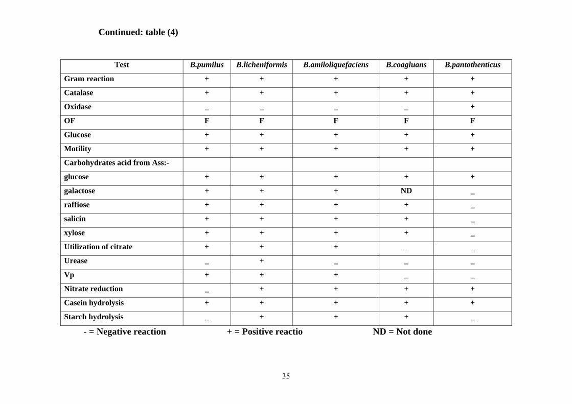
Table 4. Biochemical properties of Bacillus spp. isolated from powdered milk Test B.cereus B.mycoides B.thuringiensis B.firmus B.megaterium
Gram reaction + + + + +
Catalase + + + + +
Oxidase + + + _ _
OF F F F F F
Glucose + + + + +
Motility + _ + + +
Carbohydrates acid from Ass:-
glucose + + + + +
galactose _ ND _ _ +
raffiose _ _ _ _ _
salicin + + + _ +
xylose _ _ _ _ +
Utilization of citrate + + + _ +
Urease + + _ _ _
Vp ND ND ND _ _
Nitrate reduction + + + + _
Casein hydrolysis + + + + +
Starch hydrolysis + + + + +
35
Test B.pumilus B.licheniformis B.amiloliquefaciens B.coagluans B.pantothenticus
Gram reaction + + + + +
Catalase + + + + +
Oxidase _ _ _ _ +
OF F F F F F
Glucose + + + + +
Motility + + + + +
Carbohydrates acid from Ass:-
glucose + + + + +
galactose + + + ND _
raffiose + + + + _
salicin + + + + _
xylose + + + + _
Utilization of citrate + + + _ _
Urease _ + _ _ _
Vp + + + _ _
Nitrate reduction _ + + + +
Casein hydrolysis + + + + +
Starch hydrolysis _ + + + _
- = Negative reaction + = Positive reactio ND = Not done
Continued: table (4)

36
Fig. (1): Percentage of bacterial growth in each source
0%
10%
20%
30%
40%
50%
60%
70%
80%
90%
Percentages
AlkalaklaShambatAlsaganaOmbadaAlhag yousifDar Alsalam

37
Fig. (2): Colonies of Bacillus mycoides on nutrient agar

38
Fig. (3): Colonies of Bacillus cereus on nutrient agar

39
Fig. (4): Colonies of Bacillus thuringiensis on nutrient agar

40
CHAPTER FOUR
DISCUSSION
In the present study powdered milk samples were collected from retail
shops in six areas in Khartoum state and were investigated bacteriologically
for the presence of bacterial contaminants.
Bacterial growth was demonstrated in 56.67% of the samples. All the
bacterial isolates belonged to the genus Bacillus and this is a reflection to
their wide spread and survival in the environment due to their ability to form
resistant endospores.
The Bacillus species identified in this study included B.
amyloliquefaciens , B. licheniformis , B. cereus, B. thuringienss, B.
coagulans , B. mycoides , B. firmus , B. pantothenticus, B. pumilus , and B.
megaterium . Similar findings were reported by Vaisanen et al. (1991) who
isolated a great numbers of Bacillus strains from various diary products. The
majority of their isolates (90 %) were members of B. cereus group (B.
cereus, B. mycoides , B. thuringiensis ). Results of this study also
substantiated the previous works of Coghill and Juffs (1979); Ahmed et al.
(1983) that Bacilli, especially members of the Bacillus cereus group, are
common contaminant of milk and dairy products , as well as products based
on dried milk (Singh et al ., 1980) .
Most strains of bacilli isolated from dairy products were able to grow at
or below 10º C, which is the temperature for storage of dairy products in
retail stores. Obviously cold in dairy products has favored the colonization
of the product by psychrotrophic (Vaisanen et al., 1991). Similarly, the
bacilli isolated in powdered milk samples in the present investigation might

41
be explained by the fact bacilli initially contaminated the milk powder,
which is a good medium for bacterial growth, from the environment and
under favorable humidity and temperature bacterial multiplication occurred.
The Bacillus cereus is widely distributed in nature and foodstuffs, and
was reported to be the main cause of food intoxication (Frazier and
Westhoff, 1978). In this study 7.5 % of isolated bacilli were B. cereus in this
fact might constitute a public health hazard since the resistant spores might
germinate under appropriate conditions and liberate substantial amounts of
toxins. This finding is in agreement with those of Reves et al. (2007) who
isolated this species from dried milk and also consistent with the findings of
Holmes et al. (1981) who reported the powder milk was a source of B.
cereus in an outbreak of food borne illness.
Bacillus mycoides isolated in this investigation constituted 2.5% of the
isolates. This species was shown capable of producing diarrheagenic
enterotoxin (Griffiths, 1990) hence its presence in milk powder investigated
in the present study could be hazardous. This species was isolated from raw
milk by Johnston and Bruce (1982) and from hands and nose of food
handlers by Mohamad (2007).
Study of enterotoxin genes demonstrated the ability of B.
amloliquefaciens and B. thuringiensis to produce toxin (Phelps and Mckillip,
2002). In the present study 50 % and 10 % of the isolates were B.
amyloliquefaciens and B. thuringiensis respectively but both of them have
not been reported in the available literature concerning bacterial
contaminating powder milk, however, B. amyloliquefaciens was isolated
from air by Phelps and Mckillip (2002) and B. thuringiensis was isolated
from soil by Terranova and Blake (1978), consequently powder milk might
be contaminated from these source.

42
Bacillus licheniformis extracts was found to be ≥100 times toxic than the
emetic-toxin-producing B. cereus strains (Andersson et al., 1998). 12.5 % of
the isolates was B. licheniformis and this level of contamination plus that of
B. cereus (7.5%) might pose a serious risk to the public. This species was
also isolated by Ruckert et al. (2004) from powder milk samples and by
Ronimus et al. (2006) from factory of powder milk in New Zealand.
The highest rate of bacterial contamination of milk powder was recorded
in samples obtained from Dar Alsalam, refugee camp located in Elhaj
Yousif, and this might possibly be explained by fact that poor hygienic
measures usually prevail in such camps, in addition others factors such as
prolonged storage under suboptimal condition might also contributed in
level of contamination.
Results of the viable bacterial counts of sampled milk powder was found
to range from 1.2 × 10² to 3.5 × 10³ CFU/gm of powder and this count was
less than the levels proposed by the Sudanese Standards and Metrology
Organization (2006) which is in the order 2 ×10⁴ -3 ×10⁵ CFU/gm. The
counts were also less than the recommended levels in the USA (2001) (104 –
105 CFU/gm).
In conclusion , our study indicated that the powdered milk is
contaminated with numerous species of spore forming aerobic bacillus and
the viable bacterial counts was found ranging between 1.2×10² -3.5×10³
CFU/gm.

43
Recommendation
Based on the results of this study, we recommend the following:
• Improvement of personal hygiene of sellers.
• The use of small packages of powder milk in order to reduce the
duration of exposure and hence decrease chance of contamination.
• Further studies should investigate the effect of boiling and hot
water upon the survival of spore forming bacilli in milk powder.

44
REFERENCE
Agata, N., Ohta, M., Arakawa, Y. and Mori, M. (1995). The bceT gene of
Bacillus cereus encodes an enterotoxigenic protein. Microbiology
141:983-988.
Ahmed, A. A. H., Moustafa, M. K., and Marth, E. H. (1983). Incidence of
Bacillus cereus in milk and some milk products. J. Food Prot. 46:126 –
128
Andresson, M.A., Mikkola, R., Helin, J., Andresson , M. C. and
Sankinoja-salonen, M. S. (1998) . Anovel sensitive bioassay for
detection of Bacillus cereus emetic toxin and related depsipetide
inonphores . Appl. Environ. Microbiol. 64: 1338 – 1343.
Anon, (1985). Cited by, Gome-Lucia, J., Blaneo, L., Goyache, J.,
Delafuente, R., Vazquez, J. A., Ferri, E. F. R., and Suare, G. (1986).
Growth and enterotoxin a production by Staphylococcus auerus s6.
Manchego type cheese. J. Appl. Bacteriol. 61:499-503.
Awed alkareem, A. B. (2007). Study on Enterococci and Bacillus cereus
isolated from meat in Khartoum Stat. M. Sc. thesis. In Microbiology
University of Khartoum.
Banwart, G.I. (1981). Basic food microbiology. The Avi Publishing,
Company, Inc. West Port, Connecticut.
Barrow, G. T., and Feltham, R. K. A. (1993). Cown and Steels Manual of
the identification of medical bacteria, 3 rd ed., Cambridge University
press, Cambridge.

45
Beattie, S. H. and Williams, A. G. (1999). Detection of toxigenic strains of
Bacillus cereus and other Bacillus spp. with an impoved cytotoxcity
assay. Lett. Appl. Micrbiol .28: 221 – 225.
Beecher, D. J., Schoen, J. L., Wong, A. C. L. (1995). Enterotoxic activity of
hemolysin BL from Bacillus cereus. Infect. Immun. 63:4423- 4428.
Burges, H. D. and Hurst, J. A. (1977). Eology Bcacillus thyringiensis in
storage months. J. Invertebrate Patho. 30: 131-239.
Bylund, G. (1995). Dairy Processing Hand book. Tetra Pak processing
systems A\ B, Lund, Sweden.
Carter, G. R. (1975). Diagnostic Procedures in Veterinary Microbiology. 2nd
ed. Hlinois: Charlis C. Publisher. Campylobacter jouni infections.
Epidemiological Reviews. 5: 157-176.
Chessburgh, M. (1987). Medical Laboratory manual for tropical countries.
2nd ed. ELBS, vol II, London.
Chessburgh, M. (2000). District Laboratory Practice in Tropical Countries.
1st ed. Part2. Cambridge University press
Clause, D. and Berkeley, R. C. W. (1986). Genus Bacillus Chon 1872, 174
In Bergey’s Manual of Systematic Bacteriology. Vol. 2 ed. Sneath, P.
H. A., Mair, N. S., Sharpe, M. E. and Holt, J. G. pp. 1105-1139.
Baltimore, Williams & Wilkins.
Coghill, D. (1982). Studies on thermoduric psychrotrophic bacteria in South
East Gueensland dairy products. Australian J. Dairy Technol. 37: 147-
148.
Coghill, D., and Juffs, H. S. (1979). Incidence of psychrorophic spore-
forming bacteria in pasteurized milk and cream products and effect of
temperature in their growth. The Australian J. Dairy Technol. 34:150-
153.

46
Damgaard, P.H., Larson, H. D., Hansen, B. M., Bresciani, J. and Jergensen,
K. (1996). Enterotoxin producing strains of Bacillus thuringiensis
isolated from food. Lett. Appl. Microbiol 23:146 – 150.
Darland, G., and Brok, T. D. (1971). Bacillus acidocaldaricu sp., an
acidophilic thermophilic spore-forming bacterium. Journal of General
Bacteriology, 67: 9-15.
Davis, C. E. and Samuel, C. (1997). Detecting Bacterial Contamination.
Biol. Arch. Intern. 31: 364-396.
Delucca, A. J., Simonson, J.G. and Larson, A.D. (1981). Bacillus
thuringiensis distribuition in soils of the United States. Canadian J.
Microbiol. 27: 865-870.
Eckles, C. H., and Comb, W. B. (1980). Milk and milk products. Fourth
Edition.
Elmer, W., Stephen, D., William, M., Paul, C. and Washington, C. (1997).
Diagnostic Microbiology, Fifth Edition.
Frazier, W. C. and Westhoff, D. C. (1978). Food Microbiology, 3 rd ed.,
McGraw-Hill Book Co., New York.
Gilbert, P. J., Turnbull, P. C. B., Parry, J. K. and Kramer, J. M. (1981).
Bacillus cereus and other Bacillus species. Their part in food-poisoning
and other clinical infections. In the Aerobic Endospore-forming
Bacteria, eds. Bereley R.C.W. and Goodfellow, M., pp. 297-314.
Academic Press. London.
Glatz, B. A., Spira, W. M. and Goepfort, J. M. (1974). Alteration of
vascular permeability in rabbits by culture filtrates of Bacillus cereus
and related species. Infect .Immun. 10: 299-303.

47
Gordon, R. E., Haynes, W. C. and Pang, C. H. N. (1973). The genus
Bacillus. Agriculture hand book. No.427. Washington, D.C.US Dept .
Agriculture.
Granum, P.E. and Lund, T. (1997) .Bacillus cereus and it is food poisoning
toxins. FEMS Microbiol. Lett. 157:223 – 228.
Griffiths, M. W. (1990). Toxin production by psychrotrophic Bacillus spp.
present in milk. J. Food Prot. 53: 790-792.
Harrigan, W. F. and McCance, M. E. (1976). Laboratory Methods in food
and Dairy microbiology. Academic Press. London, pp. 31-158.
Hauge, S. (1955). Food poisoning caused by aerobic spore-forming bacilli. J.
Appl. Bacteriol. 18: 591-595.
Hobbs, B. C. and Gilbert, R. J. (1979). In: Food poisoning and food hygiene.
4th ed. Edward Arnold Great Britain.
Holmes, J. R., Plunkett, T., Pate, P., Roper, W. L. and Alexander, W. J.
(1981). Emetic food poisoning caused by Bacillus cereus. Arch. Intern.
Med. 141: 766 – 767.
In’t veld , P. H., Soentoro, P. S. and Noterman, S. H. (1993). Properties of
Bacillus cereus spores in reference materials prepared from artificially
contamination spray dried milk. Int .J. Food Microbiol 20: 23-36.
Jawez, M and Adel, B. (1990). Medical microbiology. 19th ed. United State
of America.
Jay, J. M. (1986). Modern Food Microbiology. Third Edition. New York.
Van Nostr and Reinhold Company.
Jay, J. M. (1970). Modern food microbiology. New York. Van Nostr and
Reinhold.
Jay, J. M. (2000). Modern Food Microbiology. 4th ed. New York. Van Nostr
and Reinhold

48
Johnston, D. W. and Bruce, J. (1982). Incidence of thermoduric
psychrotrophs in milk produced in the West of Scotland. J. Appl.
Bacteriol. 52:333-337.
Koneman, E. W., Allen, S. D., Janda , W. M., Schreckenberger, P. C. and
Winn, W. C. (1997) . The aerobic Gram-positive bacilli. pp. 651–708.
In Color Atals and Text Book of Diagnostic Micrbiology, 5th ed,
lippincott , New York .
Konuma, H., Shinagawa, k., and Tokumaru, M. (1998). Occurrence of
Bacillus cereus in meat products, raw meat and meat product additives.
J. of Food prot. 51:324-326.
Kotiranta, A., Lounatmaa, K., and Haapasalo, M. (2000). Epidemiology and
pathogenesis of Bacillus cereus infection. Microbes Infect. 2:189 –
198.
Kramer, J. M., and Gilbert, R. J. (1989). Bacillus cereus and other Bacillus
species. pp.21-70. In M. P. Doyle (ed), Food Borne Bacterial
Pathogens. Mareel Dekker, Inc., New York.
Lund, T., De Buyser, M. L., and Granum, P. E. (2000). A new cytotoxin
from Bacillus cereus that may cause necrotic enteritis. Mol.
Microbiol. 38: 254-261.
Meadows, M. P., Ellis, D. J., Jarrett, P. and Burges, H.D. (1992).
Distribution, frequency and diversity of Bacillus thuringiensis in an
animal feed milk. J. Appl. Bacteriol. 58: 1344-1350.
Mohamad, A. M. (2007). The impact of sanitation and hygienic conditions
of restaurants on food safety at Elsuk Alshaabi Omdurman. Thesis for
M. Sc. In Microbiology University of Khartoum.
Norris, J. R. (1969). The ecology of serotypes 4B of Bacillus thurigiensis. J.
Appl. Bacteriol. 32:261-267.

49
O’connell , J. (2002) . Staphylococcus. A vilablet at httm //www.
Cbbqa.com/articles/Food – Safety/ Staphylococcus html.
Overcast, W. W. and Atmaram, K. (1974). The role of Bacillus cereus in
sweet curdling of fluid milk. J. Milk and Food Technol. 37: 233-236.
Payne, D. N. and Wood, J. M. (1974). The incidence of enterotoxin produc-
tion in strain of Staphylococcus aureus isolated from food. J. Appl.
Bacteriol. 37:319-325.
Pereira, J. L., Salzberg, S. P. and Bergdoll, M. S. (1991). Producing of
Staphylococcal enterotoxin (D) foods by low-enterotoxin producing
Staphylococci. International J . food Microbiol 14: (1), 19-25 .
Phelps, R.J., and Mckillip, J. L. (2002) . Enterotoxin production in natural
isolate of Bacillaceae out side the Bacillus cereus group. Appl.
Environ. Microbiol. 68: 3147-3151.
Quinn, J. P., Markey, B. K., Carter, M. E., Donnelly, W. J. and Leonard,
F.C. (2002). Veterinary Microbiology and Microbial Disease. Black
well Science Ltd Edition offices. Osney Mead, Oxford OEI OEL. 25
John, London.
Raevuori, M., Kiutamo, T. and Niskanen, A. (1978). Comprative studies of
Bacillus cereus strains isolated from various foods and food poisoning
out breaks. Food Science and Technology, 10:1-20.
Reves, F. J. Bastias, H. J., Gutierrez, R. M. and Rodriguez, L. Mde. (2007).
Prevalence of Bacillus cereus in dried milk products used by Chilean
School Feeding Program. J. Food Microbiol. 24: 1-6.
Rhodehamel, E. J., and Harmon, S. M. (2001). Bacteriological Analytical
Manual. 8th Edition. Center for Food Safety and Applied Nutrition.
Robinson, R. K. (1983). Dairy microbiology. Vol. 2 : The microbiology of
milk products. A. S. Publishers, London.

50
Robinson, R. K., and Phil, M. A. (1985). Dairy microbiology. Vol.1.
Thermicrobiology, London and New York.
Ronimus , S. R., Ruckert, A. and Morgan, W. H. (2006). Survival of
thermophilic spore-forming bacteria in a 90+ year old milk powder
from Ernest Shackelton’s Cape Royds Hut in Antarctica. J. Dairy Res
73: 235-243.
Rowan, N. J., Deans, K. J., Anderson, G., Gemmell, C. G., Hunter, I. S.
and Chaithong, T. (2001). Putative virulence factor expression by
clinical and food isolates of Bacillus spp. after growth in reconstituted
infant milk formulae. Appl. Environ .Microbiol. 67: 3873 – 3881.
Rowan, N. J., Caldow, G., Gamble, G. C. and Hunter, I. S. (2003).
Production of Diarrheal Enterotoxins and other Potential Virulence
Factors by Veterinary isolates of Bacillus species Associated with
Nongastrointestinal Infections. Appl. Environ. Microbiol. 69: 2372-
2376.
Ruckert, A. S. Ronimus, R. and Morgan, W. H. (2004). Based survey of
thermophilic bacilli in milk powders from different countries. Int. J.
Food Micrbiol. 96: 263 -272.
Salkinoja-salonen, M. S., Vuorio, R., Andersson, M. A., Kampfer, P.,
Andersson, M. C., Honkanen-Buzalski, T. and Scoging, A. C. (1999).
Toxigenic strains of Bacillus licheniformis related to food poisoning.
Appl. Environ. Microbiol.65: 4637-4645.
Sanchez, M. M., Fernandez, R. E. and Mota, G. L. (1991). Demonstration of
staphylococcal thermonuclease from powdered milk. Rev. Latinoam
Microbiol. 33: 135-139.

51
Schmitt. N., Bowmer, E. J., and Willoughby, B. A. (1976) .Food poisoning
out break attributed to Bacillus cereus .Can .J. Public Health 67:418 –
422.
Schnepf, E., Crickmore. N., Van Rie, J., Lerechus, L., Buman, J., Feitelson,
j., Zeigler, D. R. and Dean, D. H. (1998). Bacillus thuringiensis and its
pesticidal crystal proteins. Microbiol. Mol. Biol. Rev. 62: 775-806.
Shaikh, M. R., Naqui, B. S., Shaikh, D. and Khan, A. F. (1986). Distribution
of Bacillus thuringiensis in Pakistan. Pakistan J. Scientific and
Industrial Res. 29: 295-296.
Singh, R. S., Singh, S., Batish, V. K. and Ranganathan, B. (1980).
Bacteriology quality of infant milk foods. J. Food Prot. 43: 340-342.
Smith, N. R., Gordon, R. E. and Clark, F. E. (1952). Aerobic spore forming
bacteria. US Department of Agriculture Monograph 16. Washington D
Government Printing Office.
Smith, R. A. and Couche, G. A. (1991). The phylloplane as a source of
Bacillus thuringiensis variants. J. Appl. Environ. Microbiol. 57: 311-
315.
Sneath, P.H.A. (1986). Endospore forming Gram-positive rods and cocci. In
Sneath, P.H.A., Mair, N.S., Sharpe, M.E. and Hoit, J.G. (eds): Bergey’s
Manual of Systematic Bacteriology, 2: 1104-4405, Baltimore, Williams
and Wilkins
Terranova, W. and Blake, P. A. (1978). Bacillus cereus food poisoning.
North England. J. Med., 298: 143-144.
The International Commission Microbiological Specifications for Foods
(1980). Milk and milk product. In: Microbial Ecology Foods, Volume
11, page 499-512 edit by Silliker Alas. Acad. Press.

52
The Sudanese Standards of Metrology Organization. (2000).
Microbiological levels of food.
Todd, E. C. D. (1978). Food Borne-disease in six countries a comparison.
J. Food Prot. 41:559-565.
Todar, K. (2005). Bacillus cereus. Toder’s online text book of
Bacteriology. University of Wisconsin – Madison Department of
Bacteriology.
Turnbull, P. C. B., Kramer, J. and Melling, J. (1990). Bacillus. pp. 188-210.
In Topley and Wilson’s principles of bacteriology, virology and
immunity, vol. 2, 8th ed. Edward Arnold, London.
Turnbull, P.C.B. (1986). Bacillus cereus toxins, pp. 397 - 448. Inf Dorner
and J. Drews (ed), Pharmacology of bacterial toxins. Pergamon press,
London.
U.S.A. (2001). Microbiology criteria, pp. 1-23. http:// www. usem bassy. cl /
agriculture / fas 05e. htm.
USAD National Nutrient Data Base for Standard references. (2005). Http //
www. nal. usda. gov./ snice / food comb./sereach.
Usera , M. A., Echeita, A., Aladuena, A., Blanco, M. C., Reymundo, R.,
Prieto, M. I., Tello, O., Cano, R., Herrera, D., Martinez-Navarro, F.
(1996) . Interregional foodborne salmonellosis out break due to
powdered infant formula contaminated with lactose-fermenting
salmonella. Eroupean journal of epidemiology 12: 377-381.
Vaisanen, O. M., Mwaisumo, N. J. and Salkinoja, M. S. (1991).
Differentiation of dairy strains of the Bacillus cereus group by phage
typing, minimum growth temperature and fatty avid analysis. J. Appl.
Bacteriol. 70: 315-324.

53
Wagner, A. I. B. and Jr, A. (2000). Bacterial food poisoning. Texas
Agricultural Extension Service. www @ Yahoo. Com.