Embed Size (px)
Citation preview

CORRESPONDENCE Anatoly T. Titov [email protected]
© 2016 Titov et al. Open Access terms of the Creative Commons Attribution 4.0 International License (http://creativecommons.org/licenses/by/4.0/) apply. The license permits unrestricted use, distribution, and reproduction in any medium, on the condition that users give exact credit to the original author(s) and the source, provide a link to the Creative Commons license, and indicate if they made any changes.
Introduction
Calcium phosphate, existing in the mineral form of HAP can either build
bones and teeth enamel and dentin or cause pathological calcifications in
humans. The behavior of calcium phosphates in the organism depends on
physicochemical conditions. In particular, the nucleation and growth of crystals
is regulated thermodynamically by Ca and P super-saturation and by local
concentrations of associate inorganic ions and organic macromolecules. Different
elements in biological fluids either facilitate or inhibit biomineralization (Demer
& Tintut, 2008). Spontaneous precipitation of HAP is often doubted (Yarbrough
Bone-like Hydroxyapatite Formation in Human Blood
Anatoly T. Titova, Peter M. Larionova, Alexandra S. Ivanovaa, Vladimir I. Zaikovskiia and Mikhail A. Chernyavskiyb
aNovosibirsk State University, Novosibirsk, RUSSIA; b Novosibirsk Research Institute of the Pathology of Blood Circulation named after academician E.N. Meshalkin, Novosibirsk, RUSSIA
ABSTRACT
The purpose of this study was to prove the mechanism of mineralization, when
hydroxyapatite (HAP) is formed in blood plasma. These observations were substantiated by
in vitro simulation of HAP crystallization in the plasma of healthy adults in a controllable
quasi-physiological environment (T = 37°C, pH = 7.4) and at concentrations of dissolved Ca
and P ions that resemble those in the blood of healthy adults. Another objective of the
study was to investigate the role of Mg2+, Na+Cl - and serum albumin, the main blood
protein, in the formation of calcium phosphate. Mineralized aortic and mitral valves of the
heart were obtained in operations on patients with rheumatic or septic acquired valvular
disease. Macroscopic and histological analyses of these samples were conducted. Primary
nanocrystals of HAP formed in the blood may take part in the mineralization of both cardiac
valves and vessels. The penetration of HAP nanocrystals may be caused by breaks in the
endothelium of blood vessels. The lowering of pH may result from substantial degradation
in the valve tissue. The experimental data make it possible to conclude that albumin and
Fetuin-A are structural proteins that are responsible for subsequent incorporation of HAP
nanocrystals in newly formed bone tissue.
KEYWORDS ARTICLE HISTORY Calcified tissue, hydroxyapatite, blood
mineralization, scanning electron microscopy, heart valves
Received 14 May 2016 Revised 10 July 2016
Accepted 22 July 2016
INTERNATIONAL JOURNAL OF ENVIRONMENTAL & SCIENCE EDUCATION
2016, VOL. 11, NO. 10, 3971-3984
OPEN ACCESS

3972 A. T. TITOV ET AL.
et al., 2010), though mammalian extracellular fluids are known to be metastable
with respect to it. However, the present research showed that such
considerations contradict experimental results (Giachelli, 1999; Titov, Larionov
& Shchukin, 2000). The nature and parameters of the tissue fluid responsible for
the physiological and pathological mineralization remain unexplained to date.
Mineralization in the cardiovascular system is known to exist solely as a
phenomenon of pathogenesis. Mineral formations on natural heart valves,
bioprostheses thereof, and blood vessels consist of calcium phosphate. Different
calcium phosphates have different solubility and crystallization
thermodynamics. The nature and parameters of the tissue fluid that is
responsible for physiological and pathological mineralization remain
undetermined. The mechanisms that control the calcification of heart valves and
vessels are studied extensively nowadays.
Literature Review
Ectopic calcification is mostly a cellular and cell-induced process, similar to
the processes that are observed in bones (London, 2013; Sage, 2011; Yiu, 2015;
Egan et al., 2011; Miller, Weiss & Heistad, 2011; Thompson & Towler, 2012).
Vascular calcification is an actively regulated process, in which cells could
assume osteoblast-like functions. At present, one of the most popular theories is
that calcium deposits form on the aorta and heart valves through a membranous
mechanism of mineralization (Leopold, 2012; New & Aikawa, 2013; Reynolds,
2004; Golub, 2011; Thompson & Towler, 2012). The most commonly accepted
hypothesis is the formation of primary calcium deposits on the aorta and heart
valves by a membrane mechanism similar to bone mineralization (Titov,
Larionov & Shchukin, 2000). Here, the role of the blood is limited to
transporting Ca and P ions. In intercellular matrix vesicles with membrane
properties, their concentration is high enough to form HAP Ca10 (PO4) 6 (OH) 2
or other calcium phosphates. Acid phosphates – octacalcium phosphate (ОСР) -
Ca8H2 (PO4) 6•5H2O or dicalcium phosphate digidrate (DCPD - brushite) -
CaHPO4•2H2O – are assumed to be precursor phases that are transformed into
HAP which is the most stable one (Titov, 2004; London, 2013; Sage, 2011).
The present research proves the existence of an alternative mechanism,
which implies that HAP forms in the blood plasma without any intermediate
phases. It is assumed that the mechanism participates in tissue mineralization,
whereas the formation of more acid calcium phosphates results from secondary
transformations.
This concept is based on: a) an investigation of the atomic structure and
morphology of calcium deposits on heart valves; b) discovered HAP microcrystals
in the human blood and lymph (Giachelli, 1999); c) experimental results from
modeling the ionic blood composition in aqueous solutions.
Aim of the Study
The aim of this study was to prove the mechanism of mineralization, when
HAP is formed in blood plasma. Investigate the role of Mg2+, Na+Cl - and serum
albumin, the main blood protein, in the formation of calcium phosphate.
Research questions
The overarching research question of this study was as follows:

INTERNATIONAL JOURNAL OF ENVIRONMENTAL & SCIENCE EDUCATION 3973
What is mechanism of mineralization, when HAP formed in blood plasma?
What is the role of Mg2+, Na+Cl - and serum albumin, the main blood protein,
in the formation of calcium phosphate?
Methods
The research was approved by the biomedical ethics committee of the J.L.
Tsivyan Novosibirsk Research Institute of Traumatology and Orthopedics of the
Ministry of Health of the Russian Federation by Minutes No. 065/15 dated
September 25, 2015. The research followed ethical principles for medical
research involving human subjects (Anon, 2013).
Mineralization Subjects in Vivo
The present study used intraoperative material – mineralized aortic and
heart valves obtained from patients with acquired valvular diseases of
rheumatic and septic genesis. A macroscopic and histological analysis of the
material was performed. In addition, blood was taken from patients with
ultrasound-confirmed heart valve mineralization. The dry residue of the blood
was studied. A similar analysis was performed for comparison purposes on donor
blood and the pulmonary lymph of a patient with disseminated sclerosis.
Extraction of HAP from Plasma
Collected sterile venous blood (50 mL) was held in glass vials for 3 hours
until it coagulated; the transparent liquid fraction was then sampled with a
pipette. Prior to extraction, the plasma samples were diluted in distilled water
(1:4) to make them less viscous. In order to avoid nucleation of apatite during
the preconditioning of the sample, the obtained precipitate was diluted with
distilled water to achieve a magnitude lower than the concentrations of ions.
During each step, the supernatant was removed, and the liquid was centrifuged
at g=45,000 for 2 hours.
After the final centrifuging, the precipitate was annealed in a muffle
furnace in air at 670°C for 1 hour. The annealing temperature was estimated
empirically against the standard samples of valve calcification HAP and
inorganic bone tissue. At this temperature, HAP did not transition to the
anhydrate state (whitlockite) and preserved its original morphology and size of
particles almost perfectly. The precipitate was diluted in distilled water in a 1
mL glass vial, for examination under a transmission electron microscope. The
suspension was then spread on carbon-coated copper grids with a diameter of 3
mm and dried for TEM examination.
Conditions of Calcium Phosphate Synthesis
Calcium phosphate mineralization in human blood was modeled in aqueous
solutions in controllable conditions (рН=7.4 0.05 and 37 0.2°C), using
analytical grade chemicals and twice-distilled water. The starting K2HPO4
(+NaCl) and CaCl2 (+MgCl2) solutions were mixed at 6.4-6.6 mL/min, the total
volume of the final solution being 800 mL. The reagents were mixed slowly to
resemble the conditions of a human organism as closely as possible. Bovine
serum albumin (BSA - fraction V, USA) was used as a model protein. The
albumin powder was added after the inorganic part of the solution had been
mixed. In all experiments, the solution was mixed continuously at the set

3974 A. T. TITOV ET AL.
temperature for 7 hours. The solid fraction was extracted by precipitation from
the aqueous solution, while the residue was diluted four times with twice-
distilled water (1:10). The calcium phosphate particles obtained with albumin
were annealed in a muffle furnace in air at 600ºC for 1 hour to remove the
inorganic component prior to electron microscopy.
Visualizations of Results
The techniques for structural and elemental analyses included high-
resolution transmission electron microscopy (HR TEM), scanning electron
microscopy (SEM), electron diffraction, and energy-dispersive X-ray spectroscopy
(ЕDХ). Electron microscopy was performed on a JEM2010 transmission electron
microscope (acceleration voltage 200 kV, resolution 1.4 Å) equipped with an
EDAX EDS detector (spectral resolution 130 eV) and a TESCAN MIRA3
scanning electron microscope with an Oxford EDS detector (resolution 128eV)
and built-in INKA ENERGY software. The electron microscopic images were
processed in Gatan Digital Micrograph. In addition, the D8 GADDS X-ray
diffractometer made by Bruker Corporation was used.
HAP stoichiometry in the heart valves was determined by X-ray
microanalysis (WDX). Synthetic apatite was used as a standard. The Са/Р molar
ratios in the deposits were in the range of 1.72-1.80. The total concentration of
Na, Mg, Cl, S, Si, Fe, and Mn in each sample did not exceed 1%.
According to IR spectroscopy data, the valve HAP contained about 4 – 5 wt
% of СО32- ions. The presence of carbonate is typical for human apatite’s, where
СО32- ions substitute РО43- groups in the structure (Yiu, 2015).
Data, Analysis, and Results
The examination of the mineralized parts of the heart valves with optical
and SEM demonstrated that calcificates existed as an aggregate of fine particles,
which formed larger particles of varying density up to several centimeters in
size.
According to TEM data, the calcificate aggregates of the heart valves consist
of primary particles of different size, morphology, and structural ordering. The
largest crystals comprising a small part of the volume in the samples of different
mineralization degree have a plate-like shape and are about 5 nm thick and 1-5
µm in cross size. They have a well-ordered structure and sometimes show
hexagonal habitus typical for apatite crystals. The most developed planes in
these crystals are basal planes (0001). The major part of the valve HAP consists
of 10-100 nm nanocrystals (Giachelli, 1999) similar to bone apatite (с-axis in the
crystal plane). Large grains normally consist of disoriented blocks 5-20 nm in
size. It is remarkable that the samples that are poorly crystallized according to
XRD data are observed as block microcrystals in the electron microscope.
The other type of mineralization was observed in some heart valves with
HAP mineralization. Plate-like crystals and aggregates of sharp-cornered
crystals growing from a common center were found (Figure 1). They are 2-10 µm
in size and are distributed throughout the entire valve tissue. These deposits are
also calcium phosphate, and only P, Ca, and O signals are registered by EDX.
The Са/Р ratios obtained from the comparison with the apatite standard are
between 0.95 and 1 for various measurements, which corresponds better to

INTERNATIONAL JOURNAL OF ENVIRONMENTAL & SCIENCE EDUCATION 3975
brushite. Brushite crystals do not adhere to the surrounding fibers in an
oriented fashion, but rather tear the interfiber connections.
Figure 1. Electron microscopic image of calcium phosphate of natural heart valve – SEM
morphology of DCPD microcrystals in the valve tissue
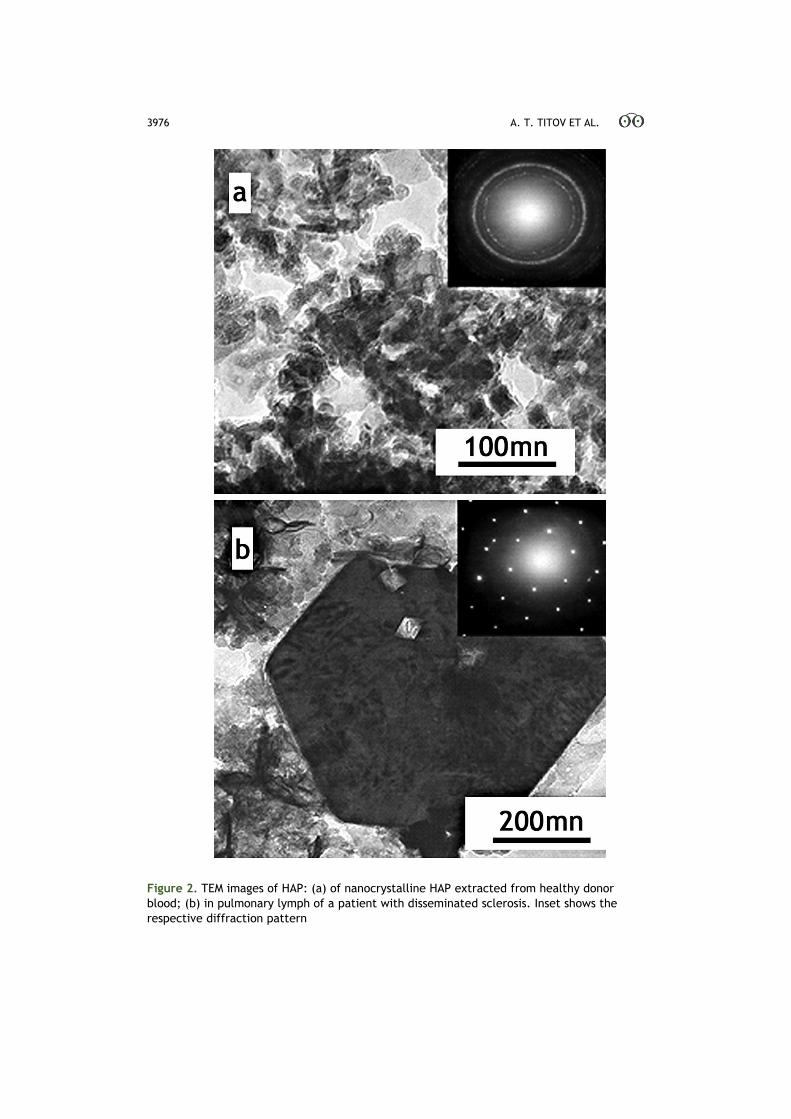
HAP crystals (10-70 nm) were discovered, which are structurally and
morphologically similar to bone apatite and the fine fraction of the heart valve
calcification. They were found both in the solid residue of the blood plasma of
patients with calcinosis, and in the blood of healthy donors (Giachelli 1999).
Figure 2a shows a TEM microphotograph of particles extracted from the donor
blood, which are similar in size, morphology, and structure to bone apatite and
to big HAP deposits in native heart valves. The respective diffraction image of
the aggregate shows several rings with spacing that corresponds to the d/n of
HAP (3.4, 3.1, 2.8, 2.3, and 1.8 Å). Furthermore, plate-like crystals up to 2.5 µm
in size (Figure 2b) were found in the lymph of the disseminated sclerosis patient;
the crystals were similar to the microcrystals with planes (0001) in the heart
valves. The blood also contained needle-like nanocrystals, but these differed
from the nanocrystals of the fine fraction of valve HAP. HAP of the solid residue
of blood and lymph was identified by electron diffraction and X-ray spectra
(EDX).
3976 A. T. TITOV ET AL.
Figure 2. TEM images of HAP: (a) of nanocrystalline HAP extracted from healthy donor
blood; (b) in pulmonary lymph of a patient with disseminated sclerosis. Inset shows the
respective diffraction pattern

INTERNATIONAL JOURNAL OF ENVIRONMENTAL & SCIENCE EDUCATION 3977
To prove further the hypothesis regarding the participation of the blood
HAP in tissue calcification and use it in the diagnostics of mineral metabolism
disorders in humans, model experiments were conducted under controlled
conditions. These observations were substantiated by in vitro simulation of HAP
crystallization in the plasma of healthy adults in a controllable quasi-
physiological environment (T = 37°C, pH = 7.4) and at concentrations of
dissolved Ca and P ions that resemble those in the blood of healthy adults (Table
1). Another objective of the study was to investigate the role of Mg2+, Na+Cl -
and serum albumin, the main blood protein, in the formation of calcium
phosphate.
Table 1. The concentration of ions measured in plasma of a healthy adult and the respective concentrations in the final aqueous solution after mixing during in vitro experiments
Concentration, mM
№ Са 2+ Р 5+ Mg2+ Na+ Cl- Albumine g/L
In blood 1.05 – 1.3 1.00 – 1.5 0.75 – 1.25 130-156 35 - 55
1 1.33 1.50
2 1.33 1.50 0.40
3 1.33 1.50 0.80
4* 1.33 1.50 1.00
5 1.33 1.50 50
6* 1.33 1.50 150
7* 1.33 1.50 1.00 150
8 1.33 1.50 38.6
9 1.33 1.50 0.40 150 38.6
10 1.33 1.50 0.75 150 38.6
X* - calcium phosphate is not formed
Magnesium inhibits the growth of calcium phosphate (Yiu, 2015); the effect
of Na+Cl- is apparently caused by the high concentration and ionizing strength
of its solutions, including plasma; albumin can adsorb onto HAP mineral
surfaces (Leopold, 2012; Egan et al., 2011) and either accelerate or delay
calcification in different conditions (Miller, Weiss & Heistad, 2011). About 45%
of the 2.5 mmol of calcium circulating in the body is free, 45% is bound to serum
proteins, chiefly albumin, and the remaining 10% is complexed with small
molecules such as citrate and phosphate (Thompson & Towler, 2012); only free
Ca ions are biologically active in extracellular fluids.
In all experiments of in vitro calcium phosphate synthesis HAP was
obtained, which formed without precursor phases at low Ca and P
concentrations and physiological pH = 7.4. The first experiment resulted in
precipitation of nanometer acicular crystals of HAP (Figure 3a) with the
crystallographic c direction along the crystal plane (see HR TEM micrograph
and respective Fourier image in Figure 3b). There were found needlelike crystals
up to 200 nm in length and 5 nm in width with a typical contrast ratio on TEM
photos – a light central line and darker edges (Figure 3a). The needlelike
crystals with a similar contrast ratio on TEM-photos were found in the patient’s

3978 A. T. TITOV ET AL.
lymph. The remaining part consisted of thin plates with different structural
ordering, varying from an aggregate of nanocrystals consisting of several
elementary cells to large crystals with a long-range structural order. As
expected, the total mass of the synthesized product reduced significantly when
the concentrations of dissolved Mg (experiments 2-4) or NaCl (experiments 5
and 6) increased, while the precipitated HAP nanocrystals were 1.5-2 times
smaller than those in experiment 1. Precipitation from an aqueous solution
laden with bovine serum albumin (BSA), but free from Mg and NaCl
(experiment 8) likewise produced acicular HAP crystals, almost twice shorter on
average (~100 nm) than with an albumin-free solution (experiment 1). The final
product looked like a mineral-organic coagulate or gel while albumin, which is
highly soluble in water, became insoluble and resisted even multiple rinsing in
bi-distilled water, i.e., BSA and HAP formed a complex. In experiment 3, when
dissolved Mg increased to 0.75 mM, the HAPs changed in both size and
morphology and turned into 40-70 nm platy crystals.

INTERNATIONAL JOURNAL OF ENVIRONMENTAL & SCIENCE EDUCATION 3979
Figure 3. a: A TEM micrograph of nanocrystalline HAP synthesized in controlled in vitro
experiments at T = 37°С and рН=7.4 (experiment 1, Table 1); b: An HR TEM image of an
HAP crystal and its Fourier pattern
Experiments 6 and 7 gave no calcium phosphate precipitation. At a NaCl
concentration of 0.15 M and a concentration of ions in the aqueous solution of Ca
-1.33 mM and P - 1.5 mM (experiment 5), the synthesized HAP had nanocrystals
with a structure that was similar to experiment 1, but significantly smaller in
size (100-150 nm). Under the conditions of experiment 6 with the NaCl
concentration of 0.15 М, and Mg concentration of 0.75 mM in the final aqueous

3980 A. T. TITOV ET AL.
solution containing 1.33 mM calcium ions, and 1.5 mM concentration of
phosphorous ions, calcium phosphate was not produced even after the solution
was aged for one year at room temperature. Thus, dissolved Mg and NaCl can
fully inhibit the nucleation of HAP within the Ca and P levels in the blood of
healthy human adults, but albumin induces HAP nucleation in similar solutions
(experiments 9 and 10).
It should be noted that the duration of the experiment (7 hours) turned out
to be insufficient to produce the amount of calcium phosphate required to
prepare samples and diagnose reliably the phase composition of the obtained
product. However, the precipitation in the solution after 7-days incubation at
room temperature was correctly identified as HAP by electron diffraction and X-
ray analysis (EDX). The TEM images (Figure 4) show HAP as thin elongated
plates, 50-150 nm in length.
Figure 4. A TEM micrograph of nanocrystalline HAP synthesized in controlled in vitro
experiments at T = 37°С and рН=7.4 (experiment 10, Table 1). Inset shows their diffraction
patterns
Discussion and Conclusion
Calcification of the forming bone tissue implies deposition of calcium
phosphates into a neogenic matrix. Pathological calcification causes calcium
phosphate deposition in the tissues of various organs. It is known that bone
apatite consists of thin-plate microcrystals elongated along the c-axis with an
average size of ~ 45×20×3nm (New & Aikawa, 2013). Based on the similarity of
the microstructure and the chemical composition between the inorganic bone

INTERNATIONAL JOURNAL OF ENVIRONMENTAL & SCIENCE EDUCATION 3981
tissue and calcinosis masses on heart valves, it was assumed that their mineral
elements are formed in the human blood. The HAP microcrystals found in the
solid blood residues of healthy donors are structurally and morphologically
similar to bone apatite (Titov, Larionov & Shchukin, 2000). Needle-like HAP
crystals found in the lymph of ill patients are similar to those obtained in
experiments 1 and 4, where there was no Mg or its concentration was much
lower (experiment 2) than the normal concentration in the human blood.
Magnesium not only inhibits HAP formation, but also makes the microcrystals
more isometric (Reynolds, 2004) and similar in shape to bone apatite
(experiment 3). Identical needle-like microcrystals were also found during in
vitro mineralization of tendon (Golub, 2011) and were formed in an aqueous
solution with polyacrylic acid (Le Geros, 2001). It is significant that these
experiments were performed without Mg.
The effect of NaCl as an inhibitor of HAP formation is evidently caused by
an increase of the ionic stress in the solution. However, sodium salt can
completely inhibit HAP formation in an aqueous solution (experiments 4 and 5).
The joint action of Mg and NaCl in an aqueous solution in concentrations
close to those of the blood plasma (experiment 6) completely inhibits HAP
formation. The blood proteins facilitate HAP formation. The presence of HAP
nanocrystals in the blood of healthy donors is indicative of their physiological
origin. The experimental findings indicate that the formation of HAP
microcrystals in the blood is possible only in the presence of albumin and,
presumably, other proteins and enzymes, which may accelerate or inhibit the
formation of HAP.
The formation of HAP in the plasma is similar to precipitation from a
colloid solution. Serum albumin, a major blood component and an important
agent in biomineralization, reduces the interfacial energy of HAP nuclei and
thus stabilizes those that are smaller than the HAP nuclei in an albumin-free
solution (Thompson & Towler, 2012). Albumin adsorbed on the surfaces of HAP
crystals blocks their active growth sites and impedes both growth and
aggregation. Thus, albumin regulates HAP nucleation, which is why changes of
its concentrations in the blood may interfere with calcification patterns.
HAP microcrystals formed in the blood may also participate in the
mineralization of heart valves and cardiovascular calcification. An endothelium
injury can facilitate the penetration of HAP microcrystals formed in the blood
into collagen fibers, which is followed by their deposition. At the late stage of
mineralization, the transformation of HAP to brushite in the valve tissue may be
accompanied by рН decrease. The latter may result from deep degrading
changes in the valve tissue related to functional abnormalities due to
calcification or septic processes.
The formation of HAP in the blood plasma is similar to precipitation from a
colloid solution. At that, albumin reduces the interfacial energy of HAP nuclei
and thus stabilizes those that are smaller than the HAP nuclei in an albumin-
free solution (Combes & Rey, 2002). Albumin adsorbed on the surfaces of HAP
crystals blocks their active growth sites and impedes both growth and
aggregation. Thus, albumin regulates HAP nucleation, which is why changes of
its concentrations in the blood may interfere with calcification patterns.
During HAP formation in the blood, the regulatory function apparently is
performed by another serum protein – Fetuin-A, which many researchers

3982 A. T. TITOV ET AL.
consider a strong inhibitor of mineralization (Smith, 2013; Dautova, 2014; Cai,
Smith & Holt, 2015; Jahnen-Dechent, 2011; Sage, 2011). Fetuin-A is known to
bind strongly calcium and forming apatite crystals. In particular, Fetuin-A
molecules can form mineral colloids with calcium and phosphorus, thus
preventing uncontrollable mineralization in pathological conditions. Paper (Wu,
2009) demonstrated mineral-protein complexes in the form of layered granules
50-500 nm in size, formed in a fetal bovine serum and an adult human serum. In
terms of chemistry and morphology, these protein-mineral particles resemble
granules discovered in the blood serum and so-called nanobacteria (NB) that
were previously found in geological samples, soil, water, and other media.
Similar 20-25 nm nanocrystals were found in vascular calcification and
mineralized heart valves (Neven, 2011; Schlieper, 2011; Miller, 2004). Their
nature is associated with biological liquids. NB were first characterized as
pathogenic agents that produce diseases, including calcification. In fact, NB are
a devitalized HAP complex with organic compounds (Raoult et al., 2008) and are
bound strongly in human tissues with plasma proteins albumin and Fetuin-A.
The presence of proteins and other inhibiting factors generally blocks the
formation of apatite nuclei and stabilizes forming nuclei in the form of
amorphous or partially crystalline spherical nanoparticles. However, such
complexes, which are formed during the interaction between calcium phosphate
complexes and albumin and Fetuin-A, become centers of origination and growth
of HAP nanocrystals when saturated with calcium and phosphorus. The dual
role of Fetuin-A in the blood plasma allows concluding that it is a systemic
regulator of mineralization (Jahnen-Dechent, 2011).
The results of this research and abovementioned data from literary sources
imply that the discovered HAP nanocrystals in the blood of patients and donor
blood (Titov & Larionov, 2007; Titov, Larionov & Shchukin, 2000; Titov, 2004)
and the protein-mineral complexes that were formed in the blood plasma have a
common nature. However, as a result of the applied technique of HAP
nanocrystal extraction from blood plasma to identify them with TEM (annealing
at T=670°C), the organic part of these complexes was destroyed.
Protein-mineral complexes play an important role in physiological and
pathological mineralization. The common property of albumin and Fetuin-A is
their participation in the formation of bone tissue. Papers show that the matrix
of a growing bone intensively consumes both albumin and Fetuin-A from the
blood. Albumin and Fetuin-A are two most common non-collagen proteins in
bone. It is also known that albumin is easily produced during bone
demineralization (Combes & Rey, 2002).
The initial stage of bone tissue formation is associated with the formation of
an extracellular matrix that mostly includes collagen fibers. The above
experimental facts allow assuming that albumin and Fetuin-A are structural
proteins responsible for subsequent integration of HAP nanocrystals into the
newly formed bone tissue.
Primary HAP nanocrystals that were formed in the blood can also
participate in the mineralization of heart valves and vessels. An endothelium
injury can facilitate the penetration of HAP microcrystals formed in the blood
into collagen fibers, which is followed by their deposition. At the late stage of
mineralization, the transformation of HAP to brushite in the valve tissue may be
accompanied by рН decrease. The latter may result from deep degrading

INTERNATIONAL JOURNAL OF ENVIRONMENTAL & SCIENCE EDUCATION 3983
changes in the valve tissue related to functional abnormalities due to
calcification or septic processes.
Acknowledgements
The authors would like to thank the Novosibirsk State University for
providing premises and equipment for this research. No conflicts of interests are
observed. This article does not contain any studies with human participants or
animals performed by any of the authors
This work was supported in part by research grant the Russian Foundation
for Basic Research – No. 13-05-00921.
Disclosure statement
No potential conflict of interest was reported by the authors.
Notes on contributors
Anatoly T. Titov holds a PhD, Associate Professor of General Physics Department,
Novosibirsk State University, Novosibirsk, Russia.
Peter M. Larionov holds Researcher, Professor of Department of Fundamental
Medicine, Novosibirsk State University, Novosibirsk, Russia.
Alexandra S. Ivanova holds a Doctor of Chemical Sciences, Head of Laboratory of
Department of Catalysis and Adsorption, Novosibirsk State University, Novosibirsk,
Russia.
Vladimir I. Zaikovskii holds a PhD, Researcher of Department of General Physics,
Novosibirsk State University, Novosibirsk, Russia.
Mikhail A. Chernyavskiy holds a PhD, Surgeon, Novosibirsk Research Institute of
the Pathology of Blood Circulation named after academician E.N. Meshalkin,
Novosibirsk, Russia.
References
Anon, A. (2013). Ethical Principles for Medical Research Involving Human Subjects. World Medical
Assotiation Declaration of Helsinki, 310(20), 206-208.
Cai, M. M., Smith, E. R. & Holt, S. G. (2015). The Role of Fetuin-A in Mineral Trafficking and
Deposition. BoneKEy reports, 4, 672.
Combes, C. & Rey, C. (2002). Adsorption of Proteins and Calcium Phosphate Materials Bioactivity.
Biomaterials, 23(13), 2817–23.
Dautova, Y. (2014). Fetuin-A and albumin alter cytotoxic effects of calcium phosphate nanoparticles
on human vascular smooth muscle cells. PloS one, 9(5), 554-565.
Demer, L. L. & Tintut, Y. (2008). Vascular Calcification: Pathobiology of a Multifaceted Disease.
Circulation, 117(22), 2938–2948.
Egan, K. P. (2011). Role for Circulating Osteogenic Precursor Cells in Aortic Valvular Disease.
Arteriosclerosis, thrombosis, and vascular biology, 31(12), 2965–2971.
Giachelli, C. M. (1999). Ectopic calcification: Gathering Hard Facts About Soft Tissue
Mineralization. The American Journal of Pathology, 154(3), 671–5.
Golub, E. E. (2011). Biomineralization and Matrix Vesicles in Biology and Pathology. Seminars in
Immunopathology, 33(5), 409–17.
Jahnen-Dechent, W. (2011). Fetuin-A Regulation of Calcified Matrix Metabolism. Circulation
Research, 108(12), 494–509.
Le Geros, R. Z. (2001). Formation and Transformation of Calcium Phosphates: Relevance to Vascular Calcification. Zeitschrift fur Kardiologie, 90(3), 116–24.
Leopold, J. A. (2012). Cellular Mechanisms of Aortic Valve Calcification. Circulation. Cardiovascular
interventions, 5(4), 605–614.

3984 A. T. TITOV ET AL.
London, G. M. (2013). Mechanisms of Arterial Calcifications and Consequences for Cardiovascular
Function. Kidney International Supplements, 3(5), 442–445.
Miller, J. D., Weiss, R. M. & Heistad, D. D. (2011). Calcific Aortic Valve Stenosis: Methods, Models,
and Mechanisms. Circulation Research, 108(11), 1392–1412.
Miller, V. M. (2004). Evidence of Nanobacterial-Like Structures in Calcified Human Arteries and
Cardiac Valves. American Journal of Physiology. Heart and Circulatory Physiology, 287(3),
1115–1124.
Neven, E. (2011). Cell Biological and Physicochemical Aspects of Arterial Calcification. Kidney
International, 79(11), 1166–1177.
New, S. E. & Aikawa, E. (2013). Role of Extracellular Vesicles in de Novo Mineralization: an
Additional Novel Mechanism of Cardiovascular Calcification. Arteriosclerosis, Thrombosis, and
Vascular Biology, 33(8), 1753–1758.
Raoult, D. (2008). Nanobacteria are Mineralo Fetuin Complexes. Plos Pathogens, 4(2), 41-53.
Reynolds, J. L. (2004). Human Vascular Smooth Muscle Cells Undergo Vesicle-Mediated
Calcification in Response to Changes in Extracellular Calcium and Phosphate Concentrations.
Journal of the American Society of Nephrology, 15(11), 2857–2867.
Sage, A. P. (2011). Hyperphosphatemia-Induced Nanocrystals Upregulate the Expression of Bone
Morphogenetic Protein-2 and Osteopontin Genes in Mouse Smooth Muscle Cells in Vitro.
Kidney International, 79(4), 414–422.
Schlieper, G. (2011). A Red Herring in Vascular Calcification: “Nanobacteria” are Protein-Mineral
Complexes Involved in Biomineralization. European Renal Association, 26(11), 3436–3439.
Smith, E.R. (2013). Serum Fetuin-A Concentration and Fetuin-A-Containing Calciprotein Particles
in Patients With Chronic Inflammatory Disease and Renal Failure. Nephrology, 18(3), 215–
221.
Thompson, B. & Towler, D. A. (2012). Arterial Calcification and Bone Physiology. Endocrinology,
8(9), 529–43.
Titov, A. & Larionov, P. 2007. Bone-like Hydroxyapatite Formation in Human Blood. Calcified
Tissue International, 80, 58.
Titov, A. (2004). Hydroxyapatite Formation in Human Blood. Sao-Paulo: ICAM-BR, 362 p.
Titov, A., Larionov, P. & Shchukin, V. (2000). Possible formation of hydroxyapatite in blood. Report of
Biochemistry, 7(2), 124-132.
Wu, C. Y. (2009). Fetuin-Albumin-Mineral Complexes Resembling Serum Calcium Granules and
Putative Nanobacteria. PloS one, 4(11), 805-816.
Yarbrough, D. K. (2010). Specific Binding and Mineralization of Calcified Surfaces By Small
Peptides. Calcified Tissue International, 86(1), 58–66.
Yiu, A. J. (2015). Vascular Calcification and Stone Disease. Journal of Cardiovascular Development
and Disease, 2(3), 141–164.