Embed Size (px)
Citation preview
JOURNAL OF VIROLOGY, June 2011, p. 6038–6048 Vol. 85, No. 120022-538X/11/$12.00 doi:10.1128/JVI.00030-11Copyright © 2011, American Society for Microbiology. All Rights Reserved.
Two Distinct Conformations of a Rinderpest Virus Epitope Presentedby Bovine Major Histocompatibility Complex Class I N*01801:
a Host Strategy To Present Featured Peptides�†Xin Li,1,2‡ Jun Liu,2,3‡ Jianxun Qi,2 Feng Gao,4,5 Qirun Li,1 Xiaoying Li,1 Nianzhi Zhang,1
Chun Xia,1,2* and George F. Gao1,2,3,6*College of Veterinary Medicine, China Agricultural University, Beijing 100094,1 CAS Key Laboratory of Pathogenic Microbiology and
Immunology, Institute of Microbiology, Chinese Academy of Sciences, Beijing 100101,2 Graduate University, Chinese Academy ofSciences, Beijing 100049,3 National Laboratory of Macromolecules, Institute of Biophysics, Chinese Academy of Sciences,
Beijing 100101,4 School of Life Sciences, Sichuan University, Chengdu 610064, Sichuan,5 and Beijing Institutes ofLife Science, Chinese Academy of Sciences, Beijing 100101,6 People’s Republic of China
Received 6 January 2011/Accepted 23 March 2011
The presentation of viral peptide epitopes to host cytotoxic T lymphocytes (CTLs) is crucial for adaptivecellular immunity to clear the virus infection, especially for some chronic viral infections. Indeed, hosts havedeveloped effective strategies to achieve this goal. The ideal scenario would be that the peptide epitopesstimulate a broad spectrum of CTL responses with diversified T-cell receptor (TCR) usage (the TCR reper-toire). It is believed that a diversified TCR repertoire requires a “featured” peptide to be presented by the hostmajor histocompatibility complex (MHC). A featured peptide can be processed and presented in a number ofways. Here, using the X-ray diffraction method, the crystal structures of an antigenic peptide derived fromrinderpest virus presented by bovine MHC class I N*01801 (BoLA-A11) have been solved, and two distinctconformations of the presented peptide are clearly displayed. A detailed analysis of the structure and com-parative sequences revealed that the polymorphic amino acid isoleucine 73 (Ile73) is extremely flexible,allowing the MHC groove to adopt different conformations to accommodate the rinderpest virus peptide. Thismakes the peptide more featured by exposing different amino acids for T-cell recognition. The crystal struc-tures also demonstrated that the N*01801 molecule has an unusually large A pocket, resulting in the specialconformation of the P1 residue at the N terminus of the peptide. We propose that this strategy of host peptidepresentation might be beneficial for creating a diversified TCR repertoire, which is important for a more-effective CTL response.
Viral diseases and other infections caused by intracellularpathogens of cattle result in large global economic losses everyyear. In the past several decades, rinderpest virus (RPV) (23,40), foot-and-mouth disease virus (FMDV) (7), and bovinetuberculosis infection (62) have brought disastrous conse-quences, including social panic and even a threat to humanhealth. Nearly 6 decades ago, most countries were committedto developing more-effective vaccines (46), and remarkableachievements were made in controlling threatening bovine vi-ral infectious diseases and other zoonoses (23, 46). In the1960s, British virologist Walter Plowright developed a live at-tenuated vaccine against rinderpest virus, which was widelyused in rinderpest eradication efforts (40). However, it stillremains extremely challenging for humans to completely erad-icate a virus. The questions of how the bovine attenuated
vaccine induces immune responses and how the viral peptidesare presented and recognized by host immune molecules arestill largely unanswered. Therefore, studies on bovine immu-nity against the rinderpest virus may provide clues for ourbattle against other viral infections.
Generally speaking, effective vaccines have common charac-teristics: they are rich in T-cell and B-cell epitopes and caninduce the immune system to generate protective immuneresponses (3, 36, 54). During this process, major histocompat-ibility complex (MHC) molecules play a pivotal role in thehost. MHC class I (MHC I) molecules form a heterotrimercomplex composed of the heavy chain of MHC, the antigenicpeptide, and �2-microglobulin (�2m). Generally, MHC I loadsendogenous peptides, including the virus-derived peptides.Briefly, at the initial stage of virus infection, antigenic proteinsare processed in a proteasome-dependent or -independentmanner. The resultant short peptides are then transported tothe endoplasmic reticulum and are loaded onto the peptide-binding grooves of MHC I molecules. Consequently, the pep-tide-loading MHC I complexes are translocated to the cellsurface and are recognized by cytotoxic T lymphocytes (CTLs)with specific T-cell receptors (TCRs) (49, 63). This immunerecognition induces an MHC-restricted CD8� T-cell responsethat is characterized by the killing and elimination of the in-fected cells by effector T cells. The majority of virus-derivedpeptides are processed and presented through this pathway
* Corresponding author. Mailing address for G. F. Gao: CAS KeyLaboratory of Pathogenic Microbiology and Immunology, Institute ofMicrobiology, Chinese Academy of Sciences, Beijing 100101, People’sRepublic of China. Phone: (86) 10-64807688. Fax: (86) 10-64807882.E-mail: [email protected]. Mailing address for C. Xia: College ofVeterinary Medicine, China Agricultural University, Beijing 100094,People’s Republic of China. Phone and fax: (86) 10-62733372. E-mail:[email protected].
† Supplemental material for this article may be found at http://jvi.asm.org/.
‡ X. Li and J. Liu contributed equally to this work.� Published ahead of print on 30 March 2011.
6038
Dow
nloa
ded
from
http
s://j
ourn
als.
asm
.org
/jour
nal/j
vi o
n 16
Dec
embe
r 20
21 b
y 77
.125
.23.
190.
and consequently trigger the CTL-specific immune responses.These virus-derived peptides presented by MHC I moleculesare known as antigenic CTL-specific epitopes.
Based on structural studies, the antigenicity of a peptide ispartially dependent on the characteristics of the peptide re-lated to its presentation by MHC molecules (31, 33, 34, 53, 55).Generally, peptides are divided into featured, featureless, andbulged peptides according to their presentation properties (25,57). Featured peptides are those with solvent-exposed, prom-inent side chains or harmonious bulged conformations, whichalways correspond to a diverse repertoire of TCRs (32, 37, 38,58). In contrast, featureless peptides have fewer or no solvent-exposed residues with prominent side chains. Bulged peptidesare usually long peptides (�12 amino acids) that contain anextreme bend in the middle of the peptide chain binding to theMHC I molecule. The structural landscapes of featureless andbulged peptides result in an immune T-cell repertoire of lim-ited diversity (58–60). Therefore, investigation of the structuralcharacteristics of a peptide presented by MHC I molecules canimprove the understanding of the antigenicity of that peptideand aid in the rational development and modification of pep-tide-based vaccines (33).
Bovine MHC I, which is called BoLA-I, has a critical role inpresenting virus-derived antigen peptides and dominating bo-vine antiviral-specific CTL immune responses. In the 1970s,BoLA-I protein was first identified and genotyped by use of aspecific serum antibody. BoLA-I was first cloned in 1988 (20),and since then, BoLA-I related research has entered the geneera. Thus far, hundreds of BoLA-I cDNA sequences have beenregistered at the National Center for Biotechnology Informa-tion (NCBI) (http://www.ncbi.nlm.nih.gov/), and 58 completeBoLA-A sequences have been uploaded to the Immuno Poly-morphism Database (IPD) (http://www.ebi.ac.uk/ipd/) (5).BoLA is located on the 23rd bovine chromosome (22), includ-ing at least six expressed gene loci (29). Further research hasdemonstrated that there are one to three gene loci that can betranscribed and expressed in one BoLA haplotype (16, 17).The fact that a breed of BoLA haplotypes expresses only oneBoLA-A gene locus (6, 15) provides an important experimen-tal model for controlling bovine viral diseases in cattle with asingle MHC I gene locus.
Due to polymorphisms in BoLA-I, there are more than 50serotypes of one BoLA-I allele locus (4, 13). The BoLA-A11serotype specifically indicates that the BoLA-A11/pBoLA-19(D18.3) gene product N*01801 is expressed (18, 28, 47, 48).Hegde and colleagues obtained the N*01801 peptide bindingmotif by monoclonal antibody purification of MHC moleculesand subsequent acid elution of peptides (28). The majority ofthe peptides that occupied the binding groove of N*01801 were
nonamers. There was clear evidence that position 2 of thepeptides was occupied by Pro, and the C-terminal amino acidanchor was Ile/Val. Among the alleles whose motifs have beenreported, the peptide motif of N*01801 is similar to those ofH-2Ld, HLA-B7, HLA-B*3501, HLA-B*5101, HLA-B*5102,HLA-B*5103, HLA-B*7801, and HLA-Cw*0401 (21, 45). Se-quence comparison of the bovine MHC I allele N*01801 tothese MHC I molecules demonstrates that key residues sharesimilar characteristics (and are conserved in some cases),which are involved in accommodating the similar anchor resi-dues in P2 and P9. This was the first report of a BoLA allele-specific peptide motif (ASPM; 28). To identify bovine-re-stricted CTL epitopes derived from pathogens, according toN*01801 ASPMs, dozens of potential bovine CTL epitopeshave been synthesized, and their antigenicity was preliminarilyanalyzed by CTL assays (27). In 2004, one of the CTL epitopeswith a typical N*01801 ASPM (peptide IPA) was identified inrinderpest virus hemagglutinin (H) protein amino acids 408 to416 (IPAYGVLTI) (51). Recently, Guzman et al. confirmedthat there is an MHC-restricted CD8� T-cell response afterFMDV infection (26). However, the mechanism of bovineantiviral-specific T-cell immunity remains unclear, partiallydue to the lack of clues as to the binding and presentation ofthe antigenic peptides by BoLA-I.
To describe the landscape of peptide presentation by bovineMHC I N*01801 and to understand the immune basis of theeradication of rinderpest virus, we determined the crystalstructures of N*01801 complexed to the rinderpest virus-de-rived peptide IPA. Analysis of the structures we determinedsuggests that compared to the structures of other MHC Imolecules, including that of bovine N*01301, which was deter-mined recently by Macdonald and colleagues (34), the struc-ture of bovine MHC I N*01801 is distinctive. The structure ofbovine N*01801 illuminates a novel presentation strategy forfeatured epitopes among mammalian MHCs and may lead toimproved understanding of the structural basis of CTL-basedimmune responses (9).
MATERIALS AND METHODS
Peptide synthesis. The peptides used in this study (Table 1), including peptideIPA (IPAYGVLTI), derived from rinderpest virus attachment glycoproteins(51), were synthesized and purified by reverse-phase high-performance liquidchromatography (HPLC) (SciLight Biotechnology). The peptide purity was de-termined to be �90% by analytical HPLC and mass spectrometry. These pep-tides were stored in lyophilized aliquots at �80°C after synthesis and weredissolved in dimethyl sulfoxide (DMSO) before use.
Preparation of the bovine MHC I N*01801-�2m-IPA complexes. Reversetranscription-PCR (RT-PCR) was used to amplify the full-length cDNA ofN*01801 (A11) and bovine �2m from bovine kidney cells. Details of the primersused can be found in Table S1 in the supplemental material. The extracellular
TABLE 1. Peptides used in this study
Name Derived protein Position Sequencea Reference
IPAb Rinderpest virus H protein 408–416 IPAYGVLTI 51APA BHV-Ic BICP0 protein 482–490 APAPISTMI 27NPM BHV-I glycoprotein B precursor 780–788 NPMKALYPI 27TPG BHV-I glycoprotein C precursor 18–26 TPGATTPV 27
a Underlined boldface residues are the typical primary anchors of the peptides presented by N*01801.b Peptide IPA was used in the structural determination of the bovine MHC class I N*01801.c BHV-I, bovine herpesvirus 1.
VOL. 85, 2011 TWO CONFORMATIONS OF A BoLA-A11 EPITOPE 6039
Dow
nloa
ded
from
http
s://j
ourn
als.
asm
.org
/jour
nal/j
vi o
n 16
Dec
embe
r 20
21 b
y 77
.125
.23.
190.
region of N*01801 (amino acids 2 to 275) was amplified from the cDNA accord-ing to its nucleotide sequence (GenBank accession no. BC151402) using primers1 and 2. Bovine �2m (amino acids 1 to 98) was cloned from bovine kidney cellcDNA according to its nucleotide sequence (GenBank accession no. BC118352)with primers 3 and 4. Mouse �2m (amino acids 1 to 99) was cloned from BALB/cmouse spleen cDNA according to its nucleotide sequence (GenBank accessionno. M84364.1) with primers 5 and 6. The amplified products were ligated into apET21a vector (Novagen) and were transformed into Escherichia coli strainBL21(DE3). The recombinant proteins were expressed as inclusion bodies andwere then purified as described previously (8).
The N*01801-�2m-peptide complexes were prepared essentially with refoldingassays as described by Zhou et al. in 2004 (64). Briefly, the N*01801 heavy chainand bovine or murine �2m inclusion bodies were separately dissolved in asolution of 10 mM Tris-HCl (pH 8.0) and 8 M urea. The N*01801 heavy chain,�2m, and the peptide in a 1:1:3 molar ratio were refolded by gradual dilution.After 48 h of incubation at 4°C, the soluble portion of the complexes wasconcentrated and then purified by size exclusion chromatography on a Superdex200 16/60 column. The comparison of the absorbance peaks of the refoldedcomplexes using different peptides is presented in Fig. 1. If the complex wasprepared for crystallization, it was further purified by Resource-Q anion-ex-change chromatography (GE Healthcare).
Crystallization and data collection. The BoLA complexes were ultimatelyconcentrated to 12 mg/ml in crystallization buffer (4.0 mM sodium formate),mixed with reservoir buffer at a 1:1 ratio, and crystallized by the hanging-dropvapor diffusion technique at 291 K. A Crystal Screen kit (Hampton Research,Riverside, CA) was used to screen for optimal crystal growth conditions. Afterseveral days, crystals of N*01801 complexed with the IPA peptide and bovine�2m (N*01801) were obtained with Crystal Screen solution 33 (4.0 mM sodiumformate). Diffraction data were collected to a resolution of 2.7 Šusing anin-house X-ray source (Rigaku MicroMax007 desktop rotating anode X-raygenerator with a Cu target operated at 40 kV and 30 mA) and an R-AXIS IV��
imaging-plate detector at a wavelength of 1.5418 Å. Similarly, the crystals ofN*01801 complexed with the IPA peptide and murine �2m (N*01801-M�) weregrown in 0.2 M ammonium sulfate, 0.1 M Tris (pH 8.5), and 25% (wt/vol)polyethylene glycol 3350 (PEG 3350) at a concentration of 12 mg/ml, and theresolution of the diffraction data was 1.9 Å. The crystals were first soaked inreservoir solution containing 25% glycerol as a cryoprotectant and were thenflash-cooled in a stream of gaseous nitrogen at 100 K (42). The collected inten-sities were indexed, integrated, corrected for absorption, scaled, and mergedusing the HKL2000 package (41).
Structure determination and refinement. The structures of the BoLA com-plexes were solved by molecular replacement using the MOLREP program withHLA-B*5101 (Protein Data Bank [PDB] code 1E27) as a search model. Exten-sive model building was performed by hand with COOT (19), and restrainedrefinement was performed using REFMAC5. Additional rounds of refinementwere performed using the phenix refine program implemented in the PHENIXpackage (2) with isotropic atomic displacement parameter (ADP) refinementand bulk solvent modeling. The stereochemical quality of the final model wasassessed with the PROCHECK program (30).
Protein structure accession numbers. The coordinates and structure factors ofN*01801 and N01801-M� have been deposited in the Protein Data Bank withaccession numbers 3PWV and 3PWU, respectively.
RESULTS
Typical peptides bound to bovine MHC I molecule N*01801revealed by in vitro refolding. A series of bovine MHC I alleleN*01801-restricted CD8� T-cell epitopes derived from differ-ent pathogens have recently been screened and characterized(14, 27, 51); from these epitopes, the peptide motif of thisMHC I molecule was defined. However, no rapid and efficientmethods to evaluate the ability of a peptide with a given motifto bind to bovine MHC I molecules in vitro have been devel-oped yet. As indicated in previous studies (31–33, 55), therefolding experiments can semiquantitatively reflect the bind-ing capability of peptides for MHCs. Four previously identifiedN*01801-restricted CD8� T-cell epitopes (Table 1) were syn-thesized and used in refolding assays to evaluate their abilitiesto bind to N*01801 molecules and to define the candidatesused for determination of the N*01801 structure. These fourN*01801-restricted peptides helped the heavy chain ofN*01801 and bovine �2m refold in vitro, as evidenced by theabsorbance peaks of the complexes (Fig. 1). All four peptidescontain a proline at position P2, while the Pc (C terminus ofthe peptide) positions were occupied by either isoleucine or
FIG. 1. Binding of peptides to bovine MHC I N*01801 indicated via in vitro refolding. Peptides with the ability to bind to N*01801 can helpthe N*01801 heavy chain and �2m refold in vitro. After correct refolding, we found that high absorbance peaks of the MHCs with the expectedmolecular mass of 45 kDa eluted at the estimated volume of 16 ml on a Superdex 200 column (GE Healthcare). Complexes were formed inrefolding with peptides IPA, APA, NPM, and TPG (which have the typical N*01801-restricted motif). IPA also aided in the renaturation of theN*01801 heavy chain with the murine �2m (IPA-M�). No binding peaks appeared when there was no peptide in the refolding system.
6040 LI ET AL. J. VIROL.
Dow
nloa
ded
from
http
s://j
ourn
als.
asm
.org
/jour
nal/j
vi o
n 16
Dec
embe
r 20
21 b
y 77
.125
.23.
190.
valine. This peptide motif was partially demonstrated previ-ously (27).
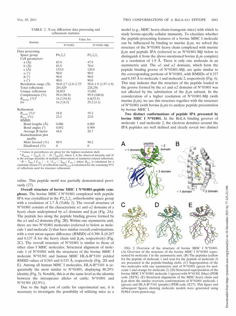
Overall structure of bovine MHC I N*01801-peptide com-plexes. The bovine MHC I N*01801 complexed with peptideIPA was crystallized in the P212121 orthorhombic space groupwith a resolution of 2.7 Å (Table 2). The overall structure ofN*01801 consists of the characteristic �1 and �2 domains of aheavy chain underpinned by �3 domains and �2m (Fig. 2A).The peptide lies along the peptide binding groove formed bythe �1 and �2 domains (Fig. 2B). Within one asymmetric unit,there are two N*01801 molecules (referred to below as mole-cule 1 and molecule 2) that have similar overall conformations,with a root mean square difference (RMSD) of 0.300 Å (0.267and 0.137 Å for the heavy chain and �2m, respectively) (Fig.2C). The overall structure of N*01801 is similar to those ofother class I MHC molecules. Structural alignment of mole-cule 1 of N*01801 with the structures of the bovine MHC Imolecule N*01301 and human MHC HLA-B*5101 yieldedRMSD values of 0.563 and 0.533 Å, respectively (Fig. 2D andE). Among all human MHC I molecules, HLA-B*5101 is se-quentially the most similar to N*01801, displaying 80.29%identity (Fig. 3). Notably, this is at the same level as the identitybetween the intraspecies alleles of cattle: N*01801 andN*01301 (82.9%).
Due to the high cost of cattle for experimental use, it isnecessary to investigate the possibility of utilizing mice as a
model (e.g., MHC heavy-chain-transgenic mice) with which tostudy bovine-specific cellular immunity. To elucidate whetherthe peptide-presenting features of a bovine MHC I moleculecan be influenced by binding to murine �2m, we solved thestructure of the N*01801 heavy chain complexed with murine�2m and peptide IPA (referred to as N*01801-M� below todistinguish it from the above-mentioned bovine �2m complex)at a resolution of 1.9 Å. There is only one molecule in anasymmetric unit. The �1 and �2 domains, which form thepeptide binding groove of N*01801-M�, are quite similar tothe corresponding portions of N*01801, with RMSDs of 0.337and 0.345 Å to molecule 1 and molecule 2, respectively (Fig. 4).This may indicate that the structure of the peptide loaded inthe groove formed by the �1 and �2 domains of N*01801 wasnot affected by the substitution of the �2m subunit. In theconsideration of a higher resolution of N*01801-M� (withmurine �2m), we use this structure together with the structureof N*01801 (with bovine �2m) to analyze peptide presentationby bovine MHC I.
Two distinct conformations of peptide IPA presented bybovine MHC I N*01801. In the BoLA binding grooves ofmolecule 1 and molecule 2, the electron densities around theIPA peptides are well defined and clearly reveal two distinct
FIG. 2. Overview of the structure of bovine MHC I N*01801.(A) Overview of the structure of the bovine MHC I N*01801 repre-sented by molecule 1 in the asymmetric unit. (B) The peptides (yellowfor the peptide of molecule 1 and cyan for the peptide of molecule 2)are presented in the peptide-binding clefts. (C) Superposition of thetwo molecules with one asymmetric unit of N*01801 (green for mol-ecule 1 and orange for molecule 2). (D) Structural superposition of thebovine MHC I N*01801 molecule 1 (green) with N*01301 (blue) (PDBcode 2XFX). (E) Structural alignments of the MHC heavy chain and�2m show the similar overview conformations of N*01801 molecule 1(green) and HLA-B*5101 (purple) (PDB code 1E27). This figure andsubsequent figures showing molecule models were generated usingPyMol (www.pymol.org).
TABLE 2. X-ray diffraction data processing andrefinement statistics
StatisticValue for:
N*01801 N*01801-M�
Data processingSpace group P41212 P212121Cell parameters
a (Å) 83.9 47.9b (Å) 83.9 70.0c (Å) 153.0 120.8� (°) 90.0 90.0� (°) 90.0 90.0� (°) 90.0 90.0
Resolution range (Å) 50.0–2.7 (2.8–2.7)a 50.0–1.9 (1.97–1.9)Total reflections 201,829 228,256Unique reflections 28,833 15,065Completeness (%) 99.4 (98.9) 99.9 (100.0)Rmerge (%)b 14.2 (54.8) 6.6(53.5)I/� 16.2 (4.3) 29.2 (3.1)
RefinementRwork (%)c 18.8 19.2Rfree (%) 23.2 23.0RMSD
Bond lengths (Å) 0.006 0.005Bond angles (°) 0.892 0.909Average B factor 44.6 28.7
Ramachandran plotquality
Most favored (%) 88.9 90.2Disallowed (%) 0 0
a Values in parentheses are given for the highest-resolution shell.b Rmerge ¥hkl¥i � Ii � I� � ¥hkl¥iIi, where Ii is the observed intensity and I�
is the average intensity of multiple observations of symmetry-related reflections.c R ¥hkl � Fobs � � k � Fcal � � ¥hkl � Fobs �, where Rfree is calculated for a
randomly chosen 5% of reflections and Rwork is calculated for the remaining 95%of reflections used for structure refinement.
VOL. 85, 2011 TWO CONFORMATIONS OF A BoLA-A11 EPITOPE 6041
Dow
nloa
ded
from
http
s://j
ourn
als.
asm
.org
/jour
nal/j
vi o
n 16
Dec
embe
r 20
21 b
y 77
.125
.23.
190.

conformations of the peptide. For simplicity, the peptides pre-sented by molecule 1 and molecule 2 are referred to asIPA-M1 and IPA-M2 below; they are shown in Fig. 5A and B,respectively. IPA-M1 and IPA-M2 have an RMSD of 1.284 Å.However, the conformation of IPA-M2 is quite similar to that
of the IPA peptide in the N*01801-M� structure, with anRMSD of 0.474. Furthermore, the interactions, including thehydrogen bonds and the nonpolar contacts, of IPA-M2 in thestructure of N*01801 are also similar to those of the IPApeptide in the N*01801-M� structure. The IPA peptide pre-
FIG. 3. Structure-based sequence alignment of N*01801 and eight different types of MHC I molecules. Cylinders indicate �-helices, and blackarrows indicate �-strands. Residues highlighted in red are completely conserved, and boxed residues are highly (�80%) conserved. Residues thatplay a critical role in peptide presentation are asterisked (residue 73, highlighted in yellow, and residue 167, highlighted in cyan). The sequencealignment was generated with Clustal X (56) and ESPript (24).
6042 LI ET AL. J. VIROL.
Dow
nloa
ded
from
http
s://j
ourn
als.
asm
.org
/jour
nal/j
vi o
n 16
Dec
embe
r 20
21 b
y 77
.125
.23.
190.
sented by N*01801-M� is referred to as IPA-M� and is shownin Fig. 5C. The B factors of the peptides in molecule 1 andN*01801-M� are 39.1 and 25.2, respectively, indicating thestructural accuracy of these two distinct conformations. Thestructure of N*01801-M� has a higher resolution (1.9 Å) thanthat of N*01801. Thus, we describe only the differences be-tween the peptide conformations of molecule 1 andN*01801-M� in detail here.
The residues at both ends of the peptides (Ile1, Pro2, andAla3 at the N terminus and Thr8 and Ile9 at the C terminus)remain the same. In contrast, the major differences betweenthe two peptides occur in the central region (Tyr4, Gly5, Val6,and Leu7) of the peptides (Fig. 5D). Within the groove ofmolecule 1, the main chain of IPA-M1 turns sharply into thefloor of the groove at the P5 (Gly5) position and then rises atP6 (Val6). In position P7, the C� of Leu7 again falls into thebinding groove, while Thr8 in position P8 lifts upward. Thetortuous main chain of the peptide presents a “double-M”conformation, which we believe to be unique among the MHCI structures solved thus far. Gly5 and Leu7 of IPA-M1 arelocated deep in the groove of molecule 1 as two secondaryanchor residues. Conversely, the side chain of Val6 is solventexposed and may function in TCR docking. However, IPA-M1and IPA-M� display a totally different conformation in thisregion. The main chain of the peptide falls only once in themiddle, with the side chain of Val6 protruding into the groove
of the �1�2 “bed” as a secondary anchor in the M-shapedpeptide. The M-shaped conformation of IPA-M� is more com-mon among the structures of mammalian MHC I molecules(12, 35).
An unusually flexible Ile73 in the �1-helix. Superposition ofall of the residues making up the peptide binding groove ofmolecule 1 and N*01801-M� shows that only Ile73 in the�1-helix of the heavy chain differs (Fig. 5E). In molecule 1, theside chain of Ile73 points up into the solvent and leaves enoughspace for the raised conformation of the P6 valine in IPA-M1.Inversely, the side chain of Ile73 in N*01801-M� protrudestoward the �2-helix and occupies the space above the middleregion of IPA-M�. In this sense, the distinct conformations ofIle73 may contribute to the two different presentation confor-mations of the IPA peptide. Indeed, residues at position 73that locate in the flanking side of the peptide binding grooveare a key factor for the peptide conformation in other MHC Imolecules (11, 34). For different MHC molecules, residueshave diverse polymorphism in position 73 (Fig. 3). Therefore,the residues in this position have different impacts on peptidepresentation. In a recently solved structure of the bovine MHCI N*01301 molecule, peptide Tp1214–224 displays an unusualbulged C-terminal conformation, which is affected by Trp73beneath the peptide (34). Another factor may also influencethe flexibility of the IPA peptide. Both of the bovine MHCspossess E97 in the bottom of the peptide binding groove.While the secondary anchor residue of K5 of Tp1214–224 formsa stable salt bridge with E97 (34), IPA does not interact withE97 due to the hydrophobicity of the middle residues of thepeptide (Gly5, Val6, and Leu7). Thus, the lack of a salt bridgemay partially contribute to the flexibility of IPA.
Different exposed areas and buried residues formed by thetwo IPA conformations. The two distinct conformations of IPAin the two molecules of the asymmetric unit of the bovineMHC I N*01801 structures may contribute to the exposure ofdifferent surfaces of the peptides to the solvent (Fig. 6). Withthe “double-M” conformation, IPA-M1 protrudes from theside chains of all of the even-numbered residues (Tyr4, Val6,and Thr8) of the C-terminal portion of the peptide (P3 to P9),which conspicuously presents their side chains for potentialTCR docking (Fig. 6A). The side chain of Ile73 protrudes intothe solvent, leaving enough space for the exposure of themiddle region of IPA-M1 (Fig. 6C). Calculation of the acces-sible areas shows that the exposed surface of the three residuesoccupies most (87.7%) of the total exposed area of IPA-M1.The odd-numbered residues in this portion of the peptide(Ala3, Gly5, Leu7, and Ile9) bury themselves deeply into thegroove. In the structure of N*01801-M�, P4 Tyr, P7 Leu, andP8 Thr are the most prominent residues and are likely impor-tant for TCR interaction (Fig. 6B). The exposed surface ofthese three residues (Tyr4, Leu7, and Thr8) accounts for81.0% of the total exposed surface of IPA-M� (Fig. 6D). Inaddition, the main chains of P5 Gly and P6 Val also partiallycontribute to the exposed area of IPA-M�. However, the sidechain of Ile73 extends over the middle region of IPA-M�,which may affect the direct docking of the TCR to the mainchain of P5 Gly and P6 Val. These distinct characteristics ofthe exposed area of the peptide presented by N*01801 may beinvolved in recognition by a greater range of the TCR reper-toire.
FIG. 4. Similar binding of murine �2m and bovine �2m to the �1�2domains of the N*01801 heavy chain. (A and B) N*01801-M� (lightblue), which was formed by the N*01801 heavy chain and murine �2m,is superposed on molecule 1 (green) (A) and molecule 2 (brown)(B) of the N*01801 complex, which was renatured with the N*01801heavy chain and bovine �2m, respectively. The red circles indicate thesimilar conformations of residues in the interface of the murine andbovine �2m during binding to the �1�2 domains of the bovine MHC IN*01801 heavy chain. (C) Similar conformations of key residues of�2m in the binding to the �1�2 domains of N*01801. Residues 31 to 34and 52 to 60 from murine �2m are presented as blue strands and loops,and the corresponding residues 31 to 34 and 51 to 59 from bovine �2mare shown as green (molecule 1) and yellow (molecule 2) strands andloops. The �-strands within the �1�2 domains of N*01801 (gray rib-bons) remain in the same conformation, which does not affect thebinding of the peptides (gray spheres). A transparent surface of the�1�2 domains of the N*01801 heavy chain in the structure ofN*01801-M� is shown in light purple.
VOL. 85, 2011 TWO CONFORMATIONS OF A BoLA-A11 EPITOPE 6043
Dow
nloa
ded
from
http
s://j
ourn
als.
asm
.org
/jour
nal/j
vi o
n 16
Dec
embe
r 20
21 b
y 77
.125
.23.
190.
Peptide presentation characteristics of bovine N*01801compared to those of other MHC I molecules. Superposition ofthe peptide presented by bovine MHC I N*01801 withN*01301 and MHC I molecules from other species revealedkey characteristics of N*01801-restricted peptide presentation.Figure 7A shows the C� backbone of IPA-M1 with other MHCI-presented nonameric peptides (excluding the 13-mer peptidepresented by N*01301). Compared to the nonamer presentedby HLA-B*5101, the main chain of the N-terminal portion (P1to P4) of the peptide IPA has a similar conformation. In P5 ofthe nonamer, valine acts as a secondary anchor residue with itsside chain inserted deep into the groove of HLA-B*5101. InN*01801, P5 is a glycine and thus lacks a side chain. However,the highly buried area of Gly5 indicates that this residue alsoacts as a secondary anchor for the peptide N*01801. Togetherwith Leu7, whose side chain protrudes into the E pocket of thegroove, we can conclude that peptide IPA contains two sec-ondary anchor residues including Gly5 in the C-terminal por-tion of the IPA-M1 peptide (P4 to Pc). This “double-M” con-formation is seldom observed among the nonameric peptidespresented by MHC I molecules. The IPA-M2 (Fig. 7B) and
IPA-M� (Fig. 7C) peptides have similar “M-shaped” confor-mations, which are common among peptides presented byother MHC molecules. The peptides within other MHCgrooves possess either the M-shaped conformation with onesecondary anchor residue (HLA-B*5101, HLA-A*0201,Mamu-A*01, and H-2Kb) or a bulged conformation withoutany secondary anchor in the P5-to-Pc region (for HLA-A*2402and N*01301).
Another characteristic resulting from the “double-M”conformation of peptide IPA-M1 is that the main chain ofthe peptide is deeply located in the groove of N*01801. TheC� backbone of the entire chain of the peptide lies deeperin the peptide binding groove than that in other MHC Imolecules. Scrutinizing the available MHC I structures, wefound that the main chain of the nonameric peptide pre-sented by H-2Kb is also buried deep in the groove. However,as shown in Fig. 7A, the C� atoms of the most exposedresidues (Tyr4 and Val6) of IPA-M1 are still below the C�of P5 of the nonamer presented by H-2Kb. In the otherconformation of peptide IPA (IPA-M1 and IPA-M�), theentire peptide is also located deep in the groove (Fig. 7B
FIG. 5. Two distinct conformations of peptides presented by N*01801. (A to C) Electron density at the 1� contour level clearly shows thepeptide conformations of the bovine MHC-specific epitope within molecule 1 (yellow) (A), molecule 2 (cyan) (B), and N*01801-M� (purple) (C).(D) Peptide alignment according to the superposition of the �1�2 domains of molecule 1, molecule 2, and M� represents two differentconformations of IPA. One is the “double-M” conformation of peptide IPA-M1 in molecule 1 (yellow), with Gly5 and Leu7 located deep in thegroove and the side chain of Val6 protruding out of the groove. The other is that of molecule 2 (IPA-M2) (cyan) and M� (IPA-M�) (purple), whichhave similar M-shaped conformations, with Val6 as a secondary anchor residue in the middle of the peptide and the side chain of Leu7 solventexposed. (E) This phenomenon is associated mainly with the conformational change of Ile73 in the �1 domain of N*01801. In molecule 1, Ile73(yellow) points upward, creating space for the middle of the peptide (yellow) of molecule 1 to bulge out of the groove. In contrast, Ile73 of molecule2 (cyan) or M� (purple) protrudes toward the groove and suppresses the middle of the peptide of molecule 2 (cyan) or M� (purple) down towardthe groove.
6044 LI ET AL. J. VIROL.
Dow
nloa
ded
from
http
s://j
ourn
als.
asm
.org
/jour
nal/j
vi o
n 16
Dec
embe
r 20
21 b
y 77
.125
.23.
190.
and C). Moreover, the highest C� atom in the backbone ofthe peptide is still lower than the C� atom of P5 of thenonamer presented by H-2Kb. The buried main chain andexposed side chains of the middle residues of IPA may
dictate the specific TCR recognition of the peptide pre-sented by N*01801.
Several factors may contribute to the low positioning of IPAin both of the conformations. First, the shorter residue Ile73may allow the portion near the C terminus of the peptide to fallinto the binding groove. In contrast, Trp73 of BoLA-A18,which forms a bulged ridge in the groove, pushes the peptideup into the solvent (34). Second, the large E pocket ofN*01801 may allow the residues in the middle of the peptide tobe inserted deeply into the groove (Val6 for IPA-M1; Leu7 forIPA-M2 and IPA-M�).
Pockets in the N*01801 peptide binding groove. ResiduesTyr59, Asn63, Tyr159, Arg163, Gly167, and Tyr171 form the Apocket of the peptide binding groove of N*01801 (Fig. 8A).The P1 Ile of the peptide is located in the A pocket and pointsits side chain out of the groove. Considering MHC I moleculesfrom different species (Fig. 3), Gly167 of N*01801 is rareamong all of the MHC I molecules (i.e., it is found only incertain alleles, such as HLA-A*2402). In the N*01801 struc-ture, the A pocket has the small residue Gly in position 167,making the space in the A pocket larger (Fig. 8B and C), whichis also seen in the structure of HLA-A*2402 (Fig. 8D) (32).The side chain of Ile1 of the IPA-M1 peptide in molecule 1points toward the N terminus (or toward the right side ifobserved from the �2 domain of the MHC). The differentconformation of the side chain of Ile1 in molecule 2 comparedto molecule 1 reveals the flexibility of Ile1 in the N*01801structure (Fig. 5A, B, and D), which reflects the large space ofthe A pocket. The most common residue at position 167 inother MHC alleles (in human, monkey, mouse, chicken, andbovine BoLA-A18) is Trp (Fig. 3), which forms the right sideof the A pocket (observed from the �2 domain of the MHC).Trp167 of these MHC I alleles reduces the size of the A
FIG. 6. Exposed area of peptide IPA presented by bovine MHC I N*01801. (A) Peptide IPA-M1 presentation in the groove of N*01801molecule 1. The residues exposed to the solvent are highlighted by an orange ball-and-stick representation (Ile1, Tyr4, Val6, and Thr8). The otherresidues, shown as yellow sticks, are located deep in the groove and act as anchors. (B) Molecule 2 (IPA-M2) and N*01801-M� (IPA-M�) presentthe IPA peptide (N*01801-M� shown here) in a different way than molecule 1. Leu7 rather than Val6 (as in molecule 1) protrudes out of thegroove. Here, Val6 acts as a secondary anchor residue. (C) The exposed area of IPA-M1 in molecule 1 is largely covered by residues Tyr4, Val6,and Thr8. Residue Ile73 (blue) of the N*01801 heavy chain points toward the solvent, leaving space in the middle of the peptide exposed. (D) Formolecule 2 and N*01801-M� (N*01801-M� shown here), the IPA peptide exposes Tyr4, Leu7, and Thr8 for potential TCR docking. Residue Ile73(blue) hangs over the main chain of the peptide and pushes Val6 into the groove.
FIG. 7. Peptide presentation characteristics of bovine MHC IN*01801 compared to those of other MHC molecules. Shown are IPAconformations (thick sticks) presented by N*01801 molecule 1 (yellow)(A) and molecule 2 (cyan) (B) and by N*01801-M� (purple) (C). Allof the other peptides, which are depicted as thin sticks, are nonamericpeptides presented by other mammalian MHC alleles, except for the11-mer peptide with bovine N*01301 restriction. The structures usedhere are bovine N*01301 (PDB code 2XFX), HLA-B*5101 (1E27),HLA-A*0201 (3I6G), HLA-A*2402 (3I6L), Mamu-A*01 (1ZLN),and H-2Kb (2VAB).
VOL. 85, 2011 TWO CONFORMATIONS OF A BoLA-A11 EPITOPE 6045
Dow
nloa
ded
from
http
s://j
ourn
als.
asm
.org
/jour
nal/j
vi o
n 16
Dec
embe
r 20
21 b
y 77
.125
.23.
190.
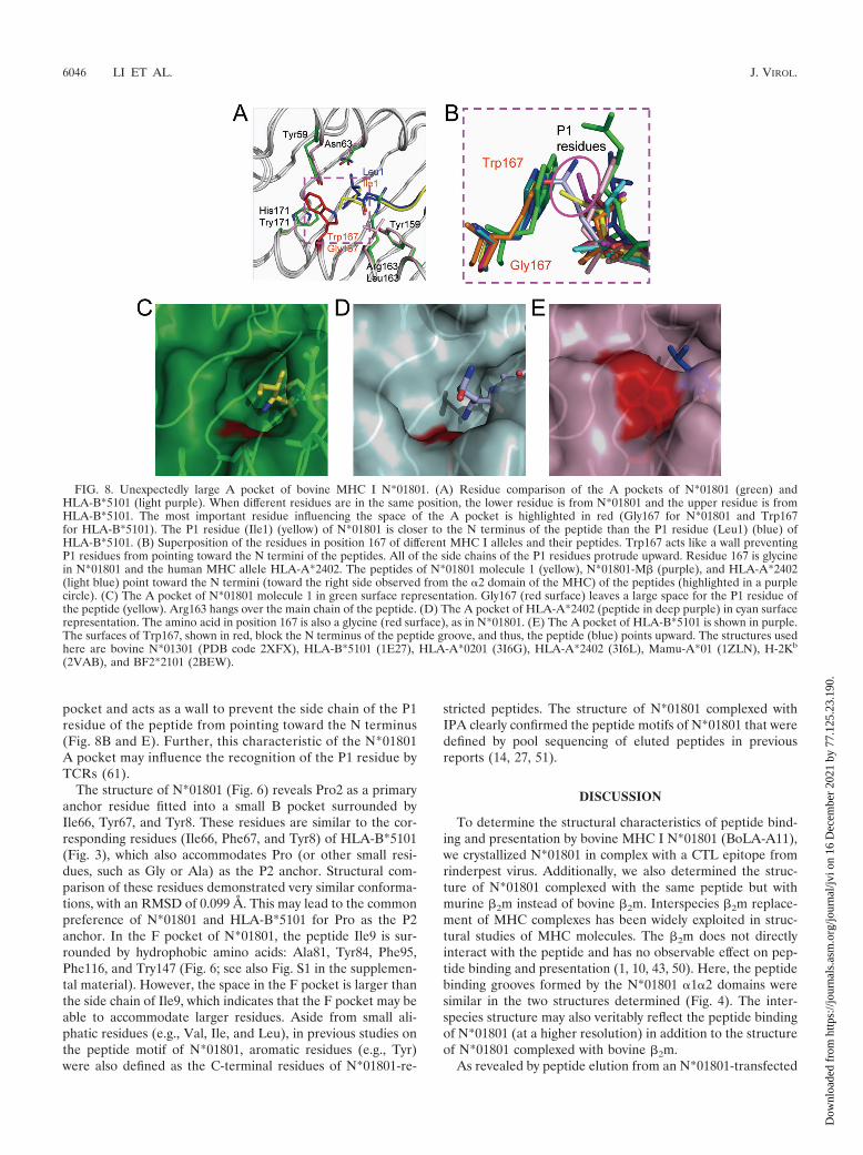
pocket and acts as a wall to prevent the side chain of the P1residue of the peptide from pointing toward the N terminus(Fig. 8B and E). Further, this characteristic of the N*01801A pocket may influence the recognition of the P1 residue byTCRs (61).
The structure of N*01801 (Fig. 6) reveals Pro2 as a primaryanchor residue fitted into a small B pocket surrounded byIle66, Tyr67, and Tyr8. These residues are similar to the cor-responding residues (Ile66, Phe67, and Tyr8) of HLA-B*5101(Fig. 3), which also accommodates Pro (or other small resi-dues, such as Gly or Ala) as the P2 anchor. Structural com-parison of these residues demonstrated very similar conforma-tions, with an RMSD of 0.099 Å. This may lead to the commonpreference of N*01801 and HLA-B*5101 for Pro as the P2anchor. In the F pocket of N*01801, the peptide Ile9 is sur-rounded by hydrophobic amino acids: Ala81, Tyr84, Phe95,Phe116, and Try147 (Fig. 6; see also Fig. S1 in the supplemen-tal material). However, the space in the F pocket is larger thanthe side chain of Ile9, which indicates that the F pocket may beable to accommodate larger residues. Aside from small ali-phatic residues (e.g., Val, Ile, and Leu), in previous studies onthe peptide motif of N*01801, aromatic residues (e.g., Tyr)were also defined as the C-terminal residues of N*01801-re-
stricted peptides. The structure of N*01801 complexed withIPA clearly confirmed the peptide motifs of N*01801 that weredefined by pool sequencing of eluted peptides in previousreports (14, 27, 51).
DISCUSSION
To determine the structural characteristics of peptide bind-ing and presentation by bovine MHC I N*01801 (BoLA-A11),we crystallized N*01801 in complex with a CTL epitope fromrinderpest virus. Additionally, we also determined the struc-ture of N*01801 complexed with the same peptide but withmurine �2m instead of bovine �2m. Interspecies �2m replace-ment of MHC complexes has been widely exploited in struc-tural studies of MHC molecules. The �2m does not directlyinteract with the peptide and has no observable effect on pep-tide binding and presentation (1, 10, 43, 50). Here, the peptidebinding grooves formed by the N*01801 �1�2 domains weresimilar in the two structures determined (Fig. 4). The inter-species structure may also veritably reflect the peptide bindingof N*01801 (at a higher resolution) in addition to the structureof N*01801 complexed with bovine �2m.
As revealed by peptide elution from an N*01801-transfected
FIG. 8. Unexpectedly large A pocket of bovine MHC I N*01801. (A) Residue comparison of the A pockets of N*01801 (green) andHLA-B*5101 (light purple). When different residues are in the same position, the lower residue is from N*01801 and the upper residue is fromHLA-B*5101. The most important residue influencing the space of the A pocket is highlighted in red (Gly167 for N*01801 and Trp167for HLA-B*5101). The P1 residue (Ile1) (yellow) of N*01801 is closer to the N terminus of the peptide than the P1 residue (Leu1) (blue) ofHLA-B*5101. (B) Superposition of the residues in position 167 of different MHC I alleles and their peptides. Trp167 acts like a wall preventingP1 residues from pointing toward the N termini of the peptides. All of the side chains of the P1 residues protrude upward. Residue 167 is glycinein N*01801 and the human MHC allele HLA-A*2402. The peptides of N*01801 molecule 1 (yellow), N*01801-M� (purple), and HLA-A*2402(light blue) point toward the N termini (toward the right side observed from the �2 domain of the MHC) of the peptides (highlighted in a purplecircle). (C) The A pocket of N*01801 molecule 1 in green surface representation. Gly167 (red surface) leaves a large space for the P1 residue ofthe peptide (yellow). Arg163 hangs over the main chain of the peptide. (D) The A pocket of HLA-A*2402 (peptide in deep purple) in cyan surfacerepresentation. The amino acid in position 167 is also a glycine (red surface), as in N*01801. (E) The A pocket of HLA-B*5101 is shown in purple.The surfaces of Trp167, shown in red, block the N terminus of the peptide groove, and thus, the peptide (blue) points upward. The structures usedhere are bovine N*01301 (PDB code 2XFX), HLA-B*5101 (1E27), HLA-A*0201 (3I6G), HLA-A*2402 (3I6L), Mamu-A*01 (1ZLN), H-2Kb
(2VAB), and BF2*2101 (2BEW).
6046 LI ET AL. J. VIROL.
Dow
nloa
ded
from
http
s://j
ourn
als.
asm
.org
/jour
nal/j
vi o
n 16
Dec
embe
r 20
21 b
y 77
.125
.23.
190.

cell line, nonameric peptides account for most of the naturallyseparated peptides (28). Proline is found almost exclusively atthe P2 position of these peptides, and as shown in the N*01801structure, the relatively small, bowl-shaped, hydrophobic Bpocket perfectly accommodates a proline at this position. TheB pocket of N*01801 is formed by residues similar to those inthe B pocket of HLA-B*5101, which may explain the samepreference for proline as the prevalent anchor residue in theP2 position. In the C-terminal position of the IPA peptide,isoleucine and valine act as the primary anchor residues, in-serting into the hydrophobic F pocket of N*01801, as indicatedin the structure of N*01801 and the refolding assays. The spacewithin the F pocket appears larger than the side chain ofisoleucine, which may indicate that larger hydrophobic andeven aromatic side chains can be accommodated. This is coin-cident with the peptide motif studies of N*01801, which indi-cated that Ile/Val and a small proportion of Leu/Tyr occupiedthe C termini of the naturally processed peptides of N*01801(28).
Studies of CD8� T-cell epitopes indicate that different pep-tides elicit diverse T-cell responses, corresponding to distinctT-cell repertoires. Recent studies have also demonstrated thateven minor modification of an epitope can lead to a profoundeffect on the antigenicity of the peptide (37, 58). Peptides witha relatively rigid conformation may result in limited types ofTCR docking, leading to a specific T-cell repertoire with lim-ited diversity. Recently, this has been thoroughly investigatedby Macdonald and colleagues in structural and function studiesof bovine MHC I N*01301 (34). Hence, a peptide with aflexible structure presented in the binding groove of an MHCmolecule may lead to a diverse TCR profile. In a number ofreported cases, a broader T-cell repertoire for the pathogen-specific epitope may facilitate a more effective host immuneresponse against the invading pathogen and may prevent theemergence of immune escape mutants (39, 44, 52). The IPApeptide in our structure unexpectedly appears to be presentedby N*01801 in two distinct conformations. The completelydifferent exposed areas in the middle of the peptide may indi-cate different manners of TCR docking. Further study shouldfocus on whether the two distinct conformations of the IPApeptide correspond to different T-cell repertoires. The contri-bution of the uncommon peptide presentation strategy ofN*01801 to the CTL-specific responses of N*01801� cattle topathogens also needs further exploration.
Generally, a featured peptide contains exposed residues pro-truding out of the landscape of the MHC complexed to pep-tide, and this can be implemented via two different strategies.First, epitopes with characteristic long side chain residues,which are solvent exposed, may act as featured peptides toelicit diverse T-cell repertoires (58). Second, moderatelybulged peptides help the short side chains rise to a suitablelevel for TCR docking (32). In the structure of bovine MHC IN*01801 complexed with IPA, N*01801 presents the samepeptide in two distinct conformations, which is associated withthe flexibility of Ile73 in the peptide binding groove. Differentexposed residues and secondary anchor residues result fromthese conformational changes. This adjustable strategy of hostantigen presentation may represent a novel type of featuredpeptide and may expand our understanding of the crucial roleof T-cell epitopes in antipathogen immunity.
BoLA-A11 is one of the most common global haplotypes indairy cattle. Studies of T-cell-specific responses against patho-gens have identified a large number of CD8� T-cell epitopeswith the BoLA-A11 restriction. Here, we demonstrated thedistinctive characteristics of peptide presentation by BoLA-A11 through the structural determination of bovine MHC IN*01801 (one of the most common alleles of BoLA-A11 hap-lotypes) complexed with a rinderpest virus-derived IPA pep-tide. Our study may lead to further definition of featured T-cellepitopes in a structural manner and, moreover, may pave theway for rational CTL-based design of vaccines for cattle orother species.
ACKNOWLEDGMENTS
This work was supported by grants from the Ministry of Science andTechnology of China (Transgenic special grant 2009ZX08009-150B;Project 973 grant 2007CB815805) and a Special Grant for ProteinScience of the National 973 Project (grant 2010CB911902). G.F.G. isa leading principal investigator of the National Natural Science Foun-dation of China (NSFC) Innovative Research Group (grant 81021003).The funders had no role in study the design, data collection andanalysis, decision to publish, or preparation of the manuscript.
We thank Joel Haywood, Yi Shi, and Hao Cheng of the Institute ofMicrobiology, Chinese Academy of Sciences, for their excellent sug-gestions on this study.
The authors declare no financial or commercial conflict of interest.
REFERENCES
1. Achour, A., et al. 2006. Structural basis of the differential stability andreceptor specificity of H-2Db in complex with murine versus human �2-microglobulin. J. Mol. Biol. 356:382–396.
2. Adams, P. D., et al. 2002. PHENIX: building new software for automatedcrystallographic structure determination. Acta Crystallogr. D Biol. Crystal-logr. 58:1948–1954.
3. Ahmed, N., and S. Gottschalk. 2009. How to design effective vaccines: les-sons from an old success story. Expert Rev. Vaccines 8:543–546.
4. Amorena, B., and W. H. Stone. 1978. Serologically defined (SD) locus incattle. Science 201:159–160.
5. Babiuk, S., et al. 2007. BoLA class I allele diversity and polymorphism in aherd of cattle. Immunogenetics 59:167–176.
6. Bensaid, A., et al. 1991. Identification of expressed bovine class I MHC genesat two loci and demonstration of physical linkage. Immunogenetics 33:247–254.
7. Birtley, J. R., et al. 2005. Crystal structure of foot-and-mouth disease virus3C protease.New insights into catalytic mechanism and cleavage specificity.J. Biol. Chem. 280:11520–11527.
8. Chen, W., et al. 2010. Crystal structure of a bony fish �2-microglobulin:insights into the evolutionary origin of immunoglobulin superfamily constantmolecules. J. Biol. Chem. 285:22505–22512.
9. Chen, Y., Y. Shi, H. Cheng, Y. Q. An, and G. F. Gao. 2009. Structuralimmunology and crystallography help immunologists see the immune systemin action: how T and NK cells touch their ligands. IUBMB Life 61:579–590.
10. Chu, F., et al. 2007. First glimpse of the peptide presentation by rhesusmacaque MHC class I: crystal structures of Mamu-A*01 complexed with twoimmunogenic SIV epitopes and insights into CTL escape. J. Immunol. 178:944–952.
11. Ciatto, C., et al. 2001. Zooming in on the hydrophobic ridge of H-2Db:implications for the conformational variability of bound peptides. J. Mol.Biol. 312:1059–1071.
12. Cole, D. K., et al. 2006. Crystal structure of HLA-A*2402 complexed with atelomerase peptide. Eur. J. Immunol. 36:170–179.
13. Davies, C. J., et al. 1994. Polymorphism of bovine MHC class I genes. Jointreport of the Fifth International Bovine Lymphocyte Antigen (BoLA) Work-shop, Interlaken, Switzerland, 1 August 1992. Eur. J. Immunogenet. 21:239–258.
14. De Groot, A. S., et al. 2003. T cell epitope identification for bovine vaccines:an epitope mapping method for BoLA A-11. Int. J. Parasitol. 33:641–653.
15. Ellis, S. 2004. The cattle major histocompatibility complex: is it unique? Vet.Immunol. Immunopathol. 102:1–8.
16. Ellis, S. A., and K. T. Ballingall. 1999. Cattle MHC: evolution in action?Immunol. Rev. 167:159–168.
17. Ellis, S. A., K. A. Staines, and W. I. Morrison. 1996. cDNA sequence ofcattle MHC class I genes transcribed in serologically defined haplotypes A18and A31. Immunogenetics 43:156–159.
VOL. 85, 2011 TWO CONFORMATIONS OF A BoLA-A11 EPITOPE 6047
Dow
nloa
ded
from
http
s://j
ourn
als.
asm
.org
/jour
nal/j
vi o
n 16
Dec
embe
r 20
21 b
y 77
.125
.23.
190.

18. Ellis, S. A., K. A. Staines, M. J. Stear, E. J. Hensen, and W. I. Morrison.1998. DNA typing for BoLA class I using sequence-specific primers (PCR-SSP). Eur. J. Immunogenet. 25:365–370.
19. Emsley, P., B. Lohkamp, W. G. Scott, and K. Cowtan. 2004. Features anddevelopment of Coot. Acta Crystallogr. D Biol. Crystallogr. 66:486–501.
20. Ennis, P. D., A. P. Jackson, and P. Parham. 1988. Molecular cloning ofbovine class I MHC cDNA. J. Immunol. 141:642–651.
21. Falk, K., et al. 1995. Peptide motifs of HLA-B51, -B52 and -B78 molecules,and implications for Behcet’s disease. Int. Immunol. 7:223–228.
22. Fries, R., R. Hediger, and G. Stranzinger. 1986. Tentative chromosomallocalization of the bovine major histocompatibility complex by in situ hy-bridization. Anim. Genet. 17:287–294.
23. Gilbert, N. 2009. Cattle disease faces total wipeout. Nature 462:709.24. Gouet, P., X. Robert, and E. Courcelle. 2003. ESPript/ENDscript: extracting
and rendering sequence and 3D information from atomic structures of pro-teins. Nucleic Acids Res. 31:3320–3323.
25. Gras, S., L. Kjer-Nielsen, S. R. Burrows, J. McCluskey, and J. Rossjohn.2008. T-cell receptor bias and immunity. Curr. Opin. Immunol. 20:119–125.
26. Guzman, E., G. Taylor, B. Charleston, M. A. Skinner, and S. A. Ellis. 2008.An MHC-restricted CD8� T-cell response is induced in cattle by foot-and-mouth disease virus (FMDV) infection and also following vaccination withinactivated FMDV. J. Gen. Virol. 89:667–675.
27. Hegde, N. R., M. S. Deshpande, D. L. Godson, L. A. Babiuk, and S. Sriku-maran. 1999. Bovine lymphocyte antigen-A11-specific peptide motif as ameans to identify cytotoxic T-lymphocyte epitopes of bovine herpesvirus 1.Viral Immunol. 12:149–161.
28. Hegde, N. R., et al. 1995. Peptide motif of the cattle MHC class I antigenBoLA-A11. Immunogenetics 42:302–303.
29. Holmes, E. C., A. F. Roberts, K. A. Staines, and S. A. Ellis. 2003. Evolutionof major histocompatibility complex class I genes in Cetartiodactyls. Immu-nogenetics 55:193–202.
30. Laskowski, R. A., D. S. Moss, and J. M. Thornton. 1993. Main-chain bondlengths and bond angles in protein structures. J. Mol. Biol. 231:1049–1067.
31. Liu, J., et al. 2010. The membrane protein of severe acute respiratorysyndrome coronavirus acts as a dominant immunogen revealed by a cluster-ing region of novel functionally and structurally defined cytotoxic T-lympho-cyte epitopes. J. Infect. Dis. 202:1171–1180.
32. Liu, J., et al. 2010. Novel immunodominant peptide presentation strategy: afeatured HLA-A*2402-restricted cytotoxic T-lymphocyte epitope stabilizedby intrachain hydrogen bonds from severe acute respiratory syndrome coro-navirus nucleocapsid protein. J. Virol. 84:11849–11857.
33. Liu, J., S. Zhang, S. Tan, B. Zhen, and G. F. Gao. 2011. Revival of theidentification of CTL epitopes for immunological diagnosis, therapy andvaccine development. Exp. Biol. Med. doi:10.1258/ebm.2010.010278.
34. Macdonald, I. K., et al. 2010. MHC class I bound to an immunodominantTheileria parva epitope demonstrates unconventional presentation to T cellreceptors. PLoS Pathog. 6:e1001149.
35. Maenaka, K., et al. 2000. Nonstandard peptide binding revealed by crystalstructures of HLA-B*5101 complexed with HIV immunodominant epitopes.J. Immunol. 165:3260–3267.
36. Markel, H. 2005. The search for effective HIV vaccines. N. Engl. J. Med.353:753–757.
37. Maryanski, J. L., et al. 1997. The diversity of antigen-specific TCR reper-toires reflects the relative complexity of epitopes recognized. Hum. Immu-nol. 54:117–128.
38. Meijers, R., et al. 2005. Crystal structures of murine MHC Class I H-2Db andKb molecules in complex with CTL epitopes from influenza A virus: impli-cations for TCR repertoire selection and immunodominance. J. Mol. Biol.345:1099–1110.
39. Messaoudi, I., J. A. Guevara Patino, R. Dyall, J. LeMaoult, and J. Nikolich-Zugich. 2002. Direct link between MHC polymorphism, T cell avidity, anddiversity in immune defense. Science 298:1797–1800.
40. Normile, D. 2008. Rinderpest. Driven to extinction. Science 319:1606–1609.41. Otwinowski, Z., and W. Minor. 1997. Processing of X-ray diffraction data
collected in oscillation mode. Methods Enzymol. 276:307–326.42. Parkin, S., and H. Hope. 1998. Macromolecular cryocrystallography: cooling,
mounting, storage and transportation of crystals. J. Appl. Crystallogr. 31:945–953.
43. Pedersen, L. O., et al. 1995. The interaction of �2-microglobulin (�2m) withmouse class I major histocompatibility antigens and its ability to supportpeptide binding. A comparison of human and mouse �2m. Eur. J. Immunol.25:1609–1616.
44. Price, D. A., et al. 2004. T cell receptor recognition motifs govern immuneescape patterns in acute SIV infection. Immunity 21:793–803.
45. Rammensee, H. G., T. Friede, and S. Stevanoviic. 1995. MHC ligands andpeptide motifs: first listing. Immunogenetics 41:178–228.
46. Rodriguez, L. L., and M. J. Grubman. 2009. Foot and mouth disease virusvaccines. Vaccine 27(Suppl. 4):D90–D94.
47. Russell, G. C., R. A. Oliver, and S. M. Sawhney. 1996. Cloning, transfection,and DNA sequence of a second gene from the BoLA-A11 haplotype. Im-munogenetics 44:315–318.
48. Sawhney, S. M., et al. 1995. Transfection, expression, and DNA sequence ofa gene encoding a BoLA-A11 antigen. Immunogenetics 41:246–250.
49. Shastri, N., S. Schwab, and T. Serwold. 2002. Producing nature’s gene-chips:the generation of peptides for display by MHC class I molecules. Annu. Rev.Immunol. 20:463–493.
50. Shields, M. J., L. E. Moffat, and R. K. Ribaudo. 1998. Functional comparisonof bovine, murine, and human �2-microglobulin: interactions with murineMHC I molecules. Mol. Immunol. 35:919–928.
51. Sinnathamby, G., S. Seth, R. Nayak, and M. S. Shaila. 2004. Cytotoxic T cellepitope in cattle from the attachment glycoproteins of rinderpest and pestedes petits ruminants viruses. Viral Immunol. 17:401–410.
52. Slifka, M. K., and J. L. Whitton. 2001. Functional avidity maturation ofCD8� T cells without selection of higher affinity TCR. Nat. Immunol. 2:711–717.
53. Sliz, P., et al. 2001. Crystal structures of two closely related but antigenicallydistinct HLA-A2/melanocyte-melanoma tumor-antigen peptide complexes.J. Immunol. 167:3276–3284.
54. Subbarao, K., B. R. Murphy, and A. S. Fauci. 2006. Development of effectivevaccines against pandemic influenza. Immunity 24:5–9.
55. Sun, Y., et al. 2010. Identification and structural definition of H5-specificCTL epitopes restricted by HLA-A*0201 derived from the H5N1 subtype ofinfluenza A viruses. J. Gen. Virol. 91:919–930.
56. Thompson, J. D., T. J. Gibson, F. Plewniak, F. Jeanmougin, and D. G.Higgins. 1997. The CLUSTAL_X windows interface: flexible strategies formultiple sequence alignment aided by quality analysis tools. Nucleic AcidsRes. 25:4876–4882.
57. Turner, S. J., P. C. Doherty, J. McCluskey, and J. Rossjohn. 2006. Structuraldeterminants of T-cell receptor bias in immunity. Nat. Rev. Immunol. 6:883–894.
58. Turner, S. J., et al. 2005. Lack of prominent peptide-major histocompatibil-ity complex features limits repertoire diversity in virus-specific CD8� T cellpopulations. Nat. Immunol. 6:382–389.
59. Tynan, F. E., et al. 2005. High resolution structures of highly bulged viralepitopes bound to major histocompatibility complex class I. Implications forT-cell receptor engagement and T-cell immunodominance. J. Biol. Chem.280:23900–23909.
60. Tynan, F. E., et al. 2005. T cell receptor recognition of a ‘super-bulged’ majorhistocompatibility complex class I-bound peptide. Nat. Immunol. 6:1114–1122.
61. Tynan, F. E., et al. 2007. A T cell receptor flattens a bulged antigenic peptidepresented by a major histocompatibility complex class I molecule. Nat. Im-munol. 8:268–276.
62. Vordermeier, H. M., et al. 2009. Adjuvants induce distinct immunologicalphenotypes in a bovine tuberculosis vaccine model. Clin. Vaccine Immunol.16:1443–1448.
63. Yewdell, J. W., and S. M. Haeryfar. 2005. Understanding presentation ofviral antigens to CD8� T cells in vivo: the key to rational vaccine design.Annu. Rev. Immunol. 23:651–682.
64. Zhou, M., et al. 2004. Complex assembly, crystallization and preliminaryX-ray crystallographic studies of MHC H-2Kd complexed with an HBV-corenonapeptide. Acta Crystallogr. D Biol. Crystallogr. 60:1473–1475.
6048 LI ET AL. J. VIROL.
Dow
nloa
ded
from
http
s://j
ourn
als.
asm
.org
/jour
nal/j
vi o
n 16
Dec
embe
r 20
21 b
y 77
.125
.23.
190.